Prediction of the Clinical Severity of Progressive Supranuclear Palsy by Diffusion Tensor Imaging
Abstract
1. Introduction
2. Materials and Methods
2.1. Study Patients
2.2. Image Acquisition
2.3. Image Processing
2.4. Statistical Analysis and Feature Reduction Process
3. Results
4. Discussion
4.1. Main Findings
4.2. Clinical Impact
4.3. Regions Related to Motor Function
4.4. Regions Related to Psychomotor Interactions
4.5. Validation of the Prediction Model
4.6. Technical Consideration and Additional Issues
4.7. Limitations
Supplementary Materials
Author Contributions
Funding
Acknowledgments
Conflicts of Interest
References
- Litvan, I.; Agid, Y.; Jankovic, J.; Goetz, C.; Brandel, J.; Lai, E.; Wenning, G.; D’olhaberriague, L.; Verny, M.; Chaudhuri, K.R. Accuracy of clinical criteria for the diagnosis of progressive supranuclear palsy (Steele-Richardson-Olszewski syndrome). Neurology 1996, 46, 922–930. [Google Scholar] [CrossRef]
- Cubo, E.; Stebbins, G.T.; Golbe, L.I.; Nieves, A.V.; Leurgans, S.; Goetz, C.G.; Kompoliti, K. Application of the unified parkinson’s disease rating scale in progressive supranuclear palsy: Factor analysis of the motor scale. Mov. Disord. Off. J. Mov. Disord. Soc. 2000, 15, 276–279. [Google Scholar] [CrossRef]
- Massey, L.A.; Micallef, C.; Paviour, D.C.; O’Sullivan, S.S.; Ling, H.; Williams, D.R.; Kallis, C.; Holton, J.L.; Revesz, T.; Burn, D.J.; et al. Conventional magnetic resonance imaging in confirmed progressive supranuclear palsy and multiple system atrophy. Mov. Disord. 2012, 27, 1754–1762. [Google Scholar] [CrossRef] [PubMed]
- Whitwell, J.L.; Hoglinger, G.U.; Antonini, A.; Bordelon, Y.; Boxer, A.L.; Colosimo, C.; van Eimeren, T.; Golbe, L.I.; Kassubek, J.; Kurz, C.; et al. Radiological biomarkers for diagnosis in PSP: Where are we and where do we need to be? Mov. Disord. 2017, 32, 955–971. [Google Scholar] [CrossRef] [PubMed]
- Lo, C.Y.; Wang, P.N.; Chou, K.H.; Wang, J.; He, Y.; Lin, C.P. Diffusion tensor tractography reveals abnormal topological organization in structural cortical networks in Alzheimer’s disease. J. Neurosci. 2010, 30, 16876–16885. [Google Scholar] [CrossRef]
- Lambin, P.; Rios-Velazquez, E.; Leijenaar, R.; Carvalho, S.; Van Stiphout, R.G.; Granton, P.; Zegers, C.M.; Gillies, R.; Boellard, R.; Dekker, A. Radiomics: Extracting more information from medical images using advanced feature analysis. Eur. J. Cancer 2012, 48, 441–446. [Google Scholar] [CrossRef]
- Gillies, R.J.; Kinahan, P.E.; Hricak, H. Radiomics: Images are more than pictures, they are data. Radiology 2015, 278, 563–577. [Google Scholar] [CrossRef]
- Ling, H. Clinical approach to progressive supranuclear palsy. J. Mov. Disord. 2016, 9, 3–13. [Google Scholar] [CrossRef]
- Chen, L.; Liu, M.; Bao, J.; Xia, Y.; Zhang, J.; Zhang, L.; Huang, X.; Wang, J. The correlation between apparent diffusion coefficient and tumor cellularity in patients: A meta-analysis. PLoS ONE 2013, 8, e79008. [Google Scholar] [CrossRef]
- Tabesh, A.; Jensen, J.H.; Ardekani, B.A.; Helpern, J.A. Estimation of tensors and tensor-derived measures in diffusional kurtosis imaging. Magn. Reson. Med. 2011, 65, 823–836. [Google Scholar] [CrossRef]
- Fan, L.; Li, H.; Zhuo, J.; Zhang, Y.; Wang, J.; Chen, L.; Yang, Z.; Chu, C.; Xie, S.; Laird, A.R.; et al. The human brainnetome atlas: A new brain atlas based on connectional architecture. Cereb. Cortex 2016, 26, 3508–3526. [Google Scholar] [CrossRef] [PubMed]
- Penny, W.D.; Friston, K.J.; Ashburner, J.T.; Kiebel, S.J.; Nichols, T.E. Statistical Parametric Mapping: The Analysis of Functional Brain Images; Elsevier: Amsterdam, The Netherlands, 2011. [Google Scholar]
- Austin, P.C.; Steyerberg, E.W. The number of subjects per variable required in linear regression analyses. J. Clin. Epidemiol. 2015, 68, 627–636. [Google Scholar] [CrossRef] [PubMed]
- Vittinghoff, E.; McCulloch, C.E. Relaxing the rule of ten events per variable in logistic and cox regression. Am. J. Epidemiol. 2007, 165, 710–718. [Google Scholar] [CrossRef] [PubMed]
- Evans, A.C.; Collins, D.L.; Mills, S.; Brown, E.; Kelly, R.; Peters, T.M. 3D statistical neuroanatomical models from 305 MRI volumes. In Proceedings of the 1993 IEEE Conference Record Nuclear Science Symposium and Medical Imaging Conference, San Francisco, CA, USA, 31 October–6 November 1993; pp. 1813–1817. [Google Scholar]
- Bluett, B.; Banks, S.; Cordes, D.; Bayram, E.; Mishra, V.; Cummings, J.; Litvan, I. Neuroimaging and neuropsychological assessment of freezing of gait in Parkinson’s disease. Alzheimers Dement (NY) 2018, 4, 387–394. [Google Scholar] [CrossRef] [PubMed]
- Halliday, G.M. Thalamic changes in Parkinson’s disease. Parkinsonism Relat. Disord. 2009, 15 (Suppl. 3), S152–S155. [Google Scholar] [CrossRef]
- Fox, P.T.; Lancaster, J.L.; Laird, A.R.; Eickhoff, S.B. Meta-analysis in human neuroimaging: Computational modeling of large-scale databases. Annu. Rev. Neurosci. 2014, 37, 409–434. [Google Scholar] [CrossRef]
- Gerardin, E.; Sirigu, A.; Lehericy, S.; Poline, J.B.; Gaymard, B.; Marsault, C.; Agid, Y.; Le Bihan, D. Partially overlapping neural networks for real and imagined hand movements. Cereb. Cortex 2000, 10, 1093–1104. [Google Scholar] [CrossRef]
- Zeidman, P.; Maguire, E.A. Anterior hippocampus: The anatomy of perception, imagination and episodic memory. Nat. Rev. Neurosci. 2016, 17, 173–182. [Google Scholar] [CrossRef]
- Christopher, L.; Duff-Canning, S.; Koshimori, Y.; Segura, B.; Boileau, I.; Chen, R.; Lang, A.E.; Houle, S.; Rusjan, P.; Strafella, A.P. Salience network and parahippocampal dopamine dysfunction in memory-impaired Parkinson disease. Ann. Neurol. 2015, 77, 269–280. [Google Scholar] [CrossRef]
- Olson, I.R.; Plotzker, A.; Ezzyat, Y. The Enigmatic temporal pole: A review of findings on social and emotional processing. Brain 2007, 130, 1718–1731. [Google Scholar] [CrossRef]
- Yang, Y.; Fan, L.; Chu, C.; Zhuo, J.; Wang, J.; Fox, P.T.; Eickhoff, S.B.; Jiang, T. Identifying functional subdivisions in the human brain using meta-analytic activation modeling-based parcellation. Neuroimage 2016, 124, 300–309. [Google Scholar] [CrossRef] [PubMed]
- Dai, Y.J.; Zhang, X.; Yang, Y.; Nan, H.Y.; Yu, Y.; Sun, Q.; Yan, L.F.; Hu, B.; Zhang, J.; Qiu, Z.Y.; et al. Gender differences in functional connectivities between insular subdivisions and selective pain-related brain structures. J. Headache Pain 2018, 19, 24. [Google Scholar] [CrossRef] [PubMed]
- Ghosh, B.C.; Rowe, J.B.; Calder, A.J.; Hodges, J.R.; Bak, T.H. Emotion recognition in progressive supranuclear palsy. J. Neurol. Neurosurg. Psychiatry 2009, 80, 1143–1145. [Google Scholar] [CrossRef] [PubMed]
- Klein, R.C.; de Jong, B.M.; de Vries, J.J.; Leenders, K.L. Direct comparison between regional cerebral metabolism in progressive supranuclear palsy and Parkinson’s disease. Mov. Disord. 2005, 20, 1021–1030. [Google Scholar] [CrossRef] [PubMed]
- Pontieri, F.E.; Assogna, F.; Stefani, A.; Pierantozzi, M.; Meco, G.; Benincasa, D.; Colosimo, C.; Caltagirone, C.; Spalletta, G. Sad and happy facial emotion recognition impairment in progressive supranuclear palsy in comparison with Parkinson’s disease. Parkinsonism Relat. Disord. 2012, 18, 871–875. [Google Scholar] [CrossRef] [PubMed]
- Chen, Y.L.; Lin, Y.J.; Lin, S.H.; Tsai, C.C.; Lin, Y.C.; Cheng, J.S.; Wang, J.J. The effect of spatial resolution on the reproducibility of diffusion imaging when controlled signal to noise ratio. Biomed. J. 2019, 42, 268–276. [Google Scholar] [CrossRef]
- Hoglinger, G.U.; Respondek, G.; Stamelou, M.; Kurz, C.; Josephs, K.A.; Lang, A.E.; Mollenhauer, B.; Muller, U.; Nilsson, C.; Whitwell, J.L.; et al. Clinical diagnosis of progressive supranuclear palsy: The movement disorder society criteria. Mov. Disord. 2017, 32, 853–864. [Google Scholar] [CrossRef]
- Constantinescu, R.; Richard, I.; Kurlan, R. Levodopa responsiveness in disorders with parkinsonism: A review of the literature. Mov. Disord. 2007, 22, 2141–2148. [Google Scholar] [CrossRef]
- Zhang, H.; Schneider, T.; Wheeler-Kingshott, C.A.; Alexander, D.C. NODDI: Practical in vivo neurite orientation dispersion and density imaging of the human brain. Neuroimage 2012, 61, 1000–1016. [Google Scholar] [CrossRef]
- Jenkinson, M.; Beckmann, C.F.; Behrens, T.E.; Woolrich, M.W.; Smith, S.M. FSL. Neuroimage 2012, 62, 782–790. [Google Scholar] [CrossRef]
- Jolliffe, I.T.; Cadima, J. Principal component analysis: A review and recent developments. Philos. Trans. R. Soc. A Math. Phys. Eng. Sci. 2016, 374, 20150202. [Google Scholar] [CrossRef] [PubMed]
- Beckmann, C.F.; Smith, S.M. Probabilistic independent component analysis for functional magnetic resonance imaging. IEEE Trans. Med. Imaging 2004, 23, 137–152. [Google Scholar] [CrossRef] [PubMed]
- Lu, Y.; Cohen, I.; Zhou, X.S.; Tian, Q. Feature selection using principal feature analysis. In Proceedings of the 15th ACM International Conference on Multimedia, Augsburg, Germany, 25–29 September 2007; pp. 301–304. [Google Scholar]
- Tibshirani, R. Regression shrinkage and selection via the lasso. J. R. Stat. Soc. Ser. B (Methodol.) 1996, 58, 267–288. [Google Scholar] [CrossRef]
- Yin, P.; Mao, N.; Zhao, C.; Wu, J.; Sun, C.; Chen, L.; Hong, N. Comparison of radiomics machine-learning classifiers and feature selection for differentiation of sacral chordoma and sacral giant cell tumour based on 3D computed tomography features. Eur. Radiol. 2019, 29, 1841–1847. [Google Scholar] [CrossRef] [PubMed]
- Gulisano, W.; Maugeri, D.; Baltrons, M.A.; Fà, M.; Amato, A.; Palmeri, A.; D’Adamio, L.; Grassi, C.; Devanand, D.; Honig, L.S. Role of amyloid-β and tau proteins in Alzheimer’s disease: Confuting the amyloid cascade. J. Alzheimer’s Dis. 2018, 64, S611–S631. [Google Scholar] [CrossRef]
- Morris, E.; Chalkidou, A.; Hammers, A.; Peacock, J.; Summers, J.; Keevil, S. Diagnostic accuracy of 18 F amyloid PET tracers for the diagnosis of Alzheimer’s disease: A systematic review and meta-analysis. Eur. J. Nucl. Med. Mol. Imaging 2016, 43, 374–385. [Google Scholar] [CrossRef]


| Protocol A | Protocol B | Protocol C | Total | |
|---|---|---|---|---|
| TE/TR (ms) | 83/7800 | 96/8200 | 108/5700 | |
| Voxel size | 2 × 2 × 2 | 2 × 2 × 2 | 2 × 2 × 3 | |
| Directions | 64 | 64 | 30 | |
| PSP | ||||
| Number of patients | 19 | 11 | 23 | 53 |
| Sex (men/women) | 7/12 | 6/5 | 8/15 | 21/32 |
| Age (years) | 63.9 ± 6.0 | 64.2 ± 6.6 | 67.8 ± 6.5 | 65.7 ± 6.5 |
| Disease duration (years) | 5.6 ± 2.3 | 4.2 ± 2.6 | 5.9 ± 3.9 | 5.4 ± 3.2 |
| Subtype (PAGF/PD/RS/CBS) | 5/8/5/1 | 7/4/0/0 | 15/3/2/3 | 27/15/7/4 |
| UPDRS-III (motor) | 29.6 ± 13.9 # | 45.8 ± 17.0 | 32.0 ± 17.3 | 36.5 ± 17.7 |
| PIGD | 10.8 ± 4.1 (NA = 3) | 9.6 ± 4.1 | 10.8 ± 3.3 | 10.5 ± 3.7 (NA = 3) |
| MHY | 4.0 ± 1.1 | 3.7 ± 1.1 | 3.8 ± 0.9 | 3.9 ± 1.0 |
| <3 | 2 | 1 | 1 | 4 |
| 3 | 5 | 4 | 9 | 18 |
| 4 | 3 | 3 | 6 | 12 |
| 5 | 9 | 3 | 7 | 19 |
| LEDD (mg/day) | 708.9 ± 311.8 | 615.0 ± 253.6 | 758.3 ± 426.6 | 724.5 ± 343.9 |
| UPDRS-III = | PIGD = | MHY = | LEDD = | ||||
|---|---|---|---|---|---|---|---|
| − | 100.6 | + | 1.2 | + | 6.0 | + | 450.9 |
| + | 48.7 × MD50_PhG_L_6_1 | + | 1.5 × MD90_INS_R_6_5 | − | 2.8 × MD50_MFG_L_7_5 | + | 2833.7 × FA90_STG_R_6_1 |
| + | 51.2 × MD10_PrG_L_6_2 | + | 210.7 × FA10_MTG_R_4_3 | − | 6.7 × FA90_IPL_R_6_4 | − | 571.6 × MD50_Amyg_R_2_2 |
| + | 28.3 × FA90_GP_L | − | 49.9 × FA90_MTG_R_4_3 | + | 9.5 × FA50_NAC_L | + | 325.4 × MD50_CG_R_7_6 |
| + | 65.2 × MD10_Tha_L_8_3 | − | 26.0 × FA90_MFG_R_7_2 | − | 3.8 × FA90_SPL_L_5_3 | − | 369.4 × MD90_PhG_L_6_3 |
| − | 23.9 × FA90_SPL_R_5_4 | + | 4.1 × MD90_PrG_R_6_2 | − | 10.5 × FA90_Tha_R_8_4 | − | 2074.1 × FA10_VM_Put_R |
| + | 98.2 × FA90_STG_R_6_1 | + | 28.4 × FA50_ITG_R_7_2 | − | 4.9 × MD10_IFG_R_6_4 | + | 2949.2 × FA50_OrG_R_6_2 |
| − | 35.9 × MD10_Amyg_L_2_1 | − | 13.7 × FA90_MTG_R_4_4 | + | 6.1 × FA50_ITG_R_7_2 | + | 1487.9 × MD10_PrG_L_6_4 |
| + | 72.8 × MD10_Tha_L_8_8 | + | 9.3 × FA90_PoG_L_4_3 | + | 18.6 × FA10_ITG_L_7_6 | − | 1568.8 × MD10_PoG_L_4_3 |
| − | 18.0 × MD90_IPL_L_6_2 | − | 5.5 × MD10_Amyg_R_2_1 | + | 2.0 × MD10_PrG_L_6_4 | − | 1112.0 × FA90_PCL_R_2_1 |
| + | 35.0 × FA90_VM_Put_R | + | 3.7 × MD90_MVOcC_L_5_3 | − | 0.6 × MD50_Amyg_L_2_1 | + | 421.3 × MD50_PoG_R_4_3 |
| − | 38.1 × FA90_MFG_L_7_6 | + | 44.6 × FA10_ITG_L_7_6 | − | 2.6 × FA90_MFG_L_7_6 | + | 5248.2 × FA10_SPL_R_5_4 |
| UPDRS-III | PIGD | MHY | LEDD | |
|---|---|---|---|---|
| Training | ||||
| Adjusted R2 (95% CI) | 0.88 (0.83~0.93) | 0.80 (0.72~0.88) | 0.85 (0.79~0.91) | 0.77 (0.69~0.85) |
| F Test | 395 | 194 | 284 | 176 |
| Cohen f2 | 3.43 | 1.78 | 2.60 | 1.46 |
| Power | 1.00 | 1.00 | 1.00 | 1.00 |
| LOOCV | ||||
| Mean Adjusted R2 | 0.884 ± 0.005 | 0.799 ± 0.010 | 0.845 ± 0.006 | 0.772 ± 0.008 |
| MAE | 6.1 ± 5.0 | 1.7 ± 1.6 | 0.4 ± 0.3 | 180.8 ± 119.9 |
| MAE in % | 5.6 ± 4.6 | 8.2 ± 7.8 | 8.0 ± 5.8 | 32.9 ± 42.6 |
| Five-fold CV | ||||
| Mean Adjusted R2 | 0.892 ± 0.016 | 0.818 ± 0.033 | 0.856 ± 0.024 | 0.739 ± 0.047 |
| MAE | 6.37 ± 0.89 | 1.845 ± 0.689 | 0.413 ± 0.056 | 223.0 ± 48.2 |
| MAE in % | 5.9 ± 0.8 | 9.2 ± 3.4 | 8.2 ± 1.1 | 40.1 ± 8.2 |
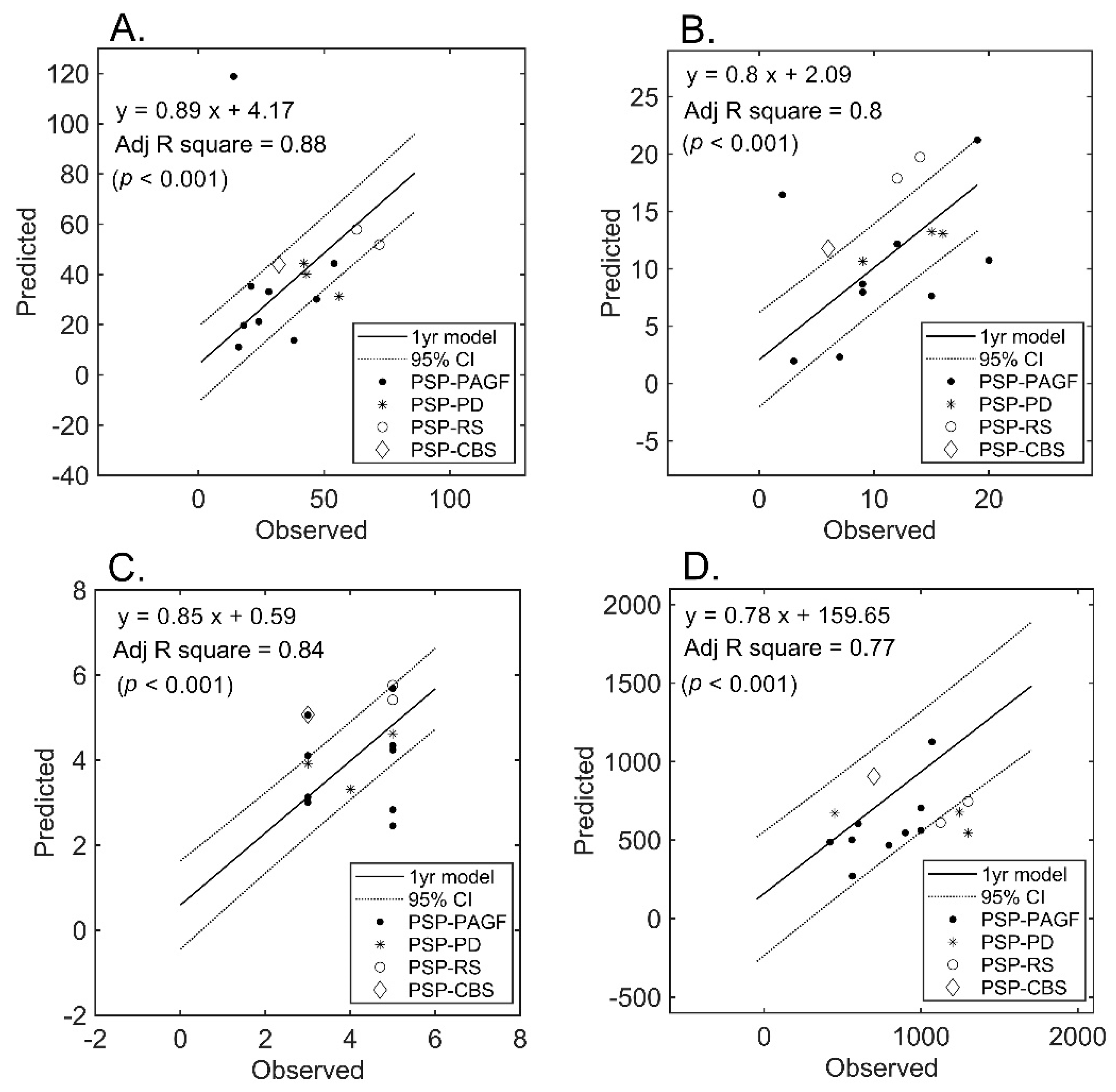
| Follow-up Validation MAE | 16.8 ± 25.6 a | 4.3 ± 3.9 b | 1.0 ± 0.8 c | 313.8 ± 220.6 d |
| MAE in % | 15.5 ± 23.7 | 21.4 ± 19.7 | 20.5 ± 16.0 | 33.9 ± 17.9 |
© 2019 by the authors. Licensee MDPI, Basel, Switzerland. This article is an open access article distributed under the terms and conditions of the Creative Commons Attribution (CC BY) license (http://creativecommons.org/licenses/by/4.0/).
Share and Cite
Chen, Y.-L.; Zhao, X.-A.; Ng, S.-H.; Lu, C.-S.; Lin, Y.-C.; Cheng, J.-S.; Tsai, C.-C.; Wang, J.-J. Prediction of the Clinical Severity of Progressive Supranuclear Palsy by Diffusion Tensor Imaging. J. Clin. Med. 2020, 9, 40. https://doi.org/10.3390/jcm9010040
Chen Y-L, Zhao X-A, Ng S-H, Lu C-S, Lin Y-C, Cheng J-S, Tsai C-C, Wang J-J. Prediction of the Clinical Severity of Progressive Supranuclear Palsy by Diffusion Tensor Imaging. Journal of Clinical Medicine. 2020; 9(1):40. https://doi.org/10.3390/jcm9010040
Chicago/Turabian StyleChen, Yao-Liang, Xiang-An Zhao, Shu-Hang Ng, Chin-Song Lu, Yu-Chun Lin, Jur-Shan Cheng, Chih-Chien Tsai, and Jiun-Jie Wang. 2020. "Prediction of the Clinical Severity of Progressive Supranuclear Palsy by Diffusion Tensor Imaging" Journal of Clinical Medicine 9, no. 1: 40. https://doi.org/10.3390/jcm9010040
APA StyleChen, Y.-L., Zhao, X.-A., Ng, S.-H., Lu, C.-S., Lin, Y.-C., Cheng, J.-S., Tsai, C.-C., & Wang, J.-J. (2020). Prediction of the Clinical Severity of Progressive Supranuclear Palsy by Diffusion Tensor Imaging. Journal of Clinical Medicine, 9(1), 40. https://doi.org/10.3390/jcm9010040


