Communication

Helicobacter pylori Is Present at Quantifiable Levels in Raw Vegetables in the Mediterranean Area of Spain

by
Miguel García-Ferrús
1,
Ana González
1,
María Consuelo Pina-Pérez
2 and
Maria Antonia Ferrús
1,*
1
Centro Avanzado de Microbiología de Alimentos, Universitat Politècnica de València, 46022 Valencia, Spain
2
Departamento Microbiologia y Ecología, Universitat de València, 46100 Valencia, Spain
*
Author to whom correspondence should be addressed.
Agriculture 2022, 12(3), 339; https://doi.org/10.3390/agriculture12030339
Submission received: 30 January 2022 / Revised: 21 February 2022 / Accepted: 25 February 2022 / Published: 27 February 2022
(This article belongs to the Section Agricultural Product Quality and Safety)

Abstract

:
Vegetables are one of the main foodstuffs consumed in the Mediterranean diet. However, raw vegetables have been associated with relevant foodborne outbreaks worldwide. Accurate knowledge of the microbiological quantitative risks associated with these matrices is crucial in order to define effective control measures, avoiding the survival and dissemination of foodborne pathogens through the different food chain stages. The aim of the present study is the assessment of the prevalence of Helicobacter pylori (a unique carcinogenic biological agent recognized to date) on leafy vegetables (spinach, lettuce, and chard) by means of the detection of the specific pathogenicity vacA gene. A real-time quantitative polymerase chain reaction (qPCR) optimized approach was used to detect H. pylori-positive samples and the concentration of this pathogen (with a limit of detection equal to 10 cells). One hundred raw vegetable samples were acquired in markets corresponding to the Spanish Mediterranean area. Sliced vegetable leaves were homogenized and centrifuged, and DNA was extracted from the homogenates. qPCR results confirmed 20 out of 100 H. pylori-positive samples, with melting temperature (Tm) values in the range of 84.8–86.5 °C (Tm vacA H. pylori = 85 °C). Amplicons were cut, purified, and sequenced to confirm the homology with the H. pylori vacA gene. A total of 17 out of 100 vegetable samples (12/45 (26.6%) lettuce, 2/21 (9.5%) spinach, and 3/34 (8.8%) chard samples) were finally confirmed as H. pylori-positive. Contamination levels were in the range of 1.5 ± 0.3 to 2.5 ± 0.1 log10 cycles (36–335 CFU/g leafy vegetables). Our results show that H. pylori is detected by qPCR at levels close to infectious doses in fresh vegetables, thus posing a food safety hazard.

1. Introduction

Current trends in adopting healthy lifestyles are leading to an increase in the demand for and consumption of fresh green-leafy vegetables [1]. This has public health consequences derived from the microbial contamination of this type of product. Beyond the gastrointestinal infections and outbreaks associated with main pathogens, such as Escherichia coli, Salmonella spp., or Listeria monocytogenes, Blau et al. (2018) remark upon the capability of these bacteria to transfer resistomes, which can interact with human microbiota, with possible fatal consequences in terms of antibiotic resistance dissemination and microbiota dysbiosis [2,3].
To date, Helicobacter pylori is one of the most concerning human pathogens [4,5]. In recent years, three factors have been considered as critical in the risk associated with these bacteria: (i) the concerning consequences associated with H. pylori chronic infection (e.g., stomach cancer and gastric mucosa-associated lymphoid tissue lymphoma, MALT), as 6.2% of gastric cancers derived from infections are attributable to H. pylori; (ii) the antibiotic resistance of this pathogen to existing therapies (rate of treatment efficacy <75%), which led the World Health Organization to include H. pylori in February 2017, for the first time, in the list of the most antibiotic-resistant pathogens, with it being a high priority to search for alternative treatments [6,7,8]; and (iii) the high capability of Helicobacter spp. to disseminate in the environment (water and foods). H. pylori survives for 4 days in water at 25 °C and for 7 days at 4 °C. This organism also resists usual wastewater disinfection procedures and survives in complex food products, such as milk, vegetables, and ready-to-eat foods [9,10,11,12,13].
Although the routes of H. pylori transmission are still poorly known, low socioeconomic and sanitization conditions have been described as significant in favoring H. pylori infection [13]. Nowadays, among the routes that are well accepted as the most probable for contamination are (i) interpersonal contact (fecal–oral and oral–oral) and (ii) the consumption of contaminated water and food products, with prevalence rates comprising a wide range, from 1 to 72%, depending on the considered product, the geographic area of analysis, and the identification/quantification technique applied [12,13,14,15,16,17,18,19].
Among the food products identified as possible vehicles of H. pylori transmission, vegetables stand out because they grow in direct contact with soil and irrigation water, which is one of the main environmental sources of contamination [20]. Moreover, they are often fertilized with organic substrates [21]. To date, scarce information has been published in relation to the prevalence of H. pylori in leafy vegetables, with available data comprising a wide H. pylori prevalence range between 10 and 34% [15,22]. According to Yahaghi et al. (2014), leek, lettuce, and cabbage are the most commonly contaminated vegetable samples (H. pylori prevalence ≈ 30%) [22].
To date, only one study has been carried out regarding the characterization of factors affecting the growth and survival of H. pylori cells in vegetables. Pina-Pérez et al. (2018) simulated the growth and survival potential of H. pylori cells artificially inoculated on lettuce, kale, spinach, and chard and reported that H. pylori was able to grow and proliferate in lettuce at 20 °C with a maximum specific growth rate (µmax) equal to 0.79 log10 (CFU/mL)/d), showing a peak of growth of 1 log10 cycle in 5 days [23].
An important question is not only the presence of the bacteria, but also the viable but not culturable (VBNC) possible state of H. pylori cells. VBNC forms represent a great threat to food safety [24,25]. According to the same study, between 0.60 and even 4.5 log10 cycles of initial bacterial load enter into the VBNC state after 3–5 days of storage at 20 °C [23].
The water activity (aw) in vegetables, quality of irrigation water, sanitation conditions after harvest, and storage conditions during commercialization have been described as the main factors influencing the infective-viable status of H. pylori cells in vegetables, from the harvest period to the moment of consumption [24,26]. Considering the capability of H. pylori cells to survive and even proliferate in leafy vegetables under the most common growth, distribution, storage, and commercialization conditions (<15 days, 5–20 °C), the present study aims to contribute to the state of knowledge regarding the assessment of the prevalence of H. pylori in this kind of agricultural product. The presence of wild H. pylori cells was assessed in commercially available Spanish leafy vegetable samples by using an optimized real-time quantitative polymerase chain reaction (q-PCR) approach.

2. Materials and Methods

2.1. Samples and Isolation of Helicobacter pylori

A total of 100 fresh leafy vegetable samples, 45 lettuce (Lactuca sativa var romana), 21 spinach (Spinaceae oleracea), and 34 chard (Beta vulgaris var. cicla), were acquired from 100 different Spanish supermarkets and groceries corresponding to Valencia city, Comunidad Valenciana (Mediterranean region, Spain), a developed geographical area, over the course of 12 months (July 2020 to June 2021). Each sample was composed of 5 individual units [27,28].
Samples were immediately transported to the laboratory and refrigerated at 4 °C. Leaves from each sample were sliced in pieces, and 10 g from each batch was aseptically disposed into sterile stomacher bags (10 g sample + 20 mL of PBS 1× (130 mM sodium chloride, 10 mM sodium phosphate, pH 7.2)). The bags were placed into a stomacher machine (Lab-Blender-400 Seward Medical, Worthing, UK) and homogenized for 5 min. Homogenized leaf samples were disposed in individual 50 mL crew cap tubes to carry out a centrifugation step (10 min, 10 °C, 8000 rpm). After centrifugation, supernatants were collected and disposed into 5 individual Eppendorf tubs (2 mL per Eppendorf).
Artificially inoculated samples were also included into the study to test the accuracy of the recovering method for detection. To carry out this task, H. pylori NCTC 11638 strain, provided by the United Kingdom National Collection of Type Cultures (London, UK), was used. H. pylori NCTC 11638 was grown in Columbia Blood Agar (CBA, Deco, Franklin Lakes, NJ, USA) plates supplemented with 10% defibrinated sterile horse blood (HB, Oxoid, Hampshire, UK) under microaerobic conditions (5% oxygen, 10% carbon dioxide, and 85% nitrogen) at 37 °C. Before inoculation, vegetable leaves were washed with sodium hypochlorite (10% (v/v)) for 30 min, dried in a sterile cabinet flow (2 h), and exposed to UV light (8 h). H. pylori 11638 NCTC grown in solid CBA-HB plates was inoculated in the range 106–108 CFU/10 g on vegetable sterile leaves by spraying. These 10 g of artificially inoculated leaves were also sliced into pieces and homogenized in PBS (1×) for 5 min. After homogenization, samples were also subjected to a centrifugation step in the same conditions as non-inoculated lettuce samples, and supernatants (10 mL) were collected and disposed into individual Eppendorf tubs for processing [23].

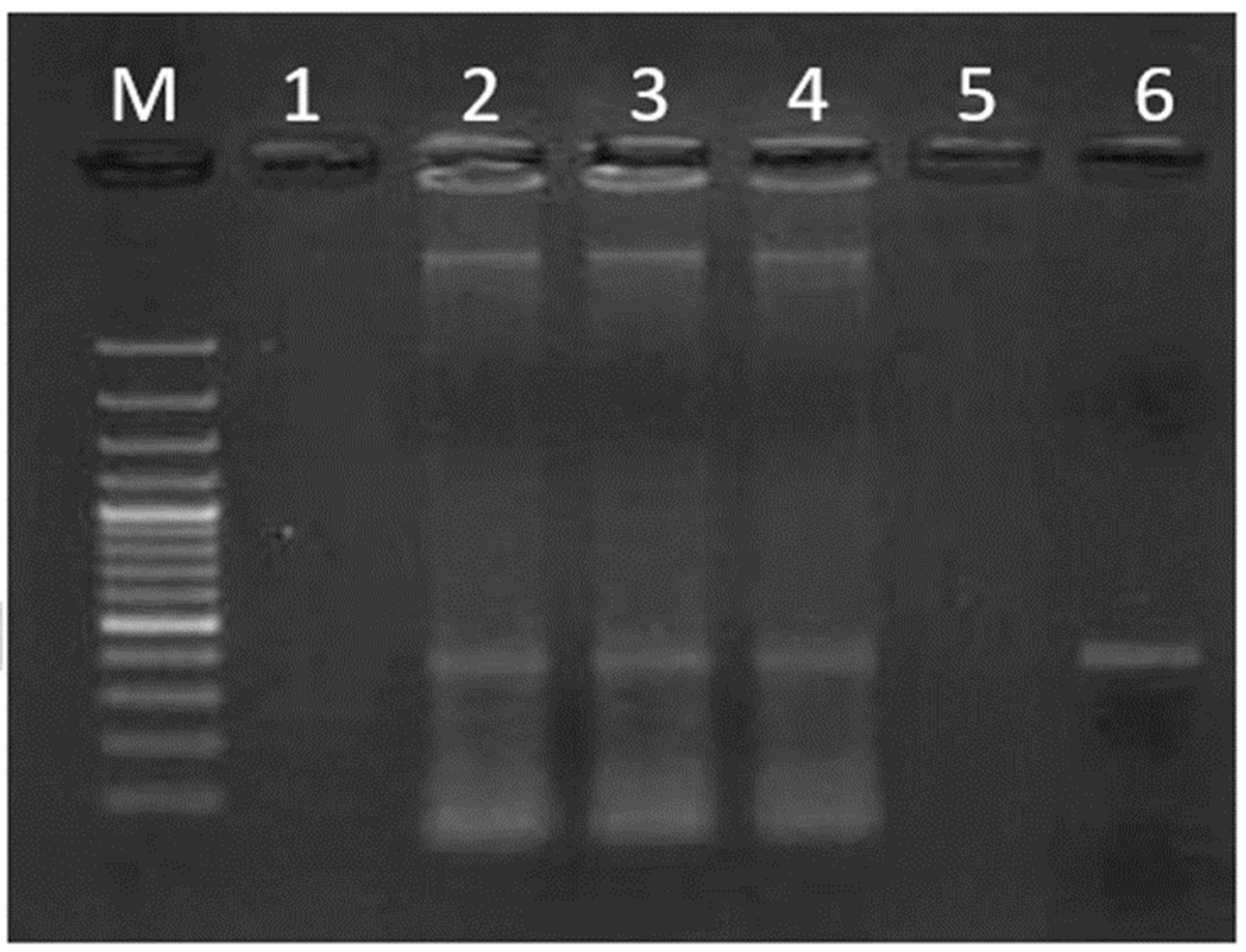
2.2. DNA Extraction and Quantification by qPCR

Supernatants from processed raw lettuces (≈10 mL) and artificially inoculated samples were used to extract bacterial DNA. The GeneJet™ genomic DNA purification kit (Fermentas, Baden-Württemberg, St. Leon-Rot, Germany) was used for the extraction of nucleic acids, following the mammalian tissue protocol, according to the manufacturer’s instructions. Aliquots of 1 mL from each sample were used in quintuplicate to obtain DNA purified samples that were stocked at −80 °C to the moment to be used.
Purified DNA samples were included in real-time quantitative polymerase chain reaction (qPCR) assays. H. pylori-specific VacA primers (Vac1 (5′-GGCACACTGGATTTGTGGCA-3′) and Vac2 (5′-CGCTCGCTTGATTGGACAGA-3′)) were used to amplify a 372-bp fragment from the vacA gene [29]. The QPCR assay was based on SYBR Green I fluorescence dye in a Light-Cycler VR 2.0 Instrument (Roche Applied Science, Barcelona, Spain). The amplification mixture consisted of 2 µL SYBR green real-time PCR master mix (Roche Applied Science, Barcelona, Spain), 0.5 µL of each primer (20 mM), 1.6 µL MgCl2 (50 mM), and 2 µL of DNA, in a final volume of 20 µL. The amplification-optimized procedure included an initial DNA denaturalization step at 95 °C for 10 min, followed by 40 cycles of 95 °C for 10 s, 62 °C for 5 s, and 72 °C for 16 s, and finally, one cycle at 72 °C for 15 s and one at 40 °C for 30 s [30]. H. pylori NCTC 11638 DNA was used as a positive control. A negative control was included by replacing DNA with an equal volume of sterile water. Assays for each sample were carried out in triplicate.
Melting curves were obtained experimentally for each sample, and crossing points (Cp) and melting temperatures (Tm) were used in each case to assess the H. pylori-positive result. The melting temperature (Tm) for the VacA primers was 85 °C. A standard curve was prepared using H. pylori NCTC 11638-purified DNA in the range of 101–106 genomic units (GU), corresponding to cycle threshold (Cp) media values ranging from 34 to 15.62 (Cp = −3.733 · Log10 (GU) + 38.98; r2 = 1)) [30]. The limit of quantitative detection by the optimized qPCR procedure was 10 cells, assuming that each gene copy of the vacA gene was equivalent to one cell [31].
The SYBR Green qPCR products obtained for vegetable samples (raw materials and artificially inoculated ones) were also submitted to electrophoresis (90 V for about 90 min) on gel-stained 1% (w/v) agarose (Pronadisa, Madrid, Spain) in 1× Tris–Acetate–EDTA (TAE) buffer prepared with 0.01% GelRed (Biotium), and visualized by UV transillumination. A 100 bp DNA ladder (GeneRuler 100 bp Plus DNA Ladder, Thermo Fisher/Fermentas, Burlington, Ontario, Canada) was used as a molecular weight marker.

2.3. Sequencing of Presumptive Positive H. pylori Samples

Vegetable leafy samples with qPCR presumptive positive H. pylori amplicons were purified with the commercial Kit Illustra GFX PCR DNA and Gel band Purification (GE Healthcare, Barcelona, Spain) according to the manufacturer’s instructions. Purified DNA was quantified spectrophotometrically (A260/A280 ratio > 1.7 for purified DNAs). Both DNA strands were sequenced commercially (Sistemas Genomicos S.L., Valencia, Spain). The Basic Logical Alignment Search Tool (BLAST) (www.ncbi.nlm.nih.gov/BLAST) (last accessed 15 October 2021) was used to determine the homology of the amplified sequences and the vacA gene fragment corresponding to the reference H. pylori NCTC 11638 strain (GenBank) [32].

3. Results

Prevalence of Helicobacter pylori in Fresh Vegetables

H. pylori cells of artificially inoculated leafy vegetable samples were positively recovered in 96% of cases, validating the applied procedure. Afterwards, by means of the optimized qPCR approach, the accuracy of H. pylori cell quantification (in the range 106–108 CFU/10 g inoculated matrices) was tested, with a correlation coefficient of r2 = 0.98 (predicted versus observed).
In the assessment of raw vegetable matrices, 20% of analyzed samples (20 out of 100) were presumptive positive by qPCR assay, corresponding to 15 lettuce, 2 spinach, and 3 chard samples (Figure 1). The Tm values for presumptive positive samples were close to 85 °C (84.8 to 86.5 °C), and Cp ranged from 34 to 29.
The qPCR presumptive positive amplicons were tested for the specific vacA amplified fragment band in an agarose gel (Figure 2). Among presumptive positive samples, 18 out of 20 showed a positive intense 372 bp band. These amplimers were used for sequencing analysis.
The sequencing results confirmed that 17 selected presumptive positive samples yielded a match of 98–100% with the H. pylori vacA gene sequence and thus were confirmed as positive for H. pylori presence (17 out of 100 samples, 17% (9.7, 24.3; CI 95%)).
The positive samples corresponded to 12 out of 45 (26.6%) lettuce, 2 out of 21 (9.5%) spinach, and 3 out of 34 (8.8%) chard samples. Contamination levels were in the range of 1.5 to 2.5 log10 cycles (36 to 335 CFU/10 g). Lettuce was confirmed as the most frequently contaminated vegetable (26.6% of positive units among tested lettuce samples).

4. Discussion

This study has detected the presence of H. pylori in unwashed fresh vegetables that are consumed raw in the Mediterranean area at rates close to 18% of samples. These results agree with those in the literature. Atapoor et al. (2014) analyzed 460 samples of salads and vegetables (leek, basil, and lettuce) by qPCR and detected that close to 20% of unwashed lettuce samples were H. pylori-positive [15].
Similar H. pylori contamination rates were found by Yahagi and co-workers (2014): salads and fresh vegetables were analyzed by qPCR using virulence gene markers (oipA, cagA, VacA, iceA1), and approximately 14% of samples were positive for H. pylori presence. Among them, 30% of them corresponded to lettuce, leek, and cabbage matrices [22].
In this work, levels of H. pylori in confirmed positive vegetable samples were in the range of 36 to 335 CFU/10 g. Although little is known about the infectious dose of H. pylori tested in vivo, some studies suggested that 102–104 cells will be necessary to set up the chronic colonization of the host [33,34]. Consumption of lettuce in the Mediterranean diet is very frequent. In fact, this leafy vegetable is generally consumed daily (5–20 g) [1,35]. Consequently, our results seem to indicate that levels of H. pylori contamination in vegetables that are consumed raw can represent a risk for consumers.
However, this is a preliminary study, and much more research should be conducted in order to assess the representativeness of these results by applying a statistical model based on which the survey results can be validated. It would also be of great interest to carry out a screening of the antimicrobial resistance of the isolates present in vegetables. Moreover, the use of other bioinformatic tools to analyze the amplified sequences should allow for calculating the nucleotide diversity, the polymorphic level (haplotype diversity) and number of variable sites, and the average number of nucleotide differences (K) of the strains, thus improving our knowledge not only about the epidemiology, but also of the genetic features of H. pylori.

5. Conclusions

A total of 17 out of 100 vegetable samples (12/45 (26.6%) lettuce, 2/21 (9.5%) spinach, and 3/34 (8.8%) chard samples) were confirmed to be positive for the presence of H. pylori. Contamination levels ranged from 36 to 335 UFC/g in leafy vegetables. According to these results, levels of H. pylori close to the estimated infectious doses (102–104 cells) are detected by qPCR in raw vegetable samples from the Mediterranean area of Spain. Considering the biological risk associated with this pathogen as a carcinogenic agent, the identification and removal of H. pylori across the food chain remains a food safety challenge. Since previous works have demonstrated that this pathogen is able to survive and proliferate in lettuce, preventive strategies such as (i) ensuring the good quality of irrigation water, (ii) correct handling and washing procedures postharvest, (iii) using a correct refrigeration step (5 °C) during distribution, and (iv) washing procedures before consumption could be a suitable way to control the pathogen, reducing the viability and load of H. pylori and, consequently, the risk for consumers.

Author Contributions

M.A.F. participated in the conceptualization of the research as the main leader, funding acquisition and project administration. M.G.-F. carried out the experimental investigation and original draft preparation; A.G. participated in the development and application of molecular techniques, validation of procedures, and supervision; M.C.P.-P. contributed to the investigation, data formal analysis, writing original draft preparation, and in supervision. All authors have read and agreed to the published version of the manuscript.

Funding

This research was funded by the Ministerio de Ciencia e Innovación, Spain, grant number PID2019-105691RB-I00. M. García-Ferrús is the recipient of a PEJ2018-003746A grant from Ministerio de Economía, Industria y Competitividad, Spain. Prof. Pina-Pérez is also grateful to the GVA202/031 grant project.

Conflicts of Interest

The authors declare no conflict of interest.

References

  1. EU Statistics. Fruits and Vegetables Consumption in EU. 2018. Available online: https://ec.europa.eu/eurostat/statistics-explained/index.php/Fruit_and_vegetable_consumption_statistics (accessed on 20 January 2022).
  2. Harvey, R.R.; Zakhour, C.M.; Gould, L.H. Foodborne disease outbreaks associated with organic foods in the United States. J. Food Prot. 2016, 79, 1953–1958. [Google Scholar] [CrossRef] [PubMed] [Green Version]
  3. Blau, K.; Bettermann, A.; Jechalke, S.; Fornefeld, E.; Vanrobaeys, Y.; Stalder, T.; Top, E.M.; Smalla, K. The transferable resistome of produce. Am. Soc. Microbiol. 2018, 9, e01300–e01318. [Google Scholar] [CrossRef] [PubMed] [Green Version]
  4. Venneman, K.; Huybrechts, I.; Gunter, M.J.; Vandendaele, L.; Herrero, R.; Van Herck, K. The epidemiology of Helicobacter pylori infection in Europe and the impact of lifestyle on its natural evolution toward stomach cancer after infection: A systematic review. Helicobacter 2018, 23, e12483. [Google Scholar] [CrossRef] [PubMed]
  5. Hooi, J.K.Y.; Lai, W.Y.; Ng, W.K.; Suen Massess, M.Y.; Underwood, F.E.; Tanyingoh, D.; Malfertheiner, P.; Graham, D.Y.; Wong, V.W.S.; Wu, J.C.Y.; et al. Global prevalence of Helicobacter pylori infection: Systematic review and meta-analysis. Gastroenterology 2017, 53, 420–429. [Google Scholar] [CrossRef] [PubMed] [Green Version]
  6. World Health Organization (WHO). WHO Publishes List of Bacteria for Which New Antibiotics Are Urgently Needed. 2017. Available online: https://www.who.int/news-room/detail/27-02-2017-who-publishes-list-of-bacteria-for-which-new-antibiotics-are-urgently-needed (accessed on 20 January 2022).
  7. Gisbert, J.P. “Rescue” regimens after Helicobacter pylori treatment failure. World J. Gastroenterol. 2008, 14, 5385–5402. [Google Scholar] [CrossRef] [PubMed]
  8. IARC Working Group on the Evaluation of Carcinogenic Risks to Humans. Biological Agents. V100 B. A review of human carcinogens. IARC Monogr. Eval. Carcinog. Risks Hum. 2012, 100, 1–441. [Google Scholar]
  9. Azevedo, N.F.; Almeida, C.; Fernandes, I.; Cerqueira, L.; Dias, S.; Keevil, C.W.; Vieira, M.J. Survival of gastric and enterohepatic Helicobacter spp. in water: Implications for transmission. Appl. Environ. Microbiol. 2008, 74, 1805–1811. [Google Scholar] [CrossRef] [Green Version]
  10. Konishi, K.; Saito, N.; Shoji, E.; Takeda, H.; Kato, M.; Asaka, M.; Ooi, H.K. Helicobacter pylori: Longer survival in deep ground water and sea water than in a nutrient-rich environment. Apmis 2007, 115, 1285–1291. [Google Scholar] [CrossRef]
  11. Bai, X.; Xi, C.; Wu, J. Survival of Helicobacter pylori in the wastewater treatment process and the receiving river in Michigan, USA. J. Water Health 2016, 14, 692–698. [Google Scholar] [CrossRef] [Green Version]
  12. Zamani, M.; Vahedi, A.; Maghdouri, Z.; Shokri-Shirvani, J. Role of food in environmental transmission of Helicobacter pylori. Caspian J. Intern. Med. 2017, 8, 146–152. [Google Scholar]
  13. Quaglia, N.C.; Dambrosio, A. Helicobacter pylori: A foodborne pathogen? World J. Gastroenterol. 2018, 24, 3472–3487. [Google Scholar] [CrossRef]
  14. Leja, M.; Grinberga-Derica, I.; Bilgilier, C.; Steininger, C. Review: Epidemiology of Helicobacter pylori infection. Helicobacter 2019, 24 (Suppl. 1), e12635. [Google Scholar] [CrossRef] [PubMed] [Green Version]
  15. Atapoor, S.; Safarpoor Dehkordi, F.; Rahimi, E. Detection of Helicobacter pylori in various types of vegetables and salads. Jundishapur J. Microbiol. 2014, 7, e10013. [Google Scholar] [CrossRef] [PubMed] [Green Version]
  16. Hamada, M.; Elbehiry, A.; Marzouk, E.; Moussa, I.M.; Hessain, A.M.; Alhaji, J.H.; Heme, H.A.; Zahran, R.; Abdeen, E. Helicobacter pylori in a poultry slaughterhouse: Prevalence, genotyping and antibiotic resistance pattern. Saudi J. Biol. Sci. 2018, 25, 1072–1078. [Google Scholar] [CrossRef] [PubMed]
  17. Moreno-Mesonero, L.; Moreno, Y.; Alonso, J.L.; Ferrús, M.A. Detection of viable Helicobacter pylori inside free-living amoebae in wastewater and drinking water samples from Eastern Spain. Environ. Microbiol. 2018, 19, 4103–4112. [Google Scholar] [CrossRef] [PubMed] [Green Version]
  18. Abdel-Moein, K.A.; Saeed, H.; Samir, A. Novel detection of Helicobacter pylori in fish: A possible public health concern. Acta Trop. 2015, 152, 141–144. [Google Scholar] [CrossRef] [PubMed]
  19. Vesga, F.J.; Moreno, Y.; Ferrús, M.A.; Campos, C.; Trespalacios, C.C. Detection of Helicobacter pylori in drinking water treatment plants in Bogotá, Colombia, using cultural and molecular techniques. Int. J. Hyg. Environ. Health 2018, 221, 595–601. [Google Scholar] [CrossRef]
  20. Allende, A.; Monaghan, J. Irrigation water quality for leafy crops: A perspective of risks and potential solutions. Int. J. Environ. Res. Public Health 2015, 12, 7457–7477. [Google Scholar] [CrossRef] [Green Version]
  21. Rahman, S.M.E.; Mele, M.A.; Lee, Y.T.; Islam, M.Z. Consumer preference, quality, and safety of organic and conventional fresh fruits, vegetables, and cereals. Foods 2021, 10, 105. [Google Scholar] [CrossRef]
  22. Yahaghi, E.; Khamesipour, F.; Mashayekhi, F.; Safarpoor Dehkordi, F.; Hossein Sakhaei, M.; Masoudimanesh, M.; Khayyat Khameneie, M. Helicobacter pylori in vegetables and salads: Genotyping and antimicrobial resistance properties. Biomed. Res. Int. 2014, 2014, 757941. [Google Scholar] [CrossRef] [Green Version]
  23. Pina-Pérez, M.C.; González, A.; Moreno, Y.; Ferrús, M.A. Helicobacter pylori growth pattern in reference media and extracts from selected minimally processed vegetables. Food Control 2018, 86, 389–396. [Google Scholar] [CrossRef]
  24. Buck, A.; Oliver, J.D. Survival of spinach-associated Helicobacter pylori in the viable but nonculturable state. Food Control 2010, 21, 1150–1154. [Google Scholar] [CrossRef]
  25. Aktas, D.; Bagirova, M.; Allahverdiyev, A.M.; Abamor, E.S.; Safarov, T.; Kocazeybek, B.S. Resuscitation of the Helicobacter pylori coccoid forms by resuscitation promoter factor obtained from Micrococcus luteus. Curr. Microbiol. 2020, 77, 2093–2103. [Google Scholar] [CrossRef] [PubMed]
  26. Ng, C.G.; Loke, M.F.; Goh, K.L.; Vadivelu, J.; Ho, B. Biofilm formation enhances Helicobacter pylori survivability in vegetables. Food Microbiol. 2017, 62, 68–76. [Google Scholar] [CrossRef] [PubMed]
  27. Huang, L. Optimization of a new mathematical model for bacterial growth. Food Control 2013, 32, 283–288. [Google Scholar] [CrossRef]
  28. Baranyi, J.; Csernus, C.; Beczner, J. Error analysis in predictive modelling demonstrated on mould data. Int. J. Food Microbiol. 2014, 170, 78–82. [Google Scholar] [CrossRef]
  29. Nilsson, H.O.; Blom, J.; Abu-Al-Soud, W.; Ljungh, A.A.; Andersen, L.; Wadström, T. Effect of cold starvation, acid stress, and nutrients on metabolic activity of Helicobacter pylori. Appl. Environ. Microbiol. 2002, 68, 11–19. [Google Scholar] [CrossRef] [Green Version]
  30. Santiago, P.; Moreno, Y.; Ferrús, M.A. Identification of viable Helicobacter pylori in drinking water supplies by cultural and molecular techniques. Helicobacter 2015, 20, 252–259. [Google Scholar] [CrossRef]
  31. Pina-Pérez, M.C.; González, A.; Moreno, Y.; Ferrús, M.A. Helicobacter pylori detection in shellfish: A real-time quantitative polymerase chain reaction approach. Foodborne Path. Dis. 2018, 16, 137–143. [Google Scholar] [CrossRef]
  32. Ye, J.; Coulouris, G.; Zaretskaya, I.; Cutcutache, I.; Rozen, S.; Madden, T.L. Primer-BLAST: A tool to design target-specific primers for polymerase chain reaction. BMC Bioinform. 2012, 13, 134. [Google Scholar] [CrossRef] [Green Version]
  33. Solnick, J.V.; Hansen, L.M.; Canfield, D.R.; Parsonnet, J. Determination of the infectious dose of Helicobacter pylori during primary and secondary infection in rhesus monkeys (Macaca mulatta). Infect. Immun. 2001, 69, 6887–6892. [Google Scholar] [CrossRef] [PubMed] [Green Version]
  34. Ferrero, R.L.; Thiberge, J.M.; Kansau, I.; Wuscher, N.; Huerre, M.; Labigne, A. The GroES homolog of Helicobacter pylori confers protective immunity against mucosal infection in mice. Proc. Natl. Acad. Sci. USA 1995, 92, 6499–6503. [Google Scholar] [CrossRef] [PubMed] [Green Version]
  35. Pagliai, G.; Sofi, F.; Vannetti, F.; Caiani, S.; Pasquini, G.; Molino Lova, R.; Cecchi, F.; Sorbi, S.; Macchi, C. Mediterranean diet, food consumption and risk of late-life depression: The mugello study. J. Nutr. Health Aging 2018, 22, 569–574. [Google Scholar] [CrossRef] [PubMed]
Figure 1. Melting curves obtained for H. pylori vacA gen quantitative detection from raw vegetables, including: curve 19, positive control (Tm 86 °C); curve 15, V91 peak detected in spinach (Tm 85.4 °C); curve 5, V17 peak detected in lettuce (85.5 °C); curve 1, VI peak detected in chard (Tm 86.1 °C).
Agriculture 12 00339 g001
Figure 2. Example of a gel electrophoresis of the amplicons of vacA H. pylori gen generated by means of qPCR. M: Molecular weight marker (100 bp DNA Ladder); lane 1: sample V3, chard; lane 2: sample V20, lettuce; lane 3, sample V2, chard; lane 4, sample V12, spinach; lane 5, sample V6, lettuce; lane 6, positive control H. pylori NCTC 11638.
Agriculture 12 00339 g002
Publisher’s Note: MDPI stays neutral with regard to jurisdictional claims in published maps and institutional affiliations.

