Article

Transcriptome-Wide Identification of Cytochrome P450s and GSTs from Spodoptera exigua Reveals Candidate Genes Involved in Camptothecin Detoxification

State Key Laboratory for Biology of Plant Diseases and Insect Pests, Institute of Plant Protection, Chinese Academy of Agricultural Sciences, Beijing 100193, China
*
Author to whom correspondence should be addressed.
Agriculture 2023, 13(8), 1494; https://doi.org/10.3390/agriculture13081494
Submission received: 20 June 2023 / Revised: 21 July 2023 / Accepted: 23 July 2023 / Published: 27 July 2023
(This article belongs to the Special Issue Evolution, Mechanisms and Management of Insecticide Resistance)

Abstract

:
The application of camptothecin (CPT) and its derivatives to control insect pests has generated significant interest. This study investigated the enzymatic response of cytochrome P450 monooxygenase (CYP) and glutathione S-transferase (GST) genes in the fat body cells of Spodoptera exigua after 10 μM CPT treatment. Additionally, we examined the effects of CPT on the growth and development of S. exigua larvae and detected the relative expression levels of selected CYP and GST genes during the CPT treatment period. Twenty-one CYP and 17 GST genes were identified from the fat body cells of S. exigua by comparative transcriptomic analyses. The relative expression of ten CYP and seven GST genes changed significantly, which suggested that these CPYs and GSTs may be involved in CPT metabolism. During exposure to CPT for 10 days, the development of S. exigua larvae was delayed and was characterized by weight inhibition and a prolonged period of development. The relative expression levels of the selected four CYP genes, CYP9A27, CYP9A186, CYP337B5, CYP321A8, and one GST gene, GSTe7, were significantly changed by CPT treatment compared to the control group. These generated data provide a basis for identifying the CPT metabolism/detoxification genes of S. exigua at the molecular level.

1. Introduction

Camptothecin (CPT), a botanical indole alkaloid, was isolated from Campototheca acuminata, which is native to China. CPT and its derivatives (CPTs) are well known for their antitumor activity and are considered to be an important class of drugs used in chemotherapy. It has been documented that its complex metabolism involves many proteins. In humans, cytochrome P450 monooxygenases (P450s or CYPs) have been documented as being involved in the degradation of CPTs, especially P450 isoforms 1A1, 3A4, and 3A5 (CYP1A1, CYP3A4, CYP3A5) [1,2,3]. In recent years, based on the knowledge of the metabolism of CPTs by CYPs, a promising CPT delivery scaffold—CPT-encapsulated PLGA (poly(lactic-co-glycolic acid), PLGA-CPT) nanoparticles—was prepared to improve the bioavailability of CPTs and reduce their side effects [4]. These studies highlight the importance of the metabolism of CPTs for drug discovery and predicting the toxicity and efficacy of CPTs in drug therapy [5].
In agriculture, much interest has been focused on developing CPTs as potential biological pesticides to control agricultural pests due to the unique action mechanisms and insecticidal activities of CPTs. It has been documented that CPTs are effective against several pest insects, including Spodoptera exigua [6,7], S. litura [8], S. frugiperda [9], Nilaparvata lugens, Brevicoryne brassicae, and Chilo suppressalis [10]. In our previous studies, CPTs induced cytotoxicity against IOZCAS-Spex-II cells derived from S. exigua by promoting apoptosis [11] and increasing intracellular oxidative stress [12,13]. In addition, a series of CPT derivatives was designed and synthesized to increase the solubility and bioavailability of CPT [6,14,15,16]. With the development of nanopesticides, CPT was conjugated with polyethylene glycol by using nanotechnology to form an amphiphilic copolymer, mPEG-CPT, which exhibited potent or superior insecticidal activities against three pests, B. brassicae, Tetranychus cinnabarinus, and Bursaphelenchus xylophilus [17]. However, to date, little is known about the metabolism of CPTs in insects. To improve the bioavailability of CPTs and promote the application of CPTs in the control of insect pests, the metabolism of CPTs should be investigated comprehensively in insects.
The detoxification systems of insects have been shown to play central roles in the metabolism of a wide range of natural and synthetic xenobiotics, including insecticides. The most frequently involved enzyme families contributing to the metabolism of xenobiotics included CYPs, glutathione S-transferases (GSTs), and carboxylesterase (CarEs), although other enzyme families may also be involved [18]. For insecticides, gene amplification, overexpression and/or the modification of genes encoding members of the CYPs and GSTs must be involved in the metabolic detoxification of multiple insecticides [19,20]. In the present study, we investigated the enzymatic response, including CYP and GST genes, in the fat body cells of S. exigua after 10 μM CPT treatment. Furthermore, the effects of CPT on the growth and development of S. exigua larvae were examined, and the relative expression levels of the selected CYP and GST genes were monitored during the CPT treatment period.

2. Materials and Methods

2.1. Chemicals, Cell Line, and Culture Conditions

CPT (98%) was supplied by Knowshine Pharmachemicals Inc., Shanghai, China. Dimethyl sulfoxide (DMSO, ≥99.5%) was purchased from Sigma Aldrich Co., Shanghai, China.

2.2. CPT Treatment of S. exigua Fat Body Cells

IOZCAS-Spex-II cells were established from the fat body of S. exigua and offered by Prof. Qilian Qing of the Institute of Zoology, Chinese Academy of Sciences, Beijing, China. The cells were grown in T25 cm2 tissue culture flasks (Corning, New York, NY, USA) and maintained in Grace’s insect culture medium supplemented with 10% fetal bovine serum. The culture was incubated at a temperature of 27 °C. Subculturing was performed every 6 days.
IOZCAS-Spex-II cells in the logarithmic phase were harvested and diluted to a density of 2 × 10⁶ cells mL−1 in the cell cultures. Following overnight incubation, the cells were exposed to CPT (10 μM well−1) at appropriate concentrations for 24 h. DMSO (0.1%) was used as a control. Each treatment was repeated at least three times.

2.3. RNA Extraction, Library Construction and Sequencing

Total RNA was extracted from cell samples using the RNeasy Mini Kit (Qiagen, Hilden, Germany) according to the manufacturer’s instructions. The integrity of the total RNA was confirmed by using both 1.5% agarose gel electrophoresis and an Agilent 2100 Bioanalyzer (Agilent Technologies, Santa Clara, CA, USA). The concentration of the total RNA was determined using a NanoDrop 1000 spectrophotometer (Thermo Fisher Scientific Inc. Waltham, MA, USA). cDNA libraries were constructed using an Illumina TruSeq RNA sample preparation kit following the manufacturer’s instructions (Illumina Inc., San Diego, CA, USA). After quantification and qualification, the cDNA libraries were sequenced with Illumina HiSeqTM 2000 in accordance with the standard procedure.
The raw reads were filtered to remove reads containing adapter, poly-N, and low-quality reads. The resulting clean reads were subjected to pool de novo transcriptome assembly using the Trinity-v2.3.2 assembler programs at default parameters without a reference genome. All de novo transcripts were predicted to have open reading frames (ORFs) with TransDecoder software (v5.5.0) and then corrected with the Pfam database. ORF annotation was performed against the NCBI nonredundant protein database (NR) and SwissProt database. The gene expression level was computed using RSEM quantitative analysis and FPKM transformation. edgeR-2020 software was used to screen the differentially expressed genes between the two samples with a fold change (FC) ≥ 1 and false discovery rate (FDR) < 0.01 as the threshold by which to judge the significance of differences in gene expression.

2.4. Identification of Detoxification Genes and Phylogenetic Analysis

The assembled transcripts associated with CYPs and GSTs were manually selected and identified from the transcriptome data of the control and CPT-treated groups. These candidate sequences were realigned by BLAST in NCBI to find homology, and preliminary classification was based on the comparison results (Table S1).
The full protein sequences of all putative detoxification enzymes were obtained and analyzed using the ExPASy (the Expert Protein Analysis System) server http://web.expasy.org/translate/ (accessed on 11 November 2022). Due to the size differences of the two detoxification gene repertoires, the corresponding protein sequences were selected from different species to build the trees (Table S2). To assign predicted S. exigua detoxification genes to different phylogenetic subgroups, multiple sequence alignments of the CYP and GST amino acids were performed using MUSCLE in MEGA 11 software. The phylogenetic tree was generated using the neighbor-joining method of the MEGA 11 program with a bootstrap value of 1000. The webserver https://www.evolgenius.info/evolview (accessed on 5 December 2022), Evolview v3, was used for the visualization, annotation, and management of phylogenetic trees [21].

2.5. Real-Time Quantitative PCR (RT-qPCR)

RT-qPCR was performed to verify the expression of candidate CYP and GST genes. The PCR primer sequences used for quantification of the 43 candidate genes are shown in Table S3. GAPDH (glyceraldehyde-3-phosphate dehydrogenase) and L7A (ribosomal protein L7A) were used as the reference genes in this study [22]. A standard curve for each gene was established to evaluate the reaction efficiency of RT-qPCR. The melt curves were examined to check the specificity of the primers. RT-qPCR was carried out using the Hieff UNICON® Universal Blue qPCR SYBR Green Master Mix (YEASEN, Shanghai, China) on an Applied Biosystems QuantStudio 3 Real-Time PCR System (ABI, Carlsbad, CA, USA). Each 20 μL reaction contained 10 μL SYBR Green, 0.6 μL of each primer, 1 μL cDNA template, and double distilled water. The cycling parameters consisted of an initial step at 95 °C for 120 s, followed by 40 cycles of 95 °C for 10 s and 55–60 °C for 30 s.

2.6. Effect of CPT on the Growth and Development of S. exigua Larvae

A stock solution of 1.00 × 10⁵ mg/L CPT was prepared with DMSO, which was then diluted with dH2O (containing 0.05% Triton X-100) to prepare the tested solution. Uniform second-instar larvae were reared on cabbage leaves dipped in CPT at concentrations of 0.02, 0.1, and 0.5 g/L for 10 days. Fresh cabbage dipped in CPT was provided every day and five replicates were made for each treatment with 20 individuals per replicate. Larvae were reared on cabbage leaves treated with dH2O (containing 0.05% Triton X-100 and 0.1% DMSO) served as the control group. The survival rate, weight, and developmental period of each individual were checked daily. The rearing conditions for S. exigua larvae were 27 ± 1 °C and 60 ± 5% relative humidity (RH) with a photoperiod of 14 h of light followed by 10 h of darkness 14:10 h (L:D).

2.7. Effect of CPT on the Expression of Detoxification Genes in S. exigua Larvae

Total RNA was extracted from S. exigua larvae with a TRNzol Universal total RNA extraction kit following the instructions provided by TIANgen Biotech Co., Ltd. Primary strand cDNA was generated from 1 μg of total RNA in a 20 μL volume using the Omniscript® Reverse Transcript Kit (Qiagen, ON, Canada) and was stored at −20 °C for future analysis. RT-qPCR was performed as described in Section 2.3.

3. Results

3.1. Identification of Candidate P450s and GSTs from the Fat Body Cells of S. exigua

As shown in Figure 1a, twenty-one putative CYP genes among the DEGs were identified from the transcriptome data obtained from the fat body cells of S. exigua. The phylogenetic tree resolved the four expected clans of insect CYPs, including CYP2, CYP3, CYP4, and the mitochondrial CYP clans. Specifically, thirteen genes were classified into the CYP3 clan, four into the CYP4 clan, three into the mitochondrial CYP clan, and one into the CYP2 clan. Additionally, the 21 CYPs belonged to 10 families and 17 subfamilies. The relative expression of nine genes, CYP6AB14, CYP6AB31, CYP6AB60, CYP337B5, CYP6AW1, CYP9A27, CYP9A186, CYP4G74 and CYP333A12, was upregulated significantly and one gene, CYP333B4, was downregulated significantly in S. exigua after treatment with 10 μM CPT for 24 h compared to the control group. The relative expression of a total of 11 genes, CYP6AN4, CYP6AE74, CYP321A8, CYP354A14, CYP9AJ1, CYP324A1, CYP4M14, CYP4CG1, CYP4G75, CYP306A1, and CYP339A1, was not changed in S. exigua after treatment with CPT compared with the control.
Seventeen GST genes were identified from the transcriptome data of S. exigua. Among these, phylogenetic analyses revealed that five GST classes were assigned, ten in epsilon, two in delta, one in theta, three in sigma, and one in unclassified (Figure 1b). The relative expression of seven genes, GSTe4, GSTe7, GSTe10, GSTe12, GSTd2, GSTu1, and GSTs2, was upregulated significantly in S. exigua after treatment with 10 μM CPT for 24 h compared to the control group. One gene, GSTd3, was significantly downregulated following CPT treatment in S. exigua.

3.2. Real-Time Quantitative PCR Validation of Candidate Genes from the Fat Body Cells of S. exigua

As shown in Figure 2, the expression patterns of the selected CYP (Figure 2a) and GST (Figure 2b) genes in the fat body cells of S. exgua changed significantly after 10 μM CPT treatment based on RT-qPCR analysis. The changes in gene expression levels based on RT-qPCR were largely consistent with the transcriptomic data. This result showed that these detoxification enzyme genes, especially CYPs, including CYP337B5, CYP4G74, CYP6AB14, CYP6AB31, CYP6AB60, CYP9A186, and CYP9A27, and GSTs, including GSTd2, GSTe10, GSTe7, GSTe12, GSTe4, and GSTu1, were potentially involved in the detoxification of CTP in S. exigua.

3.3. Effects of CPT on the Development of S. exigua Larvae

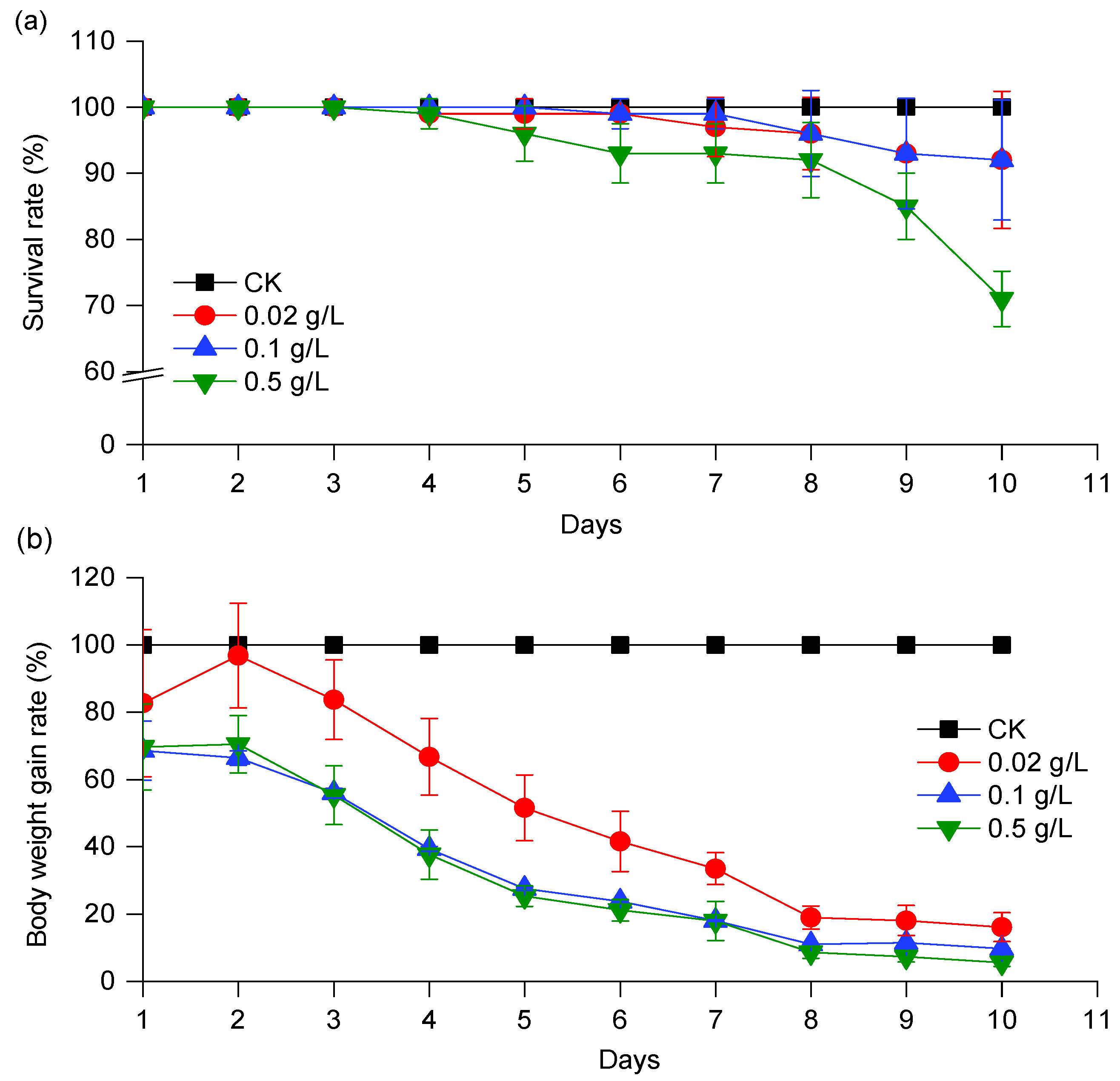
The survival rate, weight, and age-stage specific survival rate of S. exigua in different treatment groups are shown in Figure 3 and Figure 4, respectively. The survival rates of S. exigua were 92.0%, 92.0%, and 71.0% after CPT treatment for 10 days at doses of 0.02, 0.1, and 0.5 g/L, respectively (Figure 3a). The survival rate of S. exigua larvae in the 0.5 g/L concentration group was significantly lower than that in the other two treatment groups after treatment with CPT for 10 days. Compared with the control group, CPT significantly inhibited larval weight at all treatment doses. As shown in Figure 3b, the weight gain rate declined gradually from 82.7%, 68.6%, and 69.6% to 16.1%, 9.66%, and 5.54% after CPT treatment for 10 days at doses of 0.02, 0.1, and 0.5 g/L, respectively.
The age-stage survival rate curves are shown in Figure 4 to illustrate the survival rates of newly hatched larvae developing to different stages. Observation overlaps were apparent between stages because the development rate varied among individuals. In the control group, the survival rates were 7.0%, 76.0%, and 17.0% for the 4th, 5th, and 6th instars at 10 days of development, respectively. Among the three CPT-treated groups, the survival rate decreased with increasing CPT concentration (1.0%, 82.0%, and 9.0% for the 3rd, 4th, and 5th instar larvae at a dose of 0.02 g/L, respectively; 22.0%, 67.0%, and 3.0% for the 3rd, 4th, and 5th instar larvae at a dose of 0.1 g/L, respectively; 57.0% and 14.0% for the 3rd and 4th instar larvae at a dose of 0.5 g/L, respectively). These results indicated that the larvae in the CPT-treated groups developed significantly later than those in the control group.

3.4. Effect of CPT on the Expression of CYP and GST Genes during the Development of S. exigua Larvae

To study the effect of CPT on the expression of CYP and GST genes, four CYP3 clan genes (CYP9A27, CYP9A186, CYP337B5, and CYP321A8) and two GSTs (GSTe8 and GSTs3) genes were selected to conduct further studies during the development of S. exigua larvae based on both results of transcriptome analysis and RT-qPCR verification. As shown in Figure 5, the expression of CYP9A27, CYP9A186, and CYP337B5 varied with the development of S. exigua larvae in the control group (CK), which indicated that the CYP9A27, CYP9A186, and CYP337B5 genes are potentially involved in the larval development of S. exigua (Figure 5a–c). However, the relative expression of CYP321A8 remained unchanged throughout the tested period of S. exigua larval development in the control group (Figure 5d). Compared with those in the control group, the relative expression of CYP9A27, CYP9A186, CYP337B5, and CYP321A8 was changed significantly by CPT treatment at different doses. Specifically, the relative expression of the CYP9A27 gene was reduced significantly by CPT at all tested concentrations during the whole tested period.
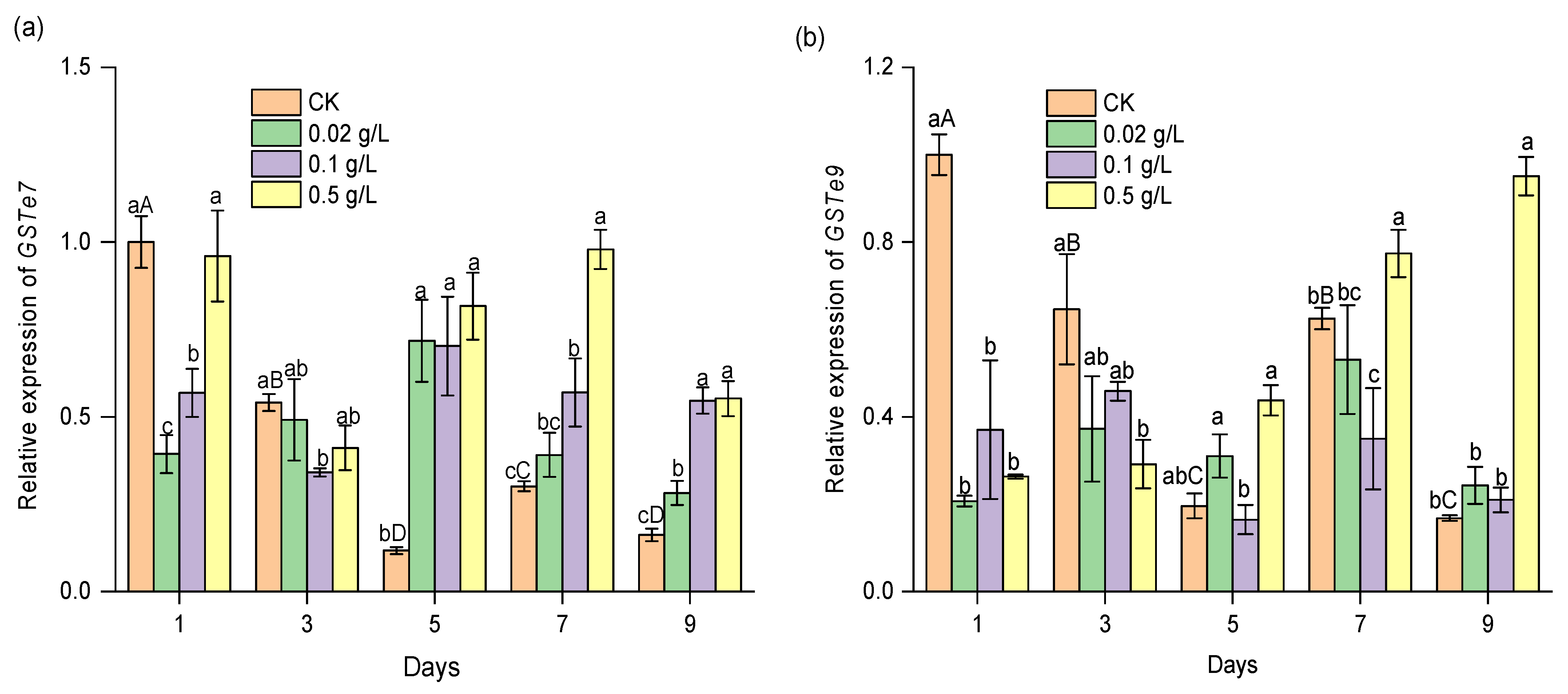
As shown in Figure 6, the relative expression levels of the GST genes GSTe7 and GSTe9 were significantly reduced during the entire testing period in the control group. From 5 days to 9 days, the relative expression of GSTe7 was increased significantly in the CPT-treated groups compared to the control group. The results show that GSTe7 expression was significantly induced by CPT.

4. Discussion

CYPs are a common and important metabolic detoxification gene family in insects. The fat body is a vital organ in insects that performs various functions, including xenobiotic metabolism and elimination. According to reports, a total of 53 CYPs were significantly differentially expressed with azadirachtin treatment in the fat bodies of S. frugiperda larvae, which indicated that these detoxification enzyme genes in fat bodies might be involved in azadirachtin detoxification in S. frugiperda [23]. In this study, a total of twenty-one putative CYP genes were identified from the transcriptomic data, of which nine and one were upregulated and downregulated, respectively, in the fat body cells established from S. exigua larvae after CPT treatment. Among them, seven genes (CYP6AB14, CYP6AB31, CYP6AB60, CYP337B5, CYP6AW1, CYP9A27, CYP9A186) belonged to the CYP3 clan and one (CYP4G74) belonged to CYP4. Compared to transcriptome analysis, the RT-qPCR results confirmed the expression level changes of six CYP3 clan genes CYP6AB14, CYP6AB31, CYP6AB60, CYP337B5, CYP9A27, CYP9A186, and one CYP4 clan gene CYP4G74. These results confirmed that members of the CYP3 and CYP4 clans are involved in the detoxification metabolism of insecticides and plant secondary metabolites in insects [19,24]. The CYP4G subfamily have been frequently associated with insecticide resistance in insects [25]. However, it has been documented that the CYP4G subfamily plays an indirect role in the insecticide resistance because they do not accept the insecticide molecule as a substrate. CYP4G enzymes are oxidative decarbonylases that catalyze the last step in cuticular hydrocarbon synthesis, making the insect cuticle thicker with a significantly reduced rate of insecticide penetration through the cuticle [26]. Subsequently, four CYP3 clan genes, CYP9A27, CYP9A186, CYP337B5, and CYP321A8, were selected to further determine the expression changes induced during CPT treatment for 10 days in S. exigua larvae.
During CPT treatment against S. exigua larvae for 10 days, it was observed that the development of S. exigua larvae was significantly delayed, characterized by the inhibition of weight gain and a prolonged period of larval development. In the control group, the expression levels of CYP9A27, CYP9A186, and CYP337B5 fluctuated significantly with the development of S. exigua larvae, which indicated that the expression of these genes was closely associated with the developmental period and may be involved in the regulation of the growth and development of S. exigua larvae. However, the expression levels of CYP321A8 remained constant during the 10 days in the control group. In the CPT-treated group, the relative expression of CYP9A27 was inhibited significantly at all three concentrations of CPT during the treatment period. The relative expression levels of CYP9A186, CYP337B5, and CYP321A8 were increased or decreased with the concentrations of CPT and the treatment period. These results illustrated the potential functions of these four genes in the metabolism/detoxication or regulation of insecticides. The CYP9 family belongs to the CYP3 clan in insects and plays key roles in insecticide resistance and xenobiotic metabolism. In the lepidopteran pests S. frugiperda, Helicoverpa armigera, and Cydia pomonella, previous studies have demonstrated that resistance to pyrethroids is conferred by the overexpression of members of the CYP9A subfamily [26,27,28]. Additionally, CYP9A1 in Heliothis virescens was overexpressed after exposure to thiodicarb and may play a key role in thiodicarb metabolism. In Locusta migratoria, the two LmCYP9A genes (LmCYP9AQ1 and LmCYP9A3) were confirmed to be involved in the detoxification of different pyrethroids [28]. Specifically, CYP9A186 confers resistance to emamectin benzoate and abamectin through enhanced metabolic detoxification in S. exigua [29]. CYP321A8 has been documented to confer resistance to organophosphate (chlorpyrifos) and pyrethroid (cypermethrin and deltamethrin) insecticides in S. exigua [30,31]. Our findings strengthened the possibility that CYP321A8 and CYP9A186 are significant for the detoxification/metabolism of insecticides in S. exigua, while some extra experiments must be carried out. Additionally, the role of CYP9A27 in the growth and development of S. eixgua provides potential new targets for pest management, and the inhibitory effects of CPT highlight the potential mechanism of CPT against insect pests.
GSTs are a diverse group of enzymes that play important roles in phase II detoxification processes. They can be further divided into seven categories, including the delta, epsilon, omega, theta, sigma, zeta, and unclassified categories [32]. In this study, a total of seventeen GST genes were identified from the transcriptomic data obtained from the fat body cells established from S. exigua larvae, among which ten belong to the epsilon class, two to delta, one to theta, three to sigma and one to an unclassified class. Six GST genes (GSTd2, GSTe10, GSTe7, GSTe12, GSTe4, and GSTu1) were significantly induced in the fat body cells established from S. exigua larvae after CPT treatment according to both DEG and RT-qPCR results. These results indicated that GSTs were associated with the detoxification of S. exigua to CPT, and those six GSTs might play important roles in insecticide detoxification or antioxidant protection in S. exigua. Previous studies have revealed that GSTs are involved in the detoxification of a variety of insecticides in insects. Specifically, the epsilon class of GSTs is involved in the metabolism of organophosphate, organochlorine, carbamate, and pyrethroid insecticides in Chilo suppressalis, Anopheles funestu, L. migratoria, and so on [33,34,35]. In this study, the relative expression levels of the two GSTs of the epsilon class, GSTe7 and GSTe9, were reduced with the development of S. exigua larvae in the control group. However, the expression of these two GSTs, especially GSTe7, was induced significantly by CPT treatment, which suggested that GSTe7 may be involved in the metabolism of CPT in S. exigua larvae.

5. Conclusions

In this study, 21 CYP and 17 GST genes were identified from the fat body cells of S. exigua using comparative transcriptomic analyses. The relative expression of ten CYP and seven GST genes changed significantly, suggesting their involvement in CPT metabolism. Exposure to CPT for 10 days, beginning at the second instar, resulted in delayed development of S. exigua larvae, as evidenced by reduced weight and extended development time. The relative expression levels of the CYP genes CYP9A27, CYP9A186, CYP337B5 and CYP321A8 and the GST gene GSTe7 were significantly altered by CPT treatment compared to the control group. These findings provide insight into the molecular-level identification of CPT metabolism/detoxification genes in S. exigua and demonstrate the potential use of CPT against insect pests.

Supplementary Materials

The following supporting information can be downloaded at https://www.mdpi.com/article/10.3390/agriculture13081494/s1: Table S1: Information for CYPs and GSTs in S. exigua obtained from the transcriptome data; Table S2: The corresponding protein sequences from different species for the phylogenetic analysis; Table S3: Information for primers used in RT-qPCR analysis.

Author Contributions

Z.Z. and L.Z. (Lan Zhang) conceived and designed the research. Z.Z. performed the experiments. Z.Z. and L.Z. (Lan Zhang) analyzed the data and wrote the manuscript. Y.Z., L.Z. (Lizhen Zhu) and H.J. reviewed the manuscript. X.L. and L.M. helped with the analysis with constructive discussions. All authors have read and agreed to the published version of the manuscript.

Funding

This research was supported by the Chinese National Natural Science Foundation (31672059).

Institutional Review Board Statement

Not applicable.

Data Availability Statement

The data that are presented in this study are available from the correspondence author upon request. The data are not publicly available due to privacy restrictions.

Acknowledgments

We would like to thank Cui for kindly providing S. exigua larvae.

Conflicts of Interest

The authors declare no conflict of interest.

References

  1. Toffoli, G.; Cecchin, E.; Corona, G.; Boiocchi, M. Pharmacogenetics of irinotecan. Curr. Med. Chem. Anticancer. Agents 2003, 3, 225–237. [Google Scholar] [CrossRef]
  2. Maier-Salamon, A.; Thalhammer, T.; Reznicek, G.; Böhmdorfer, M.; Zupkó, I.; Hartl, A.; Jaeger, W. Cytochrome P450 3A-mediated metabolism of the topoisomerase I inhibitor 9-aminocamptothecin: Impact on cancer therapy. Int. J. Oncol. 2014, 45, 877–886. [Google Scholar]
  3. Horn, J.; Milewska, M.; Arnold, S.M.; Leggas, M. Metabolic pathways of the camptothecin analog AR-67. Drug. Metab. Dispos. 2011, 39, 683–692. [Google Scholar]
  4. Bao, H.; Zhang, Q.; Yan, Z. The impact of camptothecin-encapsulated poly (lactic-co-glycolic acid) nanoparticles on the activity of cytochrome P450 in vitro. Int. J. Nanomed. 2019, 14, 383–391. [Google Scholar]
  5. Negoro, R.; Yamada, N.; Watanabe, K.; Kono, Y.; Fujita, T. Generation of Caco-2 cells stably expressing CYP3A4·POR·UGT1A1 and CYP3A4·POR·UGT1A1*6 using a PITCh system. Arch. Toxicol. 2022, 96, 499–510. [Google Scholar]
  6. Yang, F.; Wang, L.; Zhang, L.; Zhang, Y.; Mao, L.; Jiang, H. Synthesis and biological activities of two camptothecin derivatives against Spodoptera exigua. Sci. Rep. 2019, 9, 18067. [Google Scholar] [PubMed] [Green Version]
  7. Yang, F.; Zhang, L.; Zhang, Y.; Mao, L.; Zhu, L.; Liu, X.; Jiang, H. Effect of RNAi targeting SeGrx1 on the cytotoxicity and insecticide susceptibility of camptothecin in Spodoptera exigua. Agriculture 2022, 12, 930. [Google Scholar] [CrossRef]
  8. Dai, L.S.; Tian, H.F.; Hang, Y.; Wen, C.W.; Huang, Y.H.; Wang, B.F.; Hu, J.W.; Xu, J.P.; Deng, M.J. 1H NMR-based metabonomic evaluation of the pesticides camptothecin and matrine against larvae of Spodoptera litura. Pest Manag. Sci. 2021, 77, 208–216. [Google Scholar]
  9. Ngegba, P.M.; Cui, G.; Li, Y.; Zhong, G. Synergistic effects of chlorantraniliprole and camptothecin on physiological impairments, histopathological, biochemical changes, and genes responses in the larvae midgut of Spodoptera frugiperda. Pestic. Biochem. Physiol. 2023, 191, 105363. [Google Scholar]
  10. Ma, J.; Tong, S.; Wang, P.; Liao, W.; Liu, H.; Zhang, L. Insecticidal activity of camptothecin against Nilaparvata lugens, Brevicoryne brassicae, and Chilo suppressalis. J. Econ. Entomol. 2010, 103, 492–496. [Google Scholar]
  11. Zhang, L.; Zhang, Y.; He, W.; Ma, D.; Jiang, H. Effects of camptothecin and hydroxycamptothecin on insect cell lines Sf21 and IOZCAS-Spex-II. Pest Manag. Sci. 2012, 68, 652–657. [Google Scholar]
  12. Ren, X.; Zhang, L.; Zhang, Y.; Mao, L.; Jiang, H. Mitochondria response to camptothecin and hydroxycamptothecine-induced apoptosis in Spodoptera exigua cells. Pestic. Biochem. Physiol. 2017, 140, 97–104. [Google Scholar] [PubMed]
  13. Ren, X.; Zhang, L.; Zhang, Y.; Mao, L.; Jiang, H. Oxidative stress induced by camptothecin and hydroxyl-camptothecin in IOZCAS-Spex-II cells of Spodoptera exigua Hübner. Comp. Biochem. Physiol. C Toxicol. Pharmacol. 2019, 216, 52–59. [Google Scholar] [PubMed]
  14. Wang, L.; Li, Z.; Zhang, L.; Zhang, Y.; Mao, L.; Jiang, H. Synthesis, insecticidal activity and inhibition on topoisomerase I of 20(S)-t-Boc-amino acid derivatives of camptothecin. Pestic. Biochem. Physiol. 2017, 139, 46–52. [Google Scholar] [PubMed]
  15. Partridge, F.A.; Poulton, B.C.; Lake, M.A.I.; Lees, R.A.; Mann, H.J.; Lycett, G.J.; Sattelle, D.B. Actions of camptothecin derivatives on larvae and adults of the arboviral vector Aedes aegypti. Molecules 2021, 26, 6226. [Google Scholar]
  16. Jiang, Z.; Zhang, Z.; Cui, G.; Sun, Z.; Song, G.; Liu, Y.; Zhong, G. DNA Topoisomerase 1 structure-BASED design, synthesis, activity evaluation and molecular simulations study of new 7-amide camptothecin derivatives against Spodoptera frugiperda. Front. Chem. 2018, 6, 456. [Google Scholar]
  17. Zhang, Z.J.; Shang, X.F.; Yang, L.; Shi, Y.B.; Liu, Y.Q.; Li, J.C.; Yang, G.Z.; Yang, C.J. Engineering of peglayted camptothecin into nanomicelles and supramolecular hydrogels for pesticide combination control. Front. Chem. 2020, 7, 922. [Google Scholar]
  18. Amezian, D.; Nauen, R.; Le Goff, G. Comparative analysis of the detoxification gene inventory of four major Spodoptera pest species in response to xenobiotics. Insect Biochem. Mol. Biol. 2021, 138, 103646. [Google Scholar] [PubMed]
  19. Lu, K.; Song, Y.; Zeng, R. The role of cytochrome P450-mediated detoxification in insect adaptation to xenobiotics. Curr. Opin. Insect. Sci. 2021, 43, 103–107. [Google Scholar]
  20. Hu, C.; Liu, J.Y.; Wang, W.; Mota-Sanchez, D.; He, S.; Shi, Y.; Yang, X.Q. Glutathione S-transferase genes are involved in lambda-cyhalothrin resistance in Cydia pomonella via sequestration. J. Agric. Food. Chem. 2022, 70, 2265–2279. [Google Scholar]
  21. Subramanian, B.; Gao, S.; Lercher, M.J.; Hu, S.; Chen, W.H. Evolview v3: A webserver for visualization, annotation, and management of phylogenetic trees. Nucleic Acids Res. 2019, 47, W270–W275. [Google Scholar] [PubMed]
  22. Vandesompele, J.; De Preter, K.; Pattyn, F.; Poppe, B.; Van Roy, N.; De Paepe, A.; Speleman, F. Accurate normalization of real-time quantitative RT-PCR data by geometric averaging of multiple internal control genes. Genome Biol. 2002, 3, RESEARCH0034. [Google Scholar] [PubMed] [Green Version]
  23. Yu, H.; Yang, X.; Dai, J.; Li, Y.; Veeran, S.; Lin, J.; Shu, B. Effects of azadirachtin on detoxification-related gene expression in the fat bodies of the fall armyworm, Spodoptera frugiperda. Environ. Sci. Pollut. Res. Int. 2023, 30, 42587–42595. [Google Scholar] [PubMed]
  24. Dermauw, W.; Van Leeuwen, T.; Feyereisen, R. Diversity and evolution of the P450 family in arthropods. Insect Biochem. Mol. Biol. 2020, 127, 103490. [Google Scholar]
  25. Feyereisen, R. Origin and evolution of the CYP4G subfamily in insects, cytochrome P450 enzymes involved in cuticular hydrocarbon synthesis. Mol. Phylogenet. Evol. 2020, 143, 106695. [Google Scholar]
  26. Wang, Z.G.; Jiang, S.S.; Mota-Sanchez, D.; Wang, W.; Li, X.R.; Gao, Y.L.; Lu, X.P.; Yang, X.Q. Cytochrome P450-mediated λ-cyhalothrin-resistance in a field strain of Helicoverpa armigera from northeast China. J. Agric. Food Chem. 2019, 67, 3546–3553. [Google Scholar]
  27. Li, P.R.; Shi, Y.; Ju, D.; Liu, Y.X.; Wang, W.; He, Y.S.; Zhang, Y.Y.; Yang, X.Q. Metabolic functional redundancy of the CYP9A subfamily members leads to P450-mediated lambda-cyhalothrin resistance in Cydia pomonella. Pest Manag. Sci. 2023, 79, 1452–1466. [Google Scholar]
  28. Zhu, W.; Yu, R.; Wu, H.; Zhang, X.; Liu, Y.; Zhu, K.Y.; Zhang, J.; Ma, E. Identification and characterization of two CYP9A genes associated with pyrethroid detoxification in Locusta migratoria. Pestic. Biochem. Physiol. 2016, 132, 65–71. [Google Scholar]
  29. Teng, H.; Zuo, Y.; Yuan, J.; Fabrick, J.A.; Wu, Y.; Yang, Y. High frequency of ryanodine receptor and cytochrome P450 CYP9A186 mutations in insecticide-resistant field populations of Spodoptera exigua from China. Pestic. Biochem. Physiol. 2022, 186, 105153. [Google Scholar]
  30. Zhang, B.Z.; Xu, Z.; Zhen, C.A.; Lu, L.Y.; Li, Y.S.; Ge, X.; Chen, D.M.; Pei, Z.; Shi, M.W.; Chen, X.L. Silencing of cytochrome P450 in Spodoptera frugiperda (Lepidoptera: Noctuidae) by RNA interference enhances susceptibility to chlorantraniliprole. J. Insect Sci. 2020, 20, 12. [Google Scholar]
  31. Zuo, Y.; Shi, Y.; Zhang, F.; Guan, F.; Zhang, J.; Feyereisen, R.; Fabrick, J.A.; Yang, Y.; Wu, Y. Genome mapping coupled with CRISPR gene editing reveals a P450 gene confers avermectin resistance in the beet armyworm. PLoS Genet. 2021, 17, e1009680. [Google Scholar]
  32. Koirala, B.K.S.; Moural, T.; Zhu, F. Functional and structural diversity of insect glutathione S-transferases in xenobiotic adaptation. Int. J. Biol. Sci. 2022, 18, 5713–5723. [Google Scholar]
  33. Meng, X.; Wu, Z.; Jiang, C.; Guan, D.; Zhang, N.; Jiang, H.; Shen, Q.; Qian, K.; Wang, J. Identification and characterization of glutathione S-transferases and their potential roles in detoxification of abamectin in the rice stem borer, Chilo suppressalis. Pestic. Biochem. Physiol. 2022, 182, 105050. [Google Scholar] [PubMed]
  34. Zhang, J.; Ma, W.; Yin, F.; Park, Y.; Zhu, K.Y.; Zhang, X.; Qin, X.; Li, D. Evaluations of two glutathione S-transferase epsilon genes for their contributions to metabolism of three selected insecticides in Locusta migratoria. Pestic. Biochem. Physiol. 2022, 183, 105084. [Google Scholar] [PubMed]
  35. Kouamo, M.F.M.; Ibrahim, S.S.; Hearn, J.; Riveron, J.M.; Kusimo, M.; Tchouakui, M.; Ebai, T.; Tchapga, W.; Wondji, M.J.; Irving, H.; et al. Genome-wide transcriptional analysis and functional validation linked a cluster of epsilon glutathione S-transferases with insecticide resistance in the major malaria vector Anopheles funestus across Africa. Genes 2021, 12, 561. [Google Scholar] [PubMed]
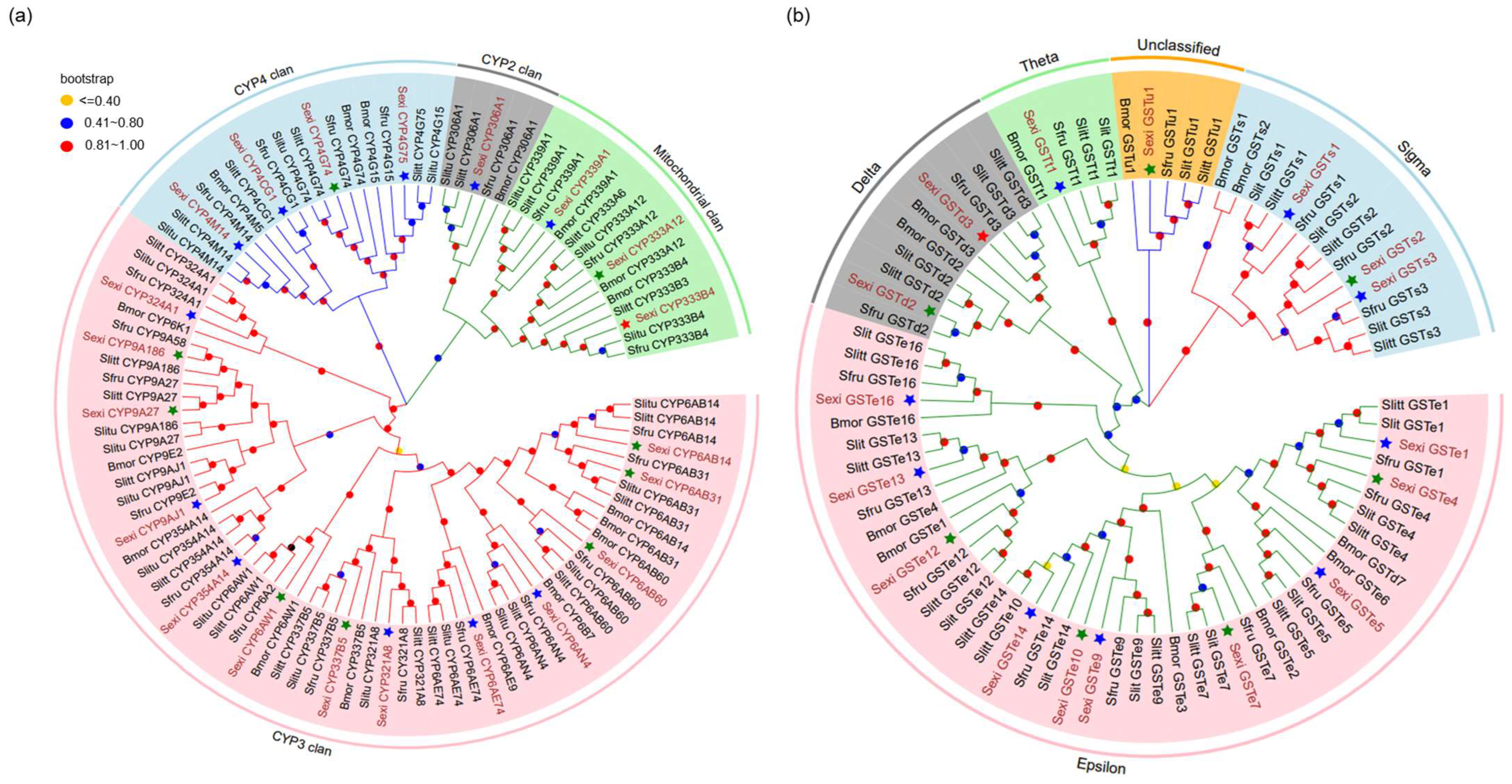
Figure 1. Phylogenetic analysis of selected CYPs (a) and GSTs (b) from S. exigua (Sexi), S. litura (Slitu), S. littoralis (Slitt), S. frugiperda (Sfru) and Bombyx mori (Bmor). The CYPs and GSTs of S. exigua are indicated in brown. The stars with different colors marked before CYP or GST proteins of S. exigua were significantly upregulated (green stars), significantly downregulated (red stars) or unchanged (blue stars) according to the transcriptome data (|Log2 FC| ≥ 1, p < 0.05).
Agriculture 13 01494 g001
Figure 2. RT-qPCR validation of the expression of selected detoxification enzyme genes in S. exigua after CPT treatment. (a) CYP genes, (b) GST genes. # indicates significant differences (|Log2 FC| ≥ 1, p < 0.05) according to the transcriptome data. RT-qPCR experiments were confirmed in at least three independent experiments and the data are presented as the mean ± standard error. * indicates significant differences compared to the control group (p < 0.05). The SPSS 26.0 Software Package (SPSS Inc.; Chicago, IL, USA) was used to perform statistical analyses with Student’s t test.
Agriculture 13 01494 g002
Figure 3. Effects of CPT on the survival (a) and body weight gain (b) of S. exigua larvae across 10 days of development. Data represent the mean ± SE.
Agriculture 13 01494 g003
Figure 4. Age-stage-specific survival rate of S. exigua after exposure to CPT at different doses. N2–N6 are the larval instars from second to sixth. The age-stage-specific survival rate of the control group (a) and the groups treated with CPT at 0.02 g/L (b), 0.1 g/L (c), and 0.5 g/L (d).
Agriculture 13 01494 g004
Figure 5. Effect of CPT on the expression of CYP genes during the development of S. exigua, including CYP9A27 (a), CYP9A186 (b), CYP337B5 (c), and CYP321A8 (d). RT-qPCR experiments were confirmed in at least three independent experiments and the data are presented as the mean ± standard error. Means followed by different small letters in the same time are significantly different (p < 0.05). To illustrate the changes in different CYP genes with the development of S. exigua larvae, the means of the relative expression in the control group were marked with different capital letters to indicate significant differences (p < 0.05). The SPSS 26.0 Software Package (SPSS Inc., Chicago, IL, USA) was used to perform statistical analyses by using Fisher’s least significant difference (LSD) test.
Agriculture 13 01494 g005
Figure 6. Effect of CPT on the expression of GST genes during the development of S. exigua, including GSTe8 (a) and GSTs3 (b). RT-qPCR experiments were confirmed in at least three independent experiments and the data are presented as the mean ± standard error. Means followed by different small letters in the same time are significantly different (p < 0.05). To illustrate the changes in different GST genes with the development of S. exigua larvae, the means of the relative expression in the control group were marked with different capital letters to indicate significant differences (p < 0.05). The SPSS 26.0 Software Package (SPSS Inc., Chicago, IL, USA) was used to perform statistical analyses by using Fisher’s least significant difference (LSD) test.
Agriculture 13 01494 g006
Disclaimer/Publisher’s Note: The statements, opinions and data contained in all publications are solely those of the individual author(s) and contributor(s) and not of MDPI and/or the editor(s). MDPI and/or the editor(s) disclaim responsibility for any injury to people or property resulting from any ideas, methods, instructions or products referred to in the content.