Article

Efficacy of Intravaginal Lactic Acid Bacteria, Cell-Free Supernatant, or Enrofloxacin on Vaginitis and Fertility in Ewes Synchronized with Progesterone-Based Protocol

1
Department of Obstetrics and Gynecology, Faculty of Veterinary Medicine, Balıkesir University, Balıkesir 10050, Türkiye
2
Department of Food Hygiene and Technology, Health Science Institute, Balıkesir University, Balıkesir 10050, Türkiye
3
Department of Physiology, Faculty of Veterinary Medicine, Balıkesir University, Balıkesir 10050, Türkiye
4
Department of Microbiology, Faculty of Veterinary Medicine, Bursa Uludag University, Bursa 16059, Türkiye
5
Department of Obstetrics and Gynecology, Faculty of Veterinary Medicine, Bursa Uludag University, Bursa 16059, Türkiye
6
Department of Animal Nutrition and Husbandry, University of Veterinary Medicine and Pharmacy, 041 81 Košice, Slovakia
*
Author to whom correspondence should be addressed.
Agriculture 2024, 14(4), 604; https://doi.org/10.3390/agriculture14040604
Submission received: 7 February 2024 / Revised: 22 March 2024 / Accepted: 9 April 2024 / Published: 11 April 2024
(This article belongs to the Special Issue Productivity, Performance and Health of Dairy Ruminants)

Abstract

:
An intravaginal sponge impregnated with progesterone is commonly used for estrus induction and synchronization in ewes. Although using an intravaginal sponge containing progesterone positively affects the synchronization rate, varying degrees of vaginitis occur during its application. This study aimed to investigate the impacts of various intravaginal treatment options on the vaginitis severity and pregnancy rate in Merino ewes synchronized with intravaginal sponges impregnated with progesterone. During the breeding period, 589 ewes, aged 2–6, received intravaginal sponges for 14 days. The control group (CON) received no treatment, whereas vaginal sponges absorbed with enrofloxacin (ENR), Lactobacillus plantarum (LAC), or Lactobacillus plantarum supernatant (CFS) were applied in the treatment groups. All groups received 500 IU of equine chorionic gonadotropin intramuscularly. The ENR group showed lower scores in vaginal discharge and sponge odor compared with the CON, LAC, and CFS groups. Although estrus responses did not differ between groups, the pregnancy rate tended to be higher in the ENR group. In conclusion, intravaginal ENR application, but not LAC or CFS, reduced vaginitis severity and tended to increase pregnancy rates in ewes synchronized with intravaginal sponges impregnated with progesterone.

1. Introduction

Several methods have been developed to control the reproduction of small ruminants in the last few decades. Intravaginal devices impregnated with progesterone are commonly used for estrus synchronization despite the availability of current synchronization methods in ewes and goats [1,2]. Progestin-containing sponges, such as fluorogestone acetate (FGA), medroxyprogesterone acetate (MAP), and controlled internal drug release (CIDR) devices, are used for synchronization in ewes and goats. Although FGA, MAP, and CIDR are approved for use in the synchronization of estrus in Türkiye, CIDR is not commercially available, and intravaginal sponges containing FGA and MAP are frequently used for progesterone-based synchronization protocols in ewes. Ewes that are synchronized with intravaginal sponges containing progesterone frequently receive an intramuscular injection of equine chorionic gonadotrophin (eCG) at sponge removal.
Intravaginal sponges generally lead to an increase in bacterial load as they remain as foreign objects in the vagina, whether for a long or short period [3]. The intravaginal sponges or devices cause physical irritation and localized inflammation with the accumulation of mucosal secretion [4]. Even though the vaginal environment normally prevents the proliferation of pathogenic microorganisms or potentially pathogenic saprophytes, factors such as temperature, humidity, pH, and the presence of nutrients may affect this control [5]. The use of an intravaginal sponge increases opportunistic Gram-negative Enterobacteriaceae, such as E.coli and Klebsiella spp., along with Gram-positive cocci, such as Staphylococcus spp. and Streptococcus spp., causing purulent and malodorous mucus accumulation [3,6,7]. Staphylococcus aureus, a resident of the vulva and vagina, is identified as the most common causative agent of purulent vaginitis in ewes [8], supporting the presence of purulent vaginal discharge during sponge removal [9]. Simultaneously, intravaginal sponges cause histological and cytological changes in the vaginal wall [10].
The proliferation of pathogenic microorganisms and changes in the composition of the vaginal microbiota trigger inflammation and contamination associated with abnormal discharges [9]. After sponge removal, abnormal vaginal discharge drains, and the increased bacteria in the vaginal flora decreases [11]. Although the vaginal flora returns to a healthy status 1 to 7 days after sponge removal [11,12,13], most opportunistic bacteria still have negative effects on sexual attractiveness. A change in the vaginal flora can lead to a decrease in attractiveness to male animals due to alterations in chemical stimuli in the vaginal environment. Vaginal discharge influenced by an intravaginal sponge negatively affects the functionality and viability of ram spermatozoa and, consequently, pregnancy rates [14]. As a practical solution, proper prophylactic management plays a crucial role, including the cleaning of all materials used in intravaginal sponge application [15]. This approach involves not only hygiene but also the application of antimicrobials or Lactobacillus spp. (an antibiotic-free strategy) at the application time of the intravaginal sponge. This study aimed to investigate the efficacy of Lactobacillus plantarum or Lactobacillus plantarum supernatant (in comparison with a control group and a positive control group, with enrofloxacin) regarding vaginitis and fertility in Merino ewes synchronized with intravaginal sponges impregnated with progesterone.

2. Materials and Methods

2.1. Animals

This study involved 620 Merino ewes aged 2–6, and the animals were housed indoors with outdoor access. The animals were clinically healthy; exhibited good appetite and no signs of lameness, nervous, or digestive disorders; and were alert to the environment as evidenced by their responsive ears. The animals were provided with a completely mixed ration, along with the opportunity to graze on natural pasture. Additionally, the ewes had free access to both water and shade throughout this study. This study was conducted during the breeding season (July–August) on a farm located in Edremit, Balikesir (39°33′ N, 26°58′ E) in Türkiye. The commercial sheep farm had 3000 mature ewes, and each paddock had a capacity for 100 ewes.

2.2. Preparation of Lactobacillus plantarum and Cell-Free Supernatants

The Lactobacillus plantarum strain (DSM 1055, DSMZ®, Braunschweig, Germany) was grown in De Man–Rogosa–Sharpe broth (MRS Broth®, Merck, Darmstadt, Germany) medium at 37 °C for 48 h. The bacterial suspension was transferred to sterile 10-mL falcon tubes. Bacteria-free (cell-free) supernatants were collected into another set of sterile tubes via centrifugation at 5000× g for 10 min. The bacterial pellet (108 CFU/mL) was resuspended in 10 mL of 0.9% sterile saline for application in the LAC group.
The cell-free supernatant was concentrated by 75% with an evaporator (55–60 °C/0.75 atm) to increase the concentration of antimicrobial activity. The cell-free supernatant was obtained after the concentrated supernatant was filtered through a 0.22 μ filter. The cell-free supernatant was prepared 2 days before for application in the CFS group.

2.3. Experimental Design

At the onset of this study, the body condition scores of each ewe were recorded. The ewes were randomly divided into four groups: CON, ENR, LAC, and CFS. Intravaginal sponges containing 60 mg of medroxyprogesterone acetate (MAP; Esponjavet®, İstanbul, HIPRA, Türkiye) were inserted into all ewes for 14 days. A total of 620 ewes were enrolled in this study, and each group consisted of 155 ewes. However, 2 ewes in the ENR group, 6 ewes in the CON group, 15 ewes in the LAC group, and 8 ewes in the CFS group were excluded from this study due to health problems or they were sold. Therefore, our study was conducted on 589 ewes.
The control group (CON; n = 149) received no treatment during intravaginal sponge application. In the other experimental groups, enrofloxacin (ENR; n = 153) was applied in the positive control group, and Lactobacillus plantarum (LAC; n = 140) and Lactobacillus plantarum supernatants (CFS; n = 147) were applied in the antibiotic-free treatment options by injecting 1 mL into the vaginal sponge with an insulin syringe (26 gauge). To prevent solution overflow, all administrations were injected into six different points underneath the sponge after placement on the sponge applicator. The vulva area was disinfected with 1% benzalkonium chloride (Zefirol, Dermosept, İstanbul, Türkiye) before sponge application, and all materials used in the sponge application were disinfected in the animals.
Vaginal discharge and sponge odor scoring were performed, and the sponges were weighed after removal. The characteristics of vaginal discharge (quantity, odor, and whether purulent or hemorrhagic) were scored as follows: 0 for negligible or no discharge; 1 for some clear discharge; and 2 for abundant, hemorrhagic, or purulent discharge at the time of sponge removal. The sponge odor (scored as 0: absent; 1: mild; and 2: abundant) was determined during sponge removal, according to Viñoles et al. [16]. The vaginal sponges of each ewe were weighed to assess the accumulation of vaginal discharge after removal. All ewes were given a 500 IU intramuscular injection of equine chorionic gonadotropin (eCG; Gonaser®, HIPRA, İstanbul, Türkiye) at the time of sponge removal. Fertile rams with crayon marks were introduced for natural mating one day later after sponge removal. The ram/ewe ratio was calculated as 1 ram per approximately 5 ewes. Estrus detection by observing marked ewes on the rump was performed for 5 days at 12 h intervals. The time of the estrus onset was assumed to be the midpoint following the detection of the first time the marked ewes after sponge removal. The pregnancy rate was assessed by transrectal ultrasonographic examination (Hasvet 838, Hasvet, Antalya, Türkiye) 28 ± 3 days after mating.

2.4. Statistical Analysis
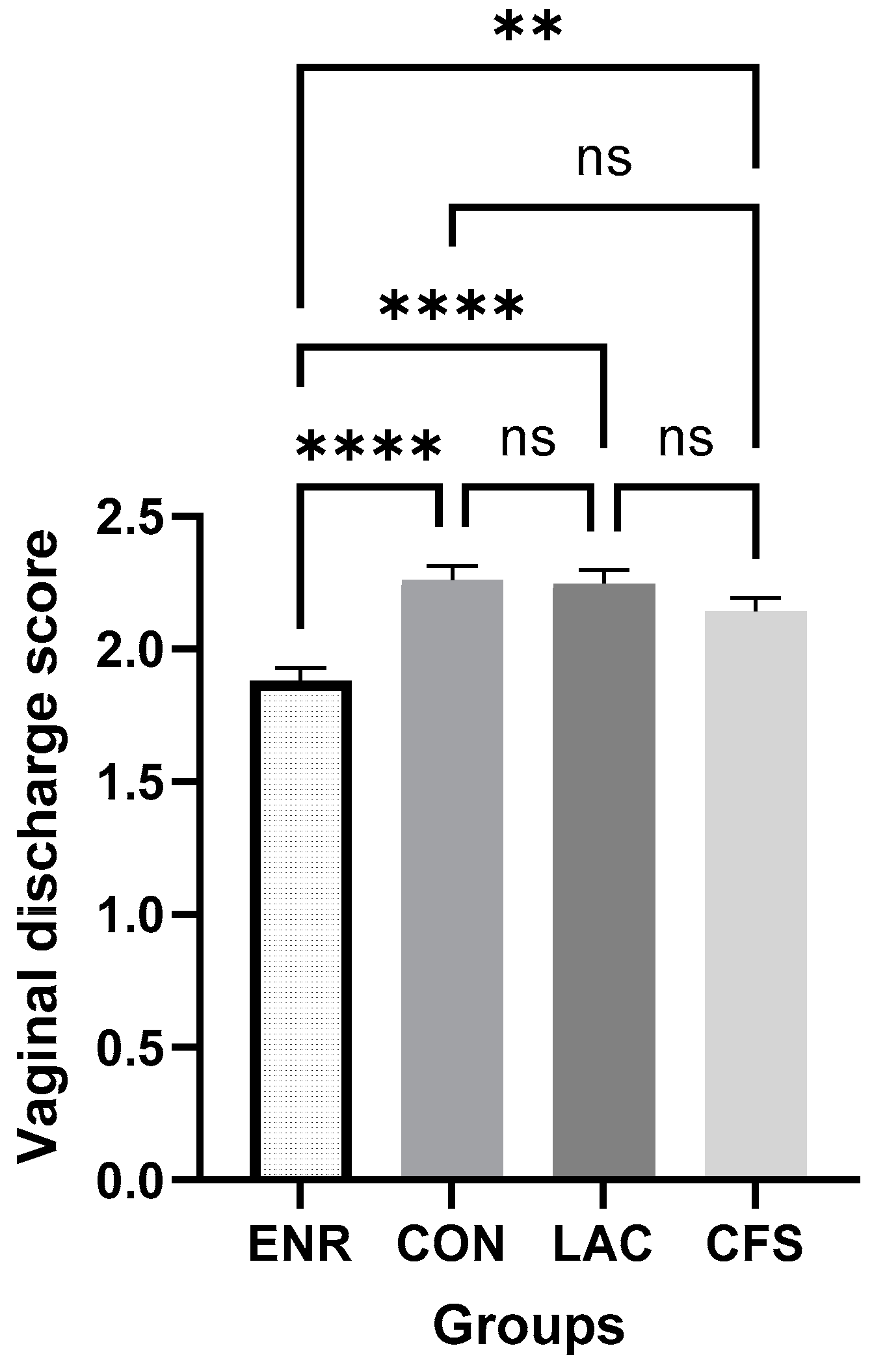
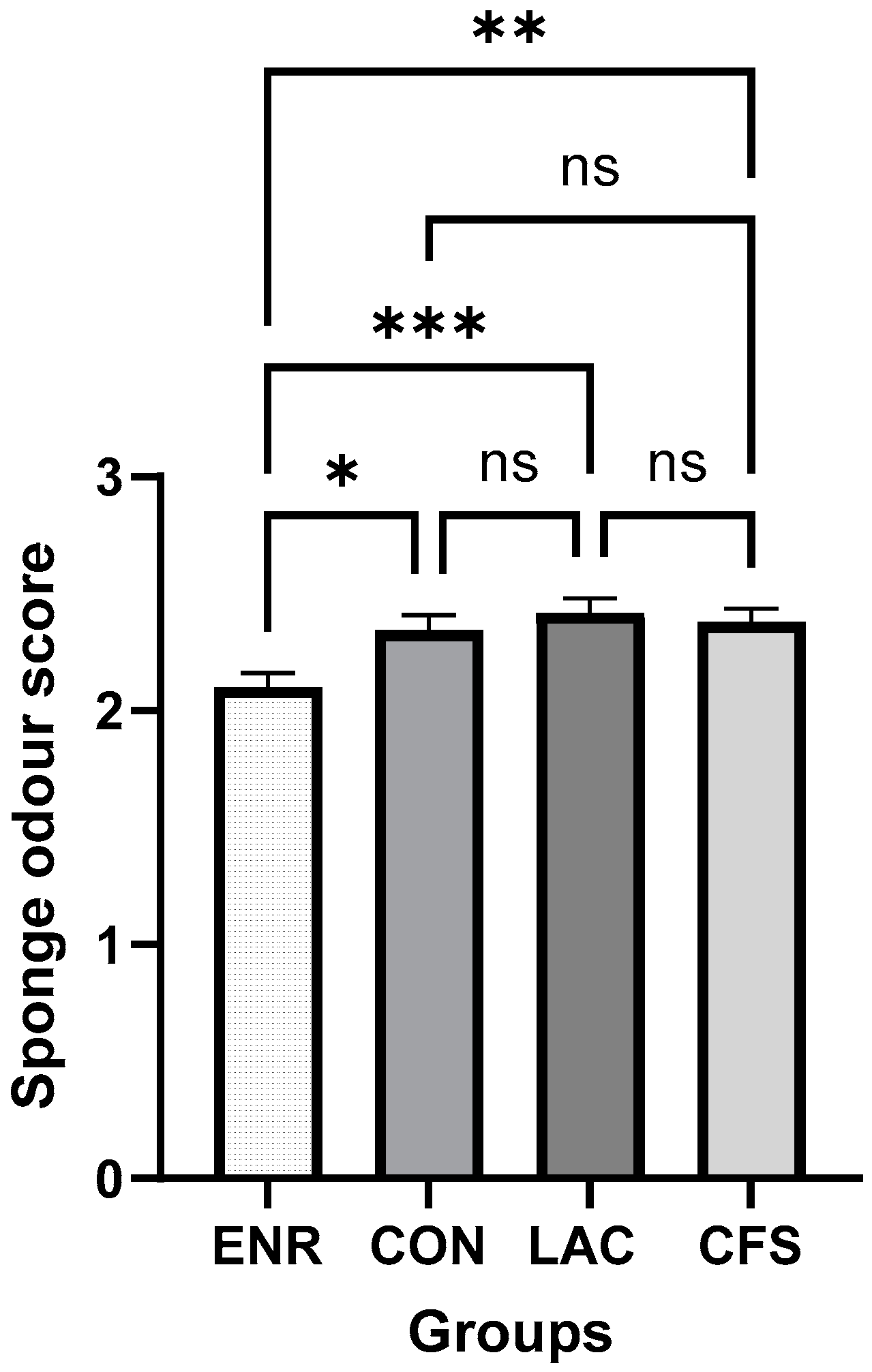
The data are presented as ±standard error of the mean (SEM). Statistical analyses were conducted utilizing SPSS® 23.0 package software (IBM Corporation, Armonk, NY, USA) and GraphPad Prism® 10 (GraphPad Software, San Diego, CA, USA). The analysis of variance test (ANOVA) was used to analyze age, vaginal discharge scores, sponge odor scores, and sponge weights. Furthermore, the post hoc Bonferroni test was performed. The chi-square test was utilized to analyze the percentages of vaginal discharge scores, estrus response, and pregnancy rates. A statistical significance level of p < 0.05 was considered significant, and statistical tendencies were defined as 0.05 < p < 0.10.

3. Results

Sponge loss occurred in three ewes in the ENR group, six ewes in the CON group, four ewes in the LAC group, and seven ewes in the CFS group. The average of the occurrence of sponge loss was 3.4% (20/589). As a result, this study initially started with 620 ewes and continued with 153 ewes in the ENR group, 149 ewes in the CON group, 140 ewes in the LAC group, and 147 ewes in the CFS group, respectively.
The average body score of the ewes was 3.21 ± 0.02, and no significant difference was observed between the groups (CON: 3.27 ± 0.05; ENR: 3.26 ± 0.05; LAC: 3.20 ± 0.05; and CFS: 3.12 ± 0.05) at the beginning of this study (p > 0.05).
Vaginal discharge was observed during sponge removal in all ewes in the groups (n = 569), except for ewes with lost sponges (n = 20). Irrespective of groups, the percentage of ewes with negligible vaginal discharge (score 0) was 13.6% (80/589), with moderate vaginal discharge (score 1) was 57.2% (337/589), and with purulent/hemorrhagic vaginal discharge (score 2) was 25.8% (152/589). For each group, the percentages of vaginal discharge score 1, score 2, and score 3 were 11.8%, 50.3%, and 37.9% in CON; 24.7%, 62.7%, and 12.6% in ENR; 7.3%, 61.0%, and 31.7% in LAC; 11.4%, 62.9%, and 25.7% in CFS, respectively. It was determined that the average vaginal discharge score in the ENR group (1.88 ± 0.5) was significantly lower compared with the CON (2.25 ± 0.5), LAC (2.24 ± 0.5), and CFS (2.14 ± 0.5) groups (p < 0.05; Figure 1).
Irrespective of groups, the percentage of ewes with no sponge odor (score 0) was 16.5% (97/589), with a mild odor (score 1) was 34.1% (201/589), and with an abundant odor (score 2) was 46.0% (271/589). For each group, the percentages of score 0, score 1, and score 2 odors were 19.0%, 27.9%, and 53.1% for CON; 24.0%, 42.0%, and 34.0% for ENR; 14.0%, 30.1%, and 55.9% for LAC; and 10.7%, 40.7%, and 48.6% for CFS, respectively. The average sponge odor score in the ENR group (2.10 ± 0.6) was statistically lower compared with the CON (2.34 ± 0.6), LAC (2.41 ± 0.6), and CFS (2.37 ± 0.6) groups (p < 0.05; Figure 2). At sponge removal, the sponge weights ranged from 3.6 g to 15.9 g. The mean (±SEM) sponge weight was 7.07 ± 0.08 g. The mean sponge weights were 6.86 g in CON, 7.19 g in ENR, 7.14 g in LAC, and 7.09 g in CFS, and there was no significant difference between the groups.
The estrus response was 86.4% (509/589), and there was no significant difference among the groups. After sponge removal, ewes expressed estrus at approximately 48 h in all groups, and the onset of estrus was not different between groups. However, there was a tendency (p = 0.08) for an increased pregnancy rate in the ENR group (56.2%) compared with the CON group (42.3%), LAC (47.1%), and CFS (45.6%) groups (Table 1). Fifty percent of ewes with sponge loss showed estrus (10/20), and ten percent of ewes were pregnant (2/20).

4. Discussion

The use of intravaginal sponges for synchronization is known to cause vaginitis in ewes. Studies focused on understanding the reasons, treatment options, and effects of the developing infection on health and reproduction. In a large-scale study, Manes et al. [3] investigated the impact of intravaginal sponge application on fertility in ewes. The study found that adding medroxyprogesterone acetate into the sponge did not alter vaginitis. However, vaginitis via sponge application for synchronization reduced the pregnancy rate in ewes [3]. Similarly, Gatti et al. [12] revealed that a bacterial increase in vaginal flora occurred regardless of the sponge content. The local application of progesterone may not contribute to vaginitis, whereas bacterial proliferation may occur due to the continuous absorption and retention of vaginal secretions by the sponge.
In our study, the vaginal discharge rate was 100%. Consistent with our results, the vaginal discharge rate percentage ranged from 94.4% to 100% in previous studies [6,9,17]. Guner et al. [17] reported that a score of 1 constituted the majority (56.7%) of vaginal discharge in Berrichon ewes that received intravaginal sponges impregnated with progesterone for 14 days. Consistent with the previous study [17], the majority (59.2%) of vaginal discharge was also scored as 1 in the Merino ewes in our study. Furthermore, the majority (82.9%) of malodorous sponge scores were consistent with the majority (85.9%) of elevated vaginal discharge scores in our study. Similar to our results, Viñoles et al. [16] found that the percentage of malodorous sponges was 89% in ewes that were administered intravaginal sponges for 14 days. The presence of foul-smelling sponges and unpleasant vaginal discharge may raise concerns among farmers or veterinary practitioners that the pregnancy rate will decrease.
In our study, the sponge was applied to the vagina for 14 days. Martinez-Ros et al. [9] investigated the impacts of different intravaginal devices (sponge or CIDR) and treatment durations with progesterone (7 or 14 days) on vaginal discharge, pH, bacteriology, and fertility. The durations of sponges or devices did not alter the percentage of vaginitis. Lower discharge scores and higher pregnancy rates were observed in the CIDR group compared with the effects of the retention periods of intravaginal devices. Approximately 15–20% of ewes treated with CIDR and 80% of ewes treated with sponges exhibited vaginal discharge, regardless of the treatment duration. Unlike an intravaginal sponge, a CIDR device allows the drainage of discharge as it does not block the vagina like a plug. This helps reduce the rate of vaginitis and its negative effects.
The fertility of spermatozoa is also affected due to the changes caused by the sponge in the vagina. Since it takes at least one week for the vaginal environment to return to its original/healthy state after sponge removal, sperm is negatively affected by this environment. Contamination with Enterobacteriaceae in vaginal flora reduces the quality of sperm. Escherichia coli is the most commonly encountered bacteria in ewes. Escherichia coli reduces sperm motility by causing the adhesion and agglutination of sperm after removing the intravaginal sponge. Additionally, the bacteria lead to significant morphological differences in human spermatozoa due to changes in their function [4,14]. Vaginal discharge in sponge-applied ewes negatively affects the functionality and viability of sperm [14]. The presence of a sponge and the subsequent discharge negatively affect the sexual attractiveness of ewes [2]. Moreover, bacterial growth and changes in vaginal microbiota have a direct impact on the sperm fertilization capability [4]. Additionally, Escherichia coli, associated with severe vaginal purulence, suppresses dominant follicle growth and estrogen production corpus luteum function [5].
Various treatment strategies for preventing vaginitis have been investigated. Antibiotics are the most commonly researched option in ewe-administered sponges. In this study, most of the ewes treated with different approaches exhibited vaginal discharge at sponge removal. A lower score was observed in the ENR group than in the CON group. Our study supports previous studies [12,18] indicating an association between antibiotic application and vaginitis. The pH of vaginal fluid affects the impacts of different antibacterial agents. Enrofloxacin is effective against Gram-negative bacteria in an acidic pH [19]. Its effectiveness against Gram-positive bacteria is not influenced by the vaginal pH [5,20]. Consistent with our results, recent studies reported that enrofloxacin was the most effective antibiotic in controlling vaginitis due to its high sensitivity to vaginal microorganisms [5,6]. Silva et al. [21] indicated that 82% of the biota collected from the vaginal mucosa of ewes were susceptible to enrofloxacin. The authors of a previous study reported that enrofloxacin could be effective in reducing vaginitis severity, but the results were not statistically different due to the limited number of animals per group [17]. The effectiveness of tetracyclines in preventing vaginitis in synchronization with sponge application was reported. Most of the previous studies highlighted the elevated number of resistant colonies [13,22,23].
The onset of estrus and the percentage of ewes that expressed estrus were consistent with previous reports [9,18,24]. The time from sponge removal to the onset of estrus was, on average, 48 h, and the onset of estrus was not different between the groups. Although the vaginal flora returns to a healthy status 1 to 7 days after sponge removal [11,12,13], an abnormal vaginal flora composition continues 48 h after sponge removal. In bacterial count analyses, several studies reported the recovery of vaginal microbiota at mating time. Guner et al. [6] determined that the bacterial count 48 h after sponge removal did not return similar values to those at sponge insertion in ewes. The rams’ sexual reluctance was expected to decrease; however, the estrus response percentage (average 86.4%) was not different between the groups.
Ozyurtlu et al. [18] revealed that adding antibiotics to sponges prevented bacterial proliferation; however, there was no significant impact on the pregnancy rate in 30 Awassi ewes. Similarly, Viñoles et al. [16] found that adding antibiotics to sponges reduced vaginal discharge in 207 Corriedale ewes. In that study, the pregnancy rate was numerically higher in the antibiotic group than in the control group by approximately 10%; however, the number of animals could have prevented the statistical increment in the pregnancy rate in the treatment group. In our study conducted on 589 Merino ewes, there was a tendency for a higher pregnancy rate (approximately 10%) in the antibiotic group compared with the other groups. Antibiotics are often recommended for the treatment of vaginitis to reduce sponge-induced adverse effects on fertility.
Probiotics have been used in women’s gynecological diseases in the last 20 years. Probiotics are preferred in cases where standard treatments have proven ineffective. Probiotics protect the vagina from pathogenic colonization via various mechanisms, such as blocking potential binding sites, producing microbiocidal substances such as hydrogen peroxide, maintaining a low pH, and inducing anti-inflammatory cytokine responses in epithelial cells [25]. Lactobacillus spp. are frequently used in women suffering from vaginitis [26,27]. All lactic acid bacteria can produce small protein molecules known as bacteriocins. Bacteriocins are molecules that exhibit a broad biological activity spectrum with different genetic origins, inhibiting the growth of bacteria, fungi, yeast, and even eukaryotic cells [28]. Lactobacillus plantarum is one of the most common Lactobacillus spp., producing numerous antimicrobial components.
Applying Lactobacillus spp. to the vaginal environment has been shown to reduce the incidence of vaginal infections in women and reduce the frequency of uterine infections in peripartum dairy cows [29,30]. The intravaginal usIe of Lactobacillus spp. can inhibit metritis pathogens [31,32]. Swartz et al. [33] noted that Lactobacillus spp. species are rarely observed in ewes aged 1 to 4 years, and the vaginal pH in ewes is close to neutral, unlike the human vaginal flora. Lactobacillus spp. are present in the vagina, which implies intensive production of lactate. As a result, the pH decreases to less than 4.5. Manes et al. [4] reported an increased vaginal pH at sponge removal compared with that in sponge application in ewes.
In a pilot study conducted by Quereda et al. [32], Lactobacillus spp. was injected into an intravaginal sponge, marking the first instance of utilizing Lactobacillus spp. in this manner in ewes. While there were no differences in bacterial density and pregnancy rates compared with the control group, the initial application of Lactobacillus spp. did not lead to adverse effects on the animals’ health status. The Enterobacteriaceae was found to be dominant during the study. No diversity in bacterial composition was observed among the control and probiotic groups. Our study also found consistent results with previous research regarding pregnancy rates and their effects on health.
In subsequent years, Guner et al. [6] and Danilova et al. [28] investigated the efficacy of Lactobacillus plantarum supernatant against pathogenic bacteria. Danilova et al. [28] found that Lactobacillus plantarum supernatant culture in a liquid nutrient medium exhibits inhibitory activity against Gram-positive and Gram-negative pathogenic microorganisms. Guner et al. [6] found that Lactobacillus plantarum cell-free supernatant administration reduced the number of Enterobacteriaceae in ewes; however, the authors did not focus on pregnancy rates. Lactic acid bacteria could have reduced the bacterial count by decreasing the vaginal pH. In our study, the CFS and LAC groups affected neither vaginitis parameters nor fertility compared with the CON group. Hence, the CFS and LAC groups were ineffective in reducing vaginal discharge and sponge odor scores and increasing fertility parameters. It appears that enrofloxacin application was more effective than the Lactobacillus plantarum and Lactobacillus plantarum cell-free supernatant groups.

5. Conclusions

The effects of intravaginal treatments consisting of ENR, CFS, and LAC on vaginitis and fertility were investigated. Varying degrees of vaginal discharge were observed in all the groups at sponge removal. The injection of enrofloxacin into the sponges reduced the vaginal discharge scores and sponge odor scores. Lactobacillus plantarum or its cell-free supernatant did not cause any adverse effects in ewes when used as an alternative strategy to antibiotic treatment. The groups receiving Lactobacillus plantarum and Lactobacillus plantarum supernatants exhibited similar pregnancy rates to the control group. However, the group treated with enrofloxacin tended to have higher pregnancy rates. It is important to note that more research is necessary due to antibiotic resistance, with a focus on alternative antibiotic-free treatment strategies, such as Lactobacillus plantarum.

Author Contributions

Project administration and funding acquisition, B.G.; conceptualization, B.G.; methodology, B.G., T.E.G., S.K.D. and A.K.; investigation, B.G., T.E.G., B.O., S.K.D., A.K. and I.K.; software, A.A.E.; formal analysis B.O., A.A.E. and Z.F.; writing of draft manuscript A.A.E., B.O. and T.E.G.; review and editing, B.G. and Z.F. All authors have read and agreed to the published version of the manuscript.

Funding

This study was supported by Balikesir University, the Scientific Research Projects Coordination Unit (BAP project no. 2022/009), and the Slovak project VEGA 1-0162-23: Reduction in antibiotic use in dairy mastitis control programs.

Institutional Review Board Statement

This study was conducted according to the guidelines of the Balıkesir University Animal Care Ethics Committee. The committee approved the experimental procedures (reference number: 2021/3-4 and date of approval: 25 March 2021).

Data Availability Statement

The data presented in this study are available upon request from the corresponding author. The data are not publicly available due to privacy.

Acknowledgments

The authors thank Mehmet OZUİCLİ for his help in this study.

Conflicts of Interest

The authors declare no conflicts of interest.

References

  1. Abecia, J.A.; Forcada, F.; González-Bulnes, A. Hormonal Control of Reproduction in Small Ruminants. Anim. Reprod. Sci. 2012, 130, 173–179. [Google Scholar] [CrossRef] [PubMed]
  2. Gatti, M.; Ungerfeld, R. Intravaginal Sponges to Synchronize Estrus Decrease Sexual Attractiveness in Ewes. Theriogenology 2012, 78, 1796–1799. [Google Scholar] [CrossRef] [PubMed]
  3. Manes, J.; Hozbor, F.; Alberio, R.; Ungerfeld, R. Intravaginal Placebo Sponges Affect Negatively the Conception Rate in Sheep. Small Rumin. Res. 2014, 120, 108–111. [Google Scholar] [CrossRef]
  4. Manes, J.; Fiorentino, M.A.; Kaiser, G.; Hozbor, F.; Alberio, R.; Sanchez, E.; Paolicchi, F. Changes in the Aerobic Vaginal Flora after Treatment with Different Intravaginal Devices in Ewes. Small Rumin. Res. 2010, 94, 201–204. [Google Scholar] [CrossRef]
  5. Ojeda-Hernández, F.; del Moral-Ventura, S.; Capataz-Tafur, J.; Peña-Castro, J.; Abad-Zavaleta, J.; Chay-Canul, A.; Ramon-Ugalde, J.; Ungerfeld, R.; Meza-Villalvazo, V. Vaginal Microbiota in Pelibuey Sheep Treated with Antimicrobials at the Removal of Intravaginal Sponges Impregnated with Flurogestone Acetate. Small Rumin. Res. 2019, 170, 116–119. [Google Scholar] [CrossRef]
  6. Guner, B.; Kisadere, İ.; Tavsanli, H.; Kahya Demirbilek, S.; Keskin, A. Comparison of the Efficacy of Enrofloxacin and Lactobacillus Plantarum Cell-Free Supernatant Treatments on Vaginitis in Ewes. J. Vet. Med. Res. 2022, 41, 105–111. [Google Scholar] [CrossRef]
  7. Mohammed, K.; Nabih, A.; Darwish, G. Efficacy of Anti-Microbial Agents on Vaginal Microorganisms and Reproductive Performance of Synchronized Estrus Ewes. Asian Pac. J. Reprod. 2017, 6, 121–127. [Google Scholar] [CrossRef]
  8. Bragança, J.F.M.; Maciel, J.M.; Girardini, L.K.; Machado, S.A.; da Rocha, J.F.X.; Tonin, A.A.; da Rocha, R.X. Influence of a Device Intravaginal to Synchronization/Induction of Estrus and Its Reuse in Sheep Vaginal Flora. Comp. Clin. Path 2017, 26, 1369–1373. [Google Scholar] [CrossRef]
  9. Martinez-Ros, P.; Lozano, M.; Hernandez, F.; Tirado, A.; Rios-Abellan, A.; López-Mendoza, M.; Gonzalez-Bulnes, A. Intravaginal Device-Type and Treatment-Length for Ovine Estrus Synchronization Modify Vaginal Mucus and Microbiota and Affect Fertility. Animals 2018, 8, 226. [Google Scholar] [CrossRef]
  10. Manes, J.; Campero, C.; Hozbor, F.; Alberio, R.; Ungerfeld, R. Vaginal Histological Changes after Using Intravaginal Sponges for Oestrous Synchronization in Anoestrous Ewes. Reprod. Domest. Anim. 2015, 50, 270–274. [Google Scholar] [CrossRef]
  11. Suárez, G.; Zunino, P.; Carol, H.; Ungerfeld, R. Changes in the Aerobic Vaginal Bacterial Mucous Load and Assessment of the Susceptibility to Antibiotics after Treatment with Intravaginal Sponges in Anestrous Ewes. Small Rumin. Res. 2006, 63, 39–43. [Google Scholar] [CrossRef]
  12. Gatti, M.; Zunino, P.; Ungerfeld, R. Changes in the Aerobic Vaginal Bacterial Mucous Load after Treatment with Intravaginal Sponges in Anoestrous Ewes: Effect of Medroxiprogesterone Acetate and Antibiotic Treatment Use. Reprod. Domest. Anim. 2011, 46, 205–208. [Google Scholar] [CrossRef] [PubMed]
  13. Oliveira, J.K.; Martins, G.; Esteves, L.V.; Penna, B.; Hamond, C.; Fonseca, J.F.; Rodrigues, A.L.; Brandão, F.Z.; Lilenbaum, W. Changes in the Vaginal Flora of Goats Following a Short-Term Protocol of Oestrus Induction and Synchronisation with Intravaginal Sponges as Well as Their Antimicrobial Sensitivity. Small Rumin. Res. 2013, 113, 162–166. [Google Scholar] [CrossRef]
  14. Manes, J.; Ríos, G.; Fiorentino, M.A.; Ungerfeld, R. Vaginal Mucus from Ewes Treated with Progestogen Sponges Affects Quality of Ram Spermatozoa. Theriogenology 2016, 85, 856–861. [Google Scholar] [CrossRef] [PubMed]
  15. Gonzalez-Bulnes, A.; Menchaca, A.; Martin, G.B.; Martinez-Ros, P. Seventy Years of Progestagen Treatments for Management of the Sheep Oestrous Cycle: Where We Are and Where We Should Go. Reprod. Fertil. Dev. 2020, 32, 441. [Google Scholar] [CrossRef]
  16. Viñoles, C.; Paganoni, B.; Milton, J.T.B.; Driancourt, M.A.; Martin, G.B. Pregnancy Rate and Prolificacy after Artificial Insemination in Ewes Following Synchronisation with Prostaglandin, Sponges, or Sponges with Bactericide. Anim. Prod. Sci. 2011, 51, 565. [Google Scholar] [CrossRef]
  17. Guner, B.; Kisadere, I. The Efficacy of Intravaginal Antibiotic or Their Combination with Different Gonadotropins on Vaginitis and Fertility in Ewes Synchronized with Progesterone Impregnated Intravaginal Sponges. Turk. J. Vet. Anim. Sci. 2022, 46, 575–581. [Google Scholar] [CrossRef]
  18. Ozyurtlu, N.; Yesilmen, S.; Kucukaslan, İ. The Effectiveness of Using Antibiotic with Intravaginal Sponge and Duration of Sponge Treatments on the Vaginal Flora and Fertility in Anestrous Ewes. J. Anim. Vet. Adv. 2008, 7, 723–727. [Google Scholar]
  19. McKellar, Q. Clinical Relevance of the Pharmacologic Properties of Fluoroquinolones. In The Compendium on Continuing Education for the Practicing Veterinarian; University of Hertfordshire: Hatfield, UK, 1996. [Google Scholar]
  20. Giguère, S.; Prescott, J.F.; Dowling, P.M.; Giguère, F.S. Antimicrobial Therapy in Veterinary Medicine. In Antimicrobial Therapy in Veterinary Medicine, 5th ed.; Wiley: Hoboken, NJ, USA, 2013; Volume 18, pp. 295–315. [Google Scholar]
  21. Silva, V.F.; Damasceno, T.E.F.; Souza, N.J.D.; Franco, I.; Costa, M.M. Microbiota Cérvico-Vaginal de Ovelhas Mestiças e Sua Susceptibilidade Aos Antibióticos. Pesqui. Veterinária Bras. 2011, 31, 586–590. [Google Scholar] [CrossRef]
  22. Manes, J.; Fiorentino, M.A.; Hozbor, F.; Paolicchi, F.; Alberio, R.; Ungerfeld, R. Changes in the Aerobic Vaginal Bacteria Load and Antimicrobial Susceptibility after Different Oestrous Synchronisation Treatments in Goats. Anim. Prod. Sci. 2013, 53, 555. [Google Scholar] [CrossRef]
  23. Martins, G.; Figueira, L.; Penna, B.; Brandão, F.; Varges, R.; Vasconcelos, C.; Lilenbaum, W. Prevalence and Antimicrobial Susceptibility of Vaginal Bacteria from Ewes Treated with Progestin-Impregnated Intravaginal Sponges. Small Rumin. Res. 2009, 81, 182–184. [Google Scholar] [CrossRef]
  24. Vasconcelos, C.O.d.P.; Brandão, F.Z.; Martins, G.; Penna, B.; Souza-Fabjan, J.M.G.d; Lilenbaum, W. Qualitative and Quantitative Analysis of Bacteria from Vaginitis Associated with Intravaginal Implants in Ewes Following Estrus Synchronization. Ciência Rural. 2016, 46, 632–636. [Google Scholar] [CrossRef]
  25. Reid, G. Probiotic Use in an Infectious Disease Setting. Expert. Rev. Anti Infect. Ther. 2017, 15, 449–455. [Google Scholar] [CrossRef]
  26. Jeng, H.; Yan, T.; Chen, J. Treating Vaginitis with Probiotics in Non-pregnant Females: A Systematic Review and Meta-analysis. Exp. Ther. Med. 2020, 20, 3749–3765. [Google Scholar] [CrossRef]
  27. van de Wijgert, J.; Verwijs, M. Lactobacilli-containing Vaginal Probiotics to Cure or Prevent Bacterial or Fungal Vaginal Dysbiosis: A Systematic Review and Recommendations for Future Trial Designs. BJOG 2020, 127, 287–299. [Google Scholar] [CrossRef]
  28. Danilova, A.; Adzhieva, A.A.; Danilina, G.A.; Polyakov, N.B.; Soloviev, A.I.; Zhukhovitsky, V.G. Antimicrobial Activity of Supernatant of Lactobacillus Plantarum against Pathogenic Microorganisms. Bull. Exp. Biol. Med. 2019, 167, 751–754. [Google Scholar] [CrossRef] [PubMed]
  29. Deng, Q.; Odhiambo, J.F.; Farooq, U.; Lam, T.; Dunn, S.M.; Ametaj, B.N. Intravaginal Lactic Acid Bacteria Modulated Local and Systemic Immune Responses and Lowered the Incidence of Uterine Infections in Periparturient Dairy Cows. PLoS ONE 2015, 10, e0124167. [Google Scholar] [CrossRef]
  30. Falagas, M.E.; Betsi, G.I.; Tokas, T.; Athanasiou, S. Probiotics for Prevention of Recurrent Urinary Tract Infections in Women. Drugs 2006, 66, 1253–1261. [Google Scholar] [CrossRef] [PubMed]
  31. Otero, M.C.; Morelli, L.; Nader-Macias, M.E. Probiotic Properties of Vaginal Lactic Acid Bacteria to Prevent Metritis in Cattle. Lett. Appl. Microbiol. 2006, 43, 91–97. [Google Scholar] [CrossRef]
  32. Quereda, J.J.; García-Roselló, E.; Barba, M.; Mocé, M.L.; Gomis, J.; Jiménez-Trigos, E.; Bataller, E.; Martínez-Boví, R.; García-Muñoz, Á.; Gómez-Martín, Á. Use of Probiotics in Intravaginal Sponges in Sheep: A Pilot Study. Animals 2020, 10, 719. [Google Scholar] [CrossRef]
  33. Swartz, J.D.; Lachman, M.; Westveer, K.; O’Neill, T.; Geary, T.; Kott, R.W.; Berardinelli, J.G.; Hatfield, P.G.; Thomson, J.M.; Roberts, A.; et al. Characterization of the Vaginal Microbiota of Ewes and Cows Reveals a Unique Microbiota with Low Levels of Lactobacilli and Near-Neutral PH. Front. Vet. Sci. 2014, 1, 19. [Google Scholar] [CrossRef] [PubMed]
Figure 1. The mean vaginal discharge score (mean ± SEM) after sponge removal in groups (ENR: enrofloxacin, CON: control, LAC: Lactobacillus plantarum, and CFS: Lactobacillus plantarum cell-free supernatant). ****: p < 0.0001, **: p < 0.01, and ns: not significant according to post hoc Bonferroni test in ANOVA.
Agriculture 14 00604 g001
Figure 2. Mean sponge odor score (mean ± SEM) after sponge removal in groups (ENR: enrofloxacin, CON: control, LAC: Lactobacillus plantarum, and CFS: Lactobacillus plantarum cell-free supernatant). ***: p < 0.001, **: p < 0.01, *: p < 0.05, ns: not significant according to post hoc Bonferroni test in ANOVA.
Agriculture 14 00604 g002
Table 1. Percentage of sponge loss, estrus response rate, pregnancy rate, and onset of estrus in ewes that received different intravaginal treatments.
VariableGroups
ENRCONLACCFSp-Value
Sponge loss (%, n/n)2.0 (3/153)4.0 (6/149)2.8 (4/140)4.8 (7/147)p > 0.05
Onset of estrus (h)48.0 ± 0.948.3 ± 0.948.4 ± 1.048.4 ± 0.9p > 0.05
Estrus response (%, n/n)89.5 (137/153)85.9 (128/149)84.2 (118/140)85.7 (126/147)p > 0.05
Pregnancy rate (%, n/n)56.2 (86/153) a42.3 (63/149) b47.1 (66/140) b45.6 (67/147) bp = 0.08
Values in rows labeled with different letters (a,b) exhibit statistical tendency (p < 0.10) (ENR: enrofloxacin, CON: control, LAC: Lactobacillus plantarum, and CFS: Lactobacillus plantarum cell-free supernatant).
Disclaimer/Publisher’s Note: The statements, opinions and data contained in all publications are solely those of the individual author(s) and contributor(s) and not of MDPI and/or the editor(s). MDPI and/or the editor(s) disclaim responsibility for any injury to people or property resulting from any ideas, methods, instructions or products referred to in the content.

