Brief Report

Relationships of Circulating and Preovulatory Follicular Fluid Hydrogen Peroxide Levels with Body Condition Score and Metabolome Profiles of Lactating Beef Cows

1
Department of Animal Science, Institute of Agriculture and AgResearch, University of Tennessee, Knoxville, TN 37996, USA
2
Department of Chemistry, University of Tennessee, Knoxville, TN 37919, USA
*
Author to whom correspondence should be addressed.
Agriculture 2024, 14(8), 1406; https://doi.org/10.3390/agriculture14081406
Submission received: 20 June 2024 / Revised: 10 August 2024 / Accepted: 12 August 2024 / Published: 20 August 2024
(This article belongs to the Special Issue Impacts of Oxidative Stress on Cattle Physiology)

Abstract

:
Nutrient requirements for lactation often lead to a negative energy balance accompanied by reduced body condition and fertility in cattle. A previous study identified an increased abundance of serum metabolites associated with tissue mobilization and reactive oxygen species (ROS) generation in postpartum beef cows with a thin versus moderate body condition. No studies, however, have measured ROS levels in the serum and follicular fluid of postpartum beef cows for comparison with body condition. We hypothesized that beef cows with a thin body condition would have elevated levels of ROS, as indicated by hydrogen peroxide (H2O2), in serum and preovulatory follicular fluid. Serum and follicular fluid samples from thin (n = 12), moderate (n = 11), and obese (n = 16) cows underwent a H2O2 bioluminescence assay. Levels of H2O2 in each biofluid were then assessed to determine relationships with cow body condition and serum or follicular fluid metabolites. Levels of H2O2 did not differ among body condition categories. In thin cows, the serum H2O2 level was positively related to the abundance of 3 metabolites with antioxidant activity. Among all animals, the follicular fluid H2O2 level was positively associated with the abundance of 13 metabolites, many of which had antioxidant roles. The results suggest an impact of postpartum beef cow metabolism on ROS levels in preovulatory follicular fluid or serum and highlight the need for additional studies to further investigate this potential impactor of reproductive efficiency and sustainable beef production.

1. Introduction

Sustainability of the cattle industry requires reproductive success to be at the forefront of beef and dairy cattle operations. Extremes in the body condition score (BCS) have been associated with negative effects on fertility and the overall productivity of beef and dairy cattle [1,2,3]. In beef specifically, cow resumption of postpartum estrous cyclicity and maintenance of a 365-day calving interval are essential for production efficiency [4]. Beef cows with a low BCS have elongated anestrous periods, a reduced conception rate, and longer calving intervals [5,6,7]. Furthermore, beef and dairy cows with a thin BCS have reduced numbers of recruited follicles and lower oocyte quality, leading to reduced embryo cleavage and blastocyst production [8,9,10].
One mechanism by which an extreme BCS likely influences fertility is through the systemic accumulation of reactive oxygen species (ROS) generated by increased oxygen-related processes such as mitochondrial oxidative metabolism and inflammation [11,12]. To maintain cellular and systemic homeostasis, normal levels of ROS production are efficiently counterbalanced by antioxidant production [13]. Dysregulated or extreme ROS levels affect numerous facets of dairy and beef cattle health, leading to mastitis, acidosis, pneumonia, and other diseases that often occur during the time of postpartum and peak lactation [14]. Elevated systemic ROS levels may also negatively influence fertility in thin cows by entering the follicle through the blood–follicle barrier and decreasing oocyte competence for embryo development. Inherently, moderate and regulated levels of ROS are not negative for fertility, as ROS serve positive roles in both ovulation and oocyte maturation [15]. Dysregulated or extreme levels of ROS in the circulation and in the follicle, however, can negatively impact reproduction [16]. Indeed, ROS accumulation in granulosa cells reduces gonadotropin sensitivity and leads to cellular apoptosis [17]. Hydrogen peroxide has also specifically been associated with apoptosis in the human embryo [18]. Additionally, the overall follicular fluid ROS level was greater in human follicles containing an oocyte that failed to fertilize or developed into a grade 3 embryo compared to follicles containing an oocyte that developed into a high-quality embryo (grade 1 or 2 [19]). Negative effects of extremely high levels of ROS on oocyte competence appear to be related to apoptotic events, a failure to complete meiotic progression to metaphase II, and reduced DNA integrity [20,21,22]. A thin BCS has been associated with increased circulating and intrafollicular ROS levels in lactating dairy cattle [23], but such relationships in beef remain to be elucidated. A previous study demonstrated that beef cattle with a thin versus moderate BCS had elevated levels of metabolites indicative of tissue mobilization, increased mitochondrial activity, and generation of ROS in serum [24]. No studies, however, have measured ROS levels in serum and follicular fluid from postpartum beef cows for comparison with body condition. Increased foundational knowledge of these factors in beef cattle is critical to relate knowledge in dairy and other species to the beef female and one day develop appropriate strategies to improve the management of ROS in beef cows if proven significant for reproductive performance. We hypothesized that preovulatory serum and follicular fluid ROS levels would differ among beef cows with thin, moderate, and obese BCS. We furthermore hypothesized that ROS levels would be related to the metabolome profiles of serum and preovulatory follicular fluid. Hydrogen peroxide (H2O2) is a common ROS, and its levels are indicative of the overall ROS content in biofluids [25,26]. Based on preliminary data indicative of increased circulating ROS levels in thin beef cows and the biological relevance of H2O2 to explore the overall ROS content, we performed a study with the primary objective to perform a H2O2 bioluminescence assay on preovulatory serum and follicular fluid collected from lactating beef cattle with varying BCS and determine if the BCS was related to the abundance of H2O2. Additionally, we aimed to test relationships between serum and follicular fluid H2O2 levels and metabolite levels in each biofluid.

2. Materials and Methods

2.1. Experimental Overview

Serum and preovulatory follicular fluid samples, cow production parameters (BCS, age, weight, and days postpartum), circulating and intrafollicular estradiol and progesterone concentrations, preovulatory follicle diameter, and circulating and intrafollicular metabolome profiles collected or generated as part of Horn et al., 2022 [24] were utilized in this study. Circulating and intrafollicular H2O2 contents from cows with a thin (n = 12), moderate (n = 11), or obese (n = 16) body condition were examined using a commercially available H2O2 luminescence assay. Serum and preovulatory follicular fluid H2O2 luminescence was compared among body conditions and assessed for relationships with serum and follicular fluid metabolite abundances.

2.2. Serum and Preovulatory Follicular Fluid Collection

Serum and preovulatory follicular fluid were collected as part of a larger study [24]. All procedures and protocols were approved by the University of Tennessee Animal Care and Use Committee. Briefly, postpartum beef cows (Angus, n = 130) underwent synchronization of preovulatory follicle development. Synchronization procedures included gonadotropin-releasing hormone (GnRH; Cystorelin; 100 μg, Boehringer Ingelheim, Ingelheim am Rhein, Germany) administration on d -9, prostaglandin F2α (PGF; 12.5 mg of dinoprost tromethamine/mL, Lutalyse HighCon, Zoetis Animal Health, Kalamazoo, MI, USA) administration on d -2, GnRH administration on d 0 (approximately 50 h post PGF), and preovulatory follicle aspiration on d 1 (approximately 19 h post GnRH).
Blood was collected at each time point, was allowed to clot at room temperature for 1 h, and was incubated at 4 °C for 24 h before centrifugation at 1200× g for 25 min at 4 °C to collect serum. Serum was collected into borosilicate glass tubes and stored at −20 °C for further analysis. Preovulatory follicular fluid was collected approximately 19 h post GnRH using transvaginal aspiration of the largest follicle of each cow, as previously performed by our lab [24,27,28]. Briefly, the cow was restrained in a head catch and squeeze chute on site, and an ultrasound-guided aspiration device containing a CF4-9 convex ultrasound probe attached to a Samsung HM70 A ultrasound, an 18-gauge needle, and a series of plastic tubing was positioned in the anterior vagina. The needle was pushed through the vaginal wall, ovarian cortex, and antrum of the preovulatory follicle. Follicular fluid was then aspirated by syringe and searched to remove the cumulus–oocyte complex. Any cells in the follicular fluid were pelleted by centrifugation at 500× g at 4 °C for 5 min. Neat follicular fluid was aliquoted into Eppendorf tubes, snap frozen in liquid nitrogen, and stored at −80 °C for further analysis.

2.3. Body Condition, Hormone, and Metabolome Data

The spread of BCS observed in Horn et al., 2022 [24] and utilized in this study were not the result of any intentional modifications in animal nutrition or husbandry. All animals were housed together and allowed ad libitum grazing of fescue and clover-based pasture and continuous access to loose mineral. Cows were also fed a haylage mixture of wheat, timothy, and orchard grass alongside protein-supplemented corn silage from early winter to study completion in mid-March. At the time of preovulatory follicle aspiration, animals were assigned a BCS based on a scale of 1 (emaciated) to 9 (obese) using visual appraisal and palpation of fat cover in the brisket, ribs, hips, pin bones, and tail head [29]. Cows assigned a BCS of 4 were categorized as thin, those assigned a BCS of 6 were categorized as moderate, and animals assigned a BCS of 8 or 9 were categorized as obese. Serum and follicular fluid hormone concentrations were assessed as indicated in Horn et al., 2022 [7] using a radioimmunoassay for serum estradiol [30], the DetectX Serum 17β-Estradiol ELISA Kit (Arbor Assays, Ann Arbor, MI, USA) for follicular fluid estradiol [31], and the ImmuChem Progesterone Double Antibody Radioimmunoassay Kit (MP Biomedicals, Costa Mesa, CA, USA) for serum and follicular fluid progesterone [32]. Ultra-High Performance Liquid Chromatography–High-Resolution Mass Spectrometry (UHPLC-HRMS) was performed at the University of Tennessee Biological and Small Molecule Mass Spectrometry Core (RRID: SCR_021368), as indicated in Horn et al., 2022 [24], to detect 50 and 38 metabolites in serum and preovulatory follicular fluid samples, respectively.

2.4. ROS Assay

A commercially available H2O2 luminescent assay (ROS-Glo™ H2O2 Assay; Promega Corporation) was performed on serum and preovulatory follicular fluid samples following the manufacturer’s instructions. This non-species-specific assay utilizes a derivatized luciferin substrate that is incubated with a sample and reacts directly with H2O2 to generate a luciferin precursor. The luciferin precursor is then converted to luciferin and provides a light signal that is proportional to the level of H2O2 present in the sample. Serum samples underwent 10× dilution in ultra-pure water prior to the assay. The H2O2 assay was performed in duplicate on diluted serum samples from cows with a thin (n = 12), moderate (n = 16), or obese (n = 11) BCS. From the samples collected in Horn et al., 2022 [24], 2 cows with a thin BCS, 2 cows with a moderate BCS, and 4 cows with an obese BCS were excluded from the serum H2O2 assay due to a lack of sample quantity. Neat follicular fluid samples from cows with a thin (n = 9), moderate (n = 18), or obese (n = 10) BCS were utilized in duplicate for the H2O2 assay. Follicular fluid from 5 cows classified as thin and 5 cows classified as obese in Horn et al., 2022 [24] were excluded from the follicular fluid H2O2 assay due to a lack of sample quantity.

2.5. Statistical Analysis

All statistical procedures were preformed using R Studio (version 4.3.2; RStudio Team 2023, Boston, MA, USA). Best fit linear models utilizing cow parameters, estradiol and progesterone concentrations, and the follicle size as covariates were used to determine relationships between the H2O2 level (luminescence) in serum or follicular fluid among body conditions and with serum or follicular fluid metabolite abundances. An interaction between the BCS class and H2O2 level was observed in 11 models relating serum H2O2 and metabolite levels. The 11 metabolites in which an interaction between the BCS class and H2O2 level was observed were further analyzed by subsetting data to assess the relationship between serum H2O2 levels and metabolite abundances in each BCS class individually. Significance in all analyses was determined at p ≤ 0.05.

3. Results

3.1. Relationship between Serum and Preovulatory Follicular Fluid H2O2 Levels and Cow BCS

There was no difference in serum or preovulatory follicular fluid H2O2 levels among cows with thin, moderate, and obese body conditions (p > 0.20, Figure 1a,b). The only significant covariate in models assessing the relationship between the cow BCS and serum or follicular fluid H2O2 level was cow days postpartum, which was positively related to the serum H2O2 level (p = 0.02, Figure 1c).

3.2. Relationship between Serum H2O2 and Metabolite Levels

No serum metabolite’s abundance was related to the serum H2O2 level when all samples were analyzed together. A significant interaction was detected between cow BCS classification and serum H2O2 abundance in the analysis of 11 serum metabolites (Table 1). Of the 11 metabolites in which an interaction between cow BCS classification and serum H2O2 abundance was detected, the abundances of succinate/methylmalonate, taurine, and methyl succinic acid were positively related to serum H2O2 levels in thin cows. On the contrary, the abundances of 2-dehydro-D-gluconate, alanine/sarcosine, creatinine, and proline were negatively associated with serum H2O2 levels in moderate conditioned cows, and no serum metabolites were significantly associated with serum H2O2 abundance in obese animals.

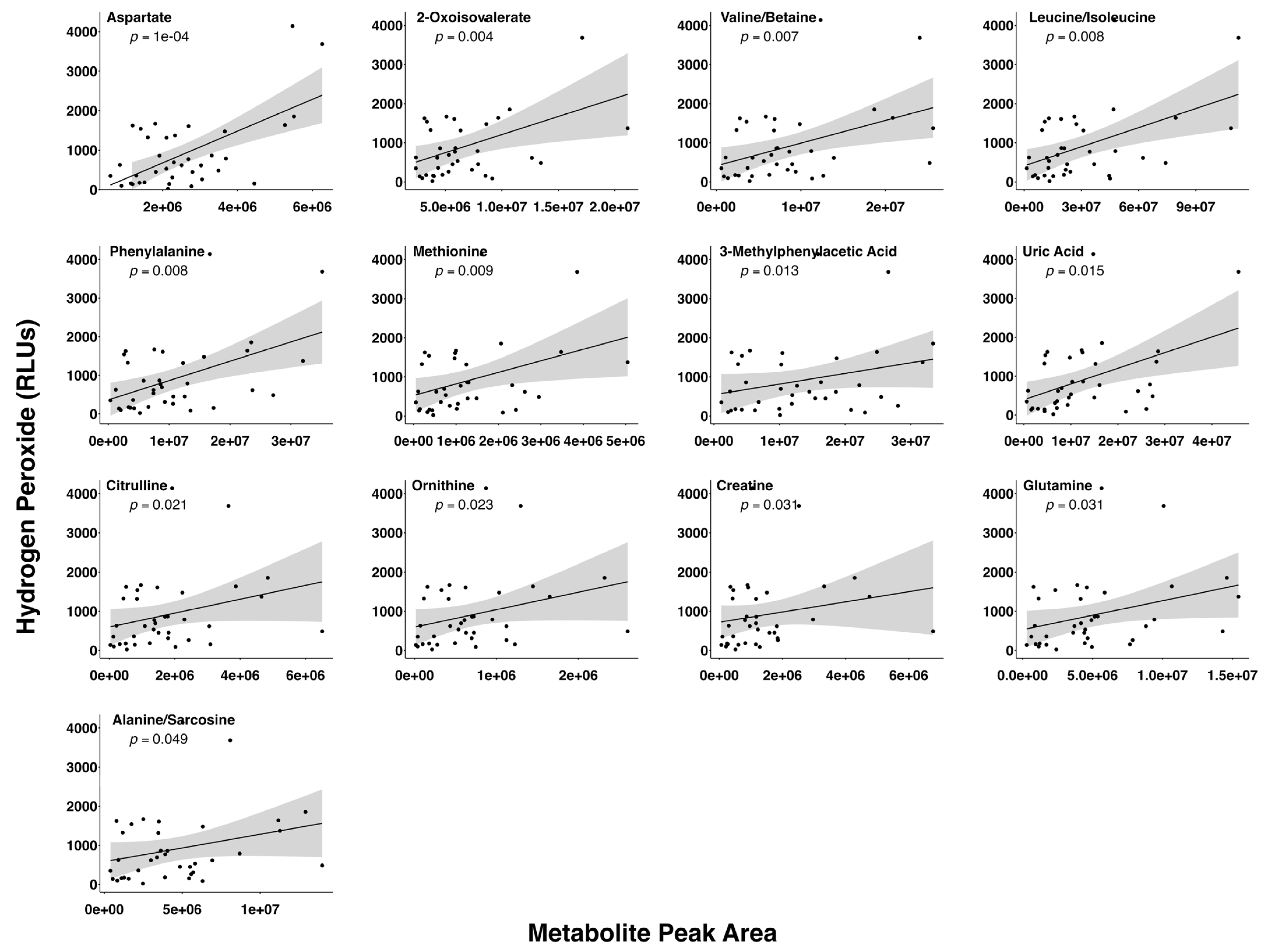
3.3. Relationship between Preovulatory Follicular Fluid H2O2 and Metabolite Levels

Thirteen follicular fluid metabolites (aspartate, 2-oxoisovalerate, valine/betaine, leucine/isoleucine, phenylalanine, methionine, 3-methylphenylacetic acid, uric acid, citrulline, ornithine, creatine, glutamine, and alanine/sarcosine) had a positive relationship with the follicular fluid H2O2 level (Figure 2).

4. Discussion

This manuscript is the first to our knowledge to relate circulating and intrafollicular ROS levels to BCS and metabolite levels in postpartum beef cows. The primary objective of this study was to determine if circulating and preovulatory follicular fluid ROS levels differed among thin, moderate, and obese conditioned beef cows. Animals of the current study were Angus in breed. Therefore, it is important to note that a limitation of the study is lack of sampling among various other beef breeds or species (Bos taurus vs. Bos indicus). It was surprising that no differences in H2O2 levels were observed among the BCS classes. Previous studies in dairy cows observed a relationship between circulating ROS levels and cow body condition [23]. This said, dairy cows experience a far greater negative energy balance and loss of body condition than beef cows. Thin cows in the present study were previously determined to have greater circulating β-hydroxybutyrate levels compared to moderate and obese animals [24]. Increased abundances of circulating β-hydroxybutyrate and numerous metabolites related to TCA cycle activity and mitochondrial function suggested that a greater negative energy balance and higher ROS levels would be observed in the thin cows of this study. The metabolic conditions that ultimately lead to a negative energy balance in these beef females mirror those observed in high-producing dairy cattle at the start of lactation in which the cow is unable to consume the dietary energy needed to maintain the metabolic requirements for lactation [33]. It is possible that the thin beef cows of the current study simply did not reach the level of negative energy balance required to observe differences in serum or follicular fluid H2O2 levels. One limitation of the current study’s design is the lack of knowledge related to length of time cows were in the respective body conditions and if weight was being gained or lost at the time of sample collections. As noted further in the discussion of serum metabolites, it is also possible that thin cows were effectively mitigating the levels of H2O2, and this contributed to lack of differences in H2O2 levels among BCS classes. It is also noteworthy that H2O2 does not fully depict the complete ROS composition of either biofluid assessed in this study. It was logical to test H2O2 in these preliminary samples because the sample quantity was limited and H2O2 is a common ROS whose levels are indicative of the overall ROS content in biofluids [25,26]. Nevertheless, the lack of a complete ROS profile was a limitation of the current study, and further investigation of a more complete ROS profile in beef animals with a greater variation in BCS could yield more significant differences.
An interesting result of the current study was the positive relationship between cow days postpartum and serum H2O2 levels. The start of peak lactation in cattle occurs at or around 30 days postpartum [34,35]. Animals in the current study were approximately 60 days postpartum when samples were collected, and were therefore lactating at peak levels that would lead to some degree of negative energy balance. It is interesting that no relationship was observed between follicular fluid H2O2 levels and days postpartum. This relationship in serum and lack of relationship in follicular fluid follows the same trend as the effect of the BCS on metabolite abundance observed in these animals and reported in Horn et al., 2022 [24]. There, we postulated that while the negative energy balance was high enough to lead to impacts in serum, the levels of metabolic stress had not reached levels adequate to affect the intrafollicular environment. The same hypothesis may hold true when considering an effect of days postpartum on H2O2 abundance within serum and follicular fluid. A limitation of the current study is that samples were collected at only one time point. This did not allow for an assessment of a relationship between serum and follicular fluid H2O2 with cow phenotypes or metabolites at varied points in lactation or allow for statistical procedures to account for distinct sampling time points in models. Future studies to examine potential relationships between days postpartum and follicular fluid or serum ROS levels at greater intervals postpartum would be of great benefit to determine if such a relationship emerges in follicular fluid after a longer duration of lactation. Additionally, determining the time point in which a positive relationship between days postpartum and serum ROS levels potentially diminishes due to a downward trend in lactation would be especially insightful when considering common beef production practices to rebreed cows at approximately 80 days postpartum to maintain a 365-day calving interval. A continued positive relationship between ROS levels and days postpartum in this ever-critical period could highlight an opportunity to develop therapeutic or management strategies to mitigate the potential negative impacts of excessive ROS levels on beef cow fertility.
Although there was no relationship between the BCS and serum H2O2 level, there was a significant interaction between the cow BCS and H2O2 level when assessing relationships between serum H2O2 and metabolite levels. Such an observation suggests that while differences in cows of each BCS category were not large enough to impact H2O2 levels directly, they did influence how H2O2 levels related to metabolite abundance. A limitation of this study related to statistical analyses and animal numbers is that the study was not designed to evaluate each BCS category individually. Preliminary results from analyses assessing each BCS class individually provide an interesting base from which to mount future studies targeted to explore relationships between serum metabolite and ROS levels in specific BCS categories. Although only statistically significant metabolites are discussed in this study, future studies should take heed of the identified statistical tendencies between metabolite abundances and H2O2 levels in the serum of each BCS category due to the limited power when data were subset by BCS category for further analyses. It is extremely intriguing that the relationship between serum H2O2 levels and significant metabolites appears to be negative in moderate conditioned animals, positive in thin cows, and potentially non-existent in obese animals.
Positive relationships between the serum abundance of H2O2 and the metabolites taurine, methyl succinic acid, and succinate/methylmalonate in thin cows suggest systemic efforts to regulate excessive ROS levels in such animals. The lack of relationships between levels of these metabolites and H2O2 in the serum of moderate and obese animals suggests that circulating ROS may have yet to reach levels in which regulation is required in these animals with a higher body condition. Such insights into the metabolic milieu of thin cows may also explain why no difference in circulating H2O2 levels was observed among the BCS categories. Taurine is a non-essential amino acid that is an effective antioxidant [36]. It plays a key role in mitochondrial integrity and has also been proven to detoxify H2O2 and other free radicals without acting as a classical scavenger of ROS formation [37]. Furthermore, although succinate could not be discriminated from methylmalonate in the current study, a positive relationship the levels of between either metabolite and H2O2 in the serum of thin animals is logical. Succinate has previously been linked to elevated ROS levels [38] and both the production and elimination of mitochondrial H2O2 [39], while an elevated methylmalonate level has been associated with disease phenotypes resulting from elevated ROS levels in humans [40]. If thin cows are actively combatting increasing H2O2 levels via the production of antioxidants such as these metabolites, our hypothesized increase in ROS levels within thin cows could be mitigated through an effective ROS scavenging system.
The positive relationship between 13 preovulatory follicular fluid metabolites and follicular fluid H2O2 could be explained by both systemic and intrafollicular events. The blood–follicle barrier is permeable to numerous metabolites and ROS [41]. Interestingly, of the preovulatory follicular fluid metabolites related to follicular fluid H2O2 levels, only 2-oxoisovalerate, ornithine, and alanine/sarcosine were significantly related or tended to be related to H2O2 levels in the serum of thin or moderately conditioned cows. Relationships between the levels of ornithine and alanine/sarcosine with H2O2 levels in serum and preovulatory follicular fluid are likely related to the mitigation of oxidative stress or accumulation of ROS, as ornithine has been demonstrated to induce signaling pathways imperative for cellular protection against ROS production, as well as to rescue cells damaged by oxidative stress [42]. While we were unable to discern the metabolite alanine from sarcosine, it is interesting that alanine has previously been utilized to induce oxidative stress and apoptosis in tumor cells [43], and sarcosine treatment increased ROS levels in dendritic cells as a means of anti-cancer treatment [44]. The lack of relationship between the same metabolites and H2O2 levels in serum and preovulatory follicular fluid and the known metabolic activity within the preovulatory follicle during the time that samples were collected [45] lead us to speculate that a majority of intrafollicular H2O2 and metabolite relationships are due to intrafollicular metabolism.
The process of oocyte maturation, which is occurring in the preovulatory follicle at the time samples were collected, requires increased mitochondrial activity and cellular metabolism, and thus leads to increased ROS production [46]. Furthermore, the metabolism of somatic follicular cells increases during this time and is associated with elevated levels of metabolites [45] and ROS [47]. Increasing ROS levels leading up to ovulation is critical for normal ovulatory events, but high fertility relies on the maintenance of the oxidative balance. Given the relationship between metabolic activity and elevated ROS levels, and the necessity to maintain oxidative balance within the follicle, it is not surprising that a number of the preovulatory follicular fluid metabolites positively associated with follicular fluid H2O2 levels have known roles in metabolism or oxidative balance. Aspartate and glutamine, metabolites associated with glucose metabolism and the TCA cycle, were positively related to H2O2 levels in follicular fluid. Aspartate concentration was positively associated with preovulatory follicle diameter in synchronized beef cows [28], a trait previously associated with cumulus–oocyte metabolism and oocyte competence for embryo development [27,48]. Furthermore, a previous study in women identified positive relationships between the follicular fluid aspartate concentration and both oocyte quality and the fertilization rate [49]. A study in dairy cows recognized a similar relationship between the follicular fluid glutamine concentration and probability of collection of an oocyte capable of producing a blastocyst, although a negative association between the follicular fluid aspartate concentration and blastocyst development was noted [50].
Similar to aspartate, the abundances of methionine, uric acid, and phenylalanine were positively correlated with preovulatory follicle diameter in lactating beef cows [28]. Levels of glutamine, uric acid, phenylalanine, methionine, and creatine, which were all positively related to H2O2 levels in follicular fluid in the current study, are associated with elevated ROS levels in various cells or biofluids. These metabolites have proven antioxidant roles [51,52,53,54,55] and were likely elevated in follicular fluid samples with increasing H2O2 levels to combat the negative impacts of oxidative stress. Uric acid is an end product of purine metabolism and acts as a potent antioxidant in the extracellular environment, such as the follicular fluid [56]. Glutamine and methionine metabolism provide necessary precursors to fuel glutathione synthesis for the oxidative balance [57,58,59]. Levels of this important antioxidant fluctuate throughout the estrous cycle but reach their peak during oocyte maturation, when samples in the current study were collected [60,61,62]. Additionally, methionine serves as a metabolic precursor to S-adenosylmethionine, an essential methyltransferase in the methylation of maternally imprinted genes in the oocyte [63], and the addition of methionine to in vitro maturing oocytes increases oocyte protein synthesis and developmental competency for embryo development [64,65].
The results described within this study are a key first step to better understand the dynamic relationships between body composition, oxidative stress, and reproduction in beef cattle. Additional explorations into the effect of increased ROS levels on postpartum anestrus, follicular recruitment, and oocyte quality may provide more insights into beef cattle rebreeding timelines. As additional foundational knowledge is gained, management strategies to mitigate the negative effects of a low BCS and excessive ROS levels on beef cow reproduction will be informed to positively impact the beef industry and production agriculture.

5. Conclusions

This study discovered no difference in serum or preovulatory follicular fluid H2O2 levels among lactating beef cows with thin, moderate, and obese BCS. Serum metabolite levels and their relationship with H2O2 levels in thin cows suggest that such individuals may be effectively mounting mechanisms to control elevated ROS levels. Thin beef cows in the current study were likely able to successfully mitigate ROS because the levels of negative energy balance and ROS generation had not reached the excessive levels observed in lactating dairy cows with a thin BCS. Preovulatory follicular fluid metabolites related to H2O2 in this study were likely byproducts of normal follicular metabolism during the preovulatory period. The positive relationships between the levels of metabolites with antioxidant roles and follicular fluid H2O2 levels demonstrates follicular mechanisms to regulate an optimal balance of ROS within the ovulatory follicle.

Author Contributions

Conceptualization, S.E.M.; methodology, K.S.H., J.L.E., R.R.P., F.N.S., S.R.C. and S.E.M.; formal analysis, K.S.H., E.A.H. and S.E.M.; investigation, K.S.H., J.L.E., R.R.P., F.N.S., S.R.C., E.A.H. and S.E.M.; resources, S.E.M.; writing—original draft preparation, K.S.H. and S.E.M.; writing—review and editing, J.L.E., R.R.P., F.N.S., S.R.C. and E.A.H.; visualization, K.S.H., E.A.H. and S.E.M.; supervision, S.E.M.; project administration, S.E.M.; funding acquisition, S.E.M. All authors have read and agreed to the published version of the manuscript.

Funding

This research was funded by the USDA NIFA MultiState project NE 2227 and the University of Tennessee Department of Animal Science and AgResearch.

Institutional Review Board Statement

All procedures involving animals for the current study were completed as part of a separate, larger study. The animal study protocol was approved by the Institutional Review Board (or Ethics Committee) of the University of Tennessee (protocol approved 20 December 2019).

Data Availability Statement

The raw data supporting the conclusions of this article will be made available by the authors on request. The raw data are not published in an online repository because studies by the PI in this area using the data contained in this manuscript are ongoing.

Acknowledgments

The authors would like to thank Sarah Howard and Hector Castro for their assistance with the UHPLC-HRMS metabolomic processing, Boyd Dingus for the donation of the Estrotect patches, and Walt Hitch and the staff of the Plateau AgResearch and Education Center for providing the cattle and their assistance in processing the cattle for this study. The authors would also like to thank Emma Horn, Justin Rhinehart, Courtnie Bridges, and Jess Klabnik for their assistance with sample collection.

Conflicts of Interest

The authors declare no conflict of interest. The funders had no role in the design of the study; in the collection, analyses, or interpretation of data; in the writing of the manuscript; or in the decision to publish the results.

References

  1. Richards, M.M.; Spitzer, J.C.; Warner, M.B. Effect of varying levels of postpartum nutrition and body condition at calving on subsequent reproductive performance in beef cattle. J. Anim. Sci. 1986, 62, 300–306. [Google Scholar] [CrossRef]
  2. Moraes, J.C.F.; Jaume, C.M.; Souza, C.J.H. Body condition score to predict the postpartum fertility of crossbred beef cows. Pesq. Agropec. 2007, 42, 741–746. [Google Scholar] [CrossRef]
  3. Gu, L.; Liu, H.; Gu, C.; Boots, C.; Moley, K.H.; Wang, Q. Metabolic control of oocyte development: Linking maternal nutrition and reproductive outcomes. Cell Mol. Life Sci. 2015, 72, 251–271. [Google Scholar] [CrossRef]
  4. Herring, A.D. Beef Cattle. In Encyclopedia of Agriculture and Food Systems; Elsevier: Amsterdam, The Netherlands, 2014; pp. 1–20. [Google Scholar] [CrossRef]
  5. Selk, G.E.; Wettemann, R.P.; Lusby, K.S.; Oltjen, J.W.; Mobley, S.L.; Rasby, R.J.; Garmendia, J.C. Relationship among weight change, body condition and reproductive performance of range beef cows. J. Anim. Sci. 1988, 66, 3153–3159. [Google Scholar] [CrossRef]
  6. DeRouen, S.M.; Franke, D.E.; Morrison, D.G.; Wyatt, W.E.; Coombs, D.F.; White, T.W.; Humes, P.E.; Greene, B.B. Prepartum body condition and weight influences on reproductive performance of first-calf beef cows. J. Anim. Sci. 1994, 72, 1119–1125. [Google Scholar] [CrossRef] [PubMed]
  7. D’Occhio, M.J.; Baruselli, P.S.; Campanile, G. Influence of nutrition, body condition, and metabolic status on reproduction in female beef cattle: A review. Theriogenology 2019, 125, 277–284. [Google Scholar] [CrossRef] [PubMed]
  8. Dominguez, M.M. Effects of body condition, reproductive status and breed on follicular population and oocyte quality in cows. Theriogenology 1995, 43, 1405–1418. [Google Scholar] [CrossRef]
  9. Snijders, S.E.M.; Dillon, P.; O’Callaghan, D.; Boland, M.P. Effect of genetic merit, milk yield, body condition and lactation number on in vitro oocyte development in dairy cows. Theriogenology 2000, 53, 981–989. [Google Scholar] [CrossRef] [PubMed]
  10. Dorice, A.K.; Ferdinand, N.; Justin, K.; Augustave, K.; Linda, K.K. Effects of breed, age, body condition score, and nutritional status on follicular population, oocyte yield, and quality in three cameroonian Zebus cattle Bos indicus. Adv. Agric. 2019, 2019, 2979740. [Google Scholar] [CrossRef]
  11. Ray, P.D.; Huang, B.W.; Tsuji, Y. Reactive oxygen species (ROS) homeostasis and redox regulation in cellular signaling. Cell Signal. 2012, 24, 981–990. [Google Scholar] [CrossRef]
  12. Lu, J.; Wang, Z.; Cao, J.; Chen, Y.; Dong, Y. A novel and compact review on the role of oxidative stress in female reproduction. Reprod. Biol. Endocrinol. 2018, 16, 80. [Google Scholar] [CrossRef] [PubMed]
  13. Long, H.; Ting, H.; Shabnam, F.; Linbao, J.; Tianyi, L.; Xi, M. Antioxidants maintain cellular redox homeostasis by elimination of reactive oxygen species. Cell Physiol. Biochem. 2017, 44, 532–553. [Google Scholar] [CrossRef]
  14. Celi, P. Oxidative stress in ruminants. In Studies on Veterinary Medicine. Oxidative Stress in Applied Basic Research and Clinical Practice; Mandelker, L., Vajdovich, P., Eds.; Humana Press: Totowa, NJ, USA, 2011. [Google Scholar] [CrossRef]
  15. Yan, F.; Zhao, Q.; Li, Y.; Zheng, Z.; Kong, X.; Shu, C.; Liu, Y.; Shi, Y. The role of oxidative stress in ovarian aging: A review. J Ovarian Res. 2022, 15, 100. [Google Scholar] [CrossRef] [PubMed]
  16. Agarwal, A.; Aponte-Mellado, A.; Premkumar, B.J.; Shaman, A.; Gupta, S. The effects of oxidative stress on female reproduction: A review. Reprod. Biol. Endocrinol. 2012, 10, 49. [Google Scholar] [CrossRef]
  17. Kala, M.; Shaikh, M.V.; Nivsarkar, M. Equilibrium between anti-oxidants and reative oxygen species: A requisite for oocyte development and maturation. Reprod. Med. Biol. 2016, 16, 28–35. [Google Scholar] [CrossRef] [PubMed]
  18. Pierce, G.B.; Parchment, R.E.; Lewellyn, A.L. Hydrogen peroxide as a mediator of programmed cell death in the blastocyst. Differentiation 1991, 46, 181–186. [Google Scholar] [CrossRef]
  19. Das, S.; Chattopadhyay, R.; Ghosh, S.; Ghosh, S.; Goswami, S.K.; Chakravarty, B.N.; Chaudhury, K. Reactive oxygen species level in follicular fluid-embryo quality marker in IVF? Hum. Reprod. 2006, 21, 2403–2407. [Google Scholar] [CrossRef]
  20. Tarín, J.J.; Vendrell, F.J.; Ten, J.; Blanes, R.; van Blerkom, J.; Cano, A. The oxidizing agent tertiary butyl hydroperoxide induces disturbances in spindle organization, c-meiosis, and aneuploidy in mouse oocytes. Mol. Hum. Reprod. 1996, 2, 895–901. [Google Scholar] [CrossRef]
  21. Orrenius, S.; Gogvadze, V.; Zhivotovsky, B. Mitochondrial oxidative stress: Implications for cell death. Annu. Rev. Pharmacol. Toxicol. 2007, 47, 143–183. [Google Scholar] [CrossRef]
  22. Tamura, H.; Takasaki, A.; Miwa, I.; Taniguchi, K.; Maekawa, R.; Asada, H.; Taketani, T.; Matsuoka, A.; Yamagata, Y.; Shimamura, K.; et al. Oxidative stress impairs oocyte quality and melatonin protects oocytes from free radical damage and improves fertilization rate. J. Pineal Res. 2008, 44, 280–287. [Google Scholar] [CrossRef]
  23. Abuelo, A.; Hernandez, J.; Benedito, J.L.; Castillo, C. The importance of the oxidative status of dairy cattle in the periparturient period: Revisiting antioxidant supplementation. J. Anim. Physiol. Anim. Nutr. 2015, 99, 1003–1016. [Google Scholar] [CrossRef] [PubMed]
  24. Horn, E.J.; Read, C.C.; Edwards, J.L.; Schrick, F.N.; Rhinehard, J.D.; Payton, R.R.; Campagna, S.R.; Klabnik, J.L.; Clark, H.M.; Myer, P.R.; et al. Preovulatory follicular fluid and serum metabolome profiles in lactating beef cows with thin, moderate, and obese body condition. J. Anim. Sci. 2022, 100, skac152. [Google Scholar] [CrossRef] [PubMed]
  25. Schieber, M.; Chandel, N.S. ROS function in redox signaling and oxidative stress. Curr. Biol. 2014, 24, 453–462. [Google Scholar] [CrossRef]
  26. Sies, H.; Belousov, V.V.; Chandel, N.S.; Davies, M.J.; Jones, D.P.; Mann, G.E.; Murphy, M.P.; Yamamoto, M.; Winterbourn, C. Defining roles of specific reactive oxygen species (ROS) in cell biology and physiology. Nat. Rev. Mol. Cell Biol. 2022, 23, 499–515. [Google Scholar] [CrossRef]
  27. Moorey, S.E.; Monning, J.M.; Smith, M.F.; Ortega, M.S.; Green, J.A.; Pohler, K.G.; Bridges, G.A.; Behura, S.K.; Geary, T.W. Differential transcript profiles in cumulus-oocyte complexes originating from pre-ovulatory follicles of varied physiological maturity in beef cows. Genes 2021, 12, 893. [Google Scholar] [CrossRef] [PubMed]
  28. Read, C.C.; Edwards, L.; Schrick, N.; Rhinehart, J.D.; Payton, R.R.; Campagna, S.R.; Castro, H.F.; Klabnik, J.L.; Horn, E.J.; Moorey, S.E. Correlation between pre-ovulatory follicle diameter and follicular fluid metabolome profiles in lactating beef cows. Metabolites 2021, 11, 623. [Google Scholar] [CrossRef]
  29. Whitman, R.W. Weight Change, Body Condition and Beef-Cow Reproduction. Colorado State University: Fort Collins, CO, USA, 1975. [Google Scholar]
  30. Kirby, C.J.; Smith, M.F.; Keisler, D.H.; Lucy, M.C. Follicular function in lactating dairy cows treated with sustained-release bovine somatotropin. J. Dairy Sci. 1997, 80, 273–285. [Google Scholar] [CrossRef]
  31. García-Guerra, A.; Canavessi, A.M.O.; Monteiro, P.L.J., Jr.; Mezera, M.A.; Sartori, R.; Kirkpatrick, B.W.; Wiltbank, M.C. Trio, a novel bovine high fecundity allele: III. acquisition of dominance and ovulatory capacity at a smaller follicle size. Biol. Reprod. 2018, 98, 350–365. [Google Scholar] [CrossRef]
  32. Pohler, K.G.; Pereira, M.H.C.; Lopes, F.R.; Lawrence, J.C.; Keisler, D.H.; Vasconcelos, J.L.M.; Green, J.A. Circulating concentrations of bovine pregnancy-associated glycoproteins and late embryonic mortality in lactating dairy herds. J. Dairy Sci. 2016, 99, 1584–1594. [Google Scholar] [CrossRef] [PubMed]
  33. Waterman, R.C.; Butler, W.R. Metabolic signals of the beef cow in negative energy balance. In Proceedings of the 4th Grazing Livestock Nutrition Conference, Ithaca, NY, USA, 9–10 July 2010. [Google Scholar]
  34. Hohenboken, W.D.; Dudley, A.; Moody, D.E. A comparison among equations to characterize lactation curves in beef cows. Anim. Sci. 1992, 55, 23–28. [Google Scholar] [CrossRef]
  35. Albertini, T.Z.; Medeiros, S.R.; Torres Junior, R.A.A.; Zocchi, S.S.; Oltjen, J.W.; Strathe, A.B.; Lanna, D.P.D. A methodological approach to estimate the lactation curve and net energy and protein requirements of beef cows using nonlinear mixed–effects modeling. J. Anim. Sci. 2012, 90, 3867–3878. [Google Scholar] [CrossRef] [PubMed]
  36. Baliou, S.; Adamaki, M.; Ioannou, P.; Pappa, A.; Panayiotidis, M.I.; Spandidos, D.A.; Christodoulou, I.; Kyriakopoulos, A.M.; Zoumpourlis, V. Protective role of taurine against oxidative stress (Review). Mol. Mel. Rep. 2021, 24, 605. [Google Scholar] [CrossRef]
  37. Cozzi, R.; Ricordy, R.; Bartolini, F.; Ramadori, L.; Perticone, P.; De Salvia, R. Taurine and el-lagic acid: Two differently-acting natural antioxidants. Environ. Mol. Mutagen. 1995, 26, 248–254. [Google Scholar] [CrossRef] [PubMed]
  38. Zhang, Y.; Zhang, M.; Zhu, W.; Yu, J.; Wang, Q.; Zhang, J.; Cui, Y.; Pan, X.; Gao, X.; Sun, H. Succinate accumulation induces mitochondrial reactive oxygen species generation and promotes status epilepticus in the kainic acid rat model. Redox. Biol. 2020, 28, 101365. [Google Scholar] [CrossRef] [PubMed]
  39. Tretter, L.; Patocs, A.; Chinopoulos, C. Succinate, an intermediate in metabolism, signal transduction, ROS, hypoxia, and tumorigenesis. Biochim. Biophys. Acta. Bioenerg. 2016, 1857, 1086–1101. [Google Scholar] [CrossRef]
  40. Stepien, K.M.; Heaton, R.; Rankin, S.; Murphy, A.; Bentley, J.; Sexton, D.; Hargreaves, I.P. Evidence of oxidative stress and secondary mitochondrial dysfunction in metabolic and non-metabolic disorders. J. Clin. Med. 2017, 6, 71. [Google Scholar] [CrossRef]
  41. Shalgi, R.; Kraicer, P.; Rimon, A.; Pinto, M.; Soferman, N. Proteins of human follicular fluid: The blood-follicle barrier. Fertil. Steril. 1973, 24, 429–434. [Google Scholar] [CrossRef]
  42. Shin, S.; Gombedza, F.C.; Bandyopadhyay, B.C. l-ornithine activates Ca2+ signaling to exert its protective function on human proximal tubular cells. Cell Signal. 2020, 67, 109484. [Google Scholar] [CrossRef]
  43. Stegman, L.D.; Zheng, H.; Neal, E.R.; Ben-Yoseph, O.; Pollegioni, L.; Pilone, M.S.; Ross, B.D. Induction of cytotoxic oxidative stress by D-alanine in brain tumor cells expressing Rhodotorula gracilis D-amino acid oxidase: A cancer gene therapy strategy. Hum. Gene Ther. 1998, 9, 185–193. [Google Scholar] [CrossRef]
  44. Dastmalchi, F.; Karachi, A.; Yang, C.; Azari, H.; Sayour, E.J.; Dechkovskaia, A.; Vlasak, A.L.; Saia, M.E.; Lovaton, R.E.; Mitchell, D.A.; et al. Sarcosine promotes trafficking of dendritic cells and improves efficacy of anti-tumor dendritic cell vaccines via CXC chemokine family signaling. J. Immunother. Cancer 2019, 7, 321. [Google Scholar] [CrossRef] [PubMed]
  45. Hessock, E.A.; Edwards, J.L.; Schrick, F.N.; Payton, R.R.; Campagna, S.R.; Pollock, A.B.; Clark, H.M.; Stokes, A.E.; Klabnik, J.L.; Hill, K.S.; et al. Metabolite abundance in bovine preovulatory follicular fluid is influenced by follicle developmental progression post estrous onset in cattle. Front. Cell Dev. Biol. 2023, 11, 1156060. [Google Scholar] [CrossRef] [PubMed]
  46. Heras, S.S.; Paramio, M.T. Impact of oxidative stress on oocyte competence for in vitro embryo production programs. Res. Vet. Sci. 2020, 132, 342–350. [Google Scholar] [CrossRef]
  47. Tamura, H.; Jozaki, M.; Tanabe, M.; Shirafuta, Y.; Mihara, Y.; Shinagawa, M.; Tamura, I.; Maekawa, R.; Sato, S.; Taketani, T.; et al. Importance of melatonin in assisted reproductive technology and ovarian aging. Int. J. Mol. Sci. 2020, 21, 1135. [Google Scholar] [CrossRef] [PubMed]
  48. Atkins, J.A.; Smith, M.F.; MacNeil, M.D.; Jinks, E.M.; Abreu, F.M.; Alexander, L.J.; Geary, T.W. Pregnancy establishment and maintenance in cattle. J. Anim. Sci. 2013, 91, 722–733. [Google Scholar] [CrossRef]
  49. D’Aniello, G.; Grieco, N.; Di Filippo, M.A.; Cappiello, F.; Topo, E.; D’Aniello, E.; Ronsini, S. Reproductive implication of D-aspartic acid in human pre-ovulatory follicular fluid. Hum. Reprod. 2007, 22, 3178–3183. [Google Scholar] [CrossRef]
  50. Marzyieh, S.; Rasoul, K.; Mohammad, H.A.A.; Nima, S.; Masoud, B.J. The relationship between bovine blastocyst formation in vitro and follicular fluid amino acids. Theriogenology 2023, 206, 197–204. [Google Scholar] [CrossRef]
  51. Arazi, H.; Eghbali, E.; Suzuki, K. Creatine supplementation, physical exercise and oxidative stress markers: A review of the mechanisms and effectiveness. Nutrients 2021, 13, 869. [Google Scholar] [CrossRef] [PubMed]
  52. Feng, L.; Li, W.; Liu, Y.; Jiang, W.D.; Kuang, S.Y.; Wu, P.; Jiang, J.; Tang, L.; Tang, W.N.; Zhang, Y.A.; et al. Protective role of phenylalanine on the ROS-induced oxidative damage, apoptosis and tight junction damage via Nrf2, TOR and NF-κB signalling molecules in the gill of fish. Fish Shellfish. Immun. 2017, 60, 185–196. [Google Scholar] [CrossRef] [PubMed]
  53. Kurajoh, M.; Fukumoto, S.; Yoshida, S. Uric acid shown to contribute to increased oxidative stress level independent of xanthine oxidoreductase activity in MedCity21 health examination registry. Sci. Rep. 2021, 11, 7378. [Google Scholar] [CrossRef] [PubMed]
  54. Luo, S.; Levine, R.L. Methionine in proteins defends against oxidative stress. FASEB J. 2009, 23, 464–472. [Google Scholar] [CrossRef]
  55. Stefani, G.P.; Nunes, R.B.; Dornelles, A.Z. Effects of creatine supplementation associated with resistance training on oxidative stress in different tissues of rats. J. Int. Soc. Sports Nutr. 2014, 11, 11. [Google Scholar] [CrossRef] [PubMed]
  56. Sánchez-Lozada, L.G.; Lanaspa, M.A.; Cristóbal-García, M.; García-Arroyo, F.; Soto, V.; Cruz-Robles, D.; Nakagawa, T.; Yu, M.A.; Kang, D.H.; Johnson, R.J. Uric acid-induced endothelial dysfunction is associated with mitochondrial alterations and decreased intracellular ATP concentrations. Nephron. Exp. Nephrol. 2012, 121, e71–e78. [Google Scholar] [CrossRef]
  57. Curi, R.; Lagranha, C.J.; Doi, S.Q.; Sellitti, D.F.; Procopio, J.; Pithon-Curi, T.C.; Corless, M.; Newsholme, P. Molecular mechanisms of glutamine action. J. Cell Physiol. 2005, 204, 392–401. [Google Scholar] [CrossRef] [PubMed]
  58. Amores-Sanchez, M.I.; Medina, M.A. Glutamine, as a precursor of glutathione, and oxidative stress. Mol. Genet. Metab. 1999, 67, 100–105. [Google Scholar] [CrossRef]
  59. Freitas, C.; Neto, A.C.; Matos, L.; Silva, E.; Ribeiro, Â.; Silva-Carvalho, J.L.; Almeida, H. Follicular fluid redox involvement for ovarian follicle growth. J. Ovarian Res. 2017, 10, 44. [Google Scholar] [CrossRef]
  60. Sutovsky, P.; Schatten, G. Depletion of glutathione during bovine oocyte maturation reversibly blocks the decondensation of the male pronucleus and pronuclear apposition during fertilization. Biol. Reprod. 1997, 56, 1503–1512. [Google Scholar] [CrossRef]
  61. Brad, A.; Bormann, C.; Swain, J.; Durkin, R.; Johnson, A.; Clifford, A.; Krisher, R.L. Glutathione and adenosine triphosphate content of in vivo and in vitro matured porcine oocytes. Mol. Reprod. Dev. 2003, 64, 492–498. [Google Scholar] [CrossRef] [PubMed]
  62. Zuelke, K.A.; Jeffay, S.C.; Zucker, R.M.; Perreault, S.D. Glutathione (GSH) concentrations vary with the cell cycle in maturing hamster oocytes, zygotes, and pre-implantation stage embryos. Mol. Reprod. Dev. 2003, 64, 106–112. [Google Scholar] [CrossRef]
  63. Ouyang, Y.; Wu, Q.; Li, J.; Sun, S.; Sun, S. S-adenosylmethionine: A metabolite critical to the regulation of autophagy. Cell Prolif. 2020, 53, 12891. [Google Scholar] [CrossRef]
  64. Saini, S.; Sharma, V.; Ansari, S.; Kumar, A.; Thakur, A.; Malik, H.; Kumar, S.; Malakar, D. Folate supplementation during oocyte maturation positively impacts the folate-methionine metabolism in pre-implantation embryos. Theriogenology 2022, 182, 63–70. [Google Scholar] [CrossRef]
  65. Ikeda, S.; Namekawa, T.; Sugimoto, M.; Kume, S. Expression of methylation pathway enzymes in bovine oocytes and preimplantation embryos. J. Exp. Zool. A Ecol. Genet. Physiol. 2010, 313, 129–136. [Google Scholar] [CrossRef] [PubMed]
Figure 1. Relationship between (a) serum hydrogen peroxide levels and body condition score (BCS), (b) follicular fluid hydrogen peroxide levels and BCS, and (c) serum hydrogen peroxide levels and cow days postpartum. RLUs, relative luminescence units; FF, follicular fluid.
Agriculture 14 01406 g001
Figure 2. Relationship between hydrogen peroxide levels and preovulatory follicular fluid metabolite abundances. The metabolite was unable to be discerned between two possible metabolites if listed with a “/” between two metabolite names. RLUs, relative luminescence units.
Agriculture 14 01406 g002
Table 1. Serum metabolites whose abundance was associated with a significant interaction between the cow body condition score and serum hydrogen peroxide level.
Interaction
BCS * H2O2
Thin
H2O2
Moderate
H2O2
Obese
H2O2
1 Metabolite Name2 p3 p (estimate)3 p (estimate)3 p (estimate)
2-Aminoadipate0.00350.0661 (112.3)0.0693 (−47.4)0.2252 (27.9)
2-Dehydro-D-Gluconate0.01150.1527 (77)0.0148 (−83)0.2466 (69)
Glyoxylate0.03100.0809 (358)0.1909 (−190)0.2092 (162)
Alanine/Sarcosine0.02550.1572 (174)0.0242 (−255)0.6234 (−61)
Creatinine0.05130.1432 (155)0.043 (−135)0.6370 (−49)
Proline0.04550.4954 (53)0.0468 (−116)0.2622 (60)
2-Oxoisovalerate0.02650.3769 (51)0.0719 (−105)0.1628 (105)
Succinate/Methylmalonate0.00080.0001 (653)0.1082 (−201)0.2775 (146)
Taurine0.00090.0095 (424)0.1436 (−135)0.0764 (240)
Methyl Succinic Acid0.00030.0016 (230)0.1572 (−60)0.6935 (−23)
Ornithine0.03190.0677 (84)0.0703 (−50)0.8930 (−4)
1 The metabolite was unable to be discerned between two possible metabolites if listed with a “/” between two metabolite names. 2 p value for the interaction between serum metabolite levels and serum H2O2 levels in the analysis including all animals. 3 p value and estimate for the relationship between serum metabolite levels and serum H2O2 levels in the respective body condition score category. BCS, body condition score; H2O2, hydrogen peroxide.
Disclaimer/Publisher’s Note: The statements, opinions and data contained in all publications are solely those of the individual author(s) and contributor(s) and not of MDPI and/or the editor(s). MDPI and/or the editor(s) disclaim responsibility for any injury to people or property resulting from any ideas, methods, instructions or products referred to in the content.

