Article

Dietary Passion Fruit Seed Oil Supplementation for Health and Performance of Laying Hens

by
Laís Garcia Cordeiro
1,*,
Paola Aparecida Damázio Rodrigues
1,
Gabrieli Andressa de Lima
1,
Elis Omar Figueroa Castillo
1,
Joyce Andrade da Silva
1,
Júlia de Lima Lopes
1,
Anna Luísa Lang
1,
Samir Moura Kadri
2,
Antônio Celso Pezzato
1 and
José Roberto Sartori
1
1
Department of Animal Breeding and Nutrition, School of Veterinary Medicine and Animal Science, São Paulo State University (UNESP), Botucatu 18618-681, SP, Brazil
2
Department of Animal Production, School of Technology and Agricultural Sciences, São Paulo State University (UNESP), Dracena 17915-899, SP, Brazil
*
Author to whom correspondence should be addressed.
Agriculture 2025, 15(8), 864; https://doi.org/10.3390/agriculture15080864
Submission received: 12 March 2025 / Revised: 5 April 2025 / Accepted: 7 April 2025 / Published: 16 April 2025

Abstract

:
Passion fruit seed oil (PFSO) is rich in bioactive compounds, which can enhance laying hens’ health and performance. The current study was conducted to investigate the effect of increasing PFSO supplementation in laying hens’ productive performance, egg quality, relative weight and length of organs, plasma lipid oxidation, antioxidant status, and gene expression of SOD, GPx, CAT, and NRF2 in the liver. One hundred ninety-two 25-week-old Lohmann Whites were randomly divided into three treatments (n = 8 replicates/diet, 8 hens/replicate). The groups were fed a corn–soybean basal diet containing 0.00%, 0.45%, and 0.90% PFSO for 16 weeks. The results indicated that increasing supplementation of PFSO decreased plasma lipid oxidation (n = 8; linear, p = 0.012) and increased CAT gene expression (n = 8; linear, p = 0.001). SOD and NRF2 genes tended to increase linearly, and GPx was not affected (n = 4; p > 0.05). The CAT activity tended to decrease linearly and the SOD and GPx were not affected (n = 8; p > 0.05) by diets. Performance and most egg quality, relative weight, and length of organs did not differ among treatments (n = 8; p > 0.05). Therefore, increasing the supplementation of PFSO in the diet may have positive effects on the laying hens’ health by decreasing oxidative stress, stimulating the antioxidant defense system, and sustaining egg production and quality.

Graphical Abstract

1. Introduction

Passion fruit seed oil (PFSO) is a vegetal oil rich in bioactive compounds, such as tocopherol, carotenoids, phytosterols and phenolic compounds, and unsaturated fatty acids (ω6, linoleic acid; ω9, oleic acid; and ω3, linolenic acid), which are important for the immune system and inflammatory response, and antioxidant system [1,2,3,4]. These compounds can modulate animals’ performance, gene expression, and final product quality [5,6].
Poultry rearing and handling conditions, even following the breeder recommendations, have a stressful effect [7]. Stress factors can lead to oxidative stress (OS), which negatively affects the performance of hens due to damage to macromolecules, immunosuppression, metabolic disorders, and reduced growth rate [8,9,10]. Egg production is also affected, as OS causes disturbances in calcium metabolism, resulting in inefficient deposition of this mineral in the eggshell, which leads to the formation of thin and fragile shells, compromising gas exchange and defense against pathogens [11,12]. Additionally, lipid and protein oxidation alters the consistency and composition of the albumen and yolk. These factors directly compromise egg quality, increasing susceptibility to breakage and shortening shelf life [13].
Given the prevalence of these agents, thought should be focused on nourishing the immune and antioxidant systems by using ingredients or additives rich in bioactive compounds and unsaturated fatty acids, such as PFSO, the properties of which can minimize the effects of oxidative stress by reducing lipid oxidation in plasma and meat, and stimulating the antioxidant enzymes activity [4,14]. The inclusion of PFSO in the diet of broilers under heat stress revealed protein modulation indicating a probable adaptive mechanism by reducing oxidative stress, activating neuroprotective mechanisms by the downregulation of neurofilament medium polypeptide subunit (NF-M), protecting against apoptosis, reducing inflammatory responses, and regulating energy metabolism [15]. All these mechanisms appear to be related to the PFSO content, especially the tocopherol, carotenoids, and phenolic compounds. Among the phenolic compounds, the flavonoids may have directly influenced the NF-M downregulation by the β-secretase activity reduction, thereby inhibiting β-amyloid formation, preventing the aggregation of toxic oligomers, and mitigating neuronal inflammation and cell death [16,17].
As PFSO has successfully been tested in broilers’ diets to improve their health and meat quality, its inclusion in laying hens’ diets may provide additional advantages for egg production and oxidative stress resistance. However, up until now, there have been few studies focused on the use of PFSO in poultry feed. Therefore, the current study was designed to assess the effect of PFSO inclusion in diets for laying hen performance, egg quality, plasma lipid oxidation, relative organ weight, antioxidant status, and relative gene expression of superoxide dismutase (SOD), catalase (CAT), glutathione peroxidase (GPx), and erythroid nuclear factor 2 (NRF2).

2. Materials and Methods

All the procedures described below were approved by the Ethics Committee on Animal Use from the São Paulo State University (UNESP), School of Veterinary Medicine and Animal Science, Botucatu, SP, Brazil, under protocol number 0097/2022.

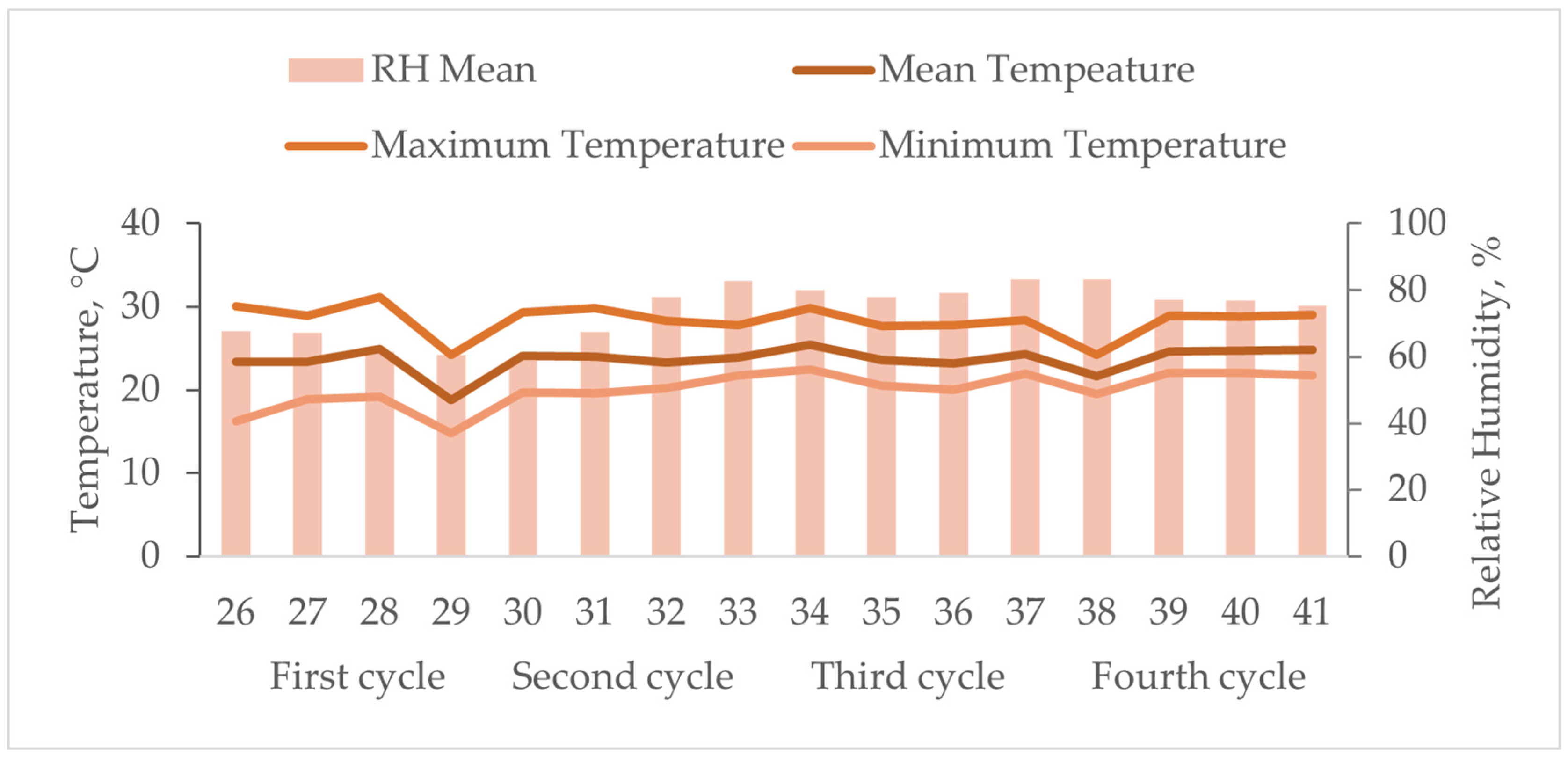
2.1. Experimental Design and Diets

The experiment was conducted in the Poultry Nutrition Laboratory at UNESP, School of Veterinary Medicine and Animal Science, Botucatu. One hundred ninety-two, 25-week-old, Lohmann White commercial laying hens with similar laying rates (98.44%) and body weight (1548.80 g) were randomly divided into three treatments. Laying hens in each treatment were designated to eight replicates with eight hens per replicate.
Mash feed and fresh water were available ad libitum during the 16 weeks of the experiment, from 25 to 41 weeks of age. The basal diet was formulated following the hen’s nutritional requirements and nutritional values of Rostagno et al. [18]. Passion fruit seed oil (PFSO) was extracted by cold pressing and purchased from Extrair-Óleos Naturais (Boa Esperança, ES, Brazil), with its composition described in Table 1.
PFSO was added to the treatments considering its apparent metabolizable energy (9378 kcal/kg) [14]. The experimental diet composition and nutritional levels are shown in Table 2.
The laying hens were placed in the two-tier battery cage with four hens per cage at 562.5 cm2/bird density. Two sequential cages with the same diet were arranged as a replicate and were equipped with trough feeders and nipple drinkers. The lighting program was applied according to the management guide [19]. The temperature and relative humidity were recorded daily using a digital thermo hygrometer (HOBO®) during the experimental period (Figure 1).

2.2. Effects of PFSO on Performance

During the experimental period, total egg weight, egg production, shell-less eggs, cracked eggs, and broken eggs were recorded daily on a replicate cage basis. Feed intake (g) was also measured and recorded based on a replicate weekly. After each 4-week cycle, the egg production (%), viable eggs (%), total egg weight (g), average egg weight (g), daily egg mass (g/bird/day), daily feed intake (g/bird/day) and feed conversion ratio (kg/dz and kg/kg) were calculated during the experimental period. Body weight was recorded at the beginning and at the end of the experiment to calculate body weight gain (g). The results were calculated and expressed as means of accumulated cycles at the end of the experiment.

2.3. Effects of PFSO on Egg Quality

To analyze egg quality, three eggs per replicate were collected with a maximum interval of three hours after laying, excluding those with fissures or cracks and just two were analyzed at the end of each cycle in three consecutive days. The eggs from each replication were identified and individually weighed on a precision scale. Thereafter, eggs were submitted to a specific gravity test through immersion in saline solution with different densities (1.060, 1.065, 1.070, 1.075, 1.080, 1.085, 1.090, 1.095, and 1.100 g/mL), measured with a densimeter, dipping the eggs from the lowest to the highest density, according to Sleigh [20].
The eggshell strength (N) and deformity (mm) were obtained with the texture analyzer (Model TA-XT2i, Stable Micro Systems LTDA., Goldalming, UK). The samples were placed horizontally on the support with a pre-test velocity of 2.0 mm/s, test speed of 1.0 mm/s, and post-test velocity of 4.0 mm/s, adapted from Montenegro et al. [21].
The eggs were broken on a flat glass surface and the heights and diameters of yolk and albumen were obtained with a digital caliper. The yolk index was determined by the ratio between the average yolk height and the diameter. Haugh unit [22] was calculated with the following formula: HU = 100 × log (H 7.57 − 1.7W0.37), where H refers to albumen height (mm) and W represents the egg weight (g). The yolk percentage was calculated by dividing the yolk weight by the egg weight. Albumen percentage was determined by the difference between the egg weight and the sum of the eggshell and yolk. The yolk color was measured by the DSM Yolk Color Fan and by a digital colorimeter, both on a scale from 0 to 16.
Eggshells were washed and dried in a dry oven at 55 °C for 24 h, weighed on a precision scale and the eggshell thickness was measured with the assistance of a digital micrometer at three points in the central region of the eggshell, after cooling. The eggshell percentage was calculated by dividing the eggshell weight by the egg weight. The shell weight per surface area (mg/cm2) was obtained with following the equation: SWUSA = {SW/[3.9782 × (EW0.7056)]} × 1000, where SW = eggshell weight, EW = egg weight; according to Abdallah et al. [23].

2.4. Internal Organs

At the end of the experimental period (112 days), eight hens per treatment were randomly selected and slaughtered by cervical dislocation after eight hours of fasting to access internal organs. The oviduct, ovary, liver, spleen, pancreas, gizzard, and total intestines were removed and weighed. Abdominal fat was also weighed. The length of the total intestines was measured and recorded considering the beginning of the duodenum from the gizzard up to the cloaca, including the length of the cecum. Oviduct length was also measured. The relative organ weight was calculated in relation to the final hen body weight.

2.5. Lipid Oxidation in Plasma

At the end of the last cycle (41-week-old), one hen per replicate (n = 8) was selected and 5.0 mL of blood was collected via brachial vein and drawn into EDTA-containing tubes. The obtained plasma samples were divided into 2.0 mL microtubes and stored at −80 °C for testing. Lipid oxidation was achieved from the results of thiobarbituric acid reactive substances (TBARS) analysis using the methodology adapted from Vyncke [24].
Hence, with 600 µL of plasma, 1.500 µL of extractor solution (TBA 0.67% + TCA 15% + HCl 0.25 N) was added into a Falcon tube, being homogenized in the vortex. The sample was centrifuged for 15 min at 3500× g at 4 °C. When the sample was cleared, the supernatant was taken, boiled for 45 min, and cooled for 5 min on ice. Afterward, the supernatant was added to a cuvette to read the absorbance in a spectrophotometer at 532 nm. A standard curve of 1,1,3,3-tetra-ethoxypropane was used to calculate the results and the data was expressed in mg of malonaldehyde (MDA) per liter of plasma.

2.6. Antioxidant Enzyme Activity in the Liver

Half of the liver’s left lobe was obtained from the same hens that were slaughtered by cervical dislocation. The samples were collected after removing all the internal organs. Livers were handled on top of ice-covered trays, preventing the enzyme’s denaturation, and immediately packed in a 5 mL microtube, immersed, and stored in liquid nitrogen, until enzyme activity determination. Briefly, 0.5 g of the liver samples were placed in a 50 mL falcon tube and homogenized in 2.5 mL of phosphate buffer (pH 7.8). The homogenate was centrifuged at 5000 rpm for 30 min at 4 °C. Supernatant was used for enzyme activity and protein content analysis.
Superoxide dismutase (SOD; EC 1.15.1.1) activity was assayed according to Beauchamp and Fridovich [25] based on the enzyme’s ability to convert superoxide radicals (O2) into hydrogen peroxide (H2O2) and molecular oxygen. This method measures one unit of SOD required to inhibit 50% of nitroblue tetrazolium, obtained by spectrophotometry at 560 nm. Catalase (CAT; EC 1.11.1.6) activity was assayed according to Sinha [26], in which dichromate in acetic acid is reduced to chromic acetate in the presence of H2O2 when heated, forming perchromic acid as an unstable intermediate. The absorbance reading was performed in a spectrophotometer at 610 nm. Glutathione peroxidase (GPx; EC 1.11.1.9) activity was assayed according to Flohé and Günzler [27]. For all the antioxidant enzyme assays, the total protein concentration was determined according to Bradford’s [28] method with Coomassie brilliant blue G-250, using bovine serum albumin as a standard. The absorbance reading was performed in a spectrophotometer at 320 nm. The results were expressed as U/mg.

2.7. RNA Extraction and Real-Time PCR Analysis

Total RNA was extracted from a 50 mg liver sample from four hens per treatment. The extraction was performed with the sample being minced with 1.000 µL of Trizol® reagent (Life Technologies, Carlsbad, CA, USA) according to the manufacturer standards, homogenized, and incubated for 5 min. Following the rupture and cell content release, 400 µL of chloroform was added, homogenized in the vortex for one minute, incubated, and centrifuged for 15 min at 12,000× g at 4 °C, to separate the phases. Afterward, the upper phase was collected and transferred to a clean tube, with the addition of 500 µL of isopropanol 100%, and homogenized.
The same process was repeated (incubated and centrifuged again), with the supernatant discarded and the precipitate washed with 1 mL of ethanol 75% in H2O DEPC 0.1%. Another centrifugation at 7500× g for 8 min at 4 °C was performed and the supernatant was discarded. The pellet was diluted in 20 µL ultrapure water (treated with DEPC 0.1%), RNAse free. The samples were heated for 10 min at 56 °C and stored at −80 °C. The extracted RNA was used for cDNA synthesis using the High-capacity cDNA Reverse Transcription kit (Thermo Fisher Scientific, Waltham, MA, USA), according to the manufacturer’s instructions.
Analysis for SOD, GPx, CAT, and NRF2 gene expression was performed with the quantitative real-time PCR technique using an Applied Biosystems StepOnePlus Real-Time PCR System (Applied Biosystems, Foster City, CA, USA) and a SYBR Green PCR Master Mix kit (Applied Biosystems, Foster City, CA, USA). The samples were evaluated in triplicates, using the β-actin gene as an internal control to normalize the expression of the target genes. The results were obtained with the Pfaffl [29] formula: (EFgoi∆CtGOI)/(EFact∆CtAct) where EFgoi = gene of interest efficiency; ∆CtGOI = cycle threshold from the gene of interest; EFact = β-actin efficiency; and ∆CtAct = cycle threshold from β-actin.
The procedures were conducted in the Biotechnology Institute (IBTEC) at UNESP, Botucatu, SP, Brazil. Primer sequences obtained from a commercial company (Thermo Fisher Scientific, Waltham, MA, USA) were tested for primer efficiency and their characteristics are described in Table 3.

2.8. Statistical Analysis

Data were submitted to the UNIVARIATE procedure and, whether the residual distribution was normal, the results were analyzed with simple regression by the REG procedure in the software SAS (version 9.4, SAS Institute Inc., Cary, NC, USA) [32], to verify which regression model (linear or quadratic) best describes the results. The means were compared using the Tukey test at the p < 0.05 value.

3. Results

3.1. Performance

In Table 4, the PFSO effect in laying hens’ productive performance is shown as means of four cycles (28 days each). No effect of dietary treatment was found in egg production (%), viable eggs (%), total egg weight (kg), daily egg weight (g/hen/day), daily egg mass (g/hen/day), daily feed intake (g), feed conversion ratio per kilograms, and body weight gain (g) (p > 0.05). There was no mortality during the trial, from weeks 26 to 41.

3.2. Egg Quality

The percentage of broken eggs calculated during the collection period for egg quality analysis was 1.05% (0.00% PFSO), 2.10% (0.45% PSFO), and 1.32% (0.90% PFSO), with no linear (p = 0.726) or quadratic (p = 0.117) effect. Egg quality data from the laying hens fed a control diet and PFSO levels are listed in Table 5. The results revealed that albumen height (mm) was reduced in linear regression (p = 0.046) with the PFSO inclusion, and there was also a trend to Haugh unit reduction (p = 0.054). The egg weight (g), albumen (%), yolk (%), eggshell (%), yolk fan color, yolk digital color, yolk index, albumen diameter (mm), eggshell strength (N), eggshell deformity (mm), eggshell thickness (mm) and SWUSA (mg/cm2) were not affected (p > 0.05) from week 26 to 41.

3.3. Relative Internal Organ Weights

Table 6 summarizes the relative organ weights (%) and the oviduct and intestine (cm) measures of the hens fed diets containing different PFSO levels. The relative abdominal fat (p = 0.081) and ovary weights (p = 0.098) presented a quadratic regression response to the PFSO supplementation. There was no difference (p > 0.05) in the body weight and relative weight of the liver, spleen, pancreas, gizzard, oviduct, and total intestines. The oviduct and intestine lengths also showed no difference (p > 0.05).

3.4. Lipid Oxidation in Plasma and Antioxidant Activity in the Liver

Lipid oxidation in plasma was evaluated considering the MDA concentration (mg/L) and the results are presented in Table 7. The responses showed that compared with the control group, the diet PFSO inclusion decreased lipid peroxidation in a linear regression (p = 0.012). The antioxidant enzyme activity in the liver is also described in Table 7. The CAT enzyme activity presented a trend (p = 0.078) to reduce linear regression. There was no effect of PFSO in the activity of SOD and GPx (p > 0.05).

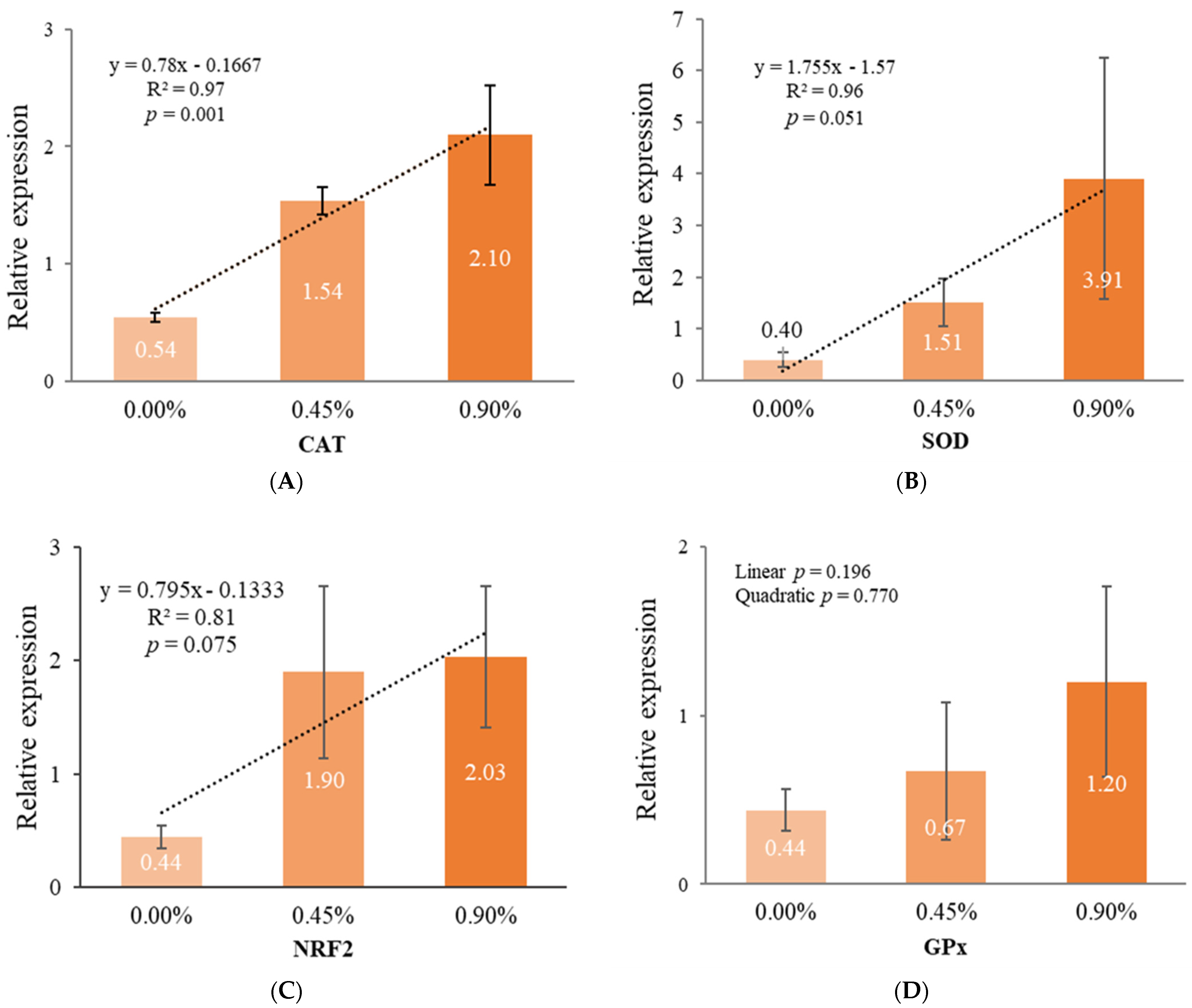
3.5. Gene Expression in the Liver

The effect of PFSO supplementation on the relative gene expression of CAT, SOD, NRF2, and GPx is shown in Figure 2. The PFSO supplementation increased CAT relative gene expression in a linear regression (p = 0.001). For the SOD (p = 0.051) and NRF2 (p = 0.075) genes, a positive trend was observed in the linear regression analysis. Nevertheless, the relative gene expression of GPx was not affected (p > 0.05) by PFSO inclusion.

4. Discussion

The inclusion of passion fruit seed oil (PFSO) in the diet stimulated the expression of antioxidant enzymes and reduced plasma oxidation in Lohmann White laying hens, even without enhancing the enzyme activity in the liver. PFSO has also sustained performance and most parameters from egg quality and relative organ weights.
Gene expression of CAT, SOD, and NRF2 linearly increased as PFSO levels were supplemented. Although the GPx gene expression was not significant, it was 52.27% (0.45% PFSO) and 172.70% (0.90% PFSO) higher than the control group. In addition, antioxidant analysis in the liver revealed that CAT enzymes tended to linearly reduce, and even without the other enzymes presenting a significative effect, the SOD was 1.65% (0.45% PFSO) and 4.02% (0.90% PFSO) lower than the control group, and GPx was 6.21% (0.45% PFSO) and 13.1% (0.90% PFSO) lower than the control group. These results indicate that the bioactive compounds of the PFSO acted in two different ways by directly acting as non-enzymatic antioxidants and by indirectly stimulating the enzymatic antioxidants gene expression. Cruvinel et al. [6], evaluating broiler chickens subjected to heat stress and fed diets containing 0.6% pequi oil, observed an increase in the NRF2 gene expression, but no effect of the enzyme activity in the liver. The NRF2 gene is linked to the antioxidant system by regulating numerous genes, such as CAT, SOD, and GPx, which is consistent with the increased mRNA expression of these enzymes in this study [30,33,34,35].
PFSO has positive effects on oxidative stress since studies evaluating broilers reared under heat stress conditions have shown an increase in the CAT enzymatic activity, along with the upregulation of proteins involved in the catabolic process of hydrogen, such as hemoglobin subunit epsilon. Additionally, PFSO influences the regulation of PGRMC1 (membrane-associated progesterone receptor component 1), which modulates the activity of cytochrome P450 which induces an increase in reactive oxygen species [36]. Assunção et al. [15] also reported that PFSO reduces the Ca2+ release in the cytosol and, consequently, the occurrence of cellular apoptosis; recovers and protects neuronal structures; inhibits NF-κB activation, as it reduces ROS with its antioxidant properties (tocopherol, phenolic compounds, and carotenoids); and reduces the expression of proteins from the glycogenolysis and gluconeogenesis pathways, as it controls oxidative stress and reduces energy demand to meet the demands of the antioxidant system. The PFSO also presents anti-inflammatory action when applied topically in rabbits [37], in hairless mice [38], in horses and Wistar rats [39], and in mice [40].
Although the bioactive compounds in PFSO, mainly tocopherol and carotenoids, have stimulated the transcription of hepatic enzymes responsible for protecting the body against oxidative stress, their action mechanisms are not yet well established [30,41,42]. Several studies have indicated that even after mRNA transcription, it is not guaranteed that these genes will be translated into proteins, depending on post-transcriptional regulation [43,44]. In addition, the protein can also be regulated at the level of translation and turnover [43]. NRF2 activity, for example, can be regulated by Keap1-NRF2 protein stability, transcriptional regulators (NF-B, ATF3, and ATF4), and post-transcription regulators (miRNAs) [45,46,47]. The CAT, beyond post-transcriptional regulation, can also be regulated by specific proteins or degraded through pexophagy, selective autophagy directed at peroxisomes, or through the ubiquitin–proteasome system [44]. In this regard, some stressors can act by reducing the target gene expression and, in the meantime, the supplementation of antioxidant substances can positively modulate their post-transcriptional process [44]. In this study, although the gene expression increases, the enzymatic activity is only slightly affected and, however, can be compared to those reported by [48], who supplemented exogenous enzymes in aged laying hens and found a lower expression of pancreatic amylase mRNA without altering its enzymatic activity, suggesting post-transcriptional regulation. Therefore, we speculate that the bioactive compounds in PFSO could indirectly regulate specific genes, through post-transcriptional mechanisms, to delay the activation of the corresponding antioxidant pathways, potentially avoiding excessive responses and maintaining balance in the organism [49].
The linear reduction in plasma lipoperoxidation due to PFSO inclusion is favorable to layers’ health, considering their predisposition to hepatic lipidosis [50,51,52]. The antioxidant system is responsible for redox balance in the body and includes enzymatic antioxidants, such as CAT, SOD, and GSPx, and non-enzymatic antioxidants including carotenoids, phenolic compounds, and vitamins (A, C, and E) [53,54]. Vitamin E is an important antioxidant molecule composed of the α, β, and γ isoforms of tocopherols and tocotrienols, with emphasis on the alpha-tocopherol action [55]. The dietary vitamin E is absorbed with the lipids and then is preferentially adsorbed in the hepatic tissue, increasing its concentration [56], which is consistent with our results of increased gene expression of antioxidant enzymes in the hepatic tissue stimulated by tocopherol [41]. From the liver, the tocopherol is secreted into plasma, especially with LDL, acting to suppress lipoperoxidation [57] and scavenging the molecules resulting from oxidative processes, such as malonaldehyde [58,59,60]. Furthermore, several studies reported that tocopherol supplementation improved antioxidant capacity by enhancing GSH level [60], GSPx [61,62,63], SOD [62,63], and CAT activity [60]. In addition, polyunsaturated fatty acids could also be associated with the reduction in LDL oxidation, as their hypocholesterolemic action results in larger chylomicrons, which appear to be eliminated more quickly than saturated fatty acids [64]. This result corroborates with [65], who found a reduction in the level of triglycerides, total cholesterol, and low-density lipoprotein-cholesterol in Wistar rats, and suggested that the PFSO hypolipidemic effect is associated with its rich content in linoleic acid.
The carotenoids (CAR) also play a crucial role in deactivating reactive species, such as peroxyl radicals (ROO) and singlet oxygen (1O2). This protective mechanism occurs through different pathways: CAR can be oxidized by a radical to form CAR radical cations, reduced to form a CAR radical anion, or donate hydrogen to form a stable radical. In addition, reactive species from lipid oxidation, e.g., peroxyl lipid (LOO), can also interact with CAR and form stable products [66,67]. Among the carotenoids, ß-carotene stands out [66] as a precursor of vitamin A that is significantly present in PFSO (75.63 mg/100g) [3], when compared to other sources of ß-carotene [68]. The improvement of lipid peroxidation can also be associated with the ß-carotene content present in the PFSO. The result corroborates the data presented by Zanetti [14] evaluating the PFSO inclusion in broiler diets. Cruvinel et al. [6], evaluating pequi oil, rich in β-carotene, also reported a malonaldehyde reduction. The author of [36] found results indicating that the use of PFSO reduced the percentage of DPPH and differentially expressed the protein spot Alcohol dehydrogenase 1 (ADH1) in broiler chickens, confirming the presence of retinol in the PFSO.
The present study aimed to improve the performance and egg quality of the layers fed with PFSO, since different oil sources can affect production variables according to the bioactive compounds concentration and the fatty acid ratio, modifying nutrient digestibility and absorption rate and the immune and antioxidant systems activity [69,70,71]. The PFSO inclusion, however, linearly reduced the albumen height, which is used to calculate the Haugh unit and explains its downward trend. In addition, the relative albumen weight decreased 0.47% (0.45% PFSO) and 0.63% (0.90% PFSO) compared to the control, while the yolk increased 0.94% (0.45% PFSO) and 1.54% (0.90% PFSO) compared to the control. These results corroborate studies that report that the lipid composition of the diet affects egg weight, especially yolk weight, with the concentration of linoleic acid being a limiting factor for yolk development [72,73]. There are also reports that Vitamin E can facilitate the release of vitellogenin, a lipoprotein specialized in transporting lipids from the liver to the ovary [74]. Thus, the high concentration of linoleic acid (69.14%) in the PFSO was used by the liver in the synthesis of lipoproteins, probably vitellogenins, which had their release stimulated by vitamin E, favoring an increase in the relative weight of the yolk. In addition, the HU results of this experiment (0.00% PFSO: 93.19; 0.45% PFSO: 92.26; and 0.90% PFSO: 91.78) exceed the minimum value determined for consumption (HU = 60), indicating quality eggs [75,76].
Despite the lack of studies using PFSO, especially in commercial layers, tests evaluating the use of oils have been carried out for years [51,77]. The studies vary from the use of soybean, canola, linseed, fish, sunflower, and rapeseed oils, among others, finding results that vary between no effect, positive effect, or negative effect on performance and egg quality [51]. The unaltered variables of this study, however, corroborate Lelis et al. [78], Ceylan et al. [79], Reddy et al. [80], and Costa et al. [81], who did not observe any effect of dietary inclusion of different oil sources on performance and egg quality. Several factors could affect oil sources, such as the vegetation stage, the climate, and the crops’ production levels, causing inconsistency in the oil’s effects [82].
The inclusion of PFSO in layers’ diet did not affect the most relative organ weights, as indicated by other phytogenic additives [83,84], which may be related to the tested product and its composition, and by the poultry rearing conditions [6]. Although oxidative stress can affect the relative weight of immunologic (spleen, bursa, and intestines) and important metabolic organs, such as the liver, the present study did not have enough challenge to affect their development [85], even with the probable increase in liver activity to produce vitellogenin, which will be deposited in the yolk, stimulated by vitamin E and the omega 6 content [72,73,74]. However, the quadratic trend observed for the relative weight of abdominal fat and ovary shows an increase in hens receiving 0.45% PFSO and a decrease in the 0.90% PFSO dose. This trend goes along with body weight gain and feed conversion ratio, which resulted in the final body weight (BW), with the hens in the 0.45% PFSO treatment (BW: 1,806.50) being 2.16% higher and those in the 0.90% PFSO treatment (BW: 1,726.63) being 2.35% lower than the average weight of the control group (BW: 1,768.25). Therefore, although this behavior can be seen in the other organs, the difference in these hens’ weight was more markedly represented by the accumulation of abdominal fat and the development of the ovary, a common characteristic in laying hens, since lipid metabolism and the development of the organ that generates the yolk are correlated [52].
The supplementation levels of PFSO used in this study did not affect the most variables analyzed. However, the PFSO bioactive compounds have been widely studied, highlighting the potential use of this oil as an additive due to its antioxidant, immunomodulatory, and anti-inflammatory properties. The results presented in this paper provide a starting point for PFSO evaluation in laying hens’ nutrition and suggest the need for further research under different conditions, such as heat stress or pathogenic challenges, to also assess the effects of PFSO on the immune system. Additionally, in-depth analyses using omics approaches are recommended to elucidate the PFSO impact on the post-transcriptional regulation of antioxidant genes.

5. Conclusions

In conclusion, PFSO at a level up to 0.90% provides beneficial responses to laying hens’ health by acting as natural antioxidants and indirectly activating the defense mechanisms to reduce the effects of molecular oxidative damage, expressed by plasma lipid oxidation decrease and antioxidant enzyme gene expression increase, even without activating the enzymatic antioxidant system in the liver. Overall, the PFSO compounds were not enough to enhance laying hens’ performance and egg quality but were able to sustain performance and most parameters from egg quality and relative organ weights and lengths.

Author Contributions

Conceptualization, L.G.C. and J.S; methodology, L.G.C., S.M.K. and J.R.S.; validation, L.G.C.; formal analysis, L.G.C.; investigation, L.G.C., P.A.D.R., G.A.d.L., E.O.F.C., J.A.d.S., J.d.L.L., A.L.L., S.M.K., A.C.P. and J.R.S.; resources, J.R.S.; data curation, L.G.C., P.A.D.R., G.A.d.L., E.O.F.C., J.d.L.L., A.L.L., S.M.K., A.C.P. and J.A.d.S.; writing—original draft preparation, L.G.C. and J.R.S.; writing—review and editing, L.G.C., P.A.D.R., G.A.d.L. and J.R.S.; visualization, L.G.C. and J.R.S.; supervision, J.R.S.; project administration, L.G.C. and J.R.S. All authors have read and agreed to the published version of the manuscript.

Funding

This research was funded by the National Council for Scientific and Technological Development—CNPq, grant number 163518/2021-1.

Institutional Review Board Statement

The animal study protocol was approved by the Ethics Committee on Animal Use of the School of Veterinary Medicine and Animal Science (protocol code 0097/2022) on 11 May 2022.

Data Availability Statement

The data presented in this study are available on request from the corresponding author.

Conflicts of Interest

The authors declare no conflicts of interest.

Abbreviations

The following abbreviations are used in this manuscript:
CARcarotenoids
CATcatalase
GPxglutathione peroxidase
HClHydrochloric acid
HUHaugh unit
IBTECBiotechnology Institute
MDAmalonaldehyde
NRF2erythroid nuclear factor 2
PFSOpassion fruit seed oil
RHrelative humidity
SODsuperoxide dismutase
SWUSAshell weight per surface area
TBAthiobarbituric acid
TBARSthiobarbituric acid reactive substances
TCATrichloroacetic acid
UNESPSão Paulo State University

References

  1. Malacrida, C.R.; Jorge, N. Yellow passion fruit seed oil (Passiflora edulis f. flavicarpa): Physical and chemical characteristics. Braz. Arch. Biol. Technol. 2012, 55, 127–134. [Google Scholar] [CrossRef]
  2. Calder, P.C. N-3 fatty acids, inflammation, and immunity: New mechanisms to explain old actions. Proc. Nutr. Soc. 2013, 72, 326–336. [Google Scholar] [CrossRef] [PubMed]
  3. Santana, F.C.; Shinagawa, F.B.; Araujo, E.S.; Costa, A.M.; Mancini-Filho, J. Chemical composition and antioxidant capacity of brazilian passiflora seed oils. J. Food Sci. 2015, 80, 2647–2654. [Google Scholar] [CrossRef] [PubMed]
  4. Zanetti, L.H.; Granero, L.; Poletto, M.; Da Luz, P.A.; Lala, B.; Delbem, N.L.C.; Sobral, N.C.; Brito, E.P.; Denadai, J.C.; Andrighetto, C.; et al. Chemical, physical, and oxidative characteristics of broilers meat supplemented with passion fruit seed oil. Int. J. Innov. Educ. Res. 2021, 9, 69–83. [Google Scholar] [CrossRef]
  5. Georganas, A.; Giamouri, E.; Pappas, A.C.; Papadomichelakis, G.; Galliou, F.; Manios, T.; Tsiplakou, E.; Fegeros, K.; Zervas, G. Bioactive Compounds in food waste: A review on the transformation of food waste to animal feed. Foods 2020, 9, 291. [Google Scholar] [CrossRef]
  6. Cruvinel, J.M.; Groff-Urayama, P.M.; Oura, C.Y.; de Lima-Krenchinski, F.K.; dos Santos, T.S.; de Souza, B.A.; Kadri, S.M.; Correa, C.R.; Sartori, J.R.; Pezzato, A.C. Pequi Oil (Caryocar brasiliense Camb.) Attenuates the Adverse Effects of Cyclical Heat Stress and Modulates the Oxidative Stress-Related Genes in Broiler Chickens. Animals 2023, 13, 1896. [Google Scholar] [CrossRef] [PubMed]
  7. Rafiei, F.; Khajali, F. Flavonoid antioxidants in chicken meat production: Potential application and future trends. Worlds Poult. Sci. J. 2021, 77, 347–361. [Google Scholar] [CrossRef]
  8. Monaghan, P.; Metcalfe, N.B.; Torres, R. Oxidative stress as a mediator of life history trade-offs: Mechanisms, measurements, and interpretation. Ecol. Lett. 2009, 12, 75–92. [Google Scholar] [CrossRef]
  9. Celi, P.; Selle, P.H.; Cowieson, A.J. Effects of organic selenium supplementation on growth performance, nutrient utilisation, oxidative stress and selenium tissue concentrations in broiler chickens. Anim. Prod. Sci. 2013, 54, 966–971. [Google Scholar] [CrossRef]
  10. Rehman, Z.U.; Meng, C.; Sun, Y.; Safdar, A.; Pasha, R.H.; Munir, M.; Ding, C. Oxidative stress in poultry: Lessons from the viral infections. Oxidative Med. Cell. Long. 2018, 2018, 5123147. [Google Scholar] [CrossRef]
  11. Fouad, A.M.; Chen, W.; Ruan, D.; Wang, S.; Xia, W.G.; Zheng, C.T. Impact of heat stress on meat, egg quality, immunity and fertility in poultry and nutritional factors that overcome these effects: A review. Int. J. Poult. Sci. 2016, 15, 81. [Google Scholar] [CrossRef]
  12. Chen, Z.; Xing, T.; Li, L.; Zhang, J.; Jiang, Y.; Gao, F. Oxidative stress impairs the meat quality of broiler by damaging mitochondrial function, affecting calcium metabolism and leading to ferroptosis. Anim. Biosci. 2022, 35, 1616. [Google Scholar] [CrossRef] [PubMed]
  13. Jin, J.; Zhou, Q.; Lan, F.; Li, J.; Yang, N.; Sun, C. Microbial composition of egg component and its association with hatchability of laying hens. Front. Microbiol. 2022, 13, 943097. [Google Scholar] [CrossRef]
  14. Zanetti, L.H. Óleo de Semente de Maracujá (Passiflora edulis) na Alimentação de Frangos de Corte. Ph.D. Thesis, São Paulo State University, São Paulo, Brazil, 2018; 100p. [Google Scholar]
  15. Assunção, A.S.; Martins, R.A.; Vieira, J.C.S.; Rocha, L.C.; Lima-Krenchinski, F.K.; Buzalaf, M.A.R.; Sartori, J.R.; Padilha, P.M. Shotgun proteomics reveals changes in the Pectoralis major muscle of broilers supplemented with passion fruit seed oil under cyclic heat stress conditions. Food Res. Int. 2023, 167, 112731. [Google Scholar] [CrossRef]
  16. Yang, S.; Liu, W.; Lu, S.; Tian, Y.Z.; Wang, W.Y.; Ling, T.J.; Liu, R.T.; Novel, A. A Novel Multifunctional Compound Camellikaempferoside B Decreases Aβ Production, Interferes with Aβ Aggregation, and Prohibits Aβ-Mediated Neurotoxicity and Neuroinflammation. ACS Chem. Neurosci. 2016, 7, 505–518. [Google Scholar] [CrossRef]
  17. dos Reis, L.C.R.; Facco, E.M.P.; Salvador, M.; Flôres, S.H.; Rios, A.d.O. Antioxidant potential and physicochemical characterization of yellow, purple and orange passion fruit. J. Food Sci. Technol. 2018, 55, 2679–2691. [Google Scholar] [CrossRef]
  18. Rostagno, H.S.; Albino, L.F.T.; Donzele, J.L.; Gomes, P.C.; Oliveira, R.F.; Lopes, D.C.; Ferreira, A.S.; Barreto, S.L.T. Tabelas Brasileiras para aves e Suínos: Composição de Alimentos e Exigências Nutricionais, 4th ed.; Departamento de Zootecnia: Viçosa, Brazil, 2017. [Google Scholar]
  19. Lohmann LSL-Lite. Manual de Manejo: Alojamento em Gaiolas. 2020. Available online: https://lohmann-breeders.com/media/2020/08/LOHMANN_MG_LSL-Lite_Portuguese.pdf (accessed on 5 August 2022).
  20. Sleigh, R.W. Egg Science and Technology, 4th ed.; Stadelman, W.J., Cotterill, O.J., Eds.; Routledge: New York, NY, USA, 1996; Volume 7, p. 341. [Google Scholar]
  21. Montenegro, A.T.; Garcia, E.A.; Molino, A.B.; Cruvinel, J.M.; Ouros, C.C.; Alves, K.S. Methods to Evaluate the Eggshell Quality of Table Eggs. Braz. J. Poult. Sci. 2019, 21, 1–6. [Google Scholar] [CrossRef]
  22. Haugh, R.R. The Haugh unit for measuring egg quality. U.S. Egg Poult. Mag. 1937, 43, 552–555. [Google Scholar]
  23. Abdallah, G.A.; Harms, R.H.; El-Husseiny, O. Various methods of measuring shell quality in relation to percentage of cracked eggs. Poult. Sci. 1993, 72, 2038–2043. [Google Scholar] [CrossRef]
  24. Vyncke, B.W. Direct determination of the thiobarbituric acid value in trichloroacetic acid extracts of fish as a measure of oxidative rancidity. Fett. Sci. Technol. 1970, 72, 1084–1087. [Google Scholar] [CrossRef]
  25. Beauchamp, C.; Fridovich, I. Superoxide dismutase: Improved assay and applicable to acrylamide gels. Anal. Biochem. 1971, 44, 276–287. [Google Scholar] [CrossRef]
  26. Sinha, A.K. Colorimetric assay of catalase. Anal. Biochem. 1972, 47, 389–394. [Google Scholar] [CrossRef] [PubMed]
  27. Flohé, L.; Günzler, W.A. Assays of glutathione peroxidase. Methods Enzymol. 1984, 105, 114. [Google Scholar] [CrossRef] [PubMed]
  28. Bradford, M.M. A rapid and sensitive method for the quantitation of microgram quantities of protein utilizing the principle of protein-dye binding. Anal. Biochem. 1976, 72, 248–254. [Google Scholar] [CrossRef] [PubMed]
  29. Pfaffl, M.W. A new mathematical model for relative quantification in real-time RT-PCR. Nucleic Acids Res. 2001, 29, e45. [Google Scholar] [CrossRef]
  30. Zhang, J.F.; Bai, K.W.; Su, W.P.; Wang, A.A.; Zhang, L.L.; Huang, K.H.; Wang, T. Curcumin attenuates heat-stress-induced oxidant damage by simultaneous activation of GSH-related antioxidant enzymes and Nrf2-mediated phase II detoxifying enzyme systems in broiler chickens. Poult. Sci. 2018, 97, 1209–1219. [Google Scholar] [CrossRef]
  31. Al-Zghoul, M.B.; Sukker, H.; Ababneh, M.M. Effect of termal manipulation of broilers embryos on the response to heat-induced oxidative stress. Poult. Sci. 2019, 98, 991–1001. [Google Scholar] [CrossRef]
  32. SAS—Statistical Analysis System. SAS OnDemand for Academics, Version 9.2; SAS Institute Inc.: Cary, NC, USA, 2023.
  33. Sahin, K.; Orhan, C.; Tuzcu, M.; Shain, N.; Hayirli, A.; Bilgili, S.; Kucuk, O. Lycopene activates antioxidant enzymes and nuclear transcription factor systems in heat-stressed broilers. Poult. Sci. 2016, 95, 1088–1095. [Google Scholar] [CrossRef]
  34. Linnewiel, K.; Ernst, H.; Caris-Veyrat, C.; Ben-Dor, A.; Kampf, A.; Salman, H.; Danilenko, M.; Levy, J.; Sharoni, Y. Structure Activity Relationship of Carotenoid Derivatives in Activation of the Electrophile/Antioxidant Response Element Transcription System. Free Radic. Biol. Med. 2009, 47, 659–667. [Google Scholar] [CrossRef]
  35. Bohn, T. Carotenoids and Markers of Oxidative Stress in Human Observational Studies and Intervention Trials: Implications for Chronic Diseases. Antioxidants 2019, 8, 179. [Google Scholar] [CrossRef]
  36. Lima-Krenchinski, F.K. Efeitos do Óleo da Semente de Maracujá nos Sistemas Imune e Antioxidante de Frangos de Corte. Master’s Thesis, São Paulo State University, São Paulo, Brazil, 2021; 137p. [Google Scholar]
  37. Marques, I.C.S. Pomada a Base de Óleo de Semente de Maracujá no Tratamento de Lesões Cutâneas em Coelhos. Master’s Thesis, São Paulo State University, São Paulo, Brazil, 2012; 76p. [Google Scholar]
  38. Rocha-Filho, P.A.; Camargo, M.F.P.; Ferrari, M.; Maruno, M. Influence of Lavander Essential Oil Addition on Passion Fruit Oil Nanoemulsions: Stability and In Vivo Study. J. Nanomed. Nanotechnol. 2014, 5, 198. [Google Scholar] [CrossRef]
  39. Marques, I.C.S. Óleo de Semente de Maracujá no Reparo de Feridas Cutâneas em Equinos e Ratos Wistar. Ph.D. Thesis, São Paulo State University, São Paulo, Brazil, 2016; 77p. [Google Scholar]
  40. Alvarenga, A.C.M. Efeitos do Óleo da Semente do Maracujá na Psoríase Experimental. Master’s Thesis, São Paulo State University, São Paulo, Brazil, 2018; 77p. [Google Scholar]
  41. Ding, X.M.; Mu, Y.D.; Zhang, K.Y.; Wang, J.P.; Bai, S.P.; Zeng, Q.F.; Peng, H.W. Vitamin E improves antioxidant status but not lipid metabolism in laying hens fed a aged corn-containing diet. Anim. Biosci. 2021, 34, 276–284. [Google Scholar] [CrossRef] [PubMed]
  42. Júnior, J.V.R.; Araújo, G.R.; Da Cruz Pádua, B.; de Brito Magalhães, C.L.; Chaves, M.M.; Pedrosa, M.L.; Silva, M.E.; Costa, D.C. Annatto extract and β-carotene enhances antioxidant status and regulate gene expression in neutrophils of diabetic rats. Free Radic. Res. 2012, 46, 329–338. [Google Scholar] [CrossRef]
  43. Zhang, L.L.; Zhang, J.R.; Yu, Z.G.; Zhao, J.; Mo, F.; Jiang, S.X. Effects of ionophores on liver CYP1A and 3A in male broilers. J. Vet. Pharmacol. Ther. 2010, 33, 551–557. [Google Scholar] [CrossRef]
  44. Nagano, S.; Unuma, K.; Aki, T.; Uemura, K. N-acetylcysteine alleviates arsenic trioxide-induced reductions in hepatic catalase gene expression both in vitro and in vivo. Leg. Med. 2024, 69, 102458. [Google Scholar] [CrossRef]
  45. Tonelli, C.; Chio, I.I.C.; Tuveson, D.A. Transcriptional Regulation by Nrf2. Antioxid. Redox Signal. 2018, 29, 1727–1745. [Google Scholar] [CrossRef] [PubMed]
  46. Bardallo, R.G.; Panisello-Roselló, A.; Sanchez-Nuno, S.; Alva, N.; Roselló-Catafau, J.; Carbonell, T. Nrf2 and oxidative stress in liver ischemia/reperfusion injury. FEBS J. 2022, 289, 5463–5479. [Google Scholar] [CrossRef]
  47. Zhou, K.; Gao, L.; Ge, P.; Wang, L.; Liu, L.; Ye, J.; Xu, H.; Wang, L.; Song, L. CgmiR307 involved in the regulation of Nrf2-dependent oxidative response in the Pacific oyster Crassostrea gigas under high-temperature stress. Dev. Comp. Immunol. 2025, 162, 105306. [Google Scholar] [CrossRef]
  48. Wen, C.; Wang, L.C.; Zhou, Y.M.; Jiang, Z.Y.; Wang, T. Effect of enzyme preparation on egg production, nutrient retention, digestive enzyme activities and pancreatic enzyme messenger RNA expression of late-phase laying hens. Anim. Feed Sci. Technol. 2012, 172, 180–186. [Google Scholar] [CrossRef]
  49. Fu, S.; Zhang, Y.; Jiao, Y.; Wang, Q.; Deng, Y.; Du, X. The role of Pm-miR-184-3p in regulating the immune response in the pearl oyster Pinctada fucata martensii. Fish Shellfish. Immunol. 2024, 150, 109658. [Google Scholar] [CrossRef]
  50. Whitehead, C.C. Nutritional and metabolic aspects of fatty liver disease in poultry. Tijdschr. Diergeneeskd. 1979, 1, 150–157. [Google Scholar] [CrossRef] [PubMed]
  51. Gao, Z.; Zhang, J.; Li, F.; Zheng, J.; Xu, G. Effect of Oils in Feed on the Production Performance and Egg Quality of Laying Hens. Animals 2021, 11, 3482. [Google Scholar] [CrossRef] [PubMed]
  52. van Eck, L.M.; Entinga, H.; Carvalhidoa, I.J.; Chen, H.; Kwakkel, R.P. Lipid metabolism and body composition in long-termproducing hens. Worlds Poult. Sci. J. 2023, 79, 243–264. [Google Scholar] [CrossRef]
  53. Ighodaro, O.M.; Akinloye, O.A. First line defence antioxidants-superoxide dismutase (SOD), catalase (CAT) and glutathione peroxidase (GPX): Their fundamental role in the entire antioxidant defence grid. Alex. J. Med. 2018, 54, 287–293. [Google Scholar] [CrossRef]
  54. Surai, P.F. Antioxidants in poultry nutrition and reproduction: An update. Antioxidants 2020, 9, 105. [Google Scholar] [CrossRef] [PubMed]
  55. Niki, E.; Noguchi, N. Antioxidant action of vitamin E in vivo as assessed from its reaction products with multiple biological oxidants. Free Radic. Res. 2020, 55, 352–363. [Google Scholar] [CrossRef]
  56. Reza, A.; Bakhshalinejad, R.; Shafiee, M. Effect of dietary zinc and α-tocopheryl acetate on broiler performance, immune responses, antioxidant enzyme activities, minerals and vitamin concentration in blood and tissues of broilers. Anim. Feed Sci. Technol. 2016, 221, 12–26. [Google Scholar] [CrossRef]
  57. Miyazawa, T.; Burdeos, G.C.; Itaya, M.; Nakagawa, K.; Miyazawa, T. Vitamin E: Regulatory redox interactions. IUBMB Life 2019, 71, 430–441. [Google Scholar] [CrossRef]
  58. Min, Y.; Sun, T.; Niu, Z.; Liu, F. Vitamin C and vitamin E supplementation alleviates oxidative stress induced by dexamethasone and improves fertility of breeder roosters. Anim. Reprod. Sci. 2016, 171, 1–6. [Google Scholar] [CrossRef]
  59. Cheng, K.; Song, Z.H.; Zheng, X.C.; Zhang, H.; Zhang, J.F.; Zhang, L.L.; Zhou, Y.M.; Wang, T. Effects of dietary vitamin E type on the growth performance and antioxidant capacity in cyclophosphamide immunosuppressed broilers. Poult. Sci. 2017, 96, 1159–1166. [Google Scholar] [CrossRef]
  60. Zhao, H.; Chen, Y.; Wang, S.; Wen, C.; Zhou, Y. Effects of dietary natural vitamin E supplementation on laying performance, egg quality, serum biochemical indices, tocopherol deposition and antioxidant capacity of laying hens. Ital. J. Anim. Sci. 2021, 20, 2254–2262. [Google Scholar] [CrossRef]
  61. Panda, A.K.; Ramarao, S.V.; Raju, M.V.; Chatterjee, R.N. Effect of dietary supplementation with vitamins E and C on production performance, immune responses and antioxidant status of White Leghorn layers under tropical summer conditions. Br. Poult. Sci. 2008, 49, 592–599. [Google Scholar] [CrossRef]
  62. Cheng, K.; Zhang, M.; Huang, X.; Zheng, X.; Song, Z.; Zhang, L.; Wang, T. An evaluation of natural and synthetic vitamin E supplementation on growth performance and antioxidant capacity of broilers in early age. Can. J. Anim. Sci. 2018, 98, 187–193. [Google Scholar] [CrossRef]
  63. El-Senousey, H.K.; Chen, B.; Wang, J.Y.; Atta, A.M.; Mohamed, F.R.; Nie, Q.H. Effects of dietary vitamin C, vitamin E, and alpha-lipoic acid supplementation on the antioxidant defense system and immune-related gene expression in broilers exposed to oxidative stress by dexamethasone. Poult. Sci. 2018, 97, 30–38. [Google Scholar] [CrossRef]
  64. Norum, K.R. Dietary fat and blood lipids. Nutr. Rev. 1992, 50, 30–37. [Google Scholar] [CrossRef] [PubMed]
  65. Takam, P.N.; Djikeng, F.T.; Kuate, D.; Kengne, A.P.N.; Tsafack, H.D.; Makamwé, I.; Oben, J.E. Passiflora edulis seed oil from west Cameroon: Chemical characterization and assessment of its hypolipidemic effect in high-fat diet–induced rats. Food Sci. Nutr. 2019, 7, 3751–3758. [Google Scholar] [CrossRef] [PubMed]
  66. Foote, C.S.; Denny, R.W. Chemistry of singlet oxygen quenching by β-carotene. J. Am. Chem. Soc. 1968, 90, 6233–6235. [Google Scholar] [CrossRef]
  67. Ribeiro, D.; Freitas, M.; Silva, A.M.S.; Carvalho, F.; Fernandes, E. Antioxidant and pro-oxidant activities of carotenoids and their oxidation products. Food Chem. Toxicol. 2018, 120, 681–699. [Google Scholar] [CrossRef]
  68. Rodriguez-Amaya, D.B.; Kimura, M.; Amaya-Farfan, J. Fontes Brasileiras de Carotenóides:Tabela Brasileira de Composição de Carotenóides Em Alimentos; Ministério do Meio Ambiente: Brasilia, Brazil, 2008.
  69. Vilchez, C.; Touchburn, S.P.; Chavez, E.R.; Chan, C.W. Effect of feeding palmitic, oleic, and linoleic acids to Japanese quail hens (Coturnix coturnix japonica): 1. Reproductive performance and tissue fatty acids. Poult. Sci. 1991, 70, 2484–2493. [Google Scholar] [CrossRef]
  70. Grobas, S.; Méndez, J.; Lázaro, R.; de Blas, C.; Mateo, G.G. Influence of source and percentage of fat added to diet on performance and fatty acid composition of egg yolks of two strains of laying hens. Poult. Sci. 2001, 80, 1171–1179. [Google Scholar] [CrossRef]
  71. Keshavarz, K.; Nakajima, S. The Effect of Dietary Manipulations of Energy, Protein, and Fat During the Growing and Laying Periods on Early Egg Weight and Egg Components. Poult. Sci. 1995, 74, 50–61. [Google Scholar] [CrossRef] [PubMed]
  72. Balnave, D. Essential fatty Acids in Poultry Nutrition. Worlds Poult. Sci. J. 1970, 26, 442–460. [Google Scholar] [CrossRef] [PubMed]
  73. March, B.E.; Macmillan, C. Linoleic Acid as a Mediator of Egg Size. Poult. Sci. 1990, 69, 634–639. [Google Scholar] [CrossRef]
  74. Bollengier-Lee, S.; Mitchell, M.A.; Utomo, D.B.; Williams, P.E.; Whitehead, C.C. Influence of high dietary vitamin E supplementation on egg production and plasma characteristics in hens subjected to heat stress. Br. Poult. Sci. 1998, 39, 106–112. [Google Scholar] [CrossRef]
  75. USDA. Egg-Grading Manual; USDA: Washington, DC, USA, 2000; 56p.
  76. Gerber, N. Factors Affecting Egg Quality in the Commercial Laying Hen: A Review; Poultry industry Association of New Zealand: Auckland, New Zealand, 2006. [Google Scholar]
  77. Wheeler, P.; Peterson, D.W.; Michaels, G.D. Fatty Acid Distribution in Egg Yolk as Influenced by Type and Level of Dietary Fat. J. Nutr. 1959, 69, 253–260. [Google Scholar] [CrossRef]
  78. Lelis, G.R.; Da Silva, M.D.; Tavernari, F.C.; Albino, L.F.T.; Rostagno, H.S. Performance of layers fed diets containing different oils. Braz. J. Poult. Sci. 2009, 11, 235–240. [Google Scholar] [CrossRef]
  79. Ceylan, N.; Ciftçi, I.; Mizrak, C.; Kahraman, Z.; Efil, H. Influence of different dietary oil sources on performance and fatty acids profile of egg yolk in laying hens. J. Anim. Feed Sci. 2011, 20, 71–83. [Google Scholar] [CrossRef]
  80. Reddy, R.V.; Lightsey, S.F.; Maurice, D.V. Research Note: Effect of Feeding Garlic Oil on Performance and Egg Yolk Cholesterol Concentration. Poult. Sci. 1991, 70, 2006–2009. [Google Scholar] [CrossRef]
  81. Costa, F.G.P.; de Souza, J.G.; da Silva, J.H.V.; Rabello, C.B.-V.; de Castro Goulart, C.; da Cunha Lima Neto, R. Influência do óleo de linhaça sobre o desempenho e a qualidade dos ovos de poedeiras semipesadas. Rev. Bras. Zootec. 2008, 37, 861–868. [Google Scholar] [CrossRef]
  82. Zaazaa, A.; Sabbah, M.; Omar, J. Effects of oil source on egg quality and yolk fatty acid profile of layer hens. Braz. J. Poult. Sci. 2022, 24, 1–8. [Google Scholar] [CrossRef]
  83. Amad, A.A.; Manner, K.; Wendler, K.R.; Neumann, K.; Zentek, J. Effects of a phytogenic feed additive on growth performance and ileal nutrient digestibility in broiler chickens. Poult. Sci. 2011, 90, 2811–2816. [Google Scholar] [CrossRef] [PubMed]
  84. Cho, J.H.; Kim, H.J.; Kim, I.H. Effects of phytogenic feed additive on growth performance, digestibility, blood metabolites, intestinal microbiota, meat color and relative organ weight after oral challenge with Clostridium perfringens in broilers. Livest. Sci. 2014, 160, 82–88. [Google Scholar] [CrossRef]
  85. Lara, L.J.; Rostagno, M.H. Impact of Heat Stress on Poultry Production. Animals 2013, 3, 356–369. [Google Scholar] [CrossRef] [PubMed]
Figure 1. Temperature and relative humidity (RH) per week: first cycle (26, 27, 28, and 29 weeks), second cycle (30, 31, 32, and 33 weeks), third cycle (34, 35, 36, and 37 weeks), and fourth cycle (38, 39, 40, and 41 weeks).
Agriculture 15 00864 g001
Figure 2. Effect of passion fruit seed oil on (A) catalase (CAT), (B) superoxide dismutase (SOD), (C) erythroid nuclear factor 2 (NRF2), and (D) glutathione peroxidase (GPx) relative expression in 41-week-old laying hens’ liver. Each bar represents the mean ± SEM (n = 4).
Agriculture 15 00864 g002
Table 1. Characterization and fatty acid profile of passion fruit seed oil based on the literature.
Antioxidant Compounds [3]
ParameterQuantity
Total carotenoids, mg of β-carotene/100 g of oil75.63
Total phenolic compounds, g GAE/100 g of oil1.47
Fatty acid profile [4]
NameQuantity, %
Myristic acid0.09 ± 0.01
Palmitic acid10.99 ± 0.03
Palmitoleic acid0.17 ± 0.01
Margaric acid0.07 ± 0.00
Stearic acid2.89 ± 0.02
Oleic acid14.89 ± 0.06
Vaccinium acid0.96 ± 0.04
Linoleic acid69.14 ± 0.01
Linolenic acid0.39 ± 0.01
Eicosanoic acid0.12 ± 0.02
Eicosenoic acid0.11 ± 0.04
Docosanoic acid0.06 ± 0.01
Erucic acid0.05 ± 0.01
Lignoceric acid0.06 ± 0.00
Saturated14.28 ± 0.01
Monounsaturated16.19 ± 0.02
Polyunsaturated69.53 ± 0.02
Table 2. Ingredients and calculated nutritional levels of the experimental diets.
Ingredients, %Passion Fruit Seed Oil
0.00%0.45%0.90%
Corn58.4958.4958.49
Soybean meal28.9028.9028.90
Soy oil2.001.521.04
Passion fruit seed oil0.000.450.90
Limestone (fine)1.601.601.60
Limestone (coarse)5.405.405.40
Dicalcium phosphate0.450.450.45
Sodium chloride0.080.080.08
DL-Methionine0.080.080.08
Supplements 13.003.003.00
Inert0.000.030.06
Nutrient composition
Metabolizable energy, kcal/kg280428042804
Crude protein, %17.5117.5117.51
Total Methionine, %0.4100.4100.410
Total Methionine + Cystine, %0.6990.6990.699
Total Lysine, %0.9660.9660.966
Total Tryptophan, %0.2120.2120.212
Total Threonine, %0.6700.6700.670
Total Isoleucine, %0.7720.7720.772
Digestible Lysine, %0.8400.8400.840
Digestible Methionine, %0.3900.3900.390
Digestible Methionine + Cystine, %0.6320.6320.632
Digestible Tryptophane, %0.2010.2010.201
Digestible Threonine, %0.5940.5940.594
Sodium, %0.1730.1730.173
Calcium, %3.6883.6883.688
Total Phosphorus, %0.6410.6410.641
Digestible Phosphorus, %0.4180.4180.418
Choline, mg/kg0.1100.1100.110
1 provided per kg of diet: Folic acid 0.45 mg; Pantothenic acid 10.5 mg; BHT 30 mg; Biotin 0.045 mg; calcium 7.5 mg; Copper 6.75 mg; Choline 180 mg; Iron 45 mg; Phytase 0.5001 ftu/g; Fluorine 13.8 mg; Phosphorus 1.47 g; Iodine 0.6 mg; Manganese 84 mg; Methionine 0.703 g; Selenium 0.18 mg; Sodium 1.35 g; Niacin 24 mg; vitamin A 6990 IU; vitamin B1 1.05 mg; vitamin B12 10.5 µg; vitamin B2 3 mg; vitamin B6 3 mg; vitamin D3 2400 IU; vitamin E 9.99 IU; vitamin K3 1.8 mg; zinc 60 mg; Virginiamycin 19.99 mg.
Table 3. Primer sequences for the target and reference genes.
Gene 1 Primers Sequence (5′–3′) 2Base PairsReferences
β-actinF:
R:
TGCTGTGTTCCCATCTATCG
TTGGTGACAATACCGTGTTCA
136[30]
GPxF:
R:
GACCAACCCGCAGTACATCA
GAGGTGCGGGCTTTCCTTTA
205[30]
NRF2F:
R:
GATGTCACCCTGCCCTTAG
CTGCCACCATGTTATTCC
215[30]
SOD2F:
R:
CTGACCTGCCTTACGACTATG
CGCCTCTTTGTATTTCTCCTCT
131[31]
CATF:
R:
GAAGCAGAGAGGTTCCCATTTA
CATACGCCATCTGTTCTACCTC
142[31]
1 GPx, glutathione peroxidase; SOD2, superoxide dismutase 2; NRF2, nuclear factor erythroid 2-related factor 2; CAT, catalase. 2 F: forward primer; R: reverse primer.
Table 4. Laying hens performance fed different passion fruit seed oil concentrations during the experimental period.
ItemPassion Fruit Seed OilSEM 1p-Value
0.00%0.45%0.90%LinearQuadratic
Egg production, %99.1499.0499.240.1070.5370.284
Viable eggs, %98.7999.0598.470.2530.4260.395
Total egg weight, kg54.2553.4053.950.5250.6900.285
Average egg weight, g/hen/day61.2560.7960.850.5430.6030.701
Daily egg mass, g/hen/day60.6360.0360.300.5400.6600.519
Daily feed intake, g/hen/day115.37115.99116.141.1910.6460.877
Feed conversion ratio, kg/kg1.9331.9831.9580.0210.4120.152
Body weight gain, g208.73218.70171.1323.9900.2790.339
1 SEM, standard error of the mean.
Table 5. Egg quality from laying hens fed different passion fruit seed oil concentrations during the experimental period.
ItemPassion Fruit Seed OilSEM 1p-Value
0.00%0.45%0.90%LinearQuadratic
Egg weight, g62.7661.8862.610.4640.8250.171
Albumen, %63.4563.1563.050.2800.3140.771
Yolk, %26.5926.8427.000.2440.2370.882
Eggshell, %9.9610.029.960.0610.9650.464
Yolk fan color7.367.357.340.0510.8210.977
Yolk digital color7.497.397.380.0530.1480.490
Yolk index0.440.440.430.0030.3780.399
Haugh unit93.1992.2691.780.4970.0540.709
Albumen diameter, mm68.8968.7369.420.4400.3960.438
Albumen height, mm8.858.628.570.0940.046 a0.459
Eggshell strength, N46.1046.1947.460.9230.3030.611
Eggshell deformity, mm0.880.880.920.0380.4800.728
Eggshell thickness, mm0.3990.4000.4000.0020.7540.965
Specific gravity, g/mL1.0961.0961.0960.0000.4650.890
SWUSA, mg/cm2 284.7084.7984.580.4770.8530.802
1 SEM, standard error of the mean. 2 SWUSA, shell weight per surface area. a y = −0.3086x + 8.8167, R2 = 0.17.
Table 6. Relative internal organ weights and lengths from hens fed diets containing PFSO for 16 weeks.
ItemPassion Fruit Seed OilSEM 1p-Value
0.00%0.45%0.90%LinearQuadratic
Body weight, g1768.251806.501726.6351.820.5750.363
Oviduct and intestine lengths
Oviduct, cm71.0071.6370.881.8940.9620.770
Intestines, cm167.50168.50174.133.8090.2240.625
Relative internal organ weights
Liver, %2.022.152.110.0920.5000.471
Spleen, %0.090.090.090.0040.3670.190
Pancreas, %0.190.210.200.0110.7460.264
Gizzard, %1.691.771.790.0890.3910.820
Abdominal Fat, %4.795.424.350.3760.4400.081 a
Ovary, %3.253.453.110.1290.4950.098 b
Oviduct, %4.364.554.500.1990.6310.632
Intestines, %4.314.364.540.1550.3140.723
1 SEM, standard error of the mean. a y = −5.3061x2 + 4.193x + 4.8721, R2 = 0.15. b y = −2.0468x2 + 1.6029x + 3,2739, R2 = 0.23.
Table 7. Effect of dietary passion fruit seed oil levels in 41-week-old laying hens plasma lipid oxidation and antioxidant enzyme activity in the liver.
Description 1Passion Fruit Seed OilSEM 2p-Value
0.00%0.45%0.90%LinearQuadratic
Plasma
MDA (mg/L)0.09840.07470.06430.008870.012 a0.549
Liver
SOD (U/mg)71.3670.1868.491.880.3020.920
GPx (U/mg)2.902.722.520.310.3840.975
CAT (U/mg)3.342.962.970.130.078 b0.270
1 MDA, malonaldehyde; SOD, superoxide dismutase; GPx, glutathione peroxidase; CAT, catalase. 2 SEM, standard error of the mean. a y = −0.0379x + 0.0962, R2 = 0.29. b y = −0.4211x + 3.2745, R2 = 0.19.
Disclaimer/Publisher’s Note: The statements, opinions and data contained in all publications are solely those of the individual author(s) and contributor(s) and not of MDPI and/or the editor(s). MDPI and/or the editor(s) disclaim responsibility for any injury to people or property resulting from any ideas, methods, instructions or products referred to in the content.

