Article

Plankton Resting Stages Distribution in Bottom Sediments along the Confinement Gradient of the Taranto Sea System (Ionian Sea, Southern Italy)

1
C.N.R. Water Research Institute (IRSA), 74100 Taranto, Italy
2
Department of Biological and Environmental Sciences and Technologies, University of the Salento, 73100 Lecce, Italy
*
Author to whom correspondence should be addressed.
J. Mar. Sci. Eng. 2024, 12(9), 1653; https://doi.org/10.3390/jmse12091653
Submission received: 20 August 2024 / Revised: 9 September 2024 / Accepted: 11 September 2024 / Published: 14 September 2024

Abstract

:
The abundance distribution and species richness of encysted plankton have been investigated in the bottom sediments of the Taranto Marine System (southern Italy) for contributing to the understanding of plankton dynamics in a confined coastal area. This confined area is characterized by four contiguous basins with different degrees of confinement. The investigation was carried out in the fall season with the aim to intercept a period of rich cyst production from the plankton before overwintering. From the analysis of a total of 36 sediment samples, from 12 sample sites, the highest abundance of cysts and species richness were registered in the confined part of the system. A total of 103 cyst morphotypes have been recognized in the whole area, with highest abundances and taxa richness in the most confined stations. These results, suggesting a benthic-pelagic exchange of living matter, more intense in confined environments than in the open sea, highlight the necessity of (i) including the analysis of sediment cyst banks in studies of plankton dynamics, and (ii) to consider the role of confined areas, where the variability of environmental conditions favours a higher planktonic biodiversity in the sediments than in the water column.

1. Introduction

Bottom sediments of confined environments host rich assemblages of resting stages (cysts) [1,2]. This richness is probably favored by the variability of environmental conditions which induces the adoption of resting strategies by most of living organisms. In general, the variable conditions of confined environments (sensu [3]) are associated with temperatures higher than in open waters, and this correlation is probably responsible of a shortage of life cycle average durations [4] and a reduction of the average body sizes of the organisms [5,6,7] even in individuals of the same species [8]. Among the adaptations evolved by organisms in confined environments, other than the shortage of life cycles, one of the most important is the capacity to interrupt the life cycle by entering a dormant phase [2] and wait for the return of conditions favorable for active stages. Dormancy encompasses two types of rest, quiescence and diapause [9]. The first indicates a state of delayed development driven by environmentally adverse conditions, that can be switched off even in a few days if the conditions return favorable, while the diapause is intended as a programmed interruption of the active life, generally of long duration, and is insensitive to variations of the environmental conditions, being normally synchronized with the seasonal cycle [2,9,10,11]. In any case, the triggering of dormancy in planktonic organisms causes the production of resting stages (cysts, morphologically distinct from the active ones) that sink to the bottom forming a cyst bank with an ecological role analogous to the seed banks of land forests [12]. The advantage of this strategy is the persistence of the species in the area and the possibility for competing species to coexist via the so-called storage effect with the consequent preservation of high levels of biodiversity [13]. The general vast abundance, even if just seasonal, of plankton in confined areas [14] fuels the cyst production and their sink to the bottom [15]. On the other hand, hostile conditions, e.g., hypoxia, sometimes developing close to the bottom and within the sediments, could impede the presence of benthic species possibly feeding on plankton resting stages arriving from the above water column [2]. Such an absence of possible consumers, together with environmental variability, and abundance of resting stage producers, give the synergic effect to maintain high the cyst bank richness in sediments of confined systems [15,16].
Rest duration, variable for each species, and factors affecting production/germination of cysts, suggest the necessity to study the community of resting stages to better understand the plankton dynamics (the so called resurrection ecology of [17]).
A recent study [15] on the cyst bank of one basin of Taranto Sea System (the first inlet of Mar Piccolo) indicated that plankton specimens in a resting phase could be even more abundant than those active in the above water column, also because the resting stages removed by cyst bank (germinated) are just a small proportion of those accumulated.
Due this situation, a spatial distribution of abundance and taxa richness of cysts parallel to that of plankton in the water column it is not so obvious. In fact, if the plankton show a species richness diminishing from open sea to more confined areas [14], the number of species able to produce resting stages is expected to grow along the same gradient.
The frame is accomplished by the existence of different strategies in cysts production/germination cycles realized by each species, and the seasonality of the phenomenon, with different species producing cysts in different periods of the year, makes the situation further puzzling [15]. In addition, even the morphology/size of the cyst can be influenced by salinity variations that can easily occur in confined environments [18].
Finally, the spatial distribution of cysts in the bottom sediments of confined or coastal areas, is not homogeneous (see for instance [19] for the Mar Piccolo of Taranto, and [20] for coastal and confined areas of the Yellow Sea), thus the resolution of such an intricated puzzle should pass through a long series of investigations. In that context, the aim of the present study is a first attempt to establish if a gradient in the community structure of the encysted plankton exists along the axis open—confined marine area, during the fall season, a period favorable to cyst production to allow the species overwintering.

2. Materials and Methods

2.1. Study Area

The Taranto Sea System consists of 4 marine basins (the Gulf of Taranto, the Mar Grande and the two basins of the Mar Piccolo) in the center of the Mediterranean, southern Italy (Figure 1). These basins are aligned along a progressive confinement grade from the open sea at south-west (Gulf, G) to a very confined area at north-east (Mar Piccolo second inlet, MPII).
These four basins are very different in their geomorphology and biological communities as well.
The Gulf basin (G), off the Cheradi islands that concur to delimit the Mar Grande basin, reaches depths of 2000 m, in its south part. The Mar Grande (MG) is a neritic area (maximum depth, 32 m and a mean annual salinity of 35.8‰), where many human activities and infrastructures are present, like the navy and industrial ports, together with aquaculture farms. The Mar Piccolo (MP) is a semi-enclosed sea, characterized since immemorial times by thriving mussel culture farms. A large promontory divides Mar Piccolo into two sub-basins, named the 1st and the 2nd Inlet and here indicated as MPI and MPII. The MPI (maximum depth, 13 m) communicates with MG only through an artificial canal 60 m large and 12 m deep, serving the transit of the Italian Navy ships and fishing boats and a natural canal 150 m large and 2.5 m deep, allowing the transit of small boats only. The MPII (maximum depth, 9 m) is the most confined part of the system with the highest degree of biological confinement for the zooplankton [14].
The Mar Piccolo has a tidal range that does not exceed 30–40 cm [21] and a limited water circulation, but has significant intakes of freshwater both by small surface watercourses and by 34 submarine springs, locally called Citri [22,23]; the largest one, the Citro Galeso, has an estimated average flux of 0.75 m3 s−1. Globally, they supply brackish water (5‰) at a constant temperature of 18 °C [21] concurring to regulate the temperature and salinity of the basin.

2.2. Experimental Design

The experimental design was set up considering the results of other studies, that pointed out both the great variability of the cyst bank in the sediments [19] and the confinement gradient characterizing the planktonic communities in the water column [14]. Three stations per basin were chosen and three replicates were planned at each station to unveil the spatial variability between the stations inside each basin and between the four basins as well (Figure 1).

2.3. Sampling

The sample collection was carried out on fall season (27 October and 3 November 2015) aboard the m/b “Attilio Cerruti” operated by the CNR-IRSA of Taranto, using a gravity corer (IdromarAmbiente, Genoa, Italy) that allowed to collect undisturbed sediment samples. At each station three independent replicates were obtained from different deployments of the corer. Physico-chemical variables of the sea water (temperature, salinity, dissolved Oxygen, pH and photosynthetically active radiation—PAR) were measured along the water column at the different stations by means of a multiparametric probe (IdromarAmbiente IP050D, Genoa, Italy).

2.4. Laboratory Procedures and Analyses

From each sediment core, the top 2 cm were extruded; each sediment sample was homogenized with a small spatula and a portion (2–3 mL of wet sediment) was weighed and screened through a 20 µm sieve (Endecotts Limited steel sieves, ISO3310-1, London, England), ultrasonicated at low frequencies for 1 min (Ultrasonic Cleaner CP102, FIOIA International, Arezzo, Italy) and further screened over a sieve chain (200, 75, and 20 µm mesh size), using natural filtered seawater (0.45 µm) to obtain a fine-grained fraction (20–75 µm), mainly containing protistan cysts, and a 75–200 µm one, with larger cysts of Dinophyta, and resting eggs of Metazoa. The material retained on the 200 µm mesh was not considered [24,25].
To avoid the dissolution of calcareous and siliceous cell walls, no chemicals were used.
Qualitative and quantitative analyses were performed under an inverted microscope (Zeiss Axiovert S100, Carl Zeiss, Jena, Germany, equipped with a Leica MIC170 HD, Leica, Weitzlar, Germany) at 320 and 400 magnifications; identification and counting were performed on both full/not damaged (i.e., with an evident inner content, and presumably viable) and empty (i.e., germinated) cysts [15]. The 75–200 µm fraction was entirely considered, while for the 20–75 µm one, the analysis was carried out until the counting of at least 200 full cyst was reached. This corresponded to an aliquot of the total sample comprised between 1/2 and 1/5, then the number of cysts present in each sample was obtained multiplying the result of each aliquot counting per the total sample.
The resting stages were identified at species level when possible, according to their morphology (shape, size, colour, type of surface structures), the published descriptions and the results of germination experiments. For organic cysts of Dinophyta, the images and the identification keys provided in Appendix B by [26] and the “Modern Dinocyst Key website” of [27] were used. Modern biological nomenclature was used, but for many Dinophyta whose active stage is still unknown, the paleontological name was maintained.
Germination experiments were performed to support the identification of the cysts under the microscope and to obtain new information for rare or not well-known species. Single cysts were collected, isolated into microwells (Nalge Nunc International, Roskilde, Denmark) containing ≈ 1 mL of natural sterilized seawater and incubated in a rearing chamber (Sanyo MLR 350-H, Sanyo Electric Co., Ltd., Gunma, Japan) at 20 °C, equinoctial photoperiod and 80 µE m−2 s−1 irradiance. The wells were examined daily until germination occurred.
An aliquot of each sample (≈10 mL wet sediment) was weighed and dried out at 70 °C overnight to estimate the water content of the sediment. This procedure allowed to obtain the dry weight of each sample [15].
The quantitative data were calculated by the following formula:
N = n × a l s d w
N: cyst abundance of the sample; n: number of cysts counted in each sample;
al: aliquot of the sample analysed; sdw: dry weight of the sample and are presented as cysts g−1 (average ± SD) both for full and empty cysts. In Table S1, the whole data matrices of full and empty cysts are reported with all the replicates values.

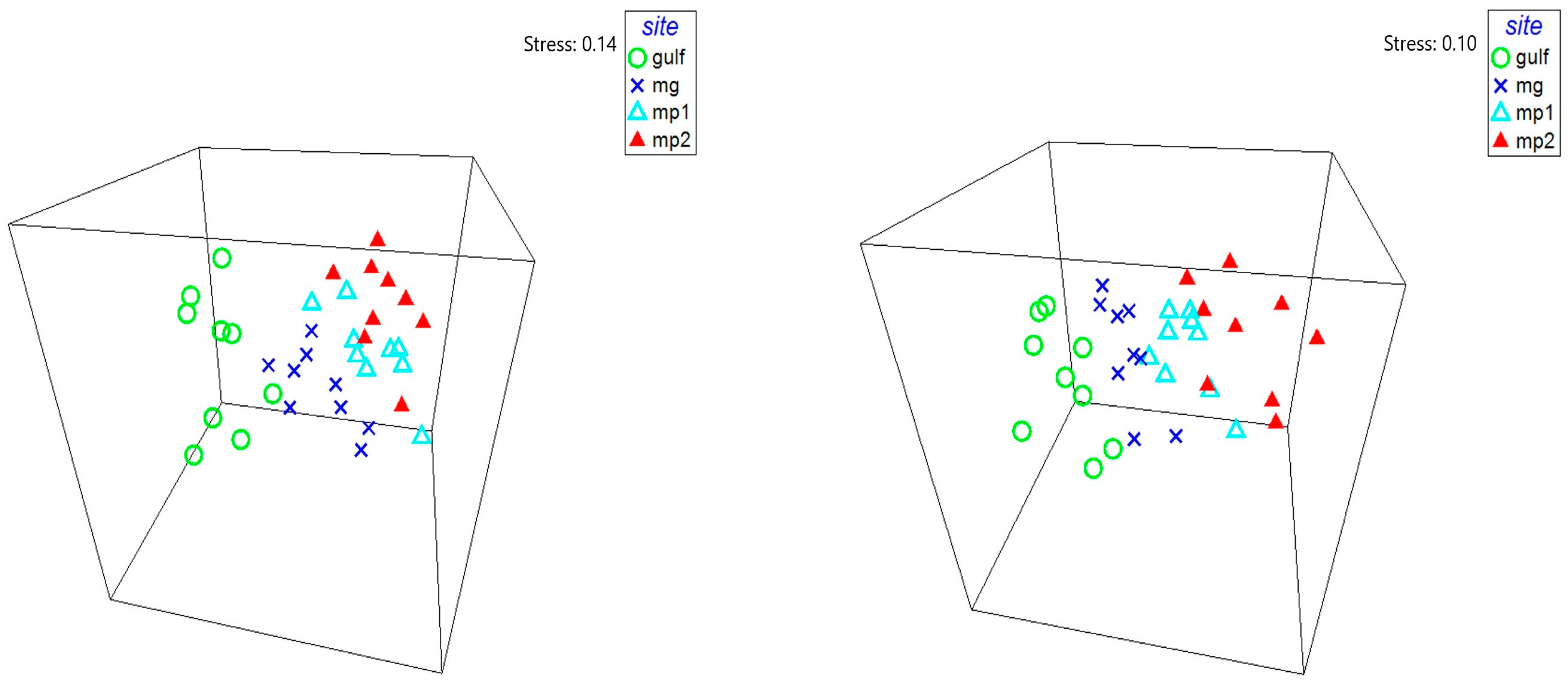
2.5. Statistical Analyses

All univariate and multivariate analyses were performed using PRIMER v.6 (Primer-E Ltd., Plymouth, UK).
A data matrix (taxa vs. stations) was realized with the average abundance values (cysts g−1) calculated from three replicates collected at each station and used to calculate the Bray-Curtis similarity after a 4th root transformation to normalize the data and downweigh the importance of the very abundant species.
The PRIMER ‘DIVERSE’ routine was used on untransformed data to calculate the taxonomic richness (S), taxon abundance (N), Pielou’s evenness index J′ [28] and Shannon-Weaver diversity index H′ [29] for each sampling site.
Bi-dimensional representations of the statistical comparisons between the sampling sites were obtained by means of non-parametric multidimensional scaling (nMDS).
The statistical significance of the spatial variations in the assemblages between sites, showed by nMDS, were tested only for full cysts, by means of a 1-way analysis of similarities (the PRIMER ‘ANOSIM’ routine), to demonstrate as the replicates from the four investigated sites were more different than the replicates within sites.
A 1-way similarity percentage procedure (the PRIMER ‘SIMPER’ routine) was performed on the full cyst assemblages, to further test the dissimilarities inside the four areas of the Taranto Sea System and to identify the most responsible taxa for these similarities/dissimilarities (cut off for low contributions 50%).
The spatial distributions of total abundance and number of taxa of full and empty cysts, together with some relevant taxa abundance were obtained as contour maps using the standard Kriging method of interpolation of the SURFER package (v.17, Golden Software Inc., Golden, CO 80402-0281, USA).

3. Results

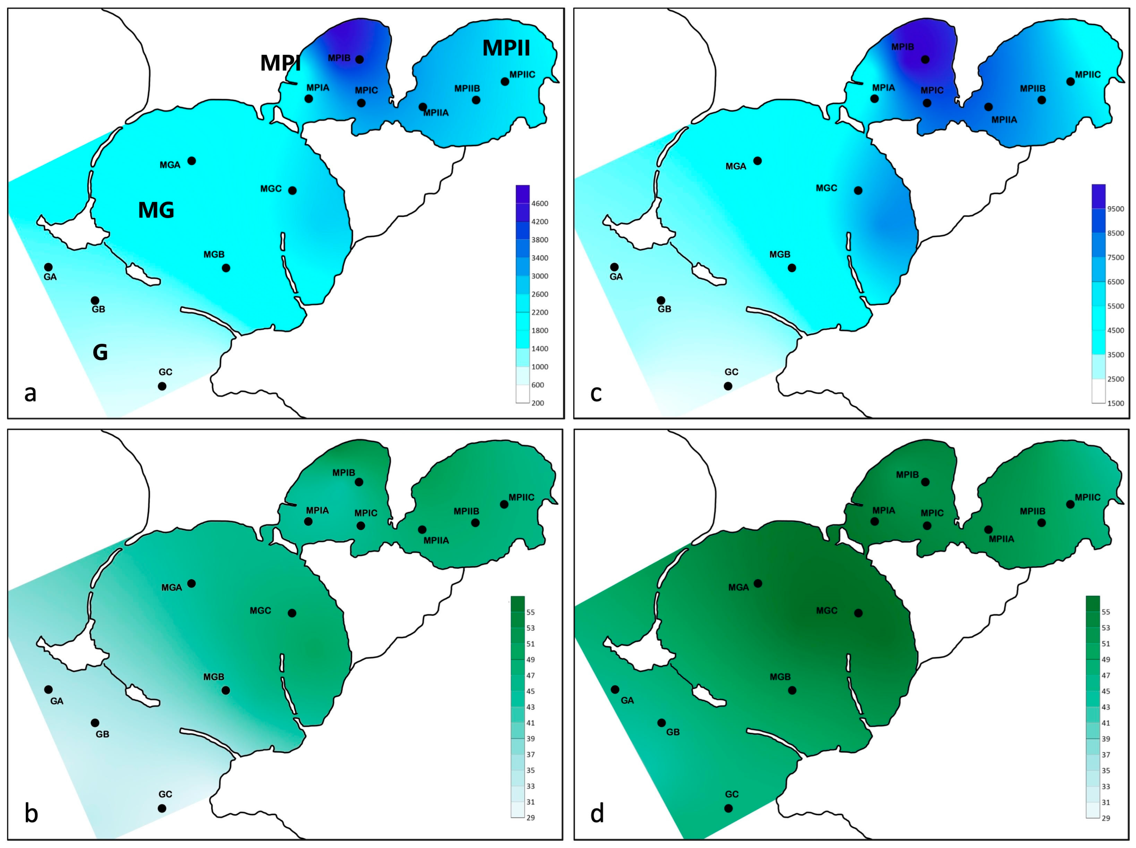
3.1. Abiotic Features of the Study Area

The physico-chemical variables of the sea water measured at 50 cm below the surface, and about 50 cm over the bottom at all the sampling stations showed values typical of the autumnal season of the Taranto sea system [14,23], with marked differences among the four basins, even more evident between the “mostly” marine part (G and MG) and the most confined part of the system (MP 1st and 2nd inlet) (Table 1).
The temperature at surface varied in a narrow range of values, with a minimum of 21.9 °C registered at the GC station in the Gulf and a maximum of 22.7 °C at the MGC station in Mar Grande. Over the bottom the values were more variable with a minimum of 17.2 °C at station GC (−74.5 m) and the maximum of 23.2 °C at the shallowest station, i.e., MPIIC (−5.7 m). Noteworthy, in the MP the temperature values were always higher over the bottom then at the surface.
The salinity (obtained from Conductivity values) showed differences between the open sea and confined sites, ranging from 38.3 at the surface and 39.1 over the bottom in G and MG, while in MP the values ranged from 37.3 to 36.8 at the surface and 38.6 to 38.0 close to the bottom.
Concerning the photosynthetically active radiation (PAR), the values at the surface ranged between 918.1 and 2551.5 μmol phot m−2 s−1, while over the bottom (at different depths in different stations) there were two orders of magnitude of difference between the open G-MG stations (range, 3.5–10.3 μmol phot m−2 s−1) and the confined MPI-MPII ones (range, 40.7–375.9 μmol phot m−2 s−1).

3.2. Cyst Bank Composition

The analysis of the collected sediments revealed a taxa richness with a total of 103 cyst morphotypes. Table 2 reports all the taxa and the cyst types that germinated during the germination experiments, allowing a more accurate taxonomic identification. A total of 96 taxa were identified, mostly at level of species, with the attribution of more than one type to the same taxon, as for Dinophyta Pentapharsodinium tyrrhenicum and Scrippsiella acuminata (Table 2). Most of the identified cysts (68 taxa) belonged to Dinophyta, 12 to Ciliophora, 3 to Rotifera, and 9 to Copepoda. Four cyst types remained unidentified.
For 88 types both full and empty cysts were found; in 8 cases, only full cysts were available.
Cysts of plankton were observed at all the sampling stations (Table S1). Total abundances of full cysts ranged from 430 ± 163 (station GB) to 4451 ± 3320 cysts g−1 (station MPIB). Empty cyst abundances resulted minimum at station GB (1688 ± 321 cysts g−1) and maximum at station MPIC (9785 ± 836 cysts g−1) (Table 3). Considering all the sites investigated in each of the four areas, both for full and empty cysts the richest area resulted MPI with an average of 3441 ± 2253 full cysts g−1 and 7787 ± 2992 empty cysts g−1, while the lowest abundances were registered in area G with 535 ± 303 full cysts g−1 and 2097 ± 622 empty cysts g−1.
This scheme of distribution was evident also in Figure 2. The color gradients show as the abundances and the species richness roughly increased moving from the Gulf to the inner part of the system.
The highest taxa richness was registered in MPII (71 taxa) for full cysts and in MPI and MG (71 taxa) for empty cysts, while the lowest taxa richness was registered in the Gulf, both for full (44 taxa) and empty (59 taxa) cysts. Abundances of specimens in the confined area MPI were sensibly higher than in the open sea G. The general richness of both taxa and specimens yielded high evenness (Pielou’s index J) also in the most crowded site MPI. Both MPI and MPII showed comparable values of Shannon-Wiener index, higher than in MG and G. In all the cases, Evenness and Shannon-Wiener diversity of empty custs showed lower values (Table 3).
Concerning the full cysts, the most abundant species resulted by far the Dinophyta Scrippsiella acuminata that represented 30.7% of the cyst assemblage in the whole study area. Other three Dinophyta species of the same family (Calciodinellaceae) slightly exceeded 5% each. The most abundant Metazoa category was Copepoda Acartiidae with a total contribution of 5.2%. Examining the empty cysts, again the most abundant taxon was S. acuminata (33.2% of the total abundance) and the second taxon resulted Copepoda Acartiidae (13.2% of the total abundance).
The nMDS representations of the statistical comparisons between the sampling sites confirmed a clear separation between the Gulf (G) and the inner basin of the Mar Piccolo (MPII), with the interposition of Mar Grande (MG) and the first basin of the Mar Piccolo (MPI) (Figure 3). This pattern was true both for full and empty cysts.
The global scheme of a gradual confinement gradient of the distribution of the resting stages in the surface sediments of the study area, was also highlighted by the ANOSIM test (R = 0.465; p = 0.001). From the pairwise tests it was evident how the basin G resulted different from each of the other basins (Table 4), especially from MPII. In addition, MG resulted significantly different from the two basins of the MP, while MPI and MPII were more similar each other.
Also the SIMPER analysis revealed the highest values of dissimilarity between the Gulf and MPI (47.65%) and the Gulf and MPII (49.84%) and the lowest dissimilarity between the two basins of the Mar Piccolo (34.68%) (Table 5) (Supplementary Material Table S2).
The taxa that mostly contributed to differentiate the Gulf from MPI were the Copepoda Acartiidae (4.26%), and the Dinophyta Scrippsiella precaria (3.64%), Gymnodinium impudicum (3.23%), and Posoniella ticarinelloides (3.08%). The same taxa were responsible for the differences between G and MPII, i.e., Acartiidae (5.61%), S. precaria (4.11%), G. impudicum (3.69%), and P. tricarinelloides (3.07%), together with Scrippsiella sp. 4 (3.57%).
On the contrary, S. acuminata was by far the species which mostly contributed to the similarity registered inside each basin (average contribution 8.69%).

4. Discussion

The study of benthic cyst assemblages produced by planktonic species allows to obtain knowledge about plankton community complementary to that obtained by classical sampling approach in the water column. In fact, viable cyst banks in the sediments represent a historical memory of the planktonic system defined by [30] as “time capsules” that represent real time series of the genetic diversity of the planktonic populations. The great advantage is that this diversity can be investigated also through a single sampling, in comparison with the complex planning of experimental designs for the sampling of plankton in the water column [2].
The present study is the first with the aim to describe the biodiversity and structure of the plankton encysted in the sediments of four different coastal marine basins aligned along a confinement grade. The analysis of the environmental variables showed large variations of bottom temperature, i.e., a driver of cysts distribution that also influences their germination (e.g., ref. [31] for Dinophyta). This high variation of bottom temperature is easily justified by the remarkable differences in the depths of the different stations considered, ranging from 75 m in the Gulf (GC) to less than 6 m in the inner basin of the Mar Piccolo (MPIIC).
As regarding the structure of the plankton community encysted in the sediments of the four basins of the Taranto Sea System, a high species richness was detected. Many taxa were common to all the basins, but other taxa were exclusive of single basins and differently contributed to their characterization, as already observed by [19] in a study considering the whole benthic diversity of the Mar Piccolo. Other studies carried out in lagoons, bays and coastal marine areas throughout the world [16,20,31,32,33,34,35,36], showed great variability both in total abundances and species richness apparently affected by specific local conditions. These conditions could include grain sediment size, oxic/anoxic conditions of the sediments, and the entity of bioturbation [37,38,39]. Our results clearly showed the existence of a gradient either of abundance and of species richness along the axis Open—Confined marine area. Highest abundances and species richness were registered in the Mar Piccolo, gradually increasing from the Gulf through the Mar Grande. Anyway, if we look at the two basins of the Mar Piccolo, it seems the biological/ecological confinement was not inversely correlated with the geographic confinement, because the maximum of resting stage abundance was registered at level of MPI. The simple difference in water column depth could justify this result (MPI water column is the double of that of MPII) due the highest abundance of plankton from where the cysts should derive. In addition, if we consider that only the bottom surface has been considered (and not the whole cyst bank) some difference could be justified by different timing in starting of cyst production by plankton among two different sites. This outcome, however, can be compared with previous studies conducted in the Mar Piccolo. Two of these studies referred to single sampling dates, although in different seasons: winter of 2011 [19], spring 2013, and spring 2014 [40]. In both these studies, the highest abundance and species richness were registered in MPII. These results, differing from the present data, suggest to consider the abundance of cysts on the sediment surface as dependent by the season other than the different confinement of Taranto basins. Concerning the species richness, in another study carried out on samples collected along one year [15], only at one site in MPI, a total of 50 cyst morphotypes were observed in surface sediments at the same year period (Autumn) considered in the present study. Our results (regarding the only MPI) of a richer species assemblage (68 full, and 71 empty cysts) was due probably to the adoption of three sample sites (vs. only one of the preceding study). Anyway, if we add to the November results of [15] also the whole year considered, the number of morphotypes in one year nearly double the number in a single date (91 vs. 50). The enhancement of species richness due to the consideration of more stations, at this point should also be increased by the multi-season effect. Thus, an even richer cyst assemblage from more complete studies is expected, and the importance to study the dormant situation of plankton seasonal species, in coastal areas, becomes more and more necessary.
Comparing the lists of taxa of all the considered studies, other points of discussion come out. In the six sampling sites in Mar Piccolo of the current study, we added 12 new morphotypes to the list of the cysts produced by plankton in the Mar Piccolo [15,19]. On the other hand, 21 cyst morphotypes observed during the previous studies were not found in the present study.
At all the sites studied in the four basins of the Taranto Sea System, the higher abundance of empty resting stages (here considered as germinated) compared to that of the full ones (presumably viable) can be explained because the analysis has regarded just the sediment surface, i.e., the most interested by cyst arrival/germination cycles. Although there are not specifical studies on empty cysts, useful to discuss the germination behaviour, some investigations exist to confirm the dominance of empty forms, also for single species, in surface sediments [32,39].
An interesting result is that the observed growth of taxa richness in the sediments, in accordance with the increase of the confinement grade, represents a trend exactly opposite to that of active/mobile stages in the plankton. This result was probably due to the highest presence, in confined environments, of species able to produce cysts, while this life cycle strategy is progressively not necessary going towards less confined or open sea areas. The differences measurable in terms of average lower salinity, higher temperature, lower dissolved Oxygen in confined areas, are probably the result of a enhanced variability of the inner environment in comparison with a more stable open sea. The shortening of life cycles of plankton organisms, and their interruption with cyst production, is driven by such an enhanced variability of the abiotic environment in the confined part. This datum agrees with the hypothesis of [14] of a biodiversity sheltered as resting stages in the sediments and not expressed in the plankton, in confined systems, where only a small part of the existing community can play an active role in the water column, in each time of the year, and environmental condition.
In literature there are very few works describing the distribution of plankton resting stages assemblages in marine environments with different confinement features and in these cases, commonly, only the group of Dinophyta is considered. Anyway, the same increase of cyst taxa has been observed (44 vs. 68) in a study of Dinophyta cyst assemblages in Patagonia between oceanic and adjacent fjord environments, going from open area to the confined one [41].
The space gradient of species richness, in any cases, has to be coupled with the seasonality because it is evident that at least among phytoplankton cysts, the production-germination differ between seasons and abiotic conditions [42].
The number of sites, and that of periods, considered for collecting cysts, need a more intense study. The final result of a complete (space-time) approach should give us a perception of biodiversity of marine confined areas, and of their functioning, increasingly different from the scientific knowledge of only few decades ago.

Supplementary Materials

The following supporting information can be downloaded at: https://www.mdpi.com/article/10.3390/jmse12091653/s1, Table S1: full and empty cyst abundances per site; Table S2: 1 way SIMPER analysis (similarity percentages—species contributions).

Author Contributions

F.R. and G.B. conceived the idea of the study; F.R. and M.B. conducted the sediment sampling and the acquisition of environmental data; F.R. and M.B. conducted the laboratory procedures and the analyses of the samples; F.R. and G.D. performed the data analyses; F.R. and G.B. wrote the original manuscript; G.B. and F.R. reviewed the original manuscripts with contributions from all authors. All authors have read and agreed to the published version of the manuscript.

Funding

The study did not receive direct funding. It, however, was carried out in the framework of the Project CyDiTSe (Cyst Distribution in Taranto Seas), funded by CNR IRSA of Taranto, and it was also indirectly sustained by the National Recovery and Resilience Plan (NRRP), Mission 4 Component 2 Investment 1.4—Call for tender n. 3138 of 16 December 2021, rectified by Decree n. 3175 of 18 December 2021 of Italian Ministry of University and Research funded by the European Union NextGenerationEU; Award Number: Project code CN_00000033, Concession Decree No. 1034 of 17 June 2022 adopted by the Italian Ministry of University and Research, CUP D33C22000960007, Project title “National Biodiversity Future Center—NBFC”.

Institutional Review Board Statement

Ethical review and approval were waived for this study due to use of resting stagfes of microinvertebrates and phytoplankton.

Informed Consent Statement

Not applicable.

Data Availability Statement

The original contributions presented in the study are included in the article/Supplementary Material, further inquiries can be directed to the corresponding author/s.

Acknowledgments

We thank Cosimo Macripò for his help aboard the m/b “A. Cerruti” operated by CNR IRSA of Taranto.

Conflicts of Interest

The authors declare no conflicts of interest.

References

  1. Belmonte, G.; Castello, P.; Piccinni, M.R.; Quarta, S.; Rubino, F.; Geraci, S.; Boero, F. Resting stages in marine sediments off the Italian coast. In Biology and Ecology of Shallow Coastal Waters; Elefteriou, A., Ansel, A.D., Smith, C.J., Eds.; Olsen and Olsen: Fredensborg, Denmark, 1995; pp. 53–58. [Google Scholar]
  2. Belmonte, G.; Rubino, F. Resting cysts from coastal marine plankton. Oceanogr. Mar. Biol. Annu. Rev. 2019, 57, 1–88. [Google Scholar]
  3. Guelorget, O.; Perthuisot, J.P. Paralic ecosystems—Biological organization and functioning. Vie Milieu 1992, 42, 215–251. [Google Scholar]
  4. Rice, E.; Dam, H.G.; Stewart, J. Impact of climate change on estuarine zooplankton: Surface water warming in Long Island Sound is associated with changes in copepod size and community structure. Estuaries Coasts 2015, 38, 13–23. [Google Scholar] [CrossRef]
  5. Ikeda, T. Relationship between respiration rate and body size in marine plankton animals as a function of the temperature of habitat. Bull. Fac. Fish. Hokkaido Univ. 1970, 21, 91–112. [Google Scholar]
  6. Uye, S.-I. Replacement of large copepods by small ones with eutrophication of embayments: Cause and consequence. Hydrobiologia 1994, 292, 513–519. [Google Scholar] [CrossRef]
  7. Hart, R.C.; Bychek, E.A. Body size in freshwater planktonic crustaceans: An overview of extrinsic determinants and modifying influences of biotic interactions. Hydrobiologia 2011, 668, 61–108. [Google Scholar] [CrossRef]
  8. Belmonte, G.; Cavallo, A. Body size and its variability in the copepod Acartia margalefi (Calanoida) from lake Acquatina (SE Italy). Ital. J. Zool. 1997, 64, 377–382. [Google Scholar] [CrossRef]
  9. Gyllström, M.; Hansson, L.-A. Dormancy in freshwater zooplankton: Induction, termination and the importance of benthic-pelagic coupling. Aquat. Sci. 2004, 66, 274–295. [Google Scholar] [CrossRef]
  10. Marcus, N.H.; Boero, F. Production and plankton community dynamics in coastal aquatic systems: The importance of benthic pelagic coupling in the forgotten role of life cycles. Limnol. Oceanogr. 1998, 43, 763–768. [Google Scholar] [CrossRef]
  11. Boero, F.; Belmonte, G.; Fanelli, G.; Piraino, S.; Rubino, F. The continuity of living matter and the discontinuities of its constituents: Do plankton and benthos really exist? Trends Ecol. Evol. 1996, 11, 177–180. [Google Scholar] [CrossRef]
  12. Leck, M.A.; Parker, V.Y.; Simpson, R.L. Ecology of Soil Seed Banks; Academic Press Inc.: San Diego, CA, USA, 1989; 445p. [Google Scholar]
  13. Lennon, J.T.; Jones, S.E. Microbial seed banks: The ecological and evolutionary implications of dormancy. Nat. Rev. Microbiol. 2011, 9, 119–130. [Google Scholar] [CrossRef] [PubMed]
  14. Belmonte, G.; Vaglio, I.; Rubino, F.; Alabiso, G. Zooplankton composition along the confinement gradient of the Taranto Sea System (Ionian Sea, south-eastern Italy). J. Mar. Syst. 2013, 128, 222–238. [Google Scholar] [CrossRef]
  15. Rubino, F.; Belmonte, G. Habitat Shift for Plankton: The Living Side of Benthic-Pelagic Coupling in the Mar Piccolo of Taranto (Southern Italy, Ionian Sea). Water 2021, 13, 3619. [Google Scholar] [CrossRef]
  16. García-Moreiras, I.; Oliveira, A.; Santos, A.I.; Oliveira, P.B.; Amorim, A. Environmental Factors Affecting Spatial Dinoflagellate Cyst Distribution in Surface Sediments off Aveiro-Figueira da Foz (Atlantic Iberian Margin). Front. Mar. Sci. 2021, 8, 699483. [Google Scholar] [CrossRef]
  17. Kerfoot, W.C.; Weider, L.J. Experimental paleoecology (resurrection ecology): Chasing Van Valen’s Red Queen hypothesis. Limnol. Oceanogr. 2004, 49, 1300–1310. [Google Scholar] [CrossRef]
  18. Rodrigues, R.V.; Patil, J.S. Salinity changes may influence dinoflagellate cyst morphometry: Data from monsoon-influenced tropical coastal ecosystems. J. Plankton Res. 2021, 43, 853–864. [Google Scholar] [CrossRef]
  19. Ferraro, L.; Rubino, F.; Belmonte, M.; Prato, S.D.; Greco, M.; Frontalini, F. A multidisciplinary approach to study confined marine basins: The holobenthic and merobenthic assemblages in the Mar Piccolo of Taranto (Ionian Sea, Mediterranean). Mar. Biodiv. 2017, 47, 887–911. [Google Scholar] [CrossRef]
  20. Wang, Z.; Zhang, Y.; Lei, M.; Ji, S.; Chen, J.; Zheng, H.; Tang, Y.; Hu, R. Distribution of Dinoflagellate cysts in surface sediments from the Qingdao coast, the Yellow Sea, China: The potential risk of harmful algal blooms. Front. Mar. Sci. 2022, 9, 910327. [Google Scholar] [CrossRef]
  21. Di Leo, A.; Annicchiarico, C.; Cardellicchio, N.; Giandomenico, S.; Conversano, M.; Castellano, G.; Basile, F.; Martinelli, W.; Scortichini, G.; Spada, L. Monitoring of PCDD/Fs and dioxin-like PCBs and seasonal variations in mussels from the Mar Grande and the Mar Piccolo of Taranto (Ionian Sea, Southern Italy). Environm. Sci. Poll. Res. 2014, 21, 13196–13207. [Google Scholar] [CrossRef]
  22. Vatova, A.R. La produttività delle acque del Mar Grande e del Mar Piccolo di Taranto (1962–1969). Boll. Pes. Pis. Idrobiol. 1972, 27, 81–103. [Google Scholar]
  23. De Pascalis, F.; Petrizzo, A.; Ghezzo, M.; Lorenzetti, G.; Manfè, G.; Alabiso, G.; Zaggia, L. Estuarine circulation in the Taranto Seas. Environm. Sci. Poll. Res. 2016, 23, 12515–12534. [Google Scholar] [CrossRef] [PubMed]
  24. Montresor, M.; Bastianini, M.; Cucchiari, E.; Giacobbe, M.; Penna, A.; Rubino, F.; Satta, C.T. Stadi di resistenza del plancton. In Metodologie di Studio del Plancton Marino, ISPRA—Istituto Superiore per la Protezione e la Ricerca Ambientale; Socal, G., Buttino, I., Cabrini, M., Mangoni, O., Penna, A., Totti, C., Eds.; Institute for Environmental Protection and Research (ISPRA): Rome, Italy, 2010; pp. 271–285. [Google Scholar]
  25. Belmonte, G.; Moscatello, S.; Rubino, F. Forme di resistenza dello zooplancton. In Metodologie di Studio del Plancton Marino, ISPRA—Istituto Superiore per la Protezione e la Ricerca Ambientale; Socal, G., Buttino, I., Cabrini, M., Mangoni, O., Penna, A., Totti, C., Eds.; Institute for Environmental Protection and Research (ISPRA): Rome, Italy, 2010; pp. 507–518. [Google Scholar]
  26. Mudie, P.J.; Marret, F.; Mertens, K.N.; Shumilovskikh, L.; Leroy, S.A.G. Atlas of modern dinoflagellate cyst distributions in the Black Sea Corridor: From Aegean to Aral Seas, including Marmara, Black, Azov and Caspian Seas. Mar. Micropaleontol. 2017, 134, 1–152. [Google Scholar] [CrossRef]
  27. Zonneveld, K.A.F.; Pospelova, V. A determination key for modern dinoflagellate cysts. Palynology 2015, 39, 387–409. [Google Scholar] [CrossRef]
  28. Pielou, E.C. Shannon’s formula as a measure of specific diversity: Its use and misuse. Am. Nat. 1966, 100, 463–465. [Google Scholar] [CrossRef]
  29. Shannon, C.E.; Weaver, W. The Mathematical Theory of Communication. University of Illinois Press: Champaign, IL, USA, 1949. [Google Scholar]
  30. Ellegaard, M.; Godhe, A.; Ribeiro, S. Time capsules in natural sediment archives—Tracking phytoplankton population genetic diversity and adaptation over multidecadal timescales in the face of environmental change. Evol. Appl. 2018, 11, 11–16. [Google Scholar] [CrossRef]
  31. Pospelova, V.; Chmura, G.L.; Walker, H.A. Environmental factors influencing the spatial distribution of dinoflagellate cyst assemblages in shallow lagoons of southern New England (USA). Rev. Palaeobot. Palynol. 2004, 128, 7–34. [Google Scholar] [CrossRef]
  32. García-Moreiras, I.; Costas, S.V.; García-Gil, S.; Sobrino, C.M. Organic-walled dinoflagellate cyst assemblages in surface sediments of the Ría de Vigo (Atlantic margin of NW Iberia) in relation to environmental gradients. Mar. Micropaleontol. 2023, 180, 102217. [Google Scholar] [CrossRef]
  33. Likumahua, S.; Sangiorgi, F.; Boer, M.K.; de Tatipatta, W.M.; Pelasula, D.D.; Polnaya, D.; Hehuwat, J.; Siahaya, D.M.; Buma, A.G.J. Dinoflagellate cyst distribution in surface sediments of Ambon Bay (eastern Indonesia): Environmental conditions and harmful blooms. Mar. Pollut. Bull. 2021, 166, 112269. [Google Scholar] [CrossRef]
  34. Price, A.M.; Pospelova, V.; Coffin, M.R.S.; Latimer, J.S.; Chmura, G.L. Biogeography of dinoflagellate cysts in northwest Atlantic estuaries. Ecol. Evol. 2016, 6, 5648–5662. [Google Scholar] [CrossRef]
  35. Rubino, F.; Belmonte, M.; Caroppo, C.; Giacobbe, M. Dinoflagellate cysts from surface sediments of Syracuse Bay (Western Ionian Sea, Mediterranean). Deep Sea Res. Part II Top. Stud. Oceanogr. 2010, 57, 243–247. [Google Scholar] [CrossRef]
  36. Rubino, F.; Belmonte, M.; Galil, B.S. Plankton resting stages in recent sediments of Haifa port, Israel (Eastern Mediterranean)-Distribution, viability and potential environmental consequences. Mar. Poll. Bull. 2017, 116, 258–269. [Google Scholar] [CrossRef] [PubMed]
  37. Kremp, A.; Anderson, D.M. Factors regulating germination of resting cysts of the spring bloom dinoflagellate Scrippsiella hangoei from the northern Baltic Sea. J. Plankton Res. 2000, 22, 1311–1327. [Google Scholar] [CrossRef]
  38. Ribeiro, S.; Berge, T.; Lundholm, N.; Ellegaard, M. Hundred years of environmental change and phytoplankton ecophysiological variability archived in coastal sediments. PLoS ONE 2013, 8, e61184-8. [Google Scholar] [CrossRef] [PubMed]
  39. Choi, S.Y.; Belmonte, G.; Hyun, B.; Jang, P.-G.; Lee, W.J.; Soh, H.Y.; Seo, M.H.; Seo, J.Y.; Shin, K.; Jang, M.-C. Effects of hypoxia on copepod egg hatching success: An in situ study. J. Mar. Syst. 2023, 245, 103979. [Google Scholar] [CrossRef]
  40. Rubino, F.; Cibic, T.; Belmonte, M.; Rogelja, M. Microbenthic community structure and trophic status of sediments in the Mar Piccolo of Taranto (Mediterranean, Ionian Sea). Environ. Sci. Poll. Res. 2016, 23, 12624–12644. [Google Scholar] [CrossRef]
  41. Rodríguez-Villegas, C.; Lee, M.R.; Salgado, P.; Figueroa, R.I.; Baldrich, A.; Pérez-Santos, I.; Tomasetti, S.J.; Niklitschek, E.; Diaz, M.; Alvarez, G.; et al. Drivers of dinoflagellate benthic cyst assemblages in the NW Patagonian fjords system and its adjacent oceanic shelf, with a focus on harmful species. Sci. Total Environ. 2021, 785, 147378. [Google Scholar] [CrossRef]
  42. Hedberg, P.; Olsson, M.; Höglander, H.; Brückert, V.; Winder, M. Climate change effects on plankton recruitment from coastal sediments. J. Plankton Res. 2024, 46, 117–125. [Google Scholar] [CrossRef]
Figure 1. Map of the study area. The position of the 12 sampling sites is indicated in the 4 basins shaping the Taranto Sea System. G, Gulf of Taranto; MG, Mar Grande; MP, Mar Piccolo (I and II inlet). A, B, C, three stations for each basin (in each station, three replicates of sediment samples have been collected).
Jmse 12 01653 g001
Figure 2. Spatial distribution of the resting stage communities in the Taranto Sea. Abundances of full (a) and empty (c) cysts; Taxa richness of full (b) and empty (d) cysts. Blue colored maps (a,c) refer to abundance of cysts (full and empty). Green colored maps (b,d) refer to taxa richness (full and empty cysts).
Jmse 12 01653 g002
Figure 3. nMDS representations of the statistical comparisons between the sampling sites. (Left) full cysts; (Right) empty cysts.
Jmse 12 01653 g003
Table 1. Geographic position, water depth and physico-chemical variables (temperature, salinity, dissolved Oxygen, pH and PAR), measured at 50 cm below the sea surface (s) and 50 cm over the bottom (b) at the sampling sites in the Taranto Sea System in October–November 2015. G: Gulf; MG: Mar Grande; MPI: Mar Piccolo 1st Inlet; MPII: Mar Piccolo 2nd Inlet.
StationLat NLong EDepth
(m)
Temperature
(°C)
Salinity
(psu)
Dissolved O2
(%)
pHPAR
(μmol phot m−2 s−1)
SurfaceBottomSurfaceBottomSurfaceBottomSurfaceBottomSurfaceBottom
G A40°26.500′17°08.531′58.422.117.338.238.995.290.48.368.311845.36.4
G B40°25.483′17°08.592′56.222.317.638.338.997.592.38.368.321768.88.8
G C40°25.006′17°10.789′74.521.917.238.238.896.592.58.358.321912.110.3
MG A40°26.191′17°12.212′26.722.521.937.939.1102.3101.38.368.381913.67.7
MG B40°27.471′17°12.275′18.022.622.438.139.189.480.98.318.321708.610.2
MG C40°26.499′17°13.463′26.822.722.238.238.8118.0115.38.408.361189.23.5
MPI A40°28.959′17°14.417′10.822.222.836.738.497.591.98.208.212218.6141.2
MPI B40°29.481′17°15.303′11.522.422.836.838.6102.292.98.228.302551.5134.4
MPI C40°28.840′17°15.102′12.122.422.837.338.6108.6100.78.248.301095.840.7
MPII A40°28.559′17°16.459′12.022.222.936.938.598.575.78.188.212050.3108.2
MPII B40°28.845′17°17.681′7.622.223.037.038.392.878.78.188.21918.174.0
MPII C40°29.019′17°18.738′5.722.223.236.938.0107.486.08.218.201521.8375.9
G 63.0 ± 10.022.1 ± 0.217.4 ± 0.238.2 ± 0.138.9 ± 0.196.4 ± 1.291.7 ± 1.28.4 ± 0.08.3 ± 0.01842.1 ± 71.78.5 ± 2.0
MG 23.8 ± 5.122.6 ± 0.122.2 ± 0.338.1 ± 0.239.0 ± 0.2103.2 ± 14.399.2 ± 17.38.4 ± 0.08.4 ± 0.01603.8 ± 373.47.1 ± 3.4
MPI 11.8 ± 0.422.3 ± 0.122.8 ± 0.036.9 ± 0.338.5 ± 0.1102.8 ± 5.695.2 ± 4.88.2 ± 0.08.3 ± 0.11955.3 ± 762.7105.4 ± 56.2
MPII 8.4 ± 3.222.2 ± 0.023.0 ± 0.236.9 ± 0.138.3 ± 0.399.6 ± 7.480.1 ± 5.38.2 ± 0.08.2 ± 0.01496.7 ± 566.5186.0 ± 165.3
Table 2. List of the cysts (taxa) identified from surface sediments collected in the Taranto sea system. For Dinophyta both paleontological (i.e., cyst based) and modern (i.e., based on planktonic stages) taxa are reported because the active stage of some species originally described as fossils is still unknown. * plankton resting stages found only as empty cysts (i.e., already germinated in situ) # cysts that successfully germinated in the germination experiments (in lab). 1 two different morphotypes were ascribed to this species; 2 four different morphotypes were ascribed to this species complex.
Dinophyta
Paleontological Taxon (Cyst Based)Modern Taxon
# Alexandrium minutum Halim
Alexandrium pseudogonyaulax (Biecheler) Horiguchi
# Alexandrium tamarense (Lebour) Balech
Alexandrium sp. 1
# Alexandrium sp. 2
Calciodinellum albatrosianum (Kamptner)Calciodinellum albatrosianum (Kamptner) Janofske & Karwath
Calciodinellum operosum (Deflandre)Calciodinellum operosum (Deflandre) Montresor
Calciperidinium asymmetricum VersteeghUnknown
Cochlodinium polykrikoides Margalef
# Diplopsalis lenticula Bergh
Follisdinellum splendidum VersteeghUnknown
Spiniferites undetGonyaulax group
# Gymnodinium impudicum (Fraga & Bravo) G. Hansen & Moestrup
Gymnodinium cf. litoralis A. Reñé
* Gymnodinium nolleri Ellegaard & Moestrup
Gymnodinium sp. 1
Levanderina fissa (Levander) Moestrup, Hakanen, Gert Hansen, Daugbjerg & M.Ellegaard
Lingulodinium machaerophorum (Deflandre & Cookson) Wall# Lingulodinium polyedra (Stein) Dodge
Lingulodinium sp.
Melodomuncula berlinensis VersteeghUnknown
Oblea acanthocysta Kawami, Iwataki & Matsuoka
# Oblea rotunda (Lebour) Balech ex Sournia
# Pentapharsodinium dalei Indelicato & Loeblich
Calcicarpinum bivalvum Versteegh# 1 Pentapharsodinium tyrrhenicum Montresor, Zingone & Marino
Calcicarpinum bivalvum VersteeghPentapharsodinium cf. tyrrhenicum Montresor, Zingone & Marino
Polykrikos kofoidii Chatton
Polykrikos schwartzii Bütschli
Bicarinellum tricarinelloides VersteeghPosoniella tricarinelloides (Versteegh) Streng et al.
Operculodinium centrocarpum (Deflandre & Cookson) WallProtoceratium reticulatum (Claparède & Lachmann) Butschli
Protoperidinium claudicans (Paulsen) Balech
Selenopemphix quanta (Bradford) MatsuokaProtoperidinium conicum (Gran) Balech
Xandarodinium xanthum Reid# Protoperidinium divaricatum (Meunier) Parke & Dodge
Votadinium calvum ReidProtoperidinium latidorsale (Balech) Balech
Quinquecuspis concretum (Reid) HarlandProtoperidinium leonis (Pavillard) Balech
Protoperidinium minutum (Kofoid) Loeblich III
Protoperidinium parthenopes Zingone & Montresor
Trinovantedinium applanatum (Bradford) Bujak & DaviesProtoperidinium shanghaiense Gu, Liu & Mertens
# Protoperidinium stellatum (Wall in Wall & Dale 1968) Head
Protoperidinium thorianum (Paulsen) Balech
Protoperidinium sp. 1
Protoperidinium sp. 4
Protoperidinium sp. 5
cf. Pseudocochlodinium profundisulcus Hu, Xu, Gu, Iwataki, Takahashi, Tang & Matsuoka
Polysphaeridium zoharyi (Rosignol) Bujak et al.Pyrodinium bahamense var. compressum (Böhm) Steidinger, Tester & Taylor
Pyrophacus horologium Stein
2 Scrippsiella acuminata complex (Ehrenb.) Kretschmann, Elbr., Zinssmeister, S. Soehner, Kirsch, Kusber & Gottschling
Scrippsiella cf. erinaceus (Kamptner) Kretschmann, Zinssmeister & Gottschling
# Scrippsiella lachrymosa Lewis
Scrippsiella precaria Montresor & Zingone
# Scrippsiella ramonii Montresor
# Scrippsiella spinifera Honsell & Cabrini
Scrippsiella trifida Lewis
# Scrippsiella sp. 1
# Scrippsiella sp. 3
# Scrippsiella sp. 4
Scrippsiella sp. 5
Scrippsiella sp. 6
Scrippsiella sp. 8
* Warnowia rosea (Pouchet) Kofoid & Schwezy
Dinophyta sp. 1
Dinophyta sp. 2
Dinophyta sp. 9
Dinophyta sp. 14
Dinophyta sp. 17
* Dinophyta sp. 24
* Dinophyta sp. 26
Dinophyta sp. 29
Dinophyta sp. 31
CiliophoraCrustacea Copepoda
Cyrtostrombidium boreale Kim, Suzuki & TaniguchiAcartia cf. italica Steuer (subitaneous egg)
Hexasterias problematica CleveAcartia sp. 2
*Laboea strobila LohmanAcartia sp. 3
Strombidium acutum (Claparéde & Lachmann) JörgensenAcartia sp. 4
Strombidium conicum (Lohmann) WulffCentropages sp. (resting egg)
Strombidium crassulum (Leegaard) Kahl* Centropages sp. (subitaneous egg)
Strombidium sp.Paracartia latisetosa (Krizcaguin)
Ciliophora sp. 1Pteriacartia josephinae (Crisafi)
Ciliophora sp. 2Copepoda sp. 1
Ciliophora sp. 5
Ciliophora sp. 6
Ciliophora sp. 7
RotiferaUnidentified
Synchaeta sp. 1 (smooth)Cyst type 2
Synchaeta sp. 2 (tubular)Cyst type 3
Synchaeta sp. 3 (spiny)* Cyst type 4
Resting egg 1
Table 3. Indices of Diversity calculated for full and empty cysts at each sampling station (averages of three replicates) and at each basin (average of three stations), in the Taranto Sea System. G: Gulf; MG: Mar Grande; MPI: Mar Piccolo 1st Inlet; MPII: Mar piccolo 2nd Inlet. S: species richness (cumulative values); N: number of individuals (means ± SD); J′: Pielou’s index: H′: Shannon-Weaver index.
Full CystsEmpty Cysts
StationSNJ′H′(loge)SNJ′H′(loge)
G A31708 ± 4370.692.38432491 ± 7900.602.27
G B33430 ± 1630.712.47481688 ± 3210.592.30
G C29467 ± 2830.682.29482113 ± 5790.572.20
MG A461774 ± 11890.682.60544292 ± 6180.652.59
MG B471687 ± 7220.732.82594067 ± 8080.662.71
MG C512382 ± 15330.722.84597115 ± 21870.622.52
MPI A451726 ± 4140.762.90574135 ± 8490.702.84
MPI B564451 ± 33200.742.97599443 ± 20660.712.88
MPI C444147 ± 15540.752.83539785 ± 8360.732.89
MPII A532860 ± 22380.742.93557941 ± 28810.702.81
MPII B502711 ± 6870.722.80566360 ± 7880.652.63
MPII C461786 ± 6810.742.83371833 ± 13500.562.03
basin
G44535 ± 3030.692.38592097 ± 6220.592.26
MG661948 ± 10860.712.75715158 ± 19020.642.61
MPI683441 ± 22530.752.90717787 ± 29920.712.87
MPII712452 ± 13190.732.85655378 ± 31820.642.49
Table 4. Results of the ANOSIM test. R value indicates the dissimilarity between the different groups (basins) in the Taranto Sea System. G, Gulf; MG, Mar Grande; MPI, Mar Piccolo 1st Inlet; MPII, Mar Piccolo 2nd Inlet.
GroupsRp Value
G, MG0.4480.001
G, MPI0.7540.001
G, MPII0.8690.001
MG, MPI0.2450.014
MG, MPII0.5800.001
MPI, MPII0.1060.094
Table 5. Results of the SIMPER test with the amount of average similarity (Aver.Simil.) inside each group of stations (3 per basin), and the average dissimilarity (Aver.Dissimil.) between the different groups in the Taranto Sea System. G: Gulf; MG: Mar Grande; MPI: Mar Piccolo 1st Inlet; MPII: Mar piccolo 2nd Inlet.
GroupAver.
Simil.
GroupsAver.Dissimil.
G58.80G, MG43.67
MG65.99G, MPI47.65
MPI65.87G, MPII49.84
MPII66.41MG, MPI36.69
MG, MPII39.98
MPI, MPII34.68
Disclaimer/Publisher’s Note: The statements, opinions and data contained in all publications are solely those of the individual author(s) and contributor(s) and not of MDPI and/or the editor(s). MDPI and/or the editor(s) disclaim responsibility for any injury to people or property resulting from any ideas, methods, instructions or products referred to in the content.

