Article

Titanium-Enriched Medium Promotes Environment-Induced Epigenetic Machinery Changes in Human Endothelial Cells

by
Célio Júnior da C. Fernandes
1,
Rodrigo A. Foganholi da Silva
2,3,
Patrícia F. Wood
1,
Marcel Rodrigues Ferreira
1,
Gerson S. de Almeida
1,
Julia Ferreira de Moraes
1,
Fábio J. Bezerra
1 and
Willian F. Zambuzzi
1,*
1
Laboratory of Bioassays and Cellular Dynamics, Department of Chemical and Biological Sciences, Institute of Biosciences, UNESP—São Paulo State University, Botucatu 18618-970, SP, Brazil
2
Department of Dentistry, University of Taubaté, Taubaté 12020-340, SP, Brazil
3
Program in Environmental and Experimental Pathology, Paulista University, São Paulo 04026-002, SP, Brazil
*
Author to whom correspondence should be addressed.
J. Funct. Biomater. 2023, 14(3), 131; https://doi.org/10.3390/jfb14030131
Submission received: 16 January 2023 / Revised: 21 February 2023 / Accepted: 22 February 2023 / Published: 27 February 2023
(This article belongs to the Section Bone Biomaterials)

Abstract

:
It is important to understand whether endothelial cells are epigenetically affected by titanium-enriched media when angiogenesis is required during bone development and it is expected to be recapitulated during osseointegration of biomaterials. To better address this issue, titanium-enriched medium was obtained from incubation of titanium discs for up to 24 h as recommended by ISO 10993-5:2016, and further used to expose human umbilical vein endothelial cells (HUVECs) for up to 72 h, when the samples were properly harvested to allow molecular analysis and epigenetics. In general, our data show an important repertoire of epigenetic players in endothelial cells responding to titanium, reinforcing protein related to the metabolism of acetyl and methyl groups, as follows: Histone deacetylases (HDACs) and NAD-dependent deacetylase sirtuin-1 (Sirt1), DNA methyltransferases (DNMTs) and ten-eleven translocation (TET) methylcytosine dioxygenases, which in conjunction culminate in driving chromatin condensation and the methylation profile of DNA strands, respectively. Taking our data into consideration, HDAC6 emerges as important player of this environment-induced epigenetic mechanism in endothelial cells, while Sirt1 is required in response to stimulation of reactive oxygen species (ROS) production, as its modulation is relevant to vasculature surrounding implanted devices. Collectively, all these findings support the hypothesis that titanium keeps the surrounding microenvironment dynamically active and so affects the performance of endothelial cells by modulating epigenetics. Specifically, this study shows the relevance of HDAC6 as a player in this process, possibly correlated with the cytoskeleton rearrangement of those cells. Furthermore, as those enzymes are druggable, it opens new perspectives to consider the use of small molecules to modulate their activities as a biotechnological tool in order to improve angiogenesis and accelerate bone growth with benefits of a fast recovery time for patients.

1. Introduction

The application of biomaterials has been widely proposed within the fields of tissue engineering and regenerative medicine, as well as to recover the functional performance in edentulism [1,2]. However, a significant number of patients may experience biological and mechanical complications characterized by surrounding tissue degradation and consequently the failure of implants [3]. In an attempt to minimize the failure of implants, numerous researchers have endeavored to elucidate the physiological and molecular mechanisms of osteointegration [4,5,6,7,8]. In this sense, biological data have guided the proposal of new surfaces presenting biomimetic properties predicted to develop necessary biological responses and so impacting the performance of surrounding host tissue during osteointegration [9,10].
Among the most commonly used biomaterials within the biomedical area, titanium has been listed as the most usual metallic alloy in biomedical applications. Indeed, titanium has well-accepted properties in biocompatibility scenarios which have repercussions on clinical success [11]. The biological performance of materials used in medicine and dentistry requires biological responses upon signaling related to the interaction of cells and the physicochemical properties of their surfaces [12,13,14,15,16,17].
Among the players in regenerative tissue surrounding implanted devices, there is a pleiotropic lineage of cells, which work together to promote new bone formation [18]. In this scenario, it is relevant to consider the coupling between endothelial cells and osteoblasts, which is known to compromise osteogenesis [19,20]. Although knowledge has been improved considering osteoblasts responding to materials [21], the relevance of endothelial cells responding within this scenario is barely known [22,23,24,25,26]. During the biomaterial-related osseointegration mechanism, it is expected that endothelial cells recapitulate the coupling with undifferentiated cells in order to optimize the osteogenesis and culminate in the success of osseointegration. Although blood vessels are not in close contact with implanted material surfaces, we have recently reported that titanium-based devices release a considerable number of molecules which interfere with cell metabolism. Importantly, the epigenetic mechanism drives the capacity of cells to respond to microenvironment changes. Epigenetics is the study of changes in gene expression without alterations of DNA itself, which include the reactions to modulate the incorporation of methyl groups into DNA strands as well as acetyl, sumoyl, and phospho groups into histones [27]. Additionally, epigenetic regulation of histone acetylation and DNA methylation are pivotal points involved in cellular responses during cell adhesion and viability [28]. Conversely, histone deacetylases (HDACs) affect gene expression in cells grown on implant surfaces [29]. Recently, differences in global DNA methylation profiles were also demonstrated with relevance to genetic expression of osteogenic pathways in mesenchymal stem cells responding to titanium [30]. Generally, this mechanism has been shown to have high performance with wound healing potential, with properties ranging from high predictability to later cellular mechanisms involved in osseointegration. We reported earlier the biological responses of cells reacting to titanium and nanohydroxyapatite-blasted surfaces [31,32,33]. In this regard, tissue integration depends on biomaterial microstructure and composition, mainly considering the properties of the surfaces of materials, and the effect of the pore size of titanium on the viability and function of fibroblasts and tenocytes in a dynamic bioreactor was evaluated [34].
Again, the knowledge of how endothelial cells react to titanium alloys remains elusive although progress has been made considering osteoblasts [15,35,36,37]. Endothelial cells are involved in angiogenesis, which is essential to deliver nutrients and oxygen during the healing process around biomaterial, as well as contribute to osteogenesis by coupling the paracrine mechanism between themselves and osteoblasts. To the best of our knowledge, this is the first study intending to explore the epigenetic machinery in endothelial cells exposed to titanium-enriched media. Altogether, it involves epigenetic changes in human endothelial cells by processing genes and enzymes and these data are gathered to draw a complimentary molecular mechanism in wound healing affected by titanium-based devices in order to better predict the mechanisms involved in osteointegration of biomaterials into bone.

2. Materials and Methods

2.1. Reagents

The titanium discs were kindly donated by S.I.N Implants System Company (SIN) (São Paulo, Brazil). Antibodies: HDAC1 (10E2) Mouse mAb #5356; HDAC2 (3F3) Mouse mAb #5113; HDAC3 (7G6C5) Mouse mAb #3949; HDAC6 (D2E5) Rabbit mAb #7558; SirT1 (1F3) Mouse mAb #8469; GAPDH (D16H11) XP® Rabbit mAb #5174, Life Technologies/Molecular Probes, Inc. (Eugene, OR, USA). DNMT1 Antibody (60B1220.1), DNMT3A Antibody (64B1446) (Novus Biologicals, LLC, Centennial, CO, USA), TET3 Antibody, Anti-DNMT3B (orb372330), TET1 (orb228563), and TET2 (orb131790) were purchased from BiorByt (San Francisco, CA, USA).

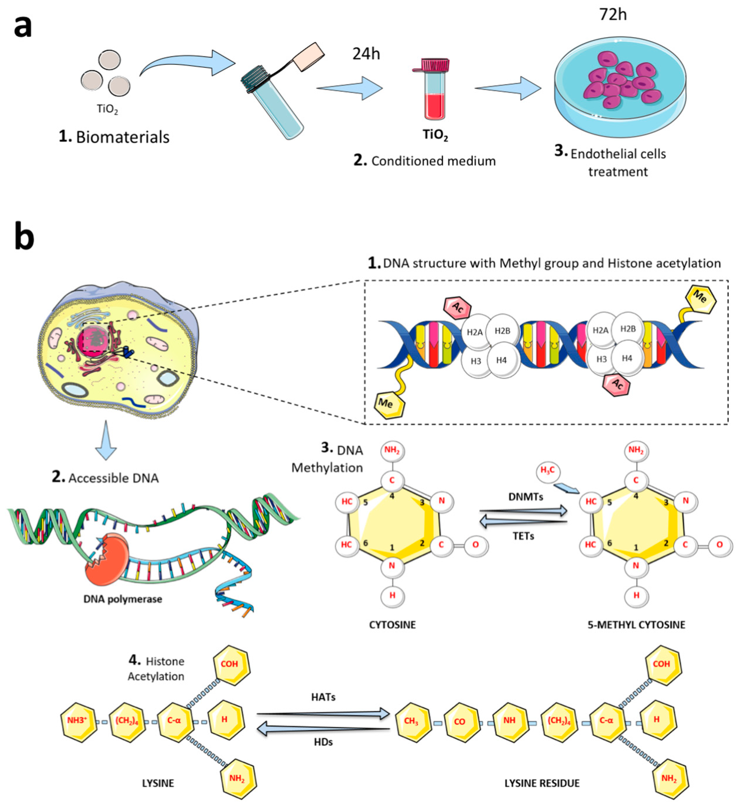
2.2. Titanium-Enriched Medium and Experimental Design

Titanium discs were previously prepared to subject their surfaces to dual acid-etching (DAE; w_Ti) and they were compared to machined surfaces (wo_Ti) [38]. Technically, to obtain the titanium-enriched medium from the samples, discs (0.6 cm diameter) were incubated within conic tubes containing cell culture medium (0.2 g/mL w/v, without fetal bovine serum (FBS)) for up to 24 h as recommended by ISO 10993-5:2016 (Figure 1a). Thereafter, this titanium-enriched medium was used to challenge human endothelial cells for up to 72 h, when the cells were harvested for biological analysis of the epigenetic mechanism (Figure 1b). Figure 1b depicts DNA methylation and histone acetylation molecular mechanisms. Additionally, both gene activation and repression are regulated by the pattern of acetylation of core histones, and the histone acetylation is mediated by coactivators (histone acetyltransferase: HATs) and corepressors (histone deacetylases: HDACs).

2.3. Cell Culture

To evaluate whether there are changes in endothelial cells responding to Ti-enriched medium and further correlation with aspects of angiogenesis, human umbilical vein endothelial cells (HUVECs; ATCC) were used in this study. Throughout the experiments, cells were grown in DMEM containing antibiotics (100 U/mL penicillin, 100 mg/mL streptomycin), and supplemented with 10% fetal bovine serum (Nutricell, Campinas, SP, Brazil). Cells were maintained at 37 °C, 5% CO2, and 95% humidity. Importantly, these cells were mycoplasma-free and were used in this study in accordance with the manufacturer’s recommendations.

2.4. Gene Expression

2.4.1. RNA Isolation and cDNA Synthesis

The total RNA was extracted using Ambion TRIzol Reagent (Life Sciences—Fisher Scientific Inc, Waltham, MA, USA) and its concentration and purity determined by measuring the absorbance at 260/280 nm (NANODROP 2000, Thermo, Waltham, MA, USA). After treatment of total RNA with DNase I (Invitrogen, Carlsbad, CA, USA), the cDNA was synthesized from 2 μg total RNA with a High-Capacity cDNA Reverse Transcription Kit (Applied Biosystems, Foster City, CA, USA) in a standard 20 μL volume according to the protocol.

2.4.2. Gene Expression

Gene expression in endothelial cells was measured using SYBR Green quantitative PCR analysis. Real-time PCR amplification was performed with the QuantStudio® 3 Real-Time PCR system (Thermo Fisher Scientific, Waltham, MA, USA). The 10 μL PCR system consisted of 1 μL cDNA (50 ng), 0.5 μmol forward primer, 0.5 μmol reverse primer, 5 μL SYBR Green qPCR mix, and 3 μL ultrapure DEPC-treated water. PCR primers were designed using Primer3 Input (version 0.4.0) software. The secondary structure and annealing temperature were analyzed by the Beacon Designer program, Free Edition (http://www.premierbiosoft.com/, accessed on 29 July 2021) and confirmed by in silico PCR (https://genome.ucsc.edu/, accessed on 29 July 2021). Primer 5.0 software and primer synthesis were acquired from Invitrogen Co. (Life Technologies, Carlsbad, Califórnia, EUA). Primer sequences and CR conditions are presented in Table 1. Gene expressions are expressed as compared to control cells by the ΔΔCT method, using Gapdh as a housekeeping control in three independent experiments in duplicate. Finally, cDNA was used to evaluate each gene’s expression (Table 1).

2.5. Western Blot

Cultures were exposed to titanium-enriched medium for up to 72 h, when the cells were lysed using lysis buffer (50 mM Tris-HCl, pH 7.4, 1% Tween 20, 0.25% sodium deoxycholate, 150 mM NaCl, 1 mM EGTA, 1 mM O-vanadate, 1 mM NaF, and protease inhibitors (1 μg/mL aprotinin, 10 μg/mL leupeptin, and 1 mM aminoethylfluorosilicon 4-fluoride hydrochloride)) for up to 2 h in ice and thereafter sonicated, then the samples were centrifuged to collect the supernatant and the pellet was discarded. The protein concentration was determined by the Lowry method and an equal volume of Laemmli buffer (2X sodium dodecyl sulfate (SDS), 100 mM Tris-HCl (pH 6.8), 200 mM dithiothreitol (DTT), 4% SDS, 0.1% bromophenol blue, and 20% glycerol) was added. The obtained pool of proteins was boiled for 5 min at 95 °C in denaturant conditions. To resolve the proteins into the gel, the proteins were resolved into SDS–PAGE (8% or 10%), then were transferred to PVDF membranes (Millipore), with sequential incubation with blocking solution containing 1% bovine serum albumin in Tris-buffered saline (TBST, Tween 20 (0.05%)) for 1 h. Thereafter, the blocked membranes were subjected to specific primary antibody incubation for up to 12 h, at 4 °C. Afterward, the membranes were extensively washed using TBS–Tween 20 (0.05%; 3X), then they were incubated with secondary antibodies for 1 h at room temperature, and washed using TBS. Enhanced chemiluminescence solution (ECL) was finally used to detect the bands. GAPDH was used as a housekeeping and loading control.

2.6. Statistical Analysis
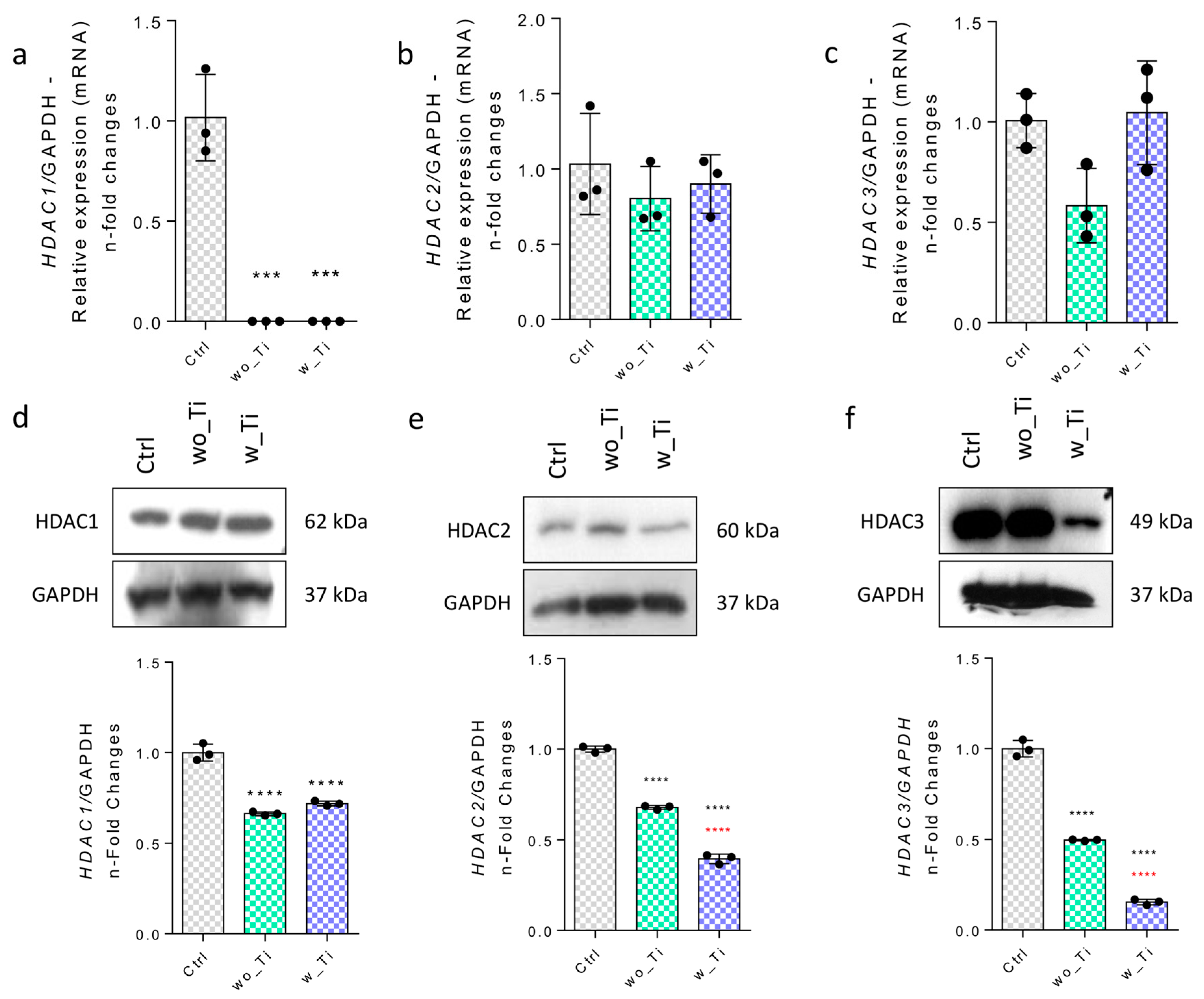
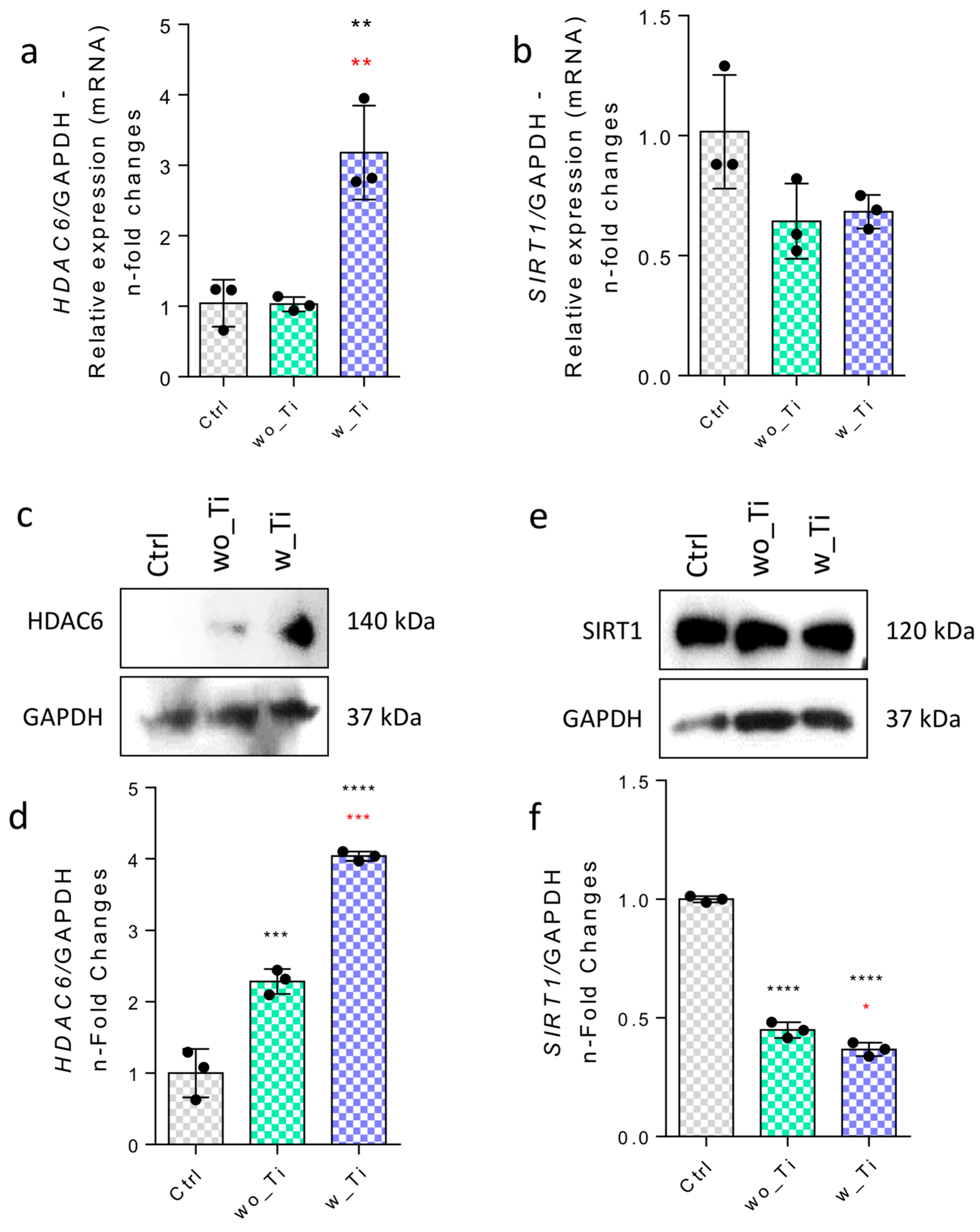
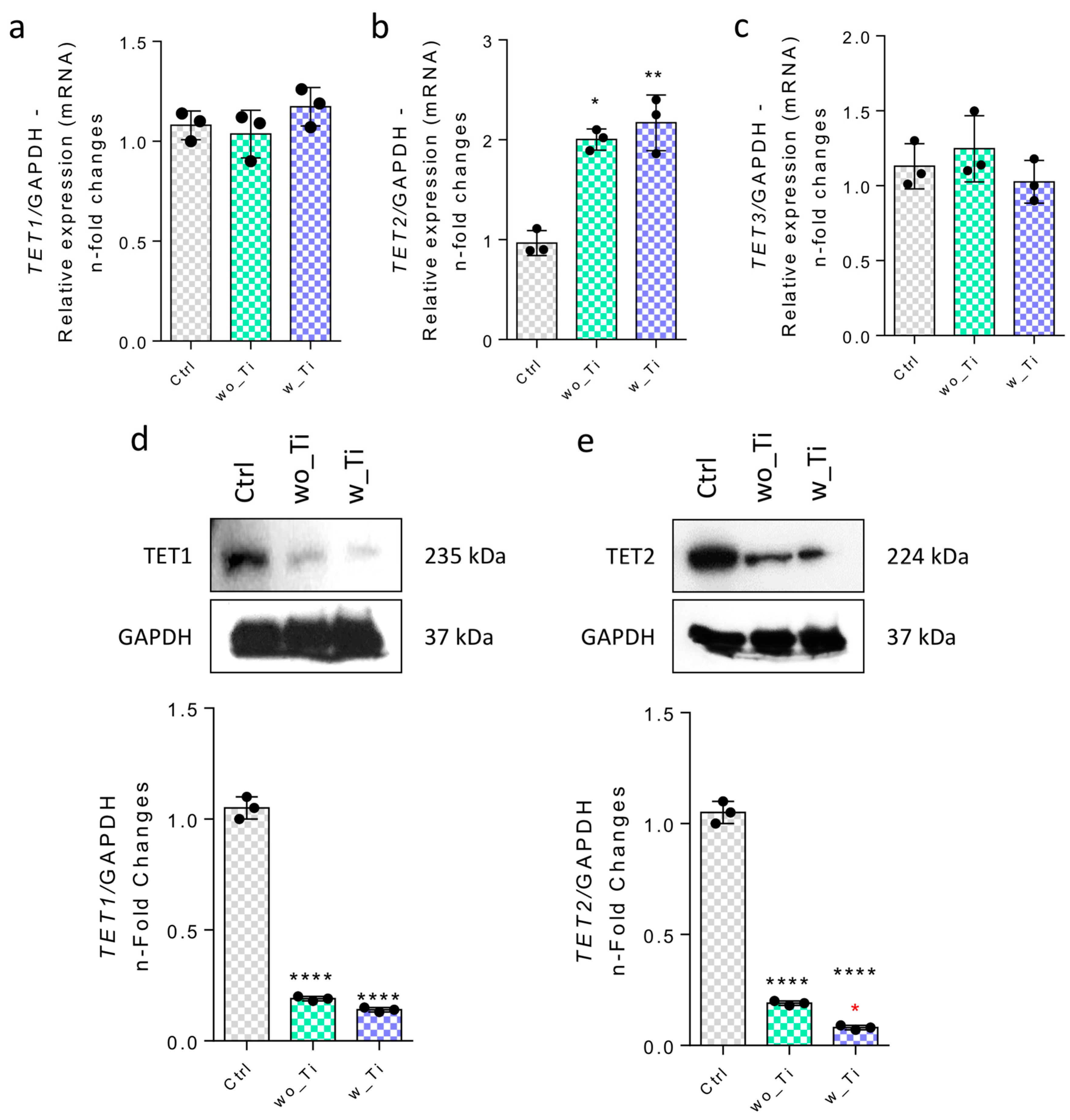
Bands of immunoblots were measured using densitometry tools and analysis to obtain arbitrary values, which represent mean ± standard deviation (SD) in the graphs. Statistical analyses were applied using Student’s t-test (2-tailed) with p < 0.05 considered statistically significant and p < 0.001 considered highly significant. Accordingly, we used one-way ANOVA with a Bonferroni post-test in order to compare all pairs of groups. In this case, the significance level was α = 0.05 (95% confidence interval). The software used was GraphPad Prism 6.

3. Results

Epigenetic mechanisms create a bridge between the microenvironmental properties and capacity to activate genes. Specifically, we have focused on investigating the epigenetic-related mechanisms in endothelial cells responding to Ti-enriched medium by evaluating the profile of expression of enzymes related to (de)methylation of DNA and histone (de)acetylation. Thus, Figure 2 shows the panorama of histone acetylation processes by exemplifying genes encoding the main HDAC enzymes involved in the epigenetic mechanisms. The family of genes encoding HDACs in endothelial cells responding to Ti-enriched medium was evaluated, and our data show a significant reduction in gene expression of HDAC1 (Figure 2a,d), HDAC2 (Figure 2b,e), and HDAC3 (Figure 2c,f) when compared to the control group. Importantly, the protein synthesis from those encoding genes presents a very similar profile to their respective transcript profiles. Conversely, there is a significantly higher expression of the HDAC6 gene in endothelial cells responding to w_Ti (Figure 3a), and this was corroborated with protein evaluation performed by Western blot (Figure 3c), which opens new perspectives to consider HDAC6′s relevance in this scenario. It is important to mention that SIRT1 was kept lower in the cultures responding to both wo_Ti and w_Ti considering its transcript profile (Figure 3b), however, the protein seems to cause a constitutive effect in endothelial cells (Figure 3d). Importantly, a significantly higher expression of H2B was observed in response to both wo_Ti and w_Ti (Figure 4).
Thereafter, we addressed the involvement in epigenetic mechanisms displayed by DNA methyltransferases (DNMTs) and TETs, both related to regulating DNA methylation (Figure 5). Our data show that the expression of the DNMT1 gene was around 6-fold higher in response to w_Ti than in the control cultures (Figure 5a), while both the DNMT3A and DNMT3B genes were downregulated (Figure 5b,c). However, this transcriptional profile does not reflect on the protein synthesis as it was lower in endothelial cells responding to w_Ti (Figure 5d,g), while DNMT3A (Figure 5e,h) and 3B (Figure 5f,i) protein levels seem to reflect the performance of those genes.
We also investigated both TET1 and TET2 genes and related proteins by using qPCR and Western blot technologies, respectively. TET1 and TET3 genes remain unchanged (Figure 6a,c) while the TET2 gene was more highly expressed (~2.5-fold change) than in the control cultures of endothelial cells responding to both wo_Ti and w_Ti (Figure 6b), although protein content shows a significant decrease in both TET1 and 2 (Figure 6d,e).

4. Discussion

During the osteointegration process of bone growth along metallic devices, well-controlled angiogenesis is expected by stimulating endothelial cell viability and proliferation, supplying the newly formed bone with cells, nutrients, and metabolite changes. Previously, we have shown that titanium-based implants induce dynamic microenvironment changes by releasing titanium molecules [39], which are very active in bone cells [40]. Considering the heterogenicity and plasticity of cells surrounding implants, it is necessary to understand the behavior of endothelial cells in this complex biological scenario related to wound healing. As seminal works have shown epigenetic marks governing the capacity of cells to respond to environmental changes [41], we have addressed this issue in this study by investigating the requirement of enzymes able to control the (de)methylation of DNA strands as well as (de)acetylation of histones, which directly compromises the capacity of cells in activating specific genes in response to the environment changes, as well as impacts their capacity to respond to biomaterial effects.
Endothelial cells compose not only a barrier in blood vessels but also play multiple roles in various pathophysiological events, such as the control of vasculature tonus and capacity to interact with the blood and surrounding cells at the same time, contributing to major signaling able to drive angiogenesis [42]. Angiogenesis is an essential event expected during biomaterial interaction with the host tissues and cells and determines the success or failure of dental implants during biological steps of osseointegration. We have, over recent years, stated that titanium-based dental implants modulate cells’ specific signaling pathways, most of the time requiring the control of reactive oxygen species (ROS) [43]. Additionally, it is known that ROS drive the activity of relevant proteins intracellularly such as protein tyrosine phosphatases (PTPs), of which PTP1B has distinguished control in osteoblast cell adhesion [43]. Although some progress has been made in this regard, the capacity of titanium-based implants in modifying the microenvironment is barely known and it brings new perspectives to know the role of epigenetic machinery in cells responding to it. Among others, our data show a potential constitutive expression of SIRT1 in endothelial cells, and it might be required to modulate ROS, as ROS production was previously reported in response to a titanium-enriched medium and oxidative stress to compromise the vascular function of SIRT1 [44].
Additionally, the capacity of endothelial cells to modulate gene expression was also investigated here by assaying HDACs, DNMTs, and TETs. Firstly, considering the involvement of histone lysine acetylation balance, our data show a likely pivotal role of HDAC6 in endothelial cells responding to titanium and it is shown to spread through the cytoplasm [45,46]. It is time to correlate HDAC6 with cytoskeleton rearrangement and consider it during cell adhesion, migration, and proliferation. In fact, HDAC6 has been previously reported as contributing to cytoskeleton rearrangement of endothelial cells with important repercussions on angiogenesis [47]. Previously, we have extensively shown that a titanium-enriched medium affects the molecular machinery related to cytoskeleton rearrangement upon integrin activation and, in conjunction, these findings reveal a critical role for HDAC6 in mediating titanium-induced cellular events.
DNMT1, DNMT3A, and TET2 genes were more highly expressed in endothelial cells responding to titanium-enriched medium, but this does not necessarily reflect the protein pattern. Mechanistically, this discrepancy between gene activity and protein profile strongly suggests an efficient post-transcriptional machinery in endothelial cells such as microRNAs and long non-coding RNA (lnc-RNA). Specifically, DNMT1 is a conserved methyltransferase necessary to maintain global methylation of DNA strands, while DNMT3A and DNMT3B are methyltransferases produced de novo [41,48,49]. On the other hand, TETs are involved in an active DNA demethylation pathway by converting 5-methylcytosine (5mC) to 5-hydroxymethylcytosine (5hmC) [50,51,52] and they are important to demethylate DNA of cells responding to environmental changes, such as titanium or other metallic biomaterials used in medicine or dentistry. Additionally, this might be explored in predictable tools such as bioreactors responding to implantable biomaterials [53,54,55,56] as cultures of blood vessels in bioreactors have been reported [57,58]. Additionally, to know the effects of titanium in cells and humans within these fields is also relevant in considering novel propositions in bioengineering research and, as these enzymes are druggable, they are targets for specific design of small molecules able to modulate their activities, aiming at enhancing angiogenesis, and they might accelerate the recovery of patients by improving the osteogenesis surrounding titanium-based implants.

5. Conclusions

These findings support the hypothesis that titanium promotes surrounding microenvironment changes and it affects the capacity of endothelial cells to respond to the microenvironment by modulating epigenetic players, among which HDAC6 seems to have a crucial role. In this perspective, whether HDAC6 modulates endothelial cell cytoskeleton rearrangement during angiogenesis during titanium osseointegration can be evaluated.

Author Contributions

Conceptualization, R.A.F.d.S. and W.F.Z.; Methodology, C.J.d.C.F., R.A.F.d.S. and G.S.d.A.; Software, R.A.F.d.S. and F.J.B.; Validation, R.A.F.d.S., M.R.F. and J.F.d.M.; Formal analysis, P.F.W. and M.R.F.; Investigation, J.F.d.M.; Data curation, C.J.d.C.F. and G.S.d.A.; Writing—original draft, P.F.W., M.R.F., G.S.d.A., J.F.d.M., F.J.B. and W.F.Z.; Writing—review and editing, C.J.d.C.F., P.F.W., F.J.B. and W.F.Z.; Project administration, W.F.Z.; Funding acquisition, W.F.Z. All authors have read and agreed to the published version of the manuscript.

Funding

This research was funded by Fundação de Amparo à Pesquisa do Estado de São Paulo (FAPESP), grant number 2019/26854-2; 2014/22689-3; 2019/21807-6, and Conselho Nacional de Desenvolvimento Científico e Tecnológico, grant number 314166/2021-1.

Data Availability Statement

Not applicable.

Conflicts of Interest

The authors declare no conflict of interest.

References

  1. Abdulghani, S.; Mitchell, G.R. Biomaterials for In Situ Tissue Regeneration: A Review. Biomolecules 2019, 9, 750. [Google Scholar] [CrossRef] [PubMed] [Green Version]
  2. Busenlechner, D.; Furhauser, R.; Haas, R.; Watzek, G.; Mailath, G.; Pommer, B. Long-Term Implant Success at the Academy for Oral Implantology: 8-Year Follow-up and Risk Factor Analysis. J. Periodontal Implant. Sci. 2014, 44, 102–108. [Google Scholar] [CrossRef] [PubMed] [Green Version]
  3. Vervaeke, S.; De Bruyn, H. Peri-implantitis. Rev. Belge. Med. Dent. 2008, 63, 161–170. [Google Scholar]
  4. Pellegrini, G.; Francetti, L.; Barbaro, B.; Del Fabbro, M. Novel Surfaces and Osseointegration in Implant Dentistry. J. Investig. Clin. Dent. 2018, 9, e12349. [Google Scholar] [CrossRef]
  5. Gemini-Piperni, S.; Takamori, E.R.; Sartoretto, S.C.; Paiva, K.B.S.; Granjeiro, J.M.; de Oliveira, R.C.; Zambuzzi, W.F. Cellular Behavior as a Dynamic Field for Exploring Bone Bioengineering: A Closer Look at Cell-Biomaterial Interface. Arch. Biochem. Biophys. 2014, 561, 88–98. [Google Scholar] [CrossRef]
  6. Zambuzzi, W.F.; Milani, R.; Teti, A. Expanding the Role of Src and Protein-Tyrosine Phosphatases Balance in Modulating Osteoblast Metabolism: Lessons from Mice. Biochimie 2010, 92, 327–332. [Google Scholar] [CrossRef]
  7. Zambuzzi, W.F.; Bonfante, E.A.; Jimbo, R.; Hayashi, M.; Andersson, M.; Alves, G.; Takamori, E.R.; Beltrao, P.J.; Coelho, P.G.; Granjeiro, J.M. Nanometer Scale Titanium Surface Texturing Are Detected by Signaling Pathways Involving Transient FAK and Src Activations. PLoS ONE 2014, 9, e95662. [Google Scholar] [CrossRef]
  8. Da Costa Fernandes, C.J.; Bezerra, F.J.B.; de Campos Souza, B.; Campos, M.A.; Zambuzzi, W.F. Titanium-Enriched Medium Drives Low Profile of ECM Remodeling as a Pre-Requisite to Pre-Osteoblast Viability and Proliferative Phenotype. J. Trace Elem. Med. Biol. 2018, 50, 339–346. [Google Scholar] [CrossRef] [Green Version]
  9. Albrektsson, T.; Wennerberg, A. On Osseointegration in Relation to Implant Surfaces. Clin. Implant Dent. Relat. Res. 2019, 21, 4–7. [Google Scholar] [CrossRef] [Green Version]
  10. Alayan, J.; Vaquette, C.; Saifzadeh, S.; Hutmacher, D.; Ivanovski, S. Comparison of Early Osseointegration of SLA((R)) and SLActive((R)) Implants in Maxillary Sinus Augmentation: A Pilot Study. Clin. Oral Implants Res. 2017, 28, 1325–1333. [Google Scholar] [CrossRef]
  11. Donos, N.; Hamlet, S.; Lang, N.P.; Salvi, G.E.; Huynh-Ba, G.; Bosshardt, D.D.; Ivanovski, S. Gene Expression Profile of Osseointegration of a Hydrophilic Compared with a Hydrophobic Microrough Implant Surface. Clin. Oral Implants Res. 2011, 22, 365–372. [Google Scholar] [CrossRef]
  12. Gomes, O.P.; Feltran, G.S.; Ferreira, M.R.; Albano, C.S.; Zambuzzi, W.F.; Lisboa-Filho, P.N. A Novel BSA Immobilizing Manner on Modified Titanium Surface Ameliorates Osteoblast Performance. Colloids Surf. B Biointerfaces 2020, 190, 110888. [Google Scholar] [CrossRef]
  13. Albano, C.S.; Moreira Gomes, A.; da Silva Feltran, G.; da Costa Fernandes, C.J.; Trino, L.D.; Zambuzzi, W.F.; Lisboa-Filho, P.N. Biofunctionalization of Titanium Surfaces with Alendronate and Albumin Modulates Osteoblast Performance. Heliyon 2020, 6, e04455. [Google Scholar] [CrossRef]
  14. Albano, C.S.; Gomes, A.M.; da Silva Feltran, G.; da Costa Fernandes, C.J.; Trino, L.D.; Zambuzzi, W.F.; Lisboa-Filho, P.N. Bisphosphonate-Based Surface Biofunctionalization Improves Titanium Biocompatibility. J. Mater. Sci. Mater. Med. 2020, 31, 109. [Google Scholar] [CrossRef]
  15. Ferreira, M.R.; Milani, R.; Rangel, E.C.; Peppelenbosch, M.; Zambuzzi, W. OsteoBLAST: Computational Routine of Global Molecular Analysis Applied to Biomaterials Development. Front. Bioeng. Biotechnol. 2020, 8, 565901. [Google Scholar] [CrossRef] [PubMed]
  16. Da S. Feltran, G.; Bezerra, F.; da Costa Fernandes, C.J.; Ferreira, M.R.; Zambuzzi, W.F. Differential Inflammatory Landscape Stimulus during Titanium Surfaces-obtained Osteogenic Phenotype. J. Biomed. Mater. Res. A 2019, 107, 1597–1604. [Google Scholar] [CrossRef] [PubMed]
  17. Baroncelli, M.; Fuhler, G.M.; Peppel, J.; Zambuzzi, W.F.; Leeuwen, J.P.; Eerden, B.C.J.; Peppelenbosch, M.P. Human Mesenchymal Stromal Cells in Adhesion to Cell-derived Extracellular Matrix and Titanium: Comparative Kinome Profile Analysis. J. Cell. Physiol. 2019, 234, 2984–2996. [Google Scholar] [CrossRef] [PubMed] [Green Version]
  18. Zambuzzi, W.F.; Coelho, P.G.; Alves, G.G.; Granjeiro, J.M. Intracellular signal transduction as a factor in the development of “smart” biomaterials for bone tissue engineering. Biotechnol Bioeng. 2011, 108, 1246–1250. [Google Scholar] [CrossRef]
  19. Li, J.; Qin, W.; Zhang, K.; Wu, F.; Yang, P.; He, Z.; Zhao, A.; Huang, N. Controlling Mesenchymal Stem Cells Differentiate into Contractile Smooth Muscle Cells on a TiO2 Micro/Nano Interface: Towards Benign Pericytes Environment for Endothelialization. Colloids Surf. B Biointerfaces 2016, 145, 410–419. [Google Scholar] [CrossRef]
  20. Klein, M.O.; Bijelic, A.; Ziebart, T.; Koch, F.; Kammerer, P.W.; Wieland, M.; Konerding, M.A.; Al-Nawas, B. Submicron Scale-Structured Hydrophilic Titanium Surfaces Promote Early Osteogenic Gene Response for Cell Adhesion and Cell Differentiation. Clin. Implant Dent. Relat. Res. 2013, 15, 166–175. [Google Scholar] [CrossRef]
  21. Gemini-Piperni, S.; Milani, R.; Bertazzo, S.; Peppelenbosch, M.; Takamori, E.R.; Granjeiro, J.M.; Ferreira, C.V.; Teti, A.; Zambuzzi, W. Kinome Profiling of Osteoblasts on Hydroxyapatite Opens New Avenues on Biomaterial Cell Signaling. Biotechnol. Bioeng. 2014, 111, 1900–1905. [Google Scholar] [CrossRef] [PubMed]
  22. Tack, L.; Schickle, K.; Böke, F.; Fischer, H. Immobilization of specific proteins to titanium surface using self-assembled monolayer technique. Dent Mater. 2015, 31, 1169–1179. [Google Scholar] [CrossRef] [PubMed]
  23. Abuna, R.P.F.; Oliveira, F.S.; Lopes, H.B.; Freitas, G.P.; Fernandes, R.R.; Rosa, A.L.; Beloti, M.M. The Wnt/β-catenin signaling pathway is regulated by titanium with nanotopography to induce osteoblast differentiation. Colloids Surf. B Biointerfaces 2019, 184, 110513. [Google Scholar] [CrossRef]
  24. Rossi, M.C.; Bezerra, F.J.B.; Silva, R.A.; Crulhas, B.P.; Fernandes, C.J.C.; Nascimento, A.S.; Pedrosa, V.A.; Padilha, P.; Zambuzzi, W.F. Titanium-Released from Dental Implant Enhances Pre-Osteoblast Adhesion by ROS Modulating Crucial Intracellular Pathways. J. Biomed. Mater. Res. A 2017, 105, 2968–2976. [Google Scholar] [CrossRef] [PubMed]
  25. Carvalho, M.S.; Silva, J.C.; Udangawa, R.N.; Cabral, J.M.S.; Ferreira, F.C.; da Silva, C.L.; Linhardt, R.J.; Vashishth, D. Co-culture cell-derived extracellular matrix loaded electrospun microfibrous scaffolds for bone tissue engineering. Mater. Sci. Eng. C Mater. Biol. Appl. 2019, 99, 479–490. [Google Scholar] [CrossRef]
  26. Bezerra, F.; Ferreira, M.R.; Fontes, G.N.; da Costa Fernandes, C.J.; Andia, D.C.; Cruz, N.C.; da Silva, R.A.; Zambuzzi, W.F. Nano Hydroxyapatite-Blasted Titanium Surface Affects Pre-Osteoblast Morphology by Modulating Critical Intracellular Pathways. Biotechnol. Bioeng. 2017, 114, 1888–1898. [Google Scholar] [CrossRef]
  27. Cortijo, S.; Wardenaar, R.; Colomé-Tatché, M.; Gilly, A.; Etcheverry, M.; Labadie, K.; Caillieux, E.; Hospital, F.; Aury, J.-M.; Wincker, P.; et al. Mapping the Epigenetic Basis of Complex Traits. Science 2014, 343, 1145–1148. [Google Scholar] [CrossRef]
  28. Larsson, L.; Pilipchuk, S.P.; Giannobile, W.V.; Castilho, R.M. When Epigenetics Meets Bioengineering-A Material Characteristics and Surface Topography Perspective. J. Biomed. Mater. Res. B Appl. Biomater. 2018, 106, 2065–2071. [Google Scholar] [CrossRef]
  29. Li, Y.; Chu, J.S.; Kurpinski, K.; Li, X.; Bautista, D.M.; Yang, L.; Sung, K.-L.P.; Li, S. Biophysical Regulation of Histone Acetylation in Mesenchymal Stem Cells. Biophys. J. 2011, 100, 1902–1909. [Google Scholar] [CrossRef] [Green Version]
  30. Lyu, M.; Zheng, Y.; Jia, L.; Zheng, Y.; Liu, Y.; Lin, Y.; Di, P. Genome-Wide DNA-Methylation Profiles in Human Bone Marrow Mesenchymal Stem Cells on Titanium Surfaces. Eur. J. Oral. Sci. 2019, 127, 196–209. [Google Scholar] [CrossRef]
  31. Goto, T.; Yoshinari, M.; Kobayashi, S.; Tanaka, T. The initial attachment and subsequent behavior of osteoblastic cells and oral epithelial cells on titanium. Biomed. Mater. Eng. 2004, 14, 537–544. [Google Scholar] [PubMed]
  32. Pinto, T.S.; Martins, B.R.; Ferreira, M.R.; Bezerra, F.; Zambuzzi, W.F. Nanohydroxyapatite-Blasted Bioactive Surface Drives Shear-Stressed Endothelial Cell Growth and Angiogenesis. Biomed. Res. Int. 2022, 2022, 1433221. [Google Scholar] [CrossRef]
  33. Fernandes, C.J.C.; Bezerra, F.; Ferreira, M.R.; Andrade, A.F.C.; Pinto, T.S.; Zambuzzi, W.F. Nano Hydroxyapatite-Blasted Titanium Surface Creates a Biointerface Able to Govern Src-Dependent Osteoblast Metabolism as Prerequisite to ECM Remodeling. Colloids Surf. B Biointerfaces 2018, 163, 321–328. [Google Scholar] [CrossRef] [PubMed] [Green Version]
  34. Markel, D.C.; Dietz, P.; Provenzano, G.; Bou-akl, T.; Ren, W.-P. Attachment and Growth of Fibroblasts and Tenocytes within a Porous Titanium Scaffold: A Bioreactor Approach. Arthroplast. Today 2022, 14, 231–236. [Google Scholar] [CrossRef] [PubMed]
  35. Marumoto, A.; Milani, R.; da Silva, R.A.; da Costa Fernandes, C.J.; Granjeiro, J.M.; Ferreira, C.V.; Peppelenbosch, M.P.; Zambuzzi, W.F. Phosphoproteome Analysis Reveals a Critical Role for Hedgehog Signalling in Osteoblast Morphological Transitions. Bone 2017, 103, 55–63. [Google Scholar] [CrossRef] [Green Version]
  36. Kang, H.R.; da Costa Fernandes, C.J.; da Silva, R.A.; Constantino, V.R.L.; Koh, I.H.J.; Zambuzzi, W.F. Mg-Al and Zn-Al Layered Double Hydroxides Promote Dynamic Expression of Marker Genes in Osteogenic Differentiation by Modulating Mitogen-Activated Protein Kinases. Adv. Healthc. Mater. 2018, 7, 1700693. [Google Scholar] [CrossRef]
  37. Da Costa Fernandes, C.J.; de Almeida, G.S.; Pinto, T.S.; Teixeira, S.A.; Bezerra, F.J.; Zambuzzi, W.F. Metabolic Effects of CoCr-Enriched Medium on Shear-Stressed Endothelial Cell and Osteoblasts: A Possible Mechanism Involving a Hypoxic Condition on Bone Healing. Mater. Sci. Eng. C 2021, 128, 112353. [Google Scholar] [CrossRef]
  38. Abuna, R.P.F.; Oliveira, F.S.; Adolpho, L.F.; Fernandes, R.R.; Rosa, A.L.; Beloti, M.M. Frizzled 6 disruption suppresses osteoblast differentiation induced by nanotopography through the canonical Wnt signaling pathway. J. Cell Physiol. 2020, 235, 8293–8303. [Google Scholar] [CrossRef]
  39. Martins, B.R.; Pinto, T.S.; da Costa Fernandes, C.J.; Bezerra, F.; Zambuzzi, W.F. PI3K/AKT Signaling Drives Titanium-Induced Angiogenic Stimulus. J. Mater. Sci. Mater. Med. 2021, 32, 18. [Google Scholar] [CrossRef]
  40. Moura, C.G.; Souza, M.; Kohal, R.J.; Dechichi, P.; Zanetta-Barbosa, D.; Jimbo, R.; Teixeira, C.C.; Teixeira, H.S.; Tovar, N.; Coelho, P.G. Evaluation of osteogenic cell culture and osteogenic/peripheral blood mononuclear human cell co-culture on modified titanium surfaces. Biomed. Mater. 2013, 8, 035002. [Google Scholar] [CrossRef] [Green Version]
  41. Robert, M.-F.; Morin, S.; Beaulieu, N.; Gauthier, F.; Chute, I.C.; Barsalou, A.; MacLeod, A.R. DNMT1 Is Required to Maintain CpG Methylation and Aberrant Gene Silencing in Human Cancer Cells. Nat. Genet. 2003, 33, 61–65. [Google Scholar] [CrossRef] [PubMed]
  42. Otrock, Z.; Mahfouz, R.; Makarem, J.; Shamseddine, A. Understanding the Biology of Angiogenesis: Review of the Most Important Molecular Mechanisms. Blood Cells Mol. Dis. 2007, 39, 212–220. [Google Scholar] [CrossRef] [PubMed]
  43. Ibrahim, M.; Schoelermann, J.; Mustafa, K.; Cimpan, M.R. TiO2 nanoparticles disrupt cell adhesion and the architecture of cytoskeletal networks of human osteoblast-like cells in a size dependent manner. J. Biomed. Mater Res. A 2018, 106, 2582–2593. [Google Scholar] [CrossRef] [PubMed]
  44. Salminen, A.; Kaarniranta, K.; Kauppinen, A. Crosstalk between Oxidative Stress and SIRT1: Impact on the Aging Process. Int. J. Mol. Sci. 2013, 14, 3834–3859. [Google Scholar] [CrossRef] [Green Version]
  45. Boyault, C.; Sadoul, K.; Pabion, M.; Khochbin, S. HDAC6, at the Crossroads between Cytoskeleton and Cell Signaling by Acetylation and Ubiquitination. Oncogene 2007, 26, 5468–5476. [Google Scholar] [CrossRef] [Green Version]
  46. Verdel, A.; Khochbin, S. Identification of a New Family of Higher Eukaryotic Histone Deacetylases. Coordinate Expression of Differentiation-Dependent Chromatin Modifiers. J. Biol. Chem. 1999, 274, 2440–2445. [Google Scholar] [CrossRef] [Green Version]
  47. Kaluza, D.; Kroll, J.; Gesierich, S.; Yao, T.-P.; Boon, R.A.; Hergenreider, E.; Tjwa, M.; Rossig, L.; Seto, E.; Augustin, H.G.; et al. Class IIb HDAC6 Regulates Endothelial Cell Migration and Angiogenesis by Deacetylation of Cortactin. EMBO J. 2011, 30, 4142–4156. [Google Scholar] [CrossRef] [Green Version]
  48. Takeshima, H.; Suetake, I.; Shimahara, H.; Ura, K.; Tate, S.; Tajima, S. Distinct DNA Methylation Activity of Dnmt3a and Dnmt3b towards Naked and Nucleosomal DNA. J. Biochem. 2006, 139, 503–515. [Google Scholar] [CrossRef]
  49. Okano, M.; Bell, D.W.; Haber, D.A.; Li, E. DNA Methyltransferases Dnmt3a and Dnmt3b Are Essential for de Novo Methylation and Mammalian Development. Cell 1999, 99, 247–257. [Google Scholar] [CrossRef] [Green Version]
  50. Seisenberger, S.; Peat, J.R.; Hore, T.A.; Santos, F.; Dean, W.; Reik, W. Reprogramming DNA Methylation in the Mammalian Life Cycle: Building and Breaking Epigenetic Barriers. Philos. Trans. R Soc. Lond. B Biol. Sci. 2013, 368, 20110330. [Google Scholar] [CrossRef] [Green Version]
  51. Logan, P.C.; Mitchell, M.D.; Lobie, P.E. DNA Methyltransferases and TETs in the Regulation of Differentiation and Invasiveness of Extra-Villous Trophoblasts. Front. Genet. 2013, 4, 265. [Google Scholar] [CrossRef] [PubMed] [Green Version]
  52. Tahiliani, M.; Koh, K.P.; Shen, Y.; Pastor, W.A.; Bandukwala, H.; Brudno, Y.; Agarwal, S.; Iyer, L.M.; Liu, D.R.; Aravind, L.; et al. Conversion of 5-Methylcytosine to 5-Hydroxymethylcytosine in Mammalian DNA by MLL Partner TET1. Science 2009, 324, 930–935. [Google Scholar] [CrossRef] [PubMed] [Green Version]
  53. Imashiro, C.; Morikura, T.; Hayama, M.; Ezura, A.; Komotori, J.; Miyata, S.; Sakaguchi, K.; Shimizu, T. Metallic Vessel with Mesh Culture Surface Fabricated Using Three-Dimensional Printing Engineers Tissue Culture Environment. Biotechnol. Bioprocess Eng. 2023. [Google Scholar] [CrossRef]
  54. De Oliveira, C.P.M.; Viana, M.M.; Amaral, M.C.S. Coupling Photocatalytic Degradation Using a Green TiO2 Catalyst to Membrane Bioreactor for Petroleum Refinery Wastewater Reclamation. J. Water Process Eng. 2020, 34, 101093. [Google Scholar] [CrossRef]
  55. Imashiro, C.; Takeshita, H.; Morikura, T.; Miyata, S.; Takemura, K.; Komotori, J. Development of Accurate Temperature Regulation Culture System with Metallic Culture Vessel Demonstrates Different Thermal Cytotoxicity in Cancer and Normal Cells. Sci. Rep. 2021, 11, 21466. [Google Scholar] [CrossRef]
  56. Fingerle, M.; Köhler, O.; Rösch, C.; Kratz, F.; Scheibe, C.; Davoudi, N.; Müller-Renno, C.; Ziegler, C.; Huster, M.; Schlegel, C.; et al. Cleaning of Titanium Substrates after Application in a Bioreactor. Biointerphases 2015, 10, 019007. [Google Scholar] [CrossRef]
  57. Dearman, B.L.; Greenwood, J.E. Scale-up of a Composite Cultured Skin Using a Novel Bioreactor Device in a Porcine Wound Model. J. Burn. Care Res. 2021, 42, 1199–1209. [Google Scholar] [CrossRef]
  58. Olmer, R.; Lange, A.; Selzer, S.; Kasper, C.; Haverich, A.; Martin, U.; Zweigerdt, R. Suspension Culture of Human Pluripotent Stem Cells in Controlled, Stirred Bioreactors. Tissue Eng. Part C Methods 2012, 18, 772–784. [Google Scholar] [CrossRef] [Green Version]
Figure 1. Experimental design performed in this study. (a) In order to analyze whether Ti-enriched medium is able to modulate the epigenetic marks in endothelial cells, discs of titanium alloys were incubated in cell culture medium without FBS for up to 24 h, as recommended by ISO 109993-5:2016. Thereafter, the conditioned medium (titanium-enriched medium) was used further to treat endothelial cells for up to 72 h, when the cells were harvested to allow molecular analysis. The experiment was performed in triplicate (n = 3); (b) the epigenetic machinery evaluated in this study: Chromatin condensation by evaluating both gene expression and protein level of HDACs (4), and the metabolism of methyl moiety by evaluating DNMTs and TETs (1–3). The discs of titanium evaluated in this study were subjected to DAE (w_Ti) or not (wo_Ti).
Jfb 14 00131 g001
Figure 2. Effect of titanium-enriched medium on HDAC1, HDAC2, and HDAC3. The effect of Ti-enriched medium on the expression of genes related to histone acetylation. HDAC1 (a), HDAC2 (b), HDAC3 (c) were evaluated by qPCR. The control group (Ctrl) values were normalized to 1 and the relative values obtained for wo_Ti or w_Ti are shown as fold changes. The effect was later evaluated regarding the protein level, and the profile was very similar to the transcripts, as follows: HDAC1 (d), HDAC2 (e), and HDAC3 (f). Representative blots as well as the analysis of the densitometry of the bands are shown. Note: HDACs: histone deacetylases; Ctrl: control group; wo_Ti: medium enriched with titanium without DAE; w_Ti: medium enriched with titanium with DAE. *** p < 0.0002, **** p < 0.0001 when compared with the control (Ctrl) group; and **** p < 0.0001 when comparing wo_Ti with w_Ti. The experiment was performed in triplicate (n = 3).
Jfb 14 00131 g002
Figure 3. Effect of titanium-enriched medium on HDAC6 and SIRT1. The effect of Ti-enriched medium on the expression of HDAC6 (a) and SIRT1 (b) was evaluated by qPCR, and the protein expression by Western blot was measured (c,d,e,f, respectively). Differences were considered statistically significant when ** p < 0.001, *** p < 0.0002, **** p < 0.0001 when compared with the control (Ctrl) group; and * p < 0.04, ** p < 0.001, *** p < 0.0002 when comparing wo_Ti with w_Ti. Note: HDACs: histone deacetylases, SIRT1: Sirtuin 1, Ctrl: control group. GADPH (gene and protein) was considered a housekeeping gene in this study. The experiment was performed in triplicate (n = 3).
Jfb 14 00131 g003
Figure 4. Effect of titanium-enriched medium on H2B. The effect of Ti-enriched medium on the expression of H2B was evaluated by performing Western blot (a), densitometry and statistics are also shown (b). GAPDH was a loading control. **** p < 0.0001 when compared with the control (Ctrl) group; and **** p < 0.0001 when comparing wo_Ti with w_Ti. The experiment was performed in triplicate (n = 3).
Jfb 14 00131 g004
Figure 5. Effect of titanium-enriched medium on DNMTs. Firstly, the gene activities were evaluated by qPCR, as follows: DNMT1 (a), DNMT3A (b), and DNMT3B (c), where GADPH gene was used to normalize the data as a housekeeping gene. Thereafter, the protein profile was evaluated by Western blot by resolving 75 μg of sample per lane and later incubating it with specific antibodies. Representative immunoblots of DNMT1 (d,g), DNMT3A (e,h), DNMT3B (f,i) are displayed. Densitometric analysis of immunoblots was normalized to the protein ratio of controls and GAPDH, here used as loading control (housekeeping control). Differences were considered statistically significant when ** p < 0.001, *** p < 0.0002, **** p < 0.0001 when compared with the control (Ctrl) group; and ** p < 0.001, *** p < 0.0002, **** p < 0.0001 when comparing wo_Ti with w_Ti. The graphs show mean ± standard deviation of three independent experiments. DNMT: DNA methyltransferase, GAPDH: glyceraldehyde-3-phosphate dehydrogenase, Ctrl: control group, wo_Ti: medium enriched with titanium without DAE, w_Ti: medium enriched with titanium with DAE. The experiment was performed in triplicate (n = 3).
Jfb 14 00131 g005
Figure 6. Effect of titanium-enriched medium on TETs. Firstly, the gene activities were evaluated by qPCR, as follows: TET1 (a), TET2 (b), and TET3 (c), where GADPH gene was used to normalize the data as a housekeeping gene. Thereafter, the protein profile was evaluated by Western blot by resolving 75 μg of sample per lane and later incubating it with specific antibodies. Representative immunoblots of TET1 (d) and TET2 (e) are presented. Densitometric analysis of immunoblots was normalized to the protein ratio of controls and GAPDH, here used as loading control (housekeeping control). Differences were considered statistically significant when * p < 0.04, ** p < 0.001, **** p < 0.0001 when compared with the control (Ctrl) group; and * p < 0.04 when comparing wo_Ti and w_Ti. The graphs show mean ± standard deviation of three independent experiments. Note: TET: ten-eleven translocation methylcytosine dioxygenase; GAPDH: glyceraldehyde-3-phosphate dehydrogenase; Ctrl: control group; wo_Ti: medium enriched with titanium without surface modification; w_Ti: medium enriched with titanium with surface modification. The experiment was performed in triplicate (n = 3).
Jfb 14 00131 g006
Table 1. Data sheet of the specific genes evaluated in this study.
Gene (ID)Primer5′–3′ SequenceReaction ConditionsProduct Size
HDAC1 (3065)ForwardCTG GCC ATC ATC TCC TTG AT95 °C—10 s; 58 °C—30 s; 72 °C—30 s216 pb
ReverseACC AGA GAC GTG GAA ACT GG
HDAC2 (3066)ForwardTTC TCA GTG CAC CCA GTC AG95 °C—10 s; 59 °C—30 s; 72 °C—30 s170 pb
ReverseCCA GTA TCC TTG GGG GAA AT
HDAC3 (8841)ForwardACG TGG GCA ACT TCC ACT AC95 °C—10 s; 58 °C—30 s; 72 °C—30 s219 pb
ReverseGAC TCT TGG TGA AGC CTT GC
HDAC6 (10013)ForwardAAG TAG GCA GAA CCC CCA GT95 °C—10 s; 59 °C—30 s; 72 °C—30 s416 pb
ReverseGTG CTT CAG CCT CAA GGT TC
SIRT 1 (23411)ForwardGCA GAT TAG TAG GCG GCT TG95 °C—15 s; 60 °C—30 s; 72 °C—30 s152 pb
ReverseTCT GGC ATG TCC CAC TAT CA
DNMT1 (1786)ForwardAGG ACC CAG ACA GAG AAG CA 95 °C—15 s; 60 °C—30 s; 72 °C—30 s201 pb
ReverseGTA CGG GAA TGC TGA GTG GT
DNMT3A (1788)ForwardAGG AAG CCC ATC CGG GTG CTA 95 °C—15 s; 60 °C—30 s; 72 °C—30 s225 pb
ReverseAGC GGT CCA CTT GGA TGC CC
DNMT3B (1789)ForwardTCG ACT TGG TGG TTA TTG TCT G 95 °C—15 s; 60 °C—30 s; 72 °C—30 s129 pb
ReverseTCG AGC TAC AAG ACT GCT TGG
TET1 (80312)ForwardGCC CCT CTT CAT TAC CAA GTC 95 °C—15 s; 60 °C—30 s; 72 °C—30 s211 pb
ReverseCGC CAG TTG CTT ATC AAA ATC
TET2 (54790)ForwardGGT GCC TCT GGA GTG ACT GT 95 °C—15 s; 60 °C—30 s; 72 °C—30 s245 pb
ReverseGGA AAA TGC AAG CCC TAT GA
TET3 (200424)ForwardGGT CAG GCT GGT TTA CAA CG95 °C—15 s; 60 °C—30 s; 72 °C—30 s198 pb
ReverseGGC ATA GAC CCA CAC ACA TCT
GAPDH (2597)ForwardAAG GTG AAG GTC GGA GTC AA95 °C—10 s; 58 °C—30 s; 72 °C—30 s345 pb
ReverseAAT GAA GGG GTC ATT GAT GG
Disclaimer/Publisher’s Note: The statements, opinions and data contained in all publications are solely those of the individual author(s) and contributor(s) and not of MDPI and/or the editor(s). MDPI and/or the editor(s) disclaim responsibility for any injury to people or property resulting from any ideas, methods, instructions or products referred to in the content.

