Recent Trends in Metal Nanoparticles Decorated 2D Materials for Electrochemical Biomarker Detection


Abstract
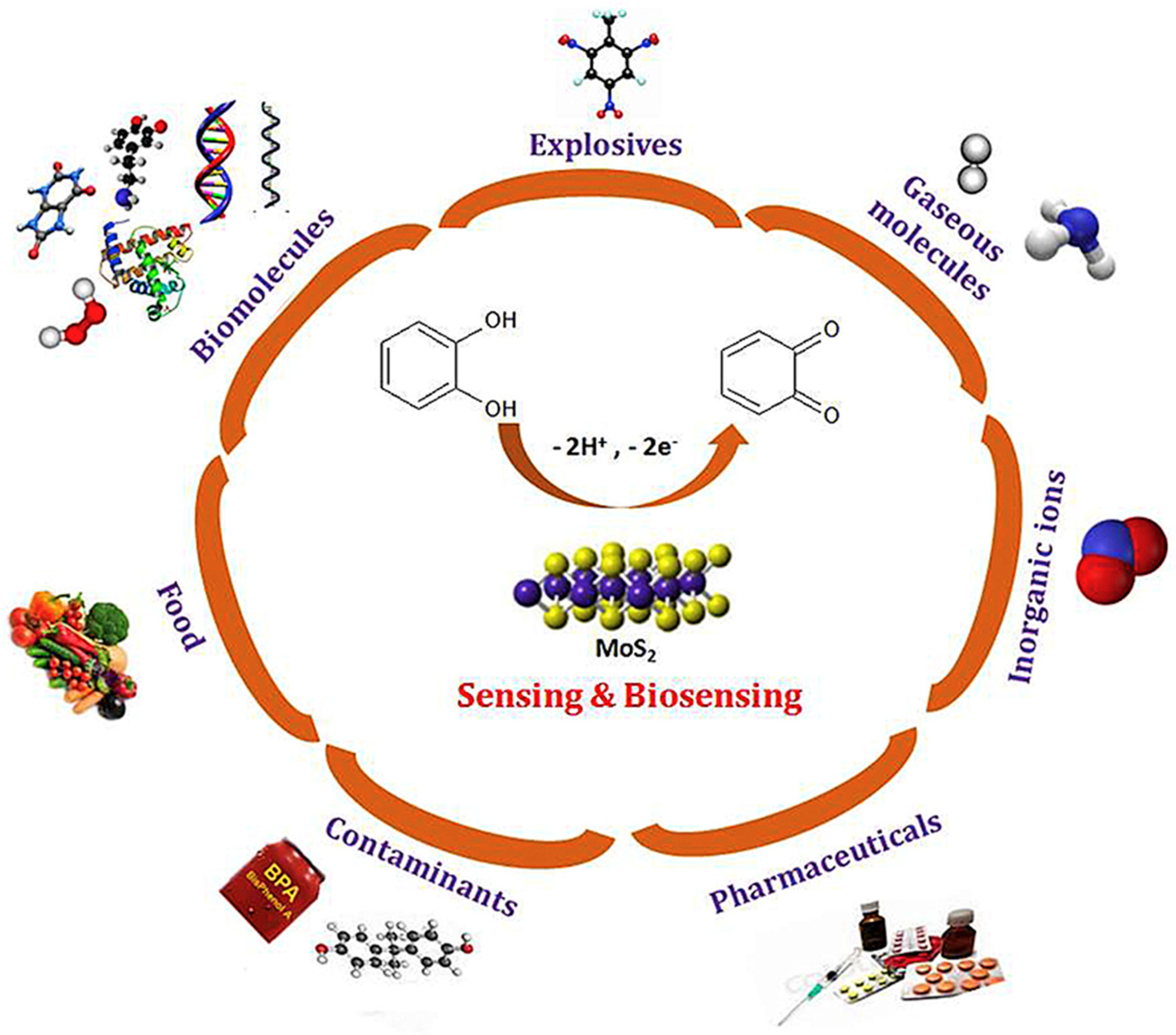
:1. Introduction
2. Metal Nanoparticles on 2D Materials for Biomarker Detection
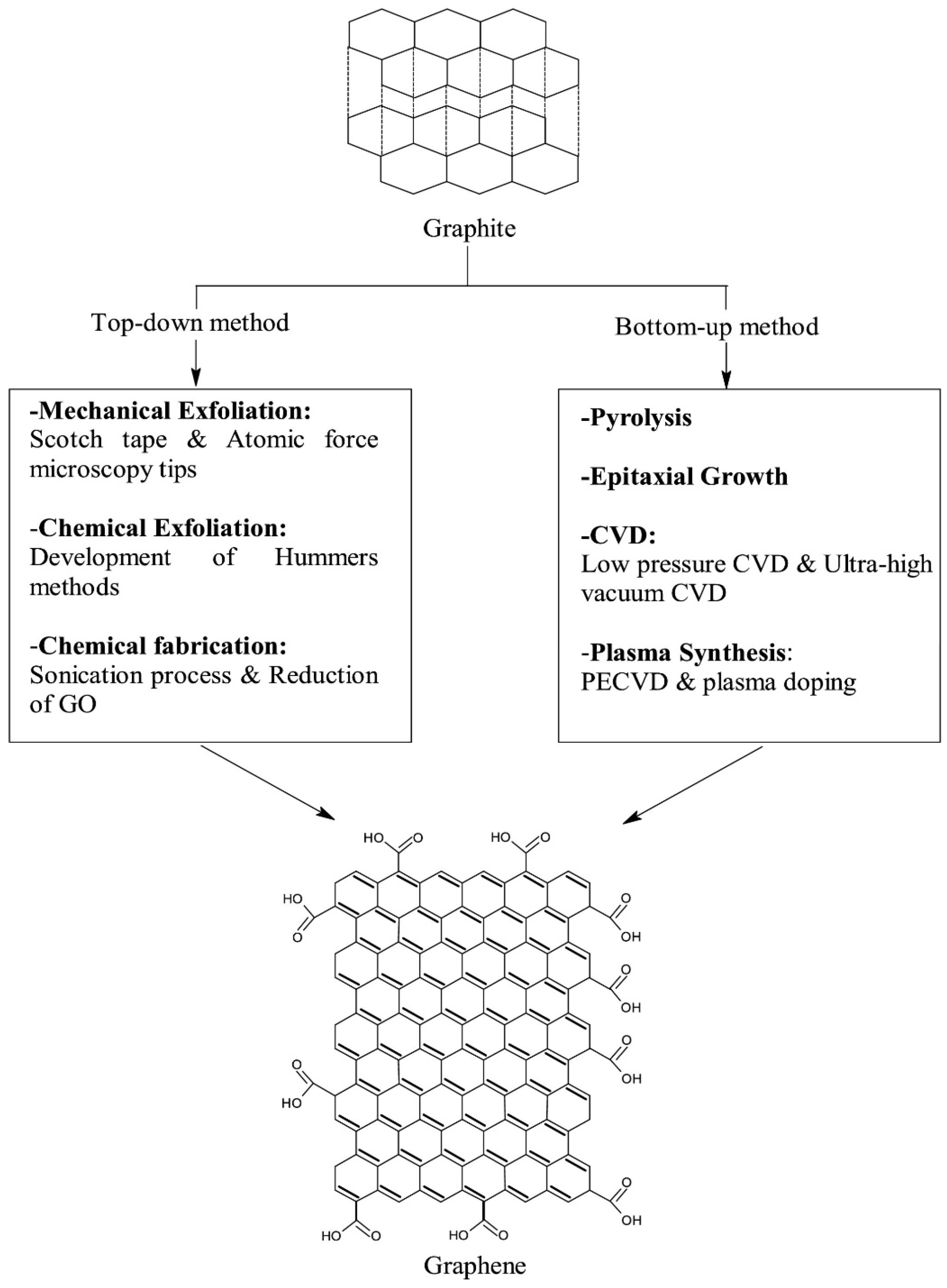
2.1. Graphene Oxide Conjugated with Nanoparticles for Electrochemical Biomarker Detection
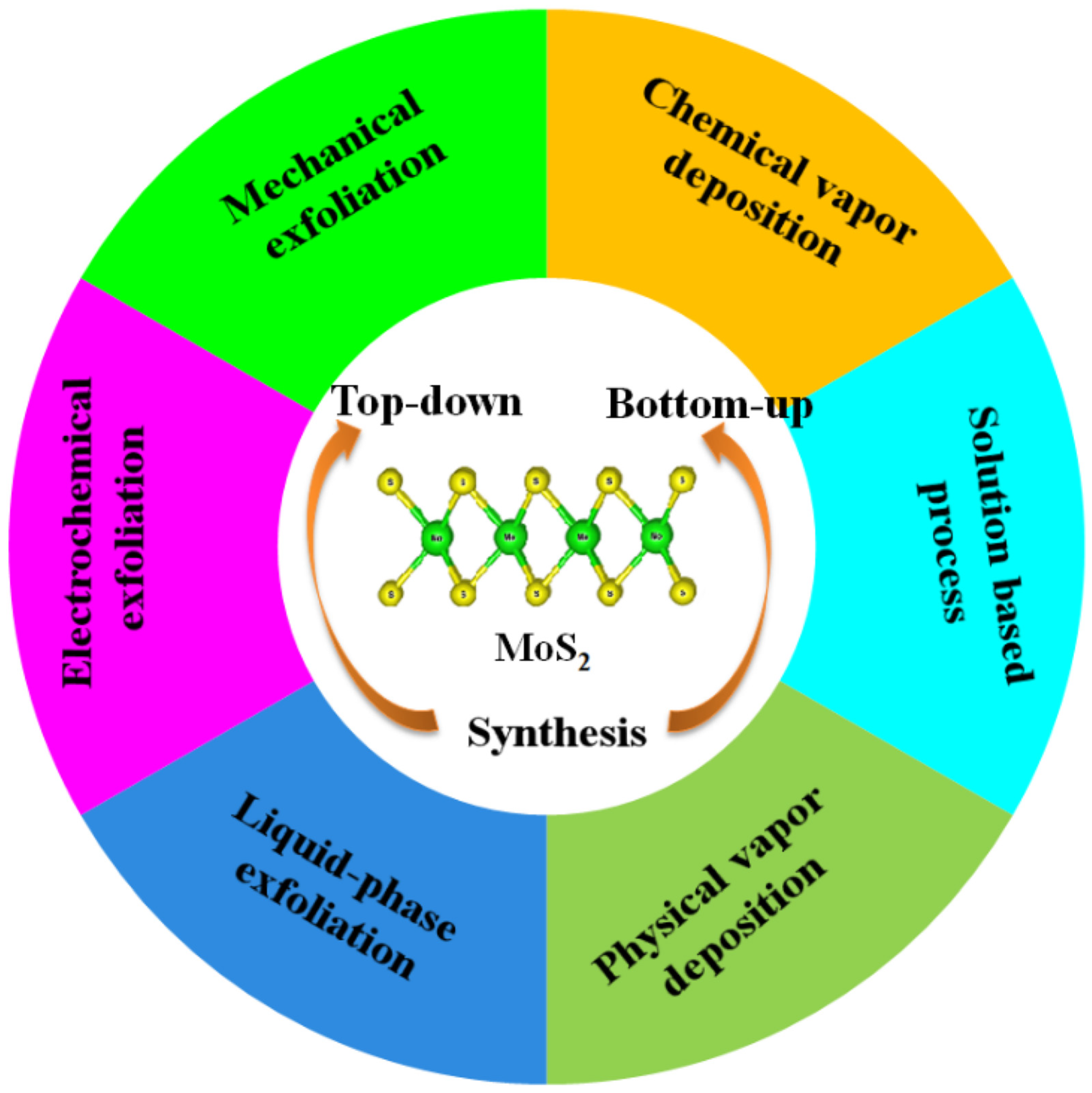
2.2. MoS2 Conjugated Nanoparticles for Electrochemical Biomarker Detection
2.3. Biomarker Detection on MXenes Conjugated with Metal Nanoparticles
2.4. MOFs Conjugated Metal Nanoparticles for Electrochemical Biomarker Detection
2.5. Biomarker Detection on Other 2D Materials Conjugated with Metal Nanoparticles
3. Conclusions
Author Contributions
Funding
Institutional Review Board Statement
Informed Consent Statement
Data Availability Statement
Conflicts of Interest
Abbreviations
| 2D | Two dimensional |
| 2D-Hbn | 2D-hexagonal boron nitride |
| AgNPs | Silver nanoparticles |
| AgNRs | Silver nanorods |
| Apt | Aptamer |
| Au | Gold |
| AuE | Gold electrode |
| AuNP-RGO | Au nanoparticle-reduced graphene oxide |
| AuNPs | Gold nanoparticles |
| AuPtBNPs | Gold platinum bimetallic nanoparticles |
| BDC | 1,4-benzenedicarboxylate |
| BiVO4 | Bismuth vanadate |
| BNNSs | Boron nitride nanosheets |
| BP | Black phosphorous |
| BSA | Bovine serum albumin |
| CA 27-29 BCA | Cancer antigen 27-29 breast cancer antigen |
| CA | Chronoamperometry |
| CA125 | Cancer antigen 125 |
| CA15-3 | Cancer antigen 15-3 |
| C-DNA | Capture DNA |
| CEA | Carcinoembryonic antigen |
| ce-MoS2 | Chemical exfoliated MoS2 |
| CGO | Carboxylic groups |
| CLB | Clenbuterol |
| CoPP | Cobalt protoporphyrin |
| CP | Capture probe |
| CPEB4 | Cytoplasmic polyadenylate element-binding protein 4 |
| Cr.6 | 18-crown-6 |
| CRP | C-reactive protein |
| CS | Chitosan |
| CTnI | Cardiac troponin I |
| CTnT | Cardiac troponin T |
| CV | Cyclic voltammetry |
| CYFRA21-1 | Cytokeratin 19 fragment |
| DNA | Deoxyribonucleic acid |
| DPV | Differential pulse voltammetry |
| ECD | Extracellular domain |
| ECL | Electrochemiluminescence |
| EIS | Electrochemical impedance spectroscopy |
| ELISA | Enzyme-linked immunosorbent assay |
| eT | Electron transfer |
| Fc | Ferrocene |
| FTO | Fluorine doped tin oxide |
| g-C3N4 | Graphitic carbon nitride |
| GCE | Glassy carbon electrode |
| GDY | Graphdiyne |
| GE | Graphite electrode |
| GFAP | Glial fibrillary acidic protein |
| GP120 | Glycoprotein GP120 |
| 4-AP | p-Aminophenol |
| HER-2 | Human epidermal growth factor receptor-2 |
| HP1 | Hairpin DNA |
| HT | Hexane thiol |
| IL8 | Interleukin-8 |
| i-t curve | Amperometric current-time response |
| ITO | Indium tin oxide |
| LA | Lactic acid |
| LBL | Layer by layer |
| L-cys | L-Cysteine |
| LOD | Limit of detection |
| LSV | Linear sweep voltammetry |
| L-Trp | L-tryptophan |
| Mb | Myoglobin |
| MCH | 6-mercaptohexanol |
| MgAl-LDH | Mg-Al-Layered double hydroxide |
| MGMT | O6-methylguanine-DNA methyltransferase |
| miRNA-141 | micro-RNA-141 |
| miRNA-21 | micro-RNA-21 |
| miRNA-377 | micro-RNA-377 |
| miRNAs | micro-RNAs |
| MNPs | Mesoporous nanoparticles |
| MOFs | Metal organic frameworks |
| MUC1 | Mucin1 |
| MWCNT | Multiwalled carbon nanotubes |
| NMP-22 | Nuclear matrix protein 22 |
| NS1 | Non-structural 1 |
| NSE | Neuron-specific enolase |
| OCV | Open circuit voltage |
| OTC | Oxytetracycline |
| PAMAM | Polyamidoamine |
| PCT | Procalcitonin |
| PDA | Polydopamine |
| PdPtBP MNPs | Pd-Pt-Black phosphorous-mesoporous nanoparticles |
| PEC | Photoelectrochemical |
| PET | Polyethylene terephthalate |
| PPY | Polypyrrole |
| PSA | Prostate specific antigen |
| PtNPs | Platinum nanoparticles |
| RAC | Ractopamine |
| rGO | Reduced graphene oxide |
| RhNPs | Rhodium nanoparticles |
| RNA | Ribonucleic acid |
| S/N | Signal-to-noise ratio |
| SAL | Salbutamol |
| SPCE | Screen-printed carbon electrode |
| SWV | Square wave voltammetry |
| TEPA | Tetraethylenepentamine |
| VEGF165 | Vascular endothelial growth factor 165 |
| YNCs | Yolk-shell nanocubes |
| β-CD | β-cyclodextrin |
References
- Filice, M.; Ruiz-Cabello, J. Nucleic Acid Nanotheranostics: Biomedical Applications; Elsevier: Amsterdam, The Netherlands, 2019. [Google Scholar]
- WHO. Biomarkers in Risk Assessment: Validity and Validation-Environmental Health Criteria 222; WHO: Geneva, Switzerland, 2001. [Google Scholar]
- National Cancer Institute. Biomarker. Available online: https://www.cancer.gov/publications/dictionaries/cancer-terms/def/biomarker (accessed on 5 October 2022).
- Biomarkers Definitions Working Group. Biomarkers and surrogate endpoints: Preferred definitions and conceptual framework. Clin. Pharmacol. Ther. 2001, 69, 89–95. [Google Scholar] [CrossRef]
- Kikkeri, K.; Wu, D.; Voldman, J. A sample-to-answer electrochemical biosensor system for biomarker detection. Lab Chip 2022, 22, 100–107. [Google Scholar] [CrossRef] [PubMed]
- Ziemssen, T.; Akgün, K.; Brück, W. Molecular biomarkers in multiple sclerosis. J. Neuroinflamm. 2019, 16, 272. [Google Scholar] [CrossRef] [PubMed] [Green Version]
- Pachner, A.R.; DiSano, K.; Royce, D.B.; Gilli, F. Clinical utility of a molecular signature in inflammatory demyelinating disease. Neurol. Neuroimmunol. Neuroinflamm. 2019, 6, e520. [Google Scholar] [CrossRef] [PubMed] [Green Version]
- Sant, G.R.; Knopf, K.B.; Albala, D.M. Live-single-cell phenotypic cancer biomarkers-future role in precision oncology? NPJ Precis. Oncol. 2017, 1, 21. [Google Scholar] [CrossRef] [PubMed] [Green Version]
- Huss, R. Chapter 19—Biomarkers. In Translational Regenerative Medicine; Atala, A., Allickson, J.G., Eds.; Academic Press: Boston, MA, USA, 2015; pp. 235–241. [Google Scholar]
- Manzanares, J.; Sala, F.; Gutiérrez, M.S.G.; Rueda, F.N. 2.30—Biomarkers. In Comprehensive Pharmacology; Kenakin, T., Ed.; Elsevier: Oxford, UK, 2022; pp. 693–724. [Google Scholar]
- Goossens, N.; Nakagawa, S.; Sun, X.; Hoshida, Y. Cancer biomarker discovery and validation. Transl. Cancer Res. 2015, 4, 256. [Google Scholar]
- Califf, R.M. Biomarker definitions and their applications. Exp. Biol. Med. 2018, 243, 213–221. [Google Scholar] [CrossRef]
- FDA-NIH Biomarker Working Group. BEST (Biomarkers, Endpoints, and Other Tools). Available online: https://www.ncbi.nlm.nih.gov/books/NBK326791/ (accessed on 7 November 2022).
- Magar, H.S.; Hassan, R.Y.A.; Mulchandani, A. Electrochemical Impedance Spectroscopy (EIS): Principles, Construction, and Biosensing Applications. Sensors 2021, 21, 6578. [Google Scholar] [CrossRef]
- Bertok, T.; Lorencova, L.; Chocholova, E.; Jane, E.; Vikartovska, A.; Kasak, P.; Tkac, J. Electrochemical Impedance Spectroscopy Based Biosensors: Mechanistic Principles, Analytical Examples and Challenges towards Commercialization for Assays of Protein Cancer Biomarkers. ChemElectroChem 2019, 6, 989–1003. [Google Scholar] [CrossRef] [Green Version]
- Lisdat, F.; Schafer, D. The use of electrochemical impedance spectroscopy for biosensing. Anal. Bioanal. Chem. 2008, 391, 1555–1567. [Google Scholar] [CrossRef]
- Grieshaber, D.; MacKenzie, R.; Voros, J.; Reimhult, E. Electrochemical biosensors—Sensor principles and architectures. Sensors 2008, 8, 1400–1458. [Google Scholar] [CrossRef]
- Rezaei, B.; Irannejad, N. Chapter 2—Electrochemical detection techniques in biosensor applications. In Electrochemical Biosensors; Ensafi, A.A., Ed.; Elsevier: Amsterdam, The Netherlands, 2019; pp. 11–43. [Google Scholar]
- Speiser, B. Electroanalytical Methods. 2. Cyclic Voltametry. Chem. Unserer. Zeit. 1981, 15, 62–67. [Google Scholar] [CrossRef]
- Huan, T.N.; Ha, V.T.T.; Hung, L.Q.; Yoon, M.Y.; Han, S.H.; Chung, H. Square wave voltammetric detection of Anthrax utilizing a peptide for selective recognition of a protein biomarker. Biosens. Bioelectron. 2009, 25, 469–474. [Google Scholar] [CrossRef]
- Kumar, S.; Kalkal, A. 3—Electrochemical detection: Cyclic voltammetry/differential pulse voltammetry/impedance spectroscopy. In Nanotechnology in Cancer Management; Khondakar, K.R., Kaushik, A.K., Eds.; Elsevier: Amsterdam, The Netherlands, 2021; pp. 43–71. [Google Scholar]
- Moore, S. Current Global Market of 2D Materials. Available online: https://www.azonano.com/article.aspx?ArticleID=6294 (accessed on 26 December 2022).
- Popov, A.; Brasiunas, B.; Kausaite-Minkstimiene, A.; Ramanaviciene, A. Metal Nanoparticle and Quantum Dot Tags for Signal Amplification in Electrochemical Immunosensors for Biomarker Detection. Chemosensors 2021, 9, 85. [Google Scholar] [CrossRef]
- Enoch, I.V.M.V.; Ramasamy, S.; Mohiyuddin, S.; Gopinath, P.; Manoharan, R. Cyclodextrin–PEG conjugate-wrapped magnetic ferrite nanoparticles for enhanced drug loading and release. Appl. Nanosci. 2018, 8, 273–284. [Google Scholar] [CrossRef] [Green Version]
- Liu, Y.; Yang, G.; Jin, S.; Xu, L.; Zhao, C.X. Development of high-drug-loading nanoparticles. ChemPlusChem 2020, 85, 2143–2157. [Google Scholar] [CrossRef]
- Aido, A.; Wajant, H.; Buzgo, M.; Simaite, A. Development of anti-TNFR antibody-conjugated nanoparticles. Multidiscip. Digit. Publ. Inst. Proc. 2020, 78, 55. [Google Scholar]
- Li, Y.; Wang, Y.; Zhang, N.; Fan, D.; Liu, L.; Yan, T.; Yang, X.; Ding, C.; Wei, Q.; Ju, H. Magnetic electrode-based electrochemical immunosensor using amorphous bimetallic sulfides of CoSnSx as signal amplifier for the NTpro BNP detection. Biosens. Bioelectron. 2019, 131, 250–256. [Google Scholar] [CrossRef]
- Zhang, M.; Hu, X.; Mei, L.; Zhang, L.; Wang, X.; Liao, X.; Qiao, X.; Hong, C. PSA detection electrochemical immunosensor based on MOF-235 nanomaterial adsorption aggregation signal amplification strategy. Microchem. J. 2021, 171, 106870. [Google Scholar] [CrossRef]
- Wang, H.; Zhang, S.; Li, S.; Qu, J. Electrochemical sensor based on palladium-reduced graphene oxide modified with gold nanoparticles for simultaneous determination of acetaminophen and 4-aminophenol. Talanta 2018, 178, 188–194. [Google Scholar] [CrossRef]
- Lim, J.Y.; Mubarak, N.M.; Abdullah, E.C.; Nizamuddin, S.; Khalid, M.; Inamuddin. Recent trends in the synthesis of graphene and graphene oxide based nanomaterials for removal of heavy metals—A review. J. Ind. Eng. Chem. 2018, 66, 29–44. [Google Scholar] [CrossRef]
- Mbayachi, V.B.; Ndayiragije, E.; Sammani, T.; Taj, S.; Mbuta, E.R.; Khan, A.U. Graphene synthesis, characterization and its applications: A review. Results Chem. 2021, 3, 100163. [Google Scholar] [CrossRef]
- Bhuyan, M.S.A.; Uddin, M.N.; Islam, M.M.; Bipasha, F.A.; Hossain, S.S. Synthesis of graphene. Int. Nano Lett. 2016, 6, 65–83. [Google Scholar] [CrossRef] [Green Version]
- Singh, R.K.; Kumar, R.; Singh, D.P. Graphene oxide: Strategies for synthesis, reduction and frontier applications. RSC Adv. 2016, 6, 64993–65011. [Google Scholar] [CrossRef]
- Yagati, A.K.; Behrent, A.; Beck, S.; Rink, S.; Goepferich, A.M.; Min, J.; Lee, M.H.; Baeumner, A.J. Laser-induced graphene interdigitated electrodes for label-free or nanolabel-enhanced highly sensitive capacitive aptamer-based biosensors. Biosens. Bioelectron. 2020, 164, 112272. [Google Scholar] [CrossRef]
- Ranjan, P.; Sadique, M.A.; Yadav, S.; Khan, R. An Electrochemical Immunosensor Based on Gold-Graphene Oxide Nanocomposites with Ionic Liquid for Detecting the Breast Cancer CD44 Biomarker. ACS Appl. Mater. Inter. 2022, 14, 20802–20812. [Google Scholar] [CrossRef]
- Yagati, A.K.; Pyun, J.C.; Min, J.; Cho, S. Label-free and direct detection of C-reactive protein using reduced graphene oxide-nanoparticle hybrid impedimetric sensor. Bioelectrochemistry 2016, 107, 37–44. [Google Scholar] [CrossRef]
- Jonous, Z.A.; Shayeh, J.S.; Yazdian, F.; Yadegari, A.; Hashemi, M.; Omidi, M. An electrochemical biosensor for prostate cancer biomarker detection using graphene oxide-gold nanostructures. Eng. Life Sci. 2019, 19, 206–216. [Google Scholar] [CrossRef] [Green Version]
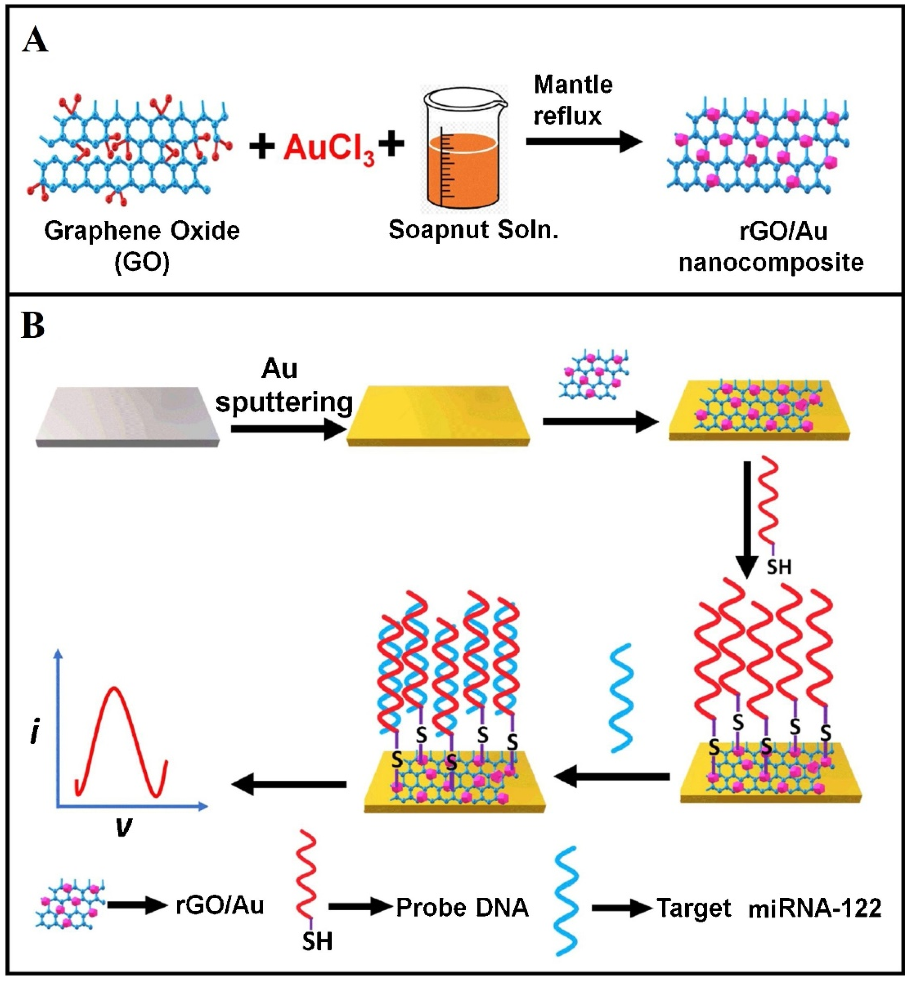
- Kasturi, S.; Eom, Y.; Torati, R.; Kim, C. Highly sensitive electrochemical biosensor based on naturally reduced rGO/Au nanocomposite for the detection of miRNA-122 biomarker. J. Ind. Eng. Chem. 2021, 93, 186–195. [Google Scholar] [CrossRef]
- Rauf, S.; Lahcen, A.A.; Aljedaibi, A.; Beduk, T.; de Oliveira, J.I.; Salama, K.N. Gold nanostructured laser-scribed graphene: A new electrochemical biosensing platform for potential point-of-care testing of disease biomarkers. Biosens. Bioelectron. 2021, 180, 113116. [Google Scholar] [CrossRef]
- Hasanjani, H.R.A.; Zarei, K. DNA/Au-Pt bimetallic nanoparticles/graphene oxide-chitosan composites modified pencil graphite electrode used as an electrochemical biosensor for sub-picomolar detection of anti-HIV drug zidovudine. Microchem. J. 2021, 164, 106005. [Google Scholar] [CrossRef]
- Kanagavalli, P.; Veerapandian, M. Opto-electrochemical functionality of Ru(II)-reinforced graphene oxide nanosheets for immunosensing of dengue virus non-structural 1 protein. Biosens. Bioelectron. 2020, 150, 111878. [Google Scholar] [CrossRef] [PubMed]
- Kanagavalli, P.; Andrew, C.; Veerapandian, M.; Jayakumar, M. In-situ redox-active hybrid graphene platform for label-free electrochemical biosensor: Insights from electrodeposition and electroless deposition. TrAC-Trend Anal. Chem. 2021, 143, 116413. [Google Scholar] [CrossRef]
- Liang, Y.; Xu, Y.; Tong, Y.Y.; Chen, Y.; Chen, X.L.; Wu, S.M. Graphene-Based Electrochemical Sensor for Detection of Hepatocellular Carcinoma Markers. Front. Chem. 2022, 10, 883627. [Google Scholar] [CrossRef] [PubMed]
- Ozkan-Ariksoysal, D. Current Perspectives in Graphene Oxide-Based Electrochemical Biosensors for Cancer Diagnostics. Biosensors 2022, 12, 607. [Google Scholar] [CrossRef]
- Rashid, J.I.A.; Kannan, V.; Ahmad, M.H.; Mon, A.A.; Taufik, S.; Miskon, A.; Ong, K.K.; Yusof, N.A. An electrochemical sensor based on gold nanoparticles-functionalized reduced graphene oxide screen printed electrode for the detection of pyocyanin biomarker in Pseudomonas aeruginosa infection. Mat. Sci. Eng. C Mater. 2021, 120, 111625. [Google Scholar] [CrossRef]
- Abolhasan, R.; Khalilzadeh, B.; Yousefi, H.; Samemaleki, S.; Chakari-Khiavi, F.; Ghorbani, F.; Pourakbari, R.; Kamrani, A.; Khataee, A.; Rad, T.S.; et al. Ultrasensitive and label free electrochemical immunosensor for detection of ROR1 as an oncofetal biomarker using gold nanoparticles assisted LDH/rGO nanocomposite. Sci. Rep. 2021, 11, 14921. [Google Scholar] [CrossRef]
- Liu, X.K.; Lin, L.Y.; Tseng, F.Y.; Tan, Y.C.; Li, J.; Feng, L.; Song, L.J.; Lai, C.F.; Li, X.H.; He, J.H.; et al. Label-free electrochemical immunosensor based on gold nanoparticle/polyethyleneimine/reduced graphene oxide nanocomposites for the ultrasensitive detection of cancer biomarker matrix metalloproteinase-1. Analyst 2021, 146, 4066–4079. [Google Scholar] [CrossRef]
- Ye, S.; Liu, Y.; Zeng, M.; Feng, W.; Yang, H.; Zheng, X. Electrochemical Immunoassay of Melanoma Biomarker CPEB4 Based on Cobalt Porphyrin Functionalized Graphene Oxide. J. Electrochem. Soc. 2022, 169, 027510. [Google Scholar] [CrossRef]
- Yagati, A.K.; Lee, G.Y.; Ha, S.; Chang, K.A.; Pyun, J.C.; Cho, S. Impedimetric Tumor Necrosis Factor-alpha Sensor Based on a Reduced Graphene Oxide Nanoparticle-Modified Electrode Array. J. Nanosci. Nanotechnol. 2016, 16, 11921–11927. [Google Scholar] [CrossRef]
- Ben Moussa, F.; Achi, F.; Meskher, H.; Henni, A.; Belkhalfa, H. Green one-step reduction approach to prepare rGO@AgNPs coupled with molecularly imprinted polymer for selective electrochemical detection of lactic acid as a cancer biomarker. Mater. Chem. Phys. 2022, 289, 126456. [Google Scholar] [CrossRef]
- Wang, H.; Zhang, Y.; Li, H.; Du, B.; Ma, H.M.; Wu, D.; Wei, Q. A silver-palladium alloy nanoparticle-based electrochemical biosensor for simultaneous detection of ractopamine, clenbuterol and salbutamol. Biosens. Bioelectron. 2013, 49, 14–19. [Google Scholar] [CrossRef] [PubMed]
- Akbarzadeh, S.; Khajehsharifi, H.; Hajihosseini, S. Detection of Oxytetracycline Using an Electrochemical Label-Free Aptamer-Based Biosensor. Biosensors 2022, 12, 468. [Google Scholar] [CrossRef] [PubMed]
- Yang, H.; Ren, J.; Zhao, M.; Chen, C.; Wang, F.; Chen, Z. Novel electrochemical immunosensor for O6-methylguanine-DNA methyltransferase gene methylation based on graphene oxide-magnetic nanoparticles-β-cyclodextrin nanocomposite. Bioelectrochemistry 2022, 146, 108111. [Google Scholar] [CrossRef]
- Pothipor, C.; Jakmunee, J.; Bamrungsap, S.; Ounnunkad, K. An electrochemical biosensor for simultaneous detection of breast cancer clinically related microRNAs based on a gold nanoparticles/graphene quantum dots/graphene oxide film. Analyst 2021, 146, 4000–4009. [Google Scholar] [CrossRef]
- Sadeghi, M.; Kashanian, S.; Naghib, S.M.; Arkan, E. A high-performance electrochemical aptasensor based on graphene-decorated rhodium nanoparticles to detect HER2-ECD oncomarker in liquid biopsy. Sci. Rep. 2022, 12, 3299. [Google Scholar] [CrossRef]
- Verma, S.; Singh, A.; Shukla, A.; Kaswan, J.; Arora, K.; Ramirez-Vick, J.; Singh, P.; Singh, S.P. Anti-IL8/AuNPs-rGO/ITO as an Immunosensing Platform for Noninvasive Electrochemical Detection of Oral Cancer. ACS Appl. Mater. Inter. 2017, 9, 27462–27474. [Google Scholar] [CrossRef]
- Barman, S.C.; Hossain, M.F.; Yoon, H.; Park, J.Y. Trimetallic Pd@Au@Pt nanocomposites platform on -COOH terminated reduced graphene oxide for highly sensitive CEA and PSA biomarkers detection. Biosens. Bioelectron. 2018, 100, 16–22. [Google Scholar] [CrossRef]
- Thunkhamrak, C.; Chuntib, P.; Ounnunkad, K.; Banet, P.; Aubert, P.H.; Saianand, G.; Gopalan, A.I.; Jakmunee, J. Highly sensitive voltammetric immunosensor for the detection of prostate specific antigen based on silver nanoprobe assisted graphene oxide modified screen printed carbon electrode. Talanta 2020, 208, 120389. [Google Scholar] [CrossRef]
- Khoshnevisan, K.; Torabi, F.; Baharifar, H.; Sajjadi-Jazi, S.M.; Afjeh, M.S.; Faridbod, F.; Larijani, B.; Khorramizadeh, M.R. Determination of the biomarker L-tryptophan level in diabetic and normal human serum based on an electrochemical sensing method using reduced graphene oxide/gold nanoparticles/18-crown-6. Anal. Bioanal. Chem. 2020, 412, 3615–3627. [Google Scholar] [CrossRef]
- Meng, F.Y.; Sun, H.X.; Huang, Y.; Tang, Y.G.; Chen, Q.; Miao, P. Peptide cleavage-based electrochemical biosensor coupling graphene oxide and silver nanoparticles. Anal. Chim. Acta 2019, 1047, 45–51. [Google Scholar] [CrossRef] [PubMed]
- Sangili, A.; Kalyani, T.; Chen, S.M.; Nanda, A.; Jana, S.K. Label-Free Electrochemical Immunosensor Based on One-Step Electrochemical Deposition of AuNP-RGO Nanocomposites for Detection of Endometriosis Marker CA 125. ACS Appl. Bio Mater. 2020, 3, 7620–7630. [Google Scholar] [CrossRef] [PubMed]
- Rostamabadi, P.F.; Heydari-Bafrooei, E. Impedimetric aptasensing of the breast cancer biomarker HER2 using a glassy carbon electrode modified with gold nanoparticles in a composite consisting of electrochemically reduced graphene oxide and single-walled carbon nanotubes. Microchim. Acta 2019, 186, 495. [Google Scholar] [CrossRef] [PubMed]
- Bharti, A.; Rana, S.; Dahiya, D.; Agnihotri, N.; Prabhakar, N. An electrochemical aptasensor for analysis of MUC1 using gold platinum bimetallic nanoparticles deposited carboxylated graphene oxide. Anal. Chim. Acta 2020, 1097, 186–195. [Google Scholar] [CrossRef] [PubMed]
- Kumar, A.; Purohit, B.; Mahato, K.; Mandal, R.; Srivastava, A.; Chandra, P. Gold-Iron Bimetallic Nanoparticles Impregnated Reduced Graphene Oxide Based Nanosensor for Label-Free Detection of Biomarker Related to Non-Alcoholic Fatty Liver Disease. Electroanalysis 2019, 31, 2417–2428. [Google Scholar] [CrossRef]
- Hu, H.W.; Zavabeti, A.; Quan, H.Y.; Zhu, W.Q.; Wei, H.Y.; Chen, D.C.; Ou, J.Z. Recent advances in two-dimensional transition metal dichalcogenides for biological sensing. Biosens. Bioelectron. 2019, 142, 111573. [Google Scholar] [CrossRef]
- Subbaiah, Y.P.V.; Saji, K.J.; Tiwari, A. Atomically Thin MoS2: A Versatile Nongraphene 2D Material. Adv. Funct. Mater. 2016, 26, 2046–2069. [Google Scholar] [CrossRef]
- Kalantar-zadeh, K.; Ou, J.Z. Biosensors Based on Two-Dimensional MoS2. ACS Sens. 2016, 1, 5–16. [Google Scholar] [CrossRef]
- Van, T.D.; Thuy, N.D.T.; Phuong, T.D.V.; Thi, N.N.; Thi, T.N.; Phuong, T.N.; Van, T.V.; Vuong-Pham, H.; Dinh, T.P. High-performance nonenzymatic electrochemical glucose biosensor based on AgNP-decorated MoS2 microflowers. Curr. Appl. Phys. 2022, 43, 116–123. [Google Scholar] [CrossRef]
- Cui, Z.L.; Li, D.J.; Yang, W.H.; Fan, K.; Liu, H.Y.; Wen, F.; Li, L.L.; Dong, L.X.; Wang, G.F.; Wu, W. An electrochemical biosensor based on few-layer MoS2 nanosheets for highly sensitive detection of tumor marker ctDNA. Anal. Methods 2022, 14, 1956–1962. [Google Scholar] [CrossRef]
- Li, L.; Zhang, D.; Gao, Y.H.; Deng, J.P.; Gou, Y.C.; Fang, J.F. Electric field driven exfoliation of MoS2. J. Alloys Compd. 2021, 862, 158551. [Google Scholar] [CrossRef]
- Shi, Z.T.; Zhao, H.B.; Chen, X.Q.; Wu, G.M.; Wei, F.; Tu, H.L. Chemical vapor deposition growth and transport properties of MoS2-2H thin layers using molybdenum and sulfur as precursors. Rare Met. 2022, 41, 3574–3578. [Google Scholar] [CrossRef]
- Jagannadham, K.; Das, K.; Reynolds, C.L.; El-Masry, N. Nature of electrical conduction in MoS2 films deposited by laser physical vapor deposition. J. Mater. Sci. Mater. Electron. 2018, 29, 14180–14191. [Google Scholar] [CrossRef]
- Gomes, F.O.V.; Pokle, A.; Marinkovic, M.; Balster, T.; Canavan, M.; Fleischer, K.; Anselmann, R.; Nicolosi, V.; Wagner, V. Influence of temperature on morphological and optical properties of MoS2 layers as grown based on solution processed precursor. Thin Solid Films 2018, 645, 38–44. [Google Scholar] [CrossRef]
- Shah, S.A.; Khan, I.; Yuan, A.H. MoS2 as a Co-Catalyst for Photocatalytic Hydrogen Production: A Mini Review. Molecules 2022, 27, 3289. [Google Scholar] [CrossRef] [PubMed]
- Li, X.; Zhu, H. Two-dimensional MoS2: Properties, preparation, and applications. J. Mater. 2015, 1, 33–44. [Google Scholar] [CrossRef] [Green Version]
- Sinha, A.; Dhanjai; Tan, B.; Huang, Y.J.; Zhao, H.M.; Dang, X.M.; Chen, J.P.; Jain, R. MoS2 nanostructures for electrochemical sensing of multidisciplinary targets: A review. TrAC-Trend Anal. Chem. 2018, 102, 75–90. [Google Scholar] [CrossRef]
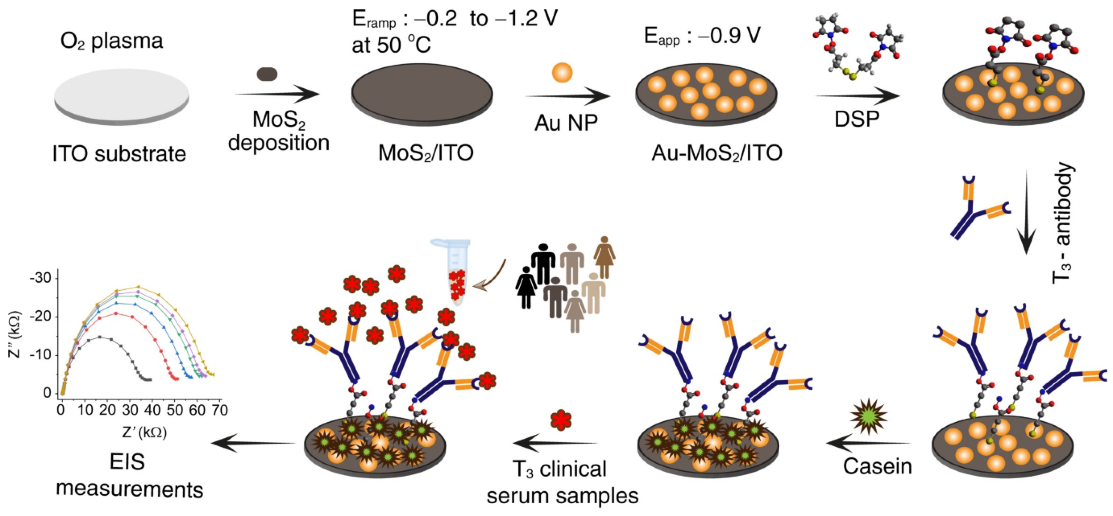
- Yagati, A.K.; Go, A.; Vu, N.H.; Lee, M.H. A MoS2-Au nanoparticle-modified immunosensor for T-3 biomarker detection in clinical serum samples. Electrochim. Acta 2020, 342, 136065. [Google Scholar] [CrossRef]
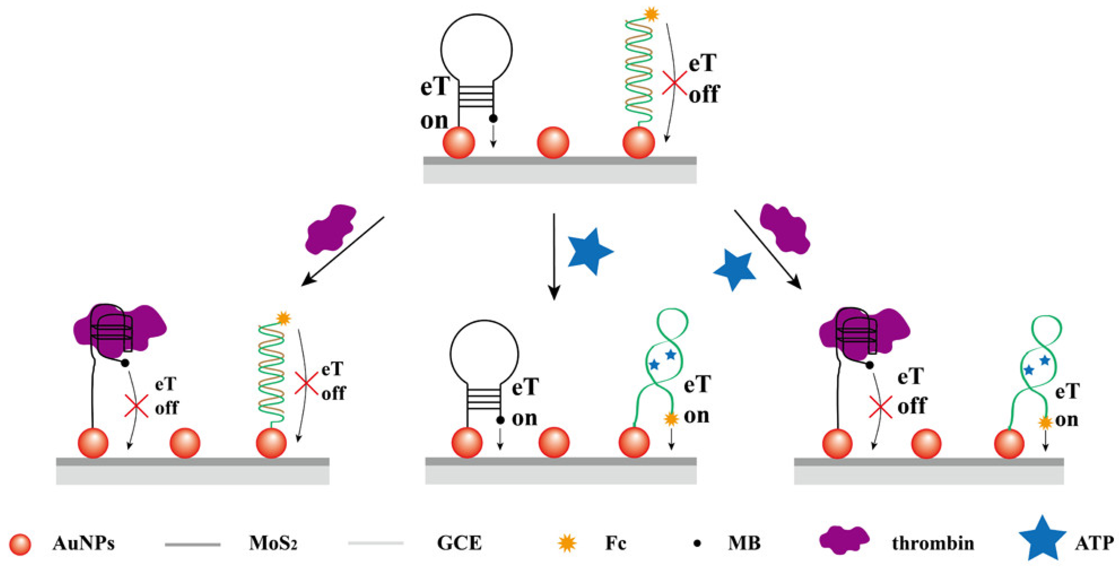
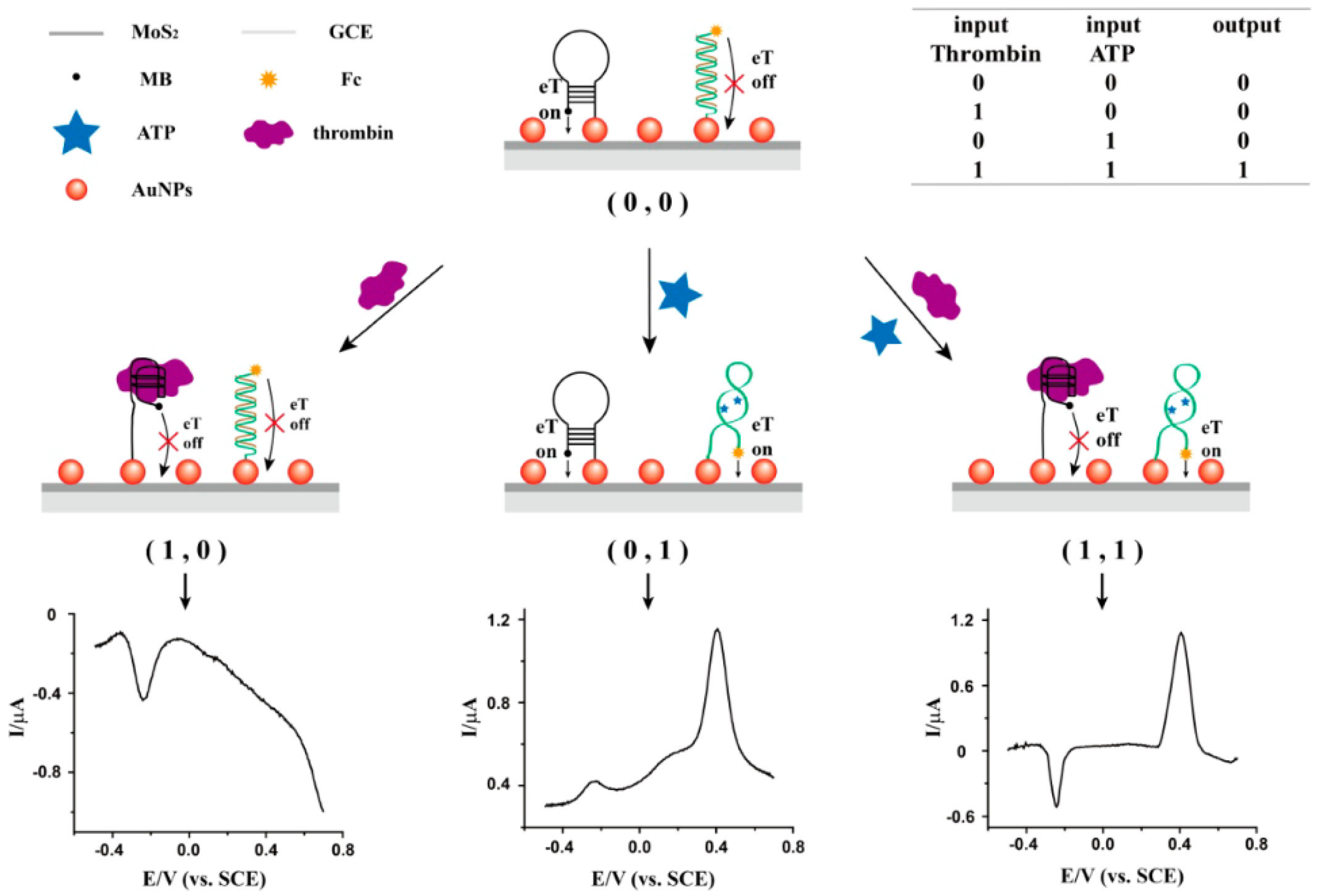
- Su, S.; Sun, H.F.; Cao, W.F.; Chao, J.; Peng, H.Z.; Zuo, X.L.; Yuwen, L.H.; Fan, C.H.; Wang, L.H. Dual-Target Electrochemical Biosensing Based on DNA Structural Switching on Gold Nanoparticle-Decorated MoS2 Nanosheets. ACS Appl. Mater. Int. 2016, 8, 6826–6833. [Google Scholar] [CrossRef]
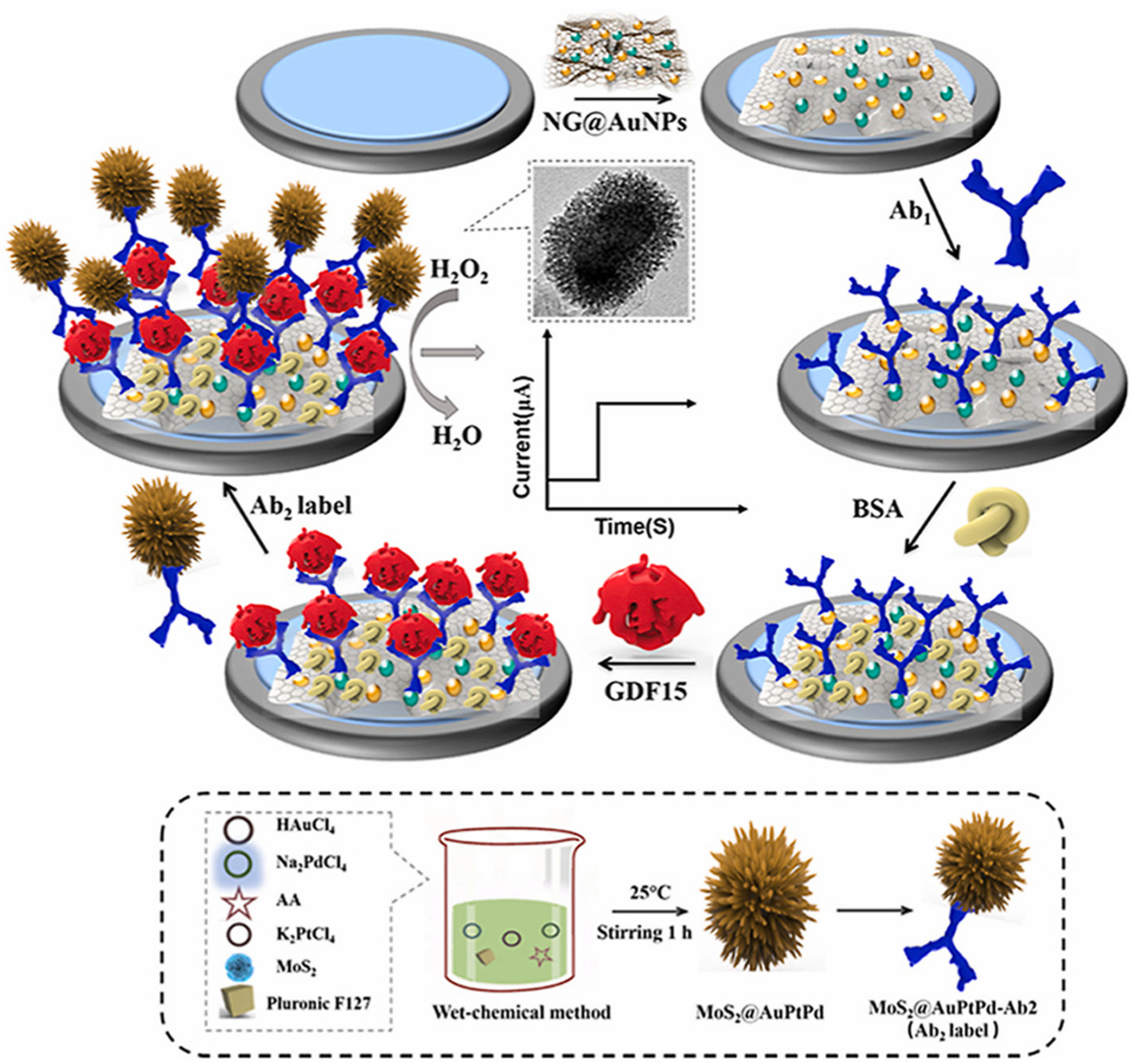
- Chen, M.; Zhao, L.; Wu, D.; Tu, S.; Chen, C.; Guo, H.; Xu, Y. Highly sensitive sandwich-type immunosensor with enhanced electrocatalytic durian-shaped MoS2/AuPtPd nanoparticles for human growth differentiation factor-15 detection. Anal. Chim. Acta 2022, 1223, 340194. [Google Scholar] [CrossRef]
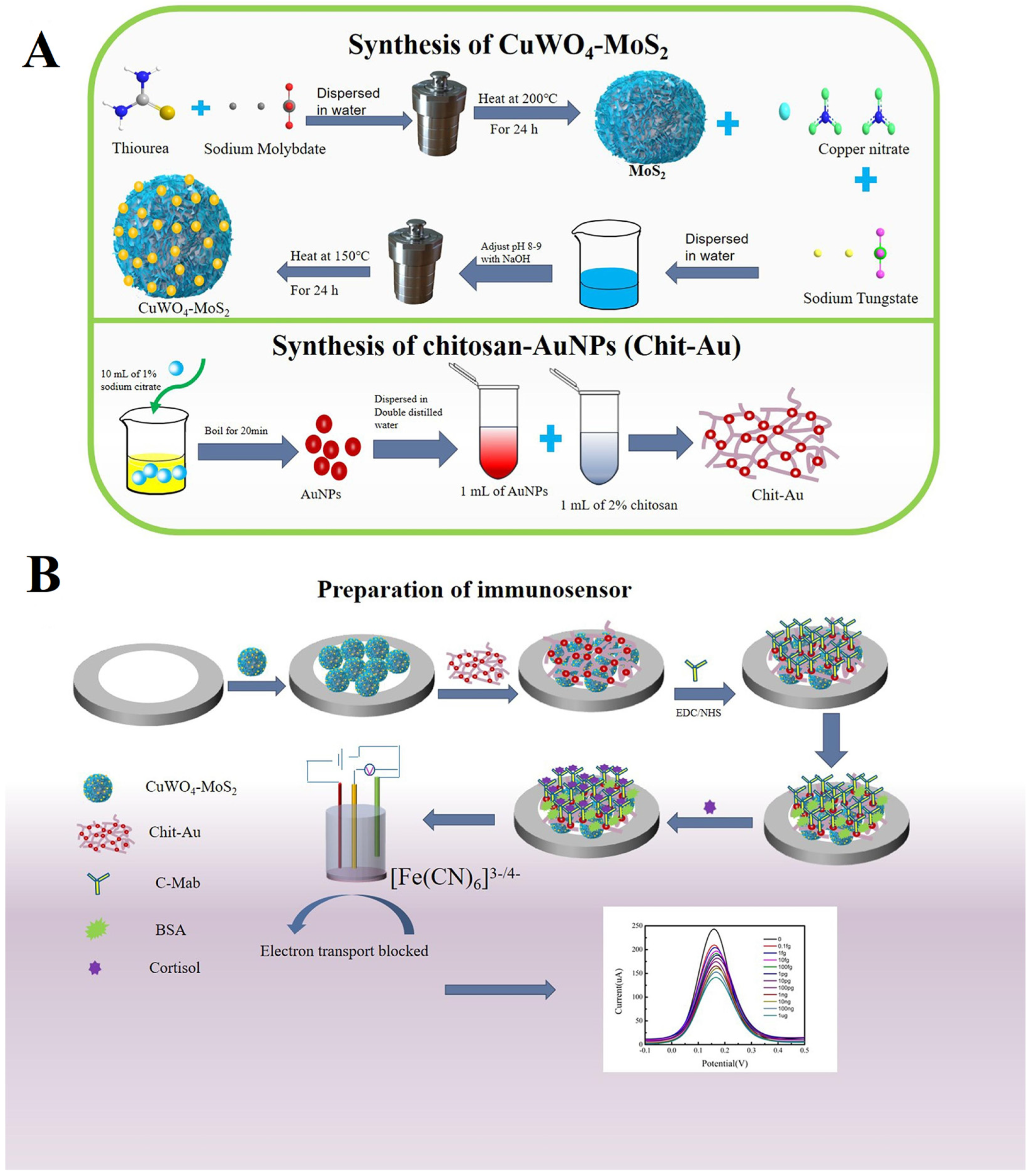
- Nong, C.J.; Yang, B.; Li, X.K.; Feng, S.X.; Cui, H.X. An ultrasensitive electrochemical immunosensor based on in-situ growth of CuWO4 nanoparticles on MoS2 and chitosan-gold nanoparticles for cortisol detection. Microchem. J. 2022, 179, 107434. [Google Scholar] [CrossRef]
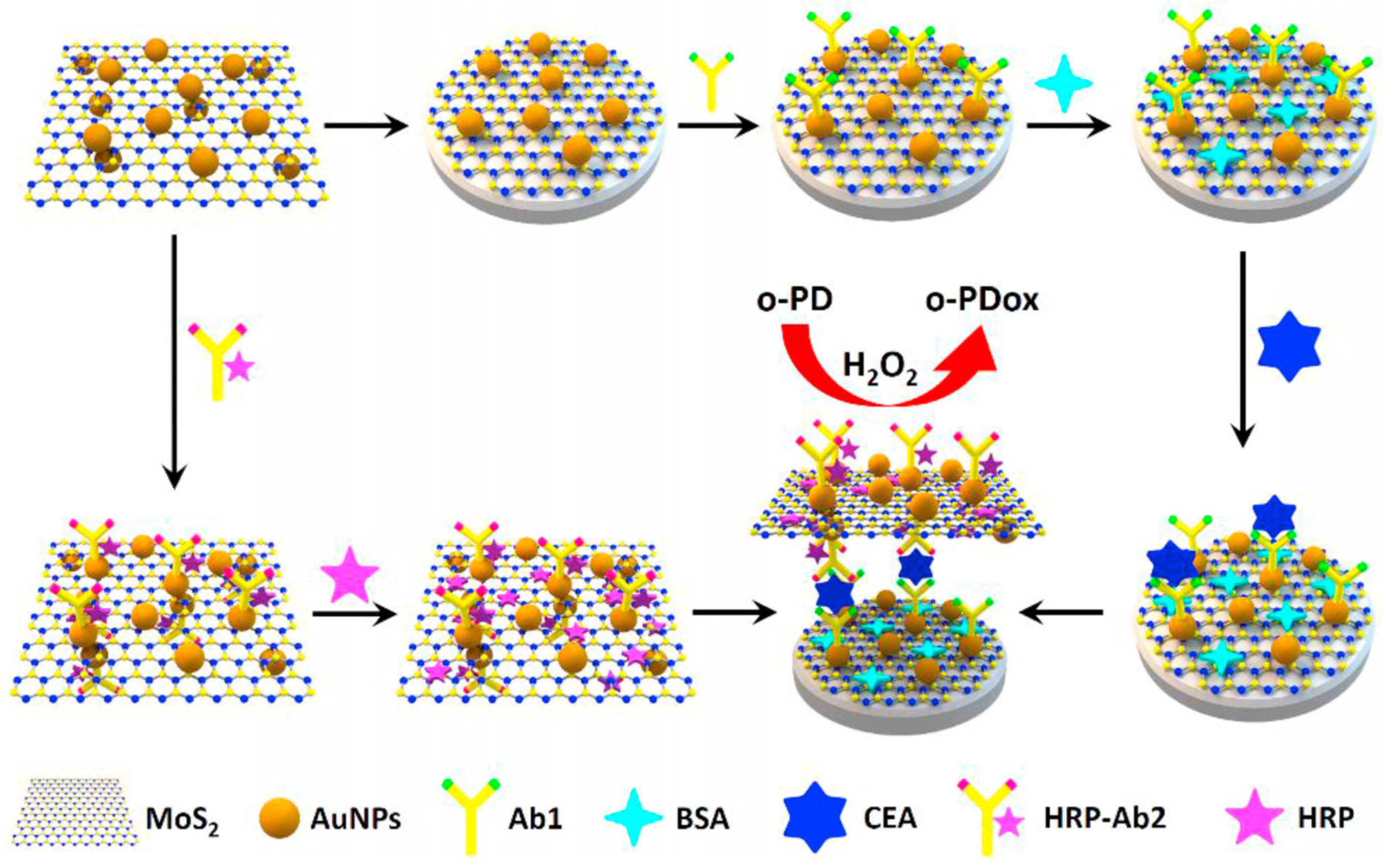
- Su, S.; Sun, Q.; Wan, L.; Gu, X.; Zhu, D.; Zhou, Y.; Chao, J.; Wang, L. Ultrasensitive analysis of carcinoembryonic antigen based on MoS2-based electrochemical immunosensor with triple signal amplification. Biosens. Bioelectron. 2019, 140, 111353. [Google Scholar] [CrossRef]
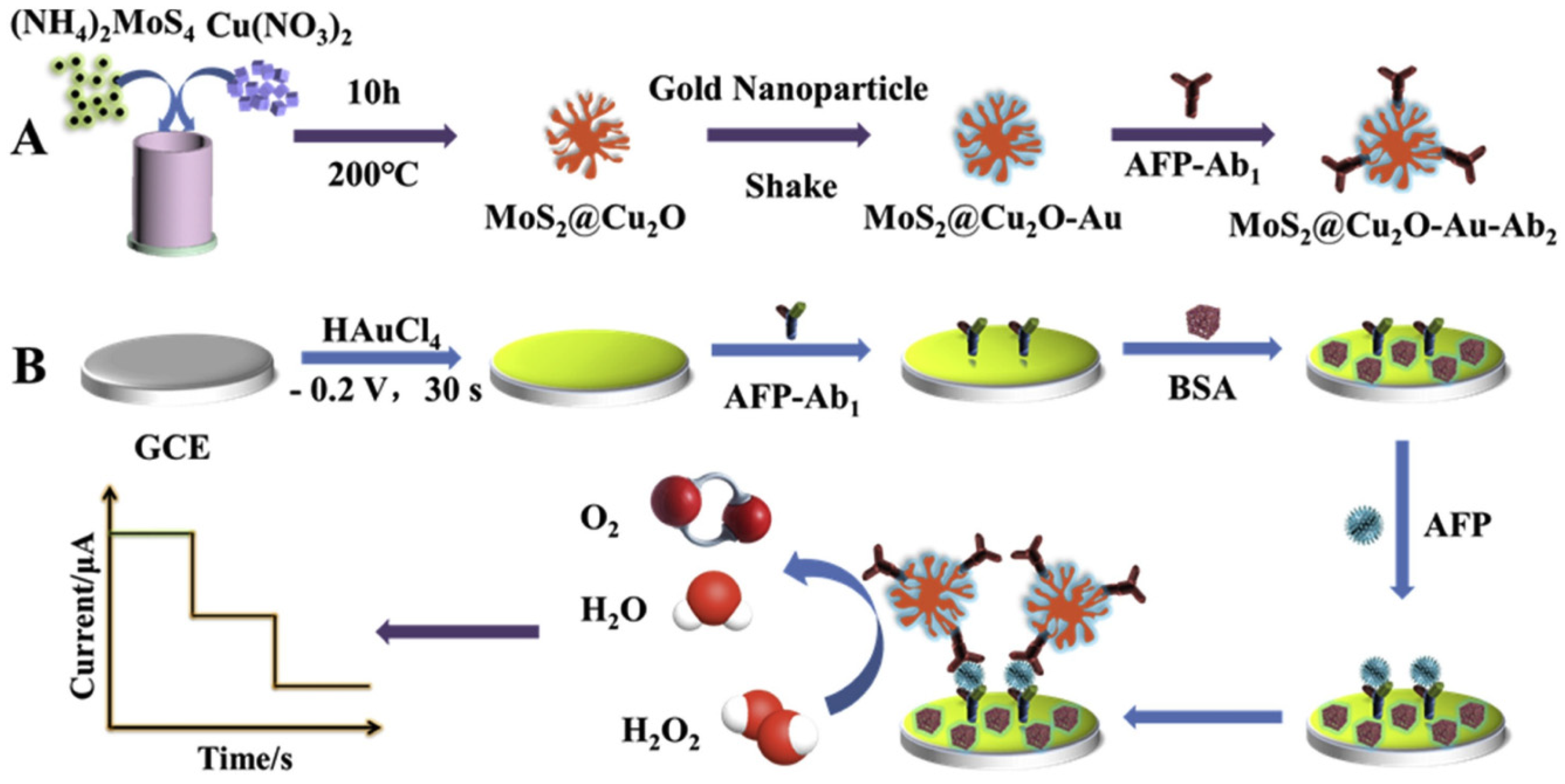
- Ma, N.; Zhang, T.; Fan, D.W.; Kuang, X.; Ali, A.; Wu, D.; Wei, Q. Triple amplified ultrasensitive electrochemical immunosensor for alpha fetoprotein detection based on MoS2@Cu2O-Au nanoparticles. Sens. Actuators B Chem. 2019, 297, 126821. [Google Scholar] [CrossRef]
- Dalila, R.N.; Arshad, M.K.M.; Gopinath, S.C.B.; Ibau, C.; Nuzaihan, M.M.N.; Fathil, M.F.M.; Azmi, U.Z.M.; Anbu, P. Faradaic electrochemical impedimetric analysis on MoS2/Au-NPs decorated surface for C-reactive protein detection. J. Taiwan Inst. Chem. E 2022, 138, 104450. [Google Scholar] [CrossRef]
- Xu, W.; Fei, J.W.; Yang, W.; Zheng, Y.N.; Dai, Y.; Sakran, M.; Zhang, J.; Zhu, W.Y.; Hong, J.L.; Zhou, X.M. A colorimetric/electrochemical dual-mode sensor based on Fe3O4@MoS2-Au NPs for high-sensitivity detection of hydrogen peroxide. Microchem. J. 2022, 181, 107825. [Google Scholar] [CrossRef]
- Shin, M.; Yoon, J.; Yi, C.Y.; Lee, T.; Choi, J.W. Flexible HIV-1 Biosensor Based on the Au/MoS2 Nanoparticles/Au Nanolayer on the PET Substrate. Nanomaterials 2019, 9, 76. [Google Scholar] [CrossRef] [Green Version]
- Li, S.; Zhang, J.W.; Tan, C.S.; Chen, C.; Hu, C.; Bai, Y.C.; Ming, D. Electrochemical immunosensor based on hybrid MoS2/Pt@Au-nanoprism/PDA for simultaneous detection of free and total prostate specific antigen in serum. Sens. Actuators B Chem. 2022, 357, 131413. [Google Scholar] [CrossRef]
- Ma, E.H.; Wang, P.; Yang, Q.S.; Yu, H.X.; Pei, F.B.; Li, Y.Y.; Liu, Q.; Dong, Y.H. Electrochemical immunosensor based on MoS2 NFs/Au@AgPt YNCs as signal amplification label for sensitive detection of CEA. Biosens. Bioelectron. 2019, 142, 111580. [Google Scholar] [CrossRef]
- Zhao, H.; Du, X.; Dong, H.; Jin, D.L.; Tang, F.; Liu, Q.; Wang, P.; Chen, L.; Zhao, P.Q.; Li, Y.Y. Electrochemical immunosensor based on Au/Co-BDC/MoS2 and DPCN/MoS2 for the detection of cardiac troponin I. Biosens. Bioelectron. 2021, 175, 112883. [Google Scholar] [CrossRef]
- Alarfaj, N.A.; El-Tohamy, M.F.; Oraby, H. New label-free ultrasensitive electrochemical immunosensor-based Au/MoS2/rGO nanocomposites for CA 27-29 breast cancer antigen detection. New J. Chem. 2018, 42, 11046–11053. [Google Scholar] [CrossRef]
- Wang, X.; Chu, C.C.; Shen, L.; Deng, W.P.; Yan, M.; Ge, S.G.; Yu, J.H.; Song, X.R. An ultrasensitive electrochemical immunosensor based on the catalytical activity of MoS2-Au composite using Ag nanospheres as labels. Sens. Actuators B Chem. 2015, 206, 30–36. [Google Scholar] [CrossRef]
- Gui, J.C.; Han, L.; Du, C.X.; Yu, X.N.; Hu, K.; Li, L.H. An efficient label-free immunosensor based on ce-MoS2/AgNR composites and screen-printed electrodes for PSA detection. J. Solid State Electrochem. 2021, 25, 973–982. [Google Scholar] [CrossRef]
- Fan, Z.Q.; Yao, B.; Ding, Y.D.; Xie, M.H.; Zhao, J.F.; Zhang, K.; Huang, W. Electrochemiluminescence aptasensor for Siglec-5 detection based on MoS2@Au nanocomposites emitter and exonuclease III-powered DNA walker. Sens. Actuators B Chem. 2021, 334, 129592. [Google Scholar] [CrossRef] [PubMed]
- Ma, K.X.; Sinha, A.; Dang, X.M.; Zhao, H.M. Electrochemical Preparation of Gold Nanoparticles-Polypyrrole Co-Decorated 2D MoS2 Nanocomposite Sensor for Sensitive Detection of Glucose. J. Electrochem. Soc. 2019, 166, B147–B154. [Google Scholar] [CrossRef]
- Zhu, X.N.; Wang, Z.G.; Gao, M.Y.; Wang, Y.Q.; Hu, J.; Song, Z.X.; Wang, Z.B.; Dong, M.D. AgPt/MoS2 hybrid as electrochemical sensor for detecting H2O2 release from living cells. New J. Chem. 2022, 46, 15032–15041. [Google Scholar] [CrossRef]
- Kim, H.; Wang, Z.; Alshareef, H.N. MXetronics: Electronic and photonic applications of MXenes. Nano Energy 2019, 60, 179–197. [Google Scholar] [CrossRef]
- Palisaitis, J.; Persson, I.; Halim, J.; Rosen, J.; Persson, P.O. On the structural stability of MXene and the role of transition metal adatoms. Nanoscale 2018, 10, 10850–10855. [Google Scholar] [CrossRef] [Green Version]
- Mehdi Aghaei, S.; Aasi, A.; Panchapakesan, B. Experimental and theoretical advances in MXene-based gas sensors. ACS Omega 2021, 6, 2450–2461. [Google Scholar] [CrossRef]
- Ibrahim, Y.; Mohamed, A.; Abdelgawad, A.M.; Eid, K.; Abdullah, A.M.; Elzatahry, A. The recent advances in the mechanical properties of self-standing two-dimensional MXene-based nanostructures: Deep insights into the supercapacitor. Nanomaterials 2020, 10, 1916. [Google Scholar] [CrossRef]
- Fu, B.; Sun, J.; Wang, C.; Shang, C.; Xu, L.; Li, J.; Zhang, H. MXenes: Synthesis, optical properties, and applications in ultrafast photonics. Small 2021, 17, 2006054. [Google Scholar] [CrossRef]
- Yu, S.; Tang, H.; Zhang, D.; Wang, S.; Qiu, M.; Song, G.; Fu, D.; Hu, B.; Wang, X. MXenes as emerging nanomaterials in water purification and environmental remediation. Sci. Total Environ. 2021, 811, 152280. [Google Scholar] [CrossRef] [PubMed]
- Zhang, Y.; Xia, W.; Wu, Y.; Zhang, P. Prediction of MXene based 2D tunable band gap semiconductors: GW quasiparticle calculations. Nanoscale 2019, 11, 3993–4000. [Google Scholar] [CrossRef] [PubMed]
- Thakur, R.; VahidMohammadi, A.; Moncada, J.; Adams, W.R.; Chi, M.; Tatarchuk, B.; Beidaghi, M.; Carrero, C.A. Insights into the thermal and chemical stability of multilayered V2CTx MXene. Nanoscale 2019, 11, 10716–10726. [Google Scholar] [CrossRef] [PubMed]
- Seredych, M.; Shuck, C.E.; Pinto, D.; Alhabeb, M.; Precetti, E.; Deysher, G.; Anasori, B.; Kurra, N.; Gogotsi, Y. High-temperature behavior and surface chemistry of carbide MXenes studied by thermal analysis. Chem. Mater. 2019, 31, 3324–3332. [Google Scholar] [CrossRef]
- Naguib, M.; Mashtalir, O.; Carle, J.; Presser, V.; Lu, J.; Hultman, L.; Gogotsi, Y.; Barsoum, M.W. Two-Dimensional Transition Metal Carbides. ACS Nano 2012, 6, 1322–1331. [Google Scholar] [CrossRef] [PubMed]
- Koyappayil, A.; Chavan, S.G.; Roh, Y.-G.; Lee, M.-H. Advances of MXenes; Perspectives on Biomedical Research. Biosensors 2022, 12, 454. [Google Scholar] [CrossRef]
- He, T.; Liu, W.; Lv, T.; Ma, M.; Liu, Z.; Vasiliev, A.; Li, X. MXene/SnO2 heterojunction based chemical gas sensors. Sens. Actuators B Chem. 2021, 329, 129275. [Google Scholar] [CrossRef]
- Zhao, F.; Yao, Y.; Jiang, C.; Shao, Y.; Barceló, D.; Ying, Y.; Ping, J. Self-reduction bimetallic nanoparticles on ultrathin MXene nanosheets as functional platform for pesticide sensing. J. Hazard. Mater. 2020, 384, 121358. [Google Scholar] [CrossRef]
- Liu, X.; Qiu, Y.; Jiang, D.; Li, F.; Gan, Y.; Zhu, Y.; Pan, Y.; Wan, H.; Wang, P. Covalently grafting first-generation PAMAM dendrimers onto MXenes with self-adsorbed AuNPs for use as a functional nanoplatform for highly sensitive electrochemical biosensing of cTnT. Microsyst. Nanoeng. 2022, 8, 35. [Google Scholar] [CrossRef]
- Medetalibeyoglu, H.; Kotan, G.; Atar, N.; Yola, M.L. A novel and ultrasensitive sandwich-type electrochemical immunosensor based on delaminated MXene@AuNPs as signal amplification for prostate specific antigen (PSA) detection and immunosensor validation. Talanta 2020, 220, 121403. [Google Scholar] [CrossRef]
- Laochai, T.; Yukird, J.; Promphet, N.; Qin, J.; Chailapakul, O.; Rodthongkum, N. Non-invasive electrochemical immunosensor for sweat cortisol based on L-cys/AuNPs/MXene modified thread electrode. Biosens. Bioelectron. 2022, 203, 114039. [Google Scholar] [CrossRef] [PubMed]
- Jiang, B.; Li, C.; Dag, Ö.; Abe, H.; Takei, T.; Imai, T.; Hossain, M.; Shahriar, A.; Islam, M.; Wood, K. Mesoporous metallic rhodium nanoparticles. Nat. Commun. 2017, 8, 15581. [Google Scholar] [CrossRef] [PubMed]
- Liu, C.; Yang, W.; Min, X.; Zhang, D.; Fu, X.; Ding, S.; Xu, W. An enzyme-free electrochemical immunosensor based on quaternary metallic/nonmetallic PdPtBP alloy mesoporous nanoparticles/MXene and conductive CuCl2 nanowires for ultrasensitive assay of kidney injury molecule-1. Sens. Actuators B Chem. 2021, 334, 129585. [Google Scholar] [CrossRef]
- Yang, X.; Zhao, L.; Lu, L.; Feng, M.; Xia, J.; Zhang, F.; Wang, Z. In Situ Reduction of Gold Nanoparticle-Decorated Ti3C2 MXene for Ultrasensitive Electrochemical Detection of MicroRNA-21 with a Cascaded Signal Amplification Strategy. J. Electrochem. Soc. 2022, 169, 057505. [Google Scholar] [CrossRef]
- Mohammadniaei, M.; Koyappayil, A.; Sun, Y.; Min, J.; Lee, M.-H. Gold nanoparticle/MXene for multiple and sensitive detection of oncomiRs based on synergetic signal amplification. Biosens. Bioelectron. 2020, 159, 112208. [Google Scholar] [CrossRef] [PubMed]
- Wang, H.; Sun, J.; Lu, L.; Yang, X.; Xia, J.; Zhang, F.; Wang, Z. Competitive electrochemical aptasensor based on a cDNA-ferrocene/MXene probe for detection of breast cancer marker Mucin1. Anal. Chim. Acta 2020, 1094, 18–25. [Google Scholar] [CrossRef]
- Cheng, J.; Hu, K.; Liu, Q.; Liu, Y.; Yang, H.; Kong, J. Electrochemical ultrasensitive detection of CYFRA21-1 using Ti3C2Tx-MXene as enhancer and covalent organic frameworks as labels. Anal. Bioanal. Chem. 2021, 413, 2543–2551. [Google Scholar] [CrossRef]
- Wei, Y.; Zhang, P.; Soomro, R.A.; Zhu, Q.; Xu, B. Advances in the Synthesis of 2D MXenes. Adv. Mater. 2021, 33, 2103148. [Google Scholar] [CrossRef]
- Wu, Q.; Li, Z.; Liang, Q.; Ye, R.; Guo, S.; Zeng, X.; Hu, J.; Li, A. Ultrasensitive electrochemical biosensor for microRNA-377 detection based on MXene-Au nanocomposite and G-quadruplex nano-amplification strategy. Electrochim. Acta 2022, 428, 140945. [Google Scholar] [CrossRef]
- Liu, J.; Tang, D. Dopamine-loaded Liposomes-amplified Electrochemical Immunoassay Based on MXene (Ti3C2)−AuNPs. Electroanalysis 2022, 34, 1329–1337. [Google Scholar] [CrossRef]
- Medetalibeyoglu, H.; Beytur, M.; Akyıldırım, O.; Atar, N.; Yola, M.L. Validated electrochemical immunosensor for ultra-sensitive procalcitonin detection: Carbon electrode modified with gold nanoparticles functionalized sulfur doped MXene as sensor platform and carboxylated graphitic carbon nitride as signal amplification. Sens. Actuators B Chem. 2020, 319, 128195. [Google Scholar] [CrossRef]
- Liu, Y.; Zeng, H.; Chai, Y.; Yuan, R.; Liu, H. Ti3C2/BiVO4 Schottky junction as a signal indicator for ultrasensitive photoelectrochemical detection of VEGF165. Chem. Commun. 2019, 55, 13729–13732. [Google Scholar] [CrossRef] [PubMed]
- He, Y.; Zhou, W.; Qian, G.; Chen, B. Methane storage in metal–organic frameworks. Chem. Soc. Rev. 2014, 43, 5657–5678. [Google Scholar] [CrossRef] [PubMed]
- Baumann, A.E.; Burns, D.A.; Liu, B.; Thoi, V.S. Metal-organic framework functionalization and design strategies for advanced electrochemical energy storage devices. Commun. Chem. 2019, 2, 86. [Google Scholar] [CrossRef] [Green Version]
- Li, J.; Ye, W.; Chen, C. Chapter 5—Removal of toxic/radioactive metal ions by metal-organic framework-based materials. In Interface Science and Technology; Chen, C., Ed.; Elsevier: Amsterdam, The Netherlands, 2019; Volume 29, pp. 217–279. [Google Scholar]
- Kreno, L.E.; Leong, K.; Farha, O.K.; Allendorf, M.; Van Duyne, R.P.; Hupp, J.T. Metal–organic framework materials as chemical sensors. Chem. Rev. 2012, 112, 1105–1125. [Google Scholar] [CrossRef]
- James, S.L. Metal-organic frameworks. Chem. Soc. Rev. 2003, 32, 276–288. [Google Scholar] [CrossRef]
- Johnson, E.M.; Ilic, S.; Morris, A.J. Design strategies for enhanced conductivity in metal–organic frameworks. ACS Cent. Sci. 2021, 7, 445–453. [Google Scholar] [CrossRef]
- Escobar-Hernandez, H.U.; Pérez, L.M.; Hu, P.; Soto, F.A.; Papadaki, M.I.; Zhou, H.-C.; Wang, Q. Thermal Stability of Metal–Organic Frameworks (MOFs): Concept, Determination, and Model Prediction Using Computational Chemistry and Machine Learning. Ind. Eng. Chem. Res. 2022, 61, 5853–5862. [Google Scholar] [CrossRef]
- Sun, Y.; Zhou, H.-C. Recent progress in the synthesis of metal–organic frameworks. Sci. Technol. Adv. Mater. 2015, 16, 054202. [Google Scholar] [CrossRef]
- Dourandish, Z.; Tajik, S.; Beitollahi, H.; Jahani, P.M.; Nejad, F.G.; Sheikhshoaie, I.; Di Bartolomeo, A. A Comprehensive Review of Metal-Organic Framework: Synthesis, Characterization, and Investigation of Their Application in Electrochemical Biosensors for Biomedical Analysis. Sensors 2022, 22, 2238. [Google Scholar] [CrossRef]
- Wu, L.-Z.; Zhou, X.-Y.; Zeng, P.-C.; Huang, J.-Y.; Zhang, M.-D.; Qin, L. Hydrothermal synthesis of Ni (II) or Co (II)-based MOF for electrocatalytic hydrogen evolution. Polyhedron 2022, 225, 116035. [Google Scholar] [CrossRef]
- Wang, F.-X.; Wang, C.-C.; Du, X.; Li, Y.; Wang, F.; Wang, P. Efficient removal of emerging organic contaminants via photo-Fenton process over micron-sized Fe-MOF sheet. Chem. Eng. J. 2022, 429, 132495. [Google Scholar] [CrossRef]
- Menon, S.S.; Chandran, S.V.; Koyappayil, A.; Berchmans, S. Copper- Based Metal-Organic Frameworks as Peroxidase Mimics Leading to Sensitive H2O2 and Glucose Detection. ChemistrySelect 2018, 3, 8319–8324. [Google Scholar] [CrossRef]
- Nazari, Z.; Taher, M.A.; Fazelirad, H. A Zn based metal organic framework nanocomposite: Synthesis, characterization and application for preconcentration of cadmium prior to its determination by FAAS. RSC Adv. 2017, 7, 44890–44895. [Google Scholar] [CrossRef] [Green Version]
- Ban, Y.; Li, Y.; Liu, X.; Peng, Y.; Yang, W. Solvothermal synthesis of mixed-ligand metal–organic framework ZIF-78 with controllable size and morphology. Microporous Mesoporous Mater. 2013, 173, 29–36. [Google Scholar] [CrossRef]
- Liu, Y.; Wei, Y.; Liu, M.; Bai, Y.; Wang, X.; Shang, S.; Chen, J.; Liu, Y. Electrochemical Synthesis of Large Area Two-Dimensional Metal–Organic Framework Films on Copper Anodes. Angew. Chem. Int. Ed. 2021, 60, 2887–2891. [Google Scholar] [CrossRef]
- Asghar, A.; Iqbal, N.; Noor, T.; Kariuki, B.M.; Kidwell, L.; Easun, T.L. Efficient electrochemical synthesis of a manganese-based metal–organic framework for H2 and CO2 uptake. Green Chem. 2021, 23, 1220–1227. [Google Scholar] [CrossRef]
- Tang, D.; Yang, X.; Wang, B.; Ding, Y.; Xu, S.; Liu, J.; Peng, Y.; Yu, X.; Su, Z.; Qin, X. One-Step Electrochemical Growth of 2D/3D Zn(II)-MOF Hybrid Nanocomposites on an Electrode and Utilization of a PtNPs@2D MOF Nanocatalyst for Electrochemical Immunoassay. ACS Appl. Mater. Interfaces 2021, 13, 46225–46232. [Google Scholar] [CrossRef]
- Wu, W.; Decker, G.E.; Weaver, A.E.; Arnoff, A.I.; Bloch, E.D.; Rosenthal, J. Facile and Rapid Room-Temperature Electrosynthesis and Controlled Surface Growth of Fe-MIL-101 and Fe-MIL-101-NH2. ACS Cent. Sci. 2021, 7, 1427–1433. [Google Scholar] [CrossRef]
- Ghoorchian, A.; Afkhami, A.; Madrakian, T.; Ahmadi, M. Chapter 9—Electrochemical synthesis of MOFs. In Metal–Organic Frameworks for Biomedical Applications; Mozafari, M., Ed.; Woodhead Publishing: Cambridge, UK, 2020; pp. 177–195. [Google Scholar]
- Ameloot, R.; Stappers, L.; Fransaer, J.; Alaerts, L.; Sels, B.F.; De Vos, D.E. Patterned growth of metal-organic framework coatings by electrochemical synthesis. Chem. Mater. 2009, 21, 2580–2582. [Google Scholar] [CrossRef]
- Bazzi, L.; Ayouch, I.; Tachallait, H.; Hankari, S.E.L. Ultrasound and microwave assisted-synthesis of ZIF-8 from zinc oxide for the adsorption of phosphate. Results Eng. 2022, 13, 100378. [Google Scholar] [CrossRef]
- Koyappayil, A.; Yeon, S.-h.; Chavan, S.G.; Jin, L.; Go, A.; Lee, M.-H. Efficient and rapid synthesis of ultrathin nickel-metal organic framework nanosheets for the sensitive determination of glucose. Microchem. J. 2022, 179, 107462. [Google Scholar] [CrossRef]
- Lee, J.H.; Ahn, Y.; Kwak, S.-Y. Facile Sonochemical Synthesis of Flexible Fe-Based Metal–Organic Frameworks and Their Efficient Removal of Organic Contaminants from Aqueous Solutions. ACS Omega 2022, 7, 23213–23222. [Google Scholar] [CrossRef]
- Li, J.; Liu, L.; Ai, Y.; Liu, Y.; Sun, H.; Liang, Q. Self-Polymerized Dopamine-Decorated Au NPs and Coordinated with Fe-MOF as a Dual Binding Sites and Dual Signal-Amplifying Electrochemical Aptasensor for the Detection of CEA. ACS Appl. Mater. Interfaces 2020, 12, 5500–5510. [Google Scholar] [CrossRef] [PubMed]
- Li, W.; Yang, Y.; Ma, C.; Song, Y.; Hong, C.; Qiao, X. A sandwich-type electrochemical immunosensor for ultrasensitive detection of CEA based on core–shell Cu2O@Cu-MOF@Au NPs nanostructure attached with HRP for triple signal amplification. J. Mater. Sci. 2020, 55, 13980–13994. [Google Scholar] [CrossRef]
- Mulchandani, A.; Myung, N.V. Conducting polymer nanowires-based label-free biosensors. Curr. Opin. Biotechnol. 2011, 22, 502–508. [Google Scholar] [CrossRef]
- Zhang, Y.; Kolmakov, A.; Lilach, Y.; Moskovits, M. Electronic Control of Chemistry and Catalysis at the Surface of an Individual Tin Oxide Nanowire. J. Phys. Chem. B 2005, 109, 1923–1929. [Google Scholar] [CrossRef]
- Li, S.; Yue, S.; Yu, C.; Chen, Y.; Yuan, D.; Yu, Q. A label-free immunosensor for the detection of nuclear matrix protein-22 based on a chrysanthemum-like Co-MOFs/CuAu NWs nanocomposite. Analyst 2019, 144, 649–655. [Google Scholar] [CrossRef]
- Feng, J.; Wang, H.; Ma, Z. Ultrasensitive amperometric immunosensor for the prostate specific antigen by exploiting a Fenton reaction induced by a metal-organic framework nanocomposite of type Au/Fe-MOF with peroxidase mimicking activity. Microchim. Acta 2020, 187, 95. [Google Scholar] [CrossRef]
- Mehmandoust, M.; Erk, E.E.; Soylak, M.; Erk, N.; Karimi, F. Metal–Organic Framework Based Electrochemical Immunosensor for Label-Free Detection of Glial Fibrillary Acidic Protein as a Biomarker. Ind. Eng. Chem. Res. 2022. [Google Scholar] [CrossRef]
- Zhao, S.; Zhang, Y.; Ding, S.; Fan, J.; Luo, Z.; Liu, K.; Shi, Q.; Liu, W.; Zang, G. A highly sensitive label-free electrochemical immunosensor based on AuNPs-PtNPs-MOFs for nuclear matrix protein 22 analysis in urine sample. J. Electroanal. Chem. 2019, 834, 33–42. [Google Scholar] [CrossRef]
- Joseph, J.; Iftekhar, S.; Srivastava, V.; Fallah, Z.; Zare, E.N.; Sillanpää, M. Iron-based metal-organic framework: Synthesis, structure and current technologies for water reclamation with deep insight into framework integrity. Chemosphere 2021, 284, 131171. [Google Scholar] [CrossRef] [PubMed]
- Silva, P.; Vilela, S.M.F.; Tomé, J.P.C.; Almeida Paz, F.A. Multifunctional metal–organic frameworks: From academia to industrial applications. Chem. Soc. Rev. 2015, 44, 6774–6803. [Google Scholar] [CrossRef] [Green Version]
- Wang, W.; Yu, Y.; Jin, Y.; Liu, X.; Shang, M.; Zheng, X.; Liu, T.; Xie, Z. Two-dimensional metal-organic frameworks: From synthesis to bioapplications. J. Nanobiotechnol. 2022, 20, 207. [Google Scholar] [CrossRef]
- Cheng, T.; Li, X.; Huang, P.; Wang, H.; Wang, M.; Yang, W. Colorimetric and electrochemical (dual) thrombin assay based on the use of a platinum nanoparticle modified metal-organic framework (type Fe-MIL-88) acting as a peroxidase mimic. Microchim. Acta 2019, 186, 94. [Google Scholar] [CrossRef]
- Sun, D.; Luo, Z.; Lu, J.; Zhang, S.; Che, T.; Chen, Z.; Zhang, L. Electrochemical dual-aptamer-based biosensor for nonenzymatic detection of cardiac troponin I by nanohybrid electrocatalysts labeling combined with DNA nanotetrahedron structure. Biosens. Bioelectron. 2019, 134, 49–56. [Google Scholar] [CrossRef]
- Miao, J.; Du, K.; Li, X.; Xu, X.; Dong, X.; Fang, J.; Cao, W.; Wei, Q. Ratiometric electrochemical immunosensor for the detection of procalcitonin based on the ratios of SiO2-Fc–COOH–Au and UiO-66-TB complexes. Biosens. Bioelectron. 2021, 171, 112713. [Google Scholar] [CrossRef]
- Dong, H.; Liu, S.; Liu, Q.; Li, Y.; Li, Y.; Zhao, Z. A dual-signal output electrochemical immunosensor based on Au–MoS2/MOF catalytic cycle amplification strategy for neuron-specific enolase ultrasensitive detection. Biosens. Bioelectron. 2022, 195, 113648. [Google Scholar] [CrossRef]
- Wang, S.; Wang, M.; Li, C.; Li, H.; Ge, C.; Zhang, X.; Jin, Y. A highly sensitive and stable electrochemiluminescence immunosensor for alpha-fetoprotein detection based on luminol-AgNPs@Co/Ni-MOF nanosheet microflowers. Sens. Actuators B Chem. 2020, 311, 127919. [Google Scholar] [CrossRef]
- Du, D.; Shu, J.; Guo, M.; Haghighatbin, M.A.; Yang, D.; Bian, Z.; Cui, H. Potential-Resolved Differential Electrochemiluminescence Immunosensor for Cardiac Troponin I Based on MOF-5-Wrapped CdS Quantum Dot Nanoluminophores. Anal. Chem. 2020, 92, 14113–14121. [Google Scholar] [CrossRef]
- Dai, L.; Li, Y.; Wang, Y.; Luo, X.; Wei, D.; Feng, R.; Yan, T.; Ren, X.; Du, B.; Wei, Q. A prostate-specific antigen electrochemical immunosensor based on Pd NPs functionalized electroactive Co-MOF signal amplification strategy. Biosens. Bioelectron. 2019, 132, 97–104. [Google Scholar] [CrossRef] [PubMed]
- Gao, X.; Liu, H.; Wang, D.; Zhang, J. Graphdiyne: Synthesis, properties, and applications. Chem. Soc. Rev. 2019, 48, 908–936. [Google Scholar] [CrossRef] [PubMed]
- Hou, Y.-Y.; Xu, J.; Wang, F.-T.; Dong, Z.; Tan, X.; Huang, K.-J.; Li, J.-Q.; Zuo, C.-Y.; Zhang, S.-Q. Construction of an Integrated Device of a Self-Powered Biosensor and Matching Capacitor Based on Graphdiyne and Multiple Signal Amplification: Ultrasensitive Method for MicroRNA Detection. Anal. Chem. 2021, 93, 15225–15230. [Google Scholar] [CrossRef] [PubMed]
- Pourali, A.; Rashidi, M.R.; Barar, J.; Pavon-Djavid, G.; Omidi, Y. Voltammetric biosensors for analytical detection of cardiac troponin biomarkers in acute myocardial infarction. TrAC-Trends Anal. Chem. 2021, 134, 116123. [Google Scholar] [CrossRef]
- Adeel, M.; Rahman, M.M.; Lee, J.-J. Label-free aptasensor for the detection of cardiac biomarker myoglobin based on gold nanoparticles decorated boron nitride nanosheets. Biosens. Bioelectron. 2019, 126, 143–150. [Google Scholar] [CrossRef]
- Cohen, M.L. Calculation of bulk moduli of diamond and zinc-blende solids. Phys. Rev. B 1985, 32, 7988–7991. [Google Scholar] [CrossRef]
- Liu, A.Y.; Cohen, M.L. Prediction of New Low Compressibility Solids. Science 1989, 245, 841–842. [Google Scholar] [CrossRef] [Green Version]
- Zhu, J.; Xiao, P.; Li, H.; Carabineiro, S.A.C. Graphitic Carbon Nitride: Synthesis, Properties, and Applications in Catalysis. ACS Appl. Mater. Interfaces 2014, 6, 16449–16465. [Google Scholar] [CrossRef]
- Bott Neto, J.L.; Martins, T.S.; Machado, S.A.; Oliveira, O.N. Enhanced photocatalysis on graphitic carbon nitride sensitized with gold nanoparticles for photoelectrochemical immunosensors. Appl. Surf. Sci. 2022, 606, 154952. [Google Scholar] [CrossRef]
- Wu, Z.; Lyu, Y.; Zhang, Y.; Ding, R.; Zheng, B.; Yang, Z.; Lau, S.P.; Chen, X.H.; Hao, J. Large-scale growth of few-layer two-dimensional black phosphorus. Nat. Mater. 2021, 20, 1203–1209. [Google Scholar] [CrossRef]
- Gaufrès, E.; Fossard, F.; Gosselin, V.; Sponza, L.; Ducastelle, F.; Li, Z.; Louie, S.G.; Martel, R.; Côté, M.; Loiseau, A. Momentum-resolved dielectric response of free-standing mono-, bi-, and trilayer black phosphorus. Nano Lett. 2019, 19, 8303–8310. [Google Scholar] [CrossRef] [Green Version]
- Youngblood, N.; Chen, C.; Koester, S.J.; Li, M. Waveguide-integrated black phosphorus photodetector with high responsivity and low dark current. Nat. Photonics 2015, 9, 247–252. [Google Scholar] [CrossRef]
- Wu, Z.; Hao, J. Electrical transport properties in group-V elemental ultrathin 2D layers. NPJ 2D Mater. Appl. 2020, 4, 4. [Google Scholar] [CrossRef] [Green Version]
- Li, Z.; Fu, Y.; Zhu, Q.; Wei, S.; Gao, J.; Zhu, Y.; Xue, T.; Bai, L.; Wen, Y. High-stable Phosphorene-supported Bimetallic Pt-Pd Nanoelectrocatalyst for p-Aminophenol, β-Galactosidase, and Escherichia coli. Int. J. Electrochem. Sci 2020, 15, 3089–3103. [Google Scholar] [CrossRef]
- Shanmuganathan, K.; Ellison, C.J. Chapter 20—Layered Double Hydroxides: An Emerging Class of Flame Retardants. In Polymer Green Flame Retardants; Papaspyrides, C.D., Kiliaris, P., Eds.; Elsevier: Amsterdam, The Netherlands, 2014; pp. 675–707. [Google Scholar]
- Wu, M.; Liu, S.; Qi, F.; Qiu, R.; Feng, J.; Ren, X.; Rong, S.; Ma, H.; Chang, D.; Pan, H. A label-free electrochemical immunosensor for CA125 detection based on CMK-3(Au/Fc@MgAl-LDH)n multilayer nanocomposites modification. Talanta 2022, 241, 123254. [Google Scholar] [CrossRef]
- Ojha, R.P.; Singh, P.; Azad, U.P.; Prakash, R. Impedimetric immunosensor for the NS1 dengue biomarker based on the gold nanorod decorated graphitic carbon nitride modified electrode. Electrochim. Acta 2022, 411, 140069. [Google Scholar] [CrossRef]
- Jalal, U.M.; Jin, G.J.; Shim, J.S. Paper-Plastic Hybrid Microfluidic Device for Smartphone-Based Colorimetric Analysis of Urine. Anal. Chem 2017, 89, 13160–13166. [Google Scholar] [CrossRef]
- Chung, S.; Breshears, L.E.; Perea, S.; Morrison, C.M.; Betancourt, W.Q.; Reynolds, K.A.; Yoon, J.Y. Smartphone-Based Paper Microfluidic Particulometry of Norovirus from Environmental Water Samples at the Single Copy Level. ACS Omega 2019, 4, 11180–11188. [Google Scholar] [CrossRef]


| Sensing Platform | Biomarker | Technique | Linear Range | LOD | Real Sample | Ref. |
|---|---|---|---|---|---|---|
| RGO-NP/ITO | CRP | EIS | 1–10,000 ng/mL | 0.08 ng/mL | Human serum | [36] |
| GO-CoPP | CPEB4 | DPV | 0.1 pg/mL–10 ng/mL | 0.074 pg/mL | Human serum | [48] |
| AuNP-RGO/ITO | TNF-α | EIS | 1–1000 pg/mL | 0.43 pg/mL | Human serum | [49] |
| rGO@AgNPs | LA | CV | 10–250 μM | 0.726 μM | Human serum | [50] |
| AgPdNPs/rGO | RAC | LSA | 0.01–100 ng/mL | 1.52 pg/mL | ---- | [51] |
| SAL | 1.44 pg/mL | |||||
| CLB | 1.38 pg/mL | |||||
| MWCNTs-AuNPs/CS-AuNPs/rGO-AuNPs | OTC | DPV | 1.00–540 nM | 30 pM | ---- | [52] |
| GO-Fe3O4-β-CD | MGMT | DPV | 0.001–1000 nM | 0.0825 pM | Human plasma | [53] |
| AuNPs/GQDs/GO/SPCE | miRNA-21 | SWV | 0.001–1000 pM | 0.04 fM | Human serum | [54] |
| miRNA-155 | 0.33 fM | |||||
| miRNA210 | 0.28 fM | |||||
| rGO/RhNPs/GE | HER-2-ECD | DPV | 10–500 ng/mL | 0.667 ng/mL | Human serum | [55] |
| AuNPs-rGO/ITO | IL8 | DPV | 500 fg/mL–4 ng/mL | 72.73 pg/mL | ---- | [56] |
| Pd@Au@Pt/rGO | CEA | DPV | 12 pg/mL–85 ng/mL | 8 pg/mL | Human serum | [57] |
| PSA | 3 pg/mL–60 ng/mL | 2 pg/mL | ||||
| AgNPs/GO/SPCE | PSA | DPV | 0.75–100 ng/mL | 0.27 ng/mL | Human serum | [58] |
| rGO-GNPs-Cr.6/GCE | L-Trp | SWV | 0.1–2.5 μM | 0.48 μM | Human serum | [59] |
| GO/AgNPs/Au | PSA | LSV | 5–20,000 pg/mL | 0.33 pg/mL | Human serum | [60] |
| AuNP/RGO/GCE | CA125 | SWV | 0.0001–300 U/mL | 0.000042 U/mL | Human serum | [61] |
| ErGO-SWCNT/AuNPs | HER2 | EIS | 0.1 pg/mL–1 ng/mL | 50 fg/mL | Human serum | [62] |
| Au-PtBNPs/CGO/FTO | MUC1 | DPV | 1 fM–100 nM | 0.79 fM | Human serum | [63] |
| BNPAu-Fe-rGO/GCE | Acetaminophen | DPV | 50–800 nM | 0.14 nM | Human urine | [64] |
| Sensing Platform | Biomarker | Technique | Linear Range | LOD | Real Sample | Ref. |
|---|---|---|---|---|---|---|
| Au-NPs/MoS2 | CRP | EIS | 1 fg/mL–1 µg/mL | 0.01 fg/mL | ---- | [83] |
| Fe3O4@MoS2-AuNPs | H2O2 | SWV | 1–120 μM | 80 nM | Human serum | [84] |
| Au/MoS2/Au/PET | GP120 | SWV | 0.1 pg/mL–10 ng/mL | 0.066 pg/mL | Human serum | [85] |
| MoS2/Pt@Au-nanoprism/PDA | free-PSA; total-PSA | DPV | 0.0001–100 ng/mL | 0.1 pg/mL; 0.0011 fg/mL | Human serum | [86] |
| MoS2 NFs/Au@AgPt YNCs | CEA | i-t curve | 10 fg/mL–100 ng/mL | 3.09 fg/mL | Human serum | [87] |
| Au/Co-BDCf/MoS2 | CTnIg | i-t curve | 10 fg/mL–100 ng/mL | 3.02 fg/mL | Human serum | [88] |
| Au/MoS2/rGO | CA 27-29 BCA | i-t curve | 0.1–100 U/mL | 0.08 U/mL | Human serum | [89] |
| MoS2-AnNPs/GCE | CEA | DPV | 1 pg/mL–50 ng/mL | 0.27 pg/mL | Human serum | [90] |
| Ce-MoS2/AgNRs | PSA | CV | 0.1–1000 ng/mL | 0.051 ng/mL | Human serum | [91] |
| MoS2@Au | Siglec-5 | ECL | 10 pM–500 pM | 8.9 pM | Human serum | [92] |
| MoS2/PPY/AuNPs | Glucose | DPV | 0.1–80 nM | 0.08 nM | Human serum | [93] |
| AgPt/MoS2 | H2O2 | i-t curve | 20 μM–4 mM | 1.0 μM | ---- | [94] |
| Sensing Platform | Biomarker | Technique | Linear Range | LOD | Real Sample | Ref. |
|---|---|---|---|---|---|---|
| AuNPs/Ti3C2@PAMAM | cTnT | DPV | 0.1–1000 ng/mL | 0.069 ng/mL | Human serum | [108] |
| Ti3C2@AuNPs | PSA | DPV | pg/mL | 3.0 fg/mL | Plasma | [109] |
| L-cys/AuNP/Ti3C2 | Cortisol | CA | 5–40 ng/mL | 0.54 ng/mL | Artificial sweat | [110] |
| PdPtBP MNPs/Ti3C2 | KIM-1 | DPV | 0.5–100 ng/mL | 86 pg/mL | Human urine | [112] |
| AuNPs-Ti3C2/AuE | miRNA-21 | DPV | 100 aM–1 nM | 50 aM | ---- | [113] |
| AuNP@MXene/Au | miRNA-21 | DPV | 500 aM–50 nM | 204 aM | Total plasma | [114] |
| miRNA-141 | 138 aM | |||||
| cDNA-Fc/MXene/Apt/Au/GCE | MUC1 | SWV | 0.001–1.0 × 10⁴ nM | 0.33 × 10⁻³ nM | Human serum | [115] |
| AuNP-Ti3C2 | CYFRA21-1 | SWV | 0.5–1.0 × 10⁴ pg/mL | 0.1 pg/mL | Human serum | [116] |
| MCH/CP/MXene-Au/GCE | miRNA-377 | SWV | 10 aM–100 pM | 1.35 aM | Human serum | [118] |
| Ti3C2-AuNPs/GCE | PSA | DPV | 1–50,000 pg/mL | 0.31 pg/mL | ---- | [119] |
| AuNPs/d-S-Ti3C2 | PCT | DPV | 0.01–1.0 | 2.0 fg/mL | ---- | [120] |
| MB/DNA/HT/HP1/AuNPs/Ti3C2/BiVO4/GCE | VEGF165 | PEC | 10 fM–100 nM | 3.3 fM | ---- | [121] |
| Sensing Platform | Biomarker | Technique | Linear Range | LOD | Real Sample | Ref. |
|---|---|---|---|---|---|---|
| Au/MOF-235/MB | PSA | DPV | 0.01–1.2 ng/mL | 3 pg/mL | Human serum | [28] |
| Co-MOFs/CuAu NWs | NMP-22 | CA | 10−4–1 ng/mL | 33 fg/mL | Human urine | [149] |
| AuNPs/Fe-MOF | PSA | SWV | 0.001–100 ng/mL | 0.13 pg/mL | Human serum | [150] |
| Au@ZIF-8@rGO/SPE | GFAP | EIS | 50–10,000 fg/mL | 50 fg/mL | Human urine | [151] |
| rGO-TEPA/AuNPs-PtNPs-MOFs | NMP-22 | DPV | 0.005–20 ng/mL | 1.7 pg/mL | Human urine | [152] |
| PtNPs/Fe-MOF | Thrombin | DPV | 1 fM–10 nM | 0.33 fM | Human serum | [156] |
| Fe3O4@UiO-66/Cu@Au | cTnI | DPV | 0.05–100 ng/mL | 16 pg/mL | Human serum | [157] |
| SiO2-Fc-COOH-Au/UiO-66-TB | PCT | DPV | 1 pg/mL–100 ng/mL | 0.3 pg/mL | Human serum | [158] |
| Au-MoS2/MOF | NSE | CA | 1 pg/mL–100 ng/mL | 0.37 pg/mL | Human serum | [159] |
| AgNPs@Co/Ni-MOF | AFP | ECL | 1 pg/mL–100 ng/mL | 0.417 pg/mL | Human serum | [160] |
| BSA/Ab-AgNPs/CdS@MOF-5/PDDA/FTO | cTnI | ECL | 0.01–1000 pg/mL | 5.01 fg/mL | Human serum | [161] |
| Pd/NH2-ZIF-67 | PSA | CA | 100 fg/mL–50 ng/mL | 0.03 pg/mL | Human serum | [162] |
| Sensing Platform | Biomarker | Technique | Linear Range | LOD | Real Sample | Ref. |
|---|---|---|---|---|---|---|
| AuNPs/GDY | miRNA-21 | OCV | 0.1–100,000 fM | 0.034 fM | Human serum | [164] |
| Au-NPs/2D-hBN/FTO | Mb | DPV | 0.1–100 μg/mL | 34.6 ng/mL | Human serum | [166] |
| AuNPs-g-C3N4 | CA15-3 | PEC | 10−7–101 ng/mL | 0.04 fg/mL | Human serum | [170] |
| Pt-Pd/BP | 4-AP | DPV | 0.02–5 μM | 14.1 nM | ---- | [175] |
| Au/Fc@MgAl-LDH | CA-125 | DPV | 0.01 U/mL–1000 U/mL | 0.004 U/mL | Human serum | [177] |
| AuNRs-g-C3N4 | NS1 | EIS | 0.6–216 ng/mL | 0.09 ng/mL | Human serum | [178] |
Disclaimer/Publisher’s Note: The statements, opinions and data contained in all publications are solely those of the individual author(s) and contributor(s) and not of MDPI and/or the editor(s). MDPI and/or the editor(s) disclaim responsibility for any injury to people or property resulting from any ideas, methods, instructions or products referred to in the content. |
© 2023 by the authors. Licensee MDPI, Basel, Switzerland. This article is an open access article distributed under the terms and conditions of the Creative Commons Attribution (CC BY) license (https://creativecommons.org/licenses/by/4.0/).
Share and Cite
Koyappayil, A.; Yagati, A.K.; Lee, M.-H. Recent Trends in Metal Nanoparticles Decorated 2D Materials for Electrochemical Biomarker Detection. Biosensors 2023, 13, 91. https://doi.org/10.3390/bios13010091
Koyappayil A, Yagati AK, Lee M-H. Recent Trends in Metal Nanoparticles Decorated 2D Materials for Electrochemical Biomarker Detection. Biosensors. 2023; 13(1):91. https://doi.org/10.3390/bios13010091
Chicago/Turabian StyleKoyappayil, Aneesh, Ajay Kumar Yagati, and Min-Ho Lee. 2023. "Recent Trends in Metal Nanoparticles Decorated 2D Materials for Electrochemical Biomarker Detection" Biosensors 13, no. 1: 91. https://doi.org/10.3390/bios13010091
APA StyleKoyappayil, A., Yagati, A. K., & Lee, M.-H. (2023). Recent Trends in Metal Nanoparticles Decorated 2D Materials for Electrochemical Biomarker Detection. Biosensors, 13(1), 91. https://doi.org/10.3390/bios13010091