Next Issue
Volume 13, February
Previous Issue
Volume 12, December


Biosensors, Volume 13, Issue 1 (January 2023) – 147 articles

Cover Story (view full-size image): 
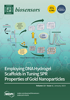
Gold nanoparticles’ (AuNPs) vivid intense color expressions are dependent on their physical properties, implying a strong influence on tuning the plasmonic properties. We have designed X-DNA base pair-controlled, size-varied Dgel scaffolds and molar ratio-based nano assemblies. The X-DNA-engineered nanoscale DNA hydrogel (Dgel) scaffolds are utilized to tune the plasmonic properties of AuNPs. The Dgel scaffolds are engineered with three different X-DNAs with varied numbers of base pairs. Dgel’s negatively charged scaffold eases positively charged AuNPs to flocculate around via electrostatic attractions. Here, we demonstrated that by altering the Dgel scaffolds and the physical properties of the nanoscale hydrogel matrix, plasmonic properties can be altered. This approach could potentially be a benefit for monitoring diverse therapeutic biomolecules. View this paper
  • Issues are regarded as officially published after their release is announced to the table of contents alert mailing list.
  • You may sign up for e-mail alerts to receive table of contents of newly released issues.
  • PDF is the official format for papers published in both, html and pdf forms. To view the papers in pdf format, click on the "PDF Full-text" link, and use the free Adobe Reader to open them.
Order results
Result details
Section
Select all
Export citation of selected articles as:
15 pages, 10515 KiB  
Article
Highly Heterogeneous Morphology of Cobalt Oxide Nanostructures for the Development of Sensitive and Selective Ascorbic Acid Non-Enzymatic Sensor
by Abdul Sattar Chang, Aneela Tahira, Fouzia Chang, Abdul Ghaffar Solangi, Muhammad Ali Bhatti, Brigitte Vigolo, Ayman Nafady and Zafar Hussain Ibupoto
Biosensors 2023, 13(1), 147; https://doi.org/10.3390/bios13010147 - 16 Jan 2023
Cited by 7 | Viewed by 2824
Abstract
The surface tailored metal oxide nanostructures for the development of non-enzymatic sensors are highly demanded, but it is a big task due to the wide range of complexities during the growth process. The presented study focused on the surface modification of the heterogeneous [...] Read more.
The surface tailored metal oxide nanostructures for the development of non-enzymatic sensors are highly demanded, but it is a big task due to the wide range of complexities during the growth process. The presented study focused on the surface modification of the heterogeneous morphology of cobalt oxide (Co3O4) prepared by the hydrothermal method. Further surface modification was conducted with the use of sodium citrate as a reducing and surface modifying agent for the Co3O4 nanostructures through the high density of oxygenated terminal groups from the citrate ions. The citrate ions enabled a significant surface modification of the Co3O4 nanostructures, which further improved the electrochemical properties of the Co3O4 material toward the design of the non-enzymatic ascorbic acid sensor in a phosphate buffer solution of pH 7.4. The morphology and crystal arrays of the Co3O4 nanostructures were studied by scanning electron microscopy (SEM) and powder X-ray diffraction (XRD) techniques. These physical characterizations showed the highly tailored surface features of Co3O4 nanostructures and a significant impact on the crystal properties. The electrochemical activity of Co3O4 was studied by chronoamperometry, linear sweep voltammetry, and cyclic voltammetry (CV) for the detection of ascorbic acid. The linear range of the proposed sensor was measured from 0.5 mM to 6.5 mM and a low limit of detection of 0.001 mM was also estimated. The presented Co3O4 nanostructures exhibited significant surface roughness and surface area, consequently playing a vital role toward the selective, sensitive, and stable detection of ascorbic acid. The use of a low cost surface modifying agent such as sodium citrate could be of great interest for the surface roughness and high surface area of nanostructured materials for the improved electrochemical properties for the biomedical, energy storage, and conversion systems. Full article
(This article belongs to the Special Issue Nanoengineering for Advanced Biosensors)
Show Figures

Figure 1

21 pages, 357 KiB  
Editorial
Acknowledgment to the Reviewers of Biosensors in 2022
by Biosensors Editorial Office
Biosensors 2023, 13(1), 146; https://doi.org/10.3390/bios13010146 - 16 Jan 2023
Viewed by 1995
Abstract
High-quality academic publishing is built on rigorous peer review [...] Full article
12 pages, 2367 KiB  
Article
Soil Microbial Fuel Cell Based Self-Powered Cathodic Biosensor for Sensitive Detection of Heavy Metals
by Shi-Hang Wang, Jian-Wei Wang, Li-Ting Zhao, Syed Zaghum Abbas, Zhugen Yang and Yang-Chun Yong
Biosensors 2023, 13(1), 145; https://doi.org/10.3390/bios13010145 - 15 Jan 2023
Cited by 3 | Viewed by 3034
Abstract
Soil microbial fuel cells (SMFCs) are an innovative device for soil-powered biosensors. However, the traditional SMFC sensors relied on anodic biosensing which might be unstable for long-term and continuous monitoring of toxic pollutants. Here, a carbon-felt-based cathodic SMFC biosensor was developed and applied [...] Read more.
Soil microbial fuel cells (SMFCs) are an innovative device for soil-powered biosensors. However, the traditional SMFC sensors relied on anodic biosensing which might be unstable for long-term and continuous monitoring of toxic pollutants. Here, a carbon-felt-based cathodic SMFC biosensor was developed and applied for soil-powered long-term sensing of heavy metal ions. The SMFC-based biosensor generated output voltage about 400 mV with the external load of 1000 Ω. Upon the injection of metal ions, the voltage of the SMFC was increased sharply and quickly reached a stable output within 2~5 min. The metal ions of Cd2+, Zn2+, Pb2+, or Hg2+ ranging from 0.5 to 30 mg/L could be quantified by using this SMFC biosensor. As the anode was immersed in the deep soil, this SMFC-based biosensor was able to monitor efficiently for four months under repeated metal ions detection without significant decrease on the output voltage. This finding demonstrated the clear potential of the cathodic SMFC biosensor, which can be further implemented as a low-cost self-powered biosensor. Full article
(This article belongs to the Section Environmental Biosensors and Biosensing)
Show Figures

Figure 1

14 pages, 3708 KiB  
Article
Zip Nucleic Acid-Based Genomagnetic Assay for Electrochemical Detection of microRNA-34a
by Arzum Erdem and Ece Eksin
Biosensors 2023, 13(1), 144; https://doi.org/10.3390/bios13010144 - 15 Jan 2023
Cited by 3 | Viewed by 2478
Abstract
Zip nucleic acid (ZNA)-based genomagnetic assay was developed herein for the electrochemical detection of microRNA-34a (miR-34a), which is related to neurological disorders and cancer. The hybridization between the ZNA probe and miR-34a target was performed in the solution phase; then, the resultant hybrids [...] Read more.
Zip nucleic acid (ZNA)-based genomagnetic assay was developed herein for the electrochemical detection of microRNA-34a (miR-34a), which is related to neurological disorders and cancer. The hybridization between the ZNA probe and miR-34a target was performed in the solution phase; then, the resultant hybrids were immobilized onto the surface of magnetic beads (MBs). After magnetic separation, the hybrids were separated from the surface of MBs and then immobilized on the surface of pencil graphite electrodes (PGEs). In the case of a full-match hybridization, the guanine oxidation signal was measured via the differential pulse voltammetry (DPV) technique. All the experimental parameters that influenced the hybridization efficiency (i.e., hybridization strategy, probe concentration, hybridization temperature, etc.) were optimized. The cross-selectivity of the genomagnetic assay was tested against two different miRNAs, miR-155 and miR-181b, individually as well as in mixture samples. To show the applicability of the ZNA-based genomagnetic assay for miR-34a detection in real samples, a batch of experiments was carried out in this study by using the total RNA samples isolated from the human hepatocellular carcinoma cell line (HUH-7). Full article
Show Figures

Graphical abstract

12 pages, 3611 KiB  
Communication
Dual-Emission Carbon-Dot Ratiometric Fluorescence Sensor for Morphine Recognition in Biological Samples
by Qinhong Yin, Yijie Wang, Xuerong Li, Dezhi Yang, Yaling Yang, Cheng Yang and Yanqin Zhu
Biosensors 2023, 13(1), 143; https://doi.org/10.3390/bios13010143 - 15 Jan 2023
Cited by 4 | Viewed by 1581
Abstract
Herein, a novel nitr[ogen-doped carbon dot (N-CD) fluorescence sensor with a dual emission ratio is developed using the microwave-assisted synthesis of m-phenylenediamine and spermidine. As a result of the fluorescence inner filtration effect (IFE) effect between morphine (MOR) and N-CD, the blue fluorescence [...] Read more.
Herein, a novel nitr[ogen-doped carbon dot (N-CD) fluorescence sensor with a dual emission ratio is developed using the microwave-assisted synthesis of m-phenylenediamine and spermidine. As a result of the fluorescence inner filtration effect (IFE) effect between morphine (MOR) and N-CD, the blue fluorescence of N-CDs at 350 nm was reduced in the presence of MOR, whereas the fluorescence of N-CDs at 456 nm increased substantially. The results demonstrated that the approach has a tremendous potential and that the linear range of MOR detection is 0.25–25 µg/mL, with a 71.8 ng/mL detection limit. Under UV light, the blue fluorescent system is easily visible to the naked eye. More significantly, the sensor proved successful in providing satisfactory results for the speciation measurement of MOR in a variety of biological samples. Full article
(This article belongs to the Section Optical and Photonic Biosensors)
Show Figures

Figure 1

30 pages, 3266 KiB  
Review
Review of Bacterial Nanocellulose-Based Electrochemical Biosensors: Functionalization, Challenges, and Future Perspectives
by Samuel Chagas de Assis, Daniella Lury Morgado, Desiree Tamara Scheidt, Samara Silva de Souza, Marco Roberto Cavallari, Oswaldo Hideo Ando Junior and Emanuel Carrilho
Biosensors 2023, 13(1), 142; https://doi.org/10.3390/bios13010142 - 14 Jan 2023
Cited by 10 | Viewed by 3104
Abstract
Electrochemical biosensing devices are known for their simple operational procedures, low fabrication cost, and suitable real-time detection. Despite these advantages, they have shown some limitations in the immobilization of biochemicals. The development of alternative materials to overcome these drawbacks has attracted significant attention. [...] Read more.
Electrochemical biosensing devices are known for their simple operational procedures, low fabrication cost, and suitable real-time detection. Despite these advantages, they have shown some limitations in the immobilization of biochemicals. The development of alternative materials to overcome these drawbacks has attracted significant attention. Nanocellulose-based materials have revealed valuable features due to their capacity for the immobilization of biomolecules, structural flexibility, and biocompatibility. Bacterial nanocellulose (BNC) has gained a promising role as an alternative to antifouling surfaces. To widen its applicability as a biosensing device, BNC may form part of the supports for the immobilization of specific materials. The possibilities of modification methods and in situ and ex situ functionalization enable new BNC properties. With the new insights into nanoscale studies, we expect that many biosensors currently based on plastic, glass, or paper platforms will rely on renewable platforms, especially BNC ones. Moreover, substrates based on BNC seem to have paved the way for the development of sensing platforms with minimally invasive approaches, such as wearable devices, due to their mechanical flexibility and biocompatibility. Full article
Show Figures

Figure 1

18 pages, 3020 KiB  
Article
NIR Luminescent Oxygen-Sensing Nanoparticles for Continuous Glucose and Lactate Monitoring
by Ananthakrishnan Soundaram Jeevarathinam, Waqas Saleem, Nya Martin, Connie Hu and Michael J. McShane
Biosensors 2023, 13(1), 141; https://doi.org/10.3390/bios13010141 - 14 Jan 2023
Cited by 2 | Viewed by 2563
Abstract
A highly sensitive, biocompatible, and scalable phosphorescent oxygen sensor formulation is designed and evaluated for use in continuous metabolite sensors for biological systems. Ethyl cellulose (EC) and polystyrene (PS) nanoparticles (NPs) stabilized with Pluronic F68 (PF 68), Polydimethylsiloxane-b-polyethyleneglycol methyl ether (PDMS-PEG), [...] Read more.
A highly sensitive, biocompatible, and scalable phosphorescent oxygen sensor formulation is designed and evaluated for use in continuous metabolite sensors for biological systems. Ethyl cellulose (EC) and polystyrene (PS) nanoparticles (NPs) stabilized with Pluronic F68 (PF 68), Polydimethylsiloxane-b-polyethyleneglycol methyl ether (PDMS-PEG), sodium dodecylsulfate (SDS), and cetyltimethylammonium bromide (CTAB) were prepared and studied. The resulting NPs with eight different surfactant–polymer matrix combinations were evaluated for physical properties, oxygen sensitivity, effect of changes in dispersion matrix, and cytotoxicity. The EC NPs exhibited a narrower size distribution and 40% higher sensitivity than PS, with Stern–Volmer constants (Ksv) 0.041–0.052 µM−1 for EC, compared to 0.029–0.034 µM−1 for PS. Notably, ethyl cellulose NPs protected with PF68 were selected as the preferred formulation, as they were not cytotoxic towards 3T3 fibroblasts and exhibited a wide phosphorescence lifetime response of >211.1 µs over 258–0 µM and ~100 µs over 2.58–0 µM oxygen, with a limit of detection (LoD) of oxygen in aqueous phase of 0.0016 µM. The EC-PF68 NPs were then efficiently encapsulated in alginate microparticles along with glucose oxidase (GOx) and catalase (CAT) to form phosphorescent nanoparticles-in-microparticle (NIMs) glucose sensing microdomains. The fabricated glucose sensors showed a sensitivity of 0.40 µs dL mg−1 with a dynamic phosphorescence lifetime range of 46.6–197.1 µs over 0–150 mg dL−1 glucose, with a glucose LoD of 18.3 mg dL−1 and maximum distinguishable concentration of 111.1 mg dL−1. Similarly, lactate sensors were prepared with NIMs microdomains containing lactate oxidase (LOx) and found to have a detection range of 0–14 mg dL−1 with LoD of 1.8 mg dL−1 and maximum concentration of 13.7 mg dL−1 with lactate sensitivity of 10.7 µs dL mg−1. Owing to its versatility, the proposed NIMs-based design can be extended to a wide range of metabolites and different oxygen-sensing dyes with different excitation wavelengths based on specific application. Full article
(This article belongs to the Special Issue New Biosensors and Nanosensors)
Show Figures

Graphical abstract

34 pages, 5210 KiB  
Review
Recent Progress in Electrochemical Nano-Biosensors for Detection of Pesticides and Mycotoxins in Foods
by Zhaoyuan Gong, Yueming Huang, Xianjing Hu, Jianye Zhang, Qilei Chen and Hubiao Chen
Biosensors 2023, 13(1), 140; https://doi.org/10.3390/bios13010140 - 14 Jan 2023
Cited by 12 | Viewed by 3326
Abstract
Pesticide and mycotoxin residues in food are concerning as they are harmful to human health. Traditional methods, such as high-performance liquid chromatography (HPLC) for such detection lack sensitivity and operation convenience. Efficient, accurate detection approaches are needed. With the recent development of nanotechnology, [...] Read more.
Pesticide and mycotoxin residues in food are concerning as they are harmful to human health. Traditional methods, such as high-performance liquid chromatography (HPLC) for such detection lack sensitivity and operation convenience. Efficient, accurate detection approaches are needed. With the recent development of nanotechnology, electrochemical biosensors based on nanomaterials have shown solid ability to detect trace pesticides and mycotoxins quickly and accurately. In this review, English articles about electrochemical biosensors in the past 11 years (2011–2022) were collected from PubMed database, and various nanomaterials are discussed, including noble metal nanomaterials, magnetic metal nanoparticles, metal–organic frameworks, carbon nanotubes, as well as graphene and its derivatives. Three main roles of such nanomaterials in the detection process are summarized, including biomolecule immobilization, signal generation, and signal amplification. The detection targets involve two types of pesticides (organophosphorus and carbamate) and six types of mycotoxins (aflatoxin, deoxynivalenol, zearalenone, fumonisin, ochratoxin A, and patulin). Although significant achievements have been made in the evolution of electrochemical nano-biosensors, many challenges remain to be overcome. Full article
Show Figures

Figure 1

19 pages, 3247 KiB  
Article
On-Chip Electromembrane Surrounded Solid Phase Microextraction for Determination of Tricyclic Antidepressants from Biological Fluids Using Poly(3,4-ethylenedioxythiophene)—Graphene Oxide Nanocomposite as a Fiber Coating
by Razieh Zamani and Yadollah Yamini
Biosensors 2023, 13(1), 139; https://doi.org/10.3390/bios13010139 - 14 Jan 2023
Cited by 6 | Viewed by 1819
Abstract
In the present study, on-chip electromembrane surrounded solid phase microextraction (EM-SPME) was employed in the determination of tricyclic antidepressants (TCAs), including amitriptyline, nortriptyline, imipramine, desipramine, maprotiline, and sertraline, from various biological fluids. In this regard, poly(3,4-ethylenedioxythiophene)–graphene oxide (PEDOT-GO) was electrodeposited on an SPME [...] Read more.
In the present study, on-chip electromembrane surrounded solid phase microextraction (EM-SPME) was employed in the determination of tricyclic antidepressants (TCAs), including amitriptyline, nortriptyline, imipramine, desipramine, maprotiline, and sertraline, from various biological fluids. In this regard, poly(3,4-ethylenedioxythiophene)–graphene oxide (PEDOT-GO) was electrodeposited on an SPME fiber as a conductive coating, then the fiber played the acceptor-electrode role during the extraction. Thus, the immigration of the analytes under the influence of an electric field and their absorption onto the fiber coating were accomplished simultaneously. Under the optimized conditions, the limits of detection for the target analytes were acquired in the range of 0.005–0.025 µg L−1 using gas chromatography–mass spectrometry. The linearity of the method was 0.010–500 µg L−1 for the imipramine and sertraline, 0.025–500 µg L−1 for the amitriptyline, nortriptyline, and desipramine, and 1.000–250 µg L−1 for the maprotiline (R2 ≥ 0.9984). Moreover, this method provided suitable precision and fiber-to-fiber reproducibility, with RSDs ≤ 8.4%. The applicability of the proposed setup was eventually investigated for extraction of the drugs from human bone marrow aspirate, urine, plasma, and well water samples, in which satisfactory relative recoveries, from 93–105%, were obtained. Full article
(This article belongs to the Special Issue Lab on a Chip for High-Throughput Drug Screening)
Show Figures

Figure 1

11 pages, 2204 KiB  
Article
CRISPR/Cas12a-Assisted Dual Visualized Detection of SARS-CoV-2 on Frozen Shrimps
by Siwenjie Qian, Yanju Chen, Xiaofu Wang, Tingzhang Wang, Yang Che, Jian Wu, Zhangying Ye and Junfeng Xu
Biosensors 2023, 13(1), 138; https://doi.org/10.3390/bios13010138 - 14 Jan 2023
Cited by 2 | Viewed by 1795
Abstract
Given the possibility that food contaminated with SARS-CoV-2 might become an infection source, there is an urgent need for us to develop a rapid and accurate nucleic acid detection method for SARS-CoV-2 in food to ensure food safety. Here, we propose a sensitive, [...] Read more.
Given the possibility that food contaminated with SARS-CoV-2 might become an infection source, there is an urgent need for us to develop a rapid and accurate nucleic acid detection method for SARS-CoV-2 in food to ensure food safety. Here, we propose a sensitive, specific, and reliable molecular detection method for SARS-CoV-2. It has a mechanism to control amplicon contamination. Swabs from spiked frozen shrimps were used as detection samples, which were processed by heating at 95 °C for 30 s. These preprocessed samples served as the templates for subsequent amplification. A colorimetric LAMP reaction was carried out to amplify both the SARS-CoV-2 target and the MS2 phage simultaneously in one tube. MS2 phage was detected by colorimetric LAMP as the internal control, while SARS-CoV-2 was detected with a CRISPR/Cas12a system. The fluorescence results could be visually detected with an ultraviolet lamp. Meanwhile, uracil was incorporated during the LAMP reaction to provide an amplicon contamination proof mechanism. This test could detect as low as 20 copies of SARS-CoV-2 in one reaction. Additionally, the detection could be finished in 45 min. The test only needs a heating block and an ultraviolet lamp, which shows the potential for field detection. Full article
(This article belongs to the Special Issue CRISPR-Cas Based Molecular Diagnostics and Their Applications)
Show Figures

Figure 1

26 pages, 4410 KiB  
Review
Semiconducting Polymer Dots for Point-of-Care Biosensing and In Vivo Bioimaging: A Concise Review
by Sile Deng, Lingfeng Li, Jiaxi Zhang, Yongjun Wang, Zhongchao Huang and Haobin Chen
Biosensors 2023, 13(1), 137; https://doi.org/10.3390/bios13010137 - 14 Jan 2023
Cited by 9 | Viewed by 2940
Abstract
In recent years, semiconducting polymer dots (Pdots) have attracted much attention due to their excellent photophysical properties and applicability, such as large absorption cross section, high brightness, tunable fluorescence emission, excellent photostability, good biocompatibility, facile modification and regulation. Therefore, Pdots have been widely [...] Read more.
In recent years, semiconducting polymer dots (Pdots) have attracted much attention due to their excellent photophysical properties and applicability, such as large absorption cross section, high brightness, tunable fluorescence emission, excellent photostability, good biocompatibility, facile modification and regulation. Therefore, Pdots have been widely used in various types of sensing and imaging in biological medicine. More importantly, the recent development of Pdots for point-of-care biosensing and in vivo imaging has emerged as a promising class of optical diagnostic technologies for clinical applications. In this review, we briefly outline strategies for the preparation and modification of Pdots and summarize the recent progress in the development of Pdots-based optical probes for analytical detection and biomedical imaging. Finally, challenges and future developments of Pdots for biomedical applications are given. Full article
Show Figures

Figure 1

23 pages, 2138 KiB  
Review
Microfluidic Gut-on-a-Chip: Fundamentals and Challenges
by Dimple Palanilkunnathil Thomas, Jun Zhang, Nam-Trung Nguyen and Hang Thu Ta
Biosensors 2023, 13(1), 136; https://doi.org/10.3390/bios13010136 - 13 Jan 2023
Cited by 15 | Viewed by 7122
Abstract
The human gut is responsible for food digestion and absorption. Recently, growing evidence has shown its vital role in the proper functioning of other organs. Advances in microfluidic technologies have made a significant impact on the biomedical field. Specifically, organ-on-a-chip technology (OoC), which [...] Read more.
The human gut is responsible for food digestion and absorption. Recently, growing evidence has shown its vital role in the proper functioning of other organs. Advances in microfluidic technologies have made a significant impact on the biomedical field. Specifically, organ-on-a-chip technology (OoC), which has become a popular substitute for animal models, is capable of imitating complex systems in vitro and has been used to study pathology and pharmacology. Over the past decade, reviews published focused more on the applications and prospects of gut-on-a-chip (GOC) technology, but the challenges and solutions to these limitations were often overlooked. In this review, we cover the physiology of the human gut and review the engineering approaches of GOC. Fundamentals of GOC models including materials and fabrication, cell types, stimuli and gut microbiota are thoroughly reviewed. We discuss the present GOC model applications, challenges, possible solutions and prospects for the GOC models and technology. Full article
Show Figures

Figure 1

18 pages, 4383 KiB  
Article
Non-Destructive and Non-Invasive Measurement of Ethanol and Toxic Alcohol Strengths in Beverages and Spirits Using Portable Raman Spectroscopy
by Panagiota Papaspyridakou, Panagiota Giannoutsou and Malvina G. Orkoula
Biosensors 2023, 13(1), 135; https://doi.org/10.3390/bios13010135 - 13 Jan 2023
Cited by 3 | Viewed by 2509
Abstract
The measurement of ethanol and toxic alcohol (methanol and isopropanol) strengths in beverages and spirits is crucial for health reasons but also for the identification of adulterated products. Many methodologies have been reported in the literature, based mainly on chromatographic and on spectroscopic [...] Read more.
The measurement of ethanol and toxic alcohol (methanol and isopropanol) strengths in beverages and spirits is crucial for health reasons but also for the identification of adulterated products. Many methodologies have been reported in the literature, based mainly on chromatographic and on spectroscopic techniques. Chromatographic techniques are laborious and time-consuming, while spectroscopic techniques are rapid and need no special sample pretreatment. All techniques were only applied to off-line or at-line manner. In the present work, Raman spectroscopy was used for fast and non-destructive measurements. A “through the container” method was developed for a non-invasive analysis, i.e., analysis without unsealing the bottles. This method, coupled with a miniature portable Raman, can serve for in-line measurements in a production line. The optimum laser focus for maximum spirit signal and minimum glass-wall signal was investigated. Calibration curves for the alcohols of interest were constructed and validated. The limits of detections were calculated and proved to be lower than the legitimate values. The influences of the liquor color and the bottle color, shape, and thickness were checked. Twenty-eight alcoholic products were studied. The concentrations found were compared against the nominal values (from the bottle labels). Full article
(This article belongs to the Special Issue Advances in Optical Biosensors and Chemical Sensors)
Show Figures

Figure 1

16 pages, 9117 KiB  
Article
Development of a Wearable Ultrasound Transducer for Sensing Muscle Activities in Assistive Robotics Applications
by Xiangming Xue, Bohua Zhang, Sunho Moon, Guo-Xuan Xu, Chih-Chung Huang, Nitin Sharma and Xiaoning Jiang
Biosensors 2023, 13(1), 134; https://doi.org/10.3390/bios13010134 - 13 Jan 2023
Cited by 13 | Viewed by 3989
Abstract
Robotic prostheses and powered exoskeletons are novel assistive robotic devices for modern medicine. Muscle activity sensing plays an important role in controlling assistive robotics devices. Most devices measure the surface electromyography (sEMG) signal for myoelectric control. However, sEMG is an integrated signal from [...] Read more.
Robotic prostheses and powered exoskeletons are novel assistive robotic devices for modern medicine. Muscle activity sensing plays an important role in controlling assistive robotics devices. Most devices measure the surface electromyography (sEMG) signal for myoelectric control. However, sEMG is an integrated signal from muscle activities. It is difficult to sense muscle movements in specific small regions, particularly at different depths. Alternatively, traditional ultrasound imaging has recently been proposed to monitor muscle activity due to its ability to directly visualize superficial and at-depth muscles. Despite their advantages, traditional ultrasound probes lack wearability. In this paper, a wearable ultrasound (US) transducer, based on lead zirconate titanate (PZT) and a polyimide substrate, was developed for a muscle activity sensing demonstration. The fabricated PZT-5A elements were arranged into a 4 × 4 array and then packaged in polydimethylsiloxane (PDMS). In vitro porcine tissue experiments were carried out by generating the muscle activities artificially, and the muscle movements were detected by the proposed wearable US transducer via muscle movement imaging. Experimental results showed that all 16 elements had very similar acoustic behaviors: the averaged central frequency, −6 dB bandwidth, and electrical impedance in water were 10.59 MHz, 37.69%, and 78.41 Ω, respectively. The in vitro study successfully demonstrated the capability of monitoring local muscle activity using the prototyped wearable transducer. The findings indicate that ultrasonic sensing may be an alternative to standardize myoelectric control for assistive robotics applications. Full article
Show Figures

Figure 1

11 pages, 2028 KiB  
Article
Sandwich-Type Electrochemiluminescence Immunosensor Based on CDs@dSiO2 Nanoparticles as Nanoprobe and Co-Reactant
by A-Ling Chen, Xiao-Yan Wang, Qing Zhang, Ning Bao and Shou-Nian Ding
Biosensors 2023, 13(1), 133; https://doi.org/10.3390/bios13010133 - 13 Jan 2023
Cited by 4 | Viewed by 1766
Abstract
In general, co-reactants are essential in highly efficient electrochemiluminescence (ECL) systems. Traditional co-reactants are usually toxic, so it is necessary to develop new environmentally friendly co-reactants. In this work, carbon dots (CDs) were assembled with dendritic silica nanospheres (CDs@dSiO2 NPs) to form [...] Read more.
In general, co-reactants are essential in highly efficient electrochemiluminescence (ECL) systems. Traditional co-reactants are usually toxic, so it is necessary to develop new environmentally friendly co-reactants. In this work, carbon dots (CDs) were assembled with dendritic silica nanospheres (CDs@dSiO2 NPs) to form a co-reactant of Ru(bpy)32+. Subsequently, a sandwich immunosensor for detecting human chorionic gonadotropin (HCG) was constructed based on CDs@dSiO2 NPs as co-reactants, the nanoprobe loaded with the secondary antibody, and Ru(bpy)32+ as a luminophore. In addition, compared to directly as a signal probe, the luminophore Ru (bpy)32+ as a part of the electrolyte solution is simpler in this work. The immunosensor has an extremely low limit of detection of 0.00019 mIU/mL. This work describes the synthesis of low-toxic, efficient, and environmentally friendly CDs, which have become ideal co-reactants of Ru(bpy)32+, and proposes an ECL immunosensor with excellent stability and selectivity, which has great potential in clinical applications. Full article
Show Figures

Figure 1

11 pages, 11137 KiB  
Article
High-Performance Nitric Oxide Gas Sensors Based on an Ultrathin Nanoporous Poly(3-hexylthiophene) Film
by Ganghoon Jeong, Seo Young Shin, Proscovia Kyokunzire, Hyeong Jun Cheon, Eunsol Wi, Minhong Woo and Mincheol Chang
Biosensors 2023, 13(1), 132; https://doi.org/10.3390/bios13010132 - 13 Jan 2023
Cited by 2 | Viewed by 2710
Abstract
Conjugated polymer (CP)-based organic field-effect transistors (OFETs) have been considered a potential sensor platform for detecting gas molecules because they can amplify sensing signals by controlling the gate voltage. However, these sensors exhibit significantly poorer oxidizing gas sensing performance than their inorganic counterparts. [...] Read more.
Conjugated polymer (CP)-based organic field-effect transistors (OFETs) have been considered a potential sensor platform for detecting gas molecules because they can amplify sensing signals by controlling the gate voltage. However, these sensors exhibit significantly poorer oxidizing gas sensing performance than their inorganic counterparts. This paper presents a high-performance nitric oxide (NO) OFET sensor consisting of a poly(3-hexylthiophene) (P3HT) film with an ultrathin nanoporous structure. The ultrathin nonporous structure of the P3HT film was created via deposition through the shear-coating-assisted phase separation of polymer blends and selective solvent etching. The ultrathin nonporous structure of the P3HT film enhanced NO gas diffusion, adsorption, and desorption, resulting in the ultrathin nanoporous P3HT-film-based OFET gas sensor exhibiting significantly better sensing performance than pristine P3HT-film-based OFET sensors. Additionally, upon exposure to 10 ppm NO at room temperature, the nanoporous P3HT-film-based OFET gas sensor exhibited significantly better sensing performance (i.e., responsivity ≈ 42%, sensitivity ≈ 4.7% ppm−1, limit of detection ≈ 0.5 ppm, and response/recovery times ≈ 6.6/8.0 min) than the pristine P3HT-film-based OFET sensors. Full article
(This article belongs to the Special Issue Recent Advances in Nanomaterial-Enhanced Gas Sensing)
Show Figures

Graphical abstract

15 pages, 4133 KiB  
Article
A Flexible Pressure Sensor Based on Silicon Nanomembrane
by Lixia Cheng, Xiaojian Hao, Guochang Liu, Wendong Zhang, Jiangong Cui, Guojun Zhang, Yuhua Yang and Renxin Wang
Biosensors 2023, 13(1), 131; https://doi.org/10.3390/bios13010131 - 12 Jan 2023
Cited by 9 | Viewed by 3272
Abstract
With advances in new materials and technologies, there has been increasing research focused on flexible sensors. However, in most flexible pressure sensors made using new materials, it is challenging to achieve high detection sensitivity across a wide pressure range. Although traditional silicon-based sensors [...] Read more.
With advances in new materials and technologies, there has been increasing research focused on flexible sensors. However, in most flexible pressure sensors made using new materials, it is challenging to achieve high detection sensitivity across a wide pressure range. Although traditional silicon-based sensors have good performance, they are not formable and, because of their rigidity and brittleness, they are not suitable for fitting with soft human skin, which limits their application in wearable devices to collect various signals. Silicon nanomembranes are ultra-thin, flexible materials with excellent piezoresistive properties, and they can be applied in various fields, such as in soft robots and flexible devices. In this study, we developed a flexible pressure sensor based on the use of silicon nanomembranes (with a thickness of only 340 nm) as piezoresistive units, which were transferred onto a flexible polydimethylsiloxane (PDMS) substrate. The flexible pressure sensor operated normally in the range of 0–200 kPa, and the sensitivity of the sensor reached 0.0185 kPa−1 in the low-pressure range of 0–5 kPa. In the high-pressure range of 5–200 kPa, the sensitivity of the sensor was maintained at 0.0023 kPa−1. The proposed sensor exhibited a fast response and excellent long-term stability and could recognize human movements, such as the bending of fingers and wrist joints, while maintaining a stable output. Thus, the developed flexible pressure sensor has promising applications in body monitoring and wearable devices. Full article
(This article belongs to the Section Wearable Biosensors)
Show Figures

Figure 1

12 pages, 9412 KiB  
Article
In Situ Deposition of Gold Nanoparticles and L-Cysteine on Screen-Printed Carbon Electrode for Rapid Electrochemical Determination of As(III) in Water and Tea
by Wenjing Wang, Zhijian Yi, Qiongxin Liang, Junjie Zhen, Rui Wang, Mei Li, Lingwen Zeng and Yongfang Li
Biosensors 2023, 13(1), 130; https://doi.org/10.3390/bios13010130 - 12 Jan 2023
Cited by 4 | Viewed by 2408
Abstract
In this study, a screen-printed carbon electrode (SPCE) based on in situ deposition modification was developed for the sensitive, rapid, easy and convenient determination of As(III) in water and tea by linear sweep anodic stripping voltammetry (LSASV). The screen-printed carbon electrodes were placed [...] Read more.
In this study, a screen-printed carbon electrode (SPCE) based on in situ deposition modification was developed for the sensitive, rapid, easy and convenient determination of As(III) in water and tea by linear sweep anodic stripping voltammetry (LSASV). The screen-printed carbon electrodes were placed in a solution consisting of As(III) solution, chlorauric acid and L-cysteine. Under certain electrical potential, the chloroauric acid was reduced to gold nanoparticles (AuNPs) on the SPCE. L-cysteine was self-assembled onto AuNPs and promoted the enrichment of As(III), thus enhancing the determination specificity and sensitivity of As(III). The method achieved a limit of determination (LOD) of 0.91 ppb (µg L−1), a linear range of 1~200 µg L−1, an inter-assay coefficient of variation of 5.3% and good specificity. The developed method was successfully applied to the determination of As(III) in tap water and tea samples, with a recovery rate of 93.8%~105.4%, and further validated by inductively coupled plasma mass spectrometry (ICP-MS). The developed method is rapid, convenient and accurate, holding great promise in the on-site determination of As(III) in tap water and tea leaves, and it can be extended to the detection of other samples. Full article
Show Figures

Figure 1

22 pages, 4791 KiB  
Review
Current Advances in Technologies for Single Extracellular Vesicle Analysis and Its Clinical Applications in Cancer Diagnosis
by Lei Qiu, Xingzhu Liu, Libo Zhu, Liqiang Luo, Na Sun and Renjun Pei
Biosensors 2023, 13(1), 129; https://doi.org/10.3390/bios13010129 - 12 Jan 2023
Cited by 4 | Viewed by 3304
Abstract
Extracellular vesicles (EVs) have been regarded as one of the most potential diagnostic biomarkers for different cancers, due to their unique physiological and pathological functions. However, it is still challenging to precisely analyze the contents and sources of EVs, due to their heterogeneity. [...] Read more.
Extracellular vesicles (EVs) have been regarded as one of the most potential diagnostic biomarkers for different cancers, due to their unique physiological and pathological functions. However, it is still challenging to precisely analyze the contents and sources of EVs, due to their heterogeneity. Herein, we summarize the advances in technologies for a single EV analysis, which may provide new strategies to study the heterogeneity of EVs, as well as their cargo, more specifically. Furthermore, the applications of a single EV analysis on cancer early diagnosis are also discussed. Full article
(This article belongs to the Special Issue Biosensing and Diagnosis of Cancer)
Show Figures

Figure 1

24 pages, 6270 KiB  
Review
Metal-Organic Frameworks-Based Optical Nanosensors for Analytical and Bioanalytical Applications
by Cong Wen, Rongsheng Li, Xiaoxia Chang and Na Li
Biosensors 2023, 13(1), 128; https://doi.org/10.3390/bios13010128 - 12 Jan 2023
Cited by 3 | Viewed by 2765
Abstract
Metal-organic frameworks (MOFs)-based optical nanoprobes for luminescence and surface-enhanced Raman spectroscopy (SERS) applications have been receiving tremendous attention. Every element in the MOF structure, including the metal nodes, the organic linkers, and the guest molecules, can be used as a source to build [...] Read more.
Metal-organic frameworks (MOFs)-based optical nanoprobes for luminescence and surface-enhanced Raman spectroscopy (SERS) applications have been receiving tremendous attention. Every element in the MOF structure, including the metal nodes, the organic linkers, and the guest molecules, can be used as a source to build single/multi-emission signals for the intended analytical purposes. For SERS applications, the MOF can not only be used directly as a SERS substrate, but can also improve the stability and reproducibility of the metal-based substrates. Additionally, the porosity and large specific surface area give MOF a sieving effect and target molecule enrichment ability, both of which are helpful for improving detection selectivity and sensitivity. This mini-review summarizes the advances of MOF-based optical detection methods, including luminescence and SERS, and also provides perspectives on future efforts. Full article
Show Figures

Graphical abstract

26 pages, 6550 KiB  
Review
Flexible Textile-Based Sweat Sensors for Wearable Applications
by Jing Yin, Jingcheng Li, Vundrala Sumedha Reddy, Dongxiao Ji, Seeram Ramakrishna and Lan Xu
Biosensors 2023, 13(1), 127; https://doi.org/10.3390/bios13010127 - 12 Jan 2023
Cited by 25 | Viewed by 6518
Abstract
The current physical health care system has gradually evolved into a form of virtual hospitals communicating with sensors, which can not only save time but can also diagnose a patient’s physical condition in real time. Textile-based wearable sensors have recently been identified as [...] Read more.
The current physical health care system has gradually evolved into a form of virtual hospitals communicating with sensors, which can not only save time but can also diagnose a patient’s physical condition in real time. Textile-based wearable sensors have recently been identified as detection platforms with high potential. They are developed for the real-time noninvasive detection of human physiological information to comprehensively analyze the health status of the human body. Sweat comprises various chemical compositions, which can be used as biomarkers to reflect the relevant information of the human physiology, thus providing references for health conditions. Combined together, textile-based sweat sensors are more flexible and comfortable than other conventional sensors, making them easily integrated into the wearable field. In this short review, the research progress of textile-based flexible sweat sensors was reviewed. Three mechanisms commonly used for textile-based sweat sensors were firstly contrasted with an introduction to their materials and preparation processes. The components of textile-based sweat sensors, which mainly consist of a sweat transportation channel and collector, a signal-selection unit, sensing elements and sensor integration and communication technologies, were reviewed. The applications of textile-based sweat sensors with different mechanisms were also presented. Finally, the existing problems and challenges of sweat sensors were summarized, which may contribute to promote their further development. Full article
(This article belongs to the Special Issue Artificial Skins and Wearable Biosensors for Healthcare Monitoring)
Show Figures

Figure 1

10 pages, 1387 KiB  
Article
Colorimetric Paper Sensor for Food Spoilage Based on Biogenic Amine Monitoring
by Maria Maddalena Calabretta, Denise Gregucci, Riccardo Desiderio and Elisa Michelini
Biosensors 2023, 13(1), 126; https://doi.org/10.3390/bios13010126 - 11 Jan 2023
Cited by 3 | Viewed by 4005
Abstract
Biogenic amines (BAs), nitrogenous molecules usually present in different foods, can be considered an indicator of freshness and food quality since their amount increases during food spoilage. Their detection, possibly in real time via the use of smart packaging, is therefore of crucial [...] Read more.
Biogenic amines (BAs), nitrogenous molecules usually present in different foods, can be considered an indicator of freshness and food quality since their amount increases during food spoilage. Their detection, possibly in real time via the use of smart packaging, is therefore of crucial importance to ensure food safety and to fulfill consumers’ demand. To this end, colorimetric sensors are considered one of the most feasible solutions. Here, we report a user-friendly colorimetric sensing paper able to detect BAs via the naked eye. The sensing molecule is the aglycone genipin, a natural cross-linking agent extracted from gardenia fruit, able to bind BAs producing water-soluble blue pigments. The paper sensor was applied to chicken meat quality monitoring and a quantitative analysis was performed with image acquisition via a smartphone camera, achieving a limit of detection equivalent to 0.1 mM of putrescine. The suitability of the BA sensing paper was assessed by integrating the sensor into smart packaging and analyzing commercial chicken meat samples stored at different temperatures; the results of the sensor paralleled the “best before date” indicated on the label, confirming the potential applicability of the sensor as a smart label. Full article
(This article belongs to the Special Issue Women in Biosensors)
Show Figures

Figure 1

24 pages, 4667 KiB  
Review
Recent Advances in Electrochemical Immunosensors with Nanomaterial Assistance for Signal Amplification
by Avinash V. Police Patil, Yu-Sheng Chuang, Chenzhong Li and Ching-Chou Wu
Biosensors 2023, 13(1), 125; https://doi.org/10.3390/bios13010125 - 11 Jan 2023
Cited by 27 | Viewed by 3846
Abstract
Electrochemical immunosensors have attracted immense attention due to the ease of mass electrode production and the high compatibility of the miniature electric reader, which is beneficial for developing point-of-care diagnostic devices. Electrochemical immunosensors can be divided into label-free and label-based sensing strategies equipped [...] Read more.
Electrochemical immunosensors have attracted immense attention due to the ease of mass electrode production and the high compatibility of the miniature electric reader, which is beneficial for developing point-of-care diagnostic devices. Electrochemical immunosensors can be divided into label-free and label-based sensing strategies equipped with potentiometric, amperometric, voltammetric, or impedimetric detectors. Emerging nanomaterials are frequently used on electrochemical immunosensors as a highly rough and conductive interface of the electrodes or on nanocarriers of immobilizing capture antibodies, electroactive mediators, or catalyzers. Adopting nanomaterials can increase immunosensor characteristics with lower detection limits and better sensitivity. Recent research has shown innovative immobilization procedures of nanomaterials which meet the requirements of different electrochemical immunosensors. This review discusses the past five years of advances in nanomaterials (metal nanoparticles, metal nanostructures, carbon nanotubes, and graphene) integrated into the electrochemical immunosensor. Furthermore, the new tendency and endeavors of nanomaterial-based electrochemical immunosensors are discussed. Full article
(This article belongs to the Special Issue Advanced Label-Free Electrochemical Affinity Biosensors)
Show Figures

Graphical abstract

15 pages, 4070 KiB  
Article
Plier Ligands for Trapping Neurotransmitters into Complexes for Sensitive Analysis by SERS Spectroscopy
by Olga E. Eremina, Olesya O. Kapitanova, Alexei V. Medved'ko, Alexandra S. Zelenetskaya, Bayirta V. Egorova, Tatyana N. Shekhovtsova, Sergey Z. Vatsadze and Irina A. Veselova
Biosensors 2023, 13(1), 124; https://doi.org/10.3390/bios13010124 - 11 Jan 2023
Cited by 4 | Viewed by 1700
Abstract
Catecholamines–dopamine, noradrenaline and adrenaline are important biomarkers of neurotransmitter metabolism, indicating neuroendocrine tumors and neurodegenerative diseases. Surface-enhanced Raman spectroscopy (SERS) is a promising analytical technique with unprecedented multiplexing capabilities. However, not all important analytes exhibit strong SERS signals on stable and robust nanostructured [...] Read more.
Catecholamines–dopamine, noradrenaline and adrenaline are important biomarkers of neurotransmitter metabolism, indicating neuroendocrine tumors and neurodegenerative diseases. Surface-enhanced Raman spectroscopy (SERS) is a promising analytical technique with unprecedented multiplexing capabilities. However, not all important analytes exhibit strong SERS signals on stable and robust nanostructured substrates. In this work, we propose a novel indicator system based on the formation of mixed ligand complexes with bispidine-based bis-azole ligands which can serve as pliers to trap Cu(II) ions and stabilize its complexes with catecholamines. Four synthesized ligands with different functional groups: carboxyl, amino, benzyl, and methoxybenzyl, were applied for forming stable complexes to shift maximum absorbance of catecholamines from the ultraviolet region to 570–600 nm. A new absorbance band in the visible range resonates with the local surface plasmon resonance (LSPR) band of metal nanoparticles and most used laser wavelengths. This match allowed use of Molecular Immobilization and Resonant Raman Amplification by Complex-Loaded Enhancers (MIRRACLE) methodology to measure intense Raman signals on a nanostructured silver-based SERS-active substrate. The synthesized plier-like ligands fixed and stabilized catecholamine complexes with Cu(II) on the SERS sensor surface, which facilitated the determination of dopamine in a 3.2 × 10⁻¹²–1 × 10⁻⁸ M concentration range. Full article
Show Figures

Figure 1

33 pages, 15061 KiB  
Review
2D Metal-Organic Frameworks: Properties, Synthesis, and Applications in Electrochemical and Optical Biosensors
by Anamika Ghosh, Sana Fathima Thanutty Kallungal and Sundara Ramaprabhu
Biosensors 2023, 13(1), 123; https://doi.org/10.3390/bios13010123 - 11 Jan 2023
Cited by 7 | Viewed by 5117
Abstract
Two-dimensional (2D) nanomaterials like graphene, layered double hydroxides, etc., have received increasing attention owing to their unique properties imparted by their 2D structure. The newest member in this family is based on metal-organic frameworks (MOFs), which have been long known for their exceptional [...] Read more.
Two-dimensional (2D) nanomaterials like graphene, layered double hydroxides, etc., have received increasing attention owing to their unique properties imparted by their 2D structure. The newest member in this family is based on metal-organic frameworks (MOFs), which have been long known for their exceptional physicochemical properties—high surface area, tunable pore size, catalytic properties, etc., to list a few. 2D MOFs are promising materials for various applications as they combine the exciting properties of 2D materials and MOFs. Recently, they have been extensively used in biosensors by virtue of their enormous surface area and abundant, accessible active sites. In this review, we provide a synopsis of the recent progress in the field of 2D MOFs for sensor applications. Initially, the properties and synthesis techniques of 2D MOFs are briefly outlined with examples. Further, electrochemical and optical biosensors based on 2D MOFs are summarized, and the associated challenges are outlined. Full article
Show Figures

Figure 1

11 pages, 2273 KiB  
Article
A Label-Free, Mix-and-Detect ssDNA-Binding Assay Based on Cationic Conjugated Polymers
by Pengbo Zhang, Mohamad Zandieh, Yuzhe Ding, Lyuyuan Wu, Xiaoyu Wang, Juewen Liu and Zhengping Li
Biosensors 2023, 13(1), 122; https://doi.org/10.3390/bios13010122 - 10 Jan 2023
Cited by 6 | Viewed by 2456
Abstract
The accurate, simple, and efficient measurement of the concentration of single-stranded DNA (ssDNA) is important for many analytical applications, such as DNA adsorption, biosensor design, and disease diagnosis, but it is still a challenge. Herein, we studied a cationic conjugated polymer (CCP)-based ssDNA [...] Read more.
The accurate, simple, and efficient measurement of the concentration of single-stranded DNA (ssDNA) is important for many analytical applications, such as DNA adsorption, biosensor design, and disease diagnosis, but it is still a challenge. Herein, we studied a cationic conjugated polymer (CCP)-based ssDNA assay taking advantage of the obvious fluorescence change of CCPs upon binding ssDNA. Poly(3-(3′-N,N,N-triethylamino-1′-propyloxy)-4-methyl-2,5-thiophene hydrochloride) (PMNT) achieved an apparent dissociation constant (Kd) of 57 ± 4 nM for ssDNA, indicating a very high binding affinity between PMNT and ssDNA. This allowed us to develop a CCP-based ssDNA biosensor with a detection limit of 0.6 nM, similar to the fluorescence-dye-based method using SYBR Green I and SYBR Gold. Our CCP-based biosensor produced smaller differences among ssDNA samples with different base compositions. In addition, the existence of double-stranded DNA (dsDNA) at different concentrations did not interfere with the fluorescence of PMNT, indicating that our CCP-based biosensor was more suitable for the measurement of ssDNA. Compared with fluorescence-intensity-based quantification, our CCP system allowed ratiometric quantification, which made the calibration easier and more robust. We then applied our method to the quantification of ssDNA on AuNPs using both unmodified and thiolated ssDNA, and the accurate quantification of ssDNA was achieved without any fluorophore modification. This method provides an alternative approach for the measurement of ssDNA. Full article
(This article belongs to the Special Issue Current Trends in Polymer-Based Biosensors)
Show Figures

Figure 1

23 pages, 4555 KiB  
Article
In Situ Investigation of Upper Airway Occlusion in Sleep Disordered Breathing Using Ultrasonic Transducer Arrays
by Mohammad Al-Abed, Donald Watenpaugh and Khosrow Behbehani
Biosensors 2023, 13(1), 121; https://doi.org/10.3390/bios13010121 - 10 Jan 2023
Viewed by 1534
Abstract
This work presents a novel application of ultrasound for the real-time, non-invasive investigation of occlusion of the upper airway during events of obstructive sleep apnea/hypopnea syndrome. It is hypothesized that ultrasonic pulses applied to the neck during apneic events produce spectral and temporal [...] Read more.
This work presents a novel application of ultrasound for the real-time, non-invasive investigation of occlusion of the upper airway during events of obstructive sleep apnea/hypopnea syndrome. It is hypothesized that ultrasonic pulses applied to the neck during apneic events produce spectral and temporal features that can detect apnea occurrence. Theoretical models of ultrasound propagation and an in vitro test were conducted to test this hypothesis in both transmission and reflection modes. Complete specifications and technical details of the system design and fabrication, which is mounted on each subject’s neck, are presented, including the methodology. Nine patients (seven male and two female, mean age of 42 years, with a range of 25 to 56 years, and body mass index 37.6 ± 6.6 kg/m2) were recruited for a full night study, which included simultaneous nocturnal polysomnography for the validation of the results. Nine temporal features and four spectral features were extracted from the envelope of the received pulse waveform. These were used to compute 26 metrics to quantify the changes in the ultrasonic waveforms between normal breathing and apneic events. The statistical analysis of the collected ultrasonic data showed that at least two or more of the proposed features could detect apneic events in all subjects. The findings establish the feasibility of the proposed method as a cost-effective and non-invasive OSAHS screening tool. Full article
Show Figures

Figure 1

16 pages, 2964 KiB  
Article
Point-of-Care Portable 3D-Printed Multispectral Sensor for Real-Time Enzyme Activity Monitoring in Healthcare Applications
by Antony Jesuraj and Umer Hassan
Biosensors 2023, 13(1), 120; https://doi.org/10.3390/bios13010120 - 10 Jan 2023
Cited by 2 | Viewed by 1627
Abstract
Absorbance spectroscopy finds many biomedical and physical applications ranging from studying the atomic and molecular details of the analyte to determination of unknown biological species and their concentration or activity in the samples. Commercially available laboratory-based spectrometers are usually bulky and require high [...] Read more.
Absorbance spectroscopy finds many biomedical and physical applications ranging from studying the atomic and molecular details of the analyte to determination of unknown biological species and their concentration or activity in the samples. Commercially available laboratory-based spectrometers are usually bulky and require high power and laborious manual processing, making them unsuitable to be deployed in portable and space-constrained environments, thereby further limiting their utility for real-time on-site monitoring. To address these challenges, here we developed a portable 3D-printed multispectral spectrophotometer based on absorbance spectroscopy for real-time monitoring of enzyme molecular activity. Monitoring enzyme (such as tyrosinase) activity is critical, as it quantifies its reaction rate, which is dependent on many factors such as the enzyme and substrate concentrations, temperature, pH, and other regulators such as inhibitors and effectors. Tyrosinase is a critical enzyme responsible for melanin synthesis in living beings and exhibits enzymatic browning in fruits and vegetables. It finds various commercial applications in the fields of healthcare (skin pigmentation, wound healing, etc.), forensics, and food processing. Here, tyrosinase activity was monitored using a 3D-printed spectral sensor at different rates and compared against measurements obtained from laboratory instruments. The enzyme activity was also studied using kojic acid (i.e., a commonly employed commercial tyrosinase inhibitor) while varying its molar and volume concentrations to control the reaction rate at discrete activity levels. For tyrosinase activity monitoring, the fabricated device has shown significant correlation (R2 = 0.9999) compared to measurements from the standard table-top spectrophotometer. We also provide a performance comparison between the 3D-printed and the laboratory spectrophotometer instruments by studying tyrosinase enzyme activity with and without the influence of an inhibitor. Such a device can be translated into various absorbance spectroscopy-based point-of-care biomedical and healthcare applications. Full article
(This article belongs to the Special Issue Feature Issue of Biosensors and Healthcare Section)
Show Figures

Figure 1

11 pages, 1566 KiB  
Article
Multiplexed smFRET Nucleic Acid Sensing Using DNA Nanotweezers
by Anisa Kaur, Roaa Mahmoud, Anoja Megalathan, Sydney Pettit and Soma Dhakal
Biosensors 2023, 13(1), 119; https://doi.org/10.3390/bios13010119 - 10 Jan 2023
Cited by 4 | Viewed by 2169
Abstract
The multiplexed detection of disease biomarkers is part of an ongoing effort toward improving the quality of diagnostic testing, reducing the cost of analysis, and accelerating the treatment processes. Although significant efforts have been made to develop more sensitive and rapid multiplexed screening [...] Read more.
The multiplexed detection of disease biomarkers is part of an ongoing effort toward improving the quality of diagnostic testing, reducing the cost of analysis, and accelerating the treatment processes. Although significant efforts have been made to develop more sensitive and rapid multiplexed screening methods, such as microarrays and electrochemical sensors, their limitations include their intricate sensing designs and semi-quantitative detection capabilities. Alternatively, fluorescence resonance energy transfer (FRET)-based single-molecule counting offers great potential for both the sensitive and quantitative detection of various biomarkers. However, current FRET-based multiplexed sensing typically requires the use of multiple excitation sources and/or FRET pairs, which complicates labeling schemes and the post-analysis of data. We present a nanotweezer (NT)-based sensing strategy that employs a single FRET pair and is capable of detecting multiple targets. Using DNA mimics of miRNA biomarkers specific to triple-negative breast cancer (TNBC), we demonstrated that the developed sensors are sensitive down to the low picomolar range (≤10 pM) and can discriminate between targets with a single-base mismatch. These simple hybridization-based sensors hold great promise for the sensitive detection of a wider spectrum of nucleic acid biomarkers. Full article
(This article belongs to the Special Issue Devices and Wearable Devices toward Innovative Applications)
Show Figures

Figure 1

13 pages, 2233 KiB  
Article
A Novel and Label-Free Chemiluminescence Detection of Zearalenone Based on a Truncated Aptamer Conjugated with a G-Quadruplex DNAzyme
by Yue Guan, Junning Ma, Jing Neng, Bolei Yang, Yan Wang and Fuguo Xing
Biosensors 2023, 13(1), 118; https://doi.org/10.3390/bios13010118 - 9 Jan 2023
Cited by 4 | Viewed by 2131
Abstract
Zearalenone (ZEN), one of the most frequently occurring mycotoxin contaminants in foods and feeds, poses considerable threat to human and animal health, owing to its acute and chronic toxicities. Thus, rapid and accurate detection of ZEN has attracted broad research interest. In this [...] Read more.
Zearalenone (ZEN), one of the most frequently occurring mycotoxin contaminants in foods and feeds, poses considerable threat to human and animal health, owing to its acute and chronic toxicities. Thus, rapid and accurate detection of ZEN has attracted broad research interest. In this work, a novel and label-free chemiluminescence aptasensor based on a ZEN aptamer and a G-quadruplex DNAzyme was constructed. It was established on a competitive assay between ZEN and an auxiliary DNA for the aptamer, leading to activation of the G-quadruplex/hemin DNAzyme and subsequent signal amplification by chemiluminescence generation after substrate addition. To maximize the detection sensitivity, numerous key parameters including truncated aptamers were optimized with molecular docking analysis. Upon optimization, our aptasensor exhibited a perfect linear relationship (R2 = 0.9996) for ZEN detection in a concentration range of 1–100 ng/mL (3.14–314.10 nM) within 40 min, achieving a detection limit of 2.85 ng/mL (8.95 nM), which was a 6.7-fold improvement over that before optimization. Most importantly, the aptasensor obtained a satisfactory recovery rate of 92.84–137.27% and 84.90–124.24% for ZEN-spiked wheat and maize samples, respectively. Overall, our label-free chemiluminescence aptasensor displayed simplicity, sensitivity, specificity and practicality in real samples, indicating high application prospects in the food supply chain for rapid detection of ZEN. Full article
Show Figures

Figure 1

Previous Issue
Next Issue
Back to TopTop