Article

Synthesis, Antimicrobial and Mutagenic Activity of a New Class of d-Xylopyranosides

1
Faculty of Chemistry, University of Gdańsk, Wita Stwosza 63, 80-308 Gdańsk, Poland
2
Department of Inorganic Chemistry, Faculty of Pharmacy, Medical University of Gdańsk, Al. Gen. J. Hallera 107, 80-416 Gdańsk, Poland
3
Department of Pharmaceutical Technology and Biochemistry, Faculty of Chemistry, Gdańsk University of Technology, G. Narutowicza 11/12, 80-233 Gdańsk, Poland
4
Department of Pharmaceutical Biochemistry, Faculty of Pharmacy, Jagiellonian University Medical College, Medyczna 9, 30-688 Krakow, Poland
*
Author to whom correspondence should be addressed.
Antibiotics 2023, 12(5), 888; https://doi.org/10.3390/antibiotics12050888
Submission received: 18 April 2023 / Revised: 4 May 2023 / Accepted: 8 May 2023 / Published: 10 May 2023

Abstract

:
Eight N-[2-(2′,3′,4′-tri-O-acetyl-α/β-d-xylopyranosyloxy)ethyl]ammonium bromides, a new class of d-xylopyranosides containing a quaternary ammonium aglycone, were obtained. Their complete structure was confirmed using NMR spectroscopy (1H, 13C, COSY and HSQC) and high-resolution mass spectrometry (HRMS). An antimicrobial activity against fungi (Candida albicans, Candida glabrata) and bacteria (Staphylococcus aureus, Escherichia coli) and a mutagenic Ames test with Salmonella typhimurium TA 98 strain were performed for the obtained compounds. The greatest activity against the tested microorganisms was shown by glycosides with the longest (octyl) hydrocarbon chain in ammonium salt. None of the tested compounds exhibited mutagenic activity in the Ames test.

1. Introduction

Taking into account the ease of obtaining from natural sources and the biological activity of its derivatives, d-xylose, next to d-glucose, should probably be considered the most important monosaccharide. Due to the fact that it can be obtained using hydrolysis of hemicellulose, constituting up to 30% of this sugar [1,2], d-xylose is the second—after d-glucose—most common monosaccharide found in nature. This puts it in the group of extremely important raw materials for obtaining chemicals and biofuels. An example is the biochemical synthesis of ethanol. The method of glucose (hexose) fermentation is widely known for its preparation. However, it turns out that equally good results are obtained using d-xylose or L-arabinose (pentoses) [3,4,5,6]. Another example of industrial use of hemicellulose—and, indirectly, d-xylose—is the production of d-xylitol by bacteria that produce enzymes necessary for d-xylose metabolism [7]. Due to the lower calorific value in relation to d-glucose (2.4 cal/g compared to 4.0 cal/g) and, above all, insulin-independent metabolism, xylose is commonly used as a sweetener. Discovered in 1891, it was used during World War II as a replacement for sucrose. Since then, a lot has been written about the benefits of its use and it is easy to find examples in the literature [8].
However, xylose is not only of industrial importance. All chiral atoms in d-xylopyranose have the same configuration as in d-glucopyranose; the only difference is the lack of one chiral center resulting from the lack of a terminal hydroxymethyl group. Perhaps, this similarity results in the high activity of naturally occurring d-xylopyranosides and this makes d-xylopyranosides good model compounds for comparative studies with d-glucopyranosides. Many examples of d-xylopyranosides exhibiting interesting biological properties have been described in the literature [9,10]. Terpene xylosides (Figure 1A–D) are commonly found in various herbs used in folk medicine, which, in addition to such properties for use as an anti-inflammatory or their ability to treat hot flashes during menopause, are also credited with the ability to treat Alzheimer’s disease.
Another type of naturally occurring d-xylopyranosides are derivatives containing polycyclic aromatic compounds such as aglycone [15,16,17]. Quercetin, a natural flavonol, is among the most common aglycones of this type (Figure 1E).
Another example of a naturally occurring d-xylopyranoside containing a polycyclic aromatic compound such as an aglycone is 4-O-(2′,3′,4′-tri-O-methyl-β-d-xylopyranosyl) diphyllin (Figure 1F).
Studies on this compound and its analogues conducted on several cell lines show that they may have anticancer properties [18].
Among the d-xylopyranosides that are worth mentioning is p-nitrophenyl-β-d-xylopyranoside (Figure 1G). Studies have shown that it can significantly affect the synthesis of proteoglycans and glycosaminoglycans [19,20].
Ellipticine is an alkaloid commonly found in plants of the Apocynaceae family. As has been shown, it exhibits antitumor activity. However, due to the hydrophobic nature of this polycyclic compound, most of its derivatives are sparingly soluble in water, which makes their use in medicine quite limited.
Honda et al. [21] synthesized a number of ellipticine derivatives in which they attached sugar derivatives, including d- and l-xylopyranose and xylofuranose (Figure 2). This was intended, among other things, to improve the solubility of the obtained derivatives. It is also worth noting that the aglycone in the obtained compounds was a quaternary ammonium salt. The obtained results of biological tests showed that two of the tested compounds, including the β-d-xylofuranose derivative, turned out to be very interesting and were to be submitted for further research.
However, in addition to d-xylopyranosides with polycyclic or aromatic aglycones, those with an aliphatic aglycone are also worth mentioning. Such xylopyranosides were reported by Wu et al. [22]. They described the synthesis of d-xylopyranosides with an aglycone containing two to four repeating oxyethyl (-OCH2CH2-) fragments (Figure 3). They used a tetra-O-acetyl derivative of d-xylose as a substrate for the preparation of glycosides. For the obtained compounds, they conducted water solubility and surface activity tests, stating a clear influence of the length of the alkyl chain on the investigated properties.
Studies on cationic surfactants have shown that they have a number of interesting biological activities. Quagliotto et al. [23] synthesized a new class of surfactants known as glucocationic compounds (Figure 4). These compounds, similar to those described above and prepared by Wu, contain a long-chain aglycone and exhibit surface activity. However, unlike the previous ones, their aglycone is a quaternary ammonium salt in which the nitrogen atom is connected to the anomeric carbon atom of the sugar moiety with an oxyethyl linker. As a substrate for the quaternization reaction, they used 2-bromoethyl-2,3,4,6-tetra-O-acetyl-β-d-glucopyranoside.
In this paper, we present the results of our research on the synthesis and biological activity of a new group of d-xylopyranosides with an aglycone possessing a quaternary ammonium salt. The presented group of xylopyranosides constitutes a group of xylocationic compounds. The aim of this work was to continue research on this group of glycocationic glycosides and to determine the effect of the lack of a terminal hydroxymethyl group in d-xylopyranosides on their biological activity compared to previously synthesized and tested glycosides [24,25] with the d-gluco and d-galacto configuration, of which only the latter showed weak mutagenicity in the Ames test.

2. Results and Discussion
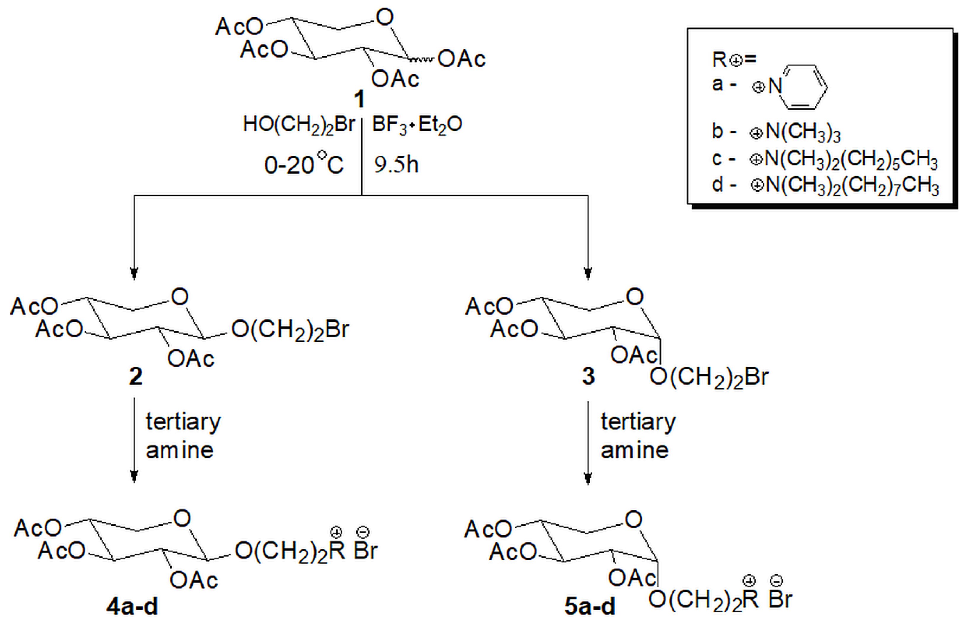
2.1. Chemistry

As a result of the classical acetylation of d-xylose, 1,2,3,4-tetra-O-acetyl-d-xylopyranose (1) was obtained as a mixture of α and β anomers. This mixture was used without further purification for the glycosylation reaction with 2-bromoethanol in the presence of BF3·Et2O (Scheme 1).
Contrary to the syntheses of the analogous glycosides of hexopyranose derivatives previously reported by us [23], it was not possible to select the reaction conditions so that only single anomer of the xylopyranoside was formed. Even though Dahmen et al. [26] reported that, in the case of per-O-acetylated d-xylopyranose, they were able to obtain only one anomer of the glycoside, which they explained by the formation of the thermodynamically more stable 1,2-trans glycoside, we were unable to obtain analogous results. Regardless of the reaction time used and the proportion of substrates, each time, a mixture of two products (α and β anomers) was obtained in a similar quantitative ratio. In the end, a synthesis time of 1 h on ice and then 8.5 h at room temperature turned out to be the optimal conditions. Anomers as isomers show large similarity in structure, which results in very similar properties and thus the difficulty in separating them. The use of chromatography for their separation leads to three fractions: two were pure anomers and the third was their mixture. This means that pure compounds are obtained in relatively low yields. In our case, chromatographic separation using flash chromatography in the n-hexane:ethyl acetate (3:1), using a column packed with silica gel, allowed us to isolate 2-bromoethyl 2′,3′,4′-tri-O-acetyl-β-d-xylopyranoside (2) in 10% yield and 2-bromoethyl 2′,3′,4′-tri-O-acetyl-α-d-xylopyranoside (3) in 7% yield.
The structure of compounds 2 and 3 was confirmed based on NMR spectroscopy (chemical shifts and coupling constants are included in the experimental section). The J1,2 coupling constant of 6.4 Hz indicates the trans proton configuration, which confirms the β configuration of compound 2, while the value of 3.6 Hz in compound 3 corresponds to the cis proton arrangement as in the α anomer. In the spectra, apart from the characteristic signals of the sugar ring protons, are signals of nine protons of methyl groups from three O-acetyl groups. The spectra also show the protons of the two methylene groups (3.5–4.1 ppm) of the aglycon. The signals m/z 405.1 [M + Na]+ appearing in the MALDI-TOF MS spectra additionally confirmed the structure of compounds 2 and 3.
2-Bromoethyl 2′,3′,4′-tri-O-acetyl-β-d-xylopyranoside (2) was used as a substrate in the quaternization reaction with selected amines (pyridine, trimethylamine, N,N-dimethylhexylamine and N,N-dimethyloctylamine). In order to determine the optimal reaction conditions, different solvents and different temperatures were tested. TLC chromatography and MALDI-TOF MS analysis were used to control the progress of the reaction. Ultimately, it turned out that for all the amines used, the best yields were obtained at 70 °C. In most cases, except for trimethylamine, the optimal reaction time was 24 h. Detailed data on the reaction conditions are provided in Table S1 (Supplementary Materials).
The synthesis of the pyridine derivative 4a did not require the use of an additional solvent as it was both a substrate and a solvent. In the case of trimethylamine, the reaction was carried out in a 30% ethanol solution of the amine. The analysis of the obtained products showed that under the reaction conditions, apart from the quaternization reaction, an undesirable partial de-O-acetylation of the resulting product took place. Therefore, in order to obtain the expected product 4b, all volatile components were removed from the post-reaction mixture under reduced pressure, and the residue was subjected to exhaustive O-acetylation. When N,N-dimethylhexylamine and N,N-dimethyloctylamine were used for the quaternization reaction, compounds 4c and 4d were obtained with similar yields. In this case, the reaction required the use of acetonitrile as a solvent. Its selection was made on the basis of a series of test reactions. Acetonitrile allowed both easy observation of the reaction progress (using TLC) and ensured good solubility of the substrates. The high boiling point of the substrate amines made their removal by evaporation under reduced pressure difficult and required extraction with diethyl ether and n-hexane after dissolving the reaction mixture in water.
1H and 13C NMR spectra were recorded to confirm the structure of the obtained salts 4a–d. The signal assignments were made based on the analysis of COSY and HSQC spectra. In all spectra, the H-1 proton signals appear in the form of a doublet with the J1,2 coupling constant with values ranging from 5.9 to 6.8 Hz, confirming the β configuration of the anomeric center. In addition to the anomeric proton signal, signals from the protons of the sugar ring are present in the spectra. The analysis of their coupling constants reveals that the pyranose ring in β-d-xylopyranosides 4a–d is in the conformational equilibrium between the 4C1 and 1C4 conformations, shifted towards the 4C1 conformation. This conclusion is supported by the J2,3 7.2–8.8 Hz, J3,4 7.5–8.4 Hz and J4,5′ 8.0–8.4 Hz coupling constants, which indicate that the respective protons are not fully axially oriented. The ethyl linkage connecting the sugar unit and the quaternary nitrogen atom occurs in the form of three groups of signals. The protons of the methylene group bounded to the oxygen atom of the sugar part appear as two signals at about 4.00 ppm and 4.25 ppm. The protons of the second methylene group of the linker directly linked to the quaternary nitrogen atom of the pyridinium salt 4a appear as a signal at 4.78 ppm. This value of the chemical shift is due to the fact that the nitrogen atom of this salt is present in the aromatic (pyridine) ring. In the case of the aliphatic amine salts 4b–d, the signal of these protons occurs at about 3.58 ppm. The signals from the aromatic pyridine ring appear as two doublets and a triplet at 8.01, 9.49 and 8.46 ppm, respectively. In the aliphatic amines salts, the methyl groups bound directly to the nitrogen atom occur at about 3.06–3.12 ppm. The proton signals of the methylene groups of the hydrocarbon chain (in compounds 4c and 4d) were observed at the 3.30, 1.72 and 1.28 ppm, where the first value is for the methylene group directly bonded to the nitrogen atom. Signal of the terminal methyl group is recorded at 0.83 ppm. A complete description of the NMR spectra signals is provided in the experimental section.
The synthesis of α-d-xylopyranosides 5a–d was performed analogously to that described above. The yields of the quaternization reaction (about 70%) were similar to those obtained for d-xylopyranoside with β configuration of the anomeric carbon atom. The exception is compound 5b, obtained by reaction with trimethylamine, which was obtained in a clearly lower (55%) yield (see Table S1, Supplementary Materials). Moreover, for these products, their structure was confirmed using NMR spectroscopy and mass spectrometry.
The H-1 proton signals in the 1H NMR spectra of 5b–d appear as doublets with a J1,2 coupling constant of 2.8–3.3 Hz, which confirms the α configuration of the anomeric center. The exception was the signal of the H-1 proton of salt 5a, which was in the form of a broad singlet. The values of 9.2–9.4 Hz of the remaining coupling constants (J2,3, J3,4 and J4,5) of the protons present in the sugar ring indicated the 4C1 conformation of the sugar ring of the d-xylopyranosides with α configuration. Proton signals from the aglycone part of 5a–d are placed similarly to those recorded for 4a–d.
All new synthesized N-[2-(2′,3′,4′-tri-O-acetyl-α/β-d-xylopyranosyloxy)ethyl]-ammonium bromides are water soluble.

2.2. Antimicrobial Activity

All synthesized salts 4a–d and 5a–d were tested for their antimicrobial activity against fungi (Candida albicans, Candida glabrata) and bacteria (Staphylococcus aureus, Escherichia coli). The minimum inhibitory concentration (MIC, μg/mL) was determined for the tested strains. Table 1 summarizes the MIC values of the studied compounds.
The data in Table 1 indicates that d-xylopyranosides 4c, 4d and 5d show the ability to inhibit the growth of both fungi and bacteria. These compounds are characterized by the presence of a relatively long alkyl chain on a quaternary nitrogen atom. Among them, compound 4c, with a hexyl group, in addition to two methyl groups, possesses the lowest activity. Two remaining glycosides, 4d and 5d, having an octyl group, in addition to two methyl groups, show greater activity in the conducted studies. Two more carbon atoms in the chain cause the molecule to be more hydrophobic. Glycosides 4d and 5d are isomers that differ solely in the configuration of the anomeric carbon atom of the sugar moiety. The obtained results indicate twofold higher activity of the compound 5d (α anomer) in three out of four tested microorganisms. We observed a different situation in the case of the shorter hexyl substituent. While the compound 4c (β anomer) shows some slight activity against most of the tested microorganisms, its α isomer (4d) shows no activity. On the basis of these results, it is possible to unequivocally indicate the significant influence of the hydrophobic nature of the substituents on the activity of the tested compounds.
Glycoconjugates, i.e., compounds in which sugar fragments are bonded to non-sugar ones, can penetrate the cell membrane in two ways. The first one is based on the active transport through the GLUT membrane proteins. The second mechanism is passive migration (diffusion of a lipophilic compound through biological membranes). Transport using GLUT proteins is more preferred for compounds with free hydroxyl groups, as is the case with intracellular glucose transport. It can be influenced by many factors, such as the structure of the sugar compound, the type of its substitution or even the type of aglycone [27,28]. In addition, studies indicate that, due to better binding to GLUT transport proteins, this mechanism prefers compounds with the β configuration of the anomeric carbon atom [29]. The passive mechanism is preferred in the case of sugar compounds containing substituted hydroxyl groups and a hydrophobic aglycone. The obtained results of biological tests seem to indicate that the tested d-xylopyranosides overcome the barrier of the cell membrane based on the mechanism of diffusion and this seems to be the right direction when designing further syntheses.
Furthermore, xylopyranosides 4d and 5d, which showed good activity against the reference strains, were tested for antistaphylococcal activity against clinical isolates collected from patients with skin and soft tissue infections as well as strains derived from subclinical bovine mastitis milk sample infections. In this study, 20 strains were used, and the MIC values are summarized in Table 2.
Considering clinical isolates, both tested compounds showed activity similar to that obtained against the reference strain of S. aureus. Compound 5d exhibits two to four times higher activity and no differences in susceptibility were observed for MRSA (methicillin-resistant Staphylococcus aureus) and MSSA (methicillin-sensitive Staphylococcus aureus) isolates.
In addition to activity against selected fungi and bacteria, all obtained d-xylopyranosides were tested for mutagenic activity using the Ames test. This test is commonly used and was recommended by OECD to detect the mutagenic potential of chemicals [30]. The results of the analyses are summarized in Table 3. In the Ames test, compounds in which a dose–response relationship is detected and an at least twofold increase in the number of mutants (MI ≥ 2) is observed with at least one concentration are considered to have mutagenic activity.
As the test results showed, none of the studied compounds show mutagenic activity in this test.

3. Summary

The obtained N-[2-(2′,3′,4′-tri-O-acetyl-d-xylopyranosyloxyethyl)]ammonium bromides (4a–d and 5a–d) were subjected to biological tests. They showed no mutagenic activity in the Ames test. On the other hand, the 4d and 5d salts, characterized by the presence of an octyl substituent on the nitrogen atom, showed antimicrobial activity against fungi (C. albicans, C. glabrata) and bacteria (S. aureus, E. coli) with an MIC value within the range of 32−256 μg/mL. These d-xylopyranosides also showed similar activity against clinical isolates of the S. aureus strain. Of these two stereoisomers, the compound with the α configuration of the anomeric center (5d) was more active.

4. Experimental Section

4.1. Synthetic Procedures and Results of Analysis

2-Bromoethyl 2,3,4-tri-O-acetyl-β-d-xylo- (2) and α-d-xylopyranosides (3)—2-bromoethanol (2.0 mL, 28 mmol) was added to a solution of compound 1 (1 g, 3.1 mmol) in dry CH2Cl2 (10 mL). The flask was cooled in an ice bath and boron trifluoride etherate solution (3.64 mL, 29.48 mmol) was slowly added dropwise. The reaction mixture was stirred for 1 h in ice and next for 8.5 h at room temperature. The reaction mixture was diluted with CH2Cl2 (10 mL) and washed with NaHCO3 aqueous solution (2 × 10 mL), water (2 × 10 mL), saturated aqueous NaCl solution (10 mL) and water (10 mL). The organic layer was dried over anhydrous MgSO4, filtered and concentrated under reduced pressure. Column chromatography (ethyl acetate-hexane 1:3) of the crude product first yielded compound 3 (85 mg, 7%, yellowish oil); Rf 0.25 (ethyl acetate-hexane 1:3); 1H-NMR (400 MHz, CDCl3): δ 5.48 (t, 1H, H-3, J3,4 10.0 Hz); 5.08 (d, 1H, H-1, J1,2 3.6 Hz); 4.96 (dt, 1H, H-4, J4,5′ 10.0 Hz, J4,5 4.4 Hz); 4.79 (dd, 1H, H-2, J2,3 10.0 Hz); 3.99 (dt, 1H, OCH2a, J 11.2 Hz); 3.78 (m, 2H, H-5, OCH2b, J 6.0 Hz); 3.69 (dd, 1H, H-5′, J5,5′ 10.8 Hz); 3.49 (t, 2H, CH2Br, J 6.4 Hz); 2.07–2.03 (3s, 9H, 3 × OAc); 13C NMR (100 MHz, CDCl3): 170.5–170.2 (3C, 3 × OOCCH3 ), 95.2 (C-1), 71.2 (C-2), 69.7 (C-3), 68.6 (2C, C-4 and O-CH2), 58.9 (C-5), 30.6 (CH2Br), 21.0–20.9 (3C, 3 × COOCH3); MALDI TOF- MS (DHB): m/z 405.1; 407.1 ([M + Na]+) and 421.1; 423.1 ([M + K]+).
Eluted second was 2 (121 mg, 10% yield, mp 127–129 °C,); Rf = 0.17 (ethyl acetate-hexane 1:3); 1H-NMR (400 MHz, CDCl3): δ 5.16 (t, 1H, H-3, J3,4 8.4 Hz); 4.94 (m, 2H, H-2, H-4, J2,3 8.4 Hz, J4,5′ 8.4 Hz); 4.55 (d, 1H, H-1, J1,2 6.4 Hz); 4.14 (dd, 1H, H-5, J4,5 5.2 Hz); 4.08 (dt, 1H, OCH2a, J 11.2 Hz); 3.48 (dt, 1H, OCH2b, J 6.4 Hz); 3.39 (t, 2H, CH2Br, J 6.0 Hz); 3.38 (dd, 1H, H-5′, J5,5′ 12.0 Hz); 2.07–2.04 (3s, 9H, 3 × OAc); 13C NMR (100 MHz, CDCl3): 170.2–169.7 (3C, 3 × OOCCH3 ), 100.9 (C-1), 71.3 (C-3), 70.6 (C-4), 69.4 (O-CH2), 69.0 (C-2), 62.3 (C-5), 30.2 (CH2Br), 21.0–20.9 (3C, 3 x COOCH3); MALDI TOF-MS (DHB): m/z 405.1; 407.1 ([M + Na]+) and 421.1; 423.1 ([M + K]+).

4.2. General Procedure for Quaternization Reaction

Amines (pyridine, 33% trimethylamine in ethanol, N,N-dimethylhexylamine or N,N-dimethyloctylamine in acetonitrile) and compounds 2 and 3, respectively, were placed in a glass screw-capped ampoule. The mixture was heated at 70 °C for a certain period of time. Next, volatile compounds were evaporated under reduced pressure and the residue, after being dissolved in water, was extracted with chloroform (diethyl ether and then with n-hexane for N,N-dimethylhexylamine or N,N-dimethyloctylamine) three times. After concentration, thick syrup was obtained. Finally, product (Rf = 0.0 (ethyl acetate-hexane 1:3)) was dried in a vacuum desiccator over P2O5.
N-[2-(2′,3′,4′-tri-O-acetyl-β-d-xylopyranosyloxy)ethyl]pyridinium bromide (4a)—A reaction between compound 2 (30.7 mg, 0.080 mmol) and dry pyridine (0.5 mL), carried out for 24 h, yielded compound 4a (25 mg, 67%, colorless oil); [ α ] 20 D −37.6° (c 1, MeOH); 1H NMR (500 MHz, D2O): δ 9.49, 8.46, 8.01, (d, t, t, 5 H, Py, J 5.6, J 7.2); 5.12 (t, 1H, H-3, J3,4 8.4 Hz); 4.88 (m, 1H, H-4, J4,5′ 8.4 Hz); 4.78 (m, 2H, H-2, CH2-N, J2,3 8.8 Hz); 4.67 (d, 1H, H-1, J1,2 6.8 Hz); 4.26 and 4.09 (2 x m, 2H, O-CH2a and O-CH2b, J 13.5 Hz J 6.0 Hz); 3.94 (dd, 1H, H-5, J4,5 4.8 Hz); 3.46 (dd, 1H, H-5′, J5,5′ 12.4 Hz); 2.01–1.98 (3s, 9H, 3 × OAc); 13C NMR (125 MHz, D2O): 173.2–172.6 (3C, 3 × OOCCH3), 146.5–145.1, 128.3 (5C, Py), 99.8 (C-1), 71.5 (C-3), 70.8 (C-2), 68.8 (C-4), 67.6 (O-CH2), 61.5 (C-5), 61.3 (CH2-N), 20.4–20.3 (3C, COOCH3); MALDI TOF-MS (CHCA): m/z 382.2 ([M-Br]+), HRMS (ESI): m/z ([M-Br]+) calculated for C18H24O8N: 382.1496; found: 382.1492.
N-[2-(2′,3′,4′-tri-O-acetyl-β-d-xylopyranosyloxy)ethyl]-N,N,N-trimethylammonium bromide (4b)—A reaction between compound 2 (37 mg, 0.097 mmol) with 33% trimethylamine in ethanol (0.5 mL) ended after 3 h. After the removal of volatile compounds, the residue was O-acetylated with Ac2O (0.5 mL) and pyridine (0.5 mL) at room temperature for 24 h. Purification of the crude product, as described in the general procedure, yielded compound 4b (36.9 mg, 87%, colorless oil); [ α ] 20 D   −40.0° (c 1, MeOH); 1H NMR (500 MHz, D2O): δ 5.17 (t, 1H, H-3, J3,4 7.6 Hz); 4.95 (td, 1H, H-4, J4,5′ 8.0 Hz); 4.89 (dd, 1H, H-2, J2,3 7.6 Hz); 4.78 (d, 1H, H-1, J1,2 6.0 Hz); 4.25 (d, 1H, O-CH2a, J 13.0 Hz); 4.13 (dd, 1H, H-5, J4,5 4.4 Hz); 4.02 (d, 1H, O-CH2b); 3.58–3.55 (m, 3H, H-5′, CH2-N, J5′,5 12.8 Hz); 3.12 (s, 9H, 3 × CH3 of N(CH3)3); 2.08, 2.05, 2.04 (3s, 9H, 3 × OAc); 13C NMR (125 MHz, D2O): 173.0, 172.9, 172.6 (3C, 3 × OOCCH3), 99.3 (C-1), 70.7 (C-3), 70.3 (C-2), 68.4 (C-4), 65.5 (CH2-N), 62.9 (O-CH2), 61.0 (C-5), 54.0, 54.0, 53.9 (3C, N(CH3)3), 20.2, 20.1 (3C, COOCH3); MALDI TOF-MS (CHCA): m/z 362.3 ([M-Br]+); HRMS (ESI): m/z ([M-Br]+) calculated for C18H28O8N: 362.1809; found: 362.1805.
N-[2-(2′,3′,4′-tri-O-acetyl-β-d-xylopyranosyloxy)ethyl]-N-hexyl-N,N-dimethylammonium bromide (4c)—A reaction between compound 2 (35 mg, 0.092 mmol) and N,N-dimethylhexylamine (0.2 mL, 1.15 mmol) in acetonitrile (1.0 mL) carried out for 24 h led to compound 4c (34 mg, 72%, colorless oil); [ α ] 20 D   −23.2° (c 1, MeOH); 1H NMR (500 MHz, D2O): δ 5.19 (t, 1H, H-3, J3,4 7.7 Hz); 4.96 (m, 1H, H-4, J4,5 4.4 Hz); 4.88 (t, 1H, H-2, J2,3 7.2 Hz); 4.79 (d, 1H, H-1, J1,2 5.9 Hz); 4.21 (d, 1H, O-CH2a, J 13.6 Hz); 4.14 (dd, 1H, H-5, J4,5′ 8.4 Hz); 4.00 (d, 1H, O-CH2b); 3.57 (m, 3H, H-5′, CH2N); 3.29 (t, 2H, N-CH2); 3.06 (s, 6H, 2 × CH3 of N(CH3)2); 2.08, 2.05, 2.04 (3s, 9H, 3 × OAc); 1.71 (bs, 2H, CH2); 1.28 (bs, 6H, 3 x CH2); 0.83 (bs, 3H, CH3); 13C NMR (125 MHz, D2O): 173.0, 172.9, 172.6 (3C, 3 × OOCCH3), 99.4 (C-1), 70.9 (C-3), 70.5 (C-2), 68.4 (C-4), 65.5 (N-CH2), 62.9 (O-CH2), 62.8 (CH2-N), 61.1 (C-5), 51.5 (2C, N(CH3)2), 30.5, 25.3, 21.9, 21.8 (4C, 4 x CH2), 13.2 (CH3), 20.3, 20.2 (3C, COOCH3); MALDI TOF-MS (CHCA): m/z 432.4 ([M-Br]+); HRMS (ESI): m/z ([M-Br]+) calculated for C21H38O8N: 432.2592; found: 432.2587.
N-[2-(2′,3′,4′-tri-O-acetyl-β-d-xylopyranosyloxy)ethyl]-N,N-dimethyl-N-octylammonium bromide (4d)—A reaction between compound 2 (35 mg, 0.092 mmol) and N,N-dimethyloctylamine (0.2 mL, 0.972 mmol) in acetonitrile (1.0 mL) carried out for 24 h yielded 4d (35 mg, 70%, yellowish oil); [ α ] 20 D   −19.4° (c 1, MeOH); 1H NMR (500 MHz, D2O): δ 5.19 (t, 1H, H-3, J3,4 7.9 Hz); 4.96 (m, 1H, H-4); 4.88 (td, 1H, H-2, J2,3 7.9 Hz); 4.79 (d, 1H, H-1, J1,2 6.3 Hz); 4.22 (bd, 1H, O-CH2a); 4.14 (dd, 1H, H-5, J4,5 4.6 Hz, J5,5′ 12.2 Hz); 4.01 (bd, 1H, O-CH2b); 3.58 (m, 3H, H-5′, CH2N); 3.29 (m, 2H, N-CH2); 3.06 (m, 8H, 2 × CH3 of N(CH3)2, CH2); 2.08, 2.05, 2.04 (3s, 9H, 3 × OAc); 1.72 (bs, 2H, CH2); 1.28 (bd, 8H, 4 × CH2); 0.82 (t, 3H, CH3); 13C NMR (125 MHz, D2O): 172.9, 172.6 (3C, 3 × OOCCH3), 99.5 (C-1), 71.0 (C-3), 70.6 (C-2), 68.5 (C-4), 65.3 (N-CH2), 62.9 (O-CH2), 62.8 (CH2-N), 61.1 (C-5), 58.0 (CH2), 51.5 (2C, N(CH3)2), 31.0, 28.2, 28.1, 25.6, 22.1, 21.9 (6 x CH2), 20.3, 20.2, 20.1 (3C, COOCH3), 13.4 (CH3); MALDI TOF-MS (CHCA): m/z 460.3 ([M-Br]+); HRMS (ESI): m/z ([M-Br]+) calculated for C23H42O8N: 460.2905; found: 460.2898.
N-[2-(2′,3′,4′-tri-O-acetyl-α-d-xylopyranosyloxy)ethyl]pyridinium bromide (5a)—A reaction between compound 3 (37 mg, 0.094 mmol) and dry pyridine (0.5 mL) carried out for 24 h yielded compound 5a (33 mg, 75% yield, colorless oil); [ α ] 20 D   +46.9° (c 1, MeOH); 1H NMR (500 MHz, D2O): δ 8.92, 8.60, 8.10 (d, t, t, 5 H, Py, J 5.9, J 7.7, J 7.7); 5.21 (t, 1H, H-3, J3,4 9.4 Hz); 4.99 (bs, 1H, H-1); 4.96 (m, 1H, H-4); 4.89 (m, 1H, H-2, J2,3 9.4 Hz); 4.89 (m, 2H, CH2-N, J 4.1 Hz); 4.22 (m, 1H, O-CH2a, J 11.1); 3.94 (m, 1H, O-CH2b J 5.2); 3.78 (dd, 1H, H-5, J4,5 5.6 Hz, J5,5′ 11.5 Hz); 3.24 (t, 1H, H-5′, J4,5′ 10.9 Hz); 2.03, 2.01, 1.90 (3s, 9H, 3 × OAc); 13C NMR (125 MHz, D2O): 173.4, 172.7 (3C, 3 × OOCCH3), 146.4, 145.1, 128.3 (5C, Py), 95.5 (C-1), 70.0 (C-2), 69.9 (C-3), 68.4 (C-4), 66.0 (O-CH2), 61.1 (CH2-N), 58.6 (C-5), 20.2, 20.1, (3C, COOCH3); MALDI TOF-MS (CHCA): m/z 382.2 ([M-Br]+); HRMS (ESI): m/z ([M-Br]+) calculated for C18H24O8N: 382.1496; found: 382.1492.
N-[2-(2′,3′,4′-tri-O-acetyl-α-d-xylopyranosyloxy)ethyl]-N,N,N-trimethylammonium bromide (5b)—A reaction between compound 3 (30 mg, 0.079 mmol) and 33% trimethylamine in ethanol (0.5 mL) ended after 3 h. After removal of all volatile compounds, the residue was O-acetylated with Ac2O (0.5 mL) and pyridine (0.5 mL) at room temperature for 24 h. Purification of the crude product, as described in the general procedure, led to compound 5b (19.3 mg, 55%, yellowish oil); [ α ] 20 D   +60.3° (c 1, MeOH); 1H NMR (500 MHz, D2O): δ 5.32 (t, 1H, H-3, J3,4 9.3 Hz); 5.12 (d, 1H, H-1, J1,2 2.8 Hz); 5.04 (m, 2H, H-2 and H-4, J2,3 9.2 Hz); 4.17 (m, 1H, O-CH2a); 3.98 (m, 1H, O-CH2b); 3.92 (dd, 1H, H-5, J4,5 5.6 Hz); 3.70 (t, 1H, H-5′, J5′,5 11.5 Hz); 3.62 (m, 2H, CH2-N); 3.19 (s, 9H, 3 × CH3 of N(CH3)3); 2.08, 2.04, 2.01 (3s, 9H, 3 × OAc); 13C NMR (125 MHz, D2O): 173.4, 172.8, 172.5 (3C, 3 × OOCCH3), 96.0 (C-1), 70.2 (C-3), 69.9 (C-2), 68.6 (C-4), 65.6 (CH2-N), 62.0 (O-CH2), 58.9 (C-5), 54.1 (3C, N(CH3)3), 20.2, 20.1, 20.0 (3C, COOCH3); MALDI TOF-MS (CHCA): m/z 362.3 ([M-Br]+); HRMS (ESI): m/z ([M-Br]+) calculated for C18H28O8N: 362.1809; found: 362.1804.
N-[2-(2′,3′,4′-tri-O-acetyl-α-d-xylopyranosyloxy)ethyl]-N,N-dimethyl-N-hexylammonium bromide (5c)—A reaction between compound 3 (35 mg, 0.092 mmol) and N,N-dimethylhexylamine (0.2 mL, 1.15 mmol) in acetonitrile (1.0 mL) carried out for 24 h yielded compound 5c (32 mg, 68%, colorless oil); [ α ] 20 D   +49.9° (c 1, MeOH); 1H NMR (500 MHz, D2O): δ 5.29 (t, 1H, H-3, J3,4 9.3 Hz); 5.11 (d, 1H, H-1, J1,2 3.3 Hz); 5.06 (m, 2H, H-2, H-4); 4.15 (dd, 1H, O-CH2a); 3.93–3.91 (m, 2H, H-5, O-CH2b); 3.69–3.57 (m, 3H, H-5′, CH2N,); 3.37 (m, 2H, N-CH2); 3.12 (s, 6H, 2 × CH3 of N(CH3)2); 2.08, 2.05, 2.03 (3s, 9H, 3 × OAc); 1.77 (m, 2H, CH2); 1.31 and 1.28 (2 x bs, 6H, 3 × CH2); 0.81 (bs, 3H, CH3); 13C NMR (125 MHz, D2O): 173.4, 172.8, 172.5 (3C, 3 × OOCCH3), 96.1 (C-1), 70.3 (C-3), 69.8 (C-2), 68.8 (C-4), 65.6 (N-CH2), 62.3 (CH2-N), 62.0 (O-CH2), 58.7 (C-5), 51.5 (2C, N(CH3)2), 30.6, 25.3, 22.1, 21.8 (4C, 4 × CH2), 20.1 (3C, COOCH3), 13.2 (CH3); MALDI TOF-MS (CHCA): m/z 432.4 ([M-Br]+); HRMS (ESI): m/z ([M-Br]+) calculated for C21H38O8N: 432.2592; found: 432.2587.
N-[2-(2′,3′,4′-tri-O-acetyl-α-d-xylopyranosyloxy)ethyl]-N,N-dimethyl-N-octylammonium bromide (5d)—A reaction between compound 3 (29 mg, 0.075 mmol) and N,N-dimethyloctylamine (0.2 mL, 0.972 mmol) in acetonitrile (1.0 mL) carried out for 24 h yielded compound 5d (28.5 mg, 70%, yellowish oil); [ α ] 20 D   +44.7° (c 1, MeOH); 1H NMR (500 MHz, D2O): δ 5.29 (t, 1H, H-3, J3,4 9.5 Hz); 5.11 (d, 1H, H-1, J1,2 3.2 Hz); 5.07–5.04 (m, 2H, H-2, H-4); 4.15 (m, 1H, O-CH2a); 3.96–3.90 (m, 2H, H-5, O-CH2b, J4,5 5.9 Hz); 3.71 (m, 1H, H-5′, J5,5′ 11.0 Hz); 3.56 (m, 2H, CH2N); 3.44 (dd, 2H, NCH2); 3.12 (s, 6H, 2 × CH3 of N(CH3)2); 2.08, 2.04, 2.03 (3s, 9H, 3 × OAc); 1.79 (m, 2H, CH2); 1.34–1.23 (m, 10H, 5 x CH2); 0.82 (t, 3H, CH3, J 6.6 Hz); 13C NMR (125 MHz, D2O): 173.2, 172.8, 172.5 (3C, 3 × OOCCH3), 96.1 (C-1), 70.4 (C-3), 69.9 (C-2), 68.8 (C-4), 65.6 (1C, NCH2), 62.2 (1C, CH2N), 62.1 (1C, O-CH2), 58.7 (C-5), 51.7 (2C, N(CH3)2), 31.0, 28.3, 28.1, 25.6, 22.3, 22.0 (6C, 6 × CH2), 20.3, 20.2, 20.1 (3C, COOCH3), 13.4 (CH3); MALDI TOF-MS (CHCA): m/z 460.3 ([M-Br]+); HRMS (ESI): m/z ([M-Br]+) calculated for C23H42O8N: 460.2905; found: 460.2899.

Supplementary Materials

The following supporting information can be downloaded at: https://www.mdpi.com/article/10.3390/antibiotics12050888/s1, Table S1. Reaction yields and solvents used for the quaternization reaction at 70 °C; Copies of NMR and HRMS spectra of new compounds [31,32,33,34,35,36].

Author Contributions

Conceptualization, B.D.; methodology, B.D. and K.S. (Karol Sikora); formal analysis, K.S. (Karol Sikora) and B.D.; investigation, K.S. (Karol Sikora), B.D., P.S. and K.S. (Karolina Słoczyńska); writing—original draft preparation, B.D.; writing—review and editing, B.D., J.M., B.L., K.S. (Karol Sikora), P.S., K.S. (Karolina Słoczyńska), J.S.-F. and E.P.; supervision, B.D.; project administration, B.D. All authors have read and agreed to the published version of the manuscript.

Funding

This study was supported by University of Gdansk, task grant no. DS/531-T100-D501-23.

Institutional Review Board Statement

Not applicable.

Informed Consent Statement

Not applicable.

Data Availability Statement

Not applicable.

Conflicts of Interest

The authors declare no conflict of interest. The funders had no role in the design of the study; in the collection, analyses, or interpretation of data; in the writing of the manuscript, or in the decision to publish the results.

References

  1. Magee, R.J.; Kosaric, N. Bioconversion of hemicellulosics. In Agricultural Feedstock and Waste Treatment and Engineering; Springer: Berlin/Heidelberg, Germany, 1985; Volume 32, pp. 61–93. [Google Scholar]
  2. Van Dyk, J.S.; Pletschke, B.I. A review of lignocellulose bioconversion using enzymatic hydrolysis and synergistic cooperation between enzymes—Factors affecting enzymes, conversion and synergy. Biotechnol. Adv. 2012, 30, 1458–1480. [Google Scholar] [CrossRef] [PubMed]
  3. Rosenberg, S.L. Fermentation of pentose sugars to ethanol and other neutral products by microorganisms. Enzyme Microb. Technol. 1980, 2, 185–193. [Google Scholar] [CrossRef]
  4. Saddler, J.N.; Yu, E.K.C.; Mes-Hartree, M.; Levitin, N.; Brownell, H.H. Utilization of Enzymatically Hydrolyzed Wood Hemicelluloses by Microorganisms for Production of Liquid Fuels. Appl. Environ. Microbiol. 1983, 45, 153–160. [Google Scholar] [CrossRef] [PubMed]
  5. Patel, G. Ethanol production during d-xylose, L-arabinose, and d-ribose fermentation by Bacteroides polypragmatus. Appl. Microbiol. Biotechnol. 1984, 20, 111–117. [Google Scholar] [CrossRef]
  6. Schepers, H.-J.; Bringer-Meyer, S.; Sahm, H. Fermentation of d-Xylose to Ethanol by Bacillus macerans. Z. Naturforsch. C 1987, 42, 401–407. [Google Scholar] [CrossRef]
  7. Espinoza-Acosta, J.L. Biotechnological production of xylitol from agricultural waste. Biotecnia 2019, 22, 126–134. [Google Scholar] [CrossRef]
  8. Umai, D.; Kayalvizhi, R.; Kumar, V.; Jacob, S. Xylitol: Bioproduction and Applications—A Review. Front. Sustain. 2022, 3, 2. [Google Scholar] [CrossRef]
  9. Ahiadorme, D.; Ande, C.; Fernandez-Botran, R.; Crich, D. Synthesis and evaluation of 1,5-dithialaminaribiose and -triose tetravalent constructs. Carbohydr. Res. 2023, 525, 108781. [Google Scholar] [CrossRef]
  10. Mohamad-Ramshan, R.; Ande, C.; Matsushita, T.; Haldimann, K.; Vasella, A.; Hobbie, S.N.; Crich, D. Synthesis of 4-O-(4-amino--4-deoxy-β-d-xylopyranosyl)paromomycin and 4-S-(β-d-xylopyranosyl)-4-deoxy-4′-thio-paromomycin and evaluation of their antiribosomal and antibacterial activity. Tetrahedron 2023, 135, 133330. [Google Scholar] [CrossRef]
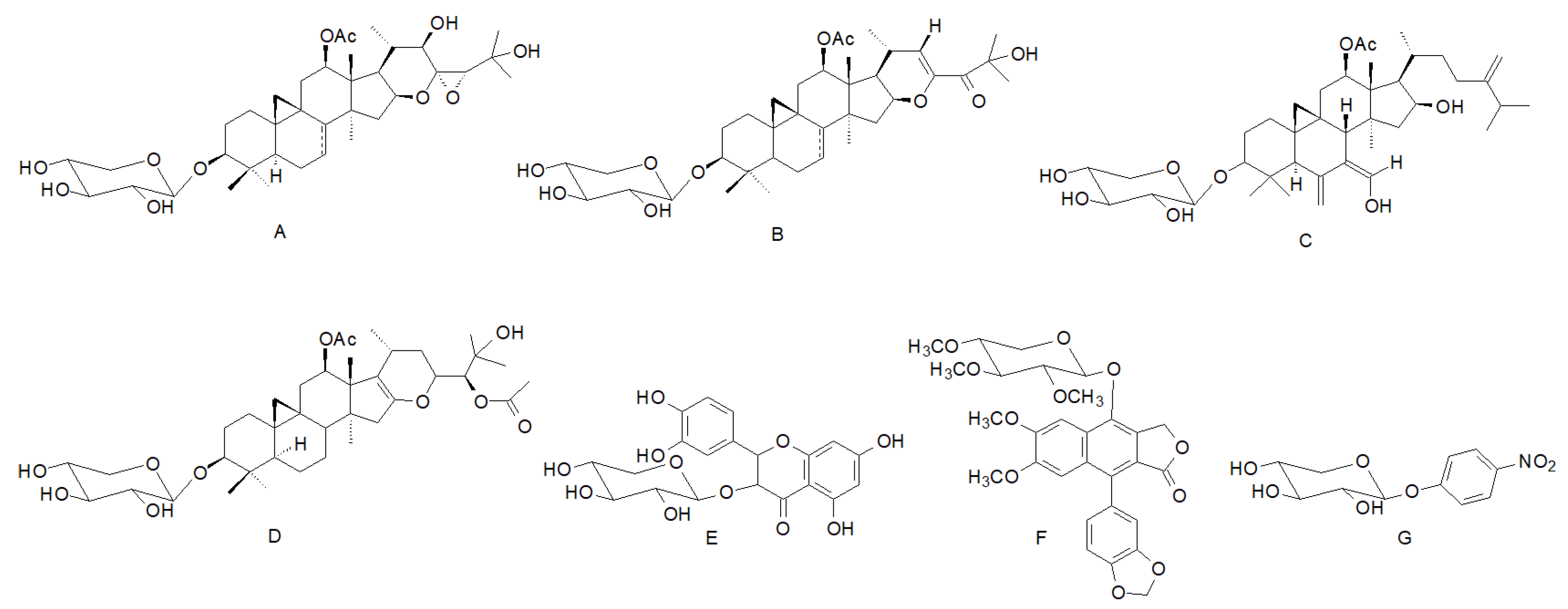
  11. Ali, Z.; Khan, S.I.; Pawar, R.S.; Ferreira, D.; Khan, I.A. 9,19-Cyclolanostane Derivatives from the Roots of Actaea pachypoda. J. Nat. Prod. 2007, 70, 107–110. [Google Scholar] [CrossRef]
  12. Gao, J.; Huang, F.; Zhang, J.; Zhu, G.; Yang, M.; Xiao, P. Cytotoxic Cycloartane Triterpene Saponins from Actaea asiatica. J. Nat. Prod. 2006, 69, 1500–1502. [Google Scholar] [CrossRef] [PubMed]
  13. Lee, I.; Kim, J.; Kim, Y.S.; Yoo, N.H.; Kim, C.-S.; Jo, K.; Kim, J.-H.; Bach, T.T.; Kim, J.S. Cycloartane-Type Triterpenes from the Leaves of Homonoia riparia with VEGF-Induced Angiogenesis Inhibitory Activity. J. Nat. Prod. 2012, 75, 1312–1318. [Google Scholar] [CrossRef] [PubMed]
  14. Findeis, M.A.; Schroeder, F.; McKee, T.D.; Yager, D.; Fraering, P.C.; Creaser, S.P.; Austin, W.F.; Clardy, J.; Wang, R.; Selkoe, D.; et al. Discovery of a Novel Pharmacological and Structural Class of Gamma Secretase Modulators Derived from the Extract of Actaea racemosa. ACS Chem. Neurosci. 2012, 3, 941–951. [Google Scholar] [CrossRef] [PubMed]
  15. Schieber, A.; Mihalev, K.; Berardini, N.; Mollov, P.; Carle, R. Flavonol Glycosides from Distilled Petals of Rosa damascena Mill. Z. Naturforsch. C 2005, 60, 379–384. [Google Scholar] [CrossRef]
  16. Szabo, K.; Mitrea, L.; Călinoiu, L.F.; Teleky, B.-E.; Martău, G.A.; Plamada, D.; Pascuta, M.S.; Nemeş, S.-A.; Varvara, R.-A.; Vodnar, D.C. Natural Polyphenol Recovery from Apple-, Cereal-, and Tomato-Processing By-Products and Related Health-Promoting Properties. Molecules 2022, 27, 7977. [Google Scholar] [CrossRef]
  17. Jeong, E.J.; Jegal, J.; Chung, K.W.; Noh, S.G.; Chung, H.Y.; Nam, Y.H.; Kang, T.H.; Kim, S.-N.; Yang, M.H. Hypolaetin-7- O -β-d-xyloside from Juniperus communis Fruits Inhibits Melanogenesis on Zebrafish Pigmentation. Nat. Prod. Commun. 2017, 12, 1934578X1701201. [Google Scholar] [CrossRef]
  18. Shen, W.; Zhao, Y.; Chen, H.; Zhang, T.; Wu, S.; Liu, P. M3, a natural lignan xyloside, exhibits potent anticancer activity in HCT116 cells. Oncol. Lett. 2018, 17, 2117–2122. [Google Scholar] [CrossRef]
  19. Kanwar, Y.S.; Rosenzweig, L.J.; Jakubowski, M.L. Xylosylated-proteoglycan-induced Golgi alterations. Proc. Natl. Acad. Sci. USA 1986, 83, 6499–6503. [Google Scholar] [CrossRef]
  20. Thompson, H.A.; Spooner, B.S. Proteoglycan and glycosaminoglycan synthesis in embryonic mouse salivary glands: Effects of beta-d-xyloside, an inhibitor of branching morphogenesis. J. Cell Biol. 1983, 96, 1443–1450. [Google Scholar] [CrossRef]
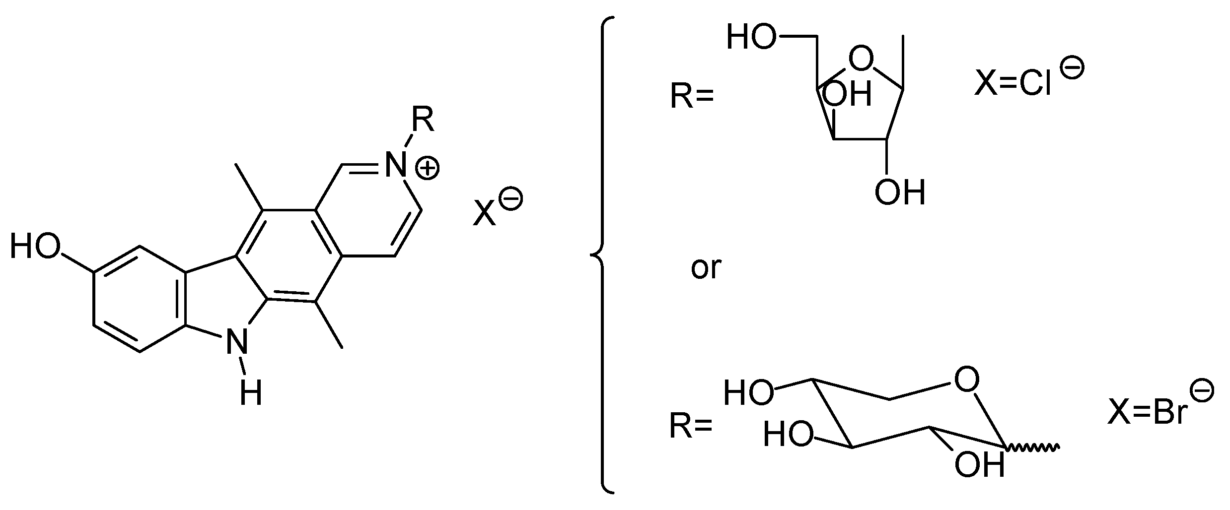
  21. Honda, T.; Kato, M.; Inoue, M.; Shimamoto, T.; Shima, K.; Nakanishi, T.; Yoshida, T.; Noguchi, T. Synthesis and antitumor activity of quaternary ellipticine glycosides, a series of novel and highly active antitumor agents. J. Med. Chem. 1988, 31, 1295–1305. [Google Scholar] [CrossRef]
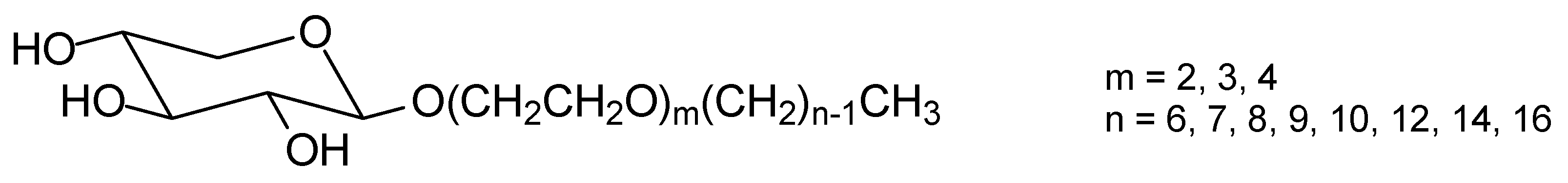
  22. Wu, X.; Chen, L.; Fan, Y.; Fu, F.; Li, J.; Zhang, J. Water Solubility and Surface Property of Alkyl Di-/Tri-/Tetraoxyethyl β-d-Xylopyranosides. J. Agric. Food Chem. 2019, 67, 10361–10372. [Google Scholar] [CrossRef]
  23. Quagliotto, P.; Viscardi, G.; Barolo, C.; D’Angelo, D.; Barni, E.; Compari, C.; Duce, E.; Fisicaro, E. Synthesis and Properties of New Glucocationic Surfactants: Model Structures for Marking Cationic Surfactants with Carbohydrates. J. Org. Chem. 2005, 70, 9857–9866. [Google Scholar] [CrossRef] [PubMed]
  24. Dmochowska, B.; Sikora, K.; Woziwodzka, A.; Piosik, J.; Podgórska, B. Mutagenic activity of quaternary ammonium salt derivatives of carbohydrates. Beilstein J. Org. Chem. 2016, 12, 1434–1439. [Google Scholar] [CrossRef] [PubMed]
  25. Dmochowska, B.; Piosik, J.; Woziwodzka, A.; Sikora, K.; Wiśniewski, A.; Węgrzyn, G. Mutagenicity of quaternary ammonium salts containing carbohydrate moieties. J. Hazard. Mater. 2011, 193, 272–278. [Google Scholar] [CrossRef] [PubMed]
  26. Dahmén, J.; Frejd, T.; Grönberg, G.; Lave, T.; Magnusson, G.; Noori, G. 2-Bromoethyl glycosides: Synthesis and characterisation. Carbohydr. Res. 1983, 116, 303–307. [Google Scholar] [CrossRef]
  27. Fu, J.; Yang, J.; Seeberger, P.H.; Yin, J. Glycoconjugates for glucose transporter-mediated cancer-specific targeting and treatment. Carbohydr. Res. 2020, 498, 108195. [Google Scholar] [CrossRef]
  28. Calvaresi, E.C.; Hergenrother, P.J. Glucose conjugation for the specific targeting and treatment of cancer. Chem. Sci. 2013, 4, 2319. [Google Scholar] [CrossRef]
  29. Barnett, J.E.G.; Holman, G.D.; Munday, K.A. Structural requirements for binding to the sugar-transport system of the human erythrocyte. Biochem. J. 1973, 131, 211–221. [Google Scholar] [CrossRef]
  30. OECD. Test No. 471: Bacterial Reverse Mutation Test; OECD Guidelines for the Testing of Chemicals, Section 4; OECD: Paris, France, 2020; ISBN 9789264071247. [Google Scholar]
  31. Clinical and Laboratory Standards Institute (CLSI). Methods for Dilution Antimicrobial Susceptibility Tests for Bacteria That Grow Aerobically; Approved Standard, 9th ed.; CLSI: Berwyn, PA, USA, 2012; p. 32. [Google Scholar]
  32. Clinical Laboratory Standards Institute. Reference Method for Broth Dilution Antifungal susceptibility testing of yeast. Clin. Lab. Stand. Inst. 2012, 32, 1–23. [Google Scholar]
  33. Maron, D.M.; Ames, B.N. Revised methods for the Salmonella mutagenicity test. Mutat. Res. Mutagen. Relat. Subj. 1983, 113, 173–215. [Google Scholar] [CrossRef]
  34. Mortelmans, K.; Zeiger, E. The Ames Salmonella/microsome mutagenicity assay. Mutat. Res. Mol. Mech. Mutagen. 2000, 455, 29–60. [Google Scholar] [CrossRef] [PubMed]
  35. Gulluce, M.; Agar, G.; Baris, O.; Karadayi, M.; Orhan, F.; Sahin, F. Mutagenic and antimutagenic effects of hexane extract of some Astragalus species grown in the eastern Anatolia region of Turkey. Phyther. Res. 2010, 24, 1014–1018. [Google Scholar] [CrossRef] [PubMed]
  36. Biso, F.I.; Rodrigues, C.M.; Rinaldo, D.; dos Reis, M.B.; Bernardi, C.C.; de Mattos, J.C.P.; Caldeira-de-Araújo, A.; Vilegas, W.; de Syllos Cólus, I.M.; Varanda, E.A. Assessment of DNA damage induced by extracts, fractions and isolated compounds of Davilla nitida and Davilla elliptica (Dilleniaceae). Mutat. Res. Toxicol. Environ. Mutagen. 2010, 702, 92–99. [Google Scholar] [CrossRef] [PubMed]
Figure 1. Selected terpenoid xylosides isolated from (A) Actaea pachypoda [11], (B) Actaea asiatica [12], (C) Homonoia riparia [13] and (D) Actaea racemose [14]; (E) structure of quercetin-3-d-xylopyranoside; (F) structure of 4-O-(2′,3′,4′-tri-O-methyl-β-d-xylopyranosyl) diphyllin; (G) structure of p-nitrophenyl-β-d-xylopyranoside.
Antibiotics 12 00888 g001
Figure 2. Ellipticine 1′,2′-trans-d-xylosides synthesized by Honda et al. [21].
Antibiotics 12 00888 g002
Figure 3. Alkyl di-/tri-/tetraoxyethyl β-d-xylopyranosides prepared by Wu et al. [22].
Antibiotics 12 00888 g003
Figure 4. Glucocationic class surfactants prepared by Quagliotto et al. [23].
Antibiotics 12 00888 g004
Scheme 1. Synthesis of N-[2-(2′,3′,4′-tri-O-acetyl-α/β-d-xylopyranosyloxy)ethyl]ammonium bromides.
Antibiotics 12 00888 sch001
Table 1. MIC values (μg/mL) for 4a–4d and 5a–5d determined against Candida albicans, Candida glabrata, Staphylococcus aureus and Escherichia coli.
CompoundMicroorganisms
C. albicansC. glabrataS. aureusE. coli
4a>4096>4096>4096>4096
5a>4096>4096>4096>4096
4b>4096>4096>4096>4096
5b>4096>4096>4096>4096
4c102410241024>4096
5c>4096>4096>4096>4096
4d1286464256
5d646432128
Table 2. MIC values (μg/mL) for compounds 4d and 5d against clinical isolates of S. aureus.
IsolatesCompound
4d5d
16432
26432
3168
46432
56432
66432
7 *6416
8 *6432
96432
1012832
1112832
1212832
136432
146432
156432
166432
176432
186432
19 *6432
20 *6432
Strains 1–8 were isolated from human infections; strains 9–20 were isolated from bovine mastitis; * MRSA strains.
Table 3. Mutagenic activity and mutagenic index (MI) of glycosides 4a–d and 5a–d in the Ames test with Salmonella typhimurium TA98 strain.
CompoundNumber of Revertants/Plate and (MI)
Concentration (µg/Plate)
4201005002000C+C−
4a28 ± 10 (0.7)26 ± 6 (0.6)41 ± 9 (1.0)26 ± 6 (0.6)38 ± 5 (0.9)760 ± 27 41 ± 13
4b50 ± 4 (1.2)39 ± 3 (1.0)44 ± 2 (1.1)49 ± 6 (1.2)38 ± 5 (0.9)
4c25 ± 3 (0.6)20 ± 1 (0.5)20 ± 3 (0.5)29 ± 7 (0.7)39 ± 4 (1.0)
4d28 ± 4 (0.7)36 ± 12 (0.9)24 ± 1 (0.6)21 ± 2 (0.5)28 ± 9 (0.7)
5a50 ± 4 (1.2)49 ± 4 (1.2)49 ± 2 (1.2)46 ± 4 (1.1)47 ± 4 (1.2)
5b41 ± 4 (1.0)25 ± 1 (0.6)23 ± 2 (0.6)38 ± 3 (0.9)41 ± 2 (1.0)
5c24 ± 2 (0.6)28 ± 6 (0.7)35 ± 2 (0.9)22 ± 6 (0.5)26 ± 5 (0.6)
5d24 ± 3 (0.6)22 ± 2 (0.5)23 ± 6 (0.6)36 ± 1 (0.9)35 ± 4 (0.9)
The results are reported as means ± SD and mutagenic index (MI, in brackets); C−: negative control (dimethylsulfoxide); C+: positive control (4-Nitro-o-phenylenediamine, 4-NPD, 10 µg/plate).
Disclaimer/Publisher’s Note: The statements, opinions and data contained in all publications are solely those of the individual author(s) and contributor(s) and not of MDPI and/or the editor(s). MDPI and/or the editor(s) disclaim responsibility for any injury to people or property resulting from any ideas, methods, instructions or products referred to in the content.

