Article

Antimicrobial Susceptibility and Toxin Gene Profiles of Commensal Clostridium perfringens Isolates from Turkeys in Hungarian Poultry Farms (2022–2023)

1
Department of Pharmacology and Toxicology, University of Veterinary Medicine, István utca 2, H-1078 Budapest, Hungary
2
National Laboratory of Infectious Animal Diseases, Antimicrobial Resistance, Veterinary Public Health and Food Chain Safety, University of Veterinary Medicine, István utca 2, H-1078 Budapest, Hungary
3
Department of Animal Hygiene, Herd Health and Mobile Clinic, University of Veterinary Medicine, István utca 2, H-1078 Budapest, Hungary
4
Poultry-Care Kft., Lehel út 21, H-5052 Újszász, Hungary
*
Author to whom correspondence should be addressed.
Antibiotics 2025, 14(4), 413; https://doi.org/10.3390/antibiotics14040413
Submission received: 20 March 2025 / Revised: 5 April 2025 / Accepted: 16 April 2025 / Published: 17 April 2025
(This article belongs to the Special Issue Antibiotics Resistance in Animals and the Environment, 2nd Edition)

Abstract

:
Background: The global spread of antimicrobial resistance (AMR) remains one of the greatest challenges of our time, necessitating collaboration among professionals in both the animal and public health sectors. One bacterial species that is developing AMR is Clostridium perfringens. It causes serious bacterial infections and continues to cause significant economic losses in the poultry industry. Methods: This study aimed to evaluate the antimicrobial susceptibility profiles of commensal C. perfringens strains isolated from large-scale turkey flocks in Hungary using minimum inhibitory concentration (MIC) determination. We complemented our research with polymerase chain reaction (PCR) analysis to detect the major and minor toxin genes that are characteristic of the species and to explore the potential associations between gene presence and antimicrobial resistance profiles. Results: A total of 146 commensal isolates were examined. Sensitivity to penicillin was reduced, with only 44.5% of isolates remaining susceptible, whereas 87.7% of isolates were sensitive to amoxicillin. The PCR results revealed that all isolates carried the alpha major toxin gene, 23.9% harbored the beta major toxin gene, 15.8% the beta2 minor toxin gene, 3.4% the NetB minor toxin gene, and 2.7% the epsilon major toxin gene. No statistically significant associations were observed between the presence of toxin genes and the antimicrobial susceptibility profiles of the isolates; the MIC values showed no correlation with the presence of toxin-producing genes. Conclusions: Clostridium perfringens isolates retained susceptibility to beta-lactam antibiotics, which remain the primary choice for treatment. Regular monitoring can aid in establishing temporal trends. Future studies should include larger sample sizes and employ next-generation sequencing to further investigate multidrug-resistant strains.

1. Introduction

Antimicrobial resistance (AMR) is currently one of the most significant challenges facing both animal and public health. The emergence and spread of resistance restrict the use of critical antimicrobials, leaving humanity vulnerable to bacterial infections [1,2]. The exact health and economic impacts of AMR on society are difficult to quantify, due to varying perspectives and the complexity and scope of available data. However, all sources agree that the issue is serious. A 2015 report estimated that resistant pathogens infect at least 400,000 people annually in Europe, resulting in over 25,000 deaths [3]. Economically, AMR is projected to cause EUR 1.5 billion in annual losses in Europe, with EUR 900 million attributed to hospital costs alone [3,4].
Broad-spectrum oral antibiotics play a significant role in selecting and maintaining resistance genes within commensal bacteria. These genes can subsequently transfer to pathogenic bacteria, meaning that not only environmental microbes but also bacteria from a healthy gut microbiome can be potential reservoirs of antimicrobial resistance genes [5]. These resistance genes, carried by bacteria, are excreted in feces and thereby introduced into the environment. Consequently, resistant bacteria can be detected in manure, wastewater, soil, and natural water systems, eventually re-entering the food chain through feed and food, which poses obvious risks to both animal and human health [6]. However, resistomes vary significantly across different ecological conditions and exhibit only a partial overlap [7].
C. perfringens is a Gram-positive, anaerobic, spore-forming microbe that causes diseases in both animals and humans. It can be found in sewage, dust, and air, as well as in the intestinal tracts of healthy humans and animals [8,9,10]. It also has a remarkable proliferation rate—under optimal environmental conditions, the bacterial population can double in less than 10 min. This remarkable virulence has been attributed to the ability of C. perfringens to produce over 20 types of toxins and hydrolytic enzymes [9,11]. Among its toxin types, A, C, and F are associated with human infections, while B, D, E, and G are of significant veterinary importance [11,12,13,14,15]. These types are associated with various diseases: for example, enterotoxin-producing strains (CPE) are classified as type F, while strains producing the critically important virulence factor NetB toxin are grouped under type G, encompassing the isolates responsible for necrotic enteritis [13].
In turkeys, C. perfringens is implicated in necrotic enteritis, similar to its role in chickens [16,17,18,19]. However, turkeys appear to be more susceptible to cellulitis, which is caused by C. perfringens Type A strains, leading to greater carcass condemnation rates and processing losses compared to broilers. Additionally, cellulitis caused by Type A strains is an emerging economic concern in the United States [20,21,22,23]; in 2011, lesions associated with cellulitis made C. perfringens the most frequent cause of turkey meat contamination in US processing plants [24]. In addition, Type A C. perfringens-induced foodborne illnesses rank as the second most common bacterial foodborne infection in the US, affecting nearly one million individuals annually and resulting in net losses of approximately USD 382 million [25,26,27].
To control C. perfringens infections in poultry, antibiotics were previously used prophylactically, for instance as feed additives. However, the ban on prophylactic antibiotic use has led to an increase in diseases caused by C. perfringens, resulting in significant economic losses [28]. Excessive use of antibiotics in the past has also contributed to the widespread dissemination of AMR in C. perfringens. Sensitivity to tetracycline, erythromycin, and lincomycin in C. perfringens strains isolated from food samples has declined over time [16,29,30].
As the efficacy of antibiotics continues to decline, the importance of alternative solutions capable of replacing them is steadily increasing, prompting an ever-growing body of research in this area. Medium-chain fatty acids and triglycerides have been shown to be effective, disrupting bacterial cell wall integrity and causing damage to microbes through cytoplasmic leakage [31]. Numerous studies have demonstrated the antimicrobial properties of peptides [32], plant extracts [33], essential oils [34], and even propolis, the flavonoid content of which exhibits exceptional effectiveness [35,36]. Equally critical is the role of proper biosecurity measures and collaborative efforts with regulatory authorities [37]. Moreover, the practical application of antimicrobial therapies today necessitates sensitivity testing as a foundation for selecting appropriate treatments, supplemented by robust pharmacokinetic and pharmacodynamic investigations [38]. Considering these factors and placing increasing emphasis on alternative solutions in the future is particularly crucial; the poultry sector ranks as the second-largest consumer of antibiotics [39] after the swine industry [40]. This is especially relevant for turkey farming, where AMR-related treatment failures and disease outbreaks can result in significant productivity losses, culling costs, and food safety concerns, underscoring the urgent need for updated resistance profiles.
Given the importance of this sector and the need for continuous updates on the current situation, we aimed to assess the antimicrobial sensitivity profile of commensal C. perfringens strains isolated from large-scale turkey flocks in Hungary as part of our national monitoring program. Additionally, polymerase chain reaction (PCR) analyses of the isolated strains were conducted to investigate potential correlations between the presence of virulence factors and AMR profiles.

2. Results

2.1. Regional Distribution and Origin of Strains Received

A total of 146 C. perfringens isolates from turkeys were analyzed, and their MIC values were determined for 14 antibiotics of significant importance to animal and public health. The isolates originated from 21 large-scale turkey farms across Hungary, with 31 isolates (21.3%) from the Dél-Alföld region, 27 isolates (18.5%) from the Dél-Dunántúl region, 17 isolates (11.6%) from the Észak-Alföld region, 12 isolates (8.2%) from the Észak-Magyarország region, 15 isolates (10.3%) from the Közép-Dunántúl region, 19 isolates (13.0%) from the Közép-Magyarország region, and 25 isolates (17.1%) from the Nyugat-Dunántúl region.

2.2. Antimicrobial Susceptibility Testing

We conducted correlation analyses to examine the proportions of resistant strains identified for each antibiotic based on MIC values. The results were visualized in a heatmap (Figure 1).
The correlation analysis revealed strong positive correlations between lincomycin and clindamycin (0.88), amoxicillin and amoxicillin-clavulanic acid (0.86), and tylosin and lincomycin (0.83), as well as between tylosin and clindamycin (0.83).
We also performed a hierarchical cluster analysis, visualizing the investigated strains based on their resistance profiles, which were determined by MIC values using a dendrogram (Figure 2). The clustering was based on binary-transformed MIC data across 10 antimicrobials, using Ward’s method. Each branch in the dendrogram reflects the similarity of their resistance profiles, while the colors below the dendrogram represent the regional origin of each isolate (see the Figure 2 legend).
The hierarchical cluster analysis of 146 C perfringens isolates was based on their MIC-derived resistance profiles against 10 antimicrobial agents. Clustering was performed using Ward’s linkage and Euclidean distance on binarized resistance data (Figure 2). Color bars beneath the dendrogram indicate the regional origin of each isolate, with the color coding defined in the right-hand legend. Based on this clustering, the isolates were assigned into three main groups. This classification supports the interpretation of Figure 3. The majority of the samples were grouped into the third and second clusters, with only a few strains being classified into the third cluster. The strains in Cluster 1 (red) exhibited exceptionally high resistance to carbapenems and lincosamides. In Cluster 2 (blue), the co-occurrence of lincosamide and fluoroquinolone resistance was predominant, whereas in Cluster 3 (green), penicillins displayed the highest resistance rates.
Notably, preliminary patterns suggest that certain clusters show partial alignment with regional origin or production type, potentially reflecting the influence of local antimicrobial usage practices or farm management strategies. These associations, however, warrant further targeted investigation.
To better visualize the distribution of isolates along a multidimensional resistance space, we performed principal component analysis (PCA) (Figure 3). In this figure, the clusters derived from hierarchical analysis are projected onto the first two principal components. The PCA does not represent a separate clustering method but, rather, illustrates the spatial separation between previously defined groups.
For variables concerning the environment and the birds themselves, significant differences were identified for amoxicillin, enrofloxacin, and clindamycin. Age groups aligned with production purposes, showing similar significant variations for the same active substances: amoxicillin (p = 0.0093) and clindamycin (p = 0.0043). Regarding farm size, significant differences were observed for enrofloxacin (p = 0.0499) and clindamycin (p = 0.0084) (Table 1).
The frequency tables present the occurrence rates and percentages of MIC values, along with the calculated MIC50 and MIC90 values for the tested antibiotics within the studied population. Among the substances with established clinical breakpoints, both the MIC50 and MIC90 values were below the clinical breakpoint for imipenem. However, for amoxicillin and clindamycin, only the MIC50 value fell below the clinical breakpoint. An official epidemiological cut-off (ECOFF) value was available solely for clindamycin (0.125 µg/mL), yet the calculated MIC50 and MIC90 values were significantly higher (Table 2).
A complete dataset summarizing the antimicrobial resistance phenotypes, geographic origin, and PCR-based toxin gene profiles of all isolates is available in the Supplementary Materials.
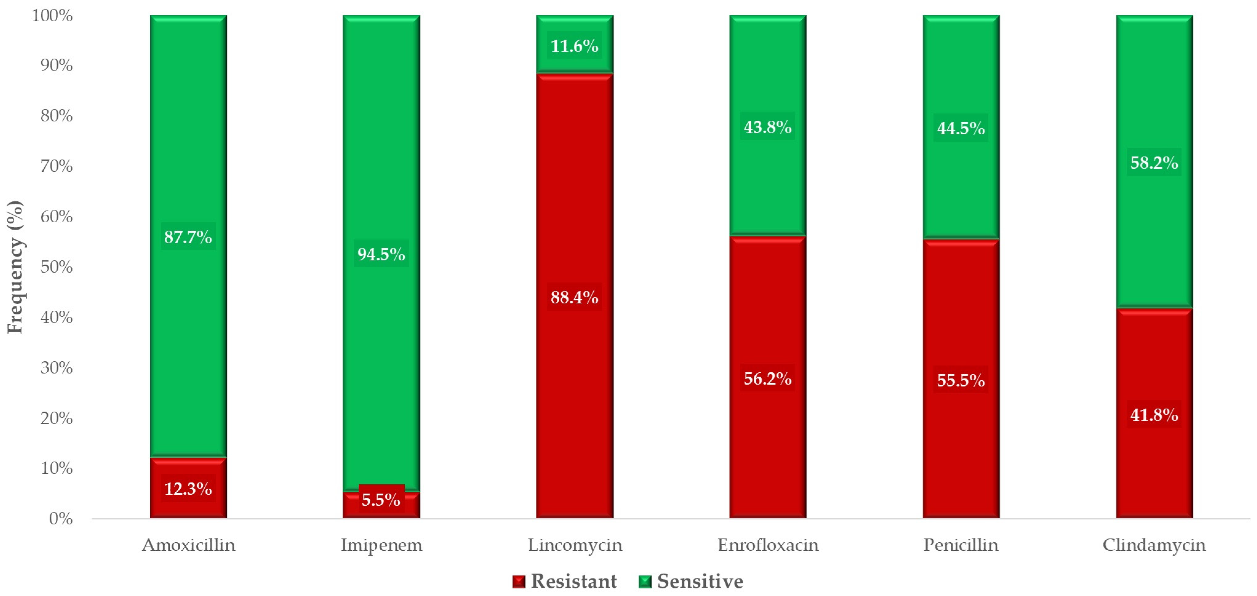
For antibiotics with established clinical breakpoints, we determined the phenotypic clinical susceptibility of the examined population (Figure 4). A significant proportion of the isolates retained susceptibility to amoxicillin (87.7%), one of the primary choices for treating C. perfringens-associated infections. However, sensitivity to penicillin was markedly reduced, with only 44.5% of isolates being susceptible. Imipenem, a critically important antibiotic for treating human multidrug-resistant infections, exhibited 94.5% susceptibility. The highest level of resistance was observed against lincomycin, with only 11.6% of isolates demonstrating susceptibility.
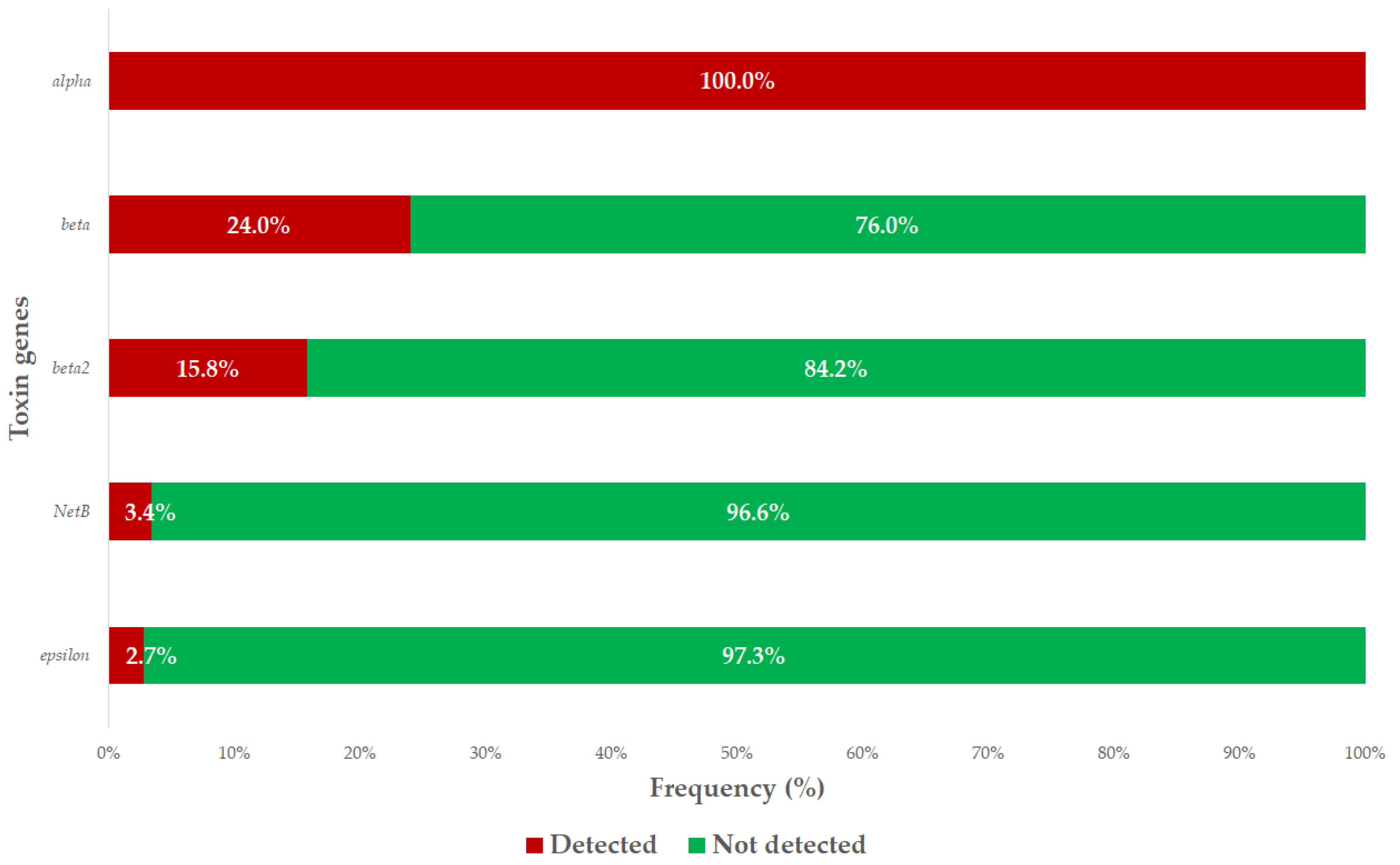
We conducted PCR analysis of the isolates to identify those genes responsible for major and minor toxin production. All examined isolates tested positive for the major alpha toxin gene, which is universally present in C. perfringens strains. Among the isolates, 23.9% were positive for the major beta toxin gene, 2.7% for the major epsilon toxin gene, 15.8% for the minor beta2 toxin gene, and 3.4% for the minor NetB toxin gene. No isolates tested positive for the major iota toxin or the minor enterotoxin genes. The prevalence of each toxin gene is summarized in Figure 5, providing a visual overview of the virulence-associated genetic potential across the tested isolates.
When examining the co-occurrence of multiple toxin genes within individual isolates (excluding the universally present major alpha gene), we observed the following: the highest number of simultaneous toxin genes (major beta, epsilon, minor beta2, and minor NetB) occurred in a single isolate. The combination of major beta, minor beta2, and minor NetB genes was detected in 4 isolates; major beta, major epsilon, and minor beta2 genes co-occurred in 3 isolates; major beta and minor beta2 genes were found together in 10 isolates; and major beta and major epsilon genes co-occurred in 1 isolate.
We also investigated potential associations between the presence of specific toxin genes and the MIC values for each antibiotic. However, no statistically significant differences in MIC values were observed between isolates carrying specific toxin genes and those without these genes for any of the antibiotics tested.

3. Discussion

In this study, we determined the MIC values of 146 C. perfringens strains isolated from turkeys for antibiotics of public and veterinary health significance. To complement our findings, we conducted PCR analyses to identify the presence of major and minor toxin-producing genes and to explore potential correlations between antimicrobial resistance patterns and the presence of these toxin genes.
Regarding beta-lactam antibiotics, our study on commensal C. perfringens strains (n = 146) revealed a 55.5% resistance rate to penicillin, with MIC50 and MIC90 values of 1 µg/mL and 32 µg/mL, respectively. In contrast, Slavic et al. did not identify any penicillin-resistant strains, reporting MIC50 and MIC90 values of 0.06 µg/mL and 0.12 µg/mL, respectively [16]. Similarly, Silva et al. found no penicillin-resistant strains in isolates from broiler chickens, with MIC values not exceeding 0.25 µg/mL [41]. For amoxicillin, we observed 12.3% resistance, whereas Martel et al. found no resistant strains in broiler-derived isolates, and Osman and Elhariri reported a 7% resistance rate [42,43]. Regarding imipenem, our study demonstrated a 5.5% resistance rate, while Akhi et al. reported a significantly higher resistance rate of 38% in C. perfringens strains isolated from human fecal samples. Akhi et al. documented MIC values reaching as high as 32 µg/mL, whereas the highest MIC value we observed for imipenem was 16 µg/mL [44]. For ceftriaxone, Akhi et al. reported MIC values ranging from 0.016 to 128 µg/mL, while our study identified strains with MIC values as high as 256 µg/mL [44].
These findings are consistent with the global AMR trends in C. perfringens reported in various regions, including Asia, North America, and the Middle East, where increasing resistance to beta-lactams, fluoroquinolones, and lincosamides has been documented. This underscores the widespread and evolving nature of AMR in both food-producing animals and the environment [45,46,47,48].
Penicillins are considered first-line antibiotics for treating infections caused by C. perfringens. The observed differences in susceptibility can likely be attributed to variations in antibiotic usage patterns and the overuse of these agents over several decades. Overall, our findings indicate a significant decrease in sensitivity to penicillin, while amoxicillin has largely retained its efficacy. This reduction in effectiveness is an undesirable trend, highlighting the importance of regular monitoring studies. Imipenem, categorized as a lifesaving “last resort” antibiotic, holds critical significance for human health, warranting constant surveillance. Although the observed resistance rate of 5.5% may seem low, the presence of any resistant strains at all is concerning. However, this figure is more likely to be a result of the instability of the antibiotic’s aqueous solution rather than true resistance [49]. For amoxicillin-clavulanic acid, we recorded MIC50 and MIC90 values of 0.5 µg/mL and 8 µg/mL, respectively. Comparative data specific to poultry are lacking; however, Park et al. reported that 84% of strains isolated from horses were susceptible to amoxicillin-clavulanic acid [50]. Since C. perfringens strains are generally not known to produce beta-lactamase enzymes, the use of clavulanic acid—already unauthorized for poultry—is not justified in practice for treating C. perfringens infections.
Among the lincosamides, only 11.6% of the strains we tested were sensitive to lincomycin, which contrasts sharply with the findings of Silva et al., who reported that 89.1% of their isolates were sensitive to this antibiotic. While Silva et al. did not observe MIC values exceeding 64 µg/mL, we recorded MIC values as high as 1024 µg/mL [41]. Gholamiandeh-Kordi et al. and Martel et al. reported resistance rates of 61.5% and 63.3%, respectively, which are closer to our observed resistance rate of 89.1% [46,47]. In contrast, Osman and Elhariri did not identify any lincomycin-sensitive strains [43]. For clindamycin, another antibiotic in this group, we observed a resistance rate of 41.8%, with MIC50 and MIC90 values of 2 µg/mL and 32 µg/mL, respectively. Slavic et al., however, reported a resistance rate of only 2%, with MIC50 and MIC90 values of 1 µg/mL and 4 µg/mL, respectively [16].
The high resistance rates that we observed for clindamycin are concerning, particularly given its broad spectrum of activity. Since clindamycin is primarily used in companion animals and human medicine, its importance is founded mainly on in vitro studies and in the context of cross-resistance with lincomycin.
For the nitroimidazole antibiotic metronidazole, the MIC50 and MIC90 values of the strains we tested were 32 µg/mL and 512 µg/mL, respectively. In contrast, Slavic et al. reported much lower MIC values of 1 µg/mL (MIC50) and 4 µg/mL (MIC90) [16]. For ronidazole, we observed MIC50 and MIC90 values of 32 µg/mL and 256 µg/mL, respectively, whereas Dutta et al. found no strains with MIC values exceeding 0.5 µg/mL in bird-derived isolates [51]. The relevance of nitroimidazoles lies primarily in research contexts, as their use is prohibited in food-producing animals.
Regarding enrofloxacin, the results differ widely. In this study, 56.2% of the strains were resistant. In human cases, second-generation fluoroquinolones, such as enrofloxacin, are effective against anaerobic bacteria like C. perfringens in only about half of the cases, which fits with our results. However, in 2013, Osman and Elhariri reported an 82% resistance rate to enrofloxacin among isolates from broiler chickens in Egypt [43]. In comparison, a 2022 study found that 98% of the C. perfringens strains tested were sensitive to enrofloxacin [52]. These notable differences in resistance suggest that AMR in C. perfringens is constantly developing in isolated colonies, which leads to varying levels of resistance. Second-generation fluoroquinolones, such as enrofloxacin, are effective against anaerobic bacteria like C. perfringens in only about half of the cases [53]. This limited efficacy likely explains the high resistance rates observed. Consequently, the use of these antibiotics should be reserved for complicated infections where a broad-spectrum agent is necessary.
For turkey-derived strains of C. perfringens, Watkins et al. reported MIC50 and MIC90 values of 2 µg/mL and 256 µg/mL, respectively, for tylosin and tilmicosin [54]. In comparison, our study found MIC50 values of 1 µg/mL for tylosin and 16 µg/mL for tilmicosin, with MIC90 values of 64 µg/mL for both antibiotics. Although the MIC50 values we calculated are more favorable on a population level, the less favorable MIC90 values indicate potential challenges in population management. The primary limitation of this analysis lies in the relatively small sample size, which may have influenced the findings. However, discrepancies with other studies may also stem from regional differences in antimicrobial usage practices, animal husbandry systems, biosecurity measures, and the genetic variability of circulating C. perfringens strains—all of which warrant further comparative investigation. Future studies incorporating larger sample sizes could provide a more accurate assessment.
Additionally, the overuse of tetracyclines over the decades has significantly impacted their effectiveness, as evidenced by the resistance patterns observed. For oxytetracycline, we observed MIC50 and MIC90 values of 16 µg/mL and 32 µg/mL, respectively, while Gharaibeh et al. reported 0.5 µg/mL for MIC50 and 256 µg/mL for MIC90 [55]. This broad range of MIC values is likely attributable to differences in sample sizes, highlighting the need for larger-scale, standardized studies.
Vancomycin is now exclusively used in public health settings for the treatment of life-threatening infections caused by Gram-positive bacteria, and resistance to this drug may indicate a troubling trend. In their experiment, Johansson et al. did not report MIC values exceeding 4 µg/mL [30], whereas our study found MIC50 and MIC90 values of 1 µg/mL and 32 µg/mL, respectively. Although there are no clinical breakpoints for the veterinary use of vancomycin, due to its critical importance in human medicine, the higher MIC in our study is concerning as it suggests that C. perfringens is developing resistance to even little-used antibiotics. As vancomycin is not approved for veterinary use, the turkeys will not have come into contact with it. Therefore, the observed resistance may be a result of genetic transfer. Next-generation sequencing would shed more light on the mechanisms driving resistance development in this case. Due to its critical importance in human medicine, no clinical breakpoints are available for veterinary use, further highlighting the need for careful stewardship.
The detection of elevated vancomycin MICs in commensal C. perfringens strains from turkeys raises important concerns, even though this antibiotic is not used in poultry. The presence of resistance may be due to horizontal gene transfer via plasmids or transposons, as demonstrated in other Gram-positive species such as Enterococcus and Staphylococcus. If such resistance elements are mobile, they may spread within the poultry microbiota or could even cross species barriers. From a food safety and One Health perspective, this could pose a significant risk, especially if resistant strains enter the human food chain. This further underlines the need for genomic surveillance and prudent antimicrobial stewardship, even in cases where antibiotics are not directly applied in animal production [56,57,58].
In our study, we examined the presence of major and minor toxin genes in 146 commensal C. perfringens isolates, including those encoding major alpha, beta, epsilon, and iota toxins, as well as minor beta2, enterotoxin, and NetB toxins. The alpha toxin gene, as expected, was present in all isolates, consistent with its universal expression in C. perfringens. Additionally, 3.4% of isolates carried the minor NetB toxin gene. Lyhs et al. investigated 212 C. perfringens isolates, of which 174 were derived from diseased turkeys and 38 from healthy individuals. Similar to our findings, Lyhs et al. detected the alpha toxin gene in all isolates. However, unlike our results, they reported the presence of the NetB toxin gene exclusively in isolates from turkeys suffering from necrotic enteritis, with a prevalence of 8%, and did not detect this gene in commensal isolates [59]. In our study, 15.8% of isolates carried the beta2 toxin gene, whereas Lyhs et al. found this in only one isolate [59]. Additionally, Lyhs et al. did not detect the genes encoding major beta, epsilon, or iota toxins in their isolates, whereas we found these genes in 23.9%, 2.7%, and 0% of our isolates, respectively. We also did not detect the minor enterotoxin gene in any isolate, which is consistent with the findings of Mohiuddin et al., who observed the NetB toxin gene in two poultry-derived isolates but did not detect the genes encoding major beta, epsilon, iota, or minor enterotoxins [28]. Slavic et al. identified the epsilon toxin gene in one turkey-derived isolate, which also harbored the beta toxin gene. Furthermore, they found the minor beta2 toxin gene in 90% of turkey-derived isolates. Although we did not observe statistically significant associations between the presence of virulence genes and resistance profiles, Slavic et al. reported that C. perfringens isolates that were resistant to clindamycin and erythromycin derived from swine were significantly less likely to carry the beta2 toxin gene. However, no significant correlations between antibiotic susceptibility and toxin gene presence were found for isolates from cattle, chickens, or turkeys [16]. While our study did not reveal significant differences between antibiotic susceptibility and the presence of toxin genes, Wei et al. reported significant differences for clindamycin among C. perfringens isolates derived from healthy chickens and from those with necrotic enteritis. The isolates from diseased chickens exhibited lower susceptibility to clindamycin and gentamicin [60].
The detection of virulence-associated genes in the isolates from turkeys in Hungary may have important implications for local poultry health. Although our study focused on apparently healthy birds, the presence of these toxin genes—especially in antimicrobial-resistant strains—suggests that subclinical reservoirs of potentially pathogenic C. perfringens are currently circulating within flocks. These strains could act as a source of infection under stress or immunosuppressive conditions, thereby increasing the risk of necrotic enteritis outbreaks. This highlights the importance of integrating toxin gene monitoring into AMR surveillance frameworks, particularly in countries like Hungary with intensive turkey production systems.
Based on the findings reported in the literature, it can be hypothesized that there are significant differences in the relationship between the presence of virulence factors and resistance profiles. One key limitation of our study was the sample size, which likely constrained our ability to detect such associations. Although our analyses did not reveal any significant differences linked to the presence or absence of virulence genes, the correlations reported in previous studies emphasize the importance of continuing investigations with larger sample sizes in the future.
Nonetheless, certain non-significant trends in our dataset, such as slightly higher resistance rates among beta2-positive isolates, may suggest a biological interplay between toxin gene carriage and resistance mechanisms. It is plausible that mobile genetic elements harboring both toxin and resistance determinants contribute to such a co-occurrence, as seen in other Clostridium species. These potential links merit further exploration using genomic approaches such as plasmid or transposon mapping.
The limitations of this study include its moderate sample size, potential sampling bias due to the opportunistic nature of strain collection from specific farms, and the cross-sectional design. Longitudinal monitoring would be necessary to track temporal resistance trends and to establish causal relationships between antimicrobial use and resistance emergence.
From a practical perspective, the resistance patterns observed in this study highlight the pressing need for improved antimicrobial stewardship in turkey production. Regular monitoring of resistance profiles at the farm level, along with regionally tailored treatment protocols, can help preserve antibiotic efficacy. Furthermore, the integration of non-antibiotic alternatives—such as competitive exclusion products, essential oils, or vaccination—should be encouraged as part of a holistic AMR management strategy. Promising non-antibiotic interventions—such as medium-chain fatty acids, essential oils, competitive exclusion cultures, and bacteriophage therapy—may offer effective preventive options, particularly when integrated with enhanced biosecurity and vaccination programs.

4. Materials and Methods

4.1. The Origin of the Strains

The examined strains were collected between February 2022 and May 2023 as part of routine diagnostic sampling conducted by veterinarians on large-scale livestock farms. For each sample, information was collected regarding the organ of origin (e.g., the trachea or cloaca) and the geographic location (the specific municipality). Based on the municipalities, the samples were categorized into one of Hungary’s seven administrative regions.
Selective isolation of the strains was performed using CHROMagar™ C. perfringens (Chebio Fejlesztő Kft., Budapest, Hungary), and anaerobic cultivation and testing were carried out using the BD GasPak™ system (VWR International Kft., Debrecen, Hungary). Pure cultures were prepared on tryptone soy agar (Biolab Zrt., Budapest, Hungary) and subsequently preserved using the Microbank™ system (Pro-Lab Diagnostics, Richmond Hill, ON, Canada) at −80 °C.
Based on the municipalities, the samples were categorized into one of Hungary’s seven administrative regions. This regional classification allows for the assessment of potential geographic patterns in antimicrobial resistance, although uneven sample distribution may influence the representativeness of certain regions.

4.2. Minimum Inhibitory Concentration (MIC) Determination

The phenotypic expression of AMR was assessed by determining the MIC values for each bacterial strain. The methodology followed the guidelines of the Clinical Laboratory Standard Institute (CLSI) [61], with breakpoints established according to the CLSI recommendations [62]. Where available, the results were also compared with the ECOFF values defined by the European Committee on Antimicrobial Susceptibility Testing (EUCAST).
Bacterial strains stored at −80 °C were resuspended in 3 mL of cation-adjusted Müller–Hinton broth (CAMHB) the day before testing and incubated at 37 °C for 18–24 h. The MIC testing was performed using 96-well microtiter plates (VWR International, LLC., Debrecen, Hungary). Except for the first column, all wells were filled with 90 µL of CAMHB. Stock solutions of the tested antimicrobial agents (Merck KGaA, Darmstadt, Germany) were prepared at 1024 µg/mL according to CLSI guidelines [63]. From the 512 µg/mL half-diluted solution, 180 µL was transferred into the first column of the microtiter plates, and a twofold serial dilution was performed across the plate. After the 10th column, 90 µL of excess solution was discarded, leaving 90 µL in each well. Using a nephelometer (ThermoFisher Scientific, Budapest, Hungary), bacterial suspensions were adjusted to a turbidity of 0.5 McFarland and inoculated into the microtiter plates starting from the 11th column, with 10 µL added per well [63]. The MIC values were determined using the Sensititre™ SWIN™ automatic MIC reader (ThermoFisher Scientific, Budapest, Hungary) and analyzed with the VIZION system software, version 3.4 (ThermoFisher Scientific, Budapest, Hungary, 2024). The reference quality control strain used was C. perfringens (ATCC 13124).

4.3. PCR Tests

We performed PCR analysis to map the major and minor toxin gene profiles of each strain. DNA extraction from bacterial suspensions was carried out using the Zymo Quick-DNA Fungal/Bacterial Miniprep Kit (Zymo Research, Murphy Ave., Irvine, CA, USA) following the manufacturer’s instructions. For bead-beating to release bacterial genetic material, we used the Qiagen TissueLyzer LT (Qiagen GmBH, Hilden, Germany) at 50 Hz for 5 min.
For PCR testing, we utilized the Kylt Clostridium perfringens real-time PCR kit (Kylt, Höltinghausen, Germany), which is designed for the detection of major (alpha, beta, epsilon, and iota) and minor (b2, entero, and NetB) toxin genes in avian samples. The kit comprises a dual multiplex real-time PCR system containing all necessary reagents, including primers, probes, positive and negative controls, and an internal amplification control, ensuring standardized detection and internal quality control. All procedures followed the manufacturer’s protocol, and the extracted nucleic acids were stored at −20 °C until use.
PCR analyses were conducted using the CFX Opus Dx real-time PCR system (Bio-Rad Hungary Ltd., Budapest, Hungary). The results were evaluated using the Bio-Rad CFX Maestro software, version 5.3.022.1030 (Bio-Rad Hungary Ltd., Budapest, Hungary).
Amplification was performed in a total volume of 20 µL, including 18 µL of Kylt Reaction-Mix (Multiplex 1 or 2) and 2 µL of sample DNA. The thermal cycling protocol consisted of initial enzyme activation at 95 °C for 10 min, followed by 42 cycles of denaturation at 95 °C for 15 s and annealing/extension at 60 °C for 60 s. Fluorescence was measured at each cycle using the FAM, Cy5, TXR, and HEX channels. Each run included positive controls for both multiplex reactions and a negative control, in accordance with the manufacturer’s instructions.
The presence of the alpha gene served as an internal amplification control, as it is universally present in all C. perfringens strains. Reactions were only deemed valid if all control curves behaved according to protocol-defined expectations.

4.4. Statistic Analysis

Analysis of the data was performed using R version 4.1.0 [64]. The normality of data distribution was assessed using the Shapiro–Wilk test [65]. For datasets that did not conform to normal distribution assumptions, non-parametric statistical methods were applied. Specifically, the Kruskal–Wallis test [66] was used to evaluate resistance levels across multiple sample groups. This non-parametric approach is particularly suited for comparing medians among groups without assuming normality. To pinpoint specific differences between the groups, post hoc analyses were conducted using pairwise comparisons via the Mann–Whitney U-test [67] and t-tests, where appropriate [68]. To mitigate the risk of Type I errors caused by multiple comparisons, Bonferroni correction was applied [69], recognizing that this method may increase the likelihood of Type II errors (overlooking true differences).
To explore the relationships among active substances, correlation analysis was employed. Additionally, PCA [70] was conducted to uncover underlying patterns and to visualize similarities or differences within the data. PCA was used to reduce the dimensionality of the dataset while retaining as much variance as possible. This approach is particularly useful for datasets with numerous variables, which can complicate analysis and visualization. PCA transforms the data into new variables called principal components, with the first component accounting for the largest variance in the dataset. Subsequent components explain progressively smaller amounts of variance. These components are orthogonal, meaning that they are uncorrelated with one another, thereby simplifying the interpretation of complex datasets.
Hierarchical cluster analysis followed, with the results presented as a dendrogram [71]. This visual representation effectively illustrates the clustering of isolates and the hierarchical relationships among them. Cluster analysis is a method for identifying patterns in data by grouping similar observations into clusters. Each cluster contains data points that share common characteristics, distinct from those in other clusters. Hierarchical clustering results were depicted using a dendrogram, a tree-like diagram illustrating the hierarchy and relationships among clusters.
The use of PCA prior to clustering served two purposes: to reduce dimensionality while preserving major variance components, and to eliminate multicollinearity among antimicrobial variables. Hierarchical clustering was performed on the principal component scores using Euclidean distance and Ward’s method, which ensures compact, interpretable clusters based on overall similarity patterns. This combined approach facilitates robust classification while minimizing noise from irrelevant variables.
Correlation analysis focuses on assessing the strength and direction of relationships between variables. It evaluates how changes in one variable correspond to changes in another. A positive correlation implies that as one variable increases, the other also increases, whereas a negative correlation indicates an inverse relationship. The strength of the correlation is quantified by the correlation coefficient, ranging from −1 to +1. A coefficient of +1 denotes a perfect positive correlation, −1 represents a perfect negative correlation, and 0 indicates no linear relationship.

5. Conclusions

Overall, our findings underscore the critical importance of the continuous monitoring of AMR. While our results were consistent with previous studies in several cases, notable discrepancies were observed. These variations are likely attributable to the low sample size in our study. However, other factors such as country-specific antibiotic usage practices, differences in flock sizes, variations in the antibiotics administered, and treatment durations during rearing may also play significant roles.
As for enrofloxacin, the varying levels of resistance to this antibiotic are likely to be the result of isolated developments. This is concerning, as the inconsistency between strains will make C. perfringens harder to treat. Consequently, to prevent resistance developing further, the use of enrofloxacin should be reserved for complicated infections where a broad-spectrum agent is necessary.
Based on these observations, future studies should aim to increase sample sizes significantly and include isolates derived from clinical cases alongside commensal strains. The inclusion of clinical strains could reveal more pronounced differences in resistance patterns, particularly concerning the presence of virulence-associated genes. Furthermore, multidrug-resistant strains should be subjected to next-generation sequencing to comprehensively explore the genetic mechanisms underlying phenotypic resistance. This approach could provide a deeper understanding of resistance patterns and contribute to the development of targeted interventions.
Incorporating clinical isolates in addition to commensal strains could provide particularly valuable insights. Comparing these datasets with an adequate sample size would likely yield even more meaningful results, enhancing our understanding of the complex interplay between virulence factors and antimicrobial resistance.
In addition to scientific insights, these findings carry important implications for policy and on-farm decision-making. Regulatory bodies should support the development of national AMR surveillance frameworks tailored to species and regional profiles, while restricting the use of critically important antibiotics, such as fluoroquinolones, in preventive protocols. Promoting antimicrobial stewardship training and incentivizing the adoption of non-antibiotic alternatives—such as probiotics, vaccines, or feed-based interventions—may also play a pivotal role in reducing selection pressure and safeguarding antimicrobial efficacy.

Supplementary Materials

The following supporting information can be downloaded at: https://www.mdpi.com/article/10.3390/antibiotics14040413/s1. Table S1: Frequency table of the minimum inhibitory concentration (MIC) values (µg/mL) for agents without breakpoints in Clostridium perfringens samples (n=146) derived from turkeys. Table S2. Additional data.

Author Contributions

Conceptualization, Á.K. and Á.J.; methodology, Á.K.; software, Á.K.; validation, Á.J.; formal analysis, Á.S.; investigation, Á.J.; resources, Á.K. and L.K.; data curation, Á.S.; writing—original draft preparation, Á.S.; writing—review and editing, Á.K.; visualization, Á.S.; supervision, Á.J.; project administration, Á.S., F.B. and B.C.; funding acquisition, Á.J. All authors have read and agreed to the published version of the manuscript.

Funding

This research was funded by project no. RRF-2.3.1-21-2022-00001 and was implemented with the support provided by the Recovery and Resilience Facility (RRF), financed under the National Recovery Fund budget estimate, the RRF-2.3.1-21 funding scheme.

Institutional Review Board Statement

Not applicable.

Informed Consent Statement

Not applicable.

Data Availability Statement

The data presented in this study are available from the corresponding author upon reasonable request.

Acknowledgments

We would like to thank Katalin Balogh and Tamásné Pénzes Imre for the preparations for the laboratory work.

Conflicts of Interest

Author László Kovács was employed by the company Poultry-Care Kft. The remaining authors declare that the research was conducted in the absence of any commercial or financial relationships that could be construed as a potential conflict of interest.

References

  1. Murray, C.J.L.; Ikuta, K.S.; Sharara, F.; Swetschinski, L.; Aguilar, G.R.; Gray, A.; Han, C.; Bisignano, C.; Rao, P.; Wool, E.; et al. Global Burden of Bacterial Antimicrobial Resistance in 2019: A Systematic Analysis. Lancet 2022, 399, 629–655. [Google Scholar] [CrossRef] [PubMed]
  2. World Bank. People, Pathogens and Our Planet: The Economics of One Health; World Bank: Washington, DC, USA, 2012. [Google Scholar]
  3. Prestinaci, F.; Pezzotti, P.; Pantosti, A. Antimicrobial Resistance: A Global Multifaceted Phenomenon. Pathog. Glob. Health 2015, 109, 309–318. [Google Scholar] [CrossRef] [PubMed]
  4. Dehove, A.; Commault, J.; Petitclerc, M.; Teissier, M.; Macé, J. Economic Analysis and Costing of Animal Health: A Literature Review of Methods and Importance. Rev. Sci. Tech. 2012, 31, 605–617+591–604. [Google Scholar] [CrossRef]
  5. Prescott, J.F. The Resistance Tsunami, Antimicrobial Stewardship, and the Golden Age of Microbiology. Vet. Microbiol. 2014, 171, 273–278. [Google Scholar] [CrossRef]
  6. Samreen; Ahmad, I.; Malak, H.A.; Abulreesh, H.H. Environmental Antimicrobial Resistance and Its Drivers: A Potential Threat to Public Health. J. Glob. Antimicrob. Resist. 2021, 27, 101–111. [Google Scholar] [CrossRef]
  7. Gibson, M.K.; Forsberg, K.J.; Dantas, G. Improved Annotation of Antibiotic Resistance Determinants Reveals Microbial Resistomes Cluster by Ecology. ISME J. 2015, 9, 207. [Google Scholar] [CrossRef]
  8. Timbermont, L.; Haesebrouck, F.; Ducatelle, R.; Van Immerseel, F. Necrotic Enteritis in Broilers: An Updated Review on the Pathogenesis. Avian Pathol. 2011, 40, 341–347. [Google Scholar] [CrossRef]
  9. Kiu, R.; Hall, L.J. An Update on the Human and Animal Enteric Pathogen Clostridium perfringens. Emerg. Microbes Infect. 2018, 7, 1–15. [Google Scholar] [CrossRef]
  10. Komatsu, H.; Inui, A.; Sogo, T.; Fujisawa, T. Clostridium perfringens. Nihon rinsho. Jpn. J. Clin. Med. 2012, 70, 1357–1361. [Google Scholar]
  11. Mehdizadeh Gohari, I.; Navarro, M.A.; Li, J.; Shrestha, A.; Uzal, F.; McClane, B.A. Pathogenicity and Virulence of Clostridium perfringens. Virulence 2021, 12, 723–753. [Google Scholar] [CrossRef]
  12. Joshy, L.; Chaudhry, R.; Dhawan, B.; Kumar, L.; Das, B.K. Incidence and Characterization of Clostridium perfringens Isolated from Antibiotic-Associated Diarrhoeal Patients: A Prospective Study in an Indian Hospital. J. Hosp. Infect. 2006, 63, 323–329. [Google Scholar] [CrossRef] [PubMed]
  13. Rood, J.I.; Adams, V.; Lacey, J.; Lyras, D.; McClane, B.A.; Melville, S.B.; Moore, R.J.; Popoff, M.R.; Sarker, M.R.; Songer, J.G.; et al. Expansion of the Clostridium perfringens Toxin-Based Typing Scheme. Anaerobe 2018, 53, 5–10. [Google Scholar] [CrossRef] [PubMed]
  14. Camargo, A.; Ramírez, J.D.; Kiu, R.; Hall, L.J.; Muñoz, M. Unveiling the Pathogenic Mechanisms of Clostridium perfringens Toxins and Virulence Factors. Emerg. Microbes Infect. 2024, 13, 2341968. [Google Scholar] [CrossRef]
  15. Ronco, T.; Stegger, M.; Ng, K.L.; Lilje, B.; Lyhs, U.; Andersen, P.S.; Pedersen, K. Genome Analysis of Clostridium perfringens Isolates from Healthy and Necrotic Enteritis Infected Chickens and Turkeys. BMC Res. Notes 2017, 10, 270. [Google Scholar] [CrossRef]
  16. Slavić, D.; Boerlin, P.; Fabri, M.; Klotins, K.C.; Zoethout, J.K.; Weir, P.E.; Bateman, D. Antimicrobial Susceptibility of Clostridium perfringens Isolates of Bovine, Chicken, Porcine, and Turkey Origin from Ontario. Can. J. Vet. Res. 2011, 75, 89–97. [Google Scholar]
  17. Gazdzinski, P.; Julian, R.J. Necrotic Enteritis in Turkeys. Avian Dis. 1992, 36, 792–798. [Google Scholar] [CrossRef]
  18. Smyth, J.A.; Mishra, N.; Shivaprasad, H.L. Toxinotyping of Clostridium perfringens Strains Recovered from U.S. Turkeys with Necrotic Enteritis. Avian Dis. 2022, 66, 193–196. [Google Scholar] [CrossRef]
  19. Hardy, S.P.; Benestad, S.L.; Hamnes, I.S.; Moldal, T.; David, B.; Barta, J.R.; Reperant, J.-M.; Kaldhusdal, M. Developing an Experimental Necrotic Enteritis Model in Turkeys—The Impact of Clostridium perfringens, Eimeria meleagrimitis and Host Age on Frequency of Severe Intestinal Lesions. BMC Vet. Res. 2020, 16, 63. [Google Scholar] [CrossRef]
  20. Thachil, A.J.; McComb, B.; Andersen, M.M.; Shaw, D.P.; Halvorson, D.A.; Nagaraja, K.V. Role of Clostridium perfringens and Clostridium septicum in Causing Turkey Cellulitis. Avian Dis. 2010, 54, 795–801. [Google Scholar] [CrossRef]
  21. Clark, S.; Porter, R.; McComb, B.; Lipper, R.; Olson, S.; Nohner, S.; Shivaprasad, H.L. Clostridial Dermatitis and Cellulitis: An Emerging Disease of Turkeys. Avian Dis. 2010, 54, 788–794. [Google Scholar] [CrossRef]
  22. Forga, A.; Robbins, K.; Smith, A.; Coles, M.; Tellez-Isaias, G.; Vuong, C.N.; Hargis, B.; Graham, D. Evaluation of Clostridium septicum Hemolytic Activity, Administration Route, and Dosage Volume of a Clostridial Dermatitis (Cellulitis) Bacterin-Toxoid on Humoral Immune Response in Commercial Turkeys. Poult. Sci. 2023, 102, 102873. [Google Scholar] [CrossRef] [PubMed]
  23. Huff, G.R.; Huff, W.E.; Rath, N.C. Dexamethasone Immunosuppression Resulting in Turkey Clostridial Dermatitis: A Retrospective Analysis of Seven Studies, 1998–2009. Avian Dis. 2013, 57, 730–736. [Google Scholar] [CrossRef] [PubMed]
  24. Agunos, A.; Carson, C.; Léger, D. Antimicrobial Therapy of Selected Diseases in Turkeys, Laying Hens, and Minor Poultry Species in Canada. Can. Vet. J. 2013, 54, 1041–1052. [Google Scholar]
  25. Yadav, J.P.; Das, S.C.; Dhaka, P.; Vijay, D.; Kumar, M.; Mukhopadhyay, A.K.; Chowdhury, G.; Chauhan, P.; Singh, R.; Dhama, K.; et al. Molecular Characterization and Antimicrobial Resistance Profile of Clostridium perfringens Type A Isolates from Humans, Animals, Fish and Their Environment. Anaerobe 2017, 47, 120–124. [Google Scholar] [CrossRef]
  26. Grass, J.E.; Gould, L.H.; Mahon, B.E. Epidemiology of Foodborne Disease Outbreaks Caused by Clostridium perfringens, United States, 1998–2010. Foodborne Pathog. Dis. 2013, 10, 131–136. [Google Scholar] [CrossRef]
  27. Scharff, R.L. Economic Burden from Health Losses Due to Foodborne Illness in the United States. J. Food Prot. 2012, 75, 123–131. [Google Scholar] [CrossRef]
  28. Mohiuddin, M.; Song, Z.; Liao, S.; Qi, N.; Li, J.; Lv, M.; Lin, X.; Cai, H.; Hu, J.; Liu, S.; et al. Animal Model Studies, Antibiotic Resistance and Toxin Gene Profile of NE Reproducing Clostridium perfringens Type A and Type G Strains Isolated from Commercial Poultry Farms in China. Microorganisms 2023, 11, 622. [Google Scholar] [CrossRef]
  29. Hassani, S.; Pakbin, B.; Brück, W.M.; Mahmoudi, R.; Mousavi, S. Prevalence, Antibiotic Resistance, Toxin-Typing and Genotyping of Clostridium perfringens in Raw Beef Meats Obtained from Qazvin City, Iran. Antibiotics 2022, 11, 340. [Google Scholar] [CrossRef]
  30. Johansson, A.; Greko, C.; Engström, B.E.; Karlsson, M. Antimicrobial Susceptibility of Swedish, Norwegian and Danish Isolates of Clostridium perfringens from Poultry, and Distribution of Tetracycline Resistance Genes. Vet. Microbiol. 2004, 99, 251–257. [Google Scholar] [CrossRef]
  31. Hetényi, N.; Bersényi, A.; Hullár, I. Physiological Effects of Medium-Chain Fatty Acids and Triglycerides, and Their Potential Use in Poultry and Swine Nutrition: A Literature Review. Magy. Állatorvosok Lapja 2024, 146, 651–659. [Google Scholar] [CrossRef]
  32. Sebők, C.; Márton, R.A.; Meckei, M.; Neogrády, Z.; Mátis, G. Antimicrobial Peptides as New Tools to Combat Infectious Diseases. Magy. Állatorvosok Lapja 2024, 146, 181–191. [Google Scholar] [CrossRef]
  33. Jerzsele, Á.; Somogyi, Z.; Szalai, M.; Kovács, D. Effects of Fermented Wheat Germ Extract on Artificial Salmonella Typhimurium Infection in Broiler Chickens. Magy. Állatorvosok Lapja 2020, 142, 77–85. [Google Scholar]
  34. Kovács, L.; Nagy, D.; Könyves, L.; Jerzsele, Á.; Kerek, Á. Antimicrobial Properties of Essential Oils—Animal Health Aspects. Magy. Állatorvosok Lapja 2023, 145, 497–510. [Google Scholar] [CrossRef]
  35. Olasz, Á.; Jerzsele, Á.; Balta, L.; Dobra, P.F.; Kerek, Á. In Vivo Efficacy of Different Extracts of Propolis in Broiler Salmonellosis. Magy. Állatorvosok Lapja 2023, 145, 461–475. [Google Scholar] [CrossRef]
  36. Kerek, Á.; Csanády, P.; Jerzsele, Á. Antibacterial Efficiency of Propolis—Part 1. Magy. Állatorvosok Lapja 2022, 144, 285–298. [Google Scholar]
  37. Farkas, M.; Könyves, L.; Csorba, S.; Farkas, Z.; Józwiák, Á.; Süth, M.; Kovács, L. Biosecurity Situation of Large-Scale Poultry Farms in Hungary According to the Databases of National Food Chain Safety Office Centre for Disease Control and Biosecurity Audit System of Poultry Product Board of Hungary in the Period of 2021–2022. Magy. Állatorvosok Lapja 2024, 146, 723–742. [Google Scholar] [CrossRef]
  38. Mag, P.; Németh, K.; Somogyi, Z.; Jerzsele, Á. Antibacterial therapy based on pharmacokinetic/ pharmacodynamic models in small animal medicine-1. Literature review. Magy. Állatorvosok Lapja 2023, 145, 419–438. [Google Scholar] [CrossRef]
  39. Essősy, M.; Fodor, I.; Ihnáth, Z.; Karancsi, Z.; Kovács, D.; Szalai, K.V.; Szentmiklósi, D.; Jerzsele, Á. The Possibilities of Antibiotic-Free Broiler-Hen Fattening, with Special Reference to the Use of Pre- and Probiotics. Magy. Állatorvosok Lapja 2020, 142, 397–407. [Google Scholar]
  40. Kovács, D.; Palkovicsné Pézsa, N.; Farkas, O.; Jerzsele, Á. Usage of Antibiotic Alternatives in Pig Farming: Literature Review. Magy. Állatorvosok Lapja 2021, 143, 281–282. [Google Scholar]
  41. Silva, R.O.S.; Salvarani, F.M.; Assis, R.A.; Martins, N.R.S.; Pires, P.S.; Lobato, F.C.F. Antimicrobial Susceptibility of Clostridium perfringens Strains Isolated from Broiler Chickens. Braz. J. Microbiol. 2009, 40, 262–264. [Google Scholar] [CrossRef]
  42. Martel, A.; Devriese, L.A.; Cauwerts, K.; De Gussem, K.; Decostere, A.; Haesebrouck, F. Susceptibility of Clostridium perfringens Strains from Broiler Chickens to Antibiotics and Anticoccidials. Avian Pathol. 2004, 33, 3–7. [Google Scholar] [CrossRef] [PubMed]
  43. Osman, K.M.; Elhariri, M. Antibiotic resistance of Clostridium perfringens isolates from broiler chickens in Egypt. Rev. Sci. Tech. 2013, 32, 841–850. [Google Scholar] [CrossRef]
  44. Akhi, M.T.; Asl, S.B.; Pirzadeh, T.; Naghili, B.; Yeganeh, F.; Memar, Y.; Mohammadzadeh, Y. Antibiotic Sensitivity of Clostridium perfringens Isolated From Faeces in Tabriz, Iran. Jundishapur J. Microbiol. 2015, 8, e20863. [Google Scholar] [CrossRef]
  45. Duc, H.M.; Hoa, T.T.K.; Ha, C.T.T.; Van Hung, L.; Van Thang, N.; Minh Son, H.; Flory, G.A. Prevalence and Antibiotic Resistance Profile of Clostridium perfringens Isolated from Pork and Chicken Meat in Vietnam. Pathogens 2024, 13, 400. [Google Scholar] [CrossRef]
  46. Archambault, M.; Rubin, J.E. Antimicrobial Resistance in Clostridium and Brachyspira spp. and Other Anaerobes. Microbiol. Spectr. 2020, 8. [Google Scholar] [CrossRef]
  47. Priya, G.B.; Srinivas, K.; Shilla, H.; Milton, A.A.P. High Prevalence of Multidrug-Resistant, Biofilm-Forming Virulent Clostridium perfringens in Broiler Chicken Retail Points in Northeast India. Foods 2023, 12, 4185. [Google Scholar] [CrossRef]
  48. Li, J.; Zhou, Y.; Yang, D.; Zhang, S.; Sun, Z.; Wang, Y.; Wang, S.; Wu, C. Prevalence and Antimicrobial Susceptibility of Clostridium perfringens in Chickens and Pigs from Beijing and Shanxi, China. Vet. Microbiol. 2020, 252, 108932. [Google Scholar] [CrossRef]
  49. de Souza Barbosa, F.; Capra Pezzi, L.; Tsao, M.; Franco de Oliveira, T.; Manoela Dias Macedo, S.; Schapoval, E.E.S.; S L Mendez, A. Stability and Degradation Products of Imipenem Applying High-Resolution Mass Spectrometry: An Analytical Study Focused on Solutions for Infusion. Biomed. Chromatogr. 2019, 33, e4471. [Google Scholar] [CrossRef]
  50. Park, C.S.; Hwang, J.Y.; Cho, G.J. The First Identification and Antibiogram of Clostridium perfringens Type C Isolated from Soil and The Feces of Dead Foals in South Korea. Animals 2019, 9, 579. [Google Scholar] [CrossRef]
  51. Gholamiandehkordi, A.; Eeckhaut, V.; Lanckriet, A.; Timbermont, L.; Bjerrum, L.; Ducatelle, R.; Haesebrouck, F.; Van Immerseel, F. Antimicrobial Resistance in Clostridium Perfringens Isolates from Broilers in Belgium. Vet Res Commun 2009, 33, 1031–1037. [Google Scholar] [CrossRef]
  52. Abd El-Hack, M.E.; El-Saadony, M.T.; Elbestawy, A.R.; El-Shall, N.A.; Saad, A.M.; Salem, H.M.; El-Tahan, A.M.; Khafaga, A.F.; Taha, A.E.; AbuQamar, S.F.; et al. Necrotic Enteritis in Broiler Chickens: Disease Characteristics and Prevention Using Organic Antibiotic Alternatives—A Comprehensive Review. Poult. Sci. 2022, 101, 101590. [Google Scholar] [CrossRef] [PubMed]
  53. Beres, C.; Colobatiu, L.; Tabaran, A.; Mihaiu, R.; Mihaiu, M. Prevalence and Characterisation of Clostridium perfringens Isolates in Food-Producing Animals in Romania. Microorganisms 2023, 11, 1373. [Google Scholar] [CrossRef] [PubMed]
  54. Watkins, K.L.; Shryock, T.R.; Dearth, R.N.; Saif, Y.M. In-Vitro Antimicrobial Susceptibility of Clostridium perfringens from Commercial Turkey and Broiler Chicken Origin. Vet. Microbiol. 1997, 54, 195–200. [Google Scholar] [CrossRef]
  55. Gharaibeh, S.; Al Rifai, R.; Al-Majali, A. Molecular Typing and Antimicrobial Susceptibility of Clostridium perfringens from Broiler Chickens. Anaerobe 2010, 16, 586–589. [Google Scholar] [CrossRef]
  56. Eubank, T.A.; Gonzales-Luna, A.J.; Hurdle, J.G.; Garey, K.W. Genetic Mechanisms of Vancomycin Resistance in Clostridioides difficile: A Systematic Review. Antibiotics 2022, 11, 258. [Google Scholar] [CrossRef]
  57. Pu, M.; Cho, J.M.; Cunningham, S.A.; Behera, G.K.; Becker, S.; Amjad, T.; Greenwood-Quaintance, K.E.; Mendes-Soares, H.; Jones-Hall, Y.; Jeraldo, P.R.; et al. Plasmid Acquisition Alters Vancomycin Susceptibility in Clostridioides difficile. Gastroenterology 2021, 160, 941–945.e8. [Google Scholar] [CrossRef]
  58. Fujiya, Y.; Harada, T.; Sugawara, Y.; Akeda, Y.; Yasuda, M.; Masumi, A.; Hayashi, J.; Tanimura, N.; Tsujimoto, Y.; Shibata, W.; et al. Transmission Dynamics of a Linear VanA-Plasmid during a Nosocomial Multiclonal Outbreak of Vancomycin-Resistant Enterococci in a Non-Endemic Area, Japan. Sci. Rep. 2021, 11, 14780. [Google Scholar] [CrossRef]
  59. Lyhs, U.; Perko-Mäkelä, P.; Kallio, H.; Brockmann, A.; Heinikainen, S.; Tuuri, H.; Pedersen, K. Characterization of Clostridium perfringens Isolates from Healthy Turkeys and from Turkeys with Necrotic Enteritis. Poult. Sci. 2013, 92, 1750–1757. [Google Scholar] [CrossRef]
  60. Wei, B.; Cha, S.-Y.; Zhang, J.-F.; Shang, K.; Park, H.-C.; Kang, J.; Lee, K.-J.; Kang, M.; Jang, H.-K. Antimicrobial Susceptibility and Association with Toxin Determinants in Clostridium perfringens Isolates from Chickens. Microorganisms 2020, 8, 1825. [Google Scholar] [CrossRef]
  61. Darcie, E.C. M11 Methods for Antimicrobial Susceptibility Testing of Anaerobic Bacteria, 9th ed.; Wiley: Hoboken, NJ, USA, 2011. [Google Scholar]
  62. Dubraska, D.-C.; Claire, B. VET01SEd7; Performance Standards for Antimicrobial Disk and Dilution Susceptibility Tests for Bacteria Isolated from Animals, 7th ed.; CLSI: Wayne, PA, USA, 2024. [Google Scholar]
  63. Clinical and Laboratory Standards Institute (CLSI). Methods for Dilution Antimicrobial Susceptibility Tests for Bacteria That Grow Aerobically, 11th ed.; Clinical and Laboratory Standards Institute: Wayne, PA, USA, 2018; Volume CLSI standards M07. [Google Scholar]
  64. R Core Team. R: A Language and Environment for Statistical Computing; R Foundation for Statistical Computing: Vienna, Austria, 2020. [Google Scholar]
  65. Habibzadeh, F. Data Distribution: Normal or Abnormal? J. Korean Med. Sci. 2024, 39, e35. [Google Scholar] [CrossRef]
  66. Kruskal, W.H.; Wallis, W.A. Use of Ranks in One-Criterion Variance Analysis. J. Am. Stat. Assoc. 1952, 47, 583–621. [Google Scholar] [CrossRef]
  67. Fay, M.P.; Proschan, M.A. Wilcoxon-Mann-Whitney or t-Test? On Assumptions for Hypothesis Tests and Multiple Interpretations of Decision Rules. Stat. Surv. 2010, 4, 1–39. [Google Scholar] [CrossRef] [PubMed]
  68. Connelly, L.M. T-Tests. Medsurg Nurs. 2011, 20, 341. [Google Scholar]
  69. Dunn, O.J. Multiple Comparisons among Means. J. Am. Stat. Assoc. 1961, 56, 52–64. [Google Scholar] [CrossRef]
  70. Jolliffe, I.T.; Cadima, J. Principal Component Analysis: A Review and Recent Developments. Philos. Trans. A Math. Phys. Eng. Sci. 2016, 374, 20150202. [Google Scholar] [CrossRef]
  71. Sibson, R. SLINK: An Optimally Efficient Algorithm for the Single-Link Cluster Method. Comput. J. 1973, 16, 30–34. [Google Scholar] [CrossRef]
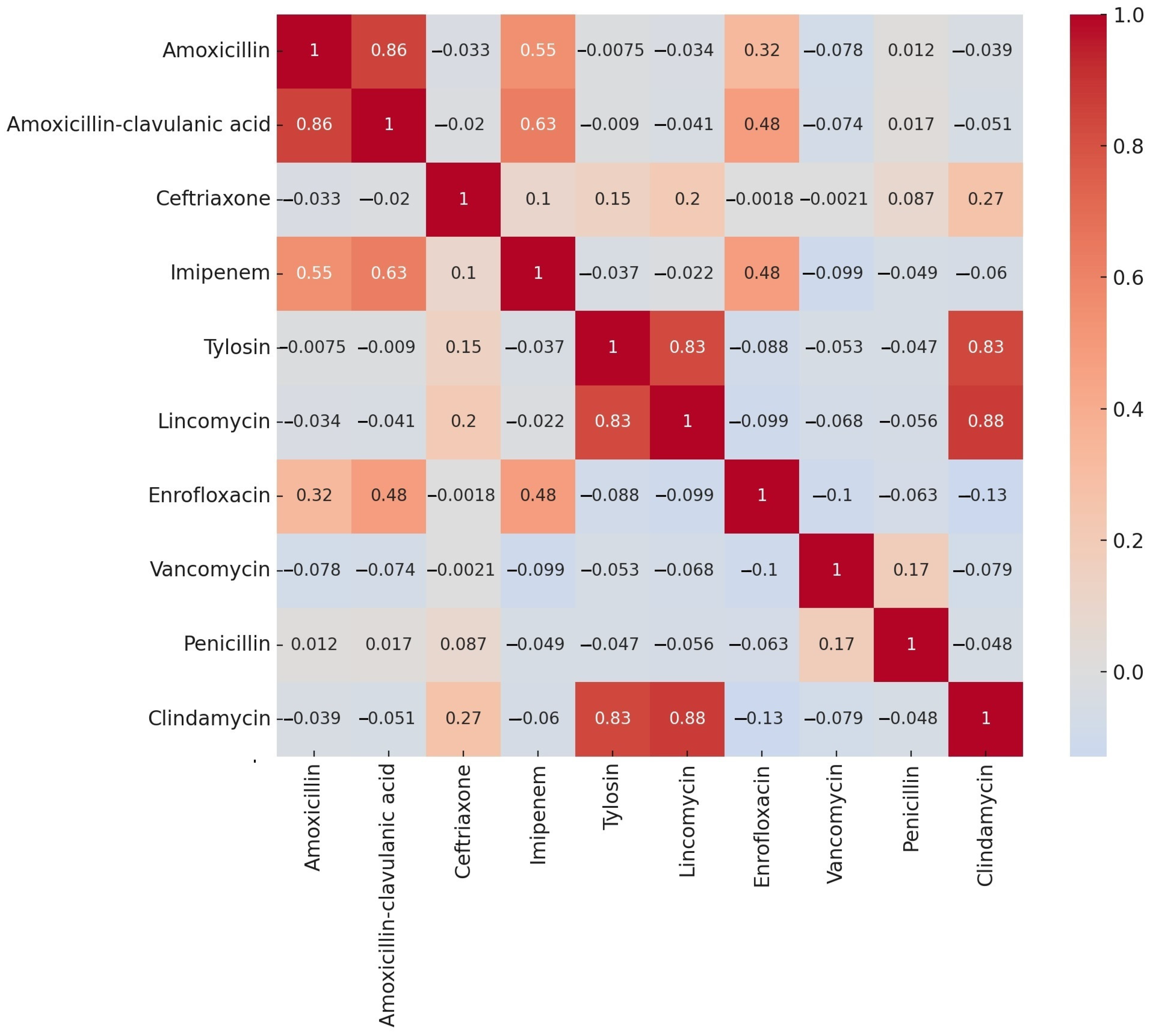
Figure 1. Correlation analysis of the resistance rates for each antibiotic in commensal Clostridium perfringens strains that were isolated from turkeys (n = 146), visualized using a heatmap.
Antibiotics 14 00413 g001
Figure 2. Cluster analysis of the commensal Clostridium perfringens strains (n = 146) isolated from turkeys. For clarity, individual samples are color-coded based on their regional origin, as displayed below the horizontal axis.
Antibiotics 14 00413 g002
Figure 3. Visualization of the principal component analysis (PCA) of turkey-derived commensal Clostridium perfringens strains (n = 146), following cluster analysis. The separation of strains into three main clusters is distinctly observable.
Antibiotics 14 00413 g003
Figure 4. Ocular susceptibility profile of Clostridium perfringens strains of turkey origin (n = 146) to antibiotic agents with clinical breakpoints.
Antibiotics 14 00413 g004
Figure 5. Prevalence of the major and minor toxin genes among Clostridium perfringens isolates (n = 146). The presence or absence of each gene was determined by real-time PCR. “Detected” indicates a positive PCR result for the corresponding gene.
Antibiotics 14 00413 g005
Table 1. Statistical comparison of the resistance profiles of the samples according to utilization direction, age group, and plant size.
AntibioticsBroiler–Breeding1 Young–2 Adult3 Small–4 Medium
Amoxicillin0.0093 *0.0093 *0.0915
Imipenem0.09730.09730.5595
Lincomycin0.63130.63130.7438
Enrofloxacin0.49410.49410.0499 *
Penicillin0.69390.69390.8216
Clindamycin0.0043 *0.0043 *0.0084 *
* Significant difference (p < 0.05); 1 younger than 13 weeks; 2 older than 13 weeks; 3 small (5001–50,000); 4 medium (50,001–100,000).
Table 2. The frequency table of minimum inhibitory concentration (MIC) values obtained for the tested antibiotics with established clinical breakpoints in turkey-derived Clostridium perfringens isolates (n = 146). For each antibiotic, the upper row indicates the number of isolates, while the lower row represents the corresponding percentage. The vertical red line indicates the clinical breakpoints.
Antibiotic1 BP *0.0010.0020.0040.0080.0160.030.060.1250.250.512481632641282565121024MIC50MIC902 ECOFF
µg/mLµg/mL
Amoxicillin161056968191826151230018 0.532-
0.7%0.0%3.4%4.1%6.2%4.1%5.5%13.0%12.3%17.8%10.3%8.2%2.1%0.0%0.0%12.3%
Imipenem1640035211014131610183218 0.58-
2.7%0.0%0.0%2.1%3.4%14.4%6.8%9.6%8.9%11.0%6.8%12.3%2.1%14.5%5.5%
Lincomycin1 300021115321168104620451664-
2.1%0.0%0.0%0.0%1.4%0.7%7.5%3.4%21.9%7.5%4.1%5.5%6.8%31.5%1.4%0.0%2.7%3.4%
Enrofloxacin2 10061415282391238 216-
0.7%0.0%0.0%4.1%9.6%10.3%19.2%15.8%6.2%8.2%26.0%
Penicillin1 1354620262818852130133 132-
0.7%2.1%3.4%2.7%4.1%13.7%17.8%19.2%12.3%5.5%3.4%1.4%8.9%0.0%0.7%2.1%2.1%
Clindamycin8 1161312812851545639121612320.125
0.7%11.0%0.7%2.1%8.2%5.5%8.2%5.5%3.4%10.3%2.7%3.4%4.1%26.7%0.7%1.4%0.7%4.1%0.7%
* BP—breakpoint; 1 Clinical Laboratory Standard Institute (CLSI); 2 Epidemiological cut-off value given by the European Committee on Antimicrobial Susceptibility Testing (EUCAST).
Disclaimer/Publisher’s Note: The statements, opinions and data contained in all publications are solely those of the individual author(s) and contributor(s) and not of MDPI and/or the editor(s). MDPI and/or the editor(s) disclaim responsibility for any injury to people or property resulting from any ideas, methods, instructions or products referred to in the content.