Article

Nonthermal Plasma Treatment Improves Uniformity and Adherence of Cyclodextrin-Based Coatings on Hydrophobic Polymer Substrates

Department of Biomedical Engineering, Case Western Reserve University, Cleveland, OH 44106, USA
*
Author to whom correspondence should be addressed.
Coatings 2020, 10(11), 1056; https://doi.org/10.3390/coatings10111056
Submission received: 10 October 2020 / Revised: 22 October 2020 / Accepted: 23 October 2020 / Published: 31 October 2020
(This article belongs to the Special Issue Polymer Coatings for Biomaterials)

Abstract

:
Low surface energy substrates, which include many plastics and polymers, present challenges toward achieving uniform, adherent coatings, thus limiting intended coating function. These inert materials are common in various applications due to favorable bulk, despite suboptimal surface, properties. The ability to functionally coat low surface energy substrates holds broad value for uses across medicine and industry. Cyclodextrin-based materials represent an emerging, widely useful class of coatings, which have previously been explored for numerous purposes involving sustained release, enhanced sorption, and reversible reuse thereof. In this study, substrate exposure to nonthermal plasma was explored as a novel means to improve uniformity and adherence of cyclodextrin-based polyurethane coatings upon unreceptive polypropylene substrates. Plasma effects on substrates were investigated using contact angle goniometry and X-ray photoelectron spectroscopy (XPS). Plasma impact on coating uniformity was assessed through visualization directly and microscopically. Plasma effects on coating adhesion and bonding were studied with mechanical lap-shear testing and XPS, respectively. Substrate surface wettability and oxygen content increased with plasma exposure, and these modifications were associated with improved coating uniformity, adhesion, and interfacial covalent bonding. Findings demonstrate utility of, and elucidate mechanisms behind, plasma-based surface activation for improving coating uniformity, adherence, and performance on inert polymeric substrates.

1. Introduction

The surface plays a critical role in the performance and success of many applied solid materials. For example, on surgically implanted devices, events such as protein adsorption, cell or bacterial attachment, biofilm formation, blood coagulation, tissue adhesion, foreign body response, and corrosion can all transpire at the host-material interface, ultimately determining the fate and function of the prosthesis, as well as the clinical outcome for the patient. Likewise, for non-medical commercial products, the surface of an item can impact its appearance, operation, and even durability, all of which influence consumer demand and profitability. Large bodies of research have been directed toward modifying material surfaces to achieve desirable function without compromising bulk properties. Within this vein, one of the most common surface modification approaches entails the application of coatings, which may serve many purposes.
Many biomedical and non-medical commercial products are comprised of polymers that possess low surface energy. Such polymeric materials include polypropylene (PP), polyethylene (PE), polydimethylsiloxane (PDMS), and polytetrafluoroethylene (PTFE). As a focus in this work, PP is a polyolefin, being one of the most abundantly used plastics today [1]. PP has many uses that range from textiles [2] (e.g., surgical meshes, sutures, clothing, and upholstery fabric), to filters/membranes [3] (e.g., in water or ventilation systems, and in surgical respirators or masks), to packaging [4] (e.g., containers for food, cosmetics, or chemicals), and beyond.
The low surface energy of such materials can be disadvantageous for several reasons. First, it decreases the receptiveness of these polymers toward coatings [5], which could otherwise allow for improved product function, appeal, longevity, safety, and in the case of implantable devices, host response. In general, the surface energy of a substrate should exceed that of a coating to achieve reasonable spreading and adhesion. If not, applied coatings suffer from lack of uniformity, adherence, and durability, limiting the intended function of the coating over the product lifetime. The second disadvantage, particularly important for implants and water treatment filters/membranes, is that the nonpolar nature of low surface energy substrates promotes non-specific adsorption and denaturation of proteins on the bare surface. It is thermodynamically favorable for proteins to displace water at a hydrophobic interface and to change conformation (core nonpolar amino acids associate with the material) [6]. On implant substrates, denatured proteins in this protein conditioning layer may assume altered functions or be misidentified by the host immune system, thus triggering adverse thrombotic, inflammatory, and foreign body responses [7,8,9]. Furthermore, this protein conditioning layer may serve as a site for undesired attachment of biofoulants such as bacteria. On water treatment filters/membranes, adherent bacteria and their biofilms can reduce flux and contaminate treated water [10,11].
An attractive solution to promote uniformity and adherence of coatings on such challenging materials involves treatment of the substrate with nonthermal plasma [12,13]. Plasma is a state of matter composed of a dynamic mixture of negative and positive ions, uncharged molecules/atoms, radicals, photons, and free electrons. It is created when sufficient energy, either in the form of heat (thermal plasma) or electromagnetic fields (nonthermal plasma) is applied to a gas. Thermal plasmas exist only at very high temperatures (>4000 K) that would destroy most polymeric materials [14,15], thus only nonthermal plasmas are used for surface modification as considered in this paper. Interaction of nonthermal plasma with a polymer surface results in cleaning or etching, bond scission or rearrangement, and the introduction of new functional groups as determined by the carrier gas composition. These plasma-induced changes can promote substrate receptiveness to coatings [16], and also directly alter biological responses at the substrate surface [17]. Furthermore, plasma treatments circumvent the need for hazardous chemicals (e.g., solvents) as adhesion promoters, are amenable for substrates with complex geometries, and can achieve desirable surface changes while minimizing impact on the substrate material bulk properties [17].
Our group and others have previously investigated polysaccharide-based materials, constructed from subunits of cyclodextrin (CD), as coatings that can serve a wide variety of unique purposes. CDs are cyclic oligosaccharides, produced through bacterial enzymatic digestion of starch, having a truncated cone geometry with a hydrophilic exterior and a lipophilic core or “pocket”. They have remarkable abilities to reversibly complex, through non-covalent interactions, with small nonpolar molecules or domains that “fit” in the pocket. Applications of these CD-based coatings include sustained delivery of antibiotics [18,19,20,21], drugs [22,23,24], pesticides [25], or fragrances [26], and efficient uptake and retention of pollutants [27,28] or dyes [29,30,31]. Additionally, due to the reversible nature of their molecular interactions, these coatings may be regenerated and reused multiple times. Furthermore, owing to hydrophilic properties imparted by the intrinsically polar exterior of CD subunits, certain formulations of polymerized CD (pCD) applied as coatings have recently shown potential for mitigating events of biofouling, through passive resistance to non-specific protein adsorption, mammalian cell adhesion, and bacterial attachment [32]. Within our group, pCD coatings have previously been applied to polyester surgical fabrics and metallic orthopedic screws [18,19,33,34]. However, given that many low surface energy polymers are used as commercial materials in medicine and industry, in addition to the perpetual need for functional and stable coatings, it would be advantageous to explore the application of pCD coatings upon such difficult substrates in efforts to maximize coating uniformity and adherence.
The objective of this work is therefore to investigate effects of nonthermal plasma activation of PP substrates on the quality of pCD coatings. PP is chosen as a model substrate material given its inherently low surface energy, and its extensive use in biomedical and non-medical commercial products. A few notable examples include: (i) PP surgical textile implants, for which pCD coatings could help mitigate inflammatory, infective, and/or post-surgical tissue adhesion complications, whether through delivery-based or passive means; (ii) PP water treatment filters/membranes, for which pCD coatings could simultaneously resist biofouling and repeatedly scavenge pollutants; and (iii) PP packaging, for which pCD coatings could benefit the quality and shelf life of food through delivery of preservatives and/or extraction of undesirable spoilage byproducts.
An overview of this work is presented in Figure 1. The hypothesis of this study is that nonthermal plasma treatment enhances the uniformity and adherence of pCD coatings on PP substrates. To test this hypothesis, the time-dependent effects of nonthermal plasma exposure on PP substrate surface characteristics were first evaluated using contact angle goniometry and X-ray photoelectron spectroscopy (XPS). Next, the effects on pCD coating uniformity and adherence were investigated. Uniformity was qualitatively assessed through both direct visualization and use of scanning electron microscopy (SEM), and semi-quantitatively investigated through spreading experiments under direct visualization. Adherence was evaluated using lap-shear testing to evaluate mechanical adhesion in accordance with ASTM standard, and XPS to survey for chemical evidence of interfacial covalent bonds.

2. Materials and Methods

2.1. Materials

Soluble, lightly epichlorohydrin-crosslinked β-CD polymer precursor (bCD) was purchased from CycloLab R&D (#CY-2009, batch CYL-4160, MW ~116 kDa; Budapest, Hungary). Hexamethylene diisocyanate (HDI) crosslinker (#52649) and 2-(trifluoromethyl) phenyl isocyanate (2-TPI) (#159379) were purchased from Sigma Aldrich (St. Louis, MO, USA). N,N-dimethylformamide (DMF) solvent (#D119-4) was purchased from Fisher Scientific (Pittsburgh, PA, USA). PP 24-well plates (#1185U58) and lids (#1185U62) for macroscopic coating observation were purchased from Thomas Scientific (Swedesboro, NJ, USA). PP sheet stock (#8742K133) for XPS and contact angle goniometry, and bar stock (#8782K11) for lap-shear testing were purchased from McMaster-Carr (Elmhurst, IL, USA). PP 4-0 Prolene blue suture (#8592G) was purchased from eSutures (Mokena, IL, USA).

2.2. Plasma Cleaning and Activation of PP Substrate Surfaces

In order to systematically examine effects of plasma treatment on pCD coatings and PP substrates, PP materials were placed in a 4 in (10 cm) diameter × 8 in (20 cm) length cylindrical quartz reaction chamber of a Branson/IPC Model #1005-248 Gas Plasma Cleaner (St. Louis, MO, USA) and treated with low-pressure nonthermal plasma (500 mTorr, 50 W, 13.56 MHz) using an inlet gas mixture of argon bubbled through water (Ar/H2O). Given equipment scheduling constraints, effects of plasma treatment on PP substrate wettability and surface chemistry were investigated using treatments of durations from 1–20 min performed within 6 or 12 h of contact angle measurement or XPS analysis, respectively. The impact of plasma treatment on pCD coating uniformity and adherence was next assessed using a substrate treatment duration of 10 min (unless otherwise specified) within 1 h of pCD coating application. Non-treated (0 min) PP samples without any known prior exposure to plasma or ultraviolet light were included as controls in all experiments.

2.3. Effects of Plasma Treatment on PP Substrates

2.3.1. Wettability—Contact Angle Goniometry

For polar pCD coatings to spread uniformly on nonpolar PP substrates, the surface must be rendered more wettable, therefore the effect of plasma treatment on wettability of PP was examined using contact angle goniometry. PP sheet stock was cut to dimensions of ~1.5 in × 1.5 in (38 mm × 38 mm), gently sanded to expose fresh surface using a graded series of SiC sandpaper (1200, 2500, and 5000 grit), and rinsed thoroughly with a stream of deionized water prior to performing plasma treatments (0, 1, 2.5, 5, 10, and 20 min). After sanding and/or plasma treatments, care was taken to ensure that faces to be analyzed were not inadvertently exposed directly to any liquid or solid materials before analysis. PP surfaces were then evaluated for wettability by static contact angle measurement using a CAM 200 Optical Contact Angle Meter (KSV Instruments, Monroe, CT, USA). Deionized water droplets (n = 9–14 unique droplets per sample) of 8 µL volume were dispensed onto each PP surface and allowed to equilibrate for 30 s prior to photographing and measurement. The measurement for each droplet reflects the average of the angles on the left and right sides. Measurements were performed using KSV CAM 2008 software. Results shown represent findings from one experiment, with the same trends having also been observed in 2 similar independent experiments.

2.3.2. Surface Chemistry—XPS

Effects of plasma treatment on PP surface chemistry were studied using XPS to better understand the mechanistic basis by which plasma impacts spreading and adhesion of pCD coatings onto PP substrates. PP sheet stock was cut to dimensions of ~7.5 mm × 7.5 mm, gently sanded to expose fresh surface using a graded series of SiC sandpaper, and rinsed thoroughly with a stream of deionized water prior to performing plasma treatments (0, 1, 2.5, 5, 10, and 20 min). After sanding and/or plasma treatments, care was taken to ensure that faces to be analyzed were not inadvertently exposed directly to any liquid or solid materials before analysis. PP surfaces were then analyzed for elemental content using a PHI Versaprobe 5000 Scanning X-ray photoelectron spectrometer (Physical Electronics, Inc., Chanhassen, MN, USA) equipped with Al Kα source (hν = 1486.6 eV). Scans were acquired on a total of n = 3–4 samples per plasma treatment duration across 2 pooled experiments, with 2 unique scan locations averaged per sample. Survey scans were collected using a 200 µm spot size, 45 W power, 15 kV acceleration voltage, 117.40 eV pass energy, 0.40 eV step size, 25 ms/step, 8 cycles, 44.7° take-off angle, and 0–1100 eV range. The C1s peak was auto-shifted to 284.8 eV, and the ratios of the elements carbon, nitrogen, and oxygen were analyzed. The areas of peaks were taken with background set using a Shirley function from 280–292 eV for C1s, 396–404 eV for N1s, and 526–538 eV for O1s. Auger peaks were not used for analysis. After survey scans, high-resolution scans were collected using a 100 µm spot size, 25.2 W power, 15 kV acceleration voltage, 23.50 eV pass energy, 0.20 eV step size, 50 ms/step, 16 cycles, 44.7° take-off angle, and 278–298 eV range for C1s, or 523–543 eV range for O1s. The vertical sampling depth ζ (from which 95% of signal originates) for take-off angle θ = 44.7° and reported inelastic mean free path of λ = 3.5 nm at a photoelectron kinetic energy of 1 keV for PP surfaces [35], is determined to be ~7.4 nm based on the relation [36] ζ = 3λcos(θ). Analysis was performed using MultiPak software version 9.8.0.19 (Physical Electronics, Inc., Chanhassen, MN, USA). For interpretation of high-resolution scans, C–C, C–O, C=O, and O–C=O peak positions [37,38,39,40,41] were constrained at 284.8 ± 0.1, 286.3 ± 0.1, 288 ± 0.2, and 289 ± 0.2 eV, respectively, and full widths at half maximum (FWHM) were constrained to be within 10% that of the C–C peak FWHM value.

2.4. pCD Synthesis and Coating onto Surfaces

pCD coatings were synthesized in three steps using HDI as a crosslinker for bCD. First, bCD was weighed and placed in a PP tube, and DMF was added to dissolve it, maintaining a ratio of 3 mL DMF per gram bCD. Second, HDI was added so as to initiate crosslinking, and this pre-polymer mixture was thoroughly vortexed. Third, pre-polymer mixtures were cast as coatings either: (i) into wells of PP multiwell plates for production of coated well surfaces for qualitative and semi-quantitative direct visualization of coatings, (ii) onto PP sutures for qualitative microscopic visualization of coating uniformity under SEM, or (iii) onto flat PP bar stock pieces (newly abraded using SiC sandpaper to expose fresh surface) for preparing coated specimens for lap-shear testing. Unless otherwise specified, the amount of HDI used in experiments was 320 µL per gram bCD to achieve an intermediate crosslink ratio of 0.32. For experiments where polymer curing was necessary, cast pre-polymer mixtures were kept covered with Parafilm and allowed to cure for between 2–6 days, as specified for each experiment, under static conditions at ambient temperature and pressure. Cured pCD coatings were used promptly and directly in subsequent experiments before any drying could take place.

2.5. Effects of Plasma Treatment on pCD Coatings

2.5.1. Qualitative Uniformity—Direct Visualization

For qualitative visual assessment of pCD coating uniformity on PP substrates following plasma treatment, pCD coatings were applied to 24-well plates with or without a prior 10 min exposure to plasma. In this experiment, to observe potential effects of pCD formulation on polymer spreading, the amount of HDI crosslinker was varied between extremes of 80 and 640 µL per gram bCD, to achieve approximate crosslink molar ratios (HDI per glucose residue) from 0.08 to 0.64, respectively, chosen to span a range from the minimum limit for gelation up to brittle materials. The volume of pre-polymer mixture added was 140 µL/well, with each unique crosslink ratio in a different column (i.e., 4 wells/formulation), then all plates were gently but thoroughly agitated to help promote complete initial coverage prior to covering them with Parafilm and allowing the polymers 2 days to cure under static conditions at ambient temperature and pressure. Plates were then photographed and representative rows were selected to demonstrate the completeness of spreading for each pCD formulation.

2.5.2. Qualitative Uniformity—SEM

SEM was performed to qualitatively study pCD coating uniformity on PP substrates with or without prior plasma surface activation. Prolene sutures were removed from sterile packaging, cut into 1 in long pieces, and either treated with nonthermal plasma for 10 min or left untreated (0 min). Sutures were then coated with pCD by dipping them in freshly prepared pre-polymer mixture, then were placed in a Parafilm-covered Teflon dish and allowed to cure for 2 days at ambient temperature and pressure. pCD-coated sutures were then gently adhered to a stub using carbon tape, and sputter-coated with 5 nm of palladium under vacuum. Sutures were characterized using a JSM-6510 series scanning electron microscope (JEOL USA, Inc., Peabody, MA, USA). Images were taken at 50× magnification and an excitation voltage of 25 kV.

2.5.3. Semi-Quantitative Uniformity—Direct Visualization

For semi-quantitative direct visual assessment of pCD coating uniformity on PP substrates following plasma treatment, pCD pre-polymer solutions were applied to PP 24-well plates treated with plasma for different lengths of time (0, 1, 2.5, 5, 10, and 20 min). PP 24-well plates were held still on a stable fixture on an isolated lab bench, and solutions were slowly and gently added to each well via pipette by the same operator. The volume of pre-polymer mixture added to each well was varied for each plate to evaluate the completeness of spreading across a narrow range of volumes. All wells were used in each plate, for a total of 24 volumes tested per plasma treatment duration, with each unique volume being tested in at least triplicate. Pre-polymer solutions were allowed to spread on well surfaces for 1 min under static conditions (no agitation), then visually determined to be either completely spread across the well surface (passing result), or not (failing result). Volumes were initially selected based on results of a preliminary study (not included), and refined slightly if needed as the experiment progressed, such that roughly half of the wells would demonstrate complete coverage, while the other half would not. Binary logistic regression was then performed on the results using a logit link function in Minitab 19 software to estimate best-fit equations, which were used to determine volumes at which 50% of wells would be expected to have a passing result after 1 min of spreading (V50).

2.5.4. Adherence—Lap-Shear Testing

To examine the impact of plasma surface activation on the adhesion of PP substrates to pCD coatings, lap-shear testing was performed. Lap-shear testing and specimen preparation were performed in accordance with the ASTM D3163-01(2014) standard [42]. pCD pre-polymer mixtures (150 µL) were cast between two rectangular strips of PP sheet substrate with overall dimensions of 4 in × 1 in × 1/8 in (101.6 mm × 25.4 mm × 3.2 mm), on a 1 in × 1 in (25.4 mm × 25.4 mm) overlap region, and allowed to cure for 6 days at ambient temperature and pressure. Paired PP strips were either left untreated (0 min), or treated with nonthermal plasma for 10 min, prior to pCD application. Overlap regions were securely held together over the curing period with the use of paired medium-size binder clips and excess pre-polymer that spilled out of the joint was carefully wiped off with a KimWipe. Lap joints (n = 5/group) were tested in tension until failure at a rate of 0.05 in/min (1.3 mm/min) and a sampling rate of 72 Hz on an Instru-Met renewed load frame (#1130) operated with Testworks 4 software and hand-tightened vice grips capable of being horizontally offset so as to minimize peel effects. Load was monitored using a 100 lbf (445 N) tension load cell (Instron, Norwood, MA, USA) (cat #2512–103). The maximum load attained during the test was divided by the overlap area and recorded as the ultimate lap-shear strength (reported in units of kPa). The work to failure was divided by the overlap area and recorded as the lap-shear toughness (reported in units of J/m2). Grips were maintained with 2.5 in (63.5 mm) between the grip edge and the bonded region, providing a 0.5 in (12.7 mm) length of specimen/grip overlap. Results shown represent findings from one experiment, with the same trend having also been observed in one similar independent experiment.

2.5.5. Interfacial Covalent Bonding—XPS

In order to better understand the mechanisms behind plasma treatment effects on PP substrate adhesion to pCD coatings, we sought to determine whether covalent bonding takes place between pCD coatings and the plasma-treated PP surface. However, to do this an isocyanate compound was needed that could be uniquely identified at the activated PP surface. This was done using XPS to detect fluorine as a unique marker of the isocyanate compound 2-TPI after exposure of this compound to PP surfaces left untreated (0 min) or treated with plasma for 10 min. PP sheet stock was first cut to dimensions of ~7.5 mm × 7.5 mm, gently sanded to expose fresh surface using a graded series of SiC sandpaper, and rinsed thoroughly with deionized water. Next, plasma treatments were performed for the 10 min samples. Sample surfaces were then directly exposed to 2-TPI overnight, followed by two sequential rinses with copious amounts of toluene, acetone, isopropanol, then deionized water. Finally, a stream of dry nitrogen gas was used to remove excess water from sample surfaces. Untreated and plasma treated PP surfaces that were not exposed to isocyanate were prepared as controls as well. After sanding, plasma, and isocyanate exposure, care was taken to ensure that faces to be analyzed were not inadvertently touched by any solid surface before or during analysis. PP surfaces were then analyzed for elemental content using XPS as above. XPS was performed within 48 h of plasma treatment. Survey scans were acquired on 2 samples per plasma treatment duration, with 2 unique scan locations per sample. The ratios of carbon, nitrogen, oxygen, and fluorine were analyzed using Multipak software (version 9.8.0.19). The areas of peaks were taken with background set using a Shirley function as before for C1s, N1s, and O1s, and from 675–695 eV for F1s. Auger peaks were not used for analysis. Increased fluorine content was attributed to covalent urethane bond formation between hydroxyl groups on the PP surface and the isocyanate group on 2-TPI.

2.6. Statistical Analysis

All data is presented as mean ± standard deviation. Unless otherwise specified, statistical analysis tests were carried out in Microsoft Excel 2016, with two-sample two-tailed Student’s t-tests with unequal variance used for all comparisons. Statistical significance was set at p < α = 0.05. Plots depicting representative data were constructed using the sample closest to the group mean value(s).

3. Results

3.1. Effects of Plasma Treatment on PP Substrate Wettability and Surface Chemistry

First, we sought to evaluate the effects of plasma on the substrate surface, particularly in terms of wettability and chemistry. Wettability was studied through contact angle goniometry using the sessile drop method. Plasma treatment for any length of time was found to increase the wettability of PP substrates (p < 0.001) (Table 1), with longer treatments correlating with decreased static water contact angles. Without plasma treatment, water contact angles on PP averaged >120°, but after 20 min of plasma exposure, this value decreased to <60°.
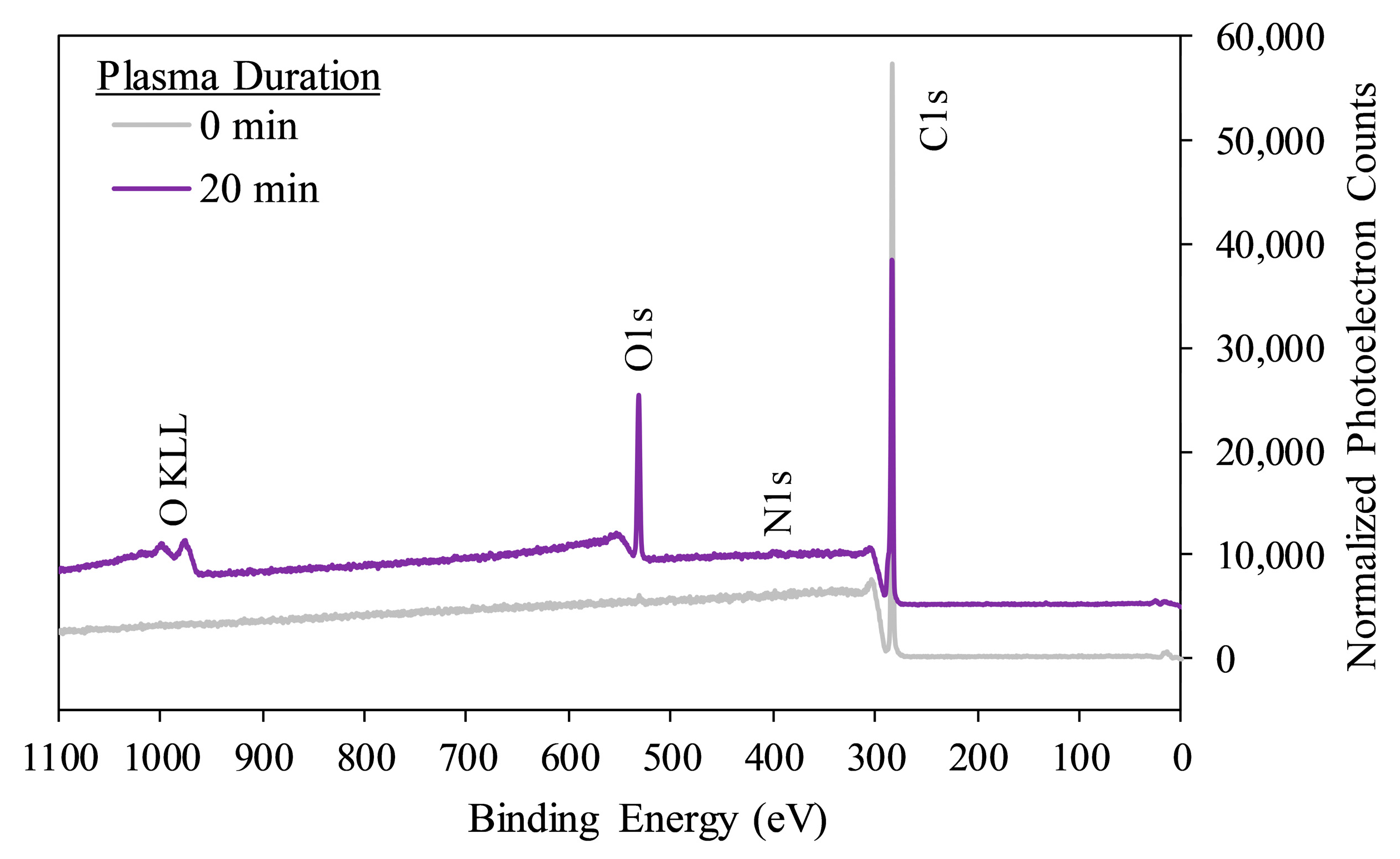
PP substrate surface chemistry was studied using XPS. Quantitative analysis of survey scan spectra (Figure 2) indicated that plasma exposure for any duration enhanced the amount of oxygen on PP surfaces (p < 0.001), and decreased the proportion of carbon (p < 0.001), with no significant impact on surface nitrogen content (p > 0.1) until 10 or 20 min (p < 0.014) of treatment (Table 1). Longer treatments were associated with increased oxygen, slightly increased nitrogen, and decreased carbon concentrations. Engraftment of oxygen-containing groups likely explains the enhanced surface wettability of PP following plasma treatment.
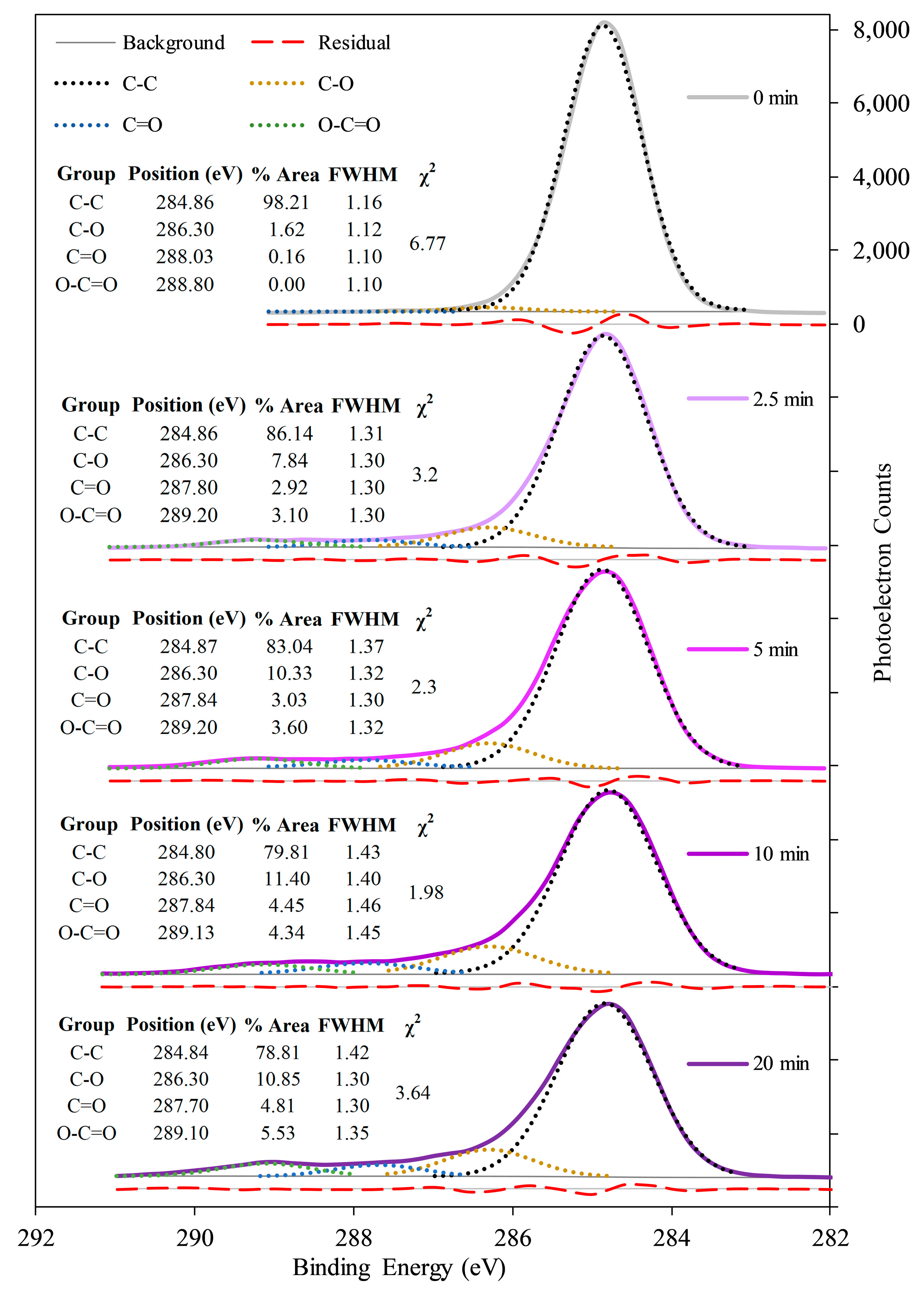
High-resolution C1s spectra were nearly symmetric for the untreated PP surface, but demonstrated an increasing skew to the left as plasma exposure time increased (Figure 3), suggesting increasing abundance of oxygen-containing functionalities including C–O, C=O, and O–C=O. Deconvolutions of high-resolution C1s scans suggested that most engrafted oxygen was incorporated as hydroxyl groups, with lesser amounts as ketones/aldehydes and carboxyls/esters, for all tested plasma durations. This finding is in agreement with previous reports [17,37]. However, the abundance of hydroxyl groups leveled off after 10 min of treatment, as existing hydroxyls were oxidized further to species such as ketones, carboxylic acids, or possibly even carbonates [38,39,41]. These latter two oxygen-rich functional groups can only be created through chain scission at the substrate surface, and such degraded polymer chains may embrittle the PP material [17]. Furthermore, loss of hydroxyl groups at the PP surface might be predicted to decrease the extent of interfacial covalent bonding with the polyurethane coating, as hydroxyls undergo expected reaction with isocyanates [43,44]. This would tend to decrease coating adherence. For these reasons, a treatment duration of 10 min was selected for most subsequent experiments.

3.2. Effects of Plasma Treatment on pCD Coating Uniformity, Adherence, and Interfacial Covalent Bonding

Having characterized the effects of plasma treatments on PP substrates, we next aimed to evaluate the effects of the plasma on pCD coating uniformity and adherence.
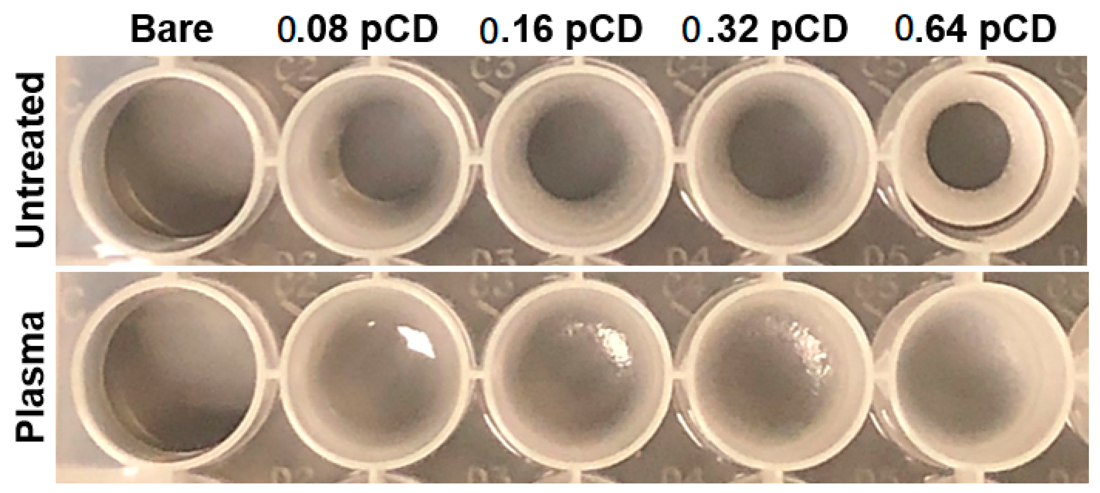
First, coating uniformity was qualitatively assessed based on direct and SEM visualizations. pCD coatings uniformly and stably covered the surfaces of PP substrates after plasma treatment, but not otherwise. This was observed both directly on initially-agitated PP 24-well plate surfaces (Figure 4), and microscopically on PP suture surfaces (Figure 5).
For PP 24-well plates, coatings tended to bead up at well edges without prior substrate plasma treatment, resulting in inadequate surface coverage at well centers. These well coatings were also observed to delaminate more easily from well surfaces, especially at higher crosslink ratios when pCD tended to contract more upon curing. Following plasma treatment, coatings applied in the same manner covered the entire surface of each well, regardless of the HDI crosslink ratio.
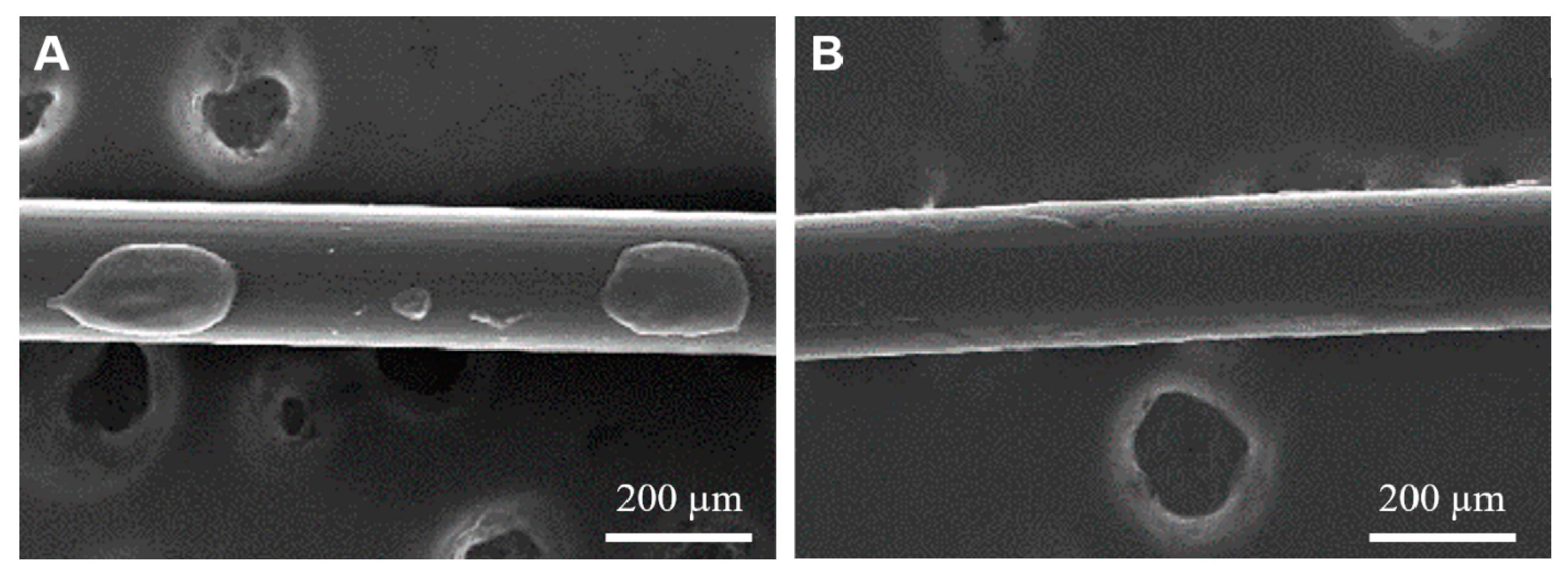
For PP sutures, SEM images demonstrated that pCD coatings beaded up and covered very little of the untreated PP suture surface (Figure 5A). At the same time, pCD coatings applied in the same manner were able to spread and coat a much larger fraction of the surface on plasma treated sutures (Figure 5B).
Next, coating uniformity was semi-quantitatively assessed by evaluating, across a range of volumes, whether or not pCD pre-polymer solutions were able to completely spread within 1 min under static conditions on PP 24-well plate surfaces that had been treated with plasma for various durations of time. Binary logistic regression was then performed on the results so as to determine the volume for each plasma exposure duration at which 50% of wells would show complete coverage, V50. In agreement with the wettability study results, V50 was lower for plasma-treated wells than for untreated wells (Table 2), implying that plasma treatment decreased the volume of pCD pre-polymer solution required to achieve complete coverage of PP wells. Additionally, V50 tended to decrease with increasing plasma duration, with the minor exception of the 20 min group (though this small deviation might reflect low sensitivity of the test method to detect such slight differences).
Taken together, results of the qualitative and semi-quantitative coating uniformity studies indicate that plasma treatments of PP substrates improve pCD coating uniformity. In light of the PP wettability findings, this improved uniformity likely reflects decreased substrate hydrophobicity, which promotes spreading of the polar pre-polymer mixtures on the surface.
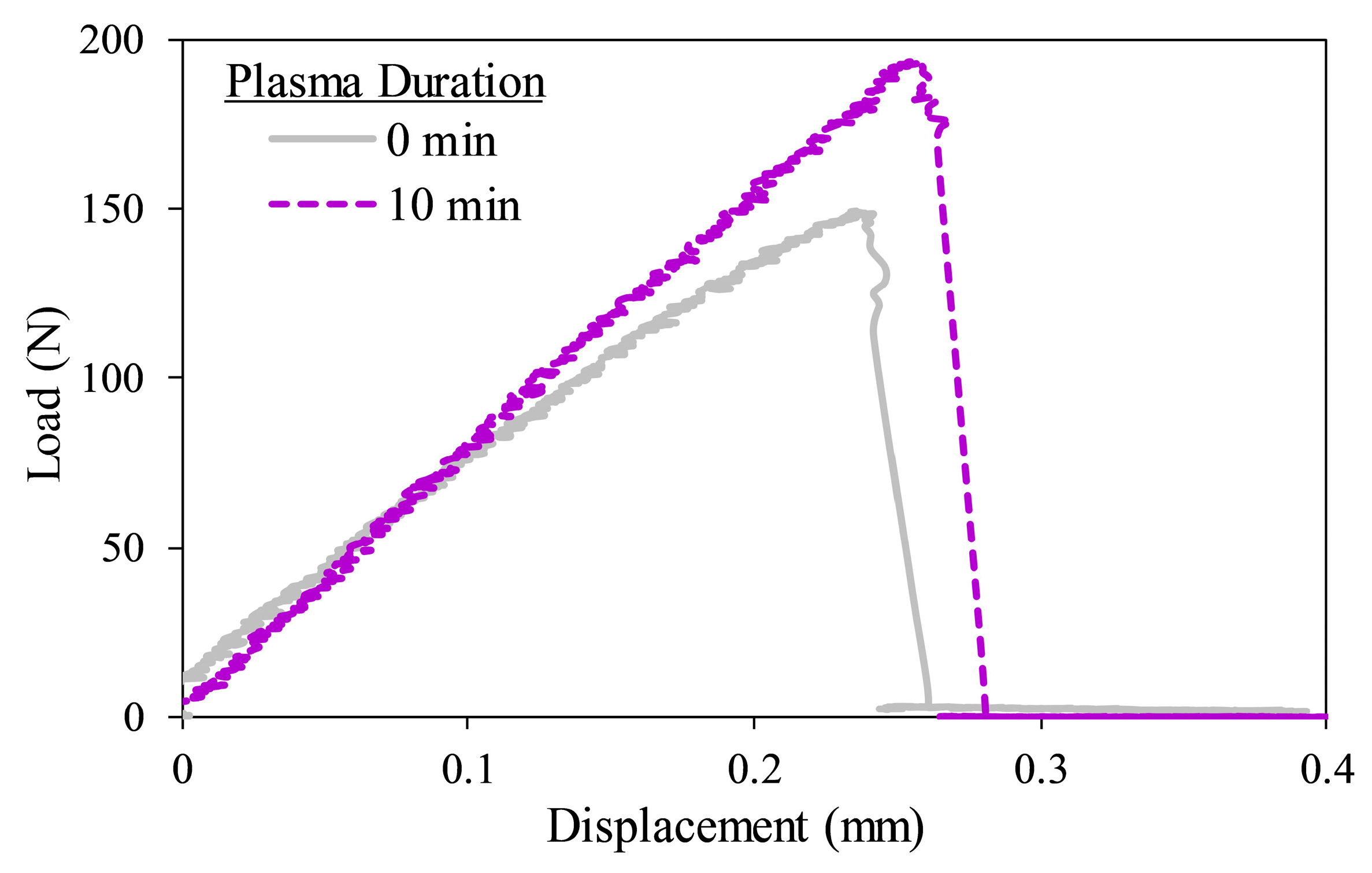
Coating adherence was next investigated using lap-shear testing, an approach used for measuring the bond shear strength of coatings and adhesives, which here involved pulling apart two rectangular PP adherends parallel to a bond line of pCD applied over a defined area. Lap-shear testing revealed that plasma treatment of PP substrates for 10 min increased average ultimate lap-shear strength by 43% (p = 0.003) and doubled average lap-shear toughness (p = 0.026) of pCD coatings (Table 3). The ultimate lap-shear strength and toughness are related to the maximum load (peak) and work (integral) for the load-displacement curve (Figure 6), respectively. For general reference, the ultimate lap-shear strength of untreated PP adherends bonded with cyanoacrylate has been reported as 0.22 MPa [45], approximately the strength observed here for untreated PP bonded with 0.32 pCD, though cyanoacrylate most likely possesses higher cohesive strength than the tested pCD. The enhanced coating adherence following plasma treatment may result from the evident engraftment of hydroxyl groups onto the PP surface, which might react to form covalent urethane linkages between the coating and substrate upon diisocyanate crosslinking [43,44].
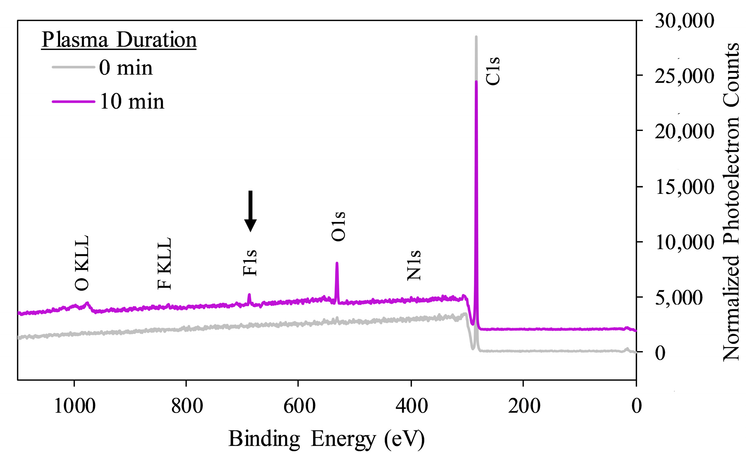
To validate this explanation that interfacial covalent bonding was partly responsible for enhanced coating adherence following plasma treatment, we next aimed to evaluate the formation of covalent urethane bonds between functional groups at the coating-substrate interface. This was done using XPS to detect fluorine as a unique marker of an isocyanate compound, 2-TPI, after exposure of this compound to untreated and plasma-treated PP surfaces followed by copious rinsing. In comparison to non-2-TPI-exposed controls, incubation of untreated PP samples with 2-TPI resulted in no significant change in surface fluorine content (p = 0.5), while incubation of plasma-treated PP samples with 2-TPI led to a significant increase in surface fluorine content (p = 0.018) (Figure 7, Table 4). This supports that plasma treatment of PP substrates enables covalent linkages between the PP surface (most likely via hydroxyl groups) and compounds containing isocyanate groups (such as pCD coatings during HDI crosslinking). This result corroborates the finding that plasma treatment improved the adherence of pCD coatings, and suggests that this effect can be attributed, at least in part, to covalent bond formation at the coating-substrate interface. Note that the 10 min plasma treated PP that was not exposed to 2-TPI in this experiment showed lower surface oxygen content than was seen before (Table 1). This likely reflects an extended period between plasma treatment and XPS (given overnight 2-TPI incubation), as well as the employed rinsing protocol.

4. Discussion

Uniformity and adherence to the substrate are crucial for the performance and function of any coating. For example, delamination of a drug-eluting polymer coating on an intravascular implant could pose risk for lethal thrombosis or embolism [46]. Similarly, improper adherence of paint to a metal component (e.g., for an automobile) could accelerate corrosion and potential structural failure of the part [47,48]. Likewise, a lack of adequate substrate coverage of an anti-biofouling coating could enable organisms to exploit coating surface defects and attach to the underlying substrate [49,50,51], creating problems such as infection on a medical device or increased drag on a marine vessel.
CD-based coatings have numerous potential applications relevant for inert polymeric substrates like PP. For example, on PP surgical textile implants, they could deliver antibiotics/drugs [19,24], or provide intrinsic resistance to protein/cell/bacterial attachment [32] to help mitigate complications related to inflammation, infection, and/or post-surgical tissue adhesion. On PP non-medical textiles, they could be used to improve fabric dyeability [30], for sustained and controlled release of insect repellent compounds [52,53,54] and fragrances [55,56,57], and for incorporating protectants against ultraviolet radiation [58,59] to which PP has known degradation susceptibility [60]. In PP water treatment filters/membranes, they could be useful as reusable adsorbents for scavenging of pollutants from water [27,61,62], and for limiting attachment and growth of biofouling microbial biofilms [32]. In PP packaging materials, they could be used to improve the quality and shelf life of food, through release of antimicrobial or preservative compounds [63,64], or through scavenging of undesirable components [65,66], either directly from the food or from the package headspace.
This study was designed to test the hypothesis that nonthermal plasma treatment enhances the uniformity and adherence of pCD coatings on PP substrates. The observations from contact angle goniometry, XPS, direct and SEM visualization, and lap-shear testing support this hypothesis. Plasma activation was found here to be a suitable strategy for improving both uniformity and adherence of polymer coatings such as pCD, on inert and otherwise incompatible polymer substrates like PP.
This conclusion is in agreement with those from prior studies that have investigated effects of plasma on uniformity and adherence of other polyurethane coating materials to low surface energy polymer substrates [44,67]. Martinez et al. determined that atmospheric plasma treatment of PDMS and acrylonitrile-butadiene-styrene substrates could enhance the adhesion of polyurethane-based paints in scratch, cross-cut, and pull-off tests [67]. Bao et al. found that atmospheric plasma treatment engrafted hydroxyl and amine groups onto styrene-butadiene-styrene rubber surfaces, and that these specific changes in surface chemistry, more so than incorporation of halogen functionalities, enabled increases in T-peel strength of polyurethane coatings after addition of an isocyanate-terminated hardener [44]. Sanbhal et al. showed that oxygen cold plasma treatment of PP mesh substrates enhanced the uniformity of pCD coatings [43]. Extending the knowledge gained from these previous investigations, through detailed surface analysis in combination with mechanical lap-shear adhesion testing, this study revealed direct evidence for formation of covalent bonds at the interface between a plasma-treated polyolefin substrate and a polyurethane coating, clarified the basis for these interfacial connections, and identified their contribution to improved strength of adhesion.
Substrate plasma activation is advantageous for improving uniformity and adherence of coatings upon inert polymer substrates when compared to conventional treatments, which utilize harsh chemicals as adhesion promoters. The former strategy represents a greener chemistry process, and eliminates the risk for residual hazardous compounds, which is especially important for any materials that may ultimately come into contact (directly or indirectly) with human cells or tissues (e.g., medical devices, water treatment filters/membranes). Furthermore, appropriate selection of plasma treatment parameters can ensure minimal impact on bulk mechanical properties of load-bearing substrates [17]. Apart from CD-based coatings, findings herein support the use of substrate plasma activation for improving uniformity and adherence of adhesives or paints (especially polyurethane-based formulations) upon low surface energy substrates such as polyolefins.
Future studies could implement these findings to achieve enhanced uniformity and adherence of coatings for improved functionality and durability in important applications, such as those mentioned previously, involving low surface energy substrates. Additionally, effects of plasma treatment on pCD coating uniformity and adherence could be investigated in detail for other polymer substrate materials, different plasma processes, and other CD-based polymer chemistries/formulations. Changes in coating uniformity or adherence over long periods after application could also be assessed, as the effects of plasma on surface chemistry of bare substrates are known to diminish (i.e., age) over time. Prior studies suggest that hydrophobic recovery of bare plasma-treated PP normally occurs over the course of several weeks, the rate being dependent on environmental factors and polymer crystallinity [68,69,70,71]. However, the application of a covalently anchored coating, such as pCD in this study, may limit conformational changes and reorientation of the activated substrate surface, in theory preserving the coating uniformity and adherence long term.

Author Contributions

Conceptualization, G.D.L. and H.A.v.R.; methodology, G.D.L.; validation, G.D.L. and E.J.L.; formal analysis, G.D.L.; investigation, G.D.L. and E.J.L.; resources, H.A.v.R.; writing—original draft preparation, G.D.L., E.J.L., and H.A.v.R.; writing—review and editing, G.D.L.; visualization, G.D.L.; supervision, G.D.L.; project administration, G.D.L.; funding acquisition, H.A.v.R. All authors have read and agreed to the published version of the manuscript.

Funding

This research was funded by the National Institutes of Health, Grant No. R01 GM121477.

Acknowledgments

The authors gratefully acknowledge support from National Institutes of Health: NIH R01 GM121477 (H.A.v.R.), and NIH Ruth L. Kirschstein NRSA T32 AR007505 Training Program in Musculoskeletal Research (G.D.L.). Additional support was provided by the Center for Stem Cell and Regenerative Medicine Student Summer Program (ENGAGE) at Case Western Reserve University (E.J.L.). Core facility services provided by the Swagelok Center for Surface Analysis of Materials and the Advanced Manufacturing and Mechanical Reliability Center at CWRU are also appreciated. The authors also thank the Advincula group for use of the contact angle goniometer, the Eppell group and Eloise Miller for use of and guidance with operating the plasma cleaner, Kevin Abbassi for expertise and assistance with XPS, Kathleen Young for expertise and assistance with SEM, Katherine Yan for assistance with sample preparation, and Erika Cyphert and Nathan Rohner for valuable writing feedback.

Conflicts of Interest

The authors declare no conflict of interest. The funders had no role in the design of the study; in the collection, analyses, or interpretation of data; in the writing of the manuscript, or in the decision to publish the results.

References

  1. Maddah, H.A. Polypropylene as a promising plastic: A review. Am. J. Polym. Sci. 2016, 6, 1–11. [Google Scholar] [CrossRef]
  2. Zhang, S.; Horrocks, A.R. A review of flame retardant polypropylene fibres. Prog. Polym. Sci. 2003, 28, 1517–1538. [Google Scholar] [CrossRef]
  3. Yang, Y.-F.; Wan, L.-S.; Xu, Z.-K. Surface engineering of microporous polypropylene membrane for antifouling: A mini-review. J. Adhes. Sci. Technol. 2011, 25, 245–260. [Google Scholar] [CrossRef]
  4. Boone, J.; Lox, F.; Pottie, S. Deficiencies of polypropylene in its use as a food-packaging material—A review. Packag. Technol. Sci. 1993, 6, 277–281. [Google Scholar] [CrossRef]
  5. Ryntz, R.A. Coating adhesion to low surface free energy substrates. Prog. Org. Coatings 1994, 25, 73–83. [Google Scholar] [CrossRef]
  6. Hoffman, A. Non-Fouling Surface Technologies. J. Biomater. Sci. Polym. Ed. 1999, 10, 1011–1014. [Google Scholar] [CrossRef]
  7. Hu, W.J.; Eaton, J.W.; Ugarova, T.P.; Tang, L. Molecular basis of biomaterial-mediated foreign body reactions. Blood 2001, 98, 1231–1238. [Google Scholar] [CrossRef] [PubMed] [Green Version]
  8. Zdolsek, J.; Eaton, J.W.; Tang, L. Histamine release and fibrinogen adsorption mediate acute inflammatory responses to biomaterial implants in humans. J. Transl. Med. 2007, 5, 31. [Google Scholar] [CrossRef] [Green Version]
  9. Koo, J.; Rafailovich, M.H.; Medved, L.; Tsurupa, G.; Kudryk, B.J.; Liu, Y.; Galanakis, D.K. Evaluation of fibrinogen self-assembly: Role of its αC region. J. Thromb. Haemost. 2010, 8, 2727–2735. [Google Scholar] [CrossRef] [Green Version]
  10. Nguyen, T.; Roddick, F.A.; Fan, L. Biofouling of water treatment membranes: A review of the underlying causes, monitoring techniques and control measures. Membranes (Basel) 2012, 2, 804–840. [Google Scholar] [CrossRef] [Green Version]
  11. Flemming, H.C. Biofouling in water systems—Cases, causes and countermeasures. Appl. Microbiol. Biotechnol. 2002, 59, 629–640. [Google Scholar] [CrossRef]
  12. Weon, J.-I.; Choi, K.-Y. Surface Characterization and morphology in Ar-plasma-treated polypropylene blend. Macromol. Res. 2009, 17, 886–893. [Google Scholar] [CrossRef]
  13. Harth, K.; Hibst, H. Surface modification of polypropylene in oxygen and nitrogen plasmas. Surf. Coatings Technol. 1993, 59, 350–355. [Google Scholar] [CrossRef]
  14. Desmet, T.; Morent, R.; De Geyter, N.; Leys, C.; Schacht, E.; Dubruel, P. Nonthermal plasma technology as a versatile strategy for polymeric biomaterials surface modification: A review. Biomacromolecules 2009, 10, 2351–2378. [Google Scholar] [CrossRef] [PubMed] [Green Version]
  15. Bogaerts, A.; Neyts, E.; Gijbels, R.; van der Mullen, J. Gas discharge plasmas and their applications. Spectrochim. Acta Part B At. Spectrosc. 2002, 57, 609–658. [Google Scholar] [CrossRef]
  16. Navaneetha Pandiyaraj, K.; Deshmukh, R.R.; Arunkumar, A.; Ramkumar, M.C.; Ruzybayev, I.; Ismat Shah, S.; Su, P.-G.; Periayah, M.H.; Halim, A.S. Evaluation of mechanism of non-thermal plasma effect on the surface of polypropylene films for enhancement of adhesive and hemo compatible properties. Appl. Surf. Sci. 2015, 347, 336–346. [Google Scholar] [CrossRef]
  17. Learn, G.D.; Lai, E.J.; Wilson, E.J.; von Recum, H.A. Nonthermal plasma treatment of polymers modulates biological fouling but can cause material embrittlement. bioRxiv 2019, 842260. [Google Scholar] [CrossRef]
  18. Thatiparti, T.R.; Shoffstall, A.J.; von Recum, H.A. Cyclodextrin-based device coatings for affinity-based release of antibiotics. Biomaterials 2010, 31, 2335–2347. [Google Scholar] [CrossRef] [PubMed]
  19. Harth, K.C.; Rosen, M.J.; Thatiparti, T.R.; Jacobs, M.R.; Halaweish, I.; Bajaksouzian, S.; Furlan, J.; Von Recum, H.A. Antibiotic-releasing mesh coating to reduce prosthetic sepsis: An in vivo study. J. Surg. Res. 2010, 163, 337–343. [Google Scholar] [CrossRef]
  20. Cyphert, E.L.; Zuckerman, S.T.; Korley, J.N.; von Recum, H.A. Affinity interactions drive post-implantation drug filling, even in the presence of bacterial biofilm. Acta Biomater. 2017, 57, 95–102. [Google Scholar] [CrossRef]
  21. Blanchemain, N.; Laurent, T.; Chai, F.; Neut, C.; Haulon, S.; Krump-konvalinkova, V.; Morcellet, M.; Martel, B.; Kirkpatrick, C.J.; Hildebrand, H.F. Polyester vascular prostheses coated with a cyclodextrin polymer and activated with antibiotics: Cytotoxicity and microbiological evaluation. Acta Biomater. 2008, 4, 1725–1733. [Google Scholar] [CrossRef] [PubMed]
  22. Haley, R.M.; Qian, V.R.; Learn, G.D.; von Recum, H.A. Use of affinity allows anti-inflammatory and anti-microbial dual release that matches suture wound resolution. J. Biomed. Mater. Res. A 2019, 107, 1434–1442. [Google Scholar] [CrossRef] [PubMed]
  23. Vermet, G.; Degoutin, S.; Chai, F.; Maton, M.; Bria, M.; Danel, C.; Hildebrand, H.F.; Blanchemain, N.; Martel, B. Visceral mesh modified with cyclodextrin for the local sustained delivery of ropivacaine. Int. J. Pharm. 2014, 476, 149–159. [Google Scholar] [CrossRef] [PubMed]
  24. Chai, F.; Maton, M.; Degoutin, S.; Vermet, G.; Simon, N.; Rousseaux, C.; Martel, B.; Blanchemain, N. In vivo evaluation of post-operative pain reduction on rat model after implantation of intraperitoneal PET meshes functionalised with cyclodextrins and loaded with ropivacaine. Biomaterials 2019, 192, 260–270. [Google Scholar] [CrossRef] [PubMed]
  25. Romi, R.; Nostro, P.L.; Bocci, E.; Ridi, F.; Baglioni, P. Bioengineering of a cellulosic fabric for insecticide delivery via grafted cyclodextrin. Biotechnol. Prog. 2005, 21, 1724–1730. [Google Scholar] [CrossRef] [PubMed]
  26. Abdelkader, M.B.; Azizi, N.; Baffoun, A.; Chevalier, Y.; Majdoub, M. Fragrant microcapsules based on β-cyclodextrin for cosmetotextile application. J. Renew. Mater. 2019, 7, 1347–1362. [Google Scholar] [CrossRef] [Green Version]
  27. Martel, B.; Le Thuaut, P.; Bertini, S.; Crini, G.; Bacquet, M.; Torri, G.; Morcellet, M. Grafting of cyclodextrins onto polypropylene nonwoven fabrics for the manufacture of reactive filters. III. Study of the sorption properties. J. Appl. Polym. Sci. 2002, 85, 1771–1778. [Google Scholar] [CrossRef]
  28. Wang, Z.; Guo, S.; Zhang, B.; Fang, J.; Zhu, L. Interfacially crosslinked β-cyclodextrin polymer composite porous membranes for fast removal of organic micropollutants from water by flow-through adsorption. J. Hazard. Mater. 2020, 384, 121187. [Google Scholar] [CrossRef]
  29. Fuhrer, R.; Herrmann, I.K.; Athanassiou, E.K.; Grass, R.N.; Stark, W.J. Immobilized β-cyclodextrin on surface-modified carbon-coated cobalt nanomagnets: Reversible organic contaminant adsorption and enrichment from water. Langmuir 2011, 27, 1924–1929. [Google Scholar] [CrossRef]
  30. Ghoul, Y.E.; Martel, B.; Achari, A.E.; Campagne, C.; Razafimahefa, L.; Vroman, I. Improved dyeability of polypropylene fabrics finished with β-cyclodextrin–citric acid polymer. Polym. J. 2010, 42, 804–811. [Google Scholar] [CrossRef] [Green Version]
  31. Park, J.S.; Kim, I.-S. Use of β-cyclodextrin in an antimigration coating for polyester fabric. Color. Technol. 2013, 129, 347–351. [Google Scholar] [CrossRef]
  32. Learn, G.D.; Lai, E.J.; von Recum, H.A. Cyclodextrin polymer coatings resist protein fouling, mammalian cell adhesion, and bacterial attachment. bioRxiv 2020. [Google Scholar] [CrossRef] [Green Version]
  33. Blatnik, J.A.; Thatiparti, T.R.; Krpata, D.M.; Zuckerman, S.T.; Rosen, M.J.; von Recum, H.A. Infection prevention using affinity polymer-coated, synthetic meshes in a pig hernia model. J. Surg. Res. 2017, 219, 5–10. [Google Scholar] [CrossRef]
  34. Grafmiller, K.T.; Zuckerman, S.T.; Petro, C.; Liu, L.; von Recum, H.A.; Rosen, M.J.; Korley, J.N. Antibiotic-releasing microspheres prevent mesh infection in vivo. J. Surg. Res. 2016, 206, 41–47. [Google Scholar] [CrossRef] [PubMed] [Green Version]
  35. Cumpson, P.J. Estimation of inelastic mean free paths for polymers and other organic materials: Use of quantitative structure–property relationships. Surf. Interface Anal. 2001, 31, 23–34. [Google Scholar] [CrossRef]
  36. Ton-That, C.; Shard, A.G.; Bradley, R.H. Thickness of spin-cast polymer thin films determined by angle-resolved XPS and AFM tip-scratch methods. Langmuir 2000, 16, 2281–2284. [Google Scholar] [CrossRef]
  37. Morent, R.; De Geyter, N.; Leys, C.; Gengembre, L.; Payen, E. Comparison between XPS- and FTIR-analysis of plasma-treated polypropylene film surfaces. Surf. Interface Anal. 2008, 40, 597–600. [Google Scholar] [CrossRef]
  38. Wang, K.; Wang, W.; Yang, D.; Huo, Y.; Wang, D. Surface modification of polypropylene non-woven fabric using atmospheric nitrogen dielectric barrier discharge plasma. Appl. Surf. Sci. 2010, 256, 6859–6864. [Google Scholar] [CrossRef]
  39. Mutel, B.; Grimblot, J.; Dessaux, O.; Goudmand, P. XPS investigations of nitrogen-plasma-treated polypropylene in a reactor coupled to the spectrometer. Surf. Interface Anal. 2000, 30, 401–406. [Google Scholar] [CrossRef]
  40. Sarani, A.; Nikiforov, A.Y.; De Geyter, N.; Morent, R.; Leys, C. Surface modification of polypropylene with an atmospheric pressure plasma jet sustained in argon and an argon/water vapour mixture. Appl. Surf. Sci. 2011, 257, 8737–8741. [Google Scholar] [CrossRef]
  41. Boyd, R.D.; Kenwright, A.M.; Badyal, J.P.S.; Briggs, D. Atmospheric nonequilibrium plasma treatment of biaxially oriented polypropylene. Macromolecules 1997, 30, 5429–5436. [Google Scholar] [CrossRef]
  42. ASTM International. ASTM D3163-01(2014) Standard Test Method for Determining Strength of Adhesively Bonded Rigid Plastic Lap-Shear Joints in Shear by Tension Loading; ASTM: West Conshohocken, PA, USA, 2014. [Google Scholar]
  43. Sanbhal, N.; Mao, Y.; Sun, G.; Xu, R.F.; Zhang, Q.; Wang, L. Surface modification of polypropylene mesh devices with cyclodextrin via cold plasma for hernia repair: Characterization and antibacterial properties. Appl. Surf. Sci. 2018, 439, 749–759. [Google Scholar] [CrossRef]
  44. Bao, L.; Fan, H.; Chen, Y.; Yan, J.; Yang, T.; Guo, Y. Effect of surface free energy and wettability on the adhesion property of waterborne polyurethane adhesive. RSC Adv. 2016, 6, 99346–99352. [Google Scholar] [CrossRef]
  45. Mühlhan, C.; Weidner, S.; Friedrich, J.; Nowack, H. Improvement of bonding properties of polypropylene by low-pressure plasma treatment. Surf. Coatings Technol. 1999, 116–119, 783–787. [Google Scholar] [CrossRef]
  46. Mehta, R.I.; Mehta, R.I. Hydrophilic polymer embolism: An update for physicians. Am. J. Med. 2017, 130, e287–e290. [Google Scholar] [CrossRef] [Green Version]
  47. Deflorian, F.; Rossi, S.; Prosseda, S. Improvement of corrosion protection system for aluminium body bus used in public transportation. Mater. Des. 2006, 27, 758–769. [Google Scholar] [CrossRef]
  48. Sørensen, P.A.; Kiil, S.; Dam-Johansen, K.; Weinell, C.E. Anticorrosive coatings:A review. J. Coatings Technol. Res. 2009, 6, 135–176. [Google Scholar] [CrossRef]
  49. Epstein, A.K.; Wong, T.-S.; Belisle, R.A.; Boggs, E.M.; Aizenberg, J. Liquid-infused structured surfaces with exceptional anti-biofouling performance. Proc. Natl. Acad. Sci. 2012, 109, 13182–13187. [Google Scholar] [CrossRef] [Green Version]
  50. Charnley, M.; Textor, M.; Acikgoz, C. Designed polymer structures with antifouling–antimicrobial properties. React. Funct. Polym. 2011, 71, 329–334. [Google Scholar] [CrossRef]
  51. Kingshott, P.; Griesser, H.J. Surfaces that resist bioadhesion. Curr. Opin. Solid State Mater. Sci. 1999, 4, 403–412. [Google Scholar] [CrossRef]
  52. Maestá Bezerra, F.; García Carmona, Ó.; García Carmona, C.; Souza Plath, A.M.; Lis, M. Biofunctional wool using β-cyclodextrins as vehiculizer of citronella oil. Process Biochem. 2019, 77, 151–158. [Google Scholar] [CrossRef]
  53. Peila, R.; Scordino, P.; Shanko, D.B.; Caldera, F.; Trotta, F.; Ferri, A. Synthesis and characterization of β-cyclodextrin nanosponges for N,N-diethyl-meta-toluamide complexation and their application on polyester fabrics. React. Funct. Polym. 2017, 119, 87–94. [Google Scholar] [CrossRef]
  54. Turan, A.C.; Özen, İ.; Gürakın, H.K.; Fatarella, E. Controlled release profile of imidacloprid-β-cyclodextrin inclusion complex embedded polypropylene filament yarns. J. Eng. Fiber. Fabr. 2017, 12. [Google Scholar] [CrossRef] [Green Version]
  55. Sricharussin, W.; Sopajaree, C.; Maneerung, T.; Sangsuriya, N. Modification of cotton fabrics with β-cyclodextrin derivative for aroma finishing. J. Text. Inst. 2009, 100, 682–687. [Google Scholar] [CrossRef]
  56. Xiao, Z.; Deng, J.; Niu, Y.; Zhu, G.; Zhu, J.; Liu, M.; Liu, S. Preparation of sustained-release fragrance based on the cavity structure of β-cyclodextrin and its application in cotton fabric. Text. Res. J. 2018, 89, 3466–3474. [Google Scholar] [CrossRef]
  57. Wang, C.X.; Chen, S.L. Fragrance-release property of β-cyclodextrin inclusion compounds and their application in aromatherapy. J. Ind. Text. 2005, 34, 157–166. [Google Scholar] [CrossRef]
  58. El-Tahlawy, K.; El-Nagar, K.; Elhendawy, A.G. Cyclodextrin-4 hydroxy benzophenone inclusion complex for UV protective cotton fabric. J. Text. Inst. 2007, 98, 453–462. [Google Scholar] [CrossRef]
  59. Liu, J.; Ma, X.; Shi, W.; Xing, J.; Ma, C.; Li, S.; Huang, Y. Anti-ultraviolet properties of β-cyclodextrin-grafted cotton fabrics dyed by broadleaf holly leaf extract. Text. Res. J. 2020, 90. [Google Scholar] [CrossRef]
  60. Schoolenberg, G.E. A fracture mechanics approach to the effects of UV-degradation on polypropylene. J. Mater. Sci. 1988, 23, 1580–1590. [Google Scholar] [CrossRef]
  61. Mhlanga, S.D.; Mamba, B.B.; Krause, R.W.; Malefetse, T.J. Removal of organic contaminants from water using nanosponge cyclodextrin polyurethanes. J. Chem. Technol. Biotechnol. 2007, 82, 382–388. [Google Scholar] [CrossRef]
  62. Yamasaki, H.; Makihata, Y.; Fukunaga, K. Efficient phenol removal of wastewater from phenolic resin plants using crosslinked cyclodextrin particles. J. Chem. Technol. Biotechnol. 2006, 81, 1271–1276. [Google Scholar] [CrossRef]
  63. Chen, H.; Li, L.; Ma, Y.; Mcdonald, T.P.; Wang, Y. Development of active packaging film containing bioactive components encapsulated in β-cyclodextrin and its application. Food Hydrocoll. 2019, 90, 360–366. [Google Scholar] [CrossRef]
  64. Siró, I.; Fenyvesi, É.; Szente, L.; De Meulenaer, B.; Devlieghere, F.; Orgoványi, J.; Sényi, J.; Barta, J. Release of alpha-tocopherol from antioxidative low-density polyethylene film into fatty food simulant: Influence of complexation in beta-cyclodextrin. Food Addit. Contam. 2006, 23, 845–853. [Google Scholar] [CrossRef] [PubMed]
  65. López-de-Dicastillo, C.; Catalá, R.; Gavara, R.; Hernández-Muñoz, P. Food applications of active packaging EVOH films containing cyclodextrins for the preferential scavenging of undesirable compounds. J. Food Eng. 2011, 104, 380–386. [Google Scholar] [CrossRef] [Green Version]
  66. López-de-Dicastillo, C.; Jordá, M.; Catalá, R.; Gavara, R.; Hernández-Muñoz, P. Development of active polyvinyl alcohol/β-cyclodextrin composites to scavenge undesirable food components. J. Agric. Food Chem. 2011, 59, 11026–11033. [Google Scholar] [CrossRef]
  67. Martinez, A.M.; Abenojar, J.; Lopez de Armentia, S. Environmentally friendly plasma activation of acrylonitrile–butadiene–styrene and polydimethylsiloxane surfaces to improve paint adhesion. Coatings 2018, 8, 428. [Google Scholar] [CrossRef] [Green Version]
  68. Carrino, L.; Polini, W.; Sorrentino, L. Ageing time of wettability on polypropylene surfaces processed by cold plasma. J. Mater. Process. Technol. 2004, 153–154, 519–525. [Google Scholar] [CrossRef]
  69. Morra, M.; Occhiello, E.; Garbassi, F. Contact angle hysteresis on oxygen plasma treated polypropylene surfaces. J. Colloid Interface Sci. 1989, 132, 504–508. [Google Scholar] [CrossRef]
  70. Jokinen, V.; Suvanto, P.; Franssila, S. Oxygen and nitrogen plasma hydrophilization and hydrophobic recovery of polymers. Biomicrofluidics 2012, 6, 16501–1650110. [Google Scholar] [CrossRef] [Green Version]
  71. Yun, Y.I.; Kim, K.S.; Uhm, S.-J.; Khatua, B.B.; Cho, K.; Kim, J.K.; Park, C.E. Aging behavior of oxygen plasma-treated polypropylene with different crystallinities. J. Adhes. Sci. Technol. 2004, 18, 1279–1291. [Google Scholar] [CrossRef]
Figure 1. Study overview. Effects of nonthermal plasma on polypropylene (PP) substrates were investigated in terms of wettability and surface chemistry, and effects on polymerized CD (pCD) coatings were explored in terms of uniformity and adherence.
Coatings 10 01056 g001
Figure 2. Representative XPS survey scans (rescaled to normalize areas under curves) of the 0 and 20 min time points demonstrate increasing height of the O1s peak with plasma treatment. KLL peaks indicate atomic relaxation via the Auger effect (with the acronym reflecting the K and L electron orbital shells involved in the energy transition), and were not used for analysis. Spectra are intentionally offset from each other along the vertical axis.
Coatings 10 01056 g002
Figure 3. Representative XPS high-resolution scans demonstrate increasing abundance of oxygen-containing functional groups with increasing plasma treatment time. Spectra are intentionally offset from each other along the vertical axis.
Coatings 10 01056 g003
Figure 4. Plasma treatment of PP substrates for 10 min improves macroscopic pCD coating uniformity on wells of a PP 24-well plate. This remained true regardless of the amount of HDI crosslinker added.
Coatings 10 01056 g004
Figure 5. Scanning electron micrographs demonstrate that plasma treatment of PP suture improves microscopic pCD coating uniformity: (A) an untreated and pCD-coated PP suture. The pCD coating beads up instead of spreading on untreated PP suture; (B) a 10 min plasma-treated and pCD-coated PP suture. The pCD coating is thin and well-spread on the plasma-treated suture, as made apparent based on the presence of cracks caused by sample drying upon sputtering and imaging.
Coatings 10 01056 g005
Figure 6. Load-displacement curves from lap-shear testing for representative pCD-coated samples of untreated and plasma-treated PP substrates.
Coatings 10 01056 g006
Figure 7. Representative XPS survey scans (rescaled to normalize areas under curves) of PP after 0 or 10 min plasma, plus equivalent exposure to 2-TPI. Increased isocyanate binding is indicated by the appearance of an F1s peak (arrow) only for the 10 min plasma sample. KLL peaks were not used for analysis. Spectra are intentionally offset from each other along the vertical axis.
Coatings 10 01056 g007
Table 1. Effect of plasma treatment duration on PP wettability, and surface chemistry.
Plasma Duration (Min)Contact Angle (°)Atomic % CarbonAtomic % NitrogenAtomic % Oxygen
0126.8 ± 4.8 †98.27 ± 0.35 †0.29 ± 0.191.45 ± 0.24 †
195.0 ± 7.488.47 ± 0.700.34 ± 0.1911.19 ± 0.51
2.587.1 ± 10.488.18 ± 0.750.45 ± 0.4011.37 ± 0.63
570.1 ± 9.585.79 ± 0.290.53 ± 0.1713.68 ± 0.28
1065.8 ± 9.982.18 ± 0.890.71 ± 0.07 *17.11 ± 0.85
2057.9 ± 9.779.93 ± 1.280.72 ± 0.15 *19.36 ± 1.20
† Significant difference to all subsequent time points. * Significant difference to 0 min control.
Table 2. Effect of plasma duration on calculated pCD pre-polymer solution volume needed for complete coverage of half of tested PP well surfaces.
Plasma Duration (Min)V50 (µL)
0246.67
1163.75
2.5163.75
5162.34
10159.65
20166.81
Table 3. Effect of plasma treatment on pCD coating adherence to PP substrates.
Plasma Duration (Min)Ultimate Lap-Shear Strength (kPa)Lap-Shear Toughness (J/m2)
0222.3 ± 31.732.9 ± 12.3
10318.7 ± 40.0 *66.4 ± 22.5 *
* Significant difference to 0 min control.
Table 4. Effect of plasma treatment on isocyanate-mediated fluorination of PP surfaces.
Plasma Duration (Min)2-TPI ExposureAtomic % CarbonAtomic % NitrogenAtomic % OxygenAtomic % Fluorine
099.34 ± 0.370.42 ± 0.020.25 ± 0.350.00 ± 0.00
0+99.55 ± 0.640.00 ± 0.000.15 ± 0.210.31 ± 0.43
1087.09 ± 0.110.89 ± 0.0611.97 ± 0.260.07 ± 0.09
10+90.41 ± 0.490.72 ± 0.327.48 ± 0.531.39 ± 0.08 #
# Significant difference to non-2-TPI-exposed control at the equivalent plasma duration.
Publisher’s Note: MDPI stays neutral with regard to jurisdictional claims in published maps and institutional affiliations.