Article

Influence of Particle Concentration on the Elemental Penetration Region and Properties of Ni-P-SiC Composite Coatings Prepared through Sandblasting and Scanning Electrodeposition on 45 Steel Surfaces

College of Engineering, Nanjing Agricultural University, Nanjing 210031, China
*
Author to whom correspondence should be addressed.
Coatings 2021, 11(10), 1237; https://doi.org/10.3390/coatings11101237
Submission received: 3 September 2021 / Revised: 5 October 2021 / Accepted: 6 October 2021 / Published: 12 October 2021

Abstract

:
Ni-P-SiC composite coating was prepared on 45 steel surfaces through sandblasting and scanning electrodeposition to explore the relationship between element penetration region and composite coating properties. The single-factor control variable method with particle concentration as the research variable was used. Results showed that with the gradually increasing concentration of SiC nanoparticles, a trend of first increasing and then gradually decreasing was observed for the surface and cross-sectional microstructure of the coating, interpenetration ability of the elements, adhesion performance, and corrosion resistance. The best deposition quality of the coating was obtained when the concentration of SiC nanoparticles was 3 g·L−1. For cross-sectional microstructure, the scratch test revealed that the maximum coating thickness was 17.3 μm, the maximum range of elemental penetration region was 28.39 μm, and the maximum adhesion of the composite coating was 36.5 N. The electrochemical test showed that the composite coating had a −0.30 V self-corrosion potential and 8.45 × 10⁻⁷ A·cm−2 self-corrosion current density, the slowest corrosion rate. In addition, the composite coating had the best corrosion resistance and the largest impedance arc radius corresponding to an equivalent impedance value R2 of 3108 Ω.

1. Introduction

With economic development and technological progress, metal materials with good strength and stiffness (such as 45 steel) have become important in modern industrial production [1,2,3,4]. However, their high susceptibility to corrosion and chemical reaction with the media in the air has shortened their life cycle and reduced their quality of use, which greatly limits their wide application [5,6,7]. Given that corrosion often occurs on their surfaces, the protection of metallic materials is currently being enhanced by improving their surface properties [8,9,10,11,12].
Compared with conventional electrodeposition, scanning electrodeposition is characterized by high speed jet impact and reciprocal cyclic scanning, which accelerate material transfer, enhance the effect of liquid phase transfer, and ultimately improve the deposition efficiency of the coating [13,14,15]. An increased ultimate current density accelerates the nucleation rate, which can promote a uniform and dense coating structure with good surface morphology and coating properties and thus has gained widespread attention [16,17,18,19]. Sandblasting is the most common pretreatment method for metal substrates in the industrial sector. This simple, efficient, and low-cost process uses compressed air to form a high-speed jet beam to spray sand particles at high speed onto the substrate surface to deform it plastically, thereby enhancing the mechanical properties and accelerating the deposition rate of the coating [20,21,22,23,24]. Most studies on electrodeposited composite coatings focused on surface characterization and performance testing such as the influence of microscopic morphology [25,26], grain structure [27,28], and element distribution [29,30] on corrosion resistance. Research on sections is only limited to the coating thickness and cross-sectional microstructure [31,32,33]; the interplay among coating morphology, element distribution, adhesion performance, and corrosion resistance is poorly examined.
In this work, on the basis of the commonly used polishing pretreatment, we put forward the sandblasting pretreatment technology. Based on the commonly used immersion ordinary electrodeposition, we proposed a scanning reciprocating electrodeposition technique to deposit Ni-base multielement nanoparticle composite coatings. The Ni-P-SiC composite coating was prepared by sandblasting–scanning electrodeposition on the surface of 45 steel, and the concentration of SiC nanoparticles was set to 0, 1, 2, 3, 4, and 5 g·L−1. The effect of nanoparticle concentration on the surface morphology and cross-sectional microstructure of the scanning electrodeposited composite coatings were studied. The influence of different nanoparticle concentrations on the penetration of elements between the coating and substrate was also analyzed, and the effect of nanoparticles on the adhesion performance and corrosion resistance of the coating was explored. This research provides a theoretical reference for the elemental penetration and surface properties of scanning electrodeposited composite coatings.

2. Experiment

2.1. Experimental Procedures

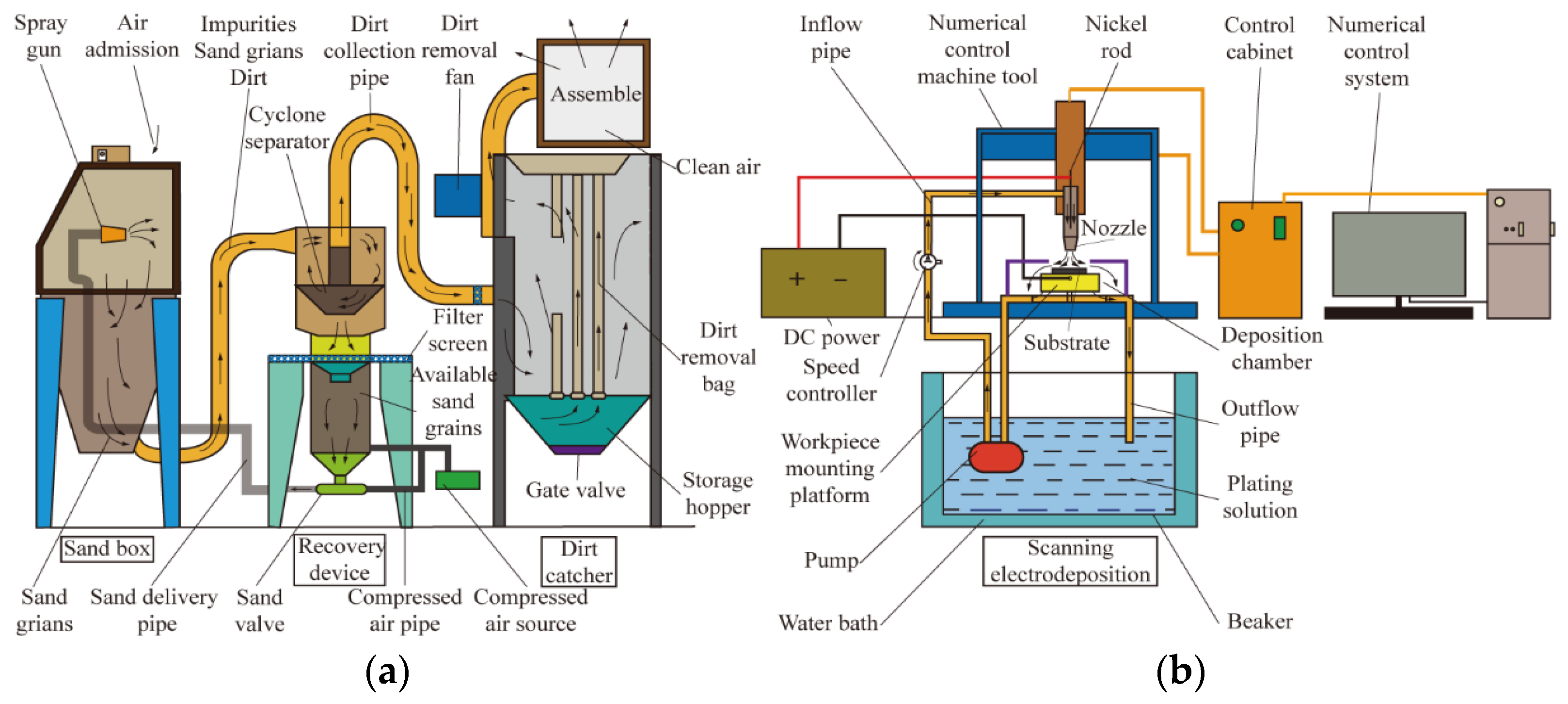
Figure 1a,b shows the experimental procedures and sandblasting pre-treatment device, respectively. The test uses a pressure fed box type sandblasting device manufactured by Shanghai Liangshi Spraying Equipment Co. (Shanghai, China). The power source of the sandblasting device is the compressed air produced by the screw air compressor, and the working pressure is generated by the compressed air in the pressure tank. The available sand grains pass through the sandblasting tube from the sand valve and are finally sprayed from the gun to the substrate surface to achieve the desired sandblasting effect. The ejected sand grains eventually flow back into the sand grain recycler device, and the resulting impurity dust is sucked into the dust collector to be purified into clean air for environmental protection purposes [21]. Figure 1b shows the scanning electrodeposition device. In this test, the anode nozzle was fixed to the machine and moved reciprocally with the numerical control machine spindle during scanning electrodeposition, and the cathode workpiece was fixed to the workpiece mounting platform. The plating solution was placed in a beaker and scanned from the nozzle through the inlet tube and sprayed onto the substrate surface and then from the deposition chamber through the outlet tube into the beaker. Finally, the coating was formed on the surface of the 45 steel substrate and gradually grew under the electric field.

2.2. Preparation of Workpiece and Coatings

The substrate material was 45 steel [16] with a specification of 25 mm × 10 mm × 8 mm and produced by Beijing Huawei Rui Ke Chemical Co., Ltd (Beijing, China). The main composition of 45 steel is Fe (elemental iron), and the composition of the remaining minor elements is shown in Table 1.
Prior to sandblasting pretreatment for the surface of the 45 steel substrate, a metallographic specimen grinding and polishing machine was used for polishing and #320, #800, and #1500 water sandpaper for mechanical grinding and polishing [23]. Sandblasting pretreatment parameters are shown in Table 2. The sandblasting grit is a mixture of sand (emery, copper ore sand, quartz sand, iron sand, and Hainan sand).
Prior to the preparation of the composite coating through scanning electrodeposition, electro-cleaning degreasing and activation treatment is required [18] and can be divided into three steps: electro-cleaning degreasing, anode activation, and pre-nickel coating. The solution formulation in this test is shown in Table 3. This process aims to remove residual oil and emery particles, remove the Fe2O3 oxide film from the surface of the workpiece, pre-plate a nickel film to prevent oxidation, and ultimately facilitate scanning electrodeposition.
The composition of the plating solution configured for the scanning electrodeposition test is shown in Table 4. The reagents used in the test were of analytical grade purity, and the solution was prepared with deionized water. After the deionized water was added, the solution was initially dissolved by hand stirring and then placed in a magnetic stirrer for full dissolution at a temperature of 60 °C, magnetic speed of 800 rpm, and stirring time of 1 h. Magnetic stirring ensures the adequate dispersion of nanoparticles, reduces nanoparticle agglomeration, and promotes the uniform and adequate dissolution of reagents and nanoparticles. During preparation, the current was adjusted to a constant 0.6 A, and the water bath was heated to a constant temperature of 60 °C. The samples were ultrasonically cleaned and dried after preparation and then post-treated (wire cutting and inlay) for characterization and performance testing.

2.3. Instruments and Characterization

Field emission scanning electron microscope (FEI-SEM, Quanta FEG250; FEI Instruments, OR, USA) was used to characterize the microscopic and cross-sectional microstructure of the plated surfaces. A secondary electron image type with an accelerating voltage of 3–15 KV, a scanning speed of 20 μs, and minimum/maximum magnification of 100×/30,000× was used for the observation. Section elemental composition was characterized using an EDS energy spectrum analyzer (XFlash Detector 5030; BrukerAXS, Inc., Berlin, Germany). Elemental composition was analyzed using elemental line scanning following the direction of the electron beam on the surface of the test sample from the coating to the substrate in a selected area to obtain a linear image of the elemental content variation. Coating adhesion was characterized using the friction method and a coating adhesion automatic scratch tester (WS-2005, Lanzhou Zhongke Kaihua Technology Development Co. Ltd., Lanzhou, China) with a load of 40 N and a scratch length of 3 mm. Electrochemical corrosion was tested using a CS350 electrochemical workstation (Wuhan Corrtest Instruments, Wuhan, China) with 3.5% NaCl solution as a corrosion medium in a scan range of −1 to 1.5 V and a scan rate of 0.5 mV·s−1.

3. Result and Discussion

3.1. Characterization of Coating Morphology

3.1.1. Coating Surface Morphology

Figure 2 shows the surface morphologies of the composite coatings with different particle concentrations prepared on 45 steel substrates through scanning electrodeposition. Figure 2a shows that when the concentration of SiC nanoparticles was 0 g·L−1, the surface of the Ni-P coating has a cytosolic protruding structure with large size and no defects on the surface (area A in Figure 2a). This finding indicates that the scanning electrodeposition technique makes the coating surface dense by increasing the current density. Figure 2b illustrates that when the concentration of SiC nanoparticles was 1 g·L−1, the Ni-P-SiC coating shows a relatively large cytosolic structure (area B in Figure 2b) because the SiC nanoparticles increase the polarization potential of the cathode and reduce the overpotential of metal nucleation, thereby promoting nucleus formation in the Ni-P coating and increasing the cytosolic structure of the nuclei [32]. However, the distribution is not uniform in the composite coating due to the low concentration of SiC nanoparticles, leading to the formation of microcracks on the stress-unconcentrated surface. Figure 2c illustrates that when the concentration of SiC nanoparticles was 2 g·L−1, the Ni-P-SiC coating shows fine cell-like structures (area C in Figure 2c) attributed to the increase in nickel crystal nucleation and growth sites with the increasing SiC nanoparticle concentration. In addition, the SiC nanoparticles further inhibit the growth of the coating grains and refine the cell structure. Figure 2d shows that when the concentration of SiC nanoparticles was 3 g·L−1, many cell-like structures are formed on the surface of the Ni-P-SiC coating. The cell structure is diversified, the coating is deposited intact without defects, and the surface is dense (area D in Figure 2d). The use of the optimum concentration of SiC nanoparticles, which act as active nucleation sites that are uniformly distributed on the surface of the composite coating, form small-sized cell structures at the cellular protrusions [34]. Figure 2e shows that when the concentration of SiC nanoparticles was 4 g·L−1, the surface cell structure of the Ni-P-SiC coating is reduced, and crystal precipitation occurs at the boundary between the cell structures (area E in Figure 2e). At this concentration, the nanoparticles exceed the degree of compounding of the coating and thus fail to uniformly disperse. The excessive grain refinement at the boundary of the cell structures leads to squeezing, overstress, and eventually to grain precipitation. Figure 2f shows that when the concentration of SiC nanoparticles was 5 g·L−1, agglomeration and porosity appeared on the surface of the Ni-P-SiC coating (area F in Figure 2f). The excessive nanoparticles reduce the current efficiency, resulting in the formation of enormous surface energy during deposition and the agglomeration of particles on the surface of the coating that are not well embedded. During the co-deposition of Ni-P cells and SiC nanoparticles, the concentration of particles was extremely large, leading to an increase in the viscosity of the plating solution and the failure of the cells to grow completely dense. When coupled with excessive internal stress, this phenomenon results in crystal precipitation and porosity.

3.1.2. Microscopic Morphology and Element Analysis of the Coating Section

Figure 3 shows the cross-sectional microstructure of the prepared composite coating. Figure 3a displays that when the concentration of SiC nanoparticles was 0 g·L−1, the Ni-P coating section is dense; however, fine pores appeared when deposited at the substrate crater, indicating that the Ni-P coating failed to thoroughly adhere onto the substrate. In addition, the coating thickness was 16.7 μm. Figure 3b shows that when the concentration of SiC nanoparticles was 1 g·L−1, the Ni-P-SiC coating adheres closely to the rough substrate, the pits are filled intact, and defects such as pores disappear. The coating thickness was 11.5 μm, which was lower than that for the coating without nanoparticles. This concentration was extremely low and contained a small amount of nanoparticles that could not be well dispersed in the composite coating. Nanoparticles can promote the formation of nuclei on the surface of the substrate, but their uneven distribution hinders the continuous growth of the coating during deposition due to internal stress. Figure 3c shows that when the concentration of SiC nanoparticles was 2 g·L−1, the adhesion degree of the Ni-P-SiC coating to the rough substrate increased, and the surface fluctuations of the coating remained consistent with those of the substrate. No defects such as cracks were found in the coating. The coating thickness was 13.6 μm, indicating that the coating thickness increased with the concentration of SiC nanoparticles. When the concentration of nanoparticles increased, the nanoparticles gradually and evenly distributed in the coating, thus promoting nucleation and growth points of the crystalline cells and prompting further growth in the coating thickness. Figure 3d shows that the edge undulations of the coating section were reduced, and the flatness of the coating surface was improved. The maximum coating thickness of 17.3 μm was obtained. Due to the uniform distribution of nanoparticles in the coating and the fastest nucleation and growth rate of the crystalline cells, the build-up of internal stresses was reduced during the growth of the coating; this phenomenon contributes to the uniform and rapid growth of the coating [35]. Figure 3e shows that when the concentration of SiC nanoparticles was 4 g·L−1, the denseness and adhesion degree of the Ni-P-SiC coating to the rough substrate decreased. When the concentration of particles continued to increase, the coating had excessive nucleation points that reduced the current efficiency and affected the deposition of the coating and the coating thickness was reduced to 14.5 μm. When the current efficiency decreased, the coating was deposited less effectively, the cells did not grow intact, and the growth rate slowed down, thus eventually leading to a reduction in the coating thickness. Figure 3f shows that when the concentration of SiC nanoparticles was 5 g·L−1, the adhesion degree of the Ni-P-SiC coating to the rough substrate further decreased, the quality of the coating section decreased, and defects such as holes and cracks appeared between the coatings. The coating thickness was further reduced to 13.1 μm. With further increase in particle concentration, the excess SiC nanoparticles agglomerated and further reduced the current efficiency, hydrogen precipitation, and oxygen absorption during deposition. This phenomenon results in uneven cell structure during growth and excessive internal stresses, which causes the generation of defects in the coating. In addition, the excessive agglomeration of particles decreases electrical conductivity, which slows down the growth rate of the grains and reduces the growing coating thickness.
The elemental scanning results of the composite coating section are shown in Figure 4. The addition of SiC nanoparticles promotes the elemental penetration between the coating and elements. The orientation of the elemental penetration region gradually increases and then decreases with the increase in nanoparticle concentration, with the maximum elemental penetration region of 28.39 μm found at a particle concentration of 3 g·L−1. When the concentration of particles increases, the distribution of nanoparticles in the coating gradually becomes uniform. Hence, the cell structure becomes refined, the internal stress of the coating is reduced, the current efficiency during deposition gradually increases, the nucleation points proliferate, and the growth rate of the cells is accelerated. The uniformity of the cell growth structure is enhanced, the penetration and diffusion of elements between the interfaces are promoted, and the adhesion of the coating to the substrate is improved. When the concentration of particles exceeds 3 g·L−1 and continues to increase, the nanoparticles agglomerate and create excessive nucleation points and internal stress during growth. Hence, defects such as pores and cracks appear in the coating section. In addition, excessive particles reduce conductivity efficiency, inhibit cell growth, create an unevenly distributed cell structure, and eventually hinder the penetration of elements between the coating and substrate. As a result, the area of elemental penetration is reduced. With the addition of nanoparticles, the trend of the section thickness of the plated layer is in agreement with that of the elemental penetration region, indicating that the increase in the coating thickness promotes elemental penetration to a certain extent.

3.1.3. Schematic of Element Penetration Mechanism

Figure 5 shows the change curve of particle concentration and composite coating thickness, and Figure 6 presents the diagram of the elemental penetration mechanism of the prepared composite coatings. When the concentration of SiC nanoparticles was 0 g·L−1, the surface of the prepared Ni-P coating has cell-like protruding structures of large size with no defects. The coating section is relatively dense, but fine pores appear when deposited in the substrate crater. In addition, the coating is not well attached to the substrate, and the elemental penetration diffusion between the attached interfaces is weak. When the concentration of SiC nanoparticles was 1 g·L−1, the nanoparticles increase the polarization potential of the cathode, lower the overpotential of metal nucleation, and promote the formation of nuclei in the coating and the cell-like structure of nuclei. The coating adheres closely to the rough substrate, the pits are filled and intact, and defects such as pores disappear. However, the concentration of nanoparticles was extremely low, and the small amount of nanoparticles are not well dispersed in the composite coating. This phenomenon results in the uneven distribution of internal stresses, which hinders the growth of the coating and reduces its thickness. The addition of nanoparticles increases the grain nucleation points, which in turn promote the enhancement of the elemental permeability between the coating and substrate. When the concentration of SiC nanoparticles was 2 g·L−1, the increase in nanoparticle concentration drives the nucleation and growth points of nickel crystals, inhibits the growth of coating grains, and refines the cell structure. The adhesion degree between the coating and rough substrate is enhanced and the coating is free from defects such as cracks. The gradual and uniform distribution of nanoparticles in the coating promotes the increase in nucleation and growth points of the crystals, contributing to the further growth of the coating thickness and the enhanced ability of the elements to penetrate and diffuse. When the concentration of SiC nanoparticles was 3 g·L−1, many cell-like structures are formed on the coating surface, the cell structure becomes highly diversified, the coating is deposited and intact without defects, and the surface is dense. The nanoparticles are evenly distributed in the coating to the best extent, and the nucleation and growth rate of the cells are the fastest. Hence, the accumulation of internal stresses during growth is slowed down, thus allowing the coating to grow uniformly and rapidly. When the concentration of SiC nanoparticles was 4 g·L−1, the degree of compounding of the coating is exceeded, and the nanoparticles are not uniformly dispersed. This phenomenon results in excessive grain refinement at the boundary of the cell structure and leads to extrusion between grains, excessive stress, and grain precipitation. When the concentration of particles continues to increase, the coating has excessive nucleation points, which reduce the current efficiency and affect the deposition of the coating. Hence, coating thickness is reduced. Some of the agglomerated nanoparticles prevent the coating from being completely deposited and attached to the substrate surface. This incomplete deposition reduces elemental penetration and shrinks the elemental penetration region. When the concentration of SiC nanoparticles was 5 g·L−1, the excessive nanoparticles reduce the current efficiency, resulting in excessive surface energy during deposition and particle agglomeration on the coating surface, which is not well embedded in the composite coating. The adhesion degree of the coating to the rough substrate is further reduced, and the quality of the coating section decreases. Excessive particle agglomeration decreases electrical conductivity, slows the growth rate of the grains, and reduces the growth coating thickness. In addition, the excessive internal stress causes defects such as pores and cracks in the coating, and the uneven growth of the cell structure hinders the mutual penetration and diffusion of elements between the interfaces, thereby reducing the region of elemental penetration.

3.2. Characterization of Coating Performance

3.2.1. Adhesion Performance

Figure 7 shows the adhesion of composite coatings with different particle concentrations prepared on 45 steel substrates through scanning electrodeposition, and Figure 8 illustrates their scratch morphologies. Figure 8a shows that when the particle concentration is 0 g·L−1, slight cracks appeared on the scratched surface and the coating adhesion was 33.8 N. Figure 8b shows that when the concentration of particles is 1 g·L−1, the scratch surface showed cracks, dense cracks, and peeling, and the coating adhesion was 22.55 N. The addition of nanoparticles increased the nucleation point of the coating and enlarged the elemental penetration region between the coating and substrate. However, because the particles were not evenly dispersed in the composite coating, the internal stress distribution was uneven, and the coating thickness was reduced. When the coating was scratched and squeezed, the adhesion degree between the coating and substrate was reduced, and the coating adhesion was reduced. When the load increased, the quality of the coating surface deposition decreased, and cracks and coating peeling occurred. Figure 8c shows that when the particle concentration is 2 g·L−1, the number of cracks on the scratched surface decreased, but coating peeling still existed, and the coating adhesion was 31.9 N. The gradual increase in nanoparticles refined the cell size, increased the nucleation point and coating thickness, expanded the region of elemental penetration, and increased the adhesion of the coating to the substrate. In addition, the distribution in the coating becomes uniform, the surface quality of the coating is improved, and the coating breaks less after being extruded. Figure 8d shows that when the concentration of particles is 3 g·L−1, the number of cracks on the scratched surface was reduced, no peeling of the coating occurred, and the coating adhesion was 36.5 N. When the concentration of added nanoparticles was 3 g·L−1, the surface quality of the coating was the best, and the coating was less likely to crack when compressed downward by the thimble squeeze. In addition, the nanoparticles were evenly distributed in the coating, and the adhesion degree between the substrate and coating, the coating thickness, the region of elemental penetration between the coating and substrate, and the adhesion force were the largest. Hence, the coating was not easily separated from the substrate when subjected to extrusion. Figure 8e shows that when the concentration of particles is 4 g·L−1, cracks and coating peeling appeared again on the scratched surface, and the coating adhesion was 32.8 N. When the particle concentration continues to increase, the high concentration leads to grain precipitation, which reduces the surface and cross-sectional microstructure and makes the coating susceptible to cracking and peeling when squeezed [35]. Excessive nanoparticles reduce the current efficiency, resulting in a decrease in the coating thickness and the range of elemental penetration. Hence, the coating failed to adhere well to the substrate, and the adhesion decreased. Figure 8f shows that when the particle concentration is 5 g·L−1, the surface cracking and coating peeling on the scratched surface increased, cracks started to appear at the less loaded locations, and the coating adhesion was 29.15 N. When the particle concentration further increased, the surface quality of the coating further deteriorated, and defects such as pores and cracks appeared, making the coating highly susceptible to damage when squeezed. Particle agglomeration slows down the growth rate of the coating and further reduces the coating thickness and the extent of elemental penetration, resulting in the low adhesion of the coating to the substrate.
Figure 9 shows the schematic of the adhesion mechanism based on element penetration for the prepared composite coatings. When the particle concentration is 0 g·L−1, slight cracks appeared on the scratched surface. When the particle concentration is 1 g·L−1, the nanoparticles enhanced the penetration and diffusion ability of the elements at the interface between the plating layer and substrate and their adhesion performance to a certain extent. However, due to the low particle concentration, the deposition distribution in the composite plating layer was not uniform, and the thickness, adhesion ability, and adhesion force of the plating layer decreased. In addition, the scratched surface showed cracks and peeling of the plating. When the concentration of particles increased, the nucleation point of the cell increased, prompting the coating thickness to increase and the elemental penetration region between the coating and substrate to expand, thus increasing the adhesion of the coating. The uniform distribution of nanoparticles in the plating layer improves the surface quality of the plating layer and prevents the formation of defects such as chipping and peeling on the scratched surface when subjected to thimble extrusion. When the concentration of nanoparticles was 3 g·L−1, the coating adhesion was the greatest, the surface quality of the scratch was the best, and only some micro cracks appeared on the scratch surface after being squeezed. When the concentration of nanoparticles continued to increase, the agglomerated particles created excessive internal stress, which reduced the surface quality and caused defects such as cracking and flaking when the plating was extruded. Hence, the thickness of the plating and the range of elemental penetration were reduced, which eventually lead to a decreased plating adhesion.

3.2.2. Corrosion Resistance

Figure 10 shows the potential polarization curves of the prepared coatings, and Table 5 lists each corrosion electrochemical parameter.
Figure 10 and Table 5 show that when the concentration of SiC nanoparticles was 0 g·L−1, the self-corrosion potential of the Ni-P coating was −0.41 V, the self-corrosion current density was 6.06 × 10⁻⁶ A·cm−2, and the corrosion rate was 0.073395. As a protective film, the Ni-P coating has a corrosion protection effect on the 45-gauge steel substrate. When the concentration of SiC nanoparticles was 1 g·L−1, the self-corrosion potential of Ni-P-SiC composite coating was −0.62 V, the self-corrosion current density was 2.20 × 10⁻⁵ A·cm−2, and the corrosion rate was 0.19241. These findings indicate that the corrosion resistance of the coating decreased, and the corrosion rate accelerated. When the nanoparticle concentration was low, the internal stress distribution was uneven, resulting in a small coating thickness. Although the elemental penetration region could enhance the corrosion resistance, the effect was minimal. The adhesion degree of the coating was low, and the corrosive medium easily penetrated the thin coating to erode into the substrate, thereby reducing the corrosion resistance of the coating. When the concentration of SiC nanoparticles was 2 g·L−1, the self-corrosion potential of the Ni-P-SiC composite coating was −0.44 V, the self-corrosion current density was 4.55 × 10⁻⁶ A·cm−2, and the corrosion rate slowed down. When the nanoparticles gradually increased, the nanoparticles were gradually distributed evenly in the coating, which promoted the growth of the coating, widened the elemental penetration region, and enhanced the coating adhesion performance. Hence, the coating protection against corrosive media enhanced the corrosion resistance of the coating. When the concentration of SiC nanoparticles was 3 g·L−1, the self-corrosion potential of the Ni-P-SiC composite coating was −0.30 V, the self-corrosion current density was 8.45 × 10⁻⁷ A·cm−2, the corrosion rate was the slowest, and the corrosion resistance was the best. At this time, the concentration of nanoparticles was the best, the degree of grain refinement was the highest, the distribution inside the coating was uniform, and the coating thickness and the elemental penetration region were the largest. The coating adhered to the substrate to the best extent, thus effectively blocking the invasion of corrosive media and showed the best corrosion resistance. When the concentration of SiC nanoparticles was 4 g·L−1, the self-corrosion potential of the Ni-P-SiC composite coating was −0.38 V, the self-corrosion current density was 1.47 × 10⁻⁶ A·cm−2, and the corrosion rate was accelerated. When the concentration of nanoparticles further increased, the denseness of the coating decreased, grain precipitation occurred on the surface, and the coating thickness, the range of elemental penetration, and the adhesion degree of the coating were reduced. Hence, the corrosion resistance of the coating decreased. When the concentration of SiC nanoparticles was 5 g·L−1, the self-corrosion potential of the Ni-P-SiC composite coating was −0.53 V, the self-corrosion current density was 1.65 × 10⁻⁵ A·cm−2, and the corrosion rate was accelerated. The extremely high concentration of nanoparticles leads to agglomeration [34], creates defects such as pores on the surface of the coating, accelerates the corrosion of the composite coating, and reduces the coating thickness and the range of elemental penetration. Hence, the substrate protection range and corrosion resistance of the coating decreased.
Figure 11 shows the Nyquist plots of the prepared coatings, Figure 12 displays their corresponding equivalent circuit diagrams, and Table 6 lists the fitted and calculated equivalent circuit parameters.
When the concentration of SiC nanoparticles was 0 g·L−1, the equivalent impedance value R2 corresponding to the impedance arc radius of the coating was 963 Ω, which is larger than that for the 45-gauge steel substrate after sandblasting. This finding indicates that scanning electrodeposition effectively improves the corrosion resistance of the metal material. When the concentration of SiC nanoparticles was 1 g·L−1, the equivalent impedance value R2 of the composite coating corresponded to an impedance arc radius of 443 Ω, which was lower than that for the coating without SiC nanoparticles. The adhesion performance decreased, which led to a decrease in the corrosion resistance of the coating and its equivalent impedance value. When the concentration of SiC nanoparticles was 2 g·L−1, the equivalent impedance value R2 corresponding to the impedance arc radius of the composite coating was 700 Ω. When the number of nanoparticles increased, the coating thickness and the elemental penetration region also increased, and the deposition quality and adhesion properties of the coating were improved. Hence, the equivalent impedance value of the coating increased. When the concentration of SiC nanoparticles was 3 g·L−1, the impedance arc radius of the composite coating corresponded to an equivalent impedance value of R2 of 3108 Ω. At the optimal nanoparticle concentration, the scanning deposition effect was the best, the coating had the greatest range of thickness and elemental penetration regions, the surface and section were dense and defect-free, and the adhesion properties were optimal, giving it the greatest equivalent impedance value. When the concentration of SiC nanoparticles was 4 g·L−1, the equivalent impedance value R2 corresponding to the impedance arc radius of the composite coating was 1505 Ω. Further increase in the concentration of nanoparticles causes agglomeration, which decreases the deposition quality, the coating thickness, elemental penetration region, and the adhesion degree of the coating to the substrate, thus contributing to the reduction in the equivalent impedance value of the composite coating. When the concentration of SiC nanoparticles was 5 g·L−1, the equivalent impedance value R2 corresponding to the impedance arc radius of the composite coating was 649 Ω. When the concentration of nanoparticles was extremely high, the deposition quality of the coating decreased again, defects such as grain precipitation and pores appeared on the surface, the coating thickness and the range of elemental penetration regions were again reduced, and the coating adhered poorly to the substrate, thus contributing to a further decrease in the equivalent impedance value of the composite coating.
A scanning electron microscope was used to observe the surface micro morphology of the composite coating after electrochemical corrosion in 3.5 wt.% NaCl solution, and the results are shown in Figure 13. Figure 13a shows that when the particle concentration was 0 g·L−1, serious corrosion occurred on the surface, a large number of corrosion holes and products were formed and accumulated. No composite deposition of nanoparticles occurred in the coating, and the nanoparticles did not play a role in refining the cell structure, and the element penetration region was small and easily corroded by corrosive media. Figure 13b shows that when the particle concentration was 1 g·L−1, microcracks appeared on the surface and local corrosion occurred. The reason is that the addition of nanoparticles promotes the formation of crystal nuclei, refines the cellular structure, increases the element penetration region, and slows down the corrosion of the coating surface. However, the internal stress was not concentrated due to the low concentration of nanoparticles, resulting in a decrease in the coating thickness. The adhesion performance was reduced, micro-cracks appeared on the surface, and the corrosion preferentially occurred in the micro-cracks in the corrosive solution. Figure 13c shows that when the particle concentration was 2 g·L−1, surface corrosion was weakened, and the local corrosion area was reduced. With the increase in nanoparticles, the surface deposition quality was improved, the unit cell structure was refined, the coating thickness and element penetration region were increased, the adhesion performance and corrosion resistance were improved, and defects such as surface cracks were reduced. Hence, only local corrosion occurred in corrosive media. Figure 13d shows that when the particle concentration was 3 g·L−1, the corrosion on the surface was significantly reduced, and slight corrosion occurred at the boundary of the protrusion structure. When the concentration of nanoparticles was the best, the surface quality of the coating was also the best. Many small-sized unit cell structures were found at the cellular protrusion structure. During corrosion, the protruding unit cell structure will be the first to contact the corrosive medium, thus effectively blocking the corrosion of the corrosive medium. At the same time, the element penetration region was the largest, and the corrosive medium did not easily penetrate the coating. Figure 13e shows that when the particle concentration was 4 g·L−1, the corrosion on the surface was aggravated and even occurred at the boundary of the cell. In addition, corrosion products were precipitated. When the concentration of nanoparticles exceeded the composite degree of the coating, crystal grains that are easily eroded and infiltrated by corrosive media will appear at the boundary of the cell structure. At the same time, the quality of the coating deposition and the element penetration region decrease, which accelerates the corrosion diffusion of the coating. Figure 13f shows that when the particle concentration was 5 g·L−1, corrosion pits and agglomeration of corrosion products appeared on the surface. When the concentration of nanoparticles was extremely high, the cell structure of the coating surface will exhibit grain precipitation, agglomeration, and porosity. Hence, the quality of the coating section was poor, the nanoparticles were not uniformly dispersed in the coating, and the element penetration region was small. During corrosion, the pores are easily penetrated by chloride ions, which accelerate corrosion diffusion to form corrosion pits that cause the agglomeration of corrosion products and aggravate this phenomenon.
4. Conclusions

  • Scanning electron microscope detection showed that with the increase in particle concentration, the coating thickness and element penetration region increased first and then gradually stabilized. When the concentration of SiC nanoparticles was 3 g·L−1, the coating on the substrate surface exhibited the best deposition quality, and various unit cell structures were formed on the surface. The coating thickness was the largest at 17.3 μm, and the element penetration region of the coating was the largest at 28.39 μm. In addition, the best inter-penetration and diffusion ability of the elements between the interfaces was observed.
  • Adhesion scratch test and scratch microscopic morphology detection showed that with the gradual increase in the concentration of SiC nanoparticles, the element penetration region first increased and then decreased, thereby prompting the surface quality of the scratches to increase first and then decrease. Coating adhesion presented a trend of first increasing and then decreasing. When the concentration of nanoparticles was 3 g·L−1, the best surface quality of scratches, 36.5 N coating adhesion, and the best adhesion performance were observed.
  • The potentiodynamic polarization curve and electrochemical impedance spectroscopy curve were detected using the electrochemical corrosion and microscopic morphology tests after corrosion. The results showed that with the increase in the concentration of SiC nanoparticles, the corrosion resistance of the composite coating showed a trend of gradually increasing first and then gradually decreasing. The optimum SiC particle concentration was 3 g·L−1. The Ni-P-SiC composite coating prepared by scanning electrodeposition had −0.30 V self-corrosion potential, 8.45 × 10⁻⁷ A·cm−2 self-corrosion current density, slowest corrosion rate, largest impedance arc radius with corresponding equivalent impedance R2 of 3108 Ω, best corrosion resistance and morphology quality after corrosion, and the least corroded surface.

Author Contributions

Conceptualization, Z.Z. and J.X.; Methodology, Z.Z. and J.X.; Software, Z.S. and H.W.; Validation, J.X. and M.J.; Formal analysis, Z.Z. and M.J.; Investigation, H.W. and Z.S.; Resources, J.X. and Z.Z.; Data curation, Z.S. and M.J.; Writing—original draft preparation, Z.Z. and Z.S.; Writing—review and editing, J.X. and Z.S.; Visualization, H.W. and M.J.; Supervision, Z.Z. and J.X.; Project ·administration, H.W. and M.J.; Funding acquisition, J.X. and Z.Z. All authors have read and agreed to the published version of the manuscript.

Funding

Financial support for this work was provided by the Jiangsu Agricultural Science and Technology Innovation Fund (grant number CX (20) 3085) and the Fundamental Research Funds for the Central Universities (grant numbers KYXJ202002 and KYXK2021001).

Institutional Review Board Statement

Not applicable.

Informed Consent Statement

Not applicable.

Data Availability Statement

Data sharing is not applicable to this article.

Conflicts of Interest

The authors declare no conflict of interest.

References

  1. Ma, T.J.; Li, W.Y.; Xu, Q.Z.; Zhang, Y.; Li, J.L.; Yang, S.Q.; Liao, H.L. Microstructure evolution and mechanical properties of linear friction welded 45 steel joint. Adv. Eng. Mater. 2007, 9, 703–707. [Google Scholar] [CrossRef]
  2. Lu, Y.Z.; Li, H.G.; Zhang, H.; Huang, G.K.; Xu, H.D.; Qin, Z.X.; Lu, X. Zr-based metallic glass coating for corrosion resistance improvement of 45 steel. Mater. Trans. 2017, 58, 1319–1321. [Google Scholar] [CrossRef] [Green Version]
  3. Gronostajski, Z.; Pater, Z.; Madej, L.; Gontarz, A.; Lisiecki, L.; Lukaszek-Solek, A.; Luksza, J.; Mroz, S.; Muskalski, Z.; Muzykiewicz, W.; et al. Recent development trends in metal forming. Arch. Civ. Mech. Eng. 2019, 19, 898–941. [Google Scholar] [CrossRef]
  4. Leszek, G.; Jacek, C.; Dalibor, B. Research of composite materials used in the construction of vehicle bodywork. Adv. Sci. Technol. Res. J. 2018, 12, 181–187. [Google Scholar]
  5. Huang, G.K.; Qu, L.D.; Lu, Y.Z.; Wang, Y.Z.; Li, H.G.; Qin, Z.X.; Lu, X. Corrosion resistance improvement of 45 steel by Fe-based amorphous coating. Vacuum 2018, 153, 39–42. [Google Scholar] [CrossRef]
  6. Miao, B.; Zhao, X.B.; Song, L.; Wei, K.X.; Hu, J. Raising the corrosion resistance of plasma-nitrided steel 45 (AISI 1045) by preliminary sandblasting. Met. Sci. Heat Treat. 2019, 60, 808–813. [Google Scholar] [CrossRef]
  7. Dudkina, N.G. Corrosion resistance of steel 45 subjected to electromechanical treatment and surface plastic deformation. metal science and heat treatment. Met. Sci. Heat Treat. 2018, 59, 584–587. [Google Scholar] [CrossRef]
  8. Luo, X.X.; Yao, Z.J.; Zhang, P.Z.; Zhou, K.Y. Corrosion resistance of Al–Cr composite strengthening layer on the surface of 45(#) steel. Rare Met. Mater. Eng. 2018, 47, 3127–3133. [Google Scholar]
  9. Li, G.; Gan, Y.Y.; Liu, C.H.; Shi, Y.; Zhao, Y.C.; Kou, S.Z. Corrosion and wear resistance of fe-based amorphous coatings. Coatings 2020, 10, 73. [Google Scholar] [CrossRef] [Green Version]
  10. Kaszuba, M.; Widomski, P.; Bialucki, P.; Lange, A.; Boryczko, B.; Walczak, M. Properties of new-generation hybrid layers combining hardfacing and nitriding dedicated to improvement in forging tools’ durability. Met. Sci. Heat Treat. 2020, 20, 70. [Google Scholar] [CrossRef]
  11. Morozow, D.; Barlak, M.; Werner, Z.; Pisarek, M.; Konarski, P.; Zagorski, J.; Rucki, M.; Chalko, L.; Lagodzinski, M.; Narojczyk, J.; et al. Wear resistance improvement of cemented tungsten carbide deep-hole drills after ion implantation. Materials 2021, 14, 239. [Google Scholar] [CrossRef] [PubMed]
  12. Latka, L.; Michalak, M.; Szala, M.; Walczak, M.; Sokolowski, P.; Ambroziak, A. Influence of 13 wt. % TiO2 content in alumina-titania powders on microstructure, sliding wear and cavitation erosion resistance of APS sprayed coatings. Surf. Coat. Technol. 2021, 410, 126979. [Google Scholar] [CrossRef]
  13. Shen, L.D.; Xu, M.Y.; Jiang, W.; Qiu, M.B.; Fan, M.Z.; Ji, G.B.; Tian, Z.J. A novel superhydrophobic Ni/Nip coating fabricated by magnetic field induced selective scanning electrodeposition. Appl. Surf. Sci. 2019, 489, 25–33. [Google Scholar] [CrossRef]
  14. Shen, L.D.; Fan, M.Z.; Qiu, M.B. Superhydrophobic nickel coating fabricated by scanning electrodeposition. Appl. Surf. Sci. 2019, 483, 706–712. [Google Scholar] [CrossRef]
  15. Xu, M.Y.; Shen, L.D.; Jiang, W.; Zhao, F.; Chen, Y.; Tian, Z.J. Fabrication of Ni-SiC superhydrophilic surface by magnetic field-assisted scanning electrodeposition. J. Alloys Compd. 2019, 799, 224–230. [Google Scholar] [CrossRef]
  16. Fu, X.Q.; Shen, Z.Y.; Chen, X.X.; Lin, J.R.; Cao, H.B. Influence of element penetration region on adhesion and corrosion performance of ni-base coatings. Coatings 2020, 10, 895. [Google Scholar] [CrossRef]
  17. Wang, G.F.; Shen, L.D.; Dou, L.M.; Tian, Z.J.; Liu, Z.D. Porous metal and method for preparation by the scanning and swinging jet electrodeposition. Int. J. Electrochem. Sci. 2014, 9, 3319–3326. [Google Scholar]
  18. Song, Z.Y.; Zhang, H.W.; Fu, X.Q.; Lin, J.R.; Shen, M.Q.; Wang, Q.Q.; Duan, S.L. Effect of current density on the performance of Ni-P–ZrO2–CeO2 composite coatings prepared by jet-electrodeposition. Coatings 2020, 10, 616. [Google Scholar] [CrossRef]
  19. Chen, J.S.; Huang, Y.H.; Qiao, B.; Yang, J.M.; He, Y.Q. A study on nanocrystalline nickel parts prepared by jet electrodeposition. Appl. Mech. Mater. 2011, 37–38, 64–67. [Google Scholar] [CrossRef]
  20. Cheng, A.Y.; Sheu, H.H.; Liu, Y.M.; Hou, K.H.; Hsieh, P.Y.; Ger, M.D. Effect of pretreatment process on the adhesion and corrosion resistance of nickel-boron coatings deposited on 8620H alloy steel. Int. J. Electrochem. Sci. 2020, 15, 61–89. [Google Scholar] [CrossRef]
  21. Reicheneder, C.; Hofrichter, B.; Faltermeier, A.; Proff, P.; Lippold, C.; Kirschneck, C. Shear bond strength of different retainer wires and bonding adhesives in consideration of the pretreatment process. Head Face Med. 2014, 10, 51. [Google Scholar] [CrossRef]
  22. Shen, X.T.; Wang, X.C.; Sun, F.H.; Ding, C.Y. Sandblasting pretreatment for deposition of diamond films on WC–Co hard metal substrates. Diamond Relat. Mater. 2017, 73, 7–14. [Google Scholar] [CrossRef]
  23. Zhang, Z.W.; Shen, Z.Y.; Wu, H.B.; Li, L.Q.; Fu, X.Q. Study on preparation of superhydrophobic Ni–Co coating and corrosion resistance by sandblasting-electrodeposition. Coatings 2020, 10, 1164. [Google Scholar] [CrossRef]
  24. Xu, W.T.; Haarberg, G.M.; Sunde, S.; Seland, F.; Ratvik, A.P.; Holmin, S.; Gustavsson, J.; Afvander, A.; Zimmerman, E.; Akre, T. Sandblasting effect on performance and durability of Ti based IrO2–Ta2O5 anode in acidic solutions. Electrochim. Acta 2019, 295, 204–214. [Google Scholar] [CrossRef]
  25. Liu, J.K.; Yang, L.J.; Song, Z.L.; Xu, C. Microstructures and capacitance performance of MnO2 films fabricated by ultrasonic-assisted electrodeposition. Appl. Surf. Sci. 2019, 478, 94–102. [Google Scholar] [CrossRef]
  26. Huang, C.; Wang, P.P.; Guan, W.; Yang, S.; Gao, L.M.; Wang, L.Q.; Song, X.P.; Murakami, R. Improved microstructure and magnetic properties of iron-cobalt nanowire via an ac electrodeposition with a multistep voltage. Mater. Lett. 2010, 64, 2465–2467. [Google Scholar] [CrossRef]
  27. Rosas-Laverde, N.M.; Pruna, A.I.; Cembrero, J.; Busquets-Mataix, D. Electrodeposition of ZnO/Cu2O heterojunctions on Ni–Mo–P electroless coating. Coatings 2020, 10, 935. [Google Scholar] [CrossRef]
  28. Huang, Y.L.; Zhu, L.A.; Ye, Y.C.; Zhang, H.; Bai, S.X. Iridium coatings with various grain structures prepared by electrodeposition from molten salts: Growth mechanism and high temperature oxidation resistance. Surf. Coat. Technol. 2017, 325, 190–199. [Google Scholar] [CrossRef]
  29. Fan, L.; Suni, I.I. Electrodeposition of elemental sulfur from dimethyl sulfoxide (DMSO). J. Electrochem. Soc. 2018, 165, D1–D5. [Google Scholar] [CrossRef]
  30. Ozdemir, R.; Karahan, I.H. Effect of solution Zn concentration on electrodeposition of CuxZn1-x alloys: Materials and resistivity characterization. Trans. Inst. Met. Finish. 2019, 97, 95–99. [Google Scholar] [CrossRef]
  31. Sheu, H.H.; Chen, Y.R.; Lin, T.T.; Ger, M.D. Effect of pretreatment process on the adhesion and corrosion resistance of chromium-carbon coatings deposited on copper substrates. Int. J. Electrochem. Sci. 2019, 14, 3281–3290. [Google Scholar] [CrossRef]
  32. Jiang, W.; Shen, L.D.; Xu, M.Y.; Wang, Z.W.; Tian, Z.J. Mechanical properties and corrosion resistance of Ni–Co–SiC composite coatings by magnetic field-induced jet electrodeposition. J. Alloys Compd. 2019, 791, 847–855. [Google Scholar] [CrossRef]
  33. Zhang, Y.; Kang, M.; Nyambura, S.M.; Yao, L.; Jin, M.F.; Zhu, J.P. Fabrication of Ni–Co–P alloy coatings using jet electrodeposition with varying reciprocating sweep speeds and jet gaps to improve wear and seawater corrosion resistance. Coatings 2020, 10, 924. [Google Scholar] [CrossRef]
  34. Jiang, W.; Shen, L.D.; Qiu, M.B.; Wang, X.; Fan, M.Z.; Tian, Z.J. Preparation of Ni–SiC composite coatings by magnetic field-enhanced jet electrodeposition. J. Alloys Compd. 2019, 762, 115–124. [Google Scholar] [CrossRef]
  35. Ji, R.J.; Han, K.; Jin, H.; Li, X.P.; Liu, Y.H.; Liu, S.G.; Dong, T.C.; Cai, B.P.; Cheng, W.H. Preparation of Ni–SiC nano-composite coating by rotating magnetic field-assisted electrodeposition. J. Manuf. Process. 2020, 57, 787–797. [Google Scholar] [CrossRef]
Figure 1. Schematic of the experimental procedure: (a) Sandblasting pretreatment device and (b) Scanning electrodeposition device.
Coatings 11 01237 g001
Figure 2. Surface morphologies of the composite coatings with different particle concentrations ((a) 0, (b) 1, (c) 2, (d) 3, (e) 4, and (f) 5 g·L−1) prepared on 45 steel substrates through scanning electrodeposition.
Coatings 11 01237 g002
Figure 3. Cross-sectional microstructure of the composite coatings with different particle concentrations ((a) 0, (b) 1, (c) 2, (d) 3, (e) 4, and (f) 5 g·L−1) prepared on 45 steel substrates through scanning electrodeposition.
Coatings 11 01237 g003aCoatings 11 01237 g003b
Figure 4. Coating section elemental scanning results of composite coatings with different particle concentrations ((a) 0, (b) 1, (c) 2, (d) 3, (e) 4, and (f) 5 g·L−1) prepared on 45 steel substrates through scanning electrodeposition.
Coatings 11 01237 g004aCoatings 11 01237 g004b
Figure 5. Curve of particle concentration and thickness of the composite coating.
Coatings 11 01237 g005
Figure 6. Schematic of the elemental penetration mechanism of the composite coatings with different particle concentrations prepared on 45 steel substrates through scanning electrodeposition.
Coatings 11 01237 g006
Figure 7. Adhesion of composite coatings with different particle concentrations prepared on 45 steel substrates through scanning electrodeposition.
Coatings 11 01237 g007
Figure 8. Scratch morphologies of the composite coatings with different particle concentrations. ((a) 0, (b) 1, (c) 2, (d) 3, (e) 4, and (f) 5 g·L−1) prepared on 45 steel substrates through scanning electrodeposition.
Coatings 11 01237 g008aCoatings 11 01237 g008b
Figure 9. Schematic of the adhesion mechanism based on element penetration for the composite coatings with different particle concentrations prepared on 45 steel substrates through scanning electrodeposition.
Coatings 11 01237 g009
Figure 10. Potential polarization curves of coatings prepared by scanning electrodeposition on 45 steel substrates with different particle concentrations.
Coatings 11 01237 g010
Figure 11. Nyquist plots of coatings with different particle concentrations prepared on 45 steel substrates through scanning electrodeposition.
Coatings 11 01237 g011
Figure 12. Equivalent circuit diagram of coatings with different particle concentrations prepared on 45 steel substrates through scanning electrodeposition.
Coatings 11 01237 g012
Figure 13. Surface morphologies after the corrosion of composite coatings with different particle concentrations prepared on 45 steel substrates through scanning electrodeposition. ((a) 0, (b) 1, (c) 2, (d) 3, (e) 4, and (f) 5 g·L−1).
Coatings 11 01237 g013aCoatings 11 01237 g013b
Table 1. The 45 steel small amount of chemical element composition (wt. %).
ElementCSiMnCrNiCu
content0.42–0.500.17–0.370.50–0.80≤0.25≤0.30≤0.25
Table 2. Sandblasting pretreatment parameters.
Sandblasting ParametersContent/(g·L−1)
Sand size80 mesh number
Sandblasting pressure0.7 MPa
Sandblasting distance15 cm
Sandblasting time20 s
Sandblasting angle90°
Table 3. Electric oil removal and activation solution composition.
StepContent/(g·L−1)
electro-cleaning degreasingNaOH 25.0 g·L−1, NaCO3 21.7 g·L−1, Na3PO4 50.0 g·L−1, NaCl 2.1 g·L−1, pH = 13
anode activationHCl 25 g·L−1, NaCl 140.1 g·L−1, pH = 0.3
pre-nickel coatingNa3C6H5O7·2H2O 141.2 g·L−1, H3C6H5O7·H2O 94.3 g·L−1, NiCl2·6H2O 3.0 g·L−1, pH = 4
Table 4. Composition of the plating solution.
CompositionsContent/(g·L−1)ProducerEffect
NiSO4·6H2O200Shanghai Jingchun Biochemical Technology Co. Ltd, Shanghai, ChinaThe main source of Ni2+
NiCl2·6H2O30Shanghai Jingchun Biochemical Technology Co. Ltd, Shanghai, ChinaAnode activator
H3PO320Shanghai Jingchun Biochemical Technology Co. Ltd, Shanghai, ChinaProvide P atoms in the coating
H3BO330Shanghai Jingchun Biochemical Technology Co. Ltd, Shanghai, ChinaAdjust the pH value of the bath
C6H8O760Shanghai Jingchun Biochemical Technology Co. Ltd, Shanghai, ChinaAccelerate deposition rate and stability
C12H25SO4Na0.08Shanghai McLean Biochemical Technology Co. Ltd, Shanghai, ChinaIncrease the amount of SiC nanoparticles suspended in the solution
CH4N2S0.01Shanghai McLean Biochemical Technology Co. Ltd, Shanghai, ChinaReduced hydrogen evolution
SiC (SiliPn carbide)0 g·L−1, 1 g·L−1, 2 g·L−1, 3 g·L−1, 4 g·L−1, 5 g·L−1Shanghai Jingchun Biochemical Technology Co. Ltd, Shanghai, ChinaPreparation of composite coatings
Table 5. Corrosion electrochemistry parameters of coatings with different particle concentrations prepared on 45 steel substrates through scanning electrodeposition.
SamplesEcorr (V)Icorr (A·cm−2)Corrosion Rate (mm/a)
0 g·L1−0.416.06 × 10−60.073395
1 g·L−1−0.622.20 × 10−50.19241
2 g·L−1−0.444.55 × 10−60.09981
3 g·L−1−0.308.45 × 10−70.041128
4 g·L−1−0.381.47 × 10−60.062861
5 g·L−1−0.531.65 × 10−50.14399
Table 6. Equivalent circuit diagram parameters of coatings with different particle concentrations prepared on 45 steel substrates through scanning electrodeposition.
R1R2R3R4R5R6CPE1/FCPE2/FCPE3/F
0 g·L−15.299631.86 × 10632.41221.9 1.19 × 10−61.31 × 10−52.89 × 10⁻⁶
1 g·L−14.074431.82 × 10645.97284.50.136.64 × 10−65.54 × 10−54.07 × 10−5
2 g·L−12.447002.87 × 10648.3348.50.344.28 × 10−61.88 × 10−52.56 × 10−5
3 g·L−11.5231084.73 × 10856.1516.40.4101.00 × 10−62.35 × 10−61.84 × 10−5
4 g·L−12.0815058.82 × 10730.7407.80.554.43 × 10−62.87 × 10−62.07 × 10−5
5 g·L−13.726492.30 × 10654.58240.20.276.58 × 10−63.53 × 10−53.42 × 10−5
Publisher’s Note: MDPI stays neutral with regard to jurisdictional claims in published maps and institutional affiliations.