Abstract
A comparative analysis of the features of UV-stimulated emission (SE) of various disordered active materials based on ZnO crystallites for a random laser (RL) was carried out. The superlinear increase in the intensity of the UV photoluminescence (PL) band of polydisperse nano-micro-crystalline (PNMC) ZnO powder at a wavelength of λ = 387 nm and some narrowing of its halfwidth in the range of 20 ÷ 15 nm with increasing pump intensity indicates random lasing with incoherent feedback (FB). The properties of similar UV PL bands under the same conditions of a thin film containing hexagonal ZnO microdisks, as well as samples of monodisperse ZnO nanopowder with nanoparticle sizes of 100 nm, indicate stimulated radiation with coherent feedback. It is shown that, among the studied materials, PNMC ZnO powder with widely dispersed crystallites ranges in size from 50 nm to several microns, which in turn, consists of nanograins with dimensions of ~25 nm, is the most suitable for creating a random laser with incoherent feedback at room temperature. The dominant factor of UV SE in PNMC ZnO powder is radiation transitions under exciton–exciton scattering conditions. The possible mechanisms of this random emission with the continuous spectrum are discussed. The average optical gain coefficient αg at λ = 387 nm in this RL system is estimated as αg~150 cm−1.
1. Introduction
The growing interest in laser-active disorder photonic systems of random lasers (RL) is due, on the one hand, to improving the quality of the light field of stimulated emission (SE), and on the other hand to simplifying and reducing the cost of microlaser manufacturing. RL is characterized by two types of feedback: coherent and incoherent, and different potential applications, accordingly. The study of the RL is important in fundamental research of light scattering and amplification in disorder systems and applications in different fields, such as in laser-precise treatment, displays, speckle-free biological imagining [1,2], surface coding [3], etc. The advantages of RL and the features of its application were covered in the reviews [4,5,6,7]. The improvement of the SE light field quality, first of all, is connected with the elimination of the speckles, which are optical noise caused by the coherence nature of the laser radiation. The problem of eliminating or reducing the speckles-negative effect is preferably resolved without the use of super-expensive computers for speckles compensation, especially for fast image changing. Based on the nature of the SE with the modeless continuous spectrum, the RL with incoherent feedback is the successful decision of speckle elimination. The most successful approach was the SE source based on a disordered medium with strong scattering and high enough photoluminescence (PL) efficiency. One such material was a medium based on ZnO crystallites with extremely high exciton bond energy Eex (60 meV) [8], in which both coherent [9,10] and incoherent feedback of UV SE [5] were obtained in microlasers. The development of the principles of RL made it possible to propose a system of eliminating or suppressing all artifacts caused by the speckles by fine-tuning the spatial coherence of illumination [10].
Another promising direction in the development of microlasers is the use of quasi-2D active media. Compared to 3D [11], they have the prospect of a lower generation threshold due to the increase in the energy of the exciton bond on the surface Eexs and the critical temperature of retention of the exciton system compared to the volume [3,12,13,14]. The RL effects were experimentally demonstrated in various quasi-2D structures, including TiO2 powder colloids [15] and polymer dispersed liquid crystals [16]. Despite the study of many disordered systems with a developed effective surface (Ti-Al2O3 powders [17], ZnO powders [4,5,6,7], polycrystalline films [18], fiber lasers [19], etc.) the influence of excitons, including surface ones, on the properties and efficiency of such nano- and microsystems has not been sufficiently studied. Perhaps this is related to the long-standing generalization about non-radiative surface defects that reduce the number of e–h pairs and, accordingly, the PL quantum efficiency. This is, of course, the case, if we do not take into account the possibility of exciton capture by near-surface centers. In addition, the influence of crystallite size dispersion in disordered photonic media (in particular, ZnO) on the type of SE feedback is not sufficiently studied.
In this work, we focused on the comparative analysis of the influence of the sizes, shapes, and distribution of ZnO crystallites in a disordered medium on the feedback mechanisms of stimulated emission in the UV band of PL, including the exciton’s contribution from the point of view of the dominance of incoherent SE.
2. Materials and Methods
The samples were prepared in the form of three types of disordered ZnO media: poydisperse nano-micro-crystalline (PNMC) compressed ZnO powder, film-forming solutions, based on one-component neutral-curing silicone sealant that cures at room temperature, and microcrystalline hexagonal plate-like films obtained by low-temperature hydrothermal method [20]. PNMC ZnO powder was “white seal”, standard BZ0M, which declared purity not less than 99.8%, crystal particle size range 50 nm ÷ 2 μm, specific surface area 6 ÷ 12 m2/g production of classical chemical and metallurgical French process in a high-temperature Welz furnace (Chemical and Metallurgical Plant “Ukrzinc”, Kyiv, Ukraine). The samples of PNMC ZnO powder were prepared in the form of layers with a thickness of 50 microns sandwiched between quartz plates. For comparison, we also used data for a low-dispersed ZnO powder with nanoparticles (NPs) of an average size of 100 nm, as described in [21]. The PL was excited by the pulsed N2 laser radiation at a wavelength of 337 nm with a pulse energy of E = 30 μJ and duration of tp = 10 ns. The irradiation intensity was controlled by a set of quartz plates and a focusing system. PL spectra were measured by a multichannel optical spectral analyzer (OMA), (SOL Instruments, Minsk, Belarus) of the Solar SL40 Duo type equipped with a high-speed 3648-pixel Toshiba CCD detector that provides a sufficiently high spectral resolution and a signal accumulation mode. Emission light from PNMC ZnO powder sample was transmitted by quartz optical fiber to the polychromator entrance slit of OMA. X-ray phase analysis was performed on a diffractometer (Philips X’Pert PRO-MRD, Almelo, The Netherlands), Cu Ka-ray source (wavelength λ = 0.15406 nm) was used. The voltage at the anode of the tube was 45 kV, and the current was 40 mA. The diffractogram was registered in symmetric (2Theta-Omega) mode, the scanning step was equal to 0.025 degrees, and the acquisition time at the point was 1 s. The Raman spectra were excited by the Nd-YAG laser radiation at a wavelength of 532 nm and recorded in the backscattering configuration on a spectrometer, which was a monochromator equipped with an Andor CCD camera. All spectral-optical measurements were carried out at room temperature conditions. The images of the ZnO powder sample s’ morphology were obtained by the auxiliary scanning electronic microscope (SEM) FESEM, FEI Nova Nano SEM 650 (FEI, Eindhoven, The Netherlands).
3. Results and Discussion
The SEM image of the PNMC ZnO cold-pressed samples obtained is shown in Figure 1a. The constituent elements of the powder are mainly elongated fragments of arbitrary shape, consisting of hexagonal nanocrystallites with a scatter of cross-sectional sizes and lengths in a wide range of 0.05 ÷ 2 μm.
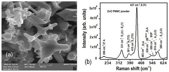
Figure 1.
(a) SEM image of morphology of the PNMC ZnO powder after cold pressing; (b) Raman scattering spectrum of the PNMC ZnO powder at room temperature; vibration modes of the first order are marked in bold.
The Raman scattering spectrum of the PNMC ZnO powder in the region of the first-order vibrational modes is shown in Figure 1b. Intense Raman bands with a relatively small half-width and the corresponding frequency position indicate that the crystalline phase of wurtzite is inherent in the ZnO nanocrystals. Even though ZnO nanocrystals of different shapes and polycrystals contribute to the Raman spectrum, as can be seen from Figure 1a, the half-width of the characteristic band with a frequency of 437 cm−1 is not significant and is only 13 cm−1. The latter indicates the good crystalline perfection of ZnO nanocrystals. Significantly higher bandwidth with a frequency of 437 cm−1 compared to one with a frequency of 584 cm−1 indicates that the average size of ZnO nanocrystals exceeds 10 nm [22], which correlates well with the data obtained from SEM images.
The diffractogram of the PNMC ZnO powder was recorded in symmetric (2θ/ω) mode, the scanning step was 0.025 degrees, and the acquisition time at the point was 1 s. All diffraction peaks on the diffractogram (Figure 2) correspond to the polycrystalline phase of ZnO (PDF 010-74-9939) with parameters a = 3.2494 Å; c = 5.2054 Å. From the angular positions and values of the half-widths of the reflections (002) and (004) using the Williamson–Hall method, the size of the coherent scattering regions (COS) and the average level of deformations (ε) were in the direction of the main optical axis c (ε = 4.8·10−4 OKR = 25.4 nm). A similar construction according to reflexes (100) and (200) gives values in the direction of the parameter a (ε = 5.3·10−4 OKR = 26.1 nm). The Williamson–Hall method is based on the graphical method of determining the OCR and ε from construction [23]:
where β is the half-width of the reflex, ϴ is the angular position, λ is the wavelength, D is the OCR size, and ε is the average level of deformations. The XRD data correspond well to the conclusions from the Raman shift spectra about the average sizes of ZnO single crystals, as components of larger crystallites, observed in SEM images.
βcosϴ = λ\D + 4εsinϴ,
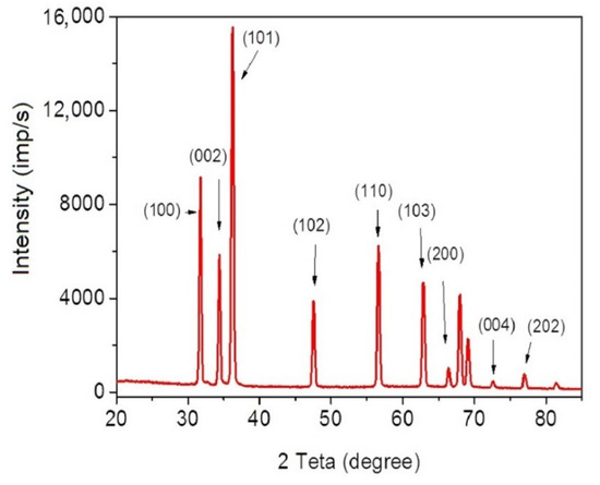
Figure 2.
Diffraction pattern of ZnO nanoparticles.
The PL spectra evolution (Figure 3) shows the significant growth in the peak intensity Ipl of the UV PL band (387 nm) of the PNMC ZnO powder samples with a simultaneous narrowing of its half-width Δλ (from 19 to 14 nm, insert). This indicates the stimulated emission of the UV band contrary to the visible bands: 405 nm (C-VZn), 502 nm (VO), 544 nm (Oi) [7] with weak increasing, and spontaneous emission. The large difference between the growth of the UV and visible bands (green and orange) with increasing excitation intensity indicates a low concentration of (Oi, VO) type defects, both a sign of a small excess and a deficiency of oxygen in the PNMC ZnO oxide powder. This is consistent with the conclusions from the Raman scattering spectra, Figure 1b, about the good crystalline quality of the PNMC ZnO powder, as well as with considerations in [24] regarding the excess or deficiency of oxygen anions in complex oxides of transition metals. The spectral position of the peak (405 nm) coincides with the position of the e–h plasma PL band at high excitation intensity Iex for the single crystalline ZnO film, obtained by MBE [4]. Nevertheless, the 405 nm peak for PNMC ZnO powder does not shift at all Iex levels. Besides, it is considerably narrower (Δλ ≤ 2 nm) as compared to e-h plasma band (Δλ ≈ 8 nm) shifted in case of the excitons decay and renormalization of the band gap. These circumstances indicate a high probability of the excitonic nature of the transmission in UV PL band (387 nm). Lasing at a wavelength of 387 nm occurs in the so-called diffusion mode due to the incoherent or “energy” FB [4]. In this case, adjacent modes spectrally overlap, which gives a continuous spectrum of emission. This result qualitatively differs from observations on the samples of the ZnO film with hexagonal microdisks formed by the method based on a low-temperature solution [20].
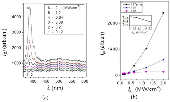
Figure 3.
(a) Evolution of the PL spectra in the compressed PNMC ZnO powder for different excitation intensities Iex (λex = 337 nm) in the range of 0.12–2 (MW/cm2) at room temperature. (b) Dependences of the PL spectral bands intensity Ipl versus the excitation intensity Iex (λex = 337 nm) for different wavelengths, nm: 387, 405, 502. Insertion: band half-width Δλ at 387 nm vs Iex.
Hexagonal ZnO microdisks had a distribution with some disordered orientation in space and size variation Figure 4b [25]. In this case, the PL spectrum occurs with a comb-like regular arrangement of ultra-narrow bands (peaks) with halfwidth Δλ ~ 0.25 nm. Superlinear growth of IPl in the range of excitation intensities Iex indicates the presence of coherent or “amplitude” FB [18]. Stimulated emission has a mode composition formed by whispering gallery modes (WGM) formed inside (on the faces) of hexagonal microdisks.
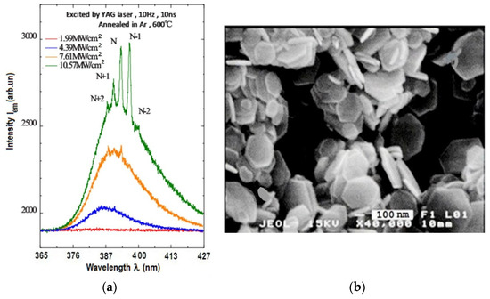
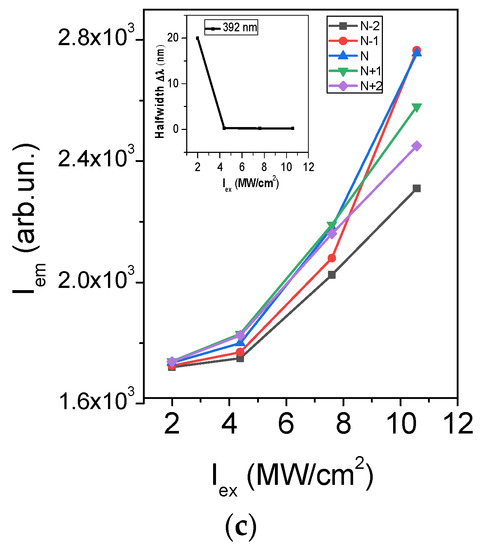
Figure 4.
(a) UV PL spectra evolution on the intensity of excitation Iex (λ = 355 nm) for the ZnO film with hexagonal microdisks; (b) SEM images of the ZnO film containing the hexagonal microdisks; (c) the PL ultra-narrow peaks’ intensity versus Iex at the wavelengths λpl (nm) = 388, 391, 392, 397, 399, constructed according to [25] for comparison. Insert: the UV PL spectral band halfwidth versus the Iex, N is mode number. The data from [25] were used with permission. It is noteworthy that the threshold of stimulated emission, in this case, coincides with the appearance of the comb of narrow peaks that confirms the non-random nature of the ultra-narrow peaks’ appearance.
A fundamentally different character of the evolution of PL spectra was observed in nanopowder with one-dimensional NPs size distribution, with an average size ~ 100 nm with strong scattering (Figure 5) [26]. The ultra-narrow bands (0.2 nm) in PL spectra appeared in a chaotic sequence with irregular spectral intervals, unlike [25]. This is associated with the stochastic nature of the resonator loop formation in the excited volume. SE, in this case, may arise initially as a result of incoherent amplification, regardless of the appearance of ultra-narrow peaks. Their occurrence does not coincide with the SE threshold, since the coherent ultra-narrow peaks arise according to the stochastic formation of a closed photon track. The superlinear growth of the UV band PL intensity Iem in the spectra of PNMC ZnO powder shown in Figure 3 on the pump intensity Iex, is qualitatively similar to the data of the film with ZnO hexagonal microdisks (Figure 4) [25] and to the integral spectrum intensity in the mono-dispersed powder containing the NPs ZnO with particle size of 100 nm, curve 1, Figure 5b, [26]. There is a tendency to sublinearity for a single ultra-narrow peak (386 nm) at the end of curve 2, Figure 5b, which can be explained by the redistribution of the SE energy on emerging random modes.
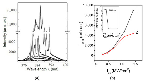
Figure 5.
(a) UV PL spectra evolution of the monodispersed ZnO powder under excitation intensity in the range (from bottom to top): 400, 562, 763, 875, and 1387 kW/cm2 (taken from reference [26] with permission). The size of the ZnO crystallite is equal 100 nm; (b) Dependencies of UV PL Iem: (1)—the spectral-integral intensity of UV-PL emission; (2)—dependence of a separate ultra-narrow peak (386 nm) from monodispersed ZnO powder on the excitation intensity Iex; inset: mean half-width of UV PL peaks as a function of Iex constructed from reference [26] with permission.
The conditions for the absence of ultra-narrow peak in the disordered laser medium can be evaluated from the threshold criterion expression for the critical volume Vcr [4]:
where lg is the length of the gain in the scattering medium and lt is the free path of the photon, at which the direction of its motion becomes unpredictable (there is a “randomization” direction) [4]:
where ls is the path between the two scattering acts, and θ is the scattering angle.
Vcr ≈ (lt · lg/3)3/2,
lt = ls/(1 − cosθ),
Given the spectrum evolution of the central UV spectral band (λmax = 387 nm) of PNMC ZnO powder, we concluded that the photon transport occurs in the so-called “diffusion mode” (L >> lt >> λ), where L is the characteristic size of random media within the critical excitation volume Vex < Vcr. The significant dispersion of particle sizes (0.05–2 µm) and shapes in PNMC ZnO powder, Figure 1a, complicates software processing data for their averaging. Therefore, in the estimations of the effective gain length [4]:
We will limit ourselves to the probable range of lt and lg from the condition that Vex ≤ Vcr, according to relation (1). We take into account the size of laser spot dex ≈ 20 µm and excited layer thick dl > 10 µm, and also that only 1 to 5% of the exciting light is absorbed by ZnO nanopowder, and the rest is scattering [4]. This corresponds to a depth of absorption from 7.14 × 10⁻⁵ cm (α = 1.4 × 104 cm−1) to 3.6 · 10−4 cm (α = 2.8 × 103 cm−1) at the wavelength of the nitrogen laser (λ = 337 nm), where α is the absorption coefficient. The value range of the exciting volume was about Vex = 3.0 × 10⁻⁸ ÷ 1.0 × 10⁻⁷ cm3. The probable range of values of the critical volume of the SE source depending on the gain length lg and length of randomization lt is shown in Figure 6. The range of the gain path of lengths lg is from 5 × 10⁻⁷ to 10−1 cm, provided that the range of lengths of «randomization» lt of the motion direction of the photon is determined within lt = 10−3 ÷ 10−2 cm. Obtained gain factors (lg)−1 = 20 ÷ 10 cm−1 were not too high. However, the gain effect between the start and endpoints of the full length of the gain path lg [26] is lamp = 0.0041–0.018 cm, which corresponds to the average value of the gain (lamp)−1 = 150 cm−1. It should be noted that in all modifications of PNMC of ZnO powder, the effect of incoherent FB is implemented. The photon transport occurs in the so-called “diffusion mode” within the critical excitation volume V < Vcr. This allows us to keep SE in the incoherent mode at all ranges of excitation by using nitrogen laser N2. The position of the UV band maximum at 387 nm fixed at all pumping levels of the PNMC ZnO powder coincides with the maximum of the so-called “P” exciton band in single-crystal ZnO films [26]. This is responsible for exciton-stimulated emission under conditions of inelastic exciton–exciton scattering [27]. Its fixed spectral position with increasing Iex in our case is caused by an increase of the exciton concentration nex, preventing them from breaking up and transitioning into e-h plasma (with a shift of the maximum to λ~405 nm). We consider two possible explanations: (1) due to the giant oscillator strength fNPex [28]:
(where fBex is the oscillator strength in the crystal bulk, a*B is Bohr radius of the exciton, and R is the NP’s radius) with the condition that the crystal size is larger than the exciton the Bohr radius (a*B ~ 2 nm) but smaller than the optical wavelength (387 nm) [29]: (2) increase the binding energy of exciton Eex and, accordingly, their concentration nex. It can be assumed that the surface capture centers of excitons exist at the interface of nanocrystals (between grains of ~25 nm) as a part of larger ZnO particles, recorded on the SEM image, Figure 1a, and are responsible for the growth of Eex. The increase of nex leads to an increase in the exciton self-scattering and SE in the PNMC ZnO. Thus, the interfaces between nanocrystals can be considered as barriers that lead to the exciton’s confinement in nanocrystal grains (~25 nm in size). We plan to study the peculiarities of the kinetics of exciton transitions in PNMC ZnO powder under these conditions in the near future.
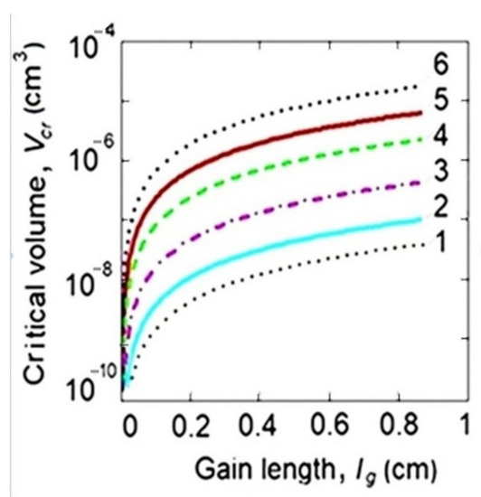
Figure 6.
Dependences of the critical volume Vcr on the gain length lg at different average randomized lengths lt, µm: 1—0.387 (1 λ); 2—0.74 (2 λ); 3—1.94 (3 λ); 4—5.85 (15 λ); 5—11.6 (30 λ); 6—15.5 (40 λ).
Summarizing the above, we can conclude that to improve the stimulated modeless emission efficiency of such a multi-element system, it is advisable to take into account and use the influence of the effective surface. The latter appears very developed due to a large number of nanocrystal surfaces that can significantly increase the concentration of the exciton capture centers without an increase in the light scattering. This aspect has not been given due attention in previous studies, including those cited in the text. The surface plays a dominant role in such multi-element systems due to several important factors: exciton capture in shallow surface centers, quantum confinement effect in 2D-like platelet nanosystems, mirror reflection forces, and correlation forces, which under certain conditions leads to an increase in Eex and, accordingly, exciton inversion population (see, for example, [12]). Clarification and confirmation of the priority of the specified mechanisms is expected in our further studies.
4. Conclusions
A comparative analysis of the optical spectral and structural properties among various types of crystallites ZnO structures allowed us to establish:
- Among considered crystalline ZnO systems, the polydisperse nano-microcrystalline (PNMC) powder with a ZnO particle size ranging from 50 nm to 2 μm showed the random UV SE (λ = 387 nm) with incoherent feedback in all ranges of excitation intensity.
- The dominant factor for UV SE at λ = 387 nm in the PNMC ZnO powder at room temperature is exciton–exciton scattering-assisted radiative transitions.
- The average value of the optical gain at λ = 387 nm in the PNMC ZnO powder RL is estimated to be as high as 150 cm−1.
- ZnO thin film with hexagonal microdisks, as well as low-dispersed ZnO nanopowder, demonstrate UV SE with coherent feedback and ultra-narrow spectral peaks: regular
In the case of the hexagonal microdisks and irregular in the case of monodisperse ZnO powder, with a particle size of ~100 nm, accordingly.
Author Contributions
Conceptualization, L.F., V.L. and H.M.; methodology, L.F., V.N., V.Y. and A.M.; validation, L.F., V.N. and V.Y.; formal analysis, L.F., D.K. and H.M.; investigation, L.F., V.N., V.Y., O.G., O.D., A.M. and H.M.; resources, D.K. and A.M.; data curation, L.F., V.N. and V.Y.; writing—original draft preparation, L.F. and A.M.; writing—review and editing, L.F., V.N., V.Y. and A.M., visualization, L.F., V.N. and A.M.; supervision, L.F. and D.K. All authors have read and agreed to the published version of the manuscript.
Funding
This research received no external funding but was supported by ongoing research programs of the National Academy of Sciences of Ukraine and funded by Riga Technical University, Latvia, project N 14508 and Research Institute of Electronics, Shizuoka University, Japan.
Institutional Review Board Statement
Not applicable.
Informed Consent Statement
Not applicable.
Data Availability Statement
The data that support the findings of this study are available from the corresponding author upon reasonable request.
Acknowledgments
This work was performed due to the scientific cooperation between the Lashkaryov Institute of Semiconductor Physics, National Academy of Sciences of Ukraine, the Institute of Technical Physics, Riga Technical University, and the Research Institute of Electronics, Shizuoka University. The authors thank Hui Cao, head of the Research Laboratory at Yale University, the USA for kindly granting permission to use the experimental data for comparison calculations.
Conflicts of Interest
The authors declare no conflict of interest.
References
- Song, Q.; Xu, Z.; Choi, S.H.; Sun, X.; Xiao, S.; Akkus, O.; Kim, Y.L. Detection of nanoscale structural changes in bone using random lasers. Biomed. Opt. Express 2010, 11, 1401–1407. [Google Scholar] [CrossRef] [PubMed]
- De Armas-Rillo, S.; Fumagallo-Reading, F.; Luis-Ravelo, D.; Abdul-Jalbar, B.; González-Hernández, T.; Lahoz, F. Random lasing detection of mutant huntingtin expression in cells. Sensors. 2021, 21, 3825. [Google Scholar] [CrossRef] [PubMed]
- Wiersma, D.S. The physics and applications of random lasers. Nat. Phys. 2008, 4, 359–367. [Google Scholar] [CrossRef]
- Cao, H. Lasing in random media. Top. Rev. Waves Random Media 2003, 13, R1–R39. [Google Scholar] [CrossRef]
- Cao, H.; Chriki, R.; Bittner, S.; Friesem, A.A.; Davidson, N. Complex lasers with controllable coherence. Nat. Rev. Phys. 2019, 1, 156–168. [Google Scholar] [CrossRef]
- Noginov, M.A. Lasers with Nonresonant Feedback and Laserlike Emission from Powders: Early Ideas and Experiments. In Solid-State Random Lasers; Springer Series in Optical Sciences; Springer: New York, NY, USA, 2005; Chapter 1; pp. 1–9. [Google Scholar] [CrossRef]
- Lu, Y.J.; Shi, Z.F.; Shan, C.X.; Shen, D.Z. ZnO nanostructures and lasers. In Nanoscale Semiconductor Lasers. Micro and Nano Technologies; Tong, C., Jagadish, C., Eds.; Elsevier: Amsterdam, The Netherlands, 2019; Chapter 4; pp. 75–108. [Google Scholar] [CrossRef]
- Bagnall, D.M.; Chen, Y.F.; Zhu, Z.; Yao, T.; Shen, M.Y.; Goto, T. High temperature excitonic stimulated emission from ZnO epitaxial layers. Appl. Phys. Lett. 1998, 73, 1038–1040. [Google Scholar] [CrossRef]
- Thareja, R.K.; Mitra, A. Random laser action in ZnO. Appl. Phys. B Lasers Opt. 2000, 71, 181–184. [Google Scholar] [CrossRef]
- Eliezer, Y.; Qu, G.; Yang, W.; Wang, Y.; Yılmaz, H.; Xiao, S.; Song, Q.; Cao, H. Suppressing meta-holographic artifacts by laser coherence tuning. Light Sci. Appl. 2021, 10, 2047–7538. [Google Scholar] [CrossRef]
- Ye, Y.; Wong, Z.J.; Lu, X.; Ni, X.; Zhu, H.; Chen, X.; Wang, Y.; Zhang, X. Monolayer excitonic laser. Nat. Photonics 2015, 9, 733–737. [Google Scholar] [CrossRef]
- Lytovchenko, V.G.; Fedorenko, L.L.; Korbutyak, D.V.; Strikha, M.V. Ordered electron-hole condensate as a perspective 2D laser environment at room temperatures. Ukr. J. Phys. 2021, 66, 612–617. [Google Scholar] [CrossRef]
- Litovchenko, V.G.; Korbutyak, D.V.; Kryuchenko, Y.V. Investigation of the collective properties of excitons in polar semiconductors (ZnO). Sov. Phys. JETP 1981, 54, 1093–1099. [Google Scholar]
- Korbutyak, D.V.; Litovchenko, V.G. Electron-hole condensate in semiconductors with high exciton energy. Sov. Phys. Solid State 1981, 23, 1411–1416. [Google Scholar]
- Lawandy, N.M.; Balachandran, R.M.; Gomes, A.S.L.; Sauvain, E. Laser action in strongly scattering media. Nature 1994, 368, 436–438. [Google Scholar] [CrossRef]
- Gottardo, S.; Cavalieri, S.; Yaroshchuk, O.; Wiersma, D.S. Quasi-Two-Dimensional Diffusive Random Laser Action. Phys. Rev. Lett. 2004, 93, 263901-1–263901-4. [Google Scholar] [CrossRef]
- Wiersma, D.S.; van Albada, M.P.; Lagendijk, A. Coherent backscattering of light amplifying random media. Phys. Rev. Lett. 1995, 75, 1739–1742. [Google Scholar] [CrossRef]
- Cao, H.; Xu, J.Y.; Zhang, D.Z.; Chang, S.-H.; Ho, S.T.; Seelig, E.W.; Liu, X.; Chang, R.P.H. Spatial confinement of laser light in active random media. Phys. Rev. Lett. 2000, 84, 5584–5587. [Google Scholar] [CrossRef]
- Gomes, A.S.L.; Moura, A.L.; de Araújo, C.B.; Raposo, E.P. Recent advances and applications of random lasers and random fiber lasers. Prog. Quantum Electron. 2021, 78, 100343. [Google Scholar] [CrossRef]
- Han, G.; Okada, M.; Xiao, Z.Y.; Neo, Y.; Aoki, T.; Mimura, H. Cathodoluminescence of single disk-like ZnO prepared by the low-temperature solution-based method. e-J. Surf. Sci. Nanotechnol. 2009, 7, 354–357. [Google Scholar] [CrossRef][Green Version]
- Wu, X.H.; Yamilov, A.; Noh, H.; Cao, H.; Seelig, E.W.; Chang, R.P.H. Random lasing in closely packed resonant scatterers. J. Opt. Soc. Am. B 2004, 21, 159–167. [Google Scholar] [CrossRef]
- Ohtomo, A.; Kawasaki, M.; Sakurai, Y.; Yoshida, Y.; Koinuma, H.; Yu, P.; Tang, Z.K.; Wong, G.K.L.; Segawa, Y. Room temperature ultraviolet laser emission from ZnO nanocrystal thin films grown by laser MBE. Mater. Sci. Eng. 1998, B54, 24–28. [Google Scholar] [CrossRef]
- Mote, V.; Purushotham, Y.; Dole, B. Williamson-Hall analysis in estimation of lattice strain in nanometer-sized ZnO particles. J. Theor. Appl. Phys. 2012, 6, 2–8. [Google Scholar] [CrossRef]
- Trukhanov, S.V.; Trukhanov, A.V.; Vasil’ev, A.N.; Maignan, A.; Szymczak, H. Critical behavior of Ga0.825Sr0.175MnO2.912 anion-deficient manganite in the magnetic phase transition region. J. Exp. Theor. Phys. Lett. 2007, 85, 507–512. [Google Scholar] [CrossRef]
- Han, G.; Shibukawa, A.; Okada, M.; Neo, Y.; Aoki, T.; Mimura, H. Nanosized hexagonal plateletlike ZnO for nanophosphor applications. J. Vac. Sci. Technol. B 2010, 28, C2C16–C2C19. [Google Scholar] [CrossRef]
- Cao, H.; Zhao, Y.G.; Ho, S.T.; Seelig, E.W.; Wang, Q.H.; Chang, R.P.H. Random laser action in semiconductor powder. Phys. Rev. Lett. 1999, 82, 2278–2281. [Google Scholar] [CrossRef]
- Klingshirn, C. The Luminescence of ZnO under High One- and Two-Quantum Excitation. Phys. Status Solidi 1975, 71, 547–556. [Google Scholar] [CrossRef]
- Kayanuma, Y. Quantum-size effects of interacting electrons and holes in semiconductor microcrystals with spherical shape. Phys. Rev. B 1988, 38, 9797–9805. [Google Scholar] [CrossRef]
- Özgür, Ü.; Alivov, Ya. I.; Liu, C.; Teke, A.; Reshchikov, M.A.; Doğan, S.; Avrutin, V.; Cho, S.-J.; and Morkoç, H. A comprehensive review of ZnO materials and devices. J. Appl. Physics 2005, 98, 041301. [Google Scholar] [CrossRef]


Publisher’s Note: MDPI stays neutral with regard to jurisdictional claims in published maps and institutional affiliations. |
© 2022 by the authors. Licensee MDPI, Basel, Switzerland. This article is an open access article distributed under the terms and conditions of the Creative Commons Attribution (CC BY) license (https://creativecommons.org/licenses/by/4.0/).