Abstract
Literature reports on Lucio Fontana’s work have focused on the study of the pictorial surface of his paintings. Consequently, to the best of our knowledge, this paper presents the first scientific investigation of gypsum objects made by this artist. These are the gypsum plaster casts made for the construction of the fifth door of the Milan Cathedral, Italy, which were discovered after 60 years, and which showed a green patina and chromatic discolorations. With the aim of understanding the microbiological or chemical nature of the stains, the surfaces of the tiles were investigated by microbiological analysis and analytical techniques including observations by optical, fluorescence and electron microscopy and spectroscopy. The investigated samples showed that the amount of microbiological cells in the stained area was negligible. Chemical analyses allowed the identification of compounds responsible for the chromatic alterations.
1. Introduction
Lucio Fontana, one of the most important artists of the 20th century, participated in the competition launched in 1950 for the construction of the fifth door of the Milan Cathedral, presenting revolutionary gypsum plaster casts. Although he won the competition, the work, made of tiles, was considered too modern to be installed, and soon the casts were set aside and forgotten. After more than 60 years the final casts completed in 1956 were discovered in the Milan Cathedral Museum repository (Figure 1). Despite being an object of contemporary art, prevention and conservation were needed, e.g., due to the cuts [or, “damage”], which had intentionally or accidentally occurred over the years. Furthermore, some of the thirteen tiles of the project showed a green patina not visible in the 1950s photographic documentation and various chromatic alterations over the surface. This outstanding work was put into an exhibit in 2019 after a conservative intervention of restoration on the gypsum figures of the tiles and removal of the deposits accumulated during their long abandonment. The surface cleaning was carried out with agar gel applied as a high viscosity liquid and then removed as a film. During this restoration phase a research investigation on the nature of the chromatic alterations over the gypsum surface was requested from the scientific team of the Cultural Heritage Centre of the University of Milan.
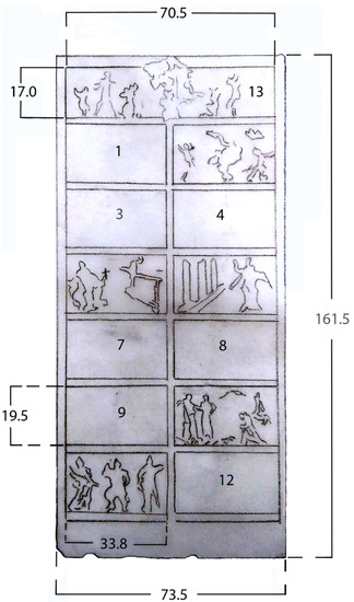
Figure 1.
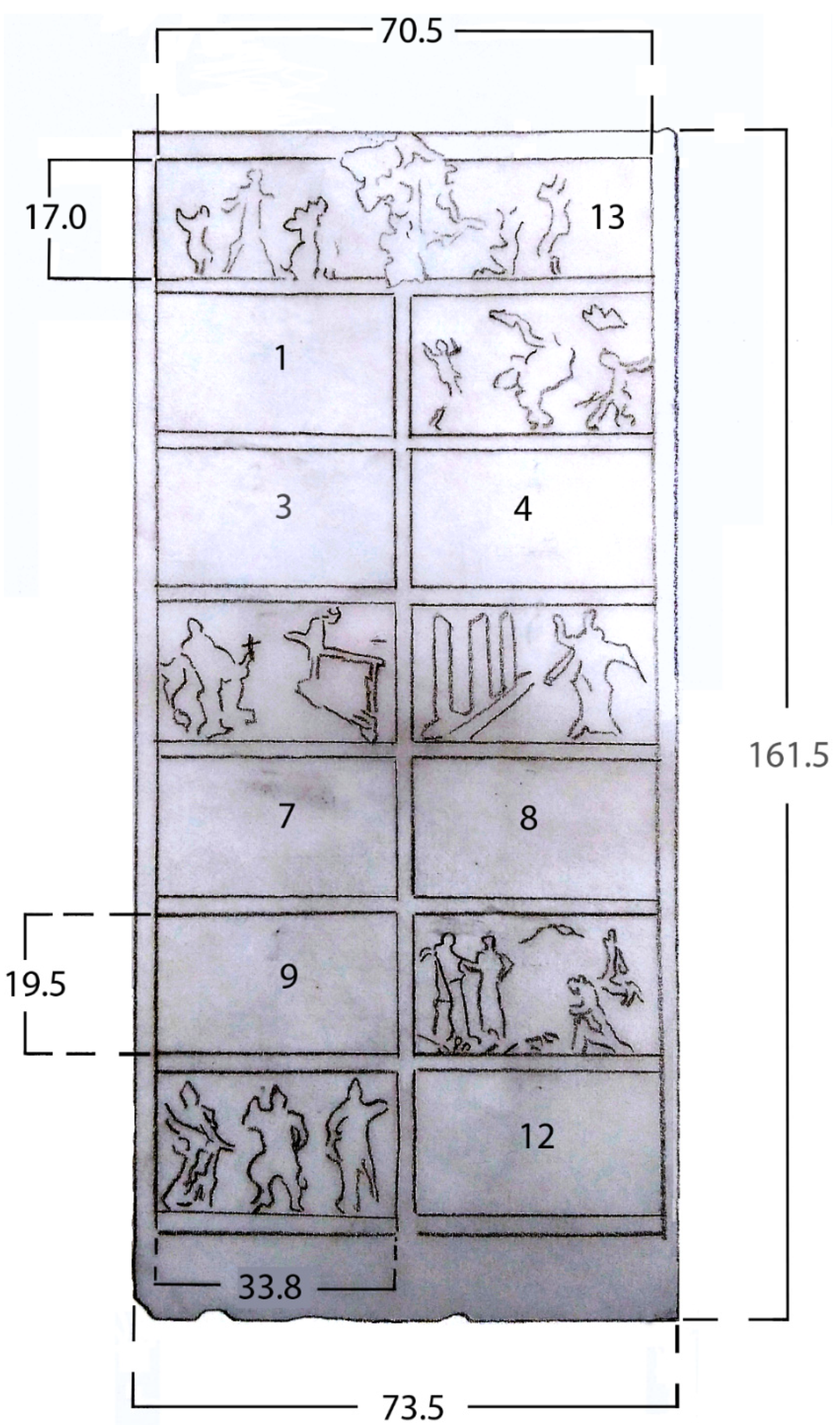
Schematic drawing of the fifth door of the Milan Cathedral, Lucio Fontana 1955–1956. Sizes are in centimeters. Tile 1: Tedeum liberation of Milan; tile 2: Archbishop Schuster among the victims of the bombing; tile 3: The plague; tile 4: Coronation of Napoleon; tile 5: Martin V and the high altar; tile 6: Saint Carlo consecrates the Cathedral; tile 7: Volunteering; tile 8: Marble transport; tile 9: Gian Galeazzo Visconti and the Milanese, the offerings; tile 10: Archbishop Antonio da Saluzzo promulgates the bull of foundation of the Cathedral; tile 11: The six Milanese popes (1); tile 12: The six Milanese popes (2); tile 13: The prayer of the city of Milan to the Virgin.
Fungi are often found where gypsum is present [1,2] and may significantly affect the physical and mechanical integrity of moisture-exposed gypsum materials, leading to up to 80% loss of tensile strength and weight [3,4]. Sometimes fungal development causing discoloration on gypsum materials can also be assessed by the naked eye [5]. Studying interior gypsum boards, Adan [6] observed that spores of Penicillium chrysogenum were capable of an instantaneous water vapor uptake in the case of relative humidity (RH) increases, suggesting that even short periods of high RH should not be neglected in evaluating the indoor climate in relation to fungal growth. The effects of biodeterioration of gypsum materials is such a known and widespread phenomenon that biocides specific to them have been devised over time, e.g., nanosilver particles [5].
From the physico-chemical point of view, gypsum is highly porous, vulnerable to scratches and mechanical damage, and can be softened or even disintegrated in the presence of moisture [7]. Traditionally, several different substances, both inorganic and organic, have been added to gypsum plaster used for casts to improve its mechanical resistance or change its aesthetic properties [7].
Before cleaning, the green patina and chromatic alterations of Fontana’s plaster cast were thoroughly studied. In particular, the green patina was examined to settle the question of whether it was part of the original artwork or due to deterioration. The materials used by L. Fontana over the years have been deeply investigated by some researchers [8,9,10]. In particular, the inks and/or pigments used by the artist in the years of cast production were identified (1955–1956). Nevertheless, his numerous experimentations and the variety of industrial products the artist used made the identification of the green patina origin challenging. The investigation reported in this paper also disclosed the nature (microbiological or chemical) of the other chromatic alterations.
The present work places itself in the scope of recent investigations on important collections of plaster casts, such as those dating back to the 19th century of the Victoria and Albert Museum in London or the Walter Copland Perry’s casts collection kept in the British Museum, also in London. Such investigations [11,12,13,14] aimed to analyze the chemical composition of any coatings, or possibly discolorations, of the plaster surface. The innovative aspects of the present study relate both to its object, being the work of a world-famous artist of the 20th century like Lucio Fontana, and to the methodological approach. In fact, microbiological analyses were performed together with chemical ones, and, to the best of our knowledge, Fourier-transform infrared (FTIR) reflection spectroscopy was applied for the first time to the in situ analysis of this type of artwork. Micro-FTIR spectroscopy and scanning electron microscopy coupled with energy-dispersive X-ray (SEM-EDX) analysis were also applied in the laboratory just to two small samples belonging, respectively, to an apparently exogenous material and to the plaster itself. The latter could be analyzed in a destructive way as it was not possible to put it back in its original place in the cast.
2. Materials and Methods
2.1. Artwork Description
The gypsum plaster casts made by Lucio Fontana, representing the most important events in the recent story of the Milan Cathedral, are thirteen tiles. The artwork’s size is 161.5 cm × 73.5 cm × 10.0 cm; each tile is 33.8 cm × 19.50 cm, while the tile that acts as the architrave is 70.5 cm × 17.0 cm. When rediscovered in the Milan Cathedral Museum repository, all the tiles were stored inside a cardboard box separated from the wood frame. Under the naked eye, the artwork showed a green patina on only three tiles and on the base as well as various types of chromatic stains. The green patina mostly covered the figures in relief on the tiles and their surroundings and it is supposed to have been applied in the 1970s during the making of a bronze copy from the original gypsum mock-up, while the other alterations were observed on the flatter surfaces of the tiles.
2.2. Microbiological Analysis
2.2.1. Sampling
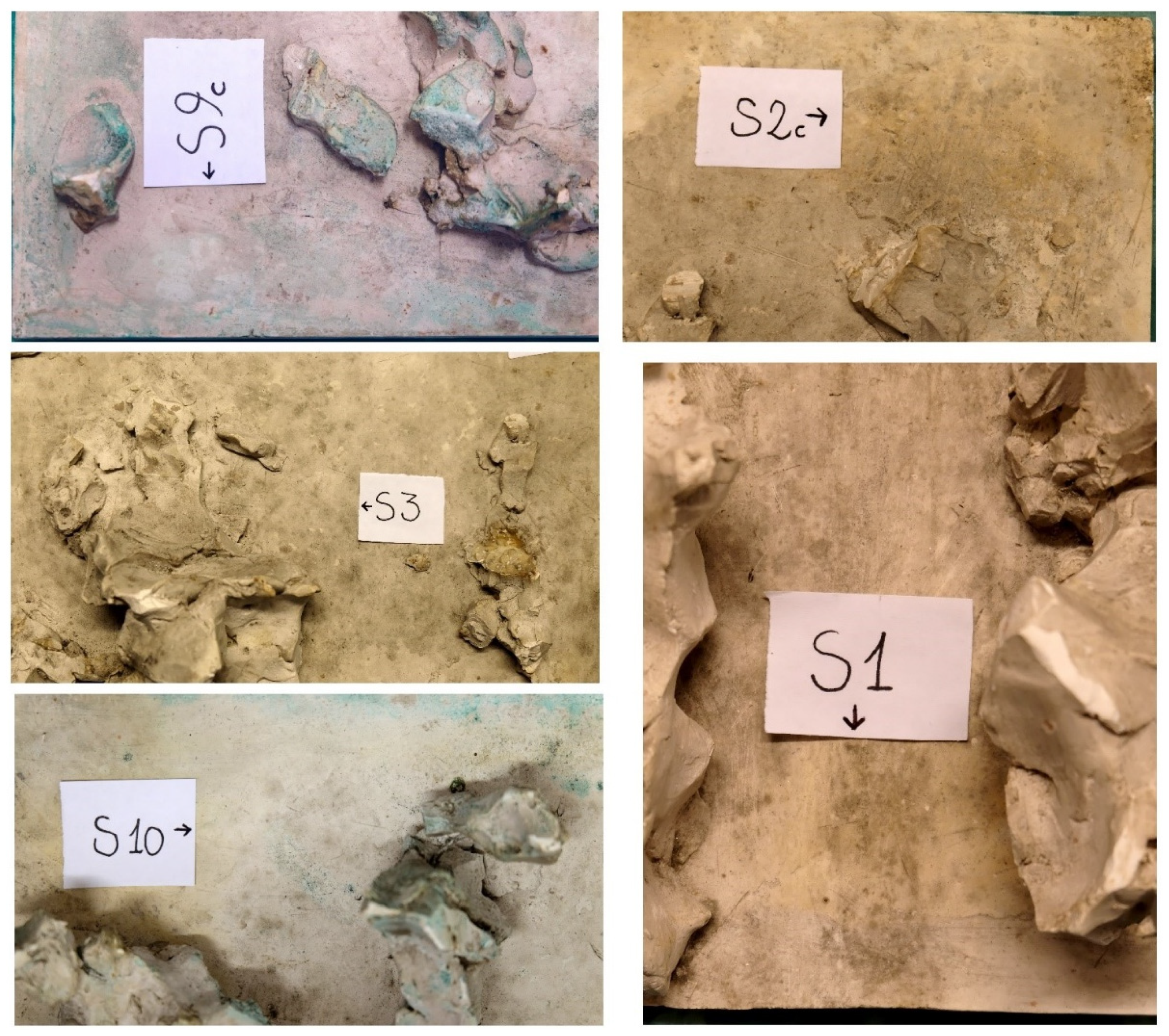
Nine of the tiles (named T1, T3, T4, T5, T7, T8, T9, T12 and T13) of gypsum plaster casts by Lucio Fontana were sampled for microbiological analysis for a total of fifteen samples representative of all the chromatic alterations on the tiles of the gypsum plaster casts. The fifteen samples were collected by using the non-invasive technique of Fungi-tape (DID Milan, Italy), to include areas with and without chromatic discolorations (Figure 2). Adhesive tape sampling coupled with microscopical analysis has been largely adopted for the study of potential/actual biodeterioration (inter alia [15,16,17]). Table 1 shows the details of the adhesive tape samples; the samples were mounted on microscope glass slides and transferred into a laboratory for optical microscopic analysis. In the laboratory all the adhesive tape samples, except for S15, S8 and S4, were fixed in 4% paraformaldehyde solution (Sigma-Aldrich) in 0.1 M PBS pH 7.2 for 2 h on ice [18]. After three washing steps with PBS 0.1 M, samples were stored at −20°C.
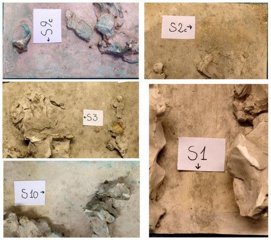
Figure 2.
Sampling points: S9 visual observation with a digital green filter of the green patina on tile T9, S2 visual observation of a yellow-reddish discoloration on tile T3, S3 black/brown discoloration from tile T3, S10 black/brown discoloration associated with greenish stains from tile T13, and S1 yellow-reddish stains from T1 tile.

Table 1.
Samples collected for microbiological (code S) and measurement areas for reflection FTIR (code F) analyses in correspondence of the discolorations on the gypsum cast plaster.
2.2.2. Microscope Observations
The microbiological strategy was to evaluate the abundance of microorganisms on the surface of the tapes (using both bright field and fluorescence microscopy) and their activity (by fluorescence microscopy). To this aim, a portion of each tape was confined by an in situ frame 1 cm2 area (Eppendorf). This portion of the adhesive tape was stained with 100 µL of the green-fluorescent nucleic acid stain SYTO 9 (10 µM) (Invitrogen, Eugene, Oregon) and 100 μL of 0.4 mg mL−1 Fluorescent Brightener 28 (Sigma-Aldrich) for 20 min in the dark at room temperature. Fluorescent Brightener 28 is a stain with a blue fluorescence, which binds specifically to chitin or cellulose [19]; SYTO 9 is a fluorescent nucleic acid stain that is widely used in fluorescence microscopy to label and count bacteria [20]. After three washes with demineralized water, all samples were observed by epifluorescence microscopy with a Leica DM 4000 B (Leica Microsystems, Milan, Italy). Images were acquired by the CoolSNAP CF camera (Photometrics Roper Scientific, Rochester, NY, USA).
In order to verify the presence of an active microbial colonization on gypsum tiles, samples S15, S8 and S4 were stained with 100 mL of 20 mg mL−1 fluorescein diacetate (FDA, green fluorescence) and observed as reported previously.
2.3. Spectroscopic Techniques
2.3.1. Reflection FTIR Spectroscopy
FTIR spectra were acquired directly on the artefact in reflection mode by means of the portable spectrometer Bruker Alpha equipped with an attachment for specular reflection measurements. Resolution was set to 4 cm−1 and each spectrum was obtained as the sum of 100 scans. The diameter of the measurement area was 0.6 cm. Reflection data were transformed in log(1/R), i.e., pseudo-absorbance [21], to take into account the fact that both specular and diffuse reflection contributed to the measured signal. For this reason, the strongest bands of inorganic components, such as gypsum, were inverted and observed as minima in the spectra, while the weaker ones and those of organic compounds appeared as maxima [22]. The measurement areas are listed in Table 1.
2.3.2. Micro-FTIR Spectroscopy
On minute fragments, detached from the plaster casts, micro-FTIR spectra were also recorded in transmission mode by placing them in a diamond compression cell. Measurements were performed by a Jasco IRT 3000 spectrometer equipped with a 32x objective. Resolution was 4 cm−1 and 256 scans were acquired for each spectrum.
In detail, two fragments were examined, one showing a green coloration and the other, a resin-like residue.
3. Results and Discussion
3.1. Green Patina
At the time of microbial sampling, dust was covering the gypsum plaster casts by Lucio Fontana; moreover a green patina stood out on tiles T1, T9 and T13. Visual observation of a green patina on tile T9 is reported in Figure 2a, the signal has been enhanced by the use of a digital green filter.
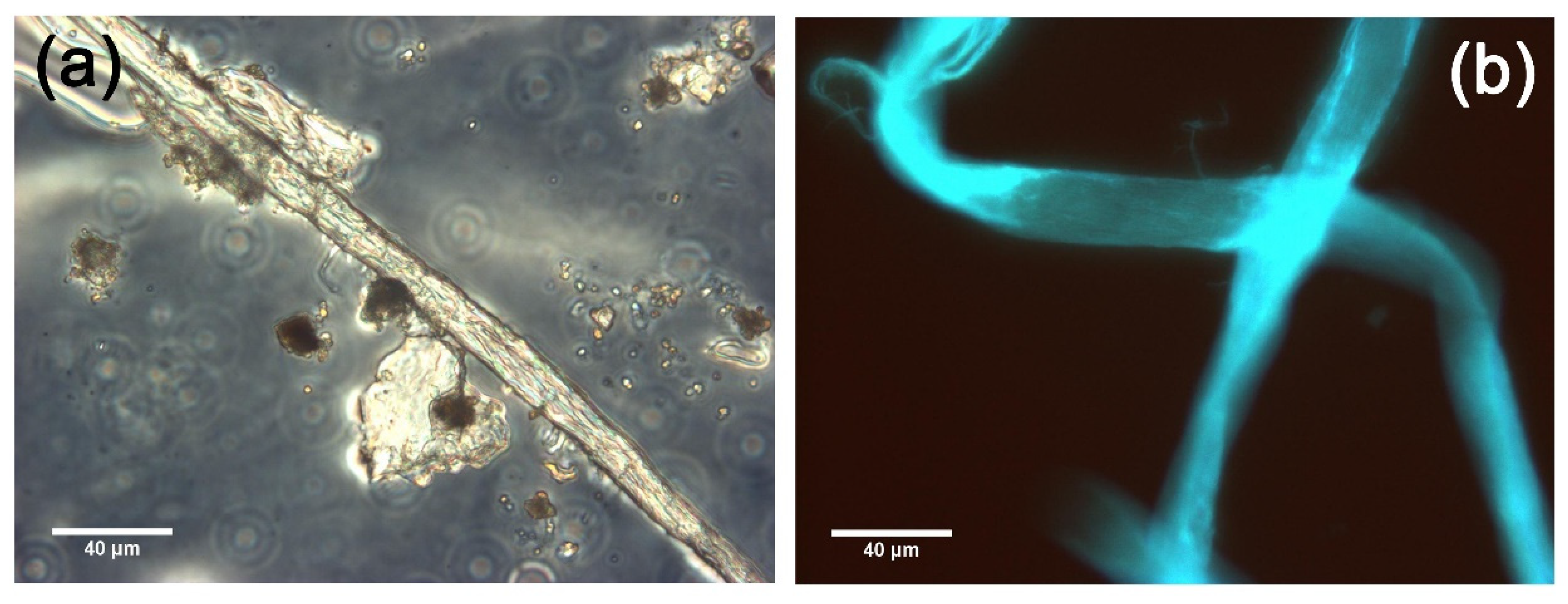
The sampling technique with Fungi-tape was used for verifying whether the green patina was due to a microbial colonization. In fact, microbial colonization of heritage materials can lead to discolorations, sometimes disfiguring the surface. Fungi-tape sampling is a non-invasive technique, widely used for monitoring microbial colonization on different materials, from human skin infections [23] to objects with cultural value like bones [24] to stone surfaces [25]. Figure 3 reports: (a) bright field; and (b) fluorescence microscope images of sample S9 from green patina on tile T9.
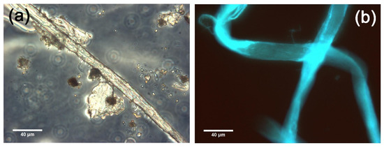
Figure 3.
Bright field (a) and epifluorescence (b) microscope images of sample S9 from green patina on tile T9.
Bright field microscope images display filamentous structures of dust with a blue fluorescence probably due to the presence of cellulose. The green fluorescence signal was absent, meaning that bacterial cells were in too low number to be detected. The absence of both bacteria and fungi led to the conclusion that no current biodeterioration was in place in areas where the green patina was present. In order to investigate the origin of the green patina, its chemical nature was investigated by spectroscopic techniques and SEM-EDX. A minute fragment, with green traces detached from the plaster, was examined by SEM-EDX and peaks due to S, Ca and Cu being detected in correspondence with the chromatic alteration (Figure S1).
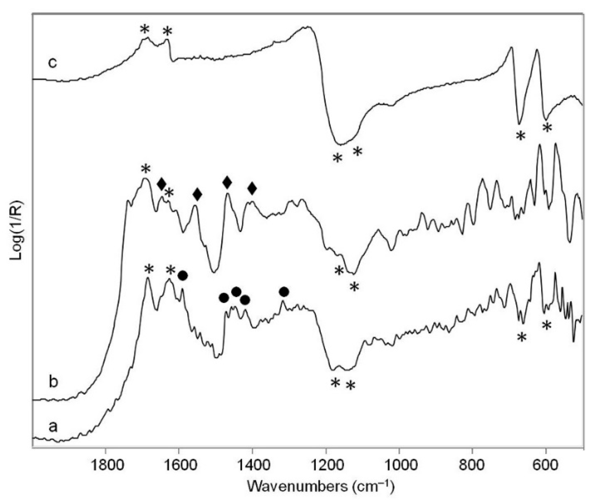
The spectra obtained “in situ” by the reflection FTIR technique on areas F9-1 and F13-1 showed first of all bands at about 1685 and 1620 cm−1 (observed as maxima in the log(1/R) traces) and at 1160, 1120, 660 and 600 cm−1 (observed as minima), all of which can be attributed to gypsum according to the literature [26]. Beside these bands, other peaks were detected at 1587, 1468, 1450, 1419 and 1316 cm−1 (Figure 4a). Such peaks can be assigned to copper carboxylates, namely palmitates or stearates [27,28]. The same signals were also observed by micro-FTIR spectroscopy on the small green fragment already analyzed by SEM-EDX.

Figure 4.
FTIR spectra acquired “in situ” in reflection mode on: (a) area with green patina F13-1; (b) yellow-reddish area F13-3; (c) unaltered area F3-1. Legend: (*) bands due to gypsum; (  ) bands due to copper carboxylates; (
  ) bands due to a proteinaceous component (possibly gelatin).
) bands due to copper carboxylates; ( ) bands due to a proteinaceous component (possibly gelatin).
These results suggest that the green color is due to a copper-containing pigment applied to the gypsum, possibly using an oil binder or even wax, as suggested in old recipes, to impart a given color to the plaster surface [7]. The interaction between the pigment and the binder gave rise to the formation of the observed metal soaps. It is interesting to note the use of a “copper soap” obtained from white soap combined with an aqueous solution of copper sulphate and then mixed with white wax and linseed oil [7,29] as a method to achieve a bronze-like appearance of the plaster surface. Furthermore, this is a rather unique case of traces of green color detected in a study on an otherwise unpainted plaster cast, as this color was never observed in the aforementioned studies on museum collections.
3.2. Yellow-Reddish Areas
After a careful observation of the surface of the artwork by Lucio Fontana, apart from the green patina, other discolorations over the gypsum artwork were present. One example is on tile T3. Over its surface an area of yellow-reddish color was observed. The microbiological or chemical origin of this stain was investigated. Adhesive tape sampling and observation under optical microscopy were conducted for microbiological analysis. Observation under optical microscopy of sample S2 taken in correspondence with the yellow-reddish stain revealed the presence of a few filamentous structures having blue fluorescence; the green fluorescence signal was absent (figures not shown). This investigation led to the conclusion that no current biodeterioration was in place in yellow-red areas.
As a consequence, samples F13-3 and F13-4 from tile T13 and sample F10-1 from tile T10 represent the areas of interest studied by spectroscopic techniques. The reflection FTIR spectra obtained on these areas (Figure 4b) are again dominated by the bands around 3500–3400 and about 1680 and 1620 cm−1 due to water molecules in the gypsum structure. Nevertheless, bands around 2930, 2860, 1640, 1550, 1460 and 1400 cm−1 suggest the presence of a proteinaceous component, in agreement with the hypothesis of the restorers that the yellow-reddish material was indeed gelatin [30]. Gelatin was commonly used before the advent of silicon to realize gypsum casts (personal communication with the conservators working on the casts [7]). Thus this material was not part of the artistic object itself. Similar residues of gelatin ascribed to the molds used were found in recesses and air pockets of some casts belonging to the Victoria and Albert Museum in London and were also identified by FTIR spectroscopy [11].
3.3. Black/Brown Discoloration
Black/brown stains were observed both alone (on tiles T3, T4, T5, T7, T8, T9 and T12) and associated with green patina (tile T13) and yellow-reddish discoloration (tile T1). To better assess if these discolorations were due to microorganisms, adhesive tape samples were collected and observed under the microscope. Bright field microscope images displayed rare circular/round and filamentous structures attributed to dust material (sample S3 on tile T3, Figure 5a); eukaryotic cells like fungal spores having blue fluorescence either appeared to be completely absent, or were only present in a very low amount (the latter situation represented by sample S3 and shown in Figure 5b); a weak green fluorescence signal was detected (Figure 5c) meaning that the bacterial cells were present in a low amount.
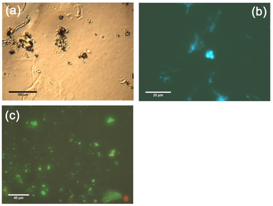
Figure 5.
Bright field (a) and epifluorescence (b,c) microscope images of sample S3 from black/brown discoloration on tile T3.
Five tiles of the gypsum plaster cast without chromatic alterations were also investigated by microbiological analysis (tiles T4, T5, T7 and T12) and spectroscopic techniques (tile T1). As for microscopic observation of areas of the tiles having discoloration, under microscopy sample S12 from an area with no discoloration showed the presence of rare microbiological structures having a filamentous shape with dust materials entrapped; no green fluorescence due to bacteria was observed.
Due to the near absence or the presence of only an extremely low number of microbiological structures in correspondence to both the chromatic alterations, and on areas without chromatic alterations on the tiles of the gypsum plaster casts, a microbiological nature for black/brown discoloration can be excluded.
As expected, the reflection IR spectra acquired from the unaltered areas F3-1 and F3-2 on tile T3 are dominated by bands due to gypsum (Figure 4c).
4. Conclusions
Fluorescence microscopic observations showed a weak signal for both green and blue fluorescence, indicating the extremely low amount of bacterial cells and fungi present respectively. Furthermore, no significant difference in the amount of microbial cells was observed in tape samples from discolored and non-discolored areas of the tiles. Moreover, no microbiological activity was detected. This meant that no current biodeterioration was the cause of the green patina and discolorations. Nevertheless, to avoid future biodeterioration we recommend routine monitoring of the temperature and relative humidity of the repository of Fontana’s plaster casts. It should be noted that a careful control of relative humidity is also important to avoid the damaging of plaster, due to the fact it is slightly soluble in water, even when hardened and its porosity makes it hygroscopic. The same feature favors the trapping of dirt and for this reason the casts should be protected from dust to avoid the risk of further discoloration [31,32]. Therefore, once having excluded biodeterioration problems in order to preserve the art object, we investigated the chemical nature of the chromatic alterations, individuating copper carboxylates in the green patina and gelatin residues in the yellow-reddish areas. The latter is a material associated with the production of casts and the former derives most probably from the use of a green copper pigment applied to the plaster with an oil or wax binder. The presence of this green coloration on some areas of the plaster was associated by the restorers [Marta Berolatti, private communication] to cutting surfaces, created in the bronze casting phase that took place in the 1970s. A similar hypothesis was put forward for other gypsum plasters made by the same artist, which also showed in many parts an obvious green shade [A. Devitini, private communication]. To the best of our knowledge, this is the first investigation of the origin of color traces on this particular category of artworks by Lucio Fontana.
Supplementary Materials
The following supporting information can be downloaded at: https://www.mdpi.com/article/10.3390/coatings12040426/s1, Figure S1: EDX spectrum obtained from SEM-EDX analysis of the green patina on a minute fragment detached from the plaster.
Author Contributions
Conceptualization, S.B., F.C. and N.G.L.; methodology, F.T. and S.B.; validation, F.C. and N.G.L.; formal analysis, N.G.L., S.B.; investigation, N.G.L., F.T., S.B., V.G. and M.L.; resources, F.C., N.G.L. and S.B.; data curation, F.T., S.B.; writing—original draft preparation, F.C., N.G.L., S.B. and M.L.; writing—review and editing, all; visualization, F.T. and M.L. All authors have read and agreed to the published version of the manuscript.
Funding
This research received no external funding.
Institutional Review Board Statement
Not applicable.
Informed Consent Statement
Not applicable.
Data Availability Statement
Not applicable.
Acknowledgments
We thank Veneranda Fabbrica del Duomo di Milano, Italy, the restorer Marta Berolatti, who gave us access to the restoration report, Teresa Signorini, communication manager of Museo del Duomo in Milan, Italy, and Michele Aversa, author of the discovery of the art work.
Conflicts of Interest
The authors declare no conflict of interest.
References
- Verdier, T.; Coutand, M.; Bertron, A.; Roques, C. A review of indoor microbial growth across building materials and sampling and analysis methods. Build. Environ. 2014, 80, 136–149. [Google Scholar] [CrossRef] [Green Version]
- Segers, F.J.J.; van Laarhoven, K.A.; Wösten, H.A.B.; Dijksterhuis, J. Growth of indoor fungi on gypsum. J. Appl. Microbiol. 2017, 123, 429–435. [Google Scholar] [CrossRef] [PubMed]
- Fiertak, M.; Stanaszek-Tomal, E. Biodeterioration of gypsum materials caused by fungi. Ochr. Przed Koroz. 2016, 59, 108–114. [Google Scholar] [CrossRef]
- Kazemian, N.; Pakpour, S.; Milani, A.S.; Klironomos, J. Environmental factors influencing fungal growth on gypsum boards and their structural biodeterioration: A university campus case study. PLoS ONE 2019, 14, e0220556. [Google Scholar] [CrossRef] [Green Version]
- Shirakawa, M.A.; Gaylarde, C.C.; Sahão, H.D.; Lima, J.R.B. Inhibition of Cladosporium growth on gypsum panels treated with nanosilver particles. Int. Biodeterior. Biodegrad. 2013, 85, 57–61. [Google Scholar] [CrossRef]
- Adan, O.C.G. On the Fungal Defacement of Interior Finishes. Ph.D. Thesis, University of Eindhoven, Eindhoven, The Netherlands, 1994; p. 62. [Google Scholar]
- Risdonne, V.; Hubbard, C.; López Borges, V.H.; Theodorakopoulos, C. Materials and techniques for the coating of nineteenth century plaster casts: A review of historical sources. Stud. Conserv. 2021, 1–23. [Google Scholar] [CrossRef]
- Gottschaller, P.; Khandekar, N.; Lee, L.F.; Kirby, D.P. The evolution of Lucio Fontana’s painting materials. Stud. Conserv. 2012, 57, 76–91. [Google Scholar] [CrossRef]
- Chiantore, O.; Ploeger, R.; Poli, T.; Ferriani, B. Materials and techniques in the pictorial oeuvre of Lucio Fontana. Stud. Conserv. 2012, 57, 92–105. [Google Scholar] [CrossRef]
- Zaffino, C.; Passaretti, A.; Poldi, G.; Fratelli, M.; Tibiletti, A.; Bestetti, R.; Saccani, I.; Guglielmi, V.; Bruni, S. A multi-technique approach to the chemical characterization of colored inks in contemporary art: The materials of Lucio Fontana. J. Cult. Herit. 2017, 23, 87–97. [Google Scholar] [CrossRef]
- Healey-Dilkes, S. Historic surface coatings on the V&A’s plaster cast collection. V&A Conserv. J. 2014, 62. Available online: http://www.vam.ac.uk/content/journals/conservation-journal/autumn-2014-issue-62/historic-surface-coatings-on-the-v-and-as-plaster-cast-collection/ (accessed on 10 March 2022).
- Risdonne, V.; Theodorakopoulos, C. Database of Results. In A Scientific and Archival Investigation of the Victoria & Albert Museum Cast Collection; Materials and Techniques for Coating of the Nineteenth-Century Plaster Casts; Northumbria University: Newcastle upon Tyne, UK, 2021. [Google Scholar] [CrossRef]
- Payne, E.M. The conservation of plaster casts in the nineteenth century. Stud. Conserv. 2020, 65, 37–58. [Google Scholar] [CrossRef]
- Risdonne, V.; Hubbard, C.; Puisto, J.; Theodorakopoulos, C. A multi-analytical study of historical coated plaster surfaces: The examination of a nineteenth-century V&A cast of a tombstone. Herit. Sci. 2021, 9, 70. [Google Scholar]
- Petraretti, M.; Duffy, K.J.; Del Mondo, A.; Pollio, A.; De Natale, A. Community composition and ex situ cultivation of fungi associated with UNESCO heritage monuments in the bay of Naples. Appl. Sci. 2021, 11, 4327. [Google Scholar] [CrossRef]
- Fiorillo, F.; Fiorentino, S.; Montanari, M.; Monaco, C.R.; Del Bianco, A.; Vandini, M. Learning from the past, intervening in the present: The role of conservation science in the challenging restoration of the wall painting Marriage at Cana by Luca Longhi (Ravenna, Italy). Herit. Sci. 2020, 8, 10. [Google Scholar] [CrossRef]
- Wu, Y.-L.; Villa, F.; Mugnai, G.; Gallinaro, M.; Spinapolice, E.E.; Zerboni, A. Geomicrobial investigations of colored outer coatings from an Ethiopian rock art gallery. Coatings 2020, 10, 536. [Google Scholar] [CrossRef]
- Troiano, F.; Polo, A.; Villa, F.; Cappitelli, F. Assessing the microbiological risk to stored sixteenth century parchment manuscripts: A holistic approach based on molecular and environmental studies. Biofouling 2014, 30, 299–311. [Google Scholar] [CrossRef]
- Widyaningrum, D.; Ohama, T. Initial study of polymer-based nanoparticles effect on carotenogenesis of Haematococcus lacustris. Proc. IOP Conf. Ser. Earth Environ. Sci. 2020, 457, 012035. [Google Scholar] [CrossRef]
- McGoverin, C.; Robertson, J.; Jonmohamadi, Y.; Swift, S.; Vanholsbeeck, F. Species dependence of SYTO 9 staining of bacteria. Front. Microbiol. 2020, 11, 545419. [Google Scholar] [CrossRef]
- Germer, T.A.; Zwinkels, J.C.; Tsai, B.K. (Eds.) Spectrophotometry: Accurate Measurement of Optical Properties of Materials; Elsevier: Amsterdam, The Netherlands, 2014; p. 434. [Google Scholar]
- Vincent, R.K.; Hunt, G.R. Infrared reflectance from mat surfaces. Appl. Opt. 1968, 7, 53–59. [Google Scholar] [CrossRef]
- Taslimi, Y.; Sadeghipour, P.; Habibzadeh, S.; Mashayekhi, V.; Mortazavi, H.; Müller, I.; Lane, M.E.; Kropf, P.; Rafati, S. A novel non-invasive diagnostic sampling technique for cutaneous leishmaniasis. PLoS Negl. Trop. Dis. 2017, 11, e0005750. [Google Scholar] [CrossRef] [Green Version]
- Pinzari, F.; Cornish, L.; Jungblut, A.D. Skeleton bones in museum indoor environments offer niches for fungi and are affected by weathering and deposition of secondary minerals. Environ. Microbiol. 2020, 22, 59–75. [Google Scholar] [CrossRef] [PubMed]
- Bartoli, F.; Thomas, N.; Ellwood, W.; Bruno, L.; Ceschin, S.; Rugnini, L.; Caneva, G. Ecological and taxonomic characterisation of Trentepohlia umbrina (Kützing) Bornet growing on stone surfaces in Lazio (Italy). Ann. Microbiol. 2019, 69, 1059–1070. [Google Scholar] [CrossRef]
- Rosi, F.; Daveri, A.; Doherty, B.; Nazzareni, S.; Brunetti, B.G.; Sgamellotti, A.; Miliani, A. On the use of overtone and combination bands for the analysis of the CaSO4–H2O system by mid-infrared reflection spectroscopy. Appl. Spectrosc. 2010, 64, 956–963. [Google Scholar] [CrossRef] [PubMed]
- Robinet, L.; Corbeil, M.-C. The characterization of metal soaps. Stud. Conserv. 2003, 48, 23–40. [Google Scholar] [CrossRef]
- Otero, V.; Sanches, D.; Montagner, C.; Vilarigues, M.; Carlyle, L.; Lopes, J.A.; Melo, M.J. Characterisation of metal carboxylates by Raman and infrared spectroscopy in works of art. J. Raman Spectrosc. 2014, 45, 1197–1206. [Google Scholar] [CrossRef]
- Turco, A. Il Gesso; Hoepli: Milan, Italy, 1990; p. 124. [Google Scholar]
- Derrick, M.R.; Stulik, D.; Landry, J.M. Infrared spectroscopy for conservation science. In The Getty Conservation Institute. Scientific Tools for Conservation; Getty Conservation: Los Angeles, CA, USA, 1999; p. 108. [Google Scholar]
- Chapman, J.; Smith-McNally, R. Storing and handling plaster objects. Conserve. O. Grams. 1997, 8, 1–4. [Google Scholar]
- Kliafa, M.; Doulgeridis, M. The contribution of plaster scupltures and casts to successful conservation interventions at the Nationale Gallery of Greece, Athens. In Plaster Casts; Frederiksen, R., Marchand, E., Eds.; De Gruyter: Berlin, Germany, 2010; pp. 403–415. [Google Scholar]

Publisher’s Note: MDPI stays neutral with regard to jurisdictional claims in published maps and institutional affiliations. |
© 2022 by the authors. Licensee MDPI, Basel, Switzerland. This article is an open access article distributed under the terms and conditions of the Creative Commons Attribution (CC BY) license (https://creativecommons.org/licenses/by/4.0/).