Article

Enhanced Electrochemical Conductivity of Surface-Coated Gold Nanoparticles/Copper Nanowires onto Screen-Printed Gold Electrode

by
Norzila Kusnin
1,
Nor Azah Yusof
1,2,*,
Nurul Asyikeen Ab Mutalib
1,
Faruq Mohammad
3,*,
Jaafar Abdullah
1,2,
Suriana Sabri
4,
Shuhaimi Mustafa
4,5,
Ahmad Farabi Mohamad Saman
1,
Fatin Nabilah Mohd Faudzi
1 and
Ahmed A. Soleiman
6
1
Institute of Nanoscience and Nanotechnology, Universiti Putra Malaysia, Serdang 43400, Selangor, Malaysia
2
Department of Chemistry, Faculty of Science, Universiti Putra Malaysia, Serdang 43400, Selangor, Malaysia
3
Department of Chemistry, College of Science, King Saud University, Riyadh 11451, Saudi Arabia
4
Department of Microbiology, Faculty of Biotechnology and Biomolecular Sciences, Universiti Putra Malaysia, Serdang 43400, Selangor, Malaysia
5
Halal Product Research Institute, Universiti Putra Malaysia, Serdang 43400, Selangor, Malaysia
6
Department of Chemistry, Southern University and A&M College, Baton Rouge, LA 70813, USA
*
Authors to whom correspondence should be addressed.
Coatings 2022, 12(5), 622; https://doi.org/10.3390/coatings12050622
Submission received: 21 March 2022 / Revised: 25 April 2022 / Accepted: 28 April 2022 / Published: 2 May 2022

Abstract

:
Electrochemical application has been widely used in the study of biosensors. Small biomolecules need a sensitive sensor, as the transducer that can relay the signal produced by biomolecule interactions. Therefore, we are improvising a sensor electrode to enhance electrochemical conductivity for the detection of small DNA molecule interaction. This work describes the enhanced electrochemical conductivity studies of copper nanowires/gold nanoparticles (CuNWs/AuNPs), using the screen-printed gold electrode (SPGE). The AuNPs were synthesized using the Turkevich method as well as characterized by the high-resolution transmission electron microscopy (HRTEM) and ultraviolet-visible (UV-Vis) analysis for the particle size and absorption nature, respectively. Further, the surface morphology and elemental analysis of a series of combinations of different ratios of CuNWs-AuNPs-modified SPGE were analyzed by field emission scanning electron microscopy (FESEM) combined with an energy dispersive X-ray (EDX). The results indicate that the nanocomposites of CuNWs-AuNPs have been randomly distributed and compacted on the surface of SPGE, with AuNPs filling the pores of CuNWs, thereby enhancing its electrochemical conductivity. The cyclic voltammetry (CV) method was used for the evaluation of SPGE performance, while the characterization of the electrochemical conductivity of the electrode modified with various concentrations of AuNPs, CuNWs, and different volumes of dithiopropionic acid (DTPA) has been conducted. Of the various parameters tested, the SPGE modified with a mixture of 5 mg/mL CuNWs and 0.25 mM AuNPs exhibited an efficient electrochemical conductivity of 20.3 µA. The effective surface area for the CuNWs-AuNPs-modified SPGE was enhanced by 2.3-fold compared with the unmodified SPGE, thereby conforming the presence of a large active biomolecule interaction area and enhanced electrochemical activity on the electrode surface, thus make it promising for biosensor application.

1. Introduction

Electrochemical sensors are a type of detection system, in which the sensitivity and limit of detection are strongly influenced by the type of sensing materials that are incorporated on the working electrode. In that view, it is highly important to accentuate the selectivity and efficiency of the sensing material used for the electrochemical measurements. The screen-printed electrode (SPE) is among the sensor types used widely for electrochemical detection. Various materials and technologies have been developed to fabricate and enhance the efficiency and performance of the SPE, which include employing the usage of different types of materials such as carbon, mercury, bismuth, and gold. In that view, the screen-printed gold electrode (SPGE) is an alternative to the conventional carbon electrodes, which has emerged as a result of the advancement of printed electrode production technologies [1]. In general, the SPGE consists of gold working electrode, gold counter electrode, and silver reference electrode, printed on a ceramic substrate. The SPGE went through huge innovation when compared to the conventional electrodes, as it is being fabricated by making use of the mass production technologies. The SPGE is easy to use and is mostly disposable after a single usage [2]. Besides, the SPGE provides great reproducibility for the electrochemical analysis, which allows for its extensive use in various sectors of sensing and electrochemical platforms [3].
In order to implement the usage of the SPGE in the biological sensor field, a plethora of modifications have been explored by researchers to enhance its electrochemical performance. Amidst the rapidity of nanoscience and nanotechnology growth, numerous kinds of new distinctive nanomaterials and their composites are being explored as electrochemical sensory materials on a sensing device [4,5,6,7,8,9,10]. For example, the performance of DNA-based electrochemical sensors in terms of conductivity and sensitivity were found to be improved using a one-dimensional (1D) nanowire structure incorporated onto the solid electrode. In a similar way, several other studies have explored the utilization of various nanowires (NWs) and nanoparticles (NPs) towards the fabrication of electrochemical sensors for multiple applications. This includes the diamond NWs [4], Au NWs [5], gallium nitride (GaN) NWs [6], polyaniline/graphene NWs [7], polyaniline NWs [8], cobalt-platinum-phosphide/gold (CoPtP/Au) NWs [9], zinc oxide (ZnO) NWs [10], etc., as they can enhance the mass transfer efficiency, improve the total surface area to volume ratio, demonstrate considerable stability, and possess several other advantageous characteristics that have contributed for their promising electrochemical performance.
As of late, owing to the desirable properties and high surface area, copper NWs (CuNWs) have been favored as the sensing materials for the fabrication of ultrasensitive sensors [11]. Zhao et al. found that the CuNWs can serve as a sophisticated candidate for future sensing applications, due to their exceptional optical and electrical properties, fast electron transfer, and good biocompatibility characteristics [12]. In addition, prior research reported that the employment of CuNWs for electrochemical sensory devices has greatly improved the conductivity of electrodes towards the detection of organophosphate pesticides [13], glucose [14], and luteolin [15]. Furthermore, a study by Song et al. found that the combination of palladium (Pd) and CuNWs with surface modification using chitosan, followed by the immobilization of acetylcholinesterase (AChE), has successfully accelerated the electron transfer [13]. Such an observation can be attributed to the resulting network-like nanostructure, while contributing to the occurrence of a synergistic electronic effect between Pd and Cu particles. Meanwhile Huang et al. discovered that the combination of CuNWs and carbon nanotubes (CNTs) can be utilized for the detection of glucose in blood samples by taking advantage of the outstanding electrocatalytic activity of CuNWs towards glucose oxidation and its biocompatibility (owing to chloride-free poisoning characteristics) [14].
Additionally, various other combinations consist of platinum NPs (PtNPs), palladium NPs (PdNPs), nickel NPs (NiNPs), and zinc oxide (ZnO) nanorods are being synthesized and widely used in the electrochemical sensing platform. The advantages of these combinations include the enhancement of stability, sensitivity, conductivity, biocompatibility, and catalyst activity, as well as amplifying the loading/conjugation of biomolecules such as proteins, enzymes, and antibodies [16,17]. Lately, the applications of gold NPs (AuNPs) have been extensively explored and utilized in devising multiple type of sensors due to the remarkable features such as the high surface to volume ratio, good compatibility, excellent adsorption properties, and high conductivity [18]. Besides, the AuNPs are considered as the most compatible material for the formation of an engineered platform in a smart sensing device [19]. For example, the citrate-capped AuNPs, with their negative surface charges, were explored for the electrostatic interactions with some positively charged biomolecules, where the outcome of the study confirmed the highly biocompatible nature of AuNPs with antibodies and antigens [20]. The surface properties of AuNPs do not contain any kind of negative effects towards the functional activity of biomolecules even after the immobilization, and this, in turn, can be advantageous for the detection of specific target analytes [21]. Due to all these features, the AuNPs are largely used in the development of electrochemical sensory devices, especially for the surface modification of electrode materials. Rashid et al. discovered that the utilization of AuNPs fabricated with silicon NWs (SiNWs) displayed excellent sensitivity and selectivity towards the detection of dengue virus DNA oligomer [22]. In their study, the presence of AuNPs enhanced the electrochemical response by improving the immobilization of DNA onto the surface of electrode and, at the same time, amplified the signal following the hybridization. Additionally, the conductivity and electron transfer between transducer and substrate can be further enhanced by using some small particles of Au that can be well dispersed on the electrode’s surface. Moreover, the other functional groups like –NH3, –CN, or –SH can be incorporated easily for effective bonding onto the surface of AuNPs, thereby making them an excellent nanomaterial candidate for modifying the surfaces of electrode materials [23]. Table 1 shows the utilization of various nanocomposite on the electrode modification with the combination of AuNPs to enhance the electrochemical conductivity. Herein, this is one of the study’s concerns (different from the literature-reported studies on CuO NWs/NPs/nanocables) that utilizes CuNWs combined with AuNPs in the electrode modification, and this electrode modification will be further useful towards the electrochemical detection of porcine DNA in the study of food adulteration.

2. Experimental

2.1. Chemicals and Supplies

The tetracholoroauric acid (HAuCl4), trisodium citrate, 3-aminopropyl triethoxysilane (APTES), 3,3-dithiodipropionic acid (DTPA), ammonium hydroxide (NH4OH), ammonium chloride (NH4Cl), ammonium molybdate ((NH4)2MoO4), and hydrogen peroxide (H2O2) used in this experiment are of analytical grade and were purchased from Sigma-Aldrich, St. Louis, MO, USA. Copper nanowires (CuNWs, 100 nm in diameter, 10 µm length, 99.5% purity) used in this work were purchased from US Research Nanomaterials Inc., Houston, TX, USA. All aqueous solutions were prepared with 18.2 MΩ·cm ultra-pure water from Barnstead Nanopure (Thermo Fisher Scientific, Waltham, MA, USA) unless otherwise specified.

2.2. Instrumental Techniques

The electrochemical measurements were performed using a µAUTOLAB (III) potentiostat (Eco Chemie, Utrecht, The Netherlands) with a screen-printed gold electrode (SPGE) consisting of a gold counter electrode, Ag/AgCl reference electrode, and gold working electrode. All measurements and analysis were operated using NOVA 1.11 software (2014, Metrohm Autolab BV, Utrecht, The Netherlands). The characterization of the CuNWs/AuNPs/SPGE surface was performed using a field emission scanning electron microscopy (FESEM) (NOVA NANOSEM 230, FEI, Diepoldsau, Switzerland), and the elemental composition of prepared samples was determined using energy dispersive X-ray (EDX) analysis (Hitachi S-3400N, Tokyo, Japan). The synthesized AuNPs were characterized using a high-resolution transmission electron microscopy (HRTEM) (JEOL JEM-2010HR, JEOL, Tokyo, Japan) and UV-Vis spectroscopy (Lambda 35, Perkin Elmer, Waltham, MA, USA).

2.3. Synthesis of AuNPs

The AuNPs suspension was prepared by the reduction in HAuCl4 in the presence of a reducing agent, trisodium citrate, and is in accordance with the literature reports [30,31]. The dependence of AuNPs size to the concentration of gold salt was tested; where five different AuNPs suspensions were prepared with 0.1 mM, 0.25 mM, 0.5 mM, 0.75 mM, and 1.0 mM of HAuCl4 solution. Briefly, HAuCl4 solutions were prepared by diluting 100 mL of ultra-pure water, followed by heating to a boil with vigorous stirring throughout. Then, 10 mL of 38.8 mM trisodium citrate solution was added quickly, and the change of solution color from pale yellow to deep red was observed. The synthesized AuNPs suspension was kept at 4 °C for storage before it was used, where the color variations in accordance with the concentration changes are provided in the Supplementary Materials, Figure S1. The synthesis reaction for the formation of AuNPs is given below:
2 HAuCl4 + 3C6H8O7 (citric acid) → 2Au + 3C5H6O5 (3-ketoglutaric acid) + 8 HCl + 3 CO2

2.4. Modification of SPGE with CuNWs and AuNPs

The gold working electrode of the SPGE was treated with H2SO4, cleaned with distilled water, and dried with nitrogen (N2) gas. The pre-treated SPGE was immersed in a solution mixture containing water, 30% NH4OH, and 30% H2O2 (5:1:1 volume ratio) for 2 min. Then, the stock solution of CuNWs was mixed with 2% APTES and sonicated for 30 min to ensure uniform dispersion. The homogenous suspension was drop casted on the SPGE surface, incubated for 3 h at room temperature, and rinsed with ethanol, followed by heating in an oven at 70 °C for 30 min. The modified SPGE (denoted as SPGE-CuNWs) was dipped in ethanolic DTPA for 2 h at room temperature in a dark condition. Later, it was soaked in AuNPs suspension for 2 h, rinsed with distilled water to remove any excess of AuNPs, and dried under N2 gas. It is commonly known that gold has a high affinity for sulfur atoms, and the formation of a self-assembled layer with disulfide on the working electrode surface is based on the following reaction reported by Shih et al. [32] and Łuczak [33]:
R–S–S–R + 2Au0n → 2 RSAu
The surface-modified SPGE (SPGE/CuNWs/DTPA/AuNPs) was electrochemically characterized using a cyclic voltammetry (CV) in 10 mL of 1.0 mM [Fe(CN)6]3−/4− containing 0.05 M KCl at a potential range of −0.4 V to 0.6 V and a scan rate of 0.1 Vs−1, with an average of 4 cycles for each materials. The morphology of the modified SPGE was characterized and examined under FESEM-EDX.

3. Results and Discussion

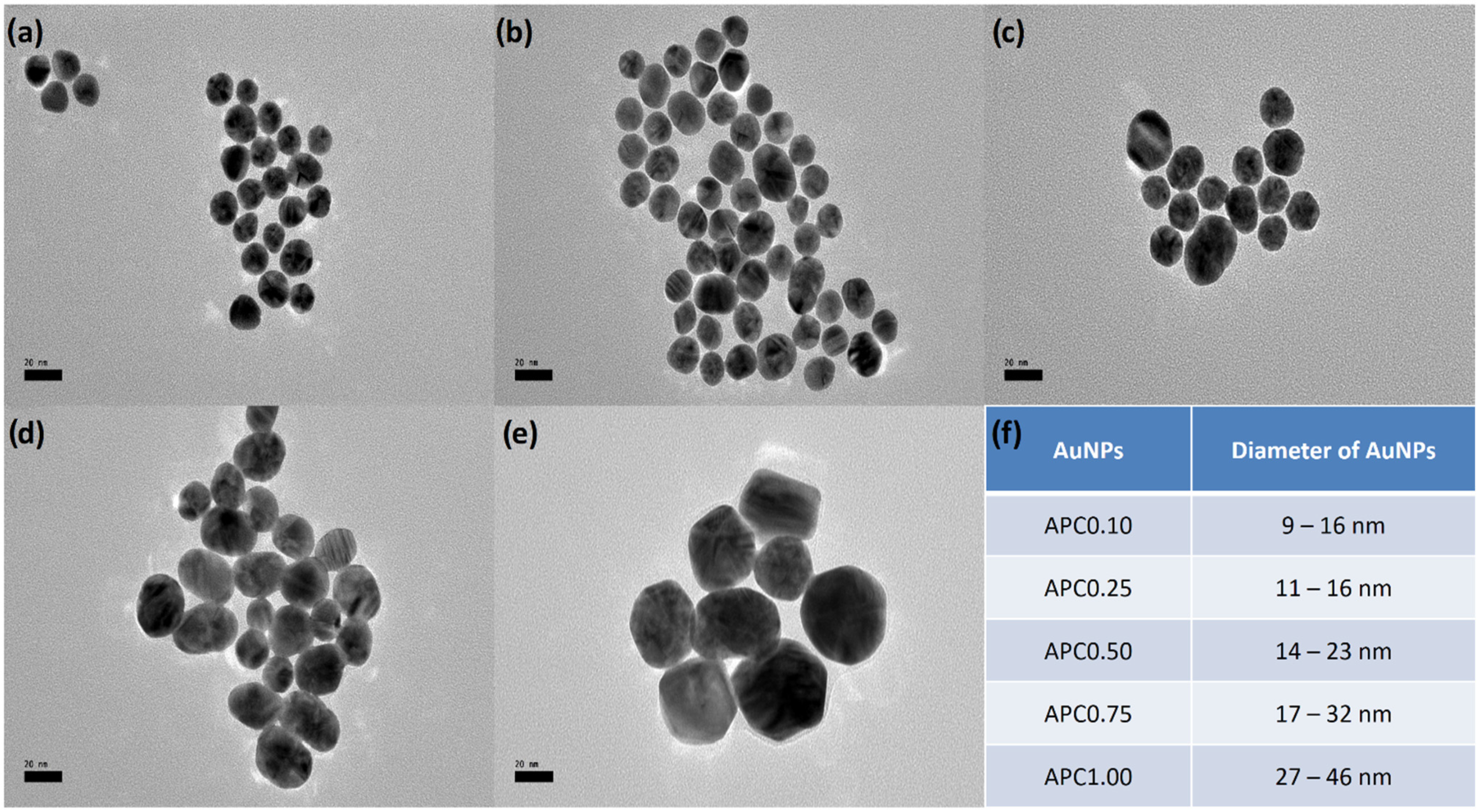
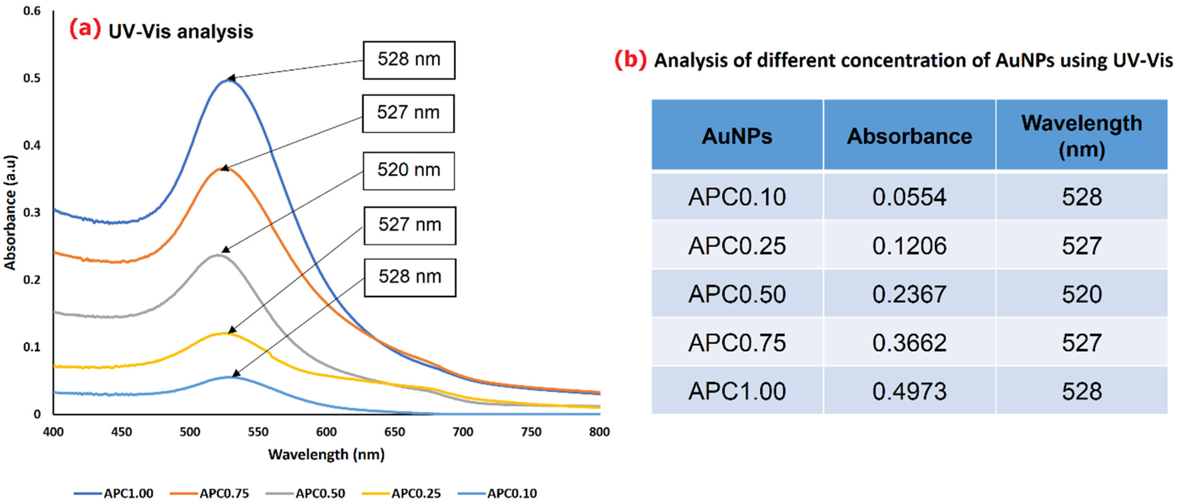
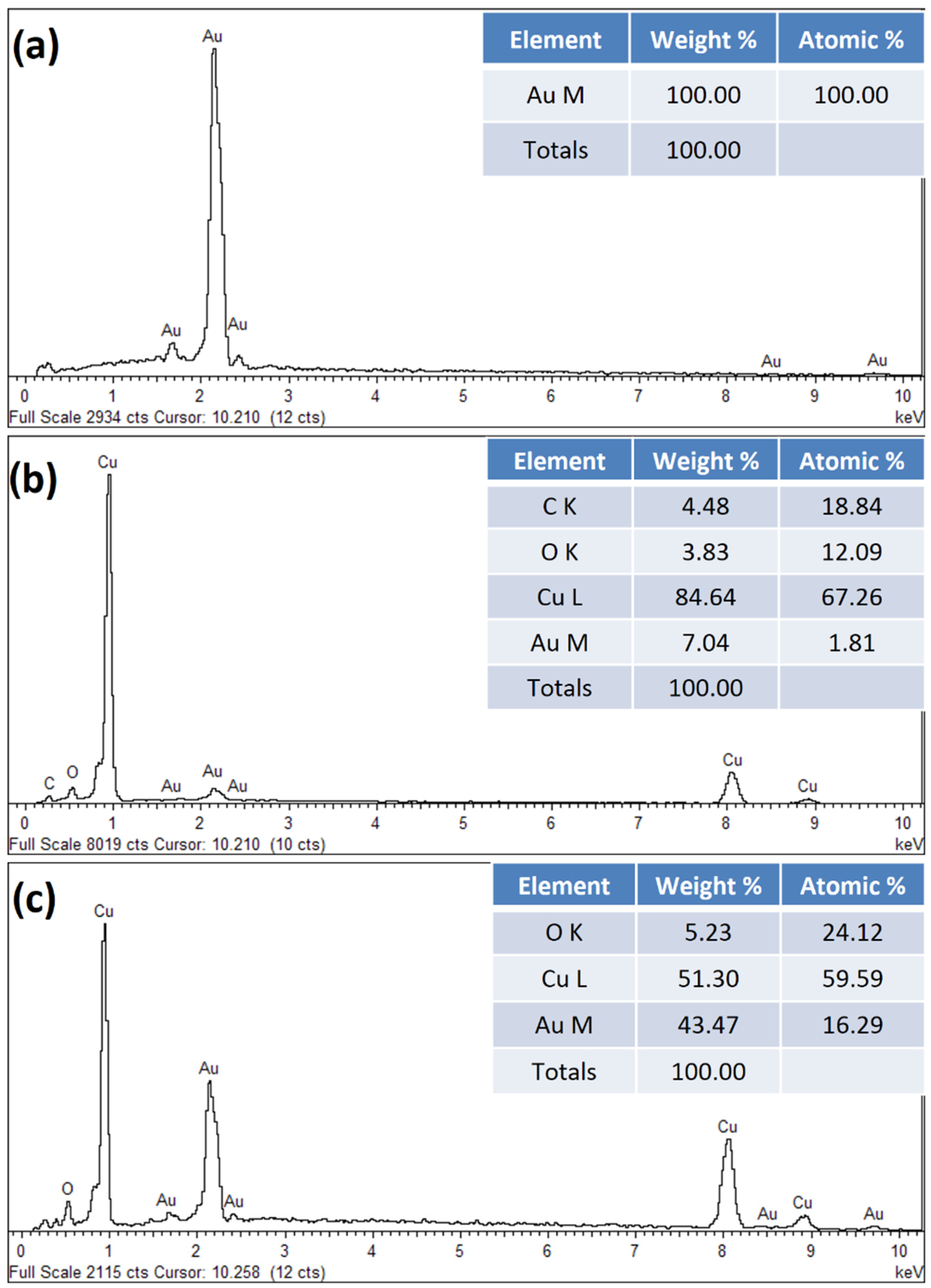
3.1. Physicochemical Characterization of Synthesized AuNPs

The AuNPs have been synthesized by using HAuCl4 solution at different concentrations. The AuNPs precursor concentration (APC) were set at 0.1, 0.25, 0.5, 0.75, and 1.0 mM, and the resulting synthesized AuNPs were labeled as APC0.10, APC0.25, APC0.50, APC0.75, and APC1.00, respectively. AuNPs formed were characterized for the morphology (HRTEM) and optical properties (UV-Vis). Figure 1 shows the HRTEM images and particle size distribution of AuNPs formed from varying concentrations of HAuCl4 solution. The sizes of AuNPs were determined by measuring the diameter of the whole particles obtained from the HRTEM images. The results indicate that the AuNPs formed are spherical in shape, uniformly distributed with specific size distribution. The particle size increased with an increase in gold salt concentration (0.1 mM to 1.0 mM), used for the formation of AuNPs. Among the particles measured for their sizes, the smallest particle diameter obtained is in the range of 9–16 nm (APC0.10), and an increase in diameter up to 46 nm was observed with the highest gold concentration (APC1.00). In addition, the observation of monodispersed state of colloidal Au particles is due to the presence of a negatively charged layer of citrate ions, making the particles repel each other and, thus, preventing AuNPs from agglomeration [34]. Similarly, the FESEM provided morphological analysis of AuNPs formed from five different concentrations in the range of 0.1 to 1.0 mM gold solution are provided in the Supplementary Materials, Figure S2.
A UV-Vis spectrophotometer is another useful tool for the characterization of AuNPs. The surface plasmon resonance (SPR) property in AuNPs plays an important role in the absorption of incident UV light, thus generating a distinct peak associated with AuNPs. AuNPs typically have a single absorption peak in the visible range between 510 and 550 nm and show a strong visible light absorption at 520 nm. This absorption range indicates the formation of AuNPs in a brilliant red color, which varies with respect to the changes in the particle size. From this study, the shift of an absorption peak towards the longer wavelength side occurs due to difference in particle size, while the broadening absorption of spectra happens because of the widened size of the distribution range. As shown in Figure 2, the colloidal gold solutions in this study only show slight differences in wavelength, as the size of the particles do not show great differences with each other with regards to changes in AuNPs concentration. The wavelengths of synthesized particles, falling in the range of 520 to 528 nm, indicate that the formed particles are purely AuNPs. Meanwhile, for the absorbance, the intensity value increases with an increase in AuNPs concentration. APC1.00 shows the highest absorbance intensity at 0.4973, while APC0.10 has the lowest absorbance intensity at 0.0554. The decrease in absorbance with the reduction in AuNPs precursor concentration is due to a reduction in the SPR property of AuNPs, which can be tuned by varying the size of NPs. As the particle size increases, the SPR-related light absorption shifts to the longer wavelength, and the red shift in the wavelength results in an increase in the absorbance value [31].

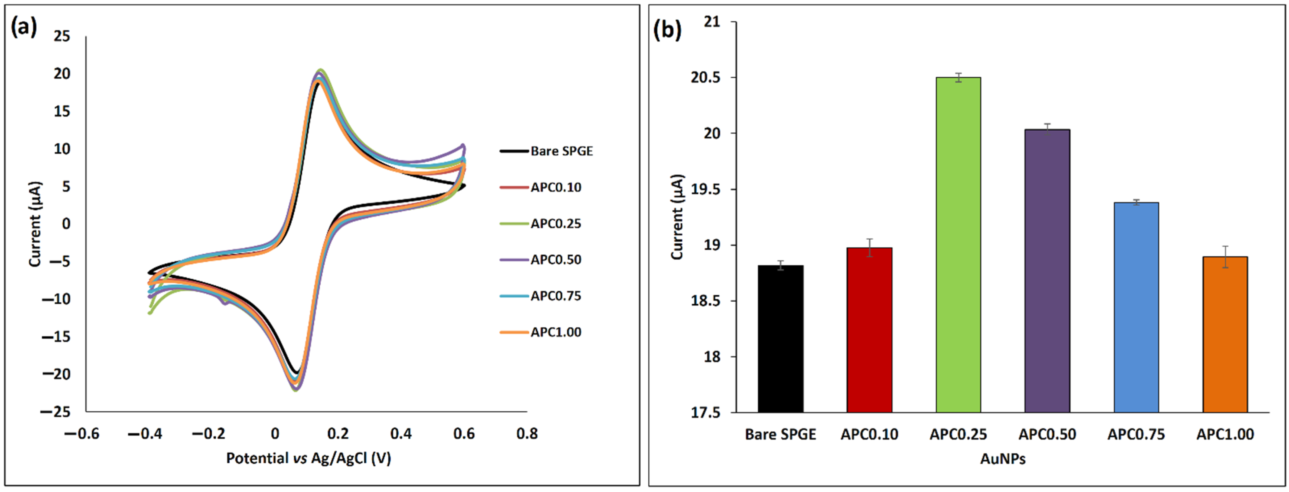
3.2. Conductivity of Bare and Crosslinked AuNPs-Modified SPGE

The AuNPs synthesized from different concentrations of gold salt were tested for electrochemical conductivity, followed by the deposition onto the SPGE, to see the effect of AuNPs concentration on the current value. In this study, the changes of electrochemical behavior for each concentration were monitored by the CV technique, which makes use of a 1.0 mM K3Fe(CN)6 solution as the redox indicator. The K3Fe(CN)6 solution has always been used as a redox solution to demonstrate the electron transfer behavior on the electrode surface during the modification process. This is due to the fact that a ferricyanide ion demonstrates a good reversibility and fast electrochemical response, as reported by the previous literature [35]. Figure 3a,b shows the CV response of the AuNPs-modified SPGE and the corresponding histogram resulting from the different AuNPs precursor concentration variation of synthesized AuNPs (respectively). From the figure, the highest peak (20.5 µA) current was obtained with APC0.25, followed by APC0.50 (20.0 µA), APC0.75 (19.4 µA), APC0.10 (18.9 µA), and APC1.00 (18.8 µA). The redox current for these five AuNPs concentrations only shows slight differences, due to the minute variations in the size of NPs. The smaller size of particles contributes to the higher value of the current. The migration of ions inside the SPGE modified with smaller AuNPs resulted in the ions reaching the whole pore volume efficiently, due to the shorter transport distance within the particle [36]. Besides, the size of AuNPs formed in the nano range of 9 to 46 nm (<100 nm) is suitable for electrode modification purposes. For further experiments, a parameter of APC0.25 was chosen, due to its better conductivity performance.
Further, DTPA was used as a crosslinker for the linking of SPGE’s surface with that of AuNPs, through self-assembled monolayer formation. Based on the previous literature, the AuNPs usually show high affinity towards the sulfur group to form a Au–S bond [37]. When DTPA is introduced to the gold surface, the S–S bond cleaves into a single thiol group and immediately binds to the gold surface through a Au–S bond. In this study, three different volumes (5, 7.5, and 10 µL) of DTPA were used to study the effect of different DTPA volume used for electrode modification. Figure 4a demonstrates the electrochemical characterization using CV, where the peak current increased linearly with the increase in DTPA volume. As observed, the 10 µL of DTPA gave the highest oxidation peak current at 20.7 µA, followed by 7.5 µL (20.2 µA) and 5 µL (13.6 µA) (Figure 4b). The increase in peak current might have happened due to the formation of a strong Au-S bond on the electrode’s surface, which may facilitate an acceleration of the electron transfer rate, thus producing high current conductivity [24]. A study by Rashid et al. in 2015 also used DTPA as the crosslinker between DTPA and SiNWs, to link AuNPs on the electrode surface via covalent bonding. This step is to discover an adequate DTPA concentration for the stable binding of AuNPs on the SiNWs electrode surface, to avoid peeling off during washing steps and, at the time, enhance the peak current signal [22].

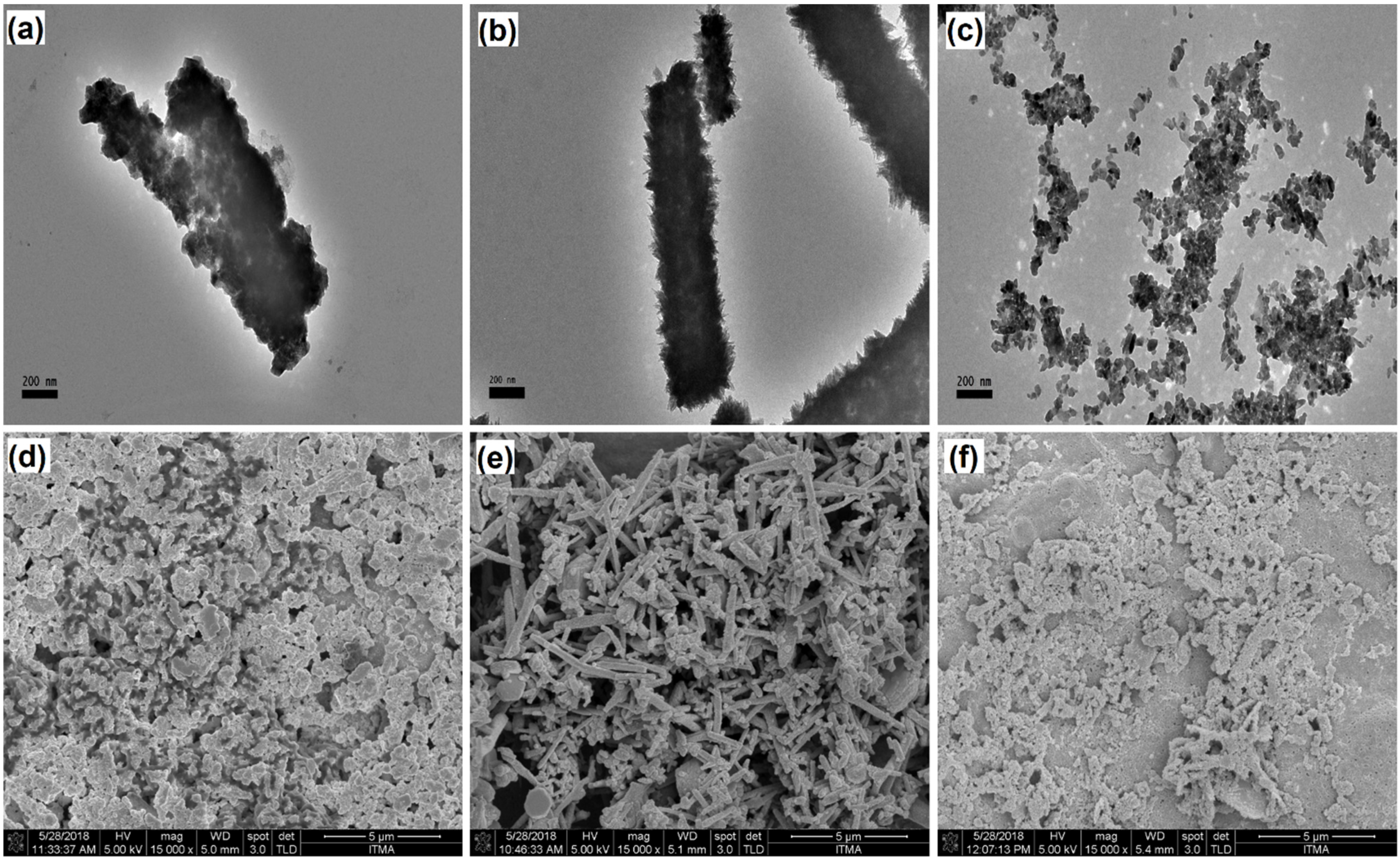
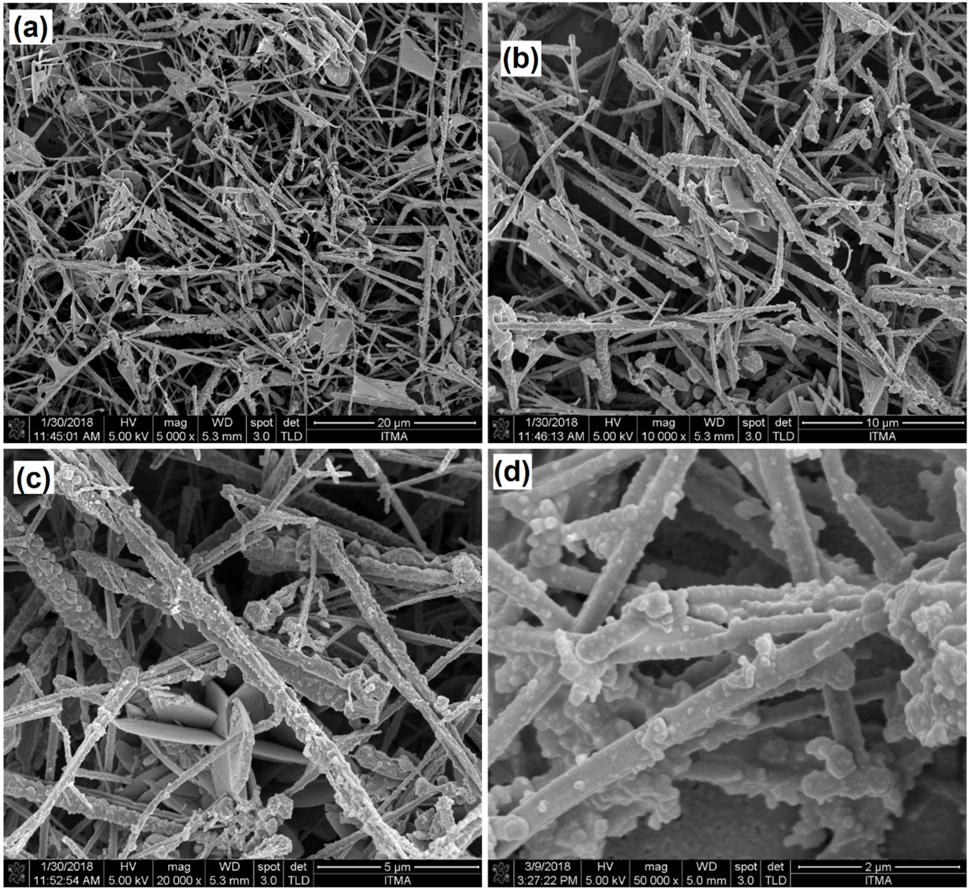
3.3. Morphological Analysis of CuNWs-Modified SPGE

The CuNWs were used for the modification of the SPGE working on an electrode’s surface, and, for that, the CuNWs were first dispersed in three different solvents. From these three different solutions, the best dispersion was selected and morphologically characterized under FESEM. The CuNWs have emerged as a promising alternative for the surface modification of electrodes due to their high conductivity, fast electron transfer, large contact area for interaction, and increased effective surface area [12]. In order to modify the electrode surface, a suitable dispersing agent must be chosen for the CuNWs deposition. Therefore, three different dispersing agents were used in this study, which include ammonium hydroxide (NH4OH), ammonium chloride (NH4Cl), and ammonium molybdate ((NH4)2MoO4). Figure 5 shows the images of CuNWs dispersion in different solvents under HRTEM Figure 5a–c with 10,000× magnification and under FESEM Figure 5d–f with 15,000× magnification. Among these three dispersing agents, NH4Cl has been shown to be the best dispersing solution. The formation of a wire shape CuNWs, when dispersed with NH4Cl, can be observed clearly under the HRTEM (Figure 5b) and FESEM (Figure 5e) analysis. Crisscrossed long and short CuNWs can also be observed, when CuNWs dispersed using the NH4OH (Figure 5a,d) and (NH4)2MoO4 (Figure 5c,f) solvents, respectively. The comparative analysis of the morphological data on CuNWs with different solvents indicates that the type of dispersion agent plays an important role while maintaining the shape and surface morphological features of CuNWs, thereby resulting in the effective finding of trace elements during the electrochemical sensor applications.
In order to produce a sensitive and stable sensor, the active surface area must be uniform to ensure the optimum reaction site provided for biomolecules immobilization. Figure 6 displays the surface morphology of CuNWs (dispersed in NH4Cl solvent) under FESEM at different magnification power. As shown in Figure 6a–c, the CuNWs were distributed randomly, forming a contact network with each wire and covering the working electrode surface of the SPGE. The average diameter of thre CuNWs used in this study is around 350 to 400 nm, and the length is up to several micrometers. In the enlarged FESEM image (Figure 6d) with 50,000× magnification, the CuNWs exhibited the porosity and roughness on its surface, thereby demonstrating the good quality of pore shape suitable for the bonding of NPs. Porosity has a significant effect on the electrode capacitance, as it provides feasible access to the ions in the electrodes to undergo redox reactions, which can further be improved by the incorporation of NPs to significantly enhance the redox current [38]. This finding was supported by a previous study by Lu et al., 2007, which found that the pores of gold nanowires on the electrode at a time will enhance the responding current, and the functionalized nanowires showed excellent electrocatalytic activity towards H2O2 [39].

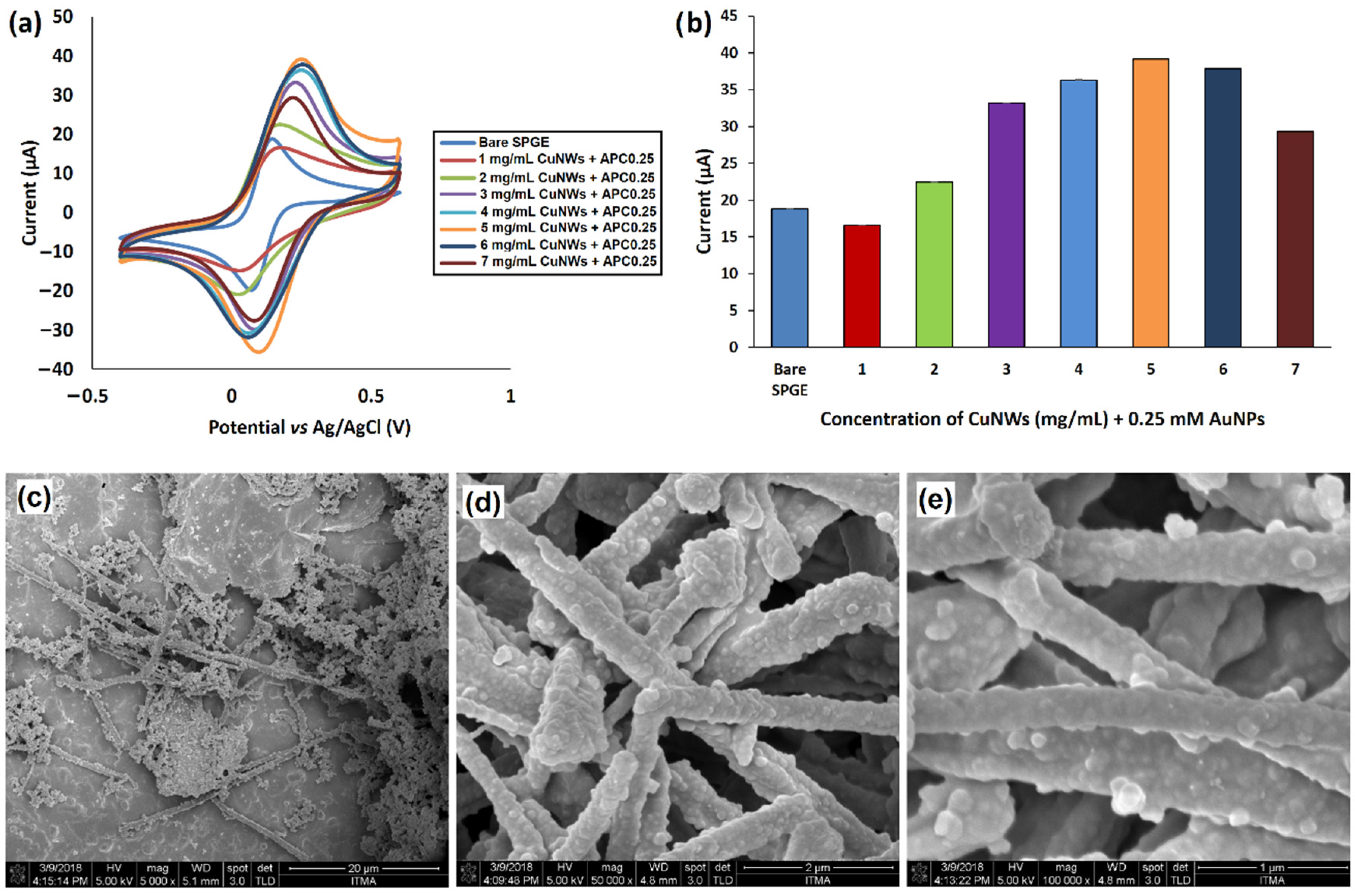
3.4. CuNWs/AuNPs Modified SPGE

Electrode surface modification is a very important step to achieve highly sensitive sensor performance. An effective and stable surface is crucial to enhance the accessibility of the target biomolecules. With the fast growth of technology, many nanomaterials have been used extensively for electrode modification [40]. As in this work, CuNWs/AuNPs composite were chosen to be implemented for the electrode modification, to investigate the effective surface modification where the maximum response can be achieved. In our case, the CuNWs/AuNPs were prepared by a crosslinking reaction using DTPA. The carboxyl group of DTPA binds to the free chloride groups of CuNWs-SPGE surface, to produce a disulfide linkage layer. Since the highest current value shown in Figure 3b is from APC0.25, this concentration value has been used for further study. Figure 7a displays the comparison of the redox peak current for different concentrations of CuNWs, with APC0.25 in the range of 0–5 mg/mL with fixed concentration of AuNPs. From the graph, the peak current was greatly enhanced at a higher concentration (5 mg/mL) of CuNWs. This is due to the higher CuNWs concentration, which provides a large surface area, thus increasing the active surface area for AuNP binding. In that way, the increase in AuNPs deposition on the surface of modified electrode resulted in a higher peak current. The increase in current response is attributed to the properties of AuNPs as a good conductor nanomaterial that can improve the electrocatalytic behavior and electroactive surface area of the electrode [41]. In general, the reaction on the modified electrode surface were greatly improved with an increase in CuNWs concentration from 1 mg/mL to 7 mg/mL. Figure 7b illustrates the oxidation peak current histogram, where the utilization of 5 mg/mL CuNWs with APC0.25 gave 39.1 µA peak current, followed by 6 mg/mL (37.8 µA), 4 mg/mL (36.3 µA), 3 mg/mL (33.2 µA), 7 mg/mL (29.3 µA), 2 mg/mL (22.4 µA), and 1 mg/mL (16.5 µA). In comparison with the bare SPGE, the CuNWs/AuNPs-modified electrode has increase the electrochemical conductivity of the sensor by 20.3 µA, suggesting that the utilization of these two nanomaterials successfully enhanced the performance of SPGE. Further, Figure 7c–e shows the FESEM images of these two nanomaterials combination with different magnification power from 5000× to 100,000×. With larger magnification (Figure 7d,e), it can be clearly seen that AuNPs occupy the pores on the CuNWs surface, which are particularly responsible for the observation of enhanced peak current values.
Further, the surface morphological feature of the CuNWs/AuNPS/SPGE electrode was characterized using FESEM analysis, where the results are provided in Figure 8. In comparison with the bare SPGE working electrode (Figure 8a,d), the presence of CuNWs/AuNPs composite on the working electrode surface has been successfully recorded under FESEM (Figure 8c,f). It can be observed that CuNWs/AuNPs are randomly distributed and compacted on the surface of SPGE. It is hard to control the orientation of CuNWs/AuNPs composite on the electrode surface when employing the drop-casting technique. The solvent evaporated leaving the CuNWs/AuNPs composite on the surface of the electrode in an irregular pattern after drying. In the previous literature, the author also reported the SEM characterization, which shows the high-density and randomly aligned nanowires [42]. With these porous and multidimensional structures of CuNWs/AuNPs composite, it can enhance the active surface area on the working electrode, thus improving the current signal produced during analysis. By making use of the FESEM morphological characterization, the average CuNWs diameter obtained was around 350–400 nm.
The CuNWs/AuNPs composite was further analysed using EDX analysis, as this has been employed to determine the elemental composition and presence of external elements at the surface of the SPGE working electrode after all the required modifications. Figure 9a shows the high weight (%) of Au (100%), as the SPGE purely contains this Au element. Similarly, from the Figure 9b, it shows the high weight (%) of Cu (84.64%), followed by Au (7.04%), C (4.48%), and the low weight (%) of O (3.83%), as a result of electrode modification with CuNWs, which contains Cu. Meanwhile for Figure 9c, the high weight (%) of Cu (51.3%), followed by Au (43.47%) and O (5.23%), was obtained, due to the addition of AuNPs on the surface of working electrode. In summary, the Cu (51.3%) and Au (43.47%) elements in Figure 9c clearly indicate that the CuNWs/AuNPs composite has been successfully deposited at the surface of the SPGE. These EDX results are consistent with the previous FESEM observation. The CuNWs/AuNPs composite modification on the working electrode has successfully enhanced the active surface area and electrochemical conductivity of the modified electrode for further biomolecule immobilization [40].

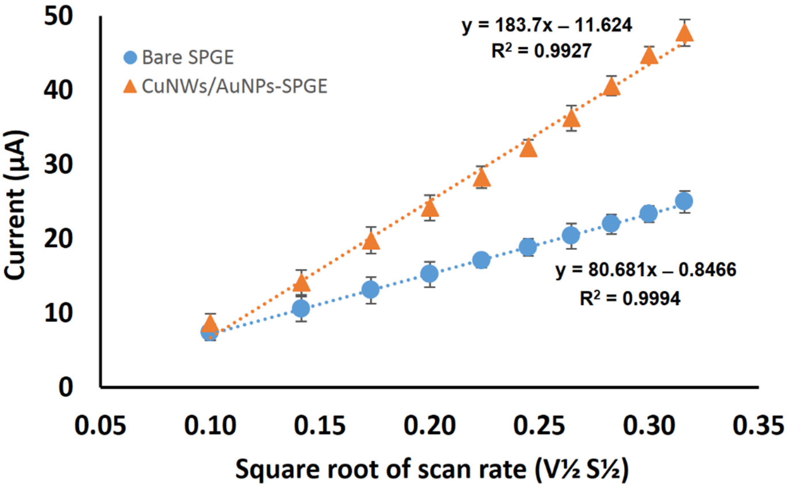
3.5. Effective Surface Area for CuNWs-AuNPs-Modified SPGE

The successful modification of the SPGE was further confirmed with the determination of the effective surface area of the modified electrode, and it can be calculated based on the Randles–Sevcik equation:
Ipa = (2.687 × 10⁵) n3/2 ν1/2 D1/2 AC
where Ipa is the oxidation peak current, n is the number of electron transfer in the redox event (n = 1), ν is the scan rate (Vs−1), D is the diffusion coefficient of [Fe(CN)6]3-/4- solution (7.6 × 10⁻⁶ cm2s−1), A is the effective surface area of the electrode (cm2), and C represents the concentration of ferricyanide solution (1.0 mM). The effective surface area of the modified and unmodified SPGE were characterized using the cyclic voltammetry technique at different scan rates (0.01 Vs–1–0.1 mVs−1), using 1.0 mM K3 [Fe(CN)6] containing 0.05 M KCl solution as the supporting electrolyte. The effective surface area of the unmodified SPGE was calculated to be 0.109 cm2, while, for the modified SPGE, it was enhanced to 0.248 cm2. The results show that the modification using CuNWs-AuNPs on the electrode surface improved the effective surface area by 2.3 times compared to the unmodified SPGE, allowing for better signal detection, while providing a high functional surface area for biomolecule immobilization. Previous studies have proven that the utilization of nanowires with nanoparticles on the surface of the electrode will increase the surface area. This fact was supported by the finding by Lu et al. in 2007, which stated gold nanowires with the combination of GO enhanced the surface 2.5 times compared to bare glassy carbon electrode (GCE) [39]. As shown in Figure 10, the redox peak current linearly increased with the square roots of the scan rates (ν1/2), indicating that the electrochemical reaction of ferricyanide is controlled by the diffusion process. The CuNWs/AuNPs-modified SPGE has shown a higher redox peak current, in the range of 0.1–0.32 Vs−1 as compared to the SPGE, which indicates accelerated redox reaction.

4. Conclusions

This work was carried out to investigate the synthesis and characterization of AuNPs, and the effect of the CuNWs-AuNPs composite modification towards SPGE conductivity enhancement. Five different concentrations of AuNPs were synthesized from the HAuCl4 precursor by the citrate reduction method. The AuNPs with the concentration of 0.25 mM shows the highest peak current using the CV analysis, and when combined with 5 mg/mL CuNWs, the redox peak current was greatly enhanced due to the large surface area of CuNWs that enhances the electron ion transfer. The effective surface area for the modified SPGE was calculated and showed a 2.3-fold increment when compared with the unmodified SPGE. The finding proved that the utilization of CuNWs/AuNPs is able to enhance the electrochemical conductivity performance of the modified SPGE and can be further used for electrochemical biosensor application.

Supplementary Materials

The following are available online at https://www.mdpi.com/article/10.3390/coatings12050622/s1, Figure S1: Color variation of AuNPs solution of 0.1 mM, 0.25 mM, 0.5 mM, 0.75 mM and 1.0 mM concentration showing increase in color intensity from pink to dark red, Figure S2: FESEM images of AuNPs formed from different concentrations, (a) 0.1 mM, (b) 0.25 mM, (c) 0.5 mM, (d) 0.75 mM, and (e) 1.0 mM of gold solution. Reference [43] is cited in the supplementary materials.

Author Contributions

Conceptualization, N.K., N.A.A.M. and N.A.Y.; investigation, N.K., N.A.A.M. and A.F.M.S.; writing—original draft preparation, N.K. and F.M.; writing—review and editing, N.A.Y., F.M., F.N.M.F. and A.A.S.; supervision, J.A., S.S., S.M. and N.A.Y. All authors have read and agreed to the published version of the manuscript.

Funding

This research was funded by Universiti Putra Malaysia, grant number GP-IPS/2017/9540500. In addition, the KSU author thanks the Researchers Supporting Project (RSP-2021/355) for the financial support.

Institutional Review Board Statement

Not applicable.

Informed Consent Statement

Not applicable.

Data Availability Statement

Data can be shared upon the request from the first/corresponding author.

Acknowledgments

The KSU author acknowledges the funding from Researchers Supporting Project (RSP-2021/355), King Saud University, Riyadh, Saudi Arabia.

Conflicts of Interest

The authors declare no conflict of interest.

References

  1. Noh, M.F.M.; Tothill, I.E. Development and characterisation of disposable gold electrodes, and their use for lead (II) analysis. Anal. Bioanal. Chem. 2006, 386, 2095–2106. [Google Scholar] [CrossRef]
  2. Sanllorente-Méndez, S.; Domínguez-Renedo, O.; Arcos-Martínez, M.J. Determination of arsenic (III) using platinum nanoparticle-modified screen-printed carbon-based electrodes. Electroanal. Int. J. Devoted Fundam. Pract. Asp. Electroanal. 2009, 21, 635–639. [Google Scholar] [CrossRef]
  3. Wang, J.; Lu, J.; Hocevar, S.B.; Ogorevc, B. Bismuth-coated screen-printed electrodes for stripping voltammetric measurements of trace lead. Electroanal. Int. J. Devoted Fundam. Pract. Asp. Electroanal. 2001, 13, 13–16. [Google Scholar] [CrossRef]
  4. Yang, N.; Uetsuka, H.; Williams, O.A.; Osawa, E.; Tokuda, N.; Nebel, C.E. Vertically aligned diamond nanowires: Fabrication, characterization, and application for DNA sensing. Phys. Status Solidi (A) 2009, 206, 2048–2056. [Google Scholar] [CrossRef]
  5. Ramulu, T.; Venu, R.; Sinha, B.; Lim, B.; Jeon, S.; Yoon, S.; Kim, C. Nanowires array modified electrode for enhanced electrochemical detection of nucleic acid. Biosens. Bioelectron. 2013, 40, 258–264. [Google Scholar] [CrossRef]
  6. Ganguly, A.; Chen, C.-P.; Lai, Y.-T.; Kuo, C.-C.; Hsu, C.-W.; Chen, K.-H.; Chen, L.-C. Functionalized GaN nanowire-based electrode for direct label-free voltammetric detection of DNA hybridization. J. Mater. Chem. 2009, 19, 928–933. [Google Scholar] [CrossRef]
  7. Bo, Y.; Yang, H.; Hu, Y.; Yao, T.; Huang, S. A novel electrochemical DNA biosensor based on graphene and polyaniline nanowires. Electrochim. Acta 2011, 56, 2676–2681. [Google Scholar] [CrossRef]
  8. Hao, Y.; Zhou, B.; Wang, F.; Li, J.; Deng, L.; Liu, Y.-N. Construction of highly ordered polyaniline nanowires and their applications in DNA sensing. Biosens. Bioelectron. 2014, 52, 422–426. [Google Scholar] [CrossRef]
  9. Ramulu, T.; Venu, R.; Sinha, B.; Yoon, S.; Kim, C. Electrodeposition of CoPtP/Au multisegment nanowires: Synthesis and DNA functionalization. Int. J. Electrochem. Sci. 2012, 7, 7762–7769. [Google Scholar]
  10. Wang, J.; Li, S.; Zhang, Y. A sensitive DNA biosensor fabricated from gold nanoparticles, carbon nanotubes, and zinc oxide nanowires on a glassy carbon electrode. Electrochim. Acta 2010, 55, 4436–4440. [Google Scholar] [CrossRef]
  11. Coffer, J. Overview of semiconducting silicon nanowires for biomedical applications. In Semiconducting Silicon Nanowires for Biomedical Applications; Elsevier: Amsterdam, The Netherlands, 2014; pp. 3–7. [Google Scholar]
  12. Zhao, S.; Han, F.; Li, J.; Meng, X.; Huang, W.; Cao, D.; Zhang, G.; Sun, R.; Wong, C.P. Advancements in copper nanowires: Synthesis, purification, assemblies, surface modification, and applications. Small 2018, 14, 1800047. [Google Scholar] [CrossRef] [PubMed]
  13. Song, D.; Li, Y.; Lu, X.; Sun, M.; Liu, H.; Yu, G.; Gao, F. Palladium-copper nanowires-based biosensor for the ultrasensitive detection of organophosphate pesticides. Anal. Chim. Acta 2017, 982, 168–175. [Google Scholar] [CrossRef] [PubMed]
  14. Huang, J.; Dong, Z.; Li, Y.; Li, J.; Wang, J.; Yang, H.; Li, S.; Guo, S.; Jin, J.; Li, R. High performance non-enzymatic glucose biosensor based on copper nanowires–carbon nanotubes hybrid for intracellular glucose study. Sens. Actuators B Chem. 2013, 182, 618–624. [Google Scholar] [CrossRef]
  15. Li, Y.; Ji, Y.; Li, Y.; Li, Y.; Ma, G.; Liu, X. Construction of Poly (methylene blue)/Copper Nanowires Modified Electrode for High-Performance Luteolin Sensing. J. Electrochem. Soc. 2020, 167, 147513. [Google Scholar] [CrossRef]
  16. Zhao, Y.; Zhang, W.; Lin, Y.; Du, D. The vital function of Fe3O4@ Au nanocomposites for hydrolase biosensor design and its application in detection of methyl parathion. Nanoscale 2013, 5, 1121–1126. [Google Scholar] [CrossRef]
  17. Huang, J.; Lin, L.; Sun, D.; Chen, H.; Yang, D.; Li, Q. Bio-inspired synthesis of metal nanomaterials and applications. Chem. Soc. Rev. 2015, 44, 6330–6374. [Google Scholar] [CrossRef] [Green Version]
  18. Rezaei, B.; Boroujeni, M.K.; Ensafi, A.A. Caffeine electrochemical sensor using imprinted film as recognition element based on polypyrrole, sol-gel, and gold nanoparticles hybrid nanocomposite modified pencil graphite electrode. Biosens. Bioelectron. 2014, 60, 77–83. [Google Scholar] [CrossRef]
  19. Maduraiveeran, G.; Sasidharan, M.; Ganesan, V. Electrochemical sensor and biosensor platforms based on advanced nanomaterials for biological and biomedical applications. Biosens. Bioelectron. 2018, 103, 113–129. [Google Scholar] [CrossRef]
  20. Ciaurriz, P.; Fernández, F.; Tellechea, E.; Moran, J.F.; Asensio, A.C. Comparison of four functionalization methods of gold nanoparticles for enhancing the enzyme-linked immunosorbent assay (ELISA). Beilstein J. Nanotechnol. 2017, 8, 244–253. [Google Scholar] [CrossRef] [Green Version]
  21. Matsumoto, M.; Kaneko, K.; Hara, M.; Matsui, M.; Morita, K.; Maruyama, T. Covalent immobilization of gold nanoparticles on a plastic substrate and subsequent immobilization of biomolecules. RSC Adv. 2021, 11, 23409–23417. [Google Scholar] [CrossRef]
  22. Rashid, J.I.A.; Yusof, N.A.; Abdullah, J.; Hashim, U.; Hajian, R. A novel disposable biosensor based on SiNWs/AuNPs modified-screen printed electrode for dengue virus DNA oligomer detection. IEEE Sens. J. 2015, 15, 4420–4427. [Google Scholar] [CrossRef] [Green Version]
  23. Lian, W.; Liu, S.; Yu, J.; Xing, X.; Li, J.; Cui, M.; Huang, J. Electrochemical sensor based on gold nanoparticles fabricated molecularly imprinted polymer film at chitosan–platinum nanoparticles/graphene–gold nanoparticles double nanocomposites modified electrode for detection of erythromycin. Biosens. Bioelectron. 2012, 38, 163–169. [Google Scholar] [CrossRef] [PubMed]
  24. Rashid, J.I.A.; Yusof, N.A.; Abdullah, J.; Hashim, U.; Hajian, R. The utilization of SiNWs/AuNPs-modified indium tin oxide (ITO) in fabrication of electrochemical DNA sensor. Mater. Sci. Eng. C 2014, 45, 270–276. [Google Scholar] [CrossRef] [PubMed]
  25. Shoaie, N.; Forouzandeh, M.; Omidfar, K. Highly sensitive electrochemical biosensor based on polyaniline and gold nanoparticles for DNA detection. IEEE Sens. J. 2017, 18, 1835–1843. [Google Scholar] [CrossRef]
  26. Fani, M.; Rezayi, M.; Meshkat, Z.; Rezaee, S.A.; Makvandi, M.; Angali, K.A. A novel electrochemical DNA biosensor based on a gold nanoparticles-reduced graphene oxide-polypyrrole nanocomposite to detect human T-lymphotropic virus-1. IEEE Sens. J. 2020, 20, 10625–10632. [Google Scholar] [CrossRef]
  27. Taei, M.; Salavati, H.; Banitaba, S.H.; Shahidi, L. A novel hydrazine electrochemical sensor based on gold nanoparticles decorated redox-active 2-amino-4H-chromene-3-carbonitrile. IEEE Sens. J. 2017, 17, 7325–7331. [Google Scholar] [CrossRef]
  28. Nazarpour, S.; Hajian, R.; Sabzvari, M.H. A novel nanocomposite electrochemical sensor based on green synthesis of reduced graphene oxide/gold nanoparticles modified screen printed electrode for determination of tryptophan using response surface methodology approach. Microchem. J. 2020, 154, 1–7. [Google Scholar] [CrossRef]
  29. Ji, D.; Xu, N.; Liu, Z.; Shi, Z.; Low, S.S.; Liu, J.; Cheng, C.; Zhu, J.; Zhang, T.; Xu, H. Smartphone-based differential pulse amperometry system for real-time monitoring of levodopa with carbon nanotubes and gold nanoparticles modified screen-printing electrodes. Biosens. Bioelectron. 2019, 129, 216–223. [Google Scholar] [CrossRef]
  30. Huang, C.-J.; Chiu, P.-H.; Wang, Y.-H.; Chen, K.-L.; Linn, J.-J.; Yang, C.-F. Electrochemically controlling the size of gold nanoparticles. J. Electrochem. Soc. 2006, 153, D193–D198. [Google Scholar] [CrossRef]
  31. Nur, H.; Nasir, S.M. Gold nanoparticles embedded on the surface of polyvinyl alcohol layer. Malays. J. Fundam. Appl. Sci. 2008, 4, 245–252. [Google Scholar] [CrossRef] [Green Version]
  32. Shih, S.-M.; Su, W.-F.; Lin, Y.-J.; Wu, C.-S.; Chen, C.-D. Two-dimensional arrays of self-assembled gold and sulfur-containing fullerene nanoparticles. Langmuir 2002, 18, 3332–3335. [Google Scholar] [CrossRef]
  33. Łuczak, T. Gold electrodes modified with self-assembled layers made of sulphur compounds and gold nanoparticles used for selective electrocatalytic oxidation of catecholamine in the presence of interfering ascorbic and uric acids. Int. J. Electrochem. 2011, 2011, 1–10. [Google Scholar] [CrossRef] [Green Version]
  34. Verma, H.N.; Singh, P.; Chavan, R. Gold nanoparticle: Synthesis and characterization. Vet. World 2014, 7, 72–77. [Google Scholar] [CrossRef] [Green Version]
  35. Han, X.; Jiang, T.; Chen, X.; Jiang, D.; Xie, K.; Jiang, Y.; Wang, Y. Electrolyte additive induced fast-charge/slow-discharge process: Potassium ferricyanide and potassium persulfate for CoO-based supercapacitors. J. Colloid Interface Sci. 2020, 576, 505–513. [Google Scholar] [CrossRef]
  36. Portet, C.; Yushin, G.; Gogotsi, Y. Effect of carbon particle size on electrochemical performance of EDLC. J. Electrochem. Soc. 2008, 155, A531–A536. [Google Scholar] [CrossRef]
  37. Łuczak, T.; Bełtowska-Brzezinska, M. Gold electrodes modified with gold nanoparticles and thio compounds for electrochemical sensing of dopamine alone and in presence of potential interferents. A comparative study. Microchim. Acta 2011, 174, 19–30. [Google Scholar] [CrossRef]
  38. Paulose, R.; Mohan, R.; Parihar, V. Nanostructured nickel oxide and its electrochemical behaviour—A brief review. Nano-Struct. Nano-Objects 2017, 11, 102–111. [Google Scholar] [CrossRef]
  39. Lu, Y.; Yang, M.; Qu, F.; Shen, G.; Yu, R. Enzyme-functionalized gold nanowires for the fabrication of biosensors. Bioelectrochemistry 2007, 71, 211–216. [Google Scholar] [CrossRef]
  40. Rashid, J.I.A.; Yusof, N.A.; Abdullah, J.; Hashim, U.; Hajian, R. Surface modifications to boost sensitivities of electrochemical biosensors using gold nanoparticles/silicon nanowires and response surface methodology approach. J. Mater. Sci. 2016, 51, 1083–1097. [Google Scholar] [CrossRef]
  41. Saberi, R.S.; Shahrokhian, S.; Marrazza, G. Amplified electrochemical DNA sensor based on polyaniline film and gold nanoparticles. Electroanalysis 2013, 25, 1373–1380. [Google Scholar] [CrossRef]
  42. Qu, F.; Yang, M.; Shen, G.; Yu, R. Electrochemical biosensing utilizing synergic action of carbon nanotubes and platinum nanowires prepared by template synthesis. Biosens. Bioelectron. 2007, 22, 1749–1755. [Google Scholar] [CrossRef]
  43. Zhang, R.; Hummelgård, M.; Olin, H. Simple and efficient gold nanoparticles deposition on carbon nanotubes with controllable particle sizes. Mater. Sci. Eng. B 2009, 158, 48–52. [Google Scholar] [CrossRef]
Figure 1. HRTEM images of AuNPs formed from different concentrations of gold salt, (a) APC0.10, (b) APC0.25, (c) APC0.50, (d) APC0.75, and (e) APC1.00 (20 nm images scale); (f) the diameter of AuNPs in nm for all five different AuNPs.
Coatings 12 00622 g001
Figure 2. UV-Vis analysis of AuNPs formed from different concentration (a) and its absorbance data (b).
Coatings 12 00622 g002
Figure 3. Effect of different concentrations of AuNPs on the modified-SPGE electrochemical conductivity (n = 3). The graphs show (a) CV response and (b) histogram of oxidation current value for different concentrations of AuNPs-modified SPGE.
Coatings 12 00622 g003
Figure 4. Effect of different volumes of DTPA on the modification of SPGE (n = 3). The graphs show (a) CV response and (b) histogram of oxidation current values for different volumes of DTPA.
Coatings 12 00622 g004
Figure 5. HRTEM images of CuNWs in different solvents (a) NH4OH, (b) NH4Cl, and (c) (NH4)2MoO4 (10,000× magnification, 200 nm images scale). In addition, (df) correspond to the FESEM images of CuNWs in respective solvents (15,000× magnification).
Coatings 12 00622 g005
Figure 6. FESEM analysis of CuNWs-modified SPGE, (a) 5000×, (b) 10,000×, (c) 20,000×, and (d) 50,000× magnification.
Coatings 12 00622 g006
Figure 7. Effect of different concentration of CuNWs (1–7 mg/mL) with APC0.25 (n = 3), (a) CV studies and (b) oxidation peak current histogram for different concentrations of CuNWs. FESEM images of 5 mg/mL CuNWs with APC0.25 at (c) 5000×, (d) 50,000×, and (e) 100,000× magnification.
Coatings 12 00622 g007
Figure 8. FESEM analysis of (a) bare SPGE, (b) CuNWs/SPGE, (c) CuNWs/AuNPs/SPGE at 5000× magnification, (d) bare SPGE, (e) CuNWs/SPGE, and (f) CuNWs/AuNPs/SPGE at 50,000× magnification.
Coatings 12 00622 g008
Figure 9. EDX analysis of (a) bare SPGE, (b) CuNWs/SPGE, and (c) CuNWs/AuNPs/SPGE. Inset: elemental analysis of modified electrode.
Coatings 12 00622 g009
Figure 10. The relationship between the peak potential and square root of scan rates for the bare and CuNWs-AuNPs-modified SPGE.
Coatings 12 00622 g010
Table 1. The utilization of nanocomposite on the fabrication of electrodes.
NanomaterialsDetectionReferences
Silicon nanowires/AuNPsDengue virus[24]
Polyaniline/AuNPsDNA molecules[25]
Reduced grapheme oxide/polypyrrole/AuNPsHuman T-Lymphotropic Virus-1 (HTLV-1)[26]
2-Amino-4H-chromene-3-carbonitrile/AuNPsHydrazine[27]
Reduced grapheme oxide/AuNPsL-tryptophan[28]
Single wall carbon nanotubes/chitosan/AuNPsLevodopa[29]
Publisher’s Note: MDPI stays neutral with regard to jurisdictional claims in published maps and institutional affiliations.

