Abstract
In this study, a durable superhydrophobic antibacterial coating was developed by a facile spraying method. A mixture of bisphenol A diglycidyl ether (BADGE) and hydrophobic SiO2 nanoparticles was sprayed on carbon steel to provide a superhydrophobic substrate. D-cysteine (D-cys) functionalized SiO2/dopamine/silver nanoparticles with multilayer core-shell structure were then sprayed on superhydrophobic substrate to enhance antibacterial performance. The results of morphology observation and X-ray photoelectron spectroscopy (XPS) indicated the successful preparation of antibacterial nanoparticles and presented the hierarchical micro/nanostructures of coating surface. The as-prepared coating exhibited superhydrophobicity, with the water contact angle of ~153°. The coating was endowed with good mechanical durability, which maintained the water contact angle of ~150° after 180 cycles in tape-peel tests. The results of electrochemical impedance spectroscopy showed satisfactory corrosion resistance of this coating during 3 days of immersion in 3.5% NaCl solution. Furthermore, the coating showed excellent antibacterial performances against Escherichia coli and Pseudomonas aeruginosa, which benefited from the synergistic actions of low wettability of superhydrophobic surface, bactericidal behavior of Ag nanoparticles, and biofilm inhibition effect of D-cys.
1. Introduction
Bacterial adhesion and contamination can lead to the failure of medical equipment and even threaten human life. For example, bacteria adhere to clinical medical devices, such as intubation and endoscope in vivo, resulting in interventional iatrogenic infection. To solve this problem, a variety of antibacterial materials have been proposed and used. Antibiotics are gradually being abandoned because they pollute the environment and increase bacterial resistance. As an alternative, metal nanoparticles have been used in the development of antibacterial materials, including silver (Ag), copper, and zinc [1,2,3]. Ag nanoparticles have widely attracted attention due to their excellent bactericidal effects against some Gram-positive and Gram-negative bacteria, such as Escherichia coli, Staphylococcus aureus, and Pseudomonas aeruginosa [4,5,6,7]. Although the bactericidal mechanism of Ag nanoparticles is not fully elucidated, it is widely believed that Ag nanoparticles can destroy the structure of cell membranes, which change the permeability of cell membranes and lead to cell death [8,9,10]. However, the rapid release of Ag nanoparticles often limits their practical application and cannot achieve the purpose of long-term antibacterial activity. Because of this, other antibacterial methods have been used in combination with Ag nanoparticles.
Inspired by natural lotus leaf, superhydrophobic surfaces show excellent water repellency with low wettability. Superhydrophobic surfaces are surfaces with a water contact angle of more than 150° and a water sliding angle of less than 10°, which are always constructed by combining appropriate micro/nano hierarchical structure with low surface energy materials [11,12,13,14]. Rough surface microstructure can be prepared by various methods, such as anodic oxidation, template, hydrothermal treatment, and sol-gel methods [15,16,17,18,19]. Due to the low surface adhesion property, superhydrophobic surfaces have been used for the application of self-cleaning, antifogging, drag reduction, anticorrosion, oil–water separation, and anti-icing fields [20,21,22,23,24,25]. Recently, superhydrophobic surfaces have attracted increasing attention as a promising strategy to resist the adhesion of bacteria for antibacterial application. Pan et al. used picosecond laser texturing method to fabricate a superhydrophobic surface on AISI 420 martensitic stainless steel [26]. It reduced the adhesion of 99% E. coli and 93% S. aureus, even under oscillation condition. Xun et al. prepared a superhydrophobic surface on the AZ31B magnesium alloy by a facile two-step in situ immersion method and subsequent modification of stearic acid, which also exhibited an antiadhesion property against S. aureus and E. coli [27]. The low microbial adhesion of the superhydrophobic surfaces mainly results from the physical barrier effect of the air film trapped in the rough microstructure. The superhydrophobic surface reduces bacterial adhesion rather than killing bacteria, which effectively avoids the problem of increasing bacterial resistance. However, the fragile microstructure of superhydrophobic surfaces limits their industrial-scale applications [28]. It is necessary to improve the durability of superhydrophobic surfaces for long-term antibacterial application.
D-amino acids are widely found in microorganisms, plants, and animals and are enantiomers of L-amino acids. D-amino acids are the main components of microbial cell wall peptidoglycans, playing an important role in maintaining cell stability and metabolism [29]. Exogenous D-amino acids have been reported to be capable of inhibiting the formation of biofilm [30,31]. Because bacterial cells do not reject exogenous D-amino acids, they can replace endogenous D-amino acids in the peptidoglycan, which causes the change in cell-wall-building proteins and leads to the destruction of the anchoring and crosslinking of intercellular fibers. Several D-amino acids have been reported to be effective in inhibiting biofilms, including D-tyrosine, D-methionine, D-leucine, and D-tryptophan [29]. Leiman et al. tested the biofilm-inhibitory behavior of soil bacterium Bacillus subtilis by D-tyrosine, D-leucine, and D-tryptophan, and found that they prevented biofilm formation at concentrations of 6 μM, 8.5 mM, and 5 mM, respectively [32]. Zilm et al. indicated that D-amino acids could inhibit the biofilm formation and even disrupt the established biofilms produced by Enterococcus faecalis [33]. Cancino-Diaz et al. evaluated the effect of D-amino acids on the biofilm formation of S. epidermidis isolated from healthy skin, conjunctiva, and ocular infection, and found that D-methionine exhibited the best biofilm inhibition property [34]. Moreover, a combination of D-amino acids and other antibacterial agents can greatly enhance the antibacterial effect and reduce the minimal inhibitory concentration of other antibacterial agents [35]. D-amino acids show great potential in the preparation of antibacterial coatings. To date, D-amino acids have been used mainly in their free form and have rarely been used in the development of antibacterial coatings. Moreover, the combination of superhydrophobic surface, Ag nanoparticles, and D-amino acids has not been reported.
This work fabricated a superhydrophobic antibacterial coating combining rough SiO2 nanoparticles/polymer substrate and D-cysteine (D-cys) functionalized SiO2/dopamine/silver nanoparticles through a facile spraying method. The mixture of hydrophobic SiO2 nanoparticles and epoxy polymer not only provided the microstructure required for superhydrophobic surface, but also endowed the coating with good mechanical durability. Antibacterial nanoparticles were further sprayed onto the substrate surface to reinforce antibacterial activity. The structure and composition of antibacterial nanoparticles were characterized with the change in preparation steps to prove the successful assembly. The coating morphology was observed to confirm the successful loading of antibacterial nanoparticles. The hydrophobicity of the coating was evaluated through water contact angle measurements, and the durability of the coating was characterized by tape-peel tests. The antibacterial performances of the coating against Escherichia coli and Pseudomonas aeruginosa were studied, and the antibacterial mechanism was proposed.
2. Experimental Section
2.1. Materials
Tetraethyl silicate, dopamine hydrochloride, and tris (hydroxymethyl) aminomethane were purchased from Aladdin Industrial Corporation (Shanghai, China). Silver nitrate (AgNO3), ethyl ethanol, and acetone were obtained from Sinopharm (Beijing, China). D-cys, bisphenol A diglycidyl ether (BADGE), and Jeffamine D230 hardener were purchased from Sigma-Aldrich (Shanghai, China). The concentrated ammonia was supplied by Beijing Chemical Works (Beijing, China). The silane coupling agent (KH550)-modified SiO2 nanoparticles with a diameter of 200 nm were purchased from China Metallurgical New Material Technology Co., Ltd. (Beijing, China). All chemicals were used as received and without further purification.
2.2. Preparation of Superhydrophobic Antibacterial Coating
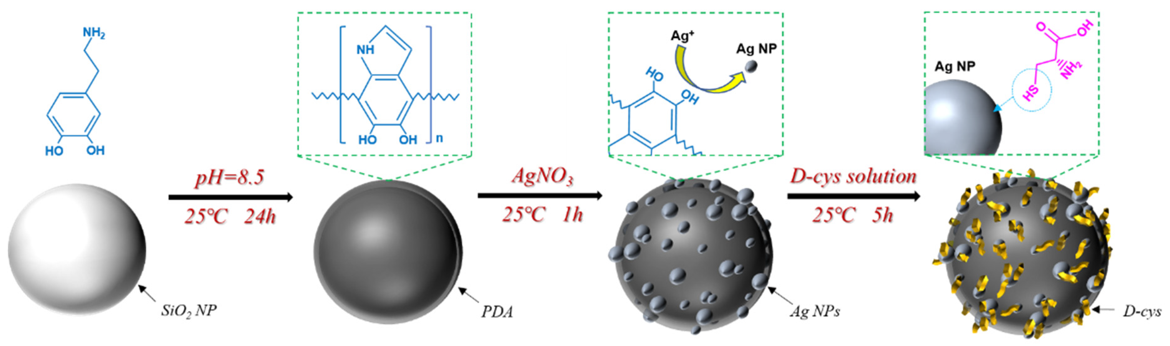
The Stöber method was used to prepare SiO2 nanoparticles with a particle size of approximately 600 nm [36]. Dry SiO2 nanoparticles were added to 400 mL Tris-HCl buffer solution (10 mM, pH = 8.5) containing dopamine hydrochloride (2.0 mg/mL), with the pH value of 8.5, and the solution was stirred slowly at a speed of 100 rpm/min for 24 h. Afterwards, the polydopamine (PDA)-coated nanoparticles were immersed in 400 mL AgNO3 solution (5.0 mg/mL) and stirred slowly at 100 rpm/min for 1 h. During the AgNO3 treatment, the conical flask containing the AgNO3 solution and nanoparticles was completely wrapped in tin foil to protect it from light. The Ag-deposited nanoparticles were then added to 400 mL D-cys solution (10.0 mmol/L) and stirred at 100 rpm/min for 5 h. After washing with deionized water, the preparation of antibacterial nanoparticles was completed. The above steps were all performed at room temperature. The preparation process is shown in Figure 1.
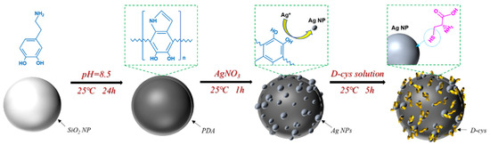
Figure 1.
Schematic illustration of the preparation procedure of antibacterial nanoparticles.
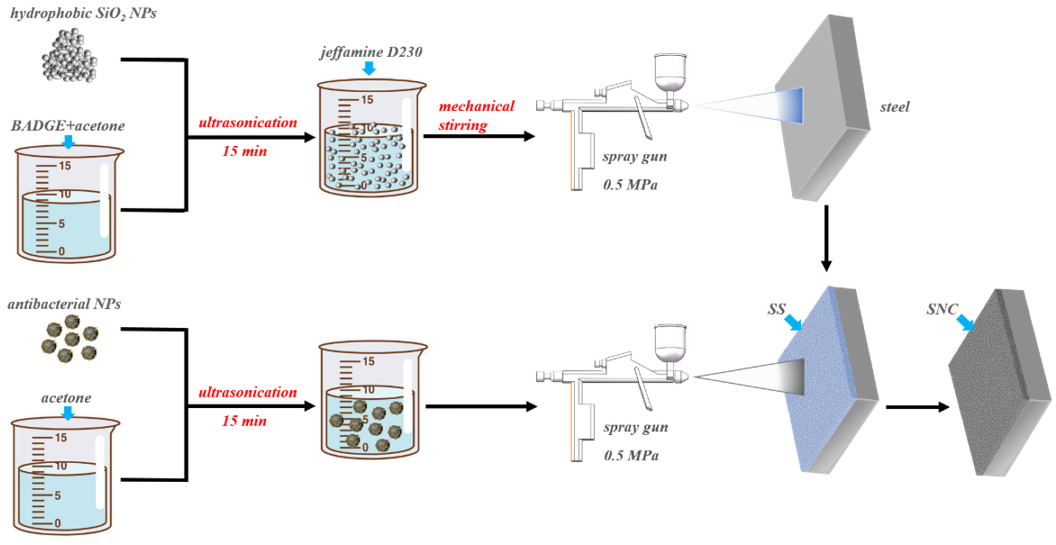
Q235 carbon steels with a dimension of 10 mm × 10 mm × 3 mm were polished by abrasive papers until reaching 800 grit, followed by washing with ethyl alcohol under ultrasonic treatment and drying in air. A total of 1.2 g of hydrophobic KH550-modified SiO2 nanoparticles and 3.0 g of BAGDE were added to 30 mL of acetone. After 20 min of sonication at temperature, 1.0 g of Jeffamine D230 was added under mechanical stirring. The mixture solution was sprayed on the carbon steel surface using a spray gun with an air pressure of 0.5 MPa. After 5 times of spraying, the rough polymer-based superhydrophobic substrate was established. Subsequently, 1.0 g of antibacterial nanoparticles was added to 20 mL of acetone and dispersed by ultrasonic treatment for 15 min at temperature, followed by spraying on the superhydrophobic substrate. After curing at 45 °C for 24 h, the superhydrophobic nanocomposite coating was obtained. The whole preparation process is shown in Figure 2. The superhydrophobic substrate and final superhydrophobic nanocomposite coating are hereinafter referred to as SS and SNC.

Figure 2.
Schematic illustration of the preparation procedure of superhydrophobic nanocomposite coating.
2.3. Surface Characterization
The surface morphologies of synthesized nanoparticles and prepared coatings were observed by field emission scanning electron microscopy (FE-SEM, JSM-7610FPlus, JEOL, Tokyo, Japan). The structure of synthesized nanoparticles was characterized by transmission electron microscope (TEM, F200X G2, Talos, City of Industry, CA, USA). The 3D profile and roughness of prepared coating were characterized by confocal laser scanning microscopy (CLSM, KEYENCE VK-X, Tokyo, Japan). The chemical composition of the synthesized nanoparticles was analyzed by X-ray photoelectron spectroscopy (XPS, Thermo ESCALAB 250Xi, USA). The surface wettability of prepared coatings was measured by a goniometer (Data physics OCA20, Germany). The sessile drop method with a 5 μL water droplet was used to measure the water contact angle of sample surface at room temperature. The sliding angle was measured by tilting the sample surface until a 10 μL water droplet slid off.
2.4. Mechanical Durability Tests
The mechanical durability of the superhydrophobic nanocomposite coating was evaluated by the tape-peel tests. The 3M tape was pressed on the surface of the prepared coating at a pressure of 30 kPa to ensure a good contact between the coating surface and the 3M tape. A 300 g weight was placed on the 1 cm × 1 cm sample surface covered with tape to control the pressure. Then, the tape was peeled off from the coating surface, and the water contact angle and sliding angle of the coating surface were measured. The above process was a cycle. The cycle was repeated 180 times, and the variation of the water contact angle and sliding angle was recorded.
2.5. Electrochemical Tests
The corrosion resistance of the SS and the final SNC samples was evaluated by electrochemical impedance spectroscopy (EIS) using an electrochemical station (PARSTAT 2273, AMETEK, Berwyn, PA, USA). A three-electrode system was adopted, in which the steels with and without coatings acted as the working electrode, the platinum sheet acted as the counter electrode, and the saturated calomel electrode acted as the reference electrode. After 1 day and 3 days of immersion in 3.5% NaCl solution, EIS measurements were performed at the open circuit potential (OCP) in the frequency range of 105 Hz to 10−2 Hz.
2.6. Antibacterial Activity Tests
The E. coli (ATCC 8739) and P. aeruginosa (MCCC 1A00099) strains were used for antibacterial activity tests. The E. coli strain was purchased from the American Type Culture Collection (ATCC), and the P. aeruginosa strain was purchased from the Marine Culture Collection of China (MCCC). Before the antibacterial activity tests, the E. coli strain was incubated in lysogeny broth (LB) medium and the P. aeruginosa strain was incubated in 2216E culture medium at 37 °C overnight. During the inoculation process, the bacterial cells were first diluted with phosphate-buffered saline (PBS) solution. The diluted solution of E. coli was inoculated into new LB liquid culture medium, and the diluted solution of P. aeruginosa was inoculated into new 2216E liquid culture medium. The initiation concentration of these bacteria cells was adjusted to 1.0 × 10⁶ cells·mL−1. Then, the SS and the final SNC samples were, respectively, immersed in different inoculated culture media and incubated at 37 °C for 1 day and 3 days.
After 1 day and 3 days of incubation, the samples were taken out from the culture medium and washed with PBS to remove planktonic bacterial cells. Then, the samples were immersed in the glutaraldehyde solution for 8 h to fix the adherent bacterial cells. Subsequently, the fixed bacterial cells were continuously dehydrated with 50%, 60%, 70%, 80%, 90%, and 100% ethanol (v/v) for 10 min at each concentration. The morphologies of the adherent bacterial cells were observed by FE-SEM (JSM-F100, JEOL, Tokyo, Japan).
The live/dead biofilm-staining tests were adopted to further observe the adhesion of the bacterial cells on different sample surfaces. After 1 day and 3 days of exposure in the culture medium, the samples with adherent bacterial cells were removed from the culture medium and stained with SYTO-9 and propidium iodide (PI) dyes in PBS for 20 min in the dark. The live/dead conditions of the adherent bacterial cells were observed by a confocal laser scanning microscope (CLSM, TCS-SP5, Leica, Bensheim, Germany). In the CLSM image, live cells and dead cells were stained with green fluorescence and red fluorescence, respectively. The coverage area of adherent bacterial cells was calculated using the Image J software (Version 1.49, 2015, National Institutes of Health, Bethesda, MD, USA). For the antibacterial activity tests method, refer to previous reports [35,37].
2.7. The Release of Ag Ions and D-cys
The SS and SNC samples were immersed in 2 mL of sterile LB culture medium at 37 °C for 1 day and 3 days. During the immersion process, the concentration of released Ag ions from the sample surfaces in the medium was monitored by an inductively coupled plasma mass spectrometry (ICP-MS, ICAP-MS-Qc, Thermo Scientific, Waltham, MA, USA). At the same time, the concentration of released D-cys from the sample surfaces was also measured by a micro cysteine content assay kit (Solarbio, Beijing, China).
3. Results and Discussion
3.1. Characterization of Antibacterial Nanoparticles
A mussel-inspired strategy was employed to fabricate multilayer antibacterial nanoparticles, as shown in Figure 1. Dopamine can spontaneously polymerize on any organic or inorganic material surfaces [38]. The formed polydopamine membrane provides the pretreatment layer and reaction site for the subsequent deposition of Ag nanoparticles. The SiO2 nanoparticles with a diameter of approximately 600 nm showed a smooth surface (Figure 3a,b). The thin and rough polydopamine layer on SiO2 nanoparticles can be distinguished by SEM and TEM, as shown in Figure 3c,d. Catechols in the polydopamine structure are capable of reducing Ag+ in AgNO3 solution to form Ag nanoparticles [39]. After immersion in AgNO3 solution, obvious tiny particles with a diameter of approximately 50 nm were homogeneously distributed on the surface of polydopamine membrane (Figure 3e,f). The size of Ag particles is related to the immersion time, and the decrease in the size of Ag particles is helpful to improve the bactericidal ability [40]. Hence, nanoscale Ag particles are often used to build antibacterial surface materials [41,42]. Subsequently, D-cys molecules were grafted to the outermost layer of antibacterial nanoparticles relying on the strong silver-thiol groups (Figure 3g,h) [35]. The whole preparation procedure is facile and easy to operate, which includes a continuous immersion process and a series of spontaneous reactions at room temperature. Through the above treatments, the surface of SiO2 nanoparticles possesses the ability of sterilization and biofilm formation inhibition.
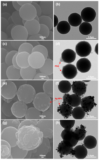
Figure 3.
SEM and TEM morphologies of (a,b) SiO2 nanoparticles, (c,d) PDA-coated nanoparticles, (e,f) Ag-deposited nanoparticles, and (g,h) D-cys-grafted nanoparticles.
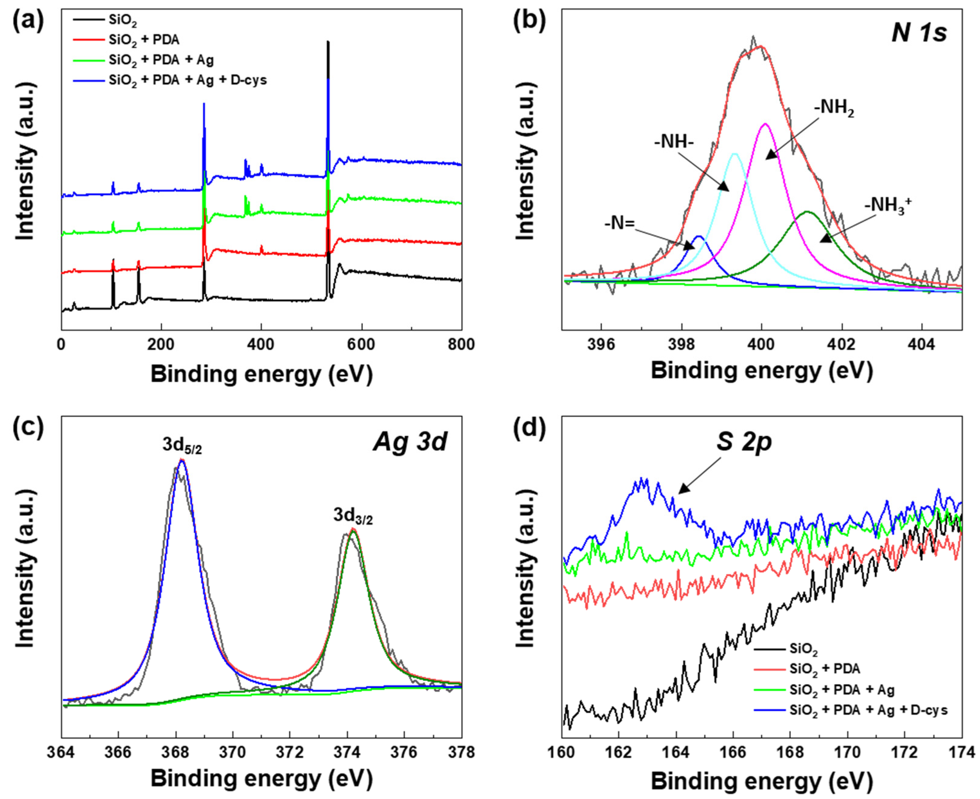
To further determine the successful fabrication of antibacterial nanoparticles, the chemical compositions of the nanoparticle surfaces after different fabrication steps were characterized by XPS, as shown in Figure 4. After treatment with dopamine hydrochloride, the peak of N appeared on the spectrum at the binding energy of approximately 400 eV (Figure 4a). The high-resolution spectrum of N element was fitted with four peaks at the binding energies of ~401.1 eV, ~400.0 eV, ~399.3 eV, and ~398.4 eV, which were associated with the –NH3+, –NH2, –NH–, and –N = groups in polydopamine structure [43,44] (Figure 4b). It indicated the successful encapsulation of polydopamine membrane. After treatments with AgNO3, the peaks of Ag can be observed clearly on the spectrum at the binding energy of approximately 370 eV. Furthermore, the high-resolution Ag 3d spectrum in Figure 4c was divided into two peaks at the binding energies of ~374.2 eV and ~368.3 eV, which were attributed to Ag3d5/2 and Ag3d3/2, respectively [45,46]. When the D-cys was modified on the surface of nanoparticles, a peak of S element appeared at the binding energy of approximately 162.5 eV (Figure 4d), which was related to the Ag-thiol groups, sulfur, and thiolate in D-cys structure [47,48]. The results of XPS demonstrated that the Ag nanoparticles and the D-cys were successfully grafted on the polydopamine surface. Based on the SEM, TEM, and XPS analysis, the successful preparation of antibacterial nanoparticles was confirmed.
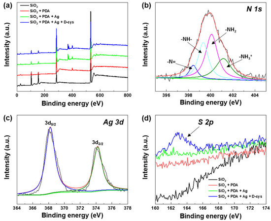
Figure 4.
XPS spectra of (a) wide scans, (b) N 1s, (c) Ag 3d, and (d) S 2p of the antibacterial nanoparticles.
3.2. Surface Morphology of Antibacterial Coating
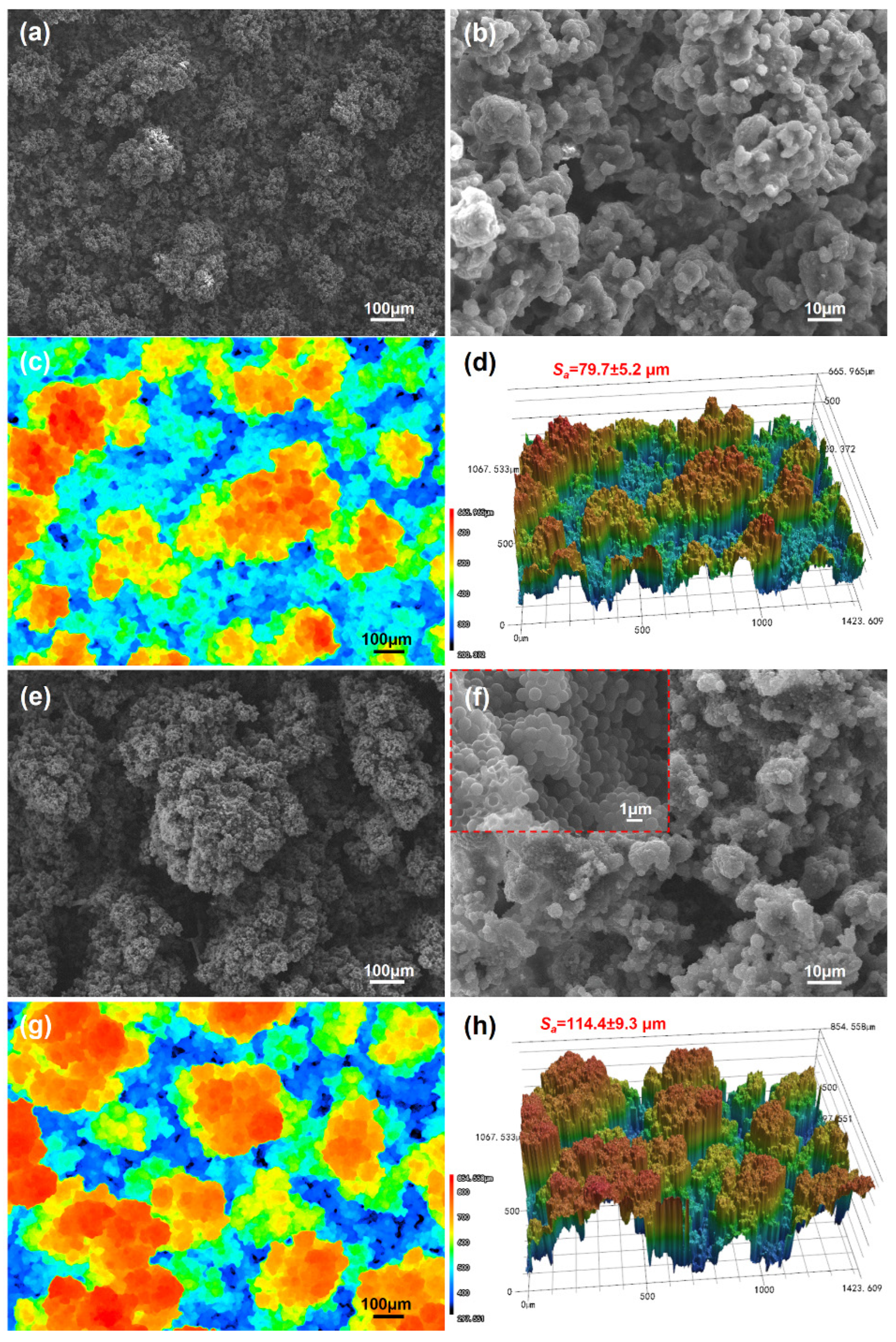
Figure 5 shows the surface morphologies and surface roughness of SS and SNC, respectively. The surface of the SS exhibited a rough microstructure with some agglomerates, which was caused by the aggregation of SiO2 nanoparticles (Figure 5a). The BADGE wrapped on the surface of SiO2 nanoparticles can be observed in Figure 5b. According to CLSM analysis, it can be seen that the size of the agglomerates was between 200 μm and 300 μm, and the surface roughness (Sa) of the SS was approximately 79.7 μm (Figure 5c,d). As shown in Figure 5e,f, after spraying the antibacterial nanoparticles, the surface morphology was significantly changed. A large number of tiny particles with the diameter of less than 1 μm uniformly covered the coating surface. Micro/nano hierarchical structures were established on the antibacterial coating surface, which was the typical physical structure required for superhydrophobic surfaces. The coating of antibacterial nanoparticles increased the cluster size to more than 300 μm, which also increased the surface roughness to approximately 114.4 μm (Figure 5g,h). In this study, antibacterial nanoparticles have dual functions: providing nanoscale morphology and enhancing antibacterial ability. The results of SEM observation indicated that the antibacterial nanoparticles were successfully loaded onto the SS, and the antibacterial SNC was prepared.
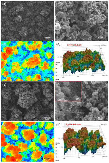
Figure 5.
(a,b) SEM images and (c,d) CLSM images of the surface of SS; (e,f) SEM images and (g,h) CLSM images of the surface of SNC.
3.3. Surface Wettability
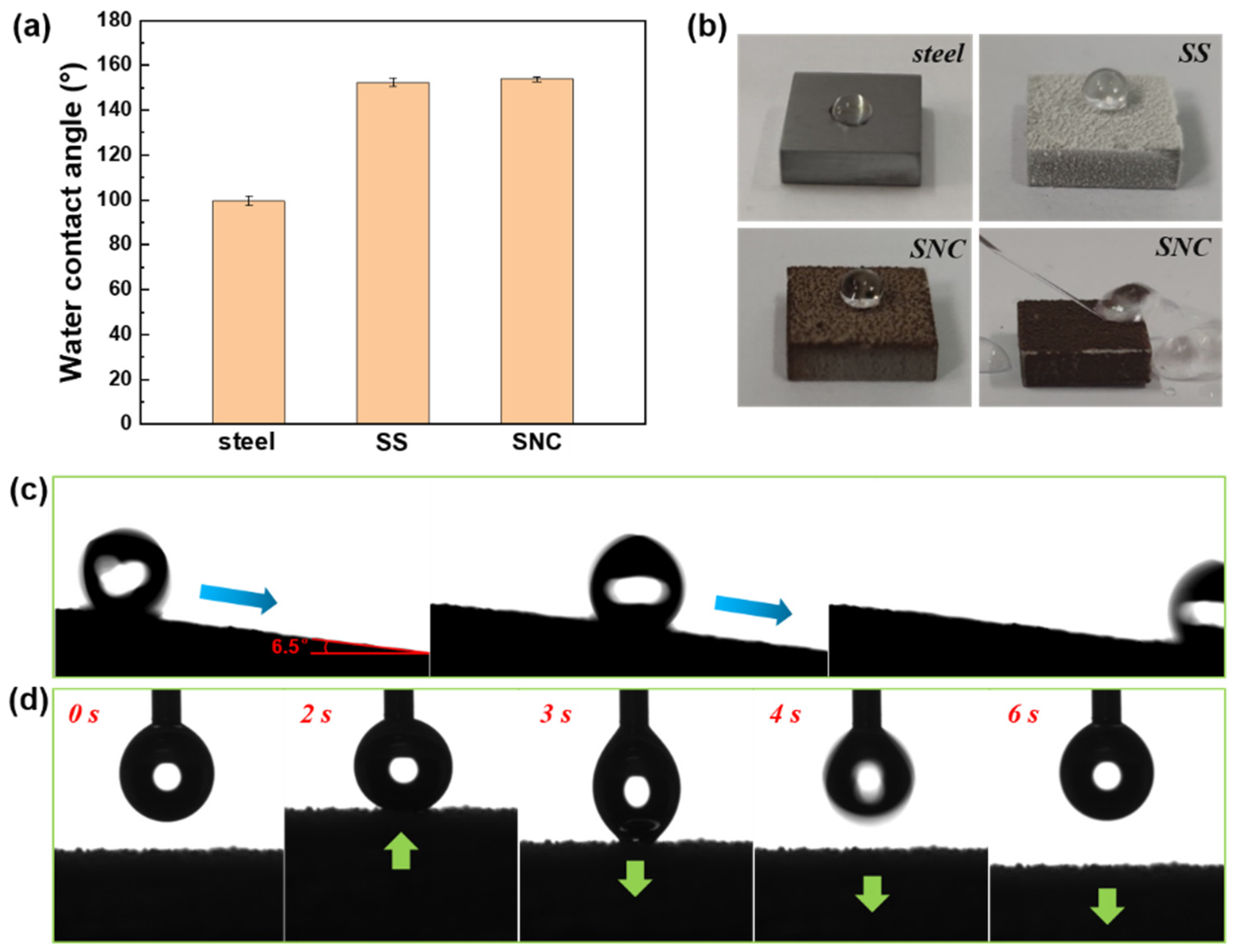
The surface wettability of steel surface, SS, and SNC is shown in Figure 6. The surface of carbon steel exhibited a water contact angle of approximately 100° (Figure 6a). The water droplet appeared hemispherical on the surface of the steel (Figure 6b). After the mixture of epoxy resin and SiO2 nanoparticles was sprayed on the carbon steel surface, the water contact angle of the sample surface was significantly increased to more than 150°, and the water droplet displayed an almost complete sphere on the sample surface. At this time, the superhydrophobic state was obtained. This resulted from the increased surface roughness by the accumulation of SiO2 nanoparticles. The air film was trapped between the liquid and solid substrate. The contact model between the water droplet and the sample surface can be explained by Cassie and Baxter model, which was calculated by the following formula [49]:
in which fSL represents the area fractions of solid/liquid interface. θSL is the intrinsic contact angles of solid/liquid interface. After the antibacterial nanoparticles were loaded, the water contact angle of the sample surface changed little. Due to abundant hydroxyl groups in catechol of polydopamine, the antibacterial nanoparticles will increase the surface energy of the coating surface slightly due to the hydrophilic property of polydopamine [50,51]. Hence, although the roughness of the sample surface was improved according to the CLSM images, the water contact angle did not change significantly. In addition, the sliding angle of the coating surface was approximately 7°, and the rolling state of a water droplet on the sloping sample surface was recorded, as shown in Figure 6c. In less than 1 s, the water droplet rolled away from the camera. The low adhesion of the water droplet can be seen from Figure 6d so that a slight contact could not bring the water droplet down from the needle tip. Even large amounts of water could not adhere to the sample surface (Figure 6b). The antibacterial coating exhibited good superhydrophobicity and excellent low wettability.
Cosθ = fSL·(cosθSL + 1) − 1

Figure 6.
(a) Water contact angle of steel surface, SS, and SNC; (b) photographs of a water droplet on steel surface, SS, and SNC and the video snapshot of the rolling state of water on SNC; (c) video snapshots of the rolling behavior of the water droplet on SNC; (d) video snapshots of the low adhesion behavior of the water droplet on SNC.
3.4. Mechanical Durability
Poor mechanical property is one of the problems restricting the practical application of superhydrophobic surfaces. The physical microstructure and low surface energy chemical composition of the superhydrophobic surface are easily destroyed by external forces or ultraviolet light. To overcome this problem, this study improved the mechanical strength of the superhydrophobic antibacterial coating from three aspects, including the crosslinking of silane coupling agent, the bonding of polymer resin, and the reinforcement of antibacterial nanoparticles. The SiO2 nanoparticles had high hardness, and the wrapping and bonding of the polymer resin kept the SiO2 framework. The crosslinking of KH550 further improved the binding force between polymer and inorganic SiO2 nanoparticles. In addition, the integration of nanostructures also contributed to the durability of superhydrophobic surfaces [28]. The spraying of antibacterial nanoparticles provided the surface nanostructures. When the micrometer scale structures were abrased, the nanostructures between micrometer scale clusters remained intact, continuing to provide the roughness required for superhydrophobicity. In this work, the mechanical durability of the SNC was evaluated through the tape-peel tests. The schematic of the tape-peel tests is shown in Figure 7a. Figure 7b shows the variation in water contact angle and sliding angle of the SNC surface as a function of tape-peeling cycles. After 180 tape-peeling cycles, the water contact angle was only slightly reduced to around 150° and the sliding angle was maintained under 10°. From further SEM observation, it can be clearly seen that the hierarchical micro/nanostructures were still retained (Figure 7c,d), which ensured the storage of the air film and the formation of Cassie and Baxter contact model between the sample surface and water droplet. The tape-peel test is a commonly used method to study the mechanical durability of antibacterial coatings. More peeling times represent good mechanical strength and adhesive force, indicating that the antibacterial coating has the ability to resist external physical damage. In this paper, the tape-peeling cycle was carried out 180 times, which was more than that of other reported works [52,53,54]. The results of tape-peel tests suggested that the surface of the SNC had good mechanical durability.
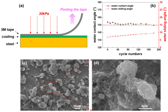
Figure 7.
(a) The schematic of the tape-peel tests; (b) variation in the water contact angle and the sliding angle with peeling cycle numbers; (c,d) SEM images of the surface morphologies of the SNC after 180 tape-peeling cycles.
3.5. Corrosion Resistance Analysis
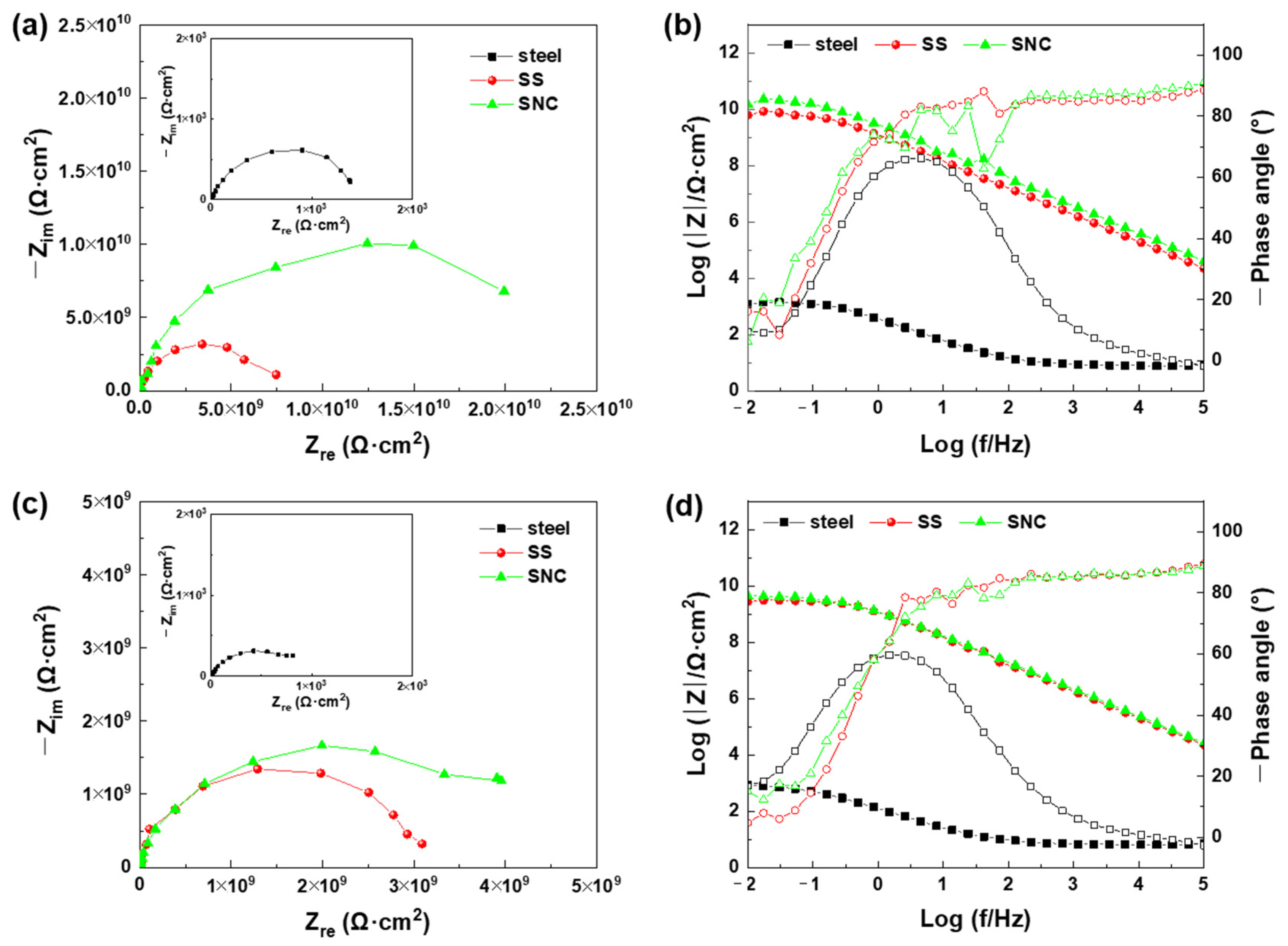
The EIS spectra for the carbon steel, SS, and SNC after 1 day and 3 days of immersion in 3.5% NaCl solutions are shown in Figure 8. After 1 day of exposure, the SNC exhibited the largest capacitive arc radius, while the carbon steel showed the minimum capacitive arc radius in the Nyquist plots (Figure 8a). In corresponding Bode plots (Figure 8b), the low-frequency impedance modulus (|Z|0.01Hz) of SNC reached 1.47 × 10¹⁰ Ω·cm2, which was almost twice as much as that of the SS (6.29 × 10⁹ Ω·cm2). |Z|0.01Hz of pure steel surface was only 1.22 × 10³ Ω·cm2. After spraying the SNC, the|Z|0.01Hz value increased by about seven orders of magnitude. The low-frequency impedance modulus is always adopted as a relatively intuitive indicator of the corrosion resistance performance of anticorrosive coatings [55]. The |Z|0.01Hz value of the SNC was much higher than that of carbon steel and close to that of the pure epoxy coating [17]. Moreover, the phase-angle plots of SS and SNC showed only one time constant, suggesting that the corrosion electrochemical reaction on the steel surface had not yet started. The SNC exhibited excellent corrosion resistance during 1 day of immersion, which was contributed by the polymer/SiO2 substrate and the air film trapped in the superhydrophobic surface. In addition, the corrosion resistance of SNC was better than that of SS, which might be caused by the coating and barrier effect of antibacterial nanoparticle layer. After 3 days of exposure, the capacitive arc radius and corresponding |Z|0.01Hz value of the SNC and the SS decreased slightly (Figure 8c). The |Z|0.01Hz value of the SNC and the SS decreased to 4.41 × 10⁹ Ω·cm2 and 2.73 × 10⁹ Ω·cm2, respectively. This decrease should be related to the reduction in the air film and the penetration of water into the coating [17]. However, the |Z|0.01Hz values were still much higher than that of carbon steel, and the phase-angle plots still showed one time constant (Figure 8d). This demonstrated that the SNC maintained good corrosion resistance after immersing for 3 days and could prevent the occurrence of steel corrosion.
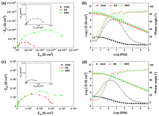
Figure 8.
EIS results of the samples after (a,b) 1 day and (c,d) 3 days of immersion in 3.5% NaCl solution.
3.6. Antibacterial Activity
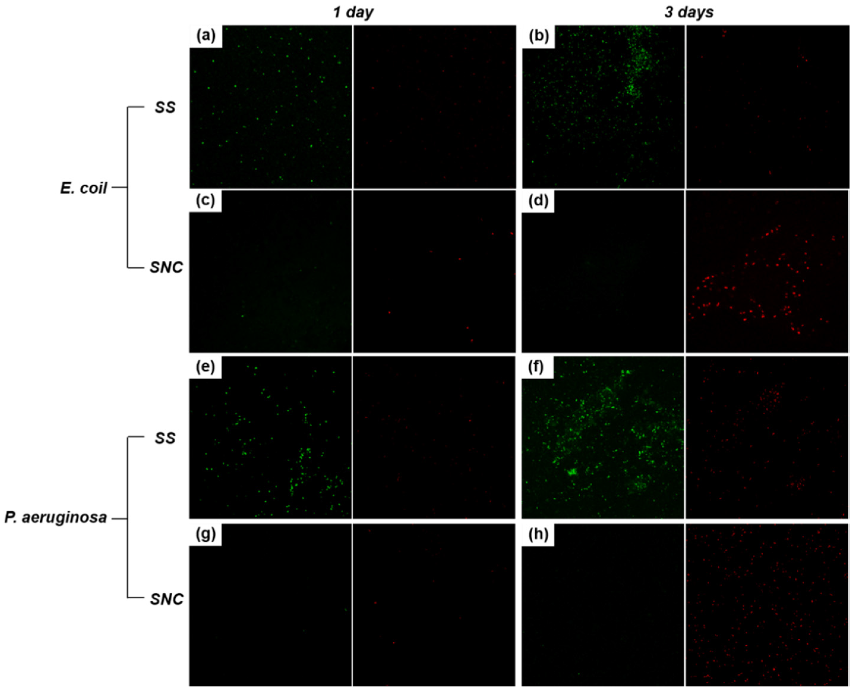
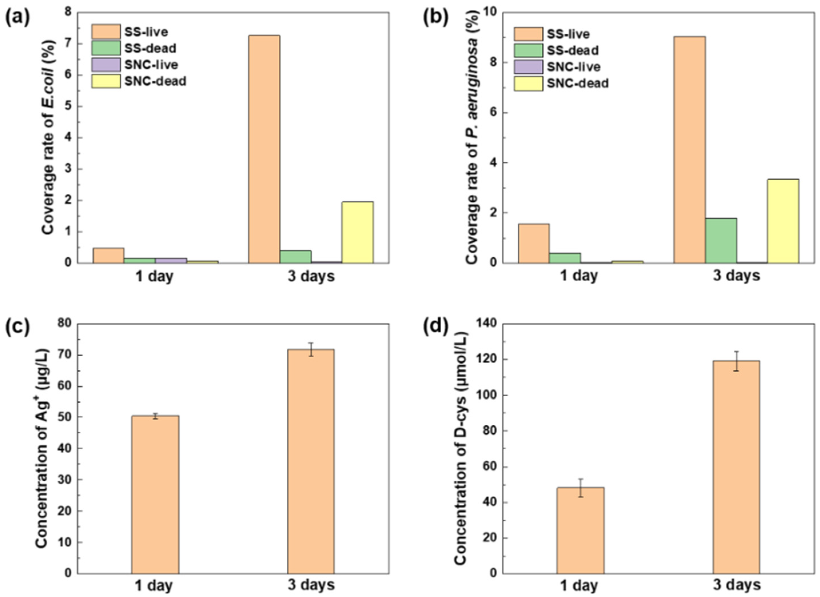
E. coli and P. aeruginosa were adopted to evaluate the antibacterial activity of the SS and the SNC. To quantitatively analyze the adhesion conditions of E. coli and P. aeruginosa, live/dead staining of bacterial cells on the sample surfaces was performed. The observation results and the calculated coverage rates are shown in Figure 9 and Figure 10a,b. Green fluorescence represented living cells, while red fluorescence represented dead cells. For the SS, green fluorescence appeared on the sample surface on the first day, indicating that living cells of E. coli and P. aeruginosa began to attach (Figure 9a,e). At this time, the coverage of living cells was only 0.5% and 1.6% for E. coli and P. aeruginosa, respectively. In our previous study, the biofilm coverage of E. coli and P. aeruginosa on steel surfaces exceeded 30.0% after only 1 day of exposure [35]. The bacterial coverage on SS surface was much smaller than that on steel surface. Hence, superhydrophobic surface exhibited good antiadhesion property during a short immersion period of 1 day. In addition, there was almost no dead cell on the surface of SS after 1 day. It proved that the superhydrophobic surface has no bactericidal effect but only reduces adhesion of bacteria. On the third day, green fluorescence gathered on localized areas, indicating that the biofilms of E. coli and P. aeruginosa began to form on the SS (Figure 9b,f). At this time, the coverage rate of living cells of E. coli and P. aeruginosa rapidly increased to approximately 7.3% and 9.0%, respectively. A few dead cells were observed, probably due to nutrients being depleted at 3 days and unable to support cell survival. Superhydrophobic surface of the SS had lost its antibacterial activity after 3 days of incubation. For the coating modified by antibacterial nanoparticles, there was almost no green and red fluorescence on the sample surfaces on the first day (Figure 9c,g), indicating that the bacterial cells were hard to adhere on the coating surface due to the superhydrophobicity. After 3 days of incubation, the green fluorescence was still hard to observe, and the coverage of living cells was nearly 0%, which was much lower than that on SS. Moreover, the number of dead cells on the SNC surface increased significantly (Figure 9d,h), and the coverage rate of dead cells reached 1.9% and 3.3% for E. coli and P. aeruginosa, respectively. The increase in dead cells resulted from the bactericidal effect of Ag nanoparticles. The dispersive distribution of red fluorescence indicated the inhibition effect of D-cys on biofilm formation.
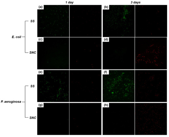
Figure 9.
Fluorescence microscope images of E.coli on the SS after (a) 1 day and (b) 3 days of immersion, and on the SNC after (c) 1 day and (d) 3 days of immersion in the culture medium; fluorescence microscope images of P. aeruginosa on the SS after (e) 1 day and (f) 3 days of immersion, and on the SNC after (g) 1 day and (h) 3 days of immersion in the culture medium.
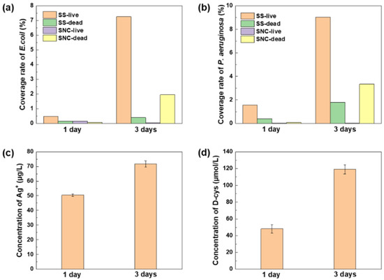
Figure 10.
The coverage rates of (a) E. coli and (b) P. aeruginosa on the SS and SNC surfaces after 1 day and 3 days of immersion in the inoculated media; the concentrations of released (c) Ag+ and (d) D-cys from the SNC surface after 1 day and 3 days of immersion in the LB medium.
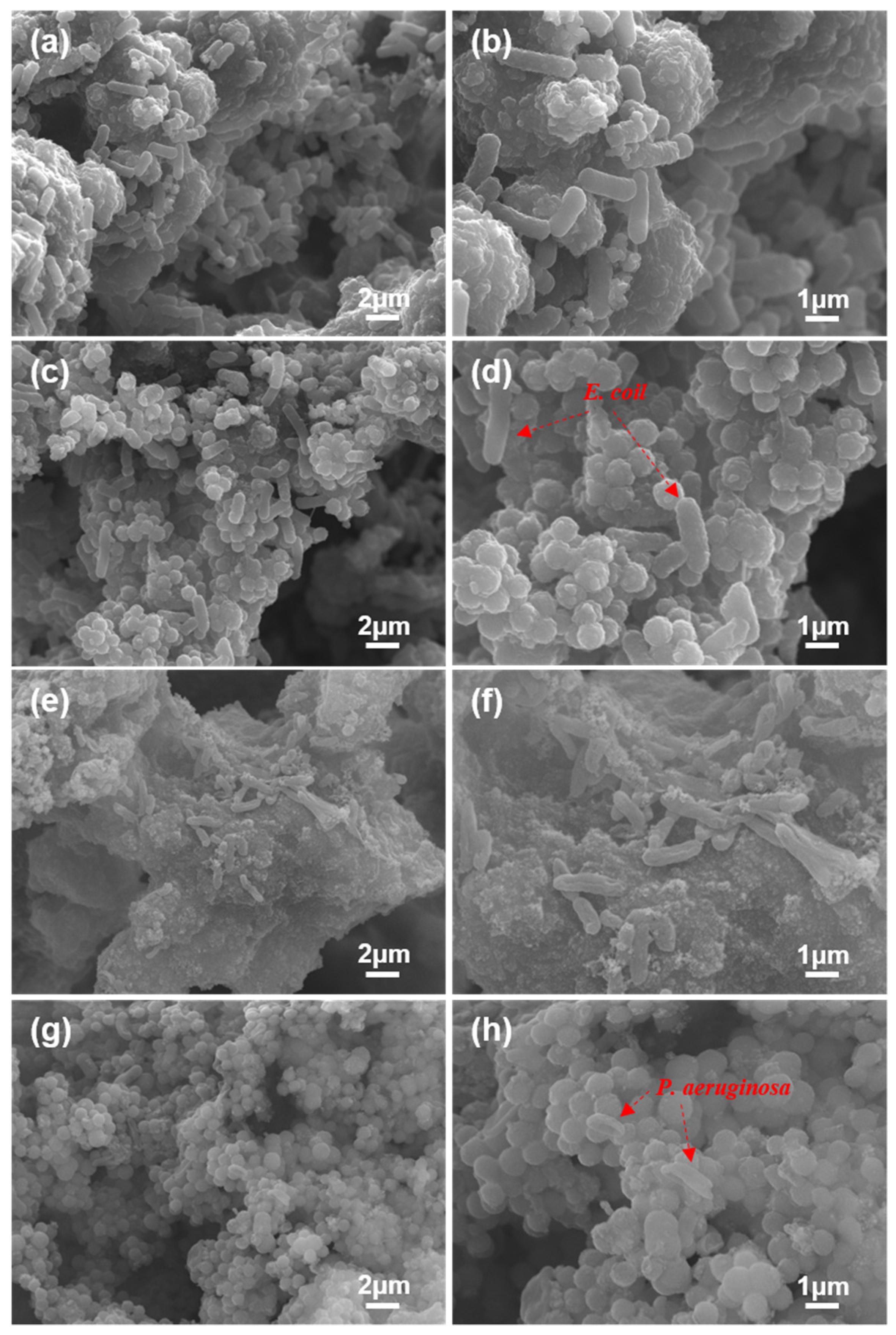
The adhesion conditions of E. coli and P. aeruginosa cells on the sample surfaces after 3 days of immersion in their culture media were also observed by SEM, which are shown in Figure 11. For the SS without antibacterial nanoparticles, obvious bacterial aggregation behavior of E. coli and P. aeruginosa cells appeared after immersing in the culture media for 3 days, and E. coli and P. aeruginosa cells all showed rod shapes with a length between 1 μm and 3 μm (Figure 11a,b,e,f). Although superhydrophobic surface can reduce protein adhesion and resist the attachment of bacterial cells, the antibacterial experimental results of the SS indicated that it cannot be maintained for a long time in the inoculated medium. When the antibacterial nanoparticles were loaded, the bacterial adhesion state on the sample surface changed significantly. After immersing for 3 days, only several adherent bacteria cells could be seen (Figure 11c,d,g,h). The results of SEM and live/death staining showed that the SNC had excellent antibacterial performance during 3 days of incubation against E. coli and P. aeruginosa.

Figure 11.
Surface SEM morphologies of the (a,b) SS and (c,d) SNC after 3 days of immersion in the E. coli-inoculated media; surface SEM morphologies of the (e,f) SS and (g,h) SNC after 3 days of immersion in the P. aeruginosa-inoculated media.
The concentrations of released Ag+ and D-cys from the SNC after 1 day and 3 days of immersion in the LB medium were also measured, which are shown in Figure 10c,d. The concentration of Ag+ after 1 day of immersion was approximately 50 μg/L. After 3 days of immersion, the concentration of released Ag+ increased to approximately 70 μg/L. This demonstrated that Ag+ was released gradually during immersion rather than explosive release at the beginning of immersion. Superhydrophobic surfaces have been reported to delay the rapid release of Ag+ [56,57]. As the superhydrophobic surface gradually failed, more and more silver nanoparticles contacted with medium solution and released silver ions. At the same time, the superhydrophobic surface in this study also showed the property of controllable release of D-cys. From the first day to the third day, the concentration of released D-cys increased from approximately 50 μmol/L to approximately 120 μmol/L. Superhydrophobic surface not only reduced bacterial adhesion, but also ensured the sustainable release of Ag+ and D-cys, which endowed the SNC with long-term antibacterial ability.
The antibacterial mechanism of the D-cys-loaded coating is presented in Figure 12. The antibacterial ability of the coating in this study is associated with the synergistic effect of the antiadhesion property of superhydrophobic surface, the bactericidal ability of nano-silver ions, and the biofilm inhibition property of D-cys. At the initial stage of incubation, the antibacterial behavior mainly depends on the low wetting property of the superhydrophobic surface, which is due to the existence of air film [58,59]. The trapped air film greatly reduces the contact area between the medium and the rough micro/nano hierarchical structure, resulting in fewer sites for bacterial colonization. Only the tip of the cluster may be coated by the extracellular polymeric substance and bacterial cells. Because of the barrier effect of air film, most of the antibacterial nanoparticles do not contact the medium, resulting in a low release of silver ions and D-cys. As the immersion progresses, the biofilms develop deeper into the ravine from the tip of the cluster. Sample surfaces covered by biofilms exhibit hydrophilicity. The increasing wetting area of the solid surface and the pressure of the medium lead to the gradual reduction in the air film, resulting in the gradual failure of superhydrophobic surface [60]. However, more antibacterial nanoparticles contact with the medium and begin to release D-cys and silver ions. The biofilm inhibition property of D-cys alleviates the attachment and aggregation of bacterial cells. The release of silver ions further kills the attached cells, completing the further elimination of adherent cells. The construction of superhydrophobic surface can realize the controlled release of silver ions and D-cys, and the antibacterial properties of D-cys and silver ions also delay the failure rate of superhydrophobic surface. This synergistic effect endowed the coating with high efficiency and durable antibacterial property. This synergistic antibacterial effect makes the coating have great application potential in the surface coating of medical devices. D-cys is relatively expensive, which will increase the cost of coating preparation. In the future work, other amino acids with low price and excellent antibacterial performance will be selected for coating preparation, which will help reduce the cost of coating preparation and increase the practical value.
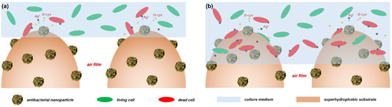
Figure 12.
The schematic figure of the antibacterial behavior of the superhydrophobic nanocomposite coating on the (a) first day and (b) third day.
4. Conclusions
In summary, a superhydrophobic antibacterial coating is fabricated combining a durable superhydrophobic substrate and antibacterial nanoparticles through a facile spraying method. The mixture of BADGE resin and KH550-modified SiO2 nanoparticles was sprayed on carbon steel surface to construct superhydrophobic substrate. D-cys functionalized SiO2/dopamine/silver nanoparticles were subsequently sprayed to enhance the antibacterial property of the coating. The prepared antibacterial coating exhibited low wettability property, with a water contact angle of more than 150° and a water sliding angle of approximately 7°. Due to the bonding of epoxy resin and the crosslinking of KH550, the coating exhibited good mechanical durability, which maintained the water contact angle of about 150° after 180 cycles in tape-peel tests. The results of EIS showed that the antibacterial coating had satisfactory corrosion resistance against 3.5% NaCl solution. Moreover, the coating showed excellent antibacterial performance against E. coli and P. aeruginosa, relying on the co-operation of superhydrophobic surface, Ag nanoparticles, and D-cys. This coating has great potential in the application of medical equipment surface coating to endow the long-term antibacterial property.
Author Contributions
Conceptualization, H.Q.; Investigation, H.Q. and W.C.; Methodology, W.L.; Visualization, W.C. and X.H.; Writing—review & editing, D.Z. All authors have read and agreed to the published version of the manuscript.
Funding
This work is supported by the National Natural Science Foundation of China (No. 52001021) and Joint Fund of Basic and Applied Basic Research Fund of Guangdong Province (2021B1515130009).
Institutional Review Board Statement
Not applicable.
Informed Consent Statement
Not applicable.
Data Availability Statement
Data sharing is not applicable to this article.
Conflicts of Interest
The authors declare no conflict of interest.
References
- Qing, Y.A.; Cheng, L.; Li, R.Y.; Liu, G.C.; Zhang, Y.B.; Tang, X.F.; Wang, J.C.; Liu, H.; Qin, Y.G. Potential antibacterial mechanism of silver nanoparticles and the optimization of orthopedic implants by advanced modification technologies. Int. J. Nanomed. 2018, 13, 3311. [Google Scholar] [CrossRef] [PubMed] [Green Version]
- Vincent, M.; Duval, R.E.; Hartemann, P.; Engels-Deutsch, M. Contact killing and antimicrobial properties of copper. J. Appl. Microbiol. 2018, 124, 1032–1046. [Google Scholar] [CrossRef] [PubMed] [Green Version]
- Sirelkhatim, A.; Mahmud, S.; Seeni, A.; Kaus, N.H.M.; Ann, L.C.; Bakhori, S.K.M.; Hasan, H.; Mohamad, D. Review on zinc oxide nanoparticles: Antibacterial activity and toxicity mechanism. Nano-Micro Lett. 2015, 7, 219–242. [Google Scholar] [CrossRef] [PubMed] [Green Version]
- Kubo, A.L.; Capjak, I.; Vrček, I.V.; Bondarenko, O.M.; Kurvet, I.; Vija, H.; Ivask, A.; Kasemets, K.; Kahru, A. Antimicrobial potency of differently coated 10 and 50 nm silver nanoparticles against clinically relevant bacteria Escherichia coli and Staphylococcus aureus. Colloids Surf. B 2018, 170, 401–410. [Google Scholar] [CrossRef]
- Qian, H.C.; Yang, J.Z.; Lou, Y.T.; ur Rahman, O.; Li, Z.Y.; Ding, X.; Gao, J.; Du, C.W.; Zhang, D.W. Mussel-inspired superhydrophilic surface with enhanced antimicrobial properties under immersed and atmospheric conditions. Appl. Surf. Sci. 2019, 465, 267–278. [Google Scholar] [CrossRef]
- Kora, A.J.; Arunachalam, J. Assessment of antibacterial activity of silver nanoparticles on Pseudomonas aeruginosa and its mechanism of action. World J. Microbiol. Biotechnol. 2011, 27, 1209–1216. [Google Scholar] [CrossRef]
- Bruna, T.; Maldonado-Bravo, F.; Jara, P.; Caro, N. Silver nanoparticles and their antibacterial applications. Int. J. Mol. Sci. 2021, 22, 7202. [Google Scholar] [CrossRef]
- Li, W.R.; Xie, X.B.; Shi, Q.S.; Zeng, H.Y.; OU-Yang, Y.S. Antibacterial activity and mechanism of silver nanoparticles on Escherichia coli. Appl. Microbiol. Biotechnol. 2010, 85, 1115–1122. [Google Scholar] [CrossRef]
- Morones, J.R.; Elechiguerra, J.L.; Camacho, A.; Holt, K.; Kouri, J.B.; Ramírez, J.T.; Yacaman, M.J. The bactericidal effect of silver nanoparticles. Nanotechnology 2005, 16, 2346. [Google Scholar] [CrossRef] [Green Version]
- Yin, I.X.; Zhang, J.; Zhao, I.S.; Mei, M.L.; Li, Q.L.; Chu, C.H. The antibacterial mechanism of silver nanoparticles and its application in dentistry. Int. J. Nanomed. 2020, 15, 2555. [Google Scholar] [CrossRef] [Green Version]
- Hooda, A.; Goyat, M.S.; Pandey, J.K.; Kumar, A.; Gupta, R. A review on fundamentals, constraints and fabrication techniques of superhydrophobic coatings. Prog. Org. Coat. 2020, 142, 105557. [Google Scholar] [CrossRef]
- Rius-Ayra, O.; Biserova-Tahchieva, A.; Llorca-Isern, N. Durable Superhydrophobic Coating for Efficient Microplastic Removal. Coatings 2021, 11, 1258. [Google Scholar] [CrossRef]
- Sebastian, D.; Yao, C.W.; Nipa, L.; Lian, I.; Twu, G. Corrosion behavior and mechanical properties of a nanocomposite superhydrophobic coating. Coatings 2021, 11, 652. [Google Scholar] [CrossRef]
- Yao, W.H.; Wu, L.; Huang, G.S.; Jiang, B.; Atrens, A.; Pan, F.S. Superhydrophobic coatings for corrosion protection of magnesium alloys. J. Mater. Sci. Technol. 2020, 52, 100–118. [Google Scholar] [CrossRef]
- Ganne, A.; Lebed, V.O.; Gavrilov, A.I. Combined wet chemical etching and anodic oxidation for obtaining the superhydrophobic meshes with anti-icing performance. Colloids Surf. A 2016, 499, 150–155. [Google Scholar] [CrossRef]
- Qian, H.C.; Xu, D.K.; Du, C.W.; Zhang, D.W.; Li, X.G.; Huang, L.Y.; Deng, L.P.; Tu, Y.C.; Mol, J.M.C.; Terryn, H.A. Dual-action smart coatings with a self-healing superhydrophobic surface and anti-corrosion properties. J. Mater. Chem. A 2017, 5, 2355–2364. [Google Scholar] [CrossRef] [Green Version]
- Zhang, D.W.; Qian, H.C.; Wang, L.T.; Li, X.G. Comparison of barrier properties for a superhydrophobic epoxy coating under different simulated corrosion environments. Corros. Sci. 2016, 103, 230–241. [Google Scholar] [CrossRef]
- Zhang, F.; Zhang, C.L.; Song, L.; Zeng, R.C.; Li, S.Q.; Cui, H.Z. Fabrication of the superhydrophobic surface on magnesium alloy and its corrosion resistance. J. Mater. Sci. Technol. 2015, 31, 1139–1143. [Google Scholar] [CrossRef]
- Vidal, K.; Gómez, E.; Goitandia, A.M.; Angulo-Ibáñez, A.; Aranzabe, E. The synthesis of a superhydrophobic and thermal stable silica coating via sol-gel process. Coatings 2019, 9, 627. [Google Scholar] [CrossRef] [Green Version]
- Zhang, D.W.; Wang, L.T.; Qian, H.C.; Li, X.G. Superhydrophobic surfaces for corrosion protection: A review of recent progresses and future directions. J. Coat. Technol. Res. 2016, 13, 11–29. [Google Scholar] [CrossRef] [Green Version]
- Varshney, P.; Mohapatra, S.S. Durable and regenerable superhydrophobic coatings for brass surfaces with excellent self-cleaning and anti-fogging properties prepared by immersion technique. Tribol. Int. 2018, 123, 17–25. [Google Scholar] [CrossRef]
- Pakzad, H.; Liravi, M.; Moosavi, A.; Nouri-Borujerdi, A.; Najafkhani, H. Fabrication of durable superhydrophobic surfaces using PDMS and beeswax for drag reduction of internal turbulent flow. Appl. Surf. Sci. 2020, 513, 145754. [Google Scholar] [CrossRef]
- Xi, Z.; Yuan, C.; Bai, X.; Wang, C.; Neville, A. Preparation of Degradable Superhydrophobic Mg/P/Z/F/H Composite Materials and Their Anticorrosion. Coatings 2021, 11, 1239. [Google Scholar] [CrossRef]
- Zhou, C.L.; Chen, Z.D.; Yang, H.; Hou, K.; Zeng, X.J.; Zheng, Y.F.; Cheng, J. Nature-inspired strategy toward superhydrophobic fabrics for versatile oil/water separation. ACS Appl. Mater. Interfaces 2017, 9, 9184–9194. [Google Scholar] [CrossRef]
- Zhu, T.X.; Cheng, Y.; Huang, J.Y.; Xiong, J.Q.; Ge, M.Z.; Mao, J.J.; Liu, Z.K.; Dong, X.L.; Chen, Z.; Lai, Y.K. A transparent superhydrophobic coating with mechanochemical robustness for anti-icing, photocatalysis and self-cleaning. Chem. Eng. J. 2020, 399, 125746. [Google Scholar] [CrossRef]
- Pan, Q.F.; Cao, Y.; Xue, W.; Zhu, D.H.; Liu, W.W. Picosecond laser-textured stainless steel superhydrophobic surface with an antibacterial adhesion property. Langmuir 2019, 35, 11414–11421. [Google Scholar] [CrossRef]
- Xun, X.W.; Wan, Y.Z.; Zhang, Q.C.; Gan, D.Q.; Hu, J.; Luo, H.L. Low adhesion superhydrophobic AZ31B magnesium alloy surface with corrosion resistant and anti-bioadhesion properties. Appl. Surf. Sci. 2020, 505, 144566. [Google Scholar] [CrossRef]
- Wang, D.H.; Sun, Q.Q.; Hokkanen, M.J.; Zhang, C.L.; Lin, F.Y.; Liu, Q.; Zhu, S.P.; Zhou, T.F.; Chang, Q.; He, B.; et al. Design of robust superhydrophobic surfaces. Nature 2020, 582, 55–59. [Google Scholar] [CrossRef]
- Kolodkin-Gal, I.; Romero, D.; Cao, S.; Clardy, J.; Kolter, R.; Losick, R. D-amino acids trigger biofilm disassembly. Science 2010, 328, 627–629. [Google Scholar] [CrossRef] [Green Version]
- Xu, H.; Liu, Y. D-Amino acid mitigated membrane biofouling and promoted biofilm detachment. J. Membr. Sci. 2011, 376, 266–274. [Google Scholar] [CrossRef]
- Hochbaum, A.I.; Kolodkin-Gal, I.; Foulston, L.; Kolter, R.; Aizenberg, J.; Losick, R. Inhibitory effects of D-amino acids on Staphylococcus aureus biofilm development. J. Bacteriol. 2011, 193, 5616–5622. [Google Scholar] [CrossRef] [PubMed] [Green Version]
- Leiman, S.A.; May, J.M.; Lebar, M.D.; Kahne, D.; Kolter, R.; Losick, R. D-amino acids indirectly inhibit biofilm formation in Bacillus subtilis by interfering with protein synthesis. J. Bacteriol. 2013, 195, 5391–5395. [Google Scholar] [CrossRef] [Green Version]
- Zilm, P.S.; Butnejski, V.; Rossi-Fedele, G.; Kidd, S.P.; Edwards, S.; Vasilev, K. D-amino acids reduce Enterococcus faecalis biofilms in vitro and in the presence of antimicrobials used for root canal treatment. PLoS ONE 2017, 12, e0170670. [Google Scholar] [CrossRef] [Green Version]
- Ramón-Peréz, M.L.; Diaz-Cedillo, F.; Ibarra, J.A.; Torales-Carden, A.; Rodríguez-Martínez, S.; Jan-Roblero, J.; Cancino-Diaz, M.E.; Cancino-Diaz, J.C. D-Amino acids inhibit biofilm formation in Staphylococcus epidermidis strains from ocular infections. J. Med. Microbiol. 2014, 63, 1369–1376. [Google Scholar] [CrossRef] [Green Version]
- Huang, L.Y.; Lou, Y.T.; Zhang, D.W.; Ma, L.W.; Qian, H.C.; Hu, Y.T.; Ju, P.F.; Xu, D.K.; Li, X.G. D-Cysteine functionalised silver nanoparticles surface with a “dispersethen-kill” antibacterial synergy. Chem. Eng. J. 2020, 381, 122662. [Google Scholar] [CrossRef]
- Stöber, W.; Fink, A.; Bohn, E. Controlled growth of monodispersesilica spheres in the micron size range. J. Colloid Interface Sci. 1968, 26, 62–69. [Google Scholar] [CrossRef]
- Zhou, E.Z.; Qiao, D.X.; Yang, Y.; Xu, D.K.; Lu, Y.P.; Wang, J.J.; Smith, J.A.; Li, H.B.; Zhao, H.L.; Liaw, P.K.; et al. A novel Cu-bearing high-entropy alloy with significant antibacterial behavior against corrosive marine biofilms. J. Mater. Sci. Technol. 2020, 46, 201–210. [Google Scholar] [CrossRef]
- Lee, H.; Dellatore, S.M.; Miller, W.M.; Messersmith, P.B. Mussel-inspired surface chemistry for multifunctional coatings. Science 2007, 318, 426–430. [Google Scholar] [CrossRef] [Green Version]
- Cong, Y.; Xia, T.; Zou, M.; Li, Z.N.; Peng, B.; Guo, D.Z.; Deng, Z.W. Mussel-inspired polydopamine coating as a versatile platform for synthesizing polystyrene/Ag nanocomposite particles with enhanced antibacterial activities. J. Mater. Chem. B 2014, 2, 3450–3461. [Google Scholar] [CrossRef]
- Zhao, R.; Lv, M.; Li, Y.; Sun, M.; Kong, W.; Wang, L.; Song, S.; Fan, C.; Jia, L.; Qiu, S.; et al. Stable nanocomposite based on PEGylated and silver nanoparticles loaded graphene oxide for long-term antibacterial activity. ACS Appl. Mater. Interfaces 2017, 9, 15328–15341. [Google Scholar] [CrossRef]
- Liu, M.L.; Liu, T.F.; Chen, X.W.; Yang, J.C.; Deng, J.; He, W.F.; Zhang, X.R.; Lei, Q.; Hu, X.H.; Luo, G.X.; et al. Nano-silver-incorporated biomimetic polydopamine coating on a thermoplastic polyurethane porous nanocomposite as an efficient antibacterial wound dressing. J. Nanobiotechnology 2018, 16, 89. [Google Scholar] [CrossRef] [PubMed]
- Qian, H.C.; Li, M.L.; Li, Z.; Lou, Y.T.; Huang, L.Y.; Zhang, D.W.; Xu, D.K.; Du, C.W.; Lu, L.; Gao, J. Mussel-inspired superhydrophobic surfaces with enhanced corrosion resistance and dual-action antibacterial properties. Mat. Sci. Eng. C 2017, 80, 566–577. [Google Scholar] [CrossRef] [PubMed]
- Wang, H.F.; Wei, L.Y.; Wang, Z.Q.; Chen, S.G. Preparation, characterization and long-term antibacterial activity of Ag–poly (dopamine)–TiO2 nanotube composites. RSC Adv. 2016, 6, 14097–14104. [Google Scholar] [CrossRef]
- Lepoittevin, B.; Bedel, S.; Dragoe, D.; Bruzaud, J.; Barthés-Labrousse, M.G.; Mazerat, S.; Herry, J.M.; Bellon-Fontaine, M.N.; Roger, P. Antibacterial surfaces obtained through dopamine and fluorination functionalizations. Prog. Org. Coat. 2015, 82, 17–25. [Google Scholar] [CrossRef]
- Xu, Q.B.; Li, R.L.; Shen, L.W.; Xu, W.; Wang, J.P.; Jiang, Q.F.; Zhang, L.; Fu, F.Y.; Fu, Y.Q.; Liu, X.D. Enhancing the surface affinity with silver nano-particles for antibacterial cotton fabric by coating carboxymethyl chitosan and L-cysteine. Appl. Surf. Sci. 2019, 497, 143673. [Google Scholar] [CrossRef]
- Song, S.; Liu, Z.; Abubaker, M.A.; Ding, L.; Zhang, J.; Yang, S.R.; Fan, Z.J. Antibacterial polyvinyl alcohol/bacterial cellulose/nano-silver hydrogels that effectively promote wound healing. Mat. Sci. Eng. C 2021, 126, 112171. [Google Scholar] [CrossRef]
- Chen, W.J.; Zheng, L.Y.; Wang, M.L.; Chi, Y.W.; Chen, G.N. Preparation of protein-like silver–cysteine hybrid nanowires and application in ultrasensitive immunoassay of cancer biomarker. Anal. Chem. 2013, 85, 9655–9663. [Google Scholar] [CrossRef]
- Bandyopadhyay, S.; Chattopadhyay, S.; Dey, A. The protonation state of thiols in self-assembled monolayers on roughened Ag/Au surfaces and nanoparticles. Phys. Chem. Chem. Phys. 2015, 17, 24866–24873. [Google Scholar] [CrossRef]
- Parvate, S.; Dixit, P.; Chattopadhyay, S. Superhydrophobic surfaces: Insights from theory and experiment. J. Phys. Chem. B 2020, 124, 1323–1360. [Google Scholar] [CrossRef] [Green Version]
- Zhang, H.; Bré, L.P.; Zhao, T.Y.; Zheng, Y.; Newland, B.; Wang, W.X. Mussel-inspired hyperbranched poly(amino ester) polymer as strong wet tissue adhesive. Biomaterials 2014, 35, 711–719. [Google Scholar] [CrossRef]
- Zhang, R.X.; Braeken, L.; Luis, P.; Wang, X.L.; Van der Bruggen, B. Novel binding procedure of TiO2 nanoparticles to thin film composite membranes via self-polymerized polydopamine. J. Membr. Sci. 2013, 437, 179–188. [Google Scholar] [CrossRef]
- Jia, S.S.; Deng, S.L.; Luo, S.; Qing, Y.; Yan, N.; Wu, Y.Q. Texturing commercial epoxy with hierarchical and porous structure for robust superhydrophobic coatings. Appl. Surf. Sci. 2019, 466, 84–91. [Google Scholar] [CrossRef]
- Sow, P.K.; Singhal, R.; Sahoo, P.; Radhakanth, S. Fabricating low-cost, robust superhydrophobic coatings with re-entrant topology for self-cleaning, corrosion inhibition, and oil-water separation. J. Colloid Interface Sci. 2021, 600, 358–372. [Google Scholar] [CrossRef]
- Zhi, D.F.; Lu, Y.; Sathasivam, S.; Parkin, I.P.; Zhang, X. Large-scale fabrication of translucent and repairable superhydrophobic spray coatings with remarkable mechanical, chemical durability and UV resistance. J. Mater. Chem. A 2017, 5, 10622–10631. [Google Scholar] [CrossRef] [Green Version]
- Liu, X.W.; Xiong, J.P.; Lv, Y.W.; Zuo, Y. Study on corrosion electrochemical behavior of several different coating systems by EIS. Prog. Org. Coat. 2009, 64, 497–503. [Google Scholar] [CrossRef]
- Liu, T.; Yin, B.; He, T.; Guo, N.; Dong, L.H.; Yin, Y.S. Complementary effects of nanosilver and superhydrophobic coatings on the prevention of marine bacterial adhesion. ACS Appl. Mater. Interfaces 2012, 4, 4683–4690. [Google Scholar] [CrossRef]
- Zhang, L.C.; Zhang, L.H.; Yang, Y.; Zhang, W.; Lv, H.C.; Yang, F.; Lin, C.J.; Tang, P.F. Inhibitory effect of super-hydrophobicity on silver release and antibacterial properties of super-hydrophobic Ag/TiO2 nanotubes. J. Biomed. Mater. Res. B 2016, 104, 1004–1012. [Google Scholar] [CrossRef]
- Zhang, X.; Wang, L.; Levänen, E. Superhydrophobic surfaces for the reduction of bacterial adhesion. RSC Adv. 2013, 3, 12003–12020. [Google Scholar] [CrossRef]
- Huang, X.; Wang, D.R.; Hu, L.Y.; Song, J.J.; Chen, Y.Q. Preparation of a novel antibacterial coating precursor and its antibacterial mechanism. Appl. Surf. Sci. 2019, 465, 478–485. [Google Scholar] [CrossRef]
- Zheng, Q.S.; Yu, Y.; Zhao, Z.H. Effects of hydraulic pressure on the stability and transition of wetting modes of superhydrophobic surfaces. Langmuir 2005, 21, 12207–12212. [Google Scholar] [CrossRef]


Publisher’s Note: MDPI stays neutral with regard to jurisdictional claims in published maps and institutional affiliations. |
© 2022 by the authors. Licensee MDPI, Basel, Switzerland. This article is an open access article distributed under the terms and conditions of the Creative Commons Attribution (CC BY) license (https://creativecommons.org/licenses/by/4.0/).