Article

Synthesis of Biomolecule Functionalized Biocompatible Silver Nanoparticles for Antioxidant and Antibacterial Applications

by
Kiseok Han
,
Anbazhagan Sathiyaseelan
,
Kandasamy Saravanakumar
and
Myeong-Hyeon Wang
*
Department of Bio-Health Convergence, Kangwon National University, Chuncheon 200-701, Korea
*
Author to whom correspondence should be addressed.
These authors contributed equally to this work.
Coatings 2022, 12(9), 1292; https://doi.org/10.3390/coatings12091292
Submission received: 19 July 2022 / Revised: 26 August 2022 / Accepted: 29 August 2022 / Published: 2 September 2022
(This article belongs to the Section Bioactive Coatings and Biointerfaces)

Abstract

:
The silver nanoparticles (AgNPs) were synthesized using quercetin (Qn) for enhanced antioxidant and antibacterial activity. The quercetin-mediated silver nanoparticles (Qn-AgNPs) were characterized by different analytical methods. The Qn-AgNPs showed maximum absorbance at 420 nm in the UV-visible spectrum. The hydrodynamic size of 92.91 ± 0.65 d.nm, polydispersity index (PDI) of 0.27 ± 0.006, and negative zeta potential of −31.36 ± 0.28 mV were measured for Qn-AgNPs. TEM analysis indicated that Qn-AgNPs were found to be homogeneous spherical particles without agglomeration. The capping of Qn and crystalline properties of Qn-AgNPs was confirmed by FTIR and XRD analysis, respectively. At a minimum concentration, Qn-AgNPs substantially inhibited the growth of bacterial pathogens, while it did not show toxicity in HEK293 cells or red blood cells and chick chorioallantoic membrane (CAM). Hence, the present results suggested that Qn could be a potent compound for the synthesis of Qn-AgNPs with promising antibacterial and antioxidant properties.

1. Introduction

Nanotechnology has a wide range of applications in electronics, material science, and medicine [1,2,3,4]. Due to their nanoscale and their large surface area, nanoparticles have various excellent characteristics such as thermal, mechanical, catalytic, electrical, and optical, which enables them to have a wide range of applications in distinct fields [5,6]. Nanomaterials are used in medicine as drug/ drug carriers for orthopedic applications, heart disease treatment, cancer treatment, and dental treatment [7,8]. The size of nanoparticles can be controlled by regulating physical and chemical factors [9,10]. Metal-based nanoparticles such as gold, silver, copper, zinc, and platinum have a great advantage due to their unique characteristics (electrical conductivity, catalytic, thermal properties, and biocompatibility) [11,12,13,14,15]. These things considered, the physical (size, shape, etc.) and biological (antimicrobial, biocompatible, etc.) properties of the metal nanoparticle can be furnished by appropriate reducing and capping agents [16].
In general, various chemical and physical techniques have been used to produce metal nanoparticles [17]. However, those methodologies are extremely expensive, and their byproducts are toxic to the environment [6]. Therefore, the green synthesis of metal nanoparticles using plant extracts has become promising [18,19]. In addition, plant-based metal nanoparticle synthesis is gaining more attention than microbial methods because of sophisticated microbial culture processes [20]. Silver nanoparticles (AgNPs) have significant antimicrobial properties among other metal nanoparticles, which attracts researchers to synthesize the AgNPs and AgNPs-based nanomaterials for antimicrobial and wound healing applications [21,22,23,24]. Plants (Rhododendron dauricum, Humulus lupulus, Jatropha curcas, etc.) have been widely used in the synthesis of AgNPs [25,26,27]. Phyto compounds such as polyphenols, alkaloids, flavonoids, fatty acids, and proteins are known to act as reducing and capping agents in metal nanoparticle synthesis [28]. However, the use of bulk reducing/capping agents for the synthesis of nanoparticles leads to unrestrained properties (size, shape, stability, toxicity, etc.) [29].
To avoid the non-specific toxicity, improve the biological activities (antimicrobial, antioxidant, anticancer, etc.), and elucidate the molecular mechanisms of AgNPs have recently synthesized using phytocompounds (flavonoids, carotenoids, phenols, etc.) [30]. Among the plant-derived molecules, quercetin (Qn) is one of the most antioxidant flavonoids found in various fruits, vegetables, and seeds [31,32]. Furthermore, Qn protects the neuronal, heart, and vascular functions improve anti-inflammatory and anti-allergic properties and reduce the risk of age-related metabolic diseases [33,34,35]. Although Qn-mediated AgNPs have been reported in a previous study, they did not evaluate their biological and toxicological properties in detail [36,37]. Hence, this study aimed to synthesize AgNPs using Qn and evaluate their physiochemical, toxicity, antibacterial, and antioxidant properties.

2. Materials and Methods

2.1. Materials

Quercetin dihydrate (HPLC grade), erythromycin, triton X-100, sodium hydroxide (NaOH) 2,2′-azino-bis-3-ethylbenzthiazoline-6-sulphonic acid (ABTS), diphenyl-2-picryl-hydrazyl (DPPH) and tetracycline hydrochloride were purchased from Sigma-Aldrich (St. Louis, MO, USA). Silver nitrate and agar powder were purchased from Daejung chemicals & metals Co., Ltd., Siheung-si, Korea. Sheep blood was purchased from Carolina, Korea. Nutrient broth (NB) and muller-Hinton agar (MHA) were purchased from BD DifcoTM, Thermo Fisher Scientific, Seoul, Korea. The cell viability assay kit was obtained from Cellomax™, MediFab (Rolleston, New Zealand).

2.2. Synthesis of Silver Nanoparticle

The 10 mL of silver nitrate solution (10 mM) was added to 90 mL of quercetin solution (3 mM). To initiate the AgNPs synthesis, 100 μL of NaOH (10 mM) was added to the reaction mixture and incubated at 28 °C overnight in the dark. The Qn-mediated AgNPs synthesis confirmed color changes in the reaction mixture from yellow to dark brown [38]. Qn-AgNPs were obtained by centrifugation at 12,000 rpm for 15 min, washed thoroughly with distilled water, and then freeze-dried at −50 °C for 48 h. Then, Qn-Ag NPs were stored at room temperature for further experiments.

2.3. Characterization of Silver Nanoparticles

The synthesis of Qn-AgNPs (1 mg/mL) was initially characterized using a UV–Visible spectrophotometer (SpectraMax® Plus 384 Microplate Reader, Molecular Devices, (San Jose, CA, USA) with a wavelength range of 200–700 nm. The functionalities of Qn and Qn-AgNPs were then investigated using an FTIR (PerkinElmer Paragon 500 (Waltham, MA, USA) with a scan range of 400–4000 cm–1. The crystalline properties of Qn-AgNPs were determined in X-ray powder diffraction (PANalytical, X′pert-pro MPD, Almelo, Netherlands) analysis with a 2θ scan range of 10°–80°. Further, Qn-Ag NPs (1 mg/mL) were dispersed in ethanol, a drop of nanoparticle solution was added to the carbon-coated copper grid, and the size and shape of Qn-AgNPs were observed using transmission electron microscopy (TEM, JEM-2100F, JEOL, Tokyo, Japan) coupled with EDS analysis. In addition, the hydrodynamic size and zeta potential of the Qn-AgNPs (1 mg/mL) dispersed in distilled water were measured by the zeta potential particle size analyzer (Malvern PANalytical, Almelo, The Netherland).

2.4. Biological Application

2.4.1. In Vitro Cytotoxicity

Cell toxicity of Qn-AgNPs in HEK293 cells was evaluated according to the methods reported in earlier work [39]. In brief, HEK293 cells were cultured in a DMEM medium containing 10% of fetal bovine serum, and 1% of antibiotics were kept at 37 °C in a CO2 (5%) incubator. The cytotoxic effects of Qn-AgNPs were tested by the WST-1 (water-soluble tetrazolium salt) assay kit. HEK293 cells were cultured in a 96-well plate in the above-mentioned condition. After reaching 70% of cell confluence, the different concentrations of serially diluted Qn-AgNPs (1.9, 3.9, 7.8, 15.6, 31.25, 62.5, 125, 250, 500, and 1000 μg/mL) were treated for 24 h. 10 μL of WST-1 was then added to each well and incubated for another 2 h. After incubation, the plate was measured at 450 nm using a microplate reader. Cell viability was calculated according to the earlier study [39].

2.4.2. Antibacterial Assay

Well Diffusion Method

The antibacterial effect of Qn-AgNPs was analyzed using the well diffusion method [40]. In brief, bacterial pathogens (Escherichia coli (ATCC 43888), Salmonella enterica (ATCC 14028), Bacillus cereus (ATCC 14579), and Staphylococcus aureus (ATCC 19095)) were initially cultured in nutrient broth. Then, the bacterial cells were inoculated on sterile MHA plates by a swabbing technique after wells were made in a sterile cork borer under an aseptic environment. After that, 50 μL of different concentrations of Qn-AgNPs (7.8, 15.6, 31.25, and 62.5 μg/mL) and Qn (62.5 μg/mL) were added to each well. Moreover, 20 μL of tetracycline-hydrochloride (1000 μg/mL) was used as the positive control. Plates were also incubated overnight at 37 °C, and the diameter of the zone inhibition around the well was measured.

Minimum Inhibitory Concentration (MIC)

Antimicrobial assays were used to determine the lowest concentration of antimicrobials that inhibits the detectable microorganism growth. The minimal inhibitory concentration (MIC50) of Qn-AgNPs was determined by the microdilution method [41]. In brief, different concentrations of Qn-AgNPs (1.9, 3.9, 7.8, 15.62, 31.25, and 62.5 μg/mL) were prepared in distilled water and added to 20 μL per each well containing sterilized fresh MHB (150 μL). Then, separately, 10 μL of E. coli and S. aureus are inoculated into each well and incubated at 37 °C. The plate was measured at 600 nm for a predetermined time interval (0, 1, 3, 6, 12, 24, and 48 h). Distilled water (20 μL) was used as the negative control.

2.4.3. Antioxidant Assay

Scavenging of DPPH and ABTS and other free radicals is the basis of determining the common antioxidant properties [42]. DPPH and ABTS free radicals’ solutions were prepared according to the earlier report [43]. For the assay, 100 μL of different concentrations of Qn-AgNPs (0.9, 1.9, 3.9, 7.8, 15.6, 31.25 62.5, 125, and 250 μg/mL) were added to 96 plates, and 100 μL of DPPH and ABTS were added, separately. Ascorbic acid and phosphate buffer (pH 7.4) was used as the positive and negative controls, respectively. After 10 min of incubation under dark conditions, the plates were measured at 517 nm and 734 nm for DPPH and ABTS, respectively. The percentage of radical scavenging ability was calculated according to the earlier method [43].

2.4.4. Hemolysis Assay

To identify the blood toxicity, an in vitro hemolysis assay was performed according to the earlier method [44]. In brief, 4% of red blood cells (RBCs) were prepared by dissolving 1 mL of sheep blood into 10 mL PBS (pH 7.2). Then, RBCs were collected through centrifugation at 2000 rpm for 10 min at 4 °C, washed thoroughly using PBS, and dispersed into PBS (10 mL). For the assay, 200 μL of different concentrations of Qn-AgNPs (0.9, 1.9, 3.9, 7.8, 15.6, 31.2, 62.5, 125, 250 μg/mL) were incubated with 200 μL of RBC at 37 °C for 1 h. Triton X-100 (1%), and PBS were used as positive and negative controls, respectively. After centrifugation, the supernatant was observed at 545 nm using a UV–vis spectrophotometer (SpectraMax® Plus 384 Microplate Reader, Molecular Devices). The percentage of hemolysis was calculated according to the earlier report [45].

2.4.5. CAM Assay

The chick embryo chorioallantoic membrane (CAM) assay has been widely performed to understand the mechanisms of angiogenesis and tumor invasion in several cancers (colon, prostate, and brain). Fertilized chicken eggs were purchased from a poultry farm in Chuncheon, Korea. After surface sterilization, the outer wall of air space champers of the egg was cut into a spherical and carefully removed. Then, 200 μL of different concentrations of Qn-AgNPs (62.5, 125, and 250 μg/mL), NaOH (1 N), and PBS were added gently and left at room temperature for 5 min. The effects (coagulations and blood vessel damage) of Qn-AgNPs, PBS, and NaOH on CAM were compared before and after the treatment.

2.5. Statistical Analysis

The collected experimental data were analyzed using a one-way analysis of variance, and the data were presented as mean ± standard error. A p-value of less than 0.05 is considered statistically significant.

3. Results and Discussion

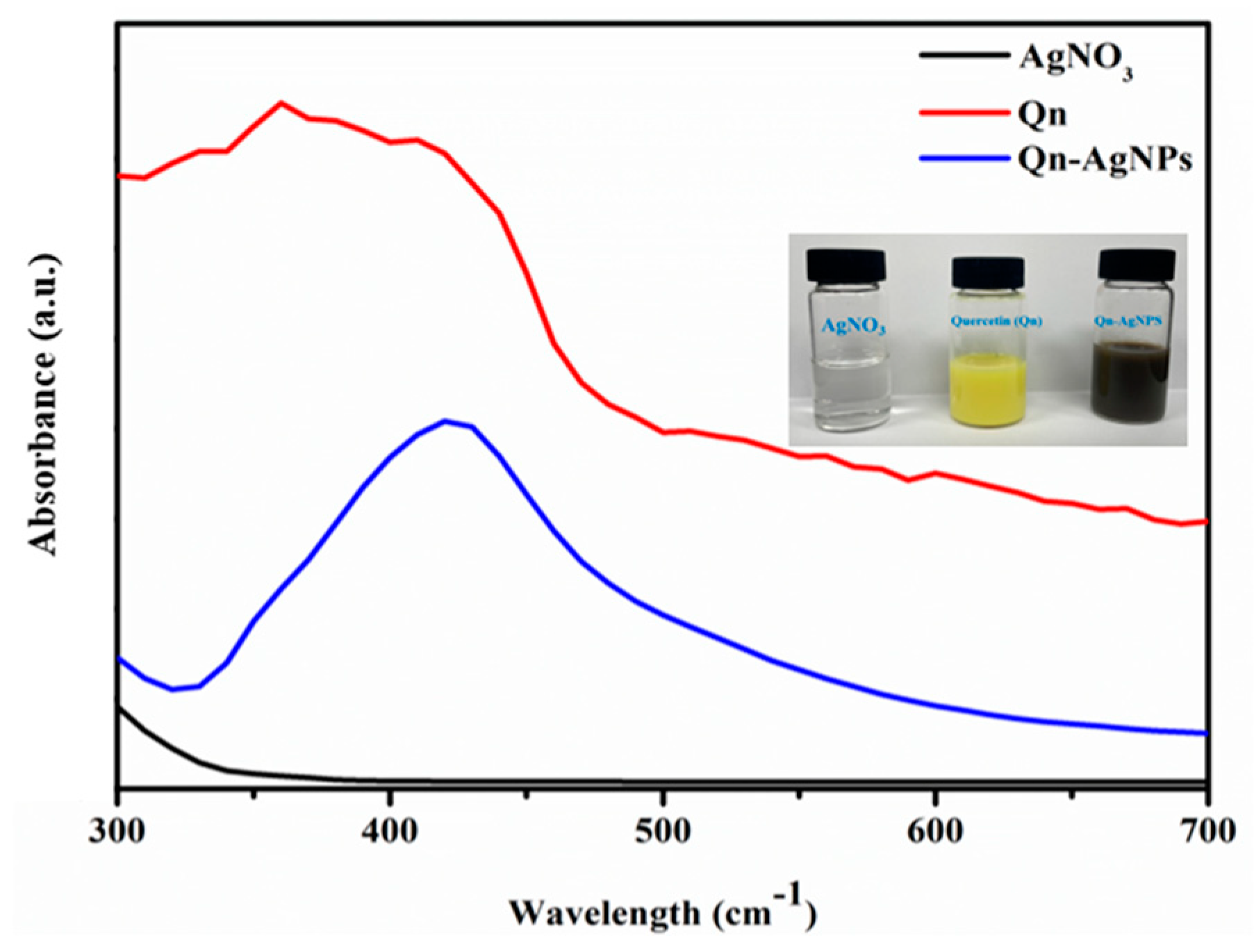
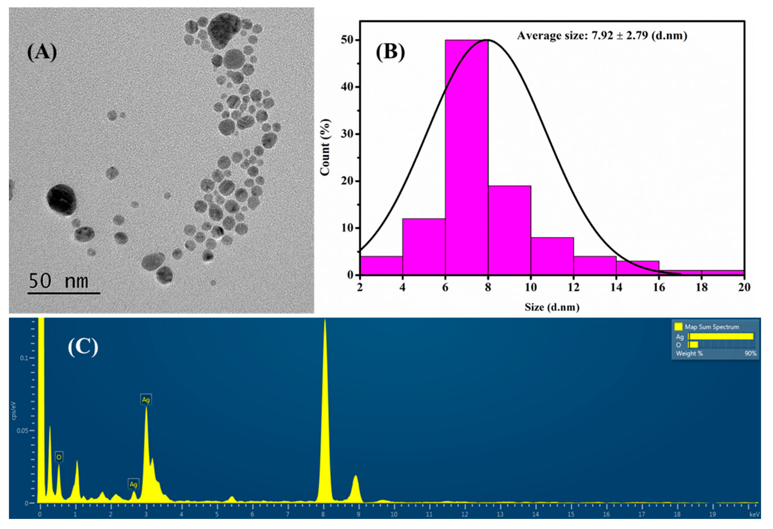
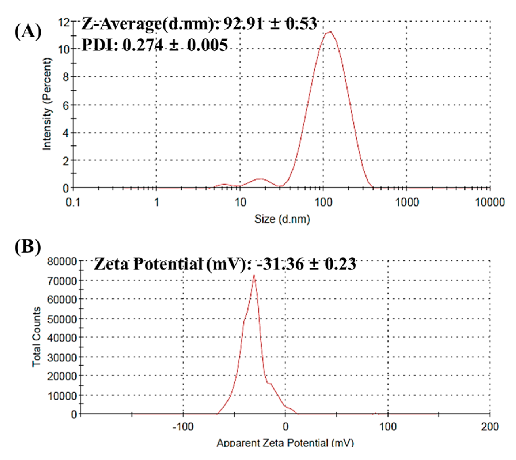
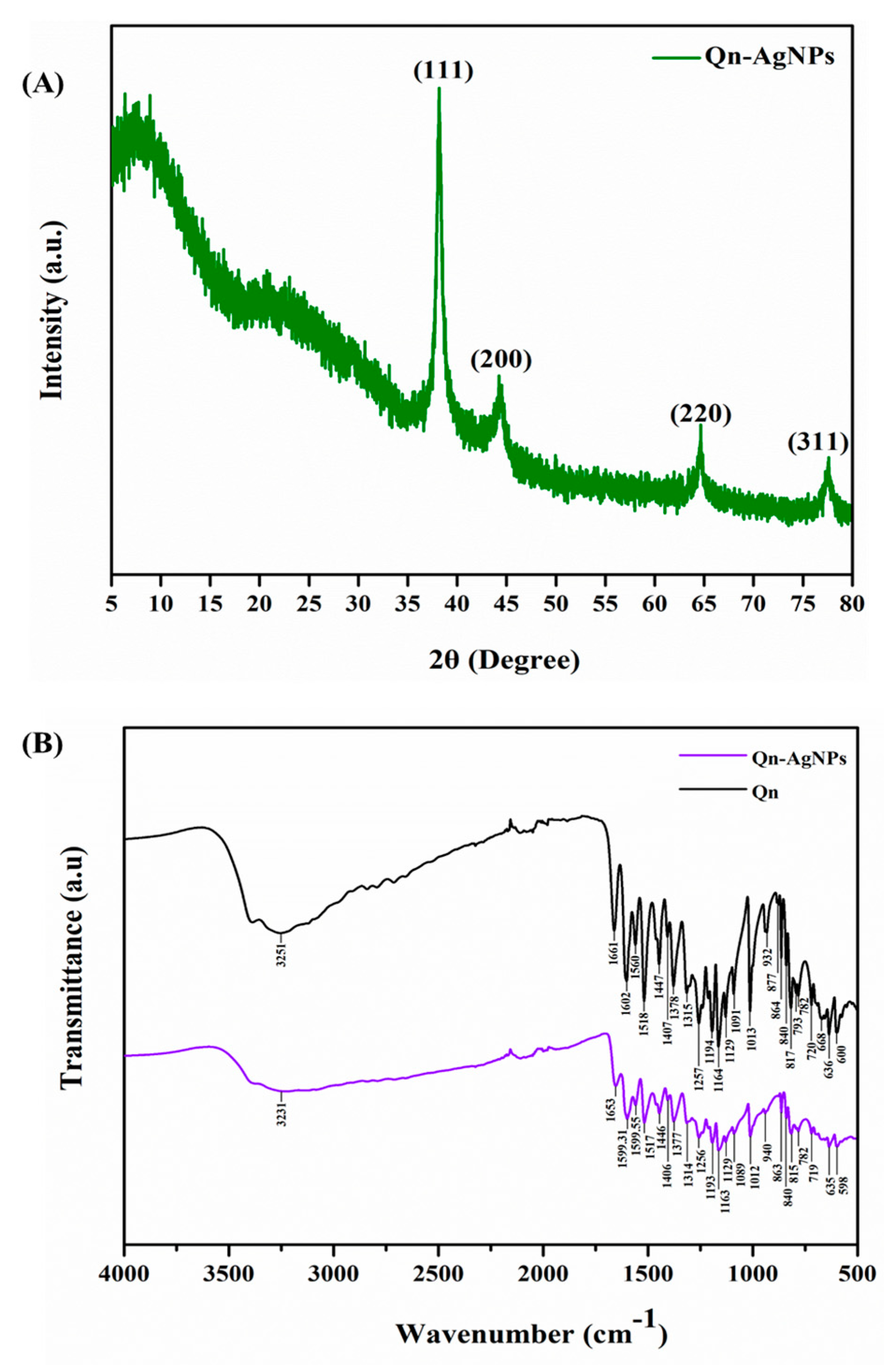
3.1. Synthesis and Characterization AgNPs

10 mM of colorless AgNO3 was mixed with yellow Qn (3 mM) and 100 μL of NaOH (10 mM). The formation of yellow into a dark brown color was evidenced the synthesis of Qn-AgNPs (Figure 1), according to the earlier report [38]. Further, AgNO3, Qn, and Qn-Ag NPs were observed in a UV-vis spectrophotometer to confirm the synthesis of An-AgNPs (Figure 1). The results showed that Qn exhibited the maximum absorbance at 300–450 nm due to the B and C rings in the cinnamoyl system of quercetin [46]. In addition, the optical spectrum of Qn-AgNPs showed the maximum absorption at 420 nm because of the Plasmon resonance effect of Ag [47,48]. TEM and EDS analyses were performed to determine the size, shape, and elemental existence of Qn-AgNPs (Figure 2). The results indicated that Qn-AgNPs were monodispersed, circular shaped, and sized <100 nm (Figure 2A). The no agglomeration of Qn-AgNPs indicates its high stability. In addition, the average particle size of 7.92 ± 2.79 d.nm for Qn-AgNPs was measured by analyzing the TEM images using ImageJ software (Figure 2B). This result was in agreement with previous studies [49,50,51]. Though AgNPs were synthesized using Qn, nanoparticles were not found to be a smaller size and uniform shape due to the variations in the synthesis, including condition, concentration, and the ratio of substrate and precursor [36,52]. The elemental analysis of Qn-Ag NPs confirmed the existence of Ag in a higher weight percentage (Figure 2C). In addition, the existence of Ag and oxygen (O) in elemental spectrum of Qn-AgNPs indicated the biomolecule capping. In addition, the elemental mapping confirmed the presence of Ag (Figure S1). The results showed that the PDI of Qn-AgNPs was 0.27 ± 0.005, and a hydrodynamic size was 92.91 ± 0.53 d.nm (Figure 3A). In addition, the zeta potential of Qn-AgNPs was exhibited −31.36 ± 0.23 (mV), which indicates the good stability of the particles (Figure 3B). The earlier reports evidenced that plant molecules mediated AgNPs have good physical properties, including hydrodynamic size and zeta potential [46,53]. Hence, the present results concluded that Qn was a good reducing as well as a stabilizing agent for the synthesis of AgNPs.
Furthermore, the crystalline properties of Qn-AgNPs were determined through the XRD analysis (Figure 4A). XRD results showed that the diffraction peaks of Qn-AgNPs appeared at 38.17°, 44.23°, 64.64°, and 77.6°, which is corresponding to the crystal plane of (111), (200), (220), and (311). The major diffraction peaks at 38.17° (111) indicated the formation of metallic Ag particles [54]. The crystal patterns of Qn-AgNPs were closely matched with the standard silver (Ag) (JCPDS No.04-0783). Moreover, previous studies evidenced that Qn successfully reduced the Ag by the reduction of AgNO3 [36,52,54]. Furthermore, the functional properties of Qn and Qn-mediated AgNPs were determined in FTIR analysis (Figure 4B). The spectrum showed that Qn and Qn-AgNPs similarly exhibited broad peaks at 3251cm−1 and 3231cm−1 attributed to the O–H stretching of the hydroxy group of Qn [55]. Moreover, Qn showed several characteristic bands representing the C=O stretching (1661 cm−1), C–H bending (1447 cm−1, 1378 cm−1, and 864 cm−1), and C–O stretching (1257 cm−1). The earlier study evidenced that the same characteristic peaks of Qn showed in the FTIR spectrum [56]. These things considered, the transmittance of Qn-AgNPs was not as strong as Qn in general, and characteristic peaks were observed in C=C stretching (1653 cm−1, 1599 cm−1, 815 cm−1), N–O stretching (1517 cm−1), and C–O stretching (1163 cm−1) related to the Qn which might exist on the surface of AgNPs due to the strong coordination with Ag [37]. Overall, Qn-AgNPs synthesized with Qn were found to have similar peaks with a minor shift which indicated that AgNPs were successfully reduced and capped by Qn.

3.2. Biological Assays

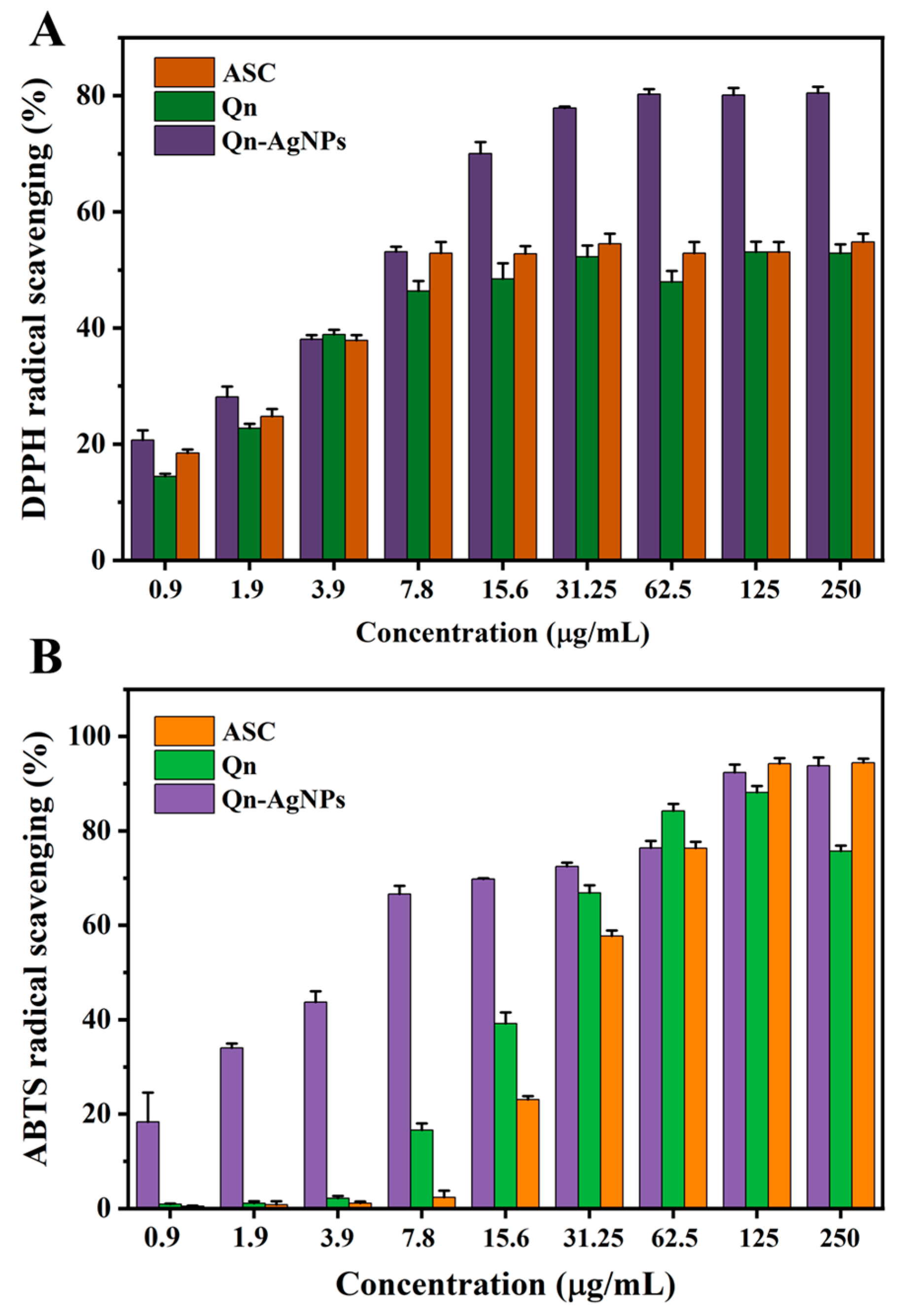
3.2.1. Antioxidant Assay

Antioxidants have been recognized as bioactive molecules that are employed as preventive agents for various diseases, but most antioxidant molecules (curcumin, quercetin, etc.) have limitations, such as less permeability or poor absorption and solubility [57,58]. Hence, several nano-drug delivery systems are developed to deliver the antioxidant molecules efficiently to the targeted site [59]. For that reason, in this study, AgNPs were synthesized using Qn and evaluated their antioxidant ability through DPPH and ABTS free radical scavenging assay (Figure 5A,B). Ascorbic acid (ASC), Qn, and Qn-AgNPs showed higher radical scavenging activity in both DPPH and ABTS free radicals, which was concentration-dependent. The DPPH assay IC50 value of 6.54 ± 0.8 μg/mL and 20.5 ± 1.2 μg/mL for ASC and Qn, respectively (Figure 5A). Similarly, the ABTS assay exhibited an IC50 value of 26 ± 1.36 μg/mL and 21.3 ± 1.2 μg/mL for ASC and Qn, respectively (Figure 5B). Nevertheless, the higher concentrations of ASC and Qn did not scavenge the DPPH radical depend on the concentration. In addition, ASC reported that was not stable in an aqueous solution [60]. However, IC50 of Qn-AgNPs was found to be 6.42 ± 0.7 μg/mL and 4.66 ± 0.4 μg/mL for DPPH and ABTS, respectively (Figure 5A,B). Qn-AgNPs showed the lowest IC50 indicating the minimum concentration enough for a higher amount of free radical scavenging because of the catalytic properties of Ag, nanoparticles, and Qn capping on the surface. The earlier studies also supported that green synthesized AgNPs have a potent antioxidant capacity depending on the phenol and flavonoid concentration [61,62].

3.2.2. Antibacterial Assay

The antibacterial properties of Qn-AgNPs were confirmed through the well diffusion method. A result of the study indicated that Qn-AgNPs have shown considerable antibacterial activity against Gram-positive bacteria (B. cereus, S. aureus, and L. monocytogenes) and Gram-negative bacteria (E. coli and S. enterica) (Table 1). The antibacterial activity of Qn-Ag NPs highly varied depending on the concentration (7.8–62.5 µg/mL). The maximum concentration of Qn (62.5 µg/mL) did not show any inhibitory activity against tested bacterial pathogens. However, the maximum concentration of Qn-AgNPs (62.5 µg/mL) showed the zone of inhibition at 12 ± 1.2 mm, 13 ± 1 mm, 13 ± 0.8 mm, 12 ± 1.4 mm, and 13 ± 1.6 mm for B. cereus, S. aureus, E. coli, S. entrica, and L. monocytogenes, respectively (Table 1). Although, did not find any difference in antibacterial activity specific to strains. Similar results were reported earlier on AgNPs that were synthesized using green synthesis method [63]. The hypothetic antimicrobial mechanisms of AgNPs were reported to damage the bacterial cell wall, protein, and nucleic acid synthesis [64]. These results confirmed that Qn-AgNPs could be an effective antibacterial agent for both Gram-positive and Gram-negative organisms. The MIC was defined as the lowest concentration that completely inhibits the visible growth of bacteria after 12 h incubation [65]. The MIC50 of Qn-AgNPs (<50 nm) was shown at 7.8 µg/mL, 3.9 µg/mL, 3.9 µg/mL, 3.9 µg/mL, and 3.9 µg/mL for E. coli, S. aureus, B. cereus, S. enterica, and L. monocytogenes, respectively (Table 1). Accordingly, Pu-erh tea leaves mediated AgNPs (4.06 nm) exhibited a similar concentration of MIC for these bacterial pathogens [66]. In addition, chemically synthesized AgNPs (5–20 nm) showed the MIC at 2.285 ± 1.492 µg/mL for multidrug-resistant Pseudomonas aeruginosa [67]. But contrastingly, chemically synthesized AgNPs (5 nm) controlled the bacterial growth at 0.625 mg/mL [68]. These results indicated that Qn-AgNPs substantially inhibited bacterial growth with effective concentration.

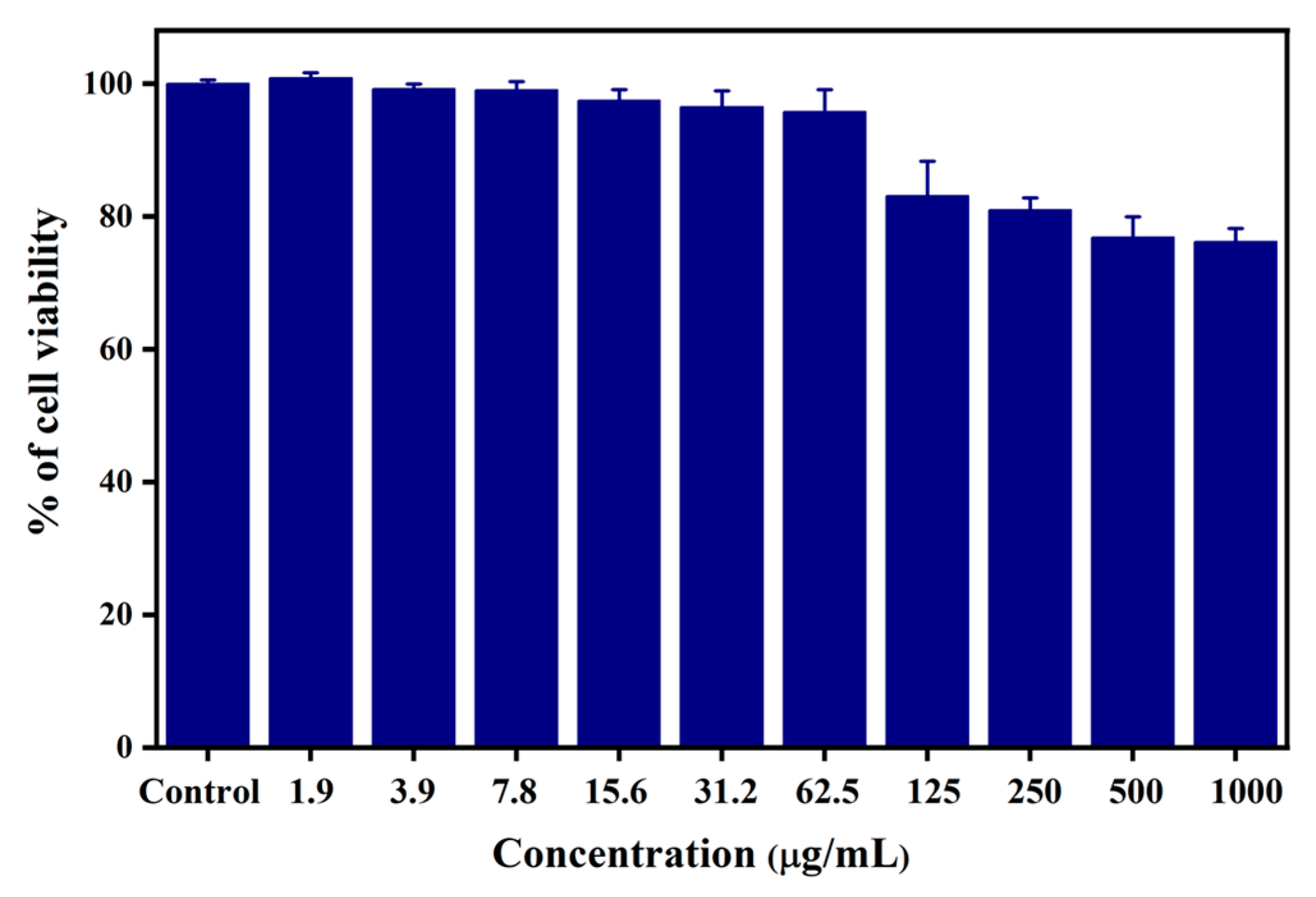
3.2.3. In Vitro Cytotoxicity

The cytotoxicity of Qn-AgNPs was determined on HEK-293 cells for 24 h and shown in Figure 6. The results showed that cytotoxicity increased depending on the concentration of Qn-AgNPs, but was not significant. HEK-293 cells showed a high cell survival rate of 95% until the concentration of 62.5 µg/mL, with a significant decrease in the cell survival rate of 76.84% at 500 µg/mL. However, no significant toxicity was not observed even at 1000 µg/mL of Qn-AgNPs, the cell survival rate was 76%. Similarly, toxicity was observed in HEK-293 cells in terms of DNA damage and mRNA expression that depend on the concentration of AgNPs [69]. Moreover, another study indicated that toxicity depends on the size, the lower size of AgNPs (10 nm) caused high toxicity due to the “Trojan-horse” mechanism [70]. The plant-mediated AgNPs significantly caused toxicity in HEK-293 cells even at lower concentrations [71]. These results indicated that nanoparticle size and reducing agent play a key role in its properties.

3.2.4. Hemolysis Assay

Nanoparticles can cause toxicity in cells and blood compared to general molecules due to their unique physical characteristics. Therefore, the hemolytic analysis was performed with various concentrations of Qn-AgNPs to confirm blood toxicity (Figure 7A). As a result of the analysis was confirmed that the degree of hemolytic activity was low (<6%) at the concentration of 31.2 μg/mL of Qn-AgNPs. The higher concentration of Qn-AgNPs (>31.2) increases, and hemolytic activity gradually increases because a larger number of particles attached to red blood cells may lead to the degradation of blood cells. Similarly, the study found that AgNPs (20 nm) showed ~19 % of hemolysis at the concentration of 40 μg/mL, hemolysis highly depends on the nanoparticles concentration [45]. In addition, another study revealed that small size AgNPs (20 nm) caused more hemolysis compared to higher sizes (≥50 nm) [72]. Therefore, low hemolytic activity is an important point in efficient nanoparticle and nanomaterial-based drug delivery system development.

3.2.5. CAM Assay

CAM assay is used as an intermediate model between cell-based and animal-based experiments. Hence, the different concentration of Qn-AgNPs was tested to identify the CAM toxicity (Figure 7B). Qn-AgNPs (7.8–62.5 μg/mL) were found to have lower toxicity than NaOH used as a positive control. The toxicity of nanoparticles on CAM was determined in terms of blood vessel damage and blood clotting [73]. Moreover, the study reported that ethanolic leaves extract of Zinnia elegans synthesized AgNPs inhibited the length, size, and blood vessel junction at the concentration of 2.29 μg/mL [74]. However, the collagen-based hybrid AgNPs did not show toxic effects on the CAM [75]. This showed that Qn-AgNPs synthesized using Qn, which protect to cause serious toxicity such as vascular tissue damage in the in-ovo model.

4. Conclusions

AgNO3 was successfully reduced into AgNPs using quercetin (Qn) as a reducing and capping agent. The functional characteristics and stability of the Qn-AgNPs were confirmed through UV-vis, FTIR, XRD, and Zeta size analysis. The small size (<50 nm) and homogenous Qn-AgNPs were determined by TEM analysis. The Qn-AgNPs considerably inhibited the pathogenic bacterial growth with minimum concentration (≤7.8 µg/mL). Qn-AgNPs exhibited promising DPPH and ABTS free radicals scavenging properties. Furthermore, a lower concentration of Qn-AgNPs (≤31.2 μg/mL) did not show significant cytotoxicity, hemolysis, and CAM damage. Therefore, the study concluded that Qn-AgNPs is a potent antioxidant and antimicrobial agent for biomedical applications.

Supplementary Materials

The following supporting information can be downloaded at: www.mdpi.com/article/10.3390/coatings12091292/s1, Figure S1: TEM and EDX mapping of Qn-AgNPs—A-B.

Author Contributions

Conceptualization, A.S.; data curation, K.H, A.S. and K.S.; formal analysis, K.H, A.S. and K.S.; investigation, K.H. and A.S.; methodology, A.S. and K.H.; visualization, K.H.; writing—original draft, K.H. and A.S.; writing—review and editing, A.S. and K.S.; software, K.H.; funding acquisition, M.-H.W.; project administration, M.-H.W.; resources, M.-H.W.; supervision, M.-H.W. All authors have read and agreed to the published version of the manuscript.

Funding

The National Research Foundation of Korea (2021R1I1A1A01057742) supported this work.

Institutional Review Board Statement

Not applicable.

Informed Consent Statement

Not applicable.

Data Availability Statement

Data sharing is not applicable to this article.

Conflicts of Interest

The authors declare no conflict of interest.

References

  1. Payal; Pandey, P. Role of Nanotechnology in Electronics: A Review of Recent Developments and Patents. Recent Pat. Nanotechnol. 2022, 16, 45–66. [Google Scholar] [CrossRef]
  2. Ikumapayi, O.M.; Akinlabi, E.T.; Adeoye, A.O.M.; Fatoba, S.O. Microfabrication and Nanotechnology in Manufacturing System–An Overview. Mater. Today Proc. 2021, 44, 1154–1162. [Google Scholar] [CrossRef]
  3. Ashfaq, A.; Khursheed, N.; Fatima, S.; Anjum, Z.; Younis, K. Application of Nanotechnology in Food Packaging: Pros and Cons. J. Agric. Food Res. 2022, 7, 100270. [Google Scholar] [CrossRef]
  4. Alghamdi, M.A.; Fallica, A.N.; Virzì, N.; Kesharwani, P.; Pittalà, V.; Greish, K. The Promise of Nanotechnology in Personalized Medicine. J. Pers. Med. 2022, 12, 673. [Google Scholar] [CrossRef]
  5. Gold, K.; Slay, B.; Knackstedt, M.; Gaharwar, A.K. Antimicrobial Activity of Metal and Metal-Oxide Based Nanoparticles. Adv. Ther. 2018, 1, 1700033. [Google Scholar] [CrossRef]
  6. Jamkhande, P.G.; Ghule, N.W.; Bamer, A.H.; Kalaskar, M.G. Metal Nanoparticles Synthesis: An Overview on Methods of Preparation, Advantages and Disadvantages, and Applications. J. Drug Deliv. Sci. Technol. 2019, 53, 101174. [Google Scholar] [CrossRef]
  7. Mohamed, N.A.; Marei, I.; Crovella, S.; Abou-Saleh, H. Recent Developments in Nanomaterials-Based Drug Delivery and Upgrading Treatment of Cardiovascular Diseases. Int. J. Mol. Sci. 2022, 23, 1404. [Google Scholar] [CrossRef]
  8. Yao, Y.; Zhou, Y.; Liu, L.; Xu, Y.; Chen, Q.; Wang, Y.; Wu, S.; Deng, Y.; Zhang, J.; Shao, A. Nanoparticle-Based Drug Delivery in Cancer Therapy and Its Role in Overcoming Drug Resistance. Front. Mol. Biosci. 2020, 7, 193. [Google Scholar] [CrossRef]
  9. Manoharan, M. Research on the Frontiers of Materials Science: The Impact of Nanotechnology on New Material Development. Technol. Soc. 2008, 30, 401–404. [Google Scholar] [CrossRef]
  10. Barkalina, N.; Charalambous, C.; Jones, C.; Coward, K. Nanotechnology in Reproductive Medicine: Emerging Applications of Nanomaterials. Nanomed. Nanotechnol. Biol. Med. 2014, 10, e921–e938. [Google Scholar] [CrossRef]
  11. Jha, D.; Thiruveedula, P.K.; Pathak, R.; Kumar, B.; Gautam, H.K.; Agnihotri, S.; Sharma, A.K.; Kumar, P. Multifunctional Biosynthesized Silver Nanoparticles Exhibiting Excellent Antimicrobial Potential against Multi-Drug Resistant Microbes along with Remarkable Anticancerous Properties. Mater. Sci. Eng. C 2017, 80, 659–669. [Google Scholar] [CrossRef]
  12. Prosposito, P.; Burratti, L.; Venditti, I. Silver Nanoparticles as Colorimetric Sensors for Water Pollutants. Chemosensors 2020, 8, 26. [Google Scholar] [CrossRef]
  13. Zayed, D.G.; Ebrahim, S.M.; Helmy, M.W.; Khattab, S.N.; Bahey-El-Din, M.; Fang, J.Y.; Elkhodairy, K.A.; Elzoghby, A.O. Combining Hydrophilic Chemotherapy and Hydrophobic Phytotherapy via Tumor-Targeted Albumin-QDs Nano-Hybrids: Covalent Coupling and Phospholipid Complexation Approaches. J. Nanobiotechnol. 2019, 17, 1–19. [Google Scholar] [CrossRef]
  14. Egger, S.; Lehmann, R.P.; Height, M.J.; Loessner, M.J.; Schuppler, M. Antimicrobial Properties of a Novel Silver-Silica Nanocomposite Material. Appl. Environ. Microbiol. 2009, 75, 2973–2976. [Google Scholar] [CrossRef]
  15. Yaqoob, S.B.; Adnan, R.; Rameez Khan, R.M.; Rashid, M. Gold, Silver, and Palladium Nanoparticles: A Chemical Tool for Biomedical Applications. Front. Chem. 2020, 8, 376. [Google Scholar] [CrossRef]
  16. Sakamoto, J.H.; van de Ven, A.L.; Godin, B.; Blanco, E.; Serda, R.E.; Grattoni, A.; Ziemys, A.; Bouamrani, A.; Hu, T.; Ranganathan, S.I.; et al. Enabling Individualized Therapy through Nanotechnology. Pharmacol. Res. 2010, 62, 57–89. [Google Scholar] [CrossRef]
  17. Nam, N.H.; Luong, N.H. Nanoparticles: Synthesis and applications. In Materials for Biomedical Engineering; Elsevier: Amsterdam, The Netherlands, 2019; pp. 211–240. [Google Scholar] [CrossRef]
  18. Anbazhagan, S.; Azeez, S.; Morukattu, G.; Rajan, R.; Venkatesan, K.; Thangavelu, K.P. Synthesis, Characterization and Biological Applications of Mycosynthesized Silver Nanoparticles. Biotech 2017, 7, 1–9. [Google Scholar] [CrossRef]
  19. Duan, H.; Wang, D.; Li, Y. Green Chemistry for Nanoparticle Synthesis. Chem. Soc. Rev. 2015, 44, 5778–5792. [Google Scholar] [CrossRef]
  20. Rónavári, A.; Igaz, N.; Adamecz, D.I.; Szerencsés, B.; Molnar, C.; Kónya, Z.; Pfeiffer, I.; Kiricsi, M. Green Silver and Gold Nanoparticles: Biological Synthesis Approaches and Potentials for Biomedical Applications. Molecules 2021, 26, 844. [Google Scholar] [CrossRef]
  21. Sathiyaseelan, A.; Shajahan, A.; Kalaichelvan, P.; Kaviyarasan, V. Fungal Chitosan Based Nanocomposites Sponges—An Alternative Medicine for Wound Dressing. Int. J. Biol. Macromol. 2017, 104, 1905–1915. [Google Scholar] [CrossRef]
  22. Sathiyaseelan, A.; Saravanakumar, K.; Manikandan, M.; Shajahan, A.; Mariadoss, A.V.A.; Wang, M.-H. Core-Shell Silver Nanoparticles: Synthesis, Characterization, and Applications. In Green Synthesis of Silver Nanomaterials; Elsevier: Amsterdam, The Netherlands, 2022; pp. 75–97. [Google Scholar] [CrossRef]
  23. Diniz, F.R.; Maia, R.C.A.P.; Rannier, L.; Andrade, L.N.; Chaud, M.V.; da Silva, C.F.; Corrêa, C.B.; de Albuquerque Junior, R.L.C.; da Costa, L.P.; Shin, S.R.; et al. Silver Nanoparticles-Composing Alginate/Gelatine Hydrogel Improves Wound Healing in Vivo. Nanomaterials 2020, 10, 390. [Google Scholar] [CrossRef]
  24. Lee, S.H.; Jun, B.H. Silver Nanoparticles: Synthesis and Application for Nanomedicine. Int. J. Mol. Sci. 2019, 20, 865. [Google Scholar] [CrossRef]
  25. Mittal, A.K.; Kaler, A.; Banerjee, U.C. Free Radical Scavenging and Antioxidant Activity of Silver Nanoparticles Synthesized from Flower Extract of Rhododendron Dauricum. Nano Biomed. Eng. 2012, 4, 118–124. [Google Scholar] [CrossRef]
  26. Das, P.; Dutta, T.; Manna, S.; Loganathan, S.; Basak, P. Facile Green Synthesis of Non-Genotoxic, Non-Hemolytic Organometallic Silver Nanoparticles Using Extract of Crushed, Wasted, and Spent Humulus Lupulus (Hops): Characterization, Anti-Bacterial, and Anti-Cancer Studies. Environ. Res. 2022, 204, 111962. [Google Scholar] [CrossRef]
  27. Bar, H.; Bhui, D.K.; Sahoo, G.P.; Sarkar, P.; Pyne, S.; Misra, A. Green Synthesis of Silver Nanoparticles Using Seed Extract of Jatropha Curcas. Colloids Surfaces A Physicochem. Eng. Asp. 2009, 348, 212–216. [Google Scholar] [CrossRef]
  28. Amini, S.M. Preparation of Antimicrobial Metallic Nanoparticles with Bioactive Compounds. Mater. Sci. Eng. C 2019, 103, 109809. [Google Scholar] [CrossRef]
  29. Makarov, V.V.; Love, A.J.; Sinitsyna, O.V.; Makarova, S.S.; Yaminsky, I.V.; Taliansky, M.E.; Kalinina, N.O. “Green” Nanotechnologies: Synthesis of Metal Nanoparticles Using Plants. Acta Naturae 2014, 6, 35. [Google Scholar] [CrossRef]
  30. Kuppusamy, P.; Yusoff, M.M.; Maniam, G.P.; Govindan, N. Biosynthesis of Metallic Nanoparticles Using Plant Derivatives and Their New Avenues in Pharmacological Applications–An Updated Report. Saudi Pharm. J. 2016, 24, 473–484. [Google Scholar] [CrossRef]
  31. Anand David, A.V.; Arulmoli, R.; Parasuraman, S. Overviews of Biological Importance of Quercetin: A Bioactive Flavonoid. Pharmacogn. Rev. 2016, 10, 84. [Google Scholar] [CrossRef]
  32. Salehi, B.; Machin, L.; Monzote, L.; Sharifi-Rad, J.; Ezzat, S.M.; Salem, M.A.; Merghany, R.M.; El Mahdy, N.M.; Klllç, C.S.; Sytar, O.; et al. Therapeutic Potential of Quercetin: New Insights and Perspectives for Human Health. ACS Omega 2020, 5, 11849–11872. [Google Scholar] [CrossRef]
  33. Ansari, M.A.; Abdul, H.M.; Joshi, G.; Opii, W.O.; Butterfield, D.A. Protective Effect of Quercetin in Primary Neurons against Aβ(1-42): Relevance to Alzheimer’s Disease. J. Nutr. Biochem. 2009, 20, 269–275. [Google Scholar] [CrossRef] [PubMed]
  34. Dok-Go, H.; Lee, K.H.; Kim, H.J.; Lee, E.H.; Lee, J.; Song, Y.S.; Lee, Y.H.; Jin, C.; Lee, Y.S.; Cho, J. Neuroprotective Effects of Antioxidative Flavonoids, Quercetin, (+)-Dihydroquercetin and Quercetin 3-Methyl Ether, Isolated from Opuntia Ficus-Indica Var. Saboten. Brain Res. 2003, 965, 130–136. [Google Scholar] [CrossRef]
  35. Deepika; Maurya, P.K. Health Benefits of Quercetin in Age-Related Diseases. Molecules 2022, 27, 2498. [Google Scholar] [CrossRef]
  36. Tasca, F.; Antiochia, R. Biocide Activity of Green Quercetin-Mediated Synthesized Silver Nanoparticles. Nanomaterials 2020, 10, 909. [Google Scholar] [CrossRef] [PubMed]
  37. Pandian, S.R.K.; Kunjiappan, S.; Ravishankar, V.; Sundarapandian, V. Synthesis of Quercetin-Functionalized Silver Nanoparticles by Rapid One-Pot Approach. BioTechnologia 2021, 102, 75–84. [Google Scholar] [CrossRef]
  38. Lee, J.H.; Lim, J.M.; Velmurugan, P.; Park, Y.J.; Park, Y.J.; Bang, K.S.; Oh, B.T. Photobiologic-Mediated Fabrication of Silver Nanoparticles with Antibacterial Activity. J. Photochem. Photobiol. B Biol. 2016, 162, 93–99. [Google Scholar] [CrossRef]
  39. Sathiyaseelan, A.; Saravanakumar, K.; Mariadoss, A.V.A.; Wang, M.-H. Antimicrobial and Wound Healing Properties of FeO Fabricated Chitosan/PVA Nanocomposite Sponge. Antibiotics 2021, 10, 524. [Google Scholar] [CrossRef]
  40. Sathiyaseelan, A.; Saravanakumar, K.; Mariadoss, A.V.A.; Wang, M.H. Biocompatible Fungal Chitosan Encapsulated Phytogenic Silver Nanoparticles Enhanced Antidiabetic, Antioxidant and Antibacterial Activity. Int. J. Biol. Macromol. 2020, 153, 63–71. [Google Scholar] [CrossRef]
  41. Wiegand, I.; Hilpert, K.; Hancock, R.E.W. Agar and Broth Dilution Methods to Determine the Minimal Inhibitory Concentration (MIC) of Antimicrobial Substances. Nat. Protoc. 2008, 3, 163–175. [Google Scholar] [CrossRef]
  42. Sathiyaseelan, A.; Saravanakumar, K.; Mariadoss, A.V.A.; Ramachandran, C.; Hu, X.; Oh, D.H.; Wang, M.H. Chitosan-Tea Tree Oil Nanoemulsion and Calcium Chloride Tailored Edible Coating Increase the Shelf Life of Fresh Cut Red Bell Pepper. Prog. Org. Coat. 2020, 151, 106010. [Google Scholar] [CrossRef]
  43. Sathiyaseelan, A.; Park, S.; Saravanakumar, K.; Mariadoss, A.V.A.; Wang, M.-H. Evaluation of Phytochemicals, Antioxidants, and Antidiabetic Efficacy of Various Solvent Fractions of Gynura Procumbens (Lour.) Merr. Process Biochem. 2021, 111, 51–62. [Google Scholar] [CrossRef]
  44. Sathiyaseelan, A.; Saravanakumar, K.; Manivasagan, P.; Jeong, M.S.; Jang, E.S.; Wang, M.H. Folic Acid Conjugated Chitosan Encapsulated Palladium Nanoclusters for NIR Triggered Photothermal Breast Cancer Treatment. Carbohydr. Polym. 2022, 280, 119021. [Google Scholar] [CrossRef] [PubMed]
  45. Huang, H.; Lai, W.; Cui, M.; Liang, L.; Lin, Y.; Fang, Q.; Liu, Y.; Xie, L. An Evaluation of Blood Compatibility of Silver Nanoparticles. Sci. Rep. 2016, 6, 1–15. [Google Scholar] [CrossRef]
  46. Zhou, Y.; Tang, R.C. Facile and Eco-Friendly Fabrication of Colored and Bioactive Silk Materials Using Silver Nanoparticles Synthesized by Two Flavonoids. Polymers 2018, 10, 404. [Google Scholar] [CrossRef]
  47. Alqadi, M.K.; Abo Noqtah, O.A.; Alzoubi, F.Y.; Alzouby, J.; Aljarrah, K. PH Effect on the Aggregation of Silver Nanoparticles Synthesized by Chemical Reduction. Mater. Sci. 2014, 32, 107–111. [Google Scholar] [CrossRef]
  48. Gloria, E.C.; Ederley, V.; Gladis, M.; César, H.; Jaime, O.; Oscar, A.; José, I.U.; Franklin, J. Synthesis of Silver Nanoparticles (AgNPs) with Antibacterial Activity. J. Phys. Conf. Ser. 2017, 850, 012023. [Google Scholar] [CrossRef]
  49. Gupta, A.; Briffa, S.M.; Swingler, S.; Gibson, H.; Kannappan, V.; Adamus, G.; Kowalczuk, M.; Martin, C.; Radecka, I. Synthesis of Silver Nanoparticles Using Curcumin-Cyclodextrins Loaded into Bacterial Cellulose-Based Hydrogels for Wound Dressing Applications. Biomacromolecules 2020, 21, 1802–1811. [Google Scholar] [CrossRef]
  50. Zhang, X.F.; Huang, F.H.; Zhang, G.L.; Bai, D.P.; de Felici, M.; Huang, Y.F.; Gurunathan, S. Novel Biomolecule Lycopene-Reduced Graphene Oxide-Silver Nanoparticle Enhances Apoptotic Potential of Trichostatin A in Human Ovarian Cancer Cells (SKOV3). Int. J. Nanomed. 2017, 12, 7551. [Google Scholar] [CrossRef] [Green Version]
  51. Venil, C.K.; Malathi, M.; Velmurugan, P.; Renuka Devi, P. Green Synthesis of Silver Nanoparticles Using Canthaxanthin from Dietzia Maris AURCCBT01 and Their Cytotoxic Properties against Human Keratinocyte Cell Line. J. Appl. Microbiol. 2021, 130, 1730–1744. [Google Scholar] [CrossRef]
  52. Chahardoli, A.; Hajmomeni, P.; Ghowsi, M.; Qalekhani, F.; Shokoohinia, Y.; Fattahi, A. Optimization of Quercetin-Assisted Silver Nanoparticles Synthesis and Evaluation of Their Hemocompatibility, Antioxidant, Anti-Inflammatory, and Antibacterial Effects. Glob. Chall. 2021, 5, 2100075. [Google Scholar] [CrossRef]
  53. Lee, Y.J.; Park, Y. Green Synthetic Nanoarchitectonics of Gold and Silver Nanoparticles Prepared Using Quercetin and Their Cytotoxicity and Catalytic Applications. J. Nanosci. Nanotechnol. 2019, 20, 2781–2790. [Google Scholar] [CrossRef] [PubMed]
  54. Yuan, Y.G.; Wang, Y.H.; Xing, H.H.; Gurunathan, S. Quercetin-Mediated Synthesis of Graphene Oxide–Silver Nanoparticle Nanocomposites: A Suitable Alternative Nanotherapy for Neuroblastoma. Int. J. Nanomed. 2017, 12, 5819. [Google Scholar] [CrossRef] [PubMed]
  55. Kato, K.; Ninomiya, M.; Tanaka, K.; Koketsu, M. Effects of Functional Groups and Sugar Composition of Quercetin Derivatives on Their Radical Scavenging Properties. J. Nat. Prod. 2016, 79, 1808–1814. [Google Scholar] [CrossRef] [PubMed]
  56. Dian, L.; Yu, E.; Chen, X.; Wen, X.; Zhang, Z.; Qin, L.; Wang, Q.; Li, G.; Wu, C. Enhancing Oral Bioavailability of Quercetin Using Novel Soluplus Polymeric Micelles. Nanoscale Res. Lett. 2014, 9, 1–11. [Google Scholar] [CrossRef]
  57. Hu, B.; Liu, X.; Zhang, C.; Zeng, X. Food Macromolecule Based Nanodelivery Systems for Enhancing the Bioavailability of Polyphenols. J. Food Drug Anal. 2017, 25, 3–15. [Google Scholar] [CrossRef]
  58. Shabbir, U.; Rubab, M.; Daliri, E.B.M.; Chelliah, R.; Javed, A.; Oh, D.H. Curcumin, Quercetin, Catechins and Metabolic Diseases: The Role of Gut Microbiota. Nutrients 2021, 13, 206. [Google Scholar] [CrossRef]
  59. Vaiserman, A.; Koliada, A.; Zayachkivska, A.; Lushchak, O. Nanodelivery of Natural Antioxidants: An Anti-Aging Perspective. Front. Bioeng. Biotechnol. 2020, 7, 447. [Google Scholar] [CrossRef]
  60. Olszowy-Tomczyk, M. How to Express the Antioxidant Properties of Substances Properly? Chem. Pap. 2021, 75, 6157–6167. [Google Scholar] [CrossRef]
  61. Mohanta, Y.K.; Panda, S.K.; Jayabalan, R.; Sharma, N.; Bastia, A.K.; Mohanta, T.K. Antimicrobial, Antioxidant and Cytotoxic Activity of Silver Nanoparticles Synthesized by Leaf Extract of Erythrina Suberosa (Roxb.). Front. Mol. Biosci. 2017, 4, 14. [Google Scholar] [CrossRef]
  62. Salari, S.; Bahabadi, S.E.; Samzadeh-Kermani, A.; Yosefzaei, F. In-Vitro Evaluation of Antioxidant and Antibacterial Potential of GreenSynthesized Silver Nanoparticles Using Prosopis Farcta Fruit Extract. Iran. J. Pharm. Res. 2019, 18, 430. [Google Scholar]
  63. Wang, C.; Kim, Y.J.; Singh, P.; Mathiyalagan, R.; Jin, Y.; Yang, D.C. Green Synthesis of Silver Nanoparticles by Bacillus Methylotrophicus, and Their Antimicrobial Activity. Artif. Cells Nanomed. Biotechnol. 2015, 44, 1127–1132. [Google Scholar] [CrossRef] [PubMed]
  64. Yin, I.X.; Zhang, J.; Zhao, I.S.; Mei, M.L.; Li, Q.; Chu, C.H. The Antibacterial Mechanism of Silver Nanoparticles and Its Application in Dentistry. Int. J. Nanomed. 2020, 15, 2555. [Google Scholar] [CrossRef] [PubMed]
  65. Andrews, J.M. Determination of Minimum Inhibitory Concentrations. J. Antimicrob. Chemother. 2001, 48, 5–16. [Google Scholar] [CrossRef] [PubMed]
  66. Loo, Y.Y.; Rukayadi, Y.; Nor-Khaizura, M.A.R.; Kuan, C.H.; Chieng, B.W.; Nishibuchi, M.; Radu, S. In Vitro Antimicrobial Activity of Green Synthesized Silver Nanoparticles against Selected Gram-Negative Foodborne Pathogens. Front. Microbiol. 2018, 9, 1555. [Google Scholar] [CrossRef]
  67. Liao, S.; Zhang, Y.; Pan, X.; Zhu, F.; Jiang, C.; Liu, Q.; Cheng, Z.; Dai, G.; Wu, G.; Wang, L.; et al. Antibacterial Activity and Mechanism of Silver Nanoparticles against Multidrug-Resistant Pseudomonas aeruginosa. Int. J. Nanomed. 2019, 14, 1469–1487. [Google Scholar] [CrossRef]
  68. Parvekar, P.; Palaskar, J.; Metgud, S.; Maria, R.; Dutta, S. The Minimum Inhibitory Concentration (MIC) and Minimum Bactericidal Concentration (MBC) of Silver Nanoparticles against Staphylococcus Aureus. Biomater. Investig. Dent. 2020, 7, 105–109. [Google Scholar] [CrossRef]
  69. Jiang, X.; Lu, C.; Tang, M.; Yang, Z.; Jia, W.; Ma, Y.; Jia, P.; Pei, D.; Wang, H. Nanotoxicity of Silver Nanoparticles on HEK293T Cells: A Combined Study Using Biomechanical and Biological Techniques. ACS Omega 2018, 3, 6770–6778. [Google Scholar] [CrossRef]
  70. Gliga, A.R.; Skoglund, S.; Odnevall Wallinder, I.; Fadeel, B.; Karlsson, H.L. Size-Dependent Cytotoxicity of Silver Nanoparticles in Human Lung Cells: The Role of Cellular Uptake, Agglomeration and Ag Release. Part. Fibre Toxicol. 2014, 11, 11. [Google Scholar] [CrossRef]
  71. Liu, X.; Shan, K.; Shao, X.; Shi, X.; He, Y.; Liu, Z.; Jacob, J.A.; Deng, L. Nanotoxic Effects of Silver Nanoparticles on Normal HEK-293 Cells in Comparison to Cancerous HeLa Cell Line. Int. J. Nanomed. 2021, 16, 753–761. [Google Scholar] [CrossRef]
  72. Chen, L.Q.; Fang, L.; Ling, J.; Ding, C.Z.; Kang, B.; Huang, C.Z. Nanotoxicity of Silver Nanoparticles to Red Blood Cells: Size Dependent Adsorption, Uptake, and Hemolytic Activity. Chem. Res. Toxicol. 2015, 28, 501–509. [Google Scholar] [CrossRef]
  73. Buhr, C.R.; Wiesmann, N.; Tanner, R.C.; Brieger, J.; Eckrich, J. The Chorioallantoic Membrane Assay in Nanotoxicological Research—An Alternative for In Vivo Experimentation. Nanomaterials 2020, 10, 2328. [Google Scholar] [CrossRef] [PubMed]
  74. Haque, S.; Norbert, C.C.; Acharyya, R.; Mukherjee, S.; Kathirvel, M.; Patra, C.R. Biosynthesized Silver Nanoparticles for Cancer Therapy and in Vivo Bioimaging. Cancers 2021, 13, 6114. [Google Scholar] [CrossRef] [PubMed]
  75. Craciunescu, O.; Seciu, A.M.; Zarnescu, O. In Vitro and in Vivo Evaluation of a Biomimetic Scaffold Embedding Silver Nanoparticles for Improved Treatment of Oral Lesions. Mater. Sci. Eng. C 2021, 123, 112015. [Google Scholar] [CrossRef] [PubMed]
Figure 1. UV-visible spectrum of silver nitrate (AgNO3), quercetin (Qn) and quercetin mediated synthesized silver nanoparticles (Qn-AgNPs). The inner image shows the color of AgNO3, Qn, and Qn-AgNPs in a glass container.
Coatings 12 01292 g001
Figure 2. Transmission electron microscopy image of Qn-AgNPs magnification at 50 nm (A); particle size distribution of Qn-AgNPs (B); and energy dispersive X-ray Spectrometer (EDS) spectrum of Qn-AgNPs (C).
Coatings 12 01292 g002
Figure 3. Zeta size analysis. Hydrodynamic particles size of Qn-AgNPs (A); and zeta potential of Qn-AgNPs (B).
Coatings 12 01292 g003
Figure 4. The XRD spectrum of quercetin mediated synthesized silver nanoparticles (Qn-AgNPs) (A) and FTIR spectrum of Qn and Qn-AgNPs (B).
Coatings 12 01292 g004
Figure 5. DPPH (A) and ABTS (B) radical scavenging activity of ascorbic acid (ASC), quercetin (Qn), and Qn-AgNPs.
Coatings 12 01292 g005
Figure 6. In vitro cytotoxicity of different concentration of Qn-AgNPs on HEK-293 cells.
Coatings 12 01292 g006
Figure 7. Hemolysis effect of Qn-AgNPs (A); and Chick chorioallantoic membrane (CAM) assay of different concentration of Qn-AgNPs (B). (−) before treatment, (+) after treatment.
Coatings 12 01292 g007
Table 1. Antibacterial activity. Zone of inhibition and MIC50 of Qn-AgNPs, against bacterial pathogens. The zone of inhibition was compared with Qn and TCH. Qn- Quercetin; TCH-tetracycline hydrochloride.
Qn-AgNPsB. cereusS. aereusE. coliS. entericaL. monocytogenes
Zone of Inhibation (mm)
7.89 ± 1.27 ± 0.88 ± 0.68 ± 1.68 ± 1.4
15.610 ± 1.48 ± 0.810 ± 0.89 ± 1.69 ± 1.4
31.211 ± 1.411 ± 1.012 ± 0.811 ± 1.211 ± 1.2
62.512 ± 1.213 ± 1.013 ± 0.812 ± 1.413 ± 1.6
Qn-----
TCH11 ± 0.610 ± 0.410 ± 0.410 ± 0.610 ± 0.8
Minimum inhibitory concentration (µg/mL)
Qn-AgNPs3.93.97.83.93.9
Publisher’s Note: MDPI stays neutral with regard to jurisdictional claims in published maps and institutional affiliations.

