Article

Improving Hydrophilicity and Adhesion of PDMS through Dopamine Modification Assisted by Carbon Dioxide Plasma

1
School of Mechanical Engineering, Jiangsu University Technology, Changzhou 213000, China
2
Jiangsu Provincial Key Laboratory of Bionic Functional Materials, College of Mechanical and Electrical Engineering, Nanjing University of Aeronautics and Astronautics, Nanjing 210016, China
*
Author to whom correspondence should be addressed.
Coatings 2023, 13(1), 126; https://doi.org/10.3390/coatings13010126
Submission received: 12 December 2022 / Revised: 28 December 2022 / Accepted: 3 January 2023 / Published: 10 January 2023

Abstract

:
In this paper, the carbon dioxide (CO2) plasma-assisted method was firstly developed for the preparation of dopamine coating polydimethylsiloxane (PDMS). The PDMS films were pre-treated by CO2 plasma at the power of 30–60 W for 5–10 min and then modified by dopamine for 18 h. The results showed that many polar groups such as C-O bonds, C=O bonds, and O-C=O bonds were introduced into the surface of PDMS films, which successfully promoted the formation of poly(dopamine) coating. Finally, the results of contact angle measurements showed that the surface of the plasma-assisted dopamine grafted samples changed from 118° to 64°. The shearing adhesion strength increased from 2.22 N/cm2 to 6.02 N/cm2, almost three times that of the original sample. This method provides a successful strategy for obtaining good poly(dopamine) coating layers on PDMS with strong hydrophilicity and shearing adhesion, which can be widely applied in the fields of medical and adhesive materials.

Graphical Abstract

1. Introduction

Polydimethylsiloxane (PDMS) is widely used in various fields such as gecko-like sole adhesive materials, electronic skin, and medical materials, due to its characteristics of easy curing, low young’s modulus, good chemical stability, and biocompatibility [1,2,3,4,5].
However, PDMS also has disadvantages of strong hydrophobicity and low surface energy, which is not conducive to adhesion and further functionalization [6,7]. Many researchers have done a lot of research on increasing the hydrophilicity and surface energy of PDMS and enhancing its adhesion and functionalization [7,8,9]. It is reported that dopamine (DA) can be codeposited into a nano-thin layer of poly(dopamine) (PDA) on the surface of the material through covalent bonds, hydrogen bonds, metal chelation, and π-π interaction under alkaline conditions, thus improving the surface energy and adhesion of the material [10,11,12]. Although acrylic acid, etc., can also be used as co-deposited coatings to improve surface energy and adhesion, the process is mostly time-consuming, relies on relatively complex setups, and is often limited to certain substrate chemistries and types of target molecules [13]. The PDA copolymer can be coated onto various materials independent of their surface properties and is stable enough to be stored for months at room temperature [14,15]. Besides, this adhesive PDA copolymer can support a variety of reactions, which also provides an attractive platform for further functionalization [14,15]. However, the experiments proved that the dopamine grafted on the surface of PDMS films by the traditional soaking methods is low content and poor binding force [16]. Plasma technology and laser beam technology are often used to activate material surfaces due to their advantages of high efficiency, simple operation, and no pollution, and it is very easy to harden PDMS with a laser beam [17,18,19]. Therefore, we consider using plasma treatment to activate the surface of PDMS to improve the grafting efficiency of dopamine. Oxygen plasma is often used to activate the surface of various materials, but it has strong oxidation and etching properties that easily damage the structure of PDMS and forms serious cracks [20,21]. In addition, PDMS modified by oxygen plasma changes rapidly in the aging process and recovers hydrophobicity in a short time [22,23,24]. By contrast, carbon dioxide (CO2) plasma, as a milder plasma, has a weak oxidizing property and easily controlled process parameters. In addition, it can also generate active particles such as O atoms and CO under radio-frequency for functionalization, so it is suitable for the surface treatment of flexible PDMS films [25,26].
In this paper, a new method to improve the grafting efficiency of dopamine on PDMS films was explored to enhance its hydrophilicity and adhesion. The grafting efficiency of dopamine onto PDMS was improved by radio-frequency (RF) CO2 plasma treatment technology, and the crack was avoided by adjusting the process parameters. Various tests were performed to characterize changes in the morphology and chemical composition of PDMS at each step. In addition, the surface wettability and adhesion (including shearing adhesion and normal adhesion) of PDMS were also analyzed to evaluate the significance of this study.

2. Materials and Methods

2.1. Preparation of PDMS Films

PMHS 184 (prepolymer) and PMVS 184 (curing agent) purchased from Dow Corning Corp. were mixed in a 10:1 ratio by weight and vigorously stirred for 10 min using a glass rod. The solution was poured onto a flat substrate after degassing with a vacuum pump for 20 min. Next, the PDMS film was demolded after curing at 80 °C for 1 h in a drying oven. Finally, the film (with the thickness of 1.2 ± 0.1 mm) was cut evenly into 1 cm × 1 cm squares.

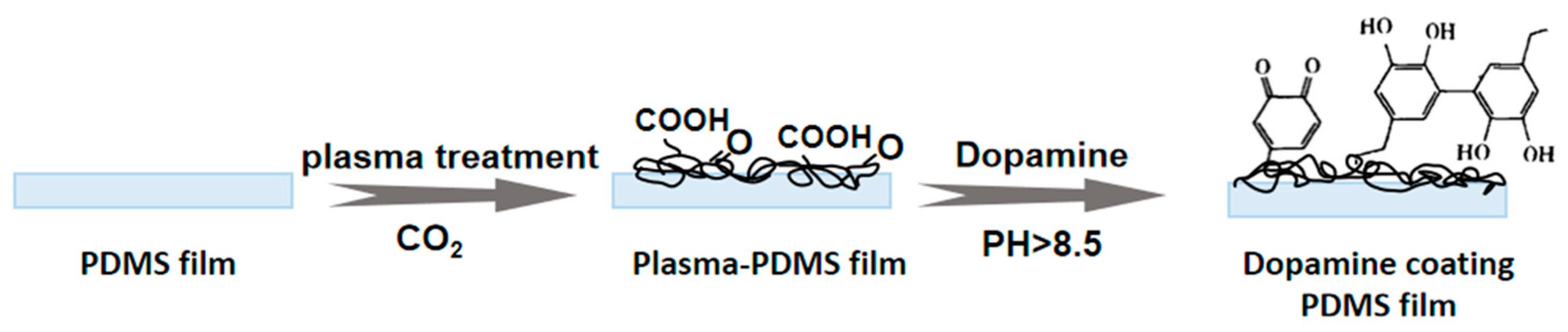
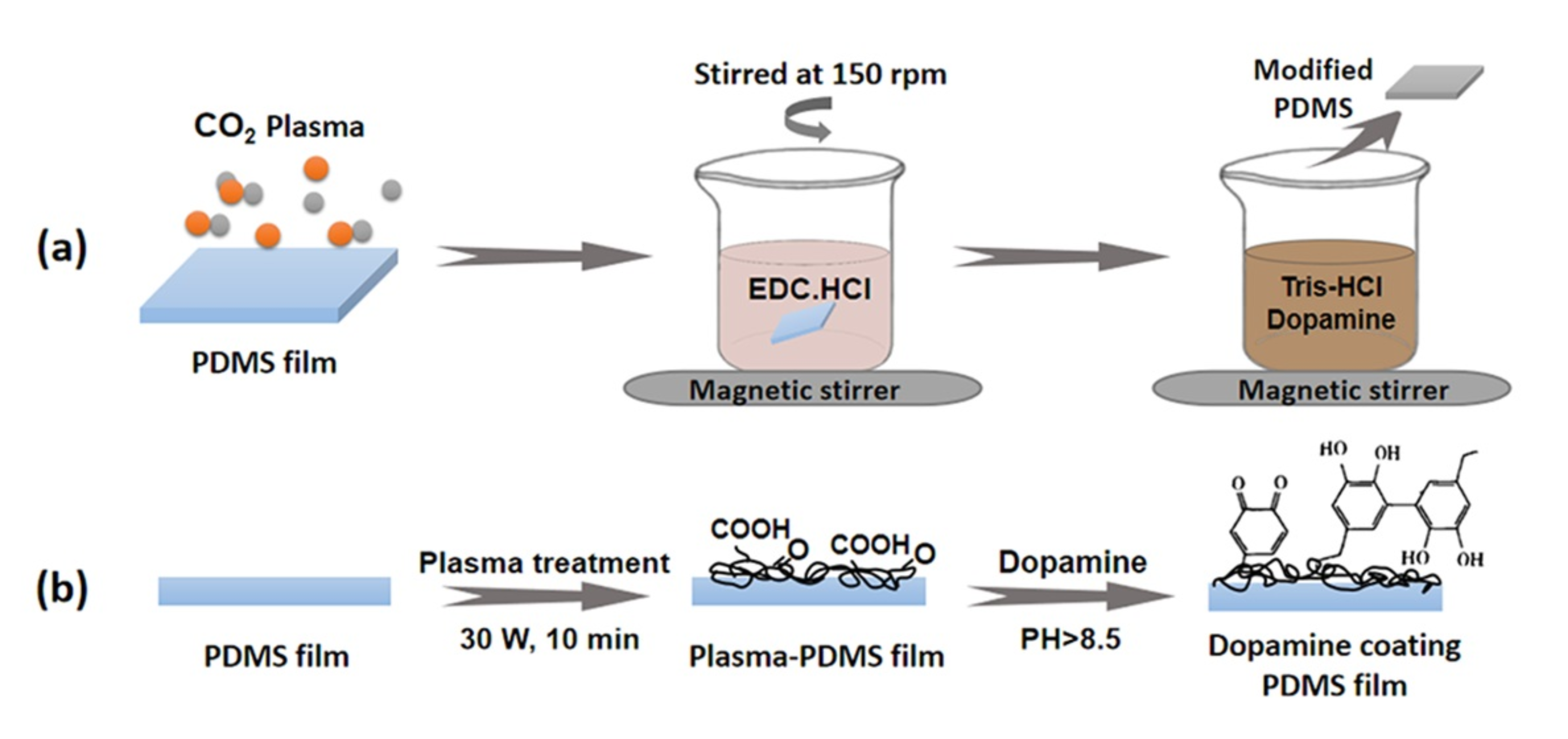
2.2. Carbon Dioxide Plasma Treatment

The prepared PDMS films were pre-treated with CO2 plasma using a radio frequency apparatus (Ion40, PVA TePla, Wettenberg, German), operating at 13.56 MHz. After the samples were put into the chamber and vacuumized, CO2 gas was introduced at a flow of 250 sccm and a pressure of 300 mTorr. Simultaneously, the power and exposure time were adjusted to 30–60 W and 5–10 min, respectively. As shown in Table 1, the samples before and after CO2 plasma treatment were labeled as pristine (original), C1 (30 W, 5 min), C2 (30 W, 10 min), and C3 (60 W, 10 min), respectively. These four groups of samples will be labeled as P, C1-P, C2-P, and C3-P, respectively, after further modification of dopamine.

2.3. Modification of Dopamine

The samples treated by CO2 plasma were immersed in deionized water. Then, 100 mg of 1-ethyl-(3-dimethylaminopropyl) carbodiimide hydrochloride (EDC. HCl) was added and stirred at 150 rpm for 30 min, to activate the functional groups such as carbonyl on the surface of the samples. For the preparation of dopamine buffer, trimethylolaminomethane was first dissolved in distilled water and then adjusted by dilute hydrochloric acid to obtain tris-HCl buffer (PH = 8.5, 0.05 mol/L). After that, dopamine hydrochloride was rapidly added in the tris-HCl buffer to obtain dopamine buffer (2 mg/mL). Subsequently, the dopamine buffer solution was poured into the EDC. HCl reaction solution and stirred at 150 rpm for 18 h, to fully carry out self-polymerization reaction (as shown in Figure 1). Finally, the modified samples were taken out and washed with deionized water at least three times to remove unbound molecules and then dried at room temperature for 24 h. All samples were sealed for storage and marked according to Table 1.

2.4. Characterization and Testing

The surface morphology of PDMS films was observed by scanning electron microscopy (SEM, ZEISS SIGMA, Jena, Germany). The chemical composition of PDMS films before and after CO2 plasma treatment was studied using X-ray photoelectron spectroscopy (XPS, PHI Quantera II, Tokyo, Japan). The formation of the PDA on PDMS films was analyzed by Fourier transform infrared spectroscopy (FTIR, Perkin-Elmer, Waltham, MA, USA). The surface wettability of PDMS films was characterized by an automatic contact angle instrument (JC2000D7M, Shanghai, China). The surface roughness of PDMS films was investigated by atomic force microscopy (AFM) using a SmartSPM-1000 atomic force microscope (AIST-NT, Novato, CA, USA).
The adhesion of PDMS films was measured using a multifunctional material friction and adhesion test platform (IBSS-2, NBIT Co. Ltd., Nanjing, China), as shown in Figure 2. The sample contacted the surface of the substrate (glass) at a speed of 0.1 μm/s until a preload force of 5 N was detected. After contacting for 5 s under this preload, the sample was peeled off in the shear direction or the vertical direction, respectively. Five samples were tested in each group to ensure the accuracy of the results.

3. Results and Discussion

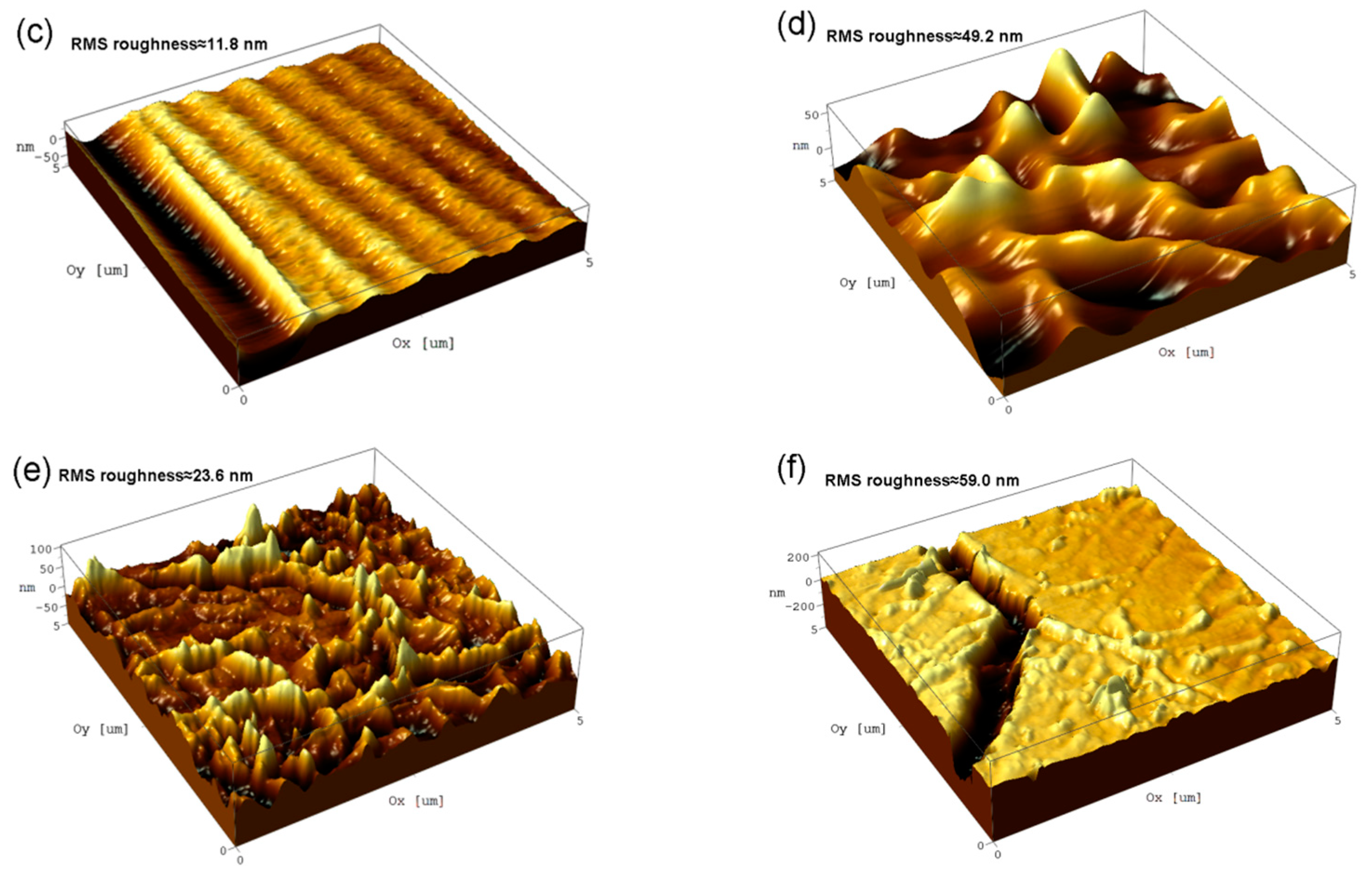
3.1. SEM Observation and EDS Test
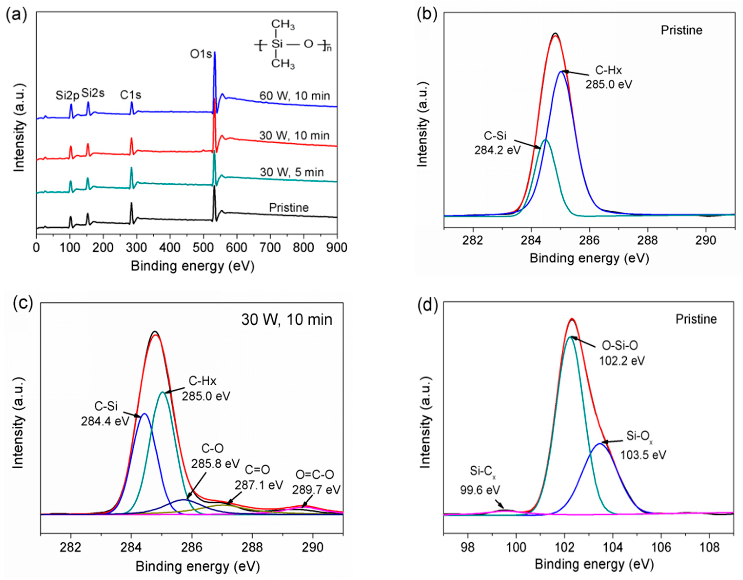
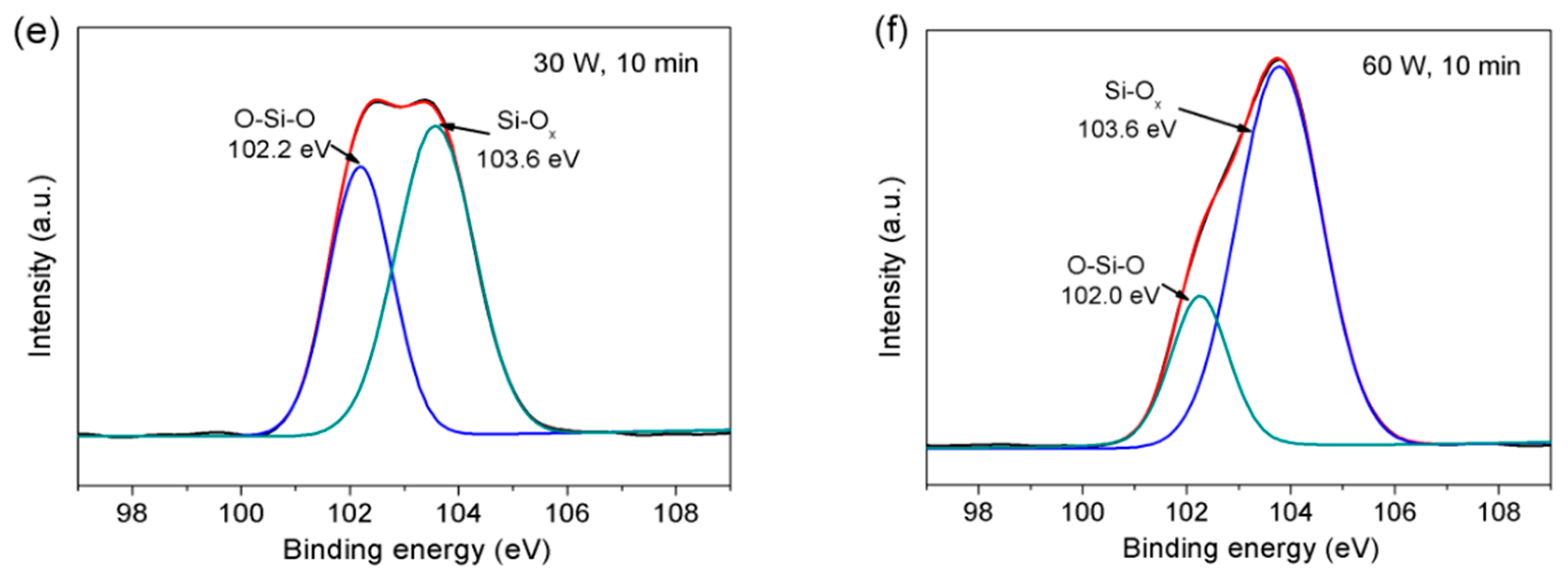
Figure 3 shows the SEM images of the pristine and the dopamine-modified PDMS assisted by CO2 plasma treatment. We can see that the surface of the pristine PDMS is very smooth and clean (Figure 3a). After plasma-assisted dopamine modification, numerous white particles appeared on the surface, which is due to the self-polymerization and crosslinking reaction of dopamine to form poly(dopamine) aggregates [27,28]. These particles gradually became denser with the increase of exposure time and discharge power, so that a poly(dopamine) coating layer was deposited on the surface (as shown in Figure 3b–d). This may be because many polar groups were generated on the surface of PDMS after plasma treatment (which will be explained in Section 3.2), which promoted the grafting and polymerization of dopamine. However, tight network-like cracks appeared on the PDMS surface under high discharge power (60 W), which may be due to the brittle and hard polymers caused by the plasma-assisted process or dopamine modification [28]. These cracks will reduce the effective contact area between the PDMS and the substrate, thus possibly weakening the adhesion force. Figure 3e,f are cross-sectional views of pristine and dopamine-modified PDMS film (C2-P), respectively. The cross-sectional view of the pristine PDMS film shows only one smooth and flat layer (Figure 3e). With the assistance of plasma technology, the surface of PDMS film modified by dopamine is covered with a poly(dopamine) composite layer with a thickness of about 800 nm (Figure 3f). Thus, the coating rate of the poly(dopamine) layer (44.4 nm/h, 0.74 nm/min) is obviously faster than that reported in previous studies, such as 4.5 nm/h recorded in reference [29] and 0.6 nm/min recorded in reference [30]. Moreover, the coating thickness of the finally formed poly(dopamine) layer (800 nm) is also significantly greater than the thickness of 50 nm recorded in reference [31].
In addition, the atomic content of nitrogen (N) was analyzed by EDS spectroscopy to characterize and compare the grafting efficiency of dopamine in different samples (the pristine sample does not contain the N element). As shown in Table 2, the content of N on the surface of samples directly modified with dopamine without plasma assistance is 5.04 at.% (P). However, with the assistance of plasma technology, the content of the N element on the surface of dopamine-modified samples is 8.03 at.% (C1-P group), 10.04 at.% (C2-P), and 10.26 at.% (C3-P), respectively, which indicates that CO2 plasma treatment greatly improves the grafting efficiency of dopamine.

3.2. XPS Analysis

It can be preliminarily revealed from the above that the CO2 plasma treatment process can significantly promote the coating of dopamine on PDMS. The effect of CO2 plasma treatment on the specific chemical composition on surface of PDMS can be further analyzed by XPS. Figure 4a shows the general XPS spectra of different samples, and the contents of different elements in these samples are shown in Table 3. It can be seen that XPS spectra and elemental composition of the samples treated by CO2 plasma for 5 min at 30 W have only slightly changed. The lower the discharge power, the lower the plasma energy, and the shorter the treatment time, so there is little reaction on the surface. In addition, PDMS treated by CO2 plasma will undergo an aging reaction with storage time, so the data measured by XPS are similar. With the increase of treatment time and power, the Si2s and Si2p peaks remain almost unchanged, but the C1s peak decreases and the O1s peak increases, which indicates that a large number of oxygen-containing functional groups are introduced.
Figure 4b,c show the high-resolution C1s XPS spectra of the samples before and after CO2 plasma treatment (30 W, 10 min), respectively. It can be seen that the C1s peak of the pristine sample mainly consists of two components, including 284.2 ± 0.2 eV and 285.1 ± 0.2 eV, corresponding to the C-Si bonds and the C-Hx bonds [32,33], respectively (Figure 4b). After CO2 plasma treatment (Figure 4c), three more components appeared, including 285.8 ± 0.2 eV, 287.1 ± 0.2 eV, and 289.7 ± 0.2 eV, corresponding to C-O bonds, C=O bonds, and O-C=O bonds [33,34], respectively, which confirmed the introduction of a large number of oxygen-containing polar groups on the surface. These polar groups can carry out an amidation reaction with amine groups of dopamine to promote the formation and bonding of poly(dopamine) layer onto the surface of PDMS interlayer, thus improving the surface energy and frictional adhesion [35]. Figure 4d–f show high-resolution Si2p XPS spectra of different samples, and their corresponding components are shown in Table 4. The Si2p XPS spectra of all of the samples contain two components, O-Si-O at 102.0 ± 0.2 eV and Si-Ox (x = 3 or 4) at 103.6 ± 0.2 eV [36,37]. For the pristine sample (Figure 4d), the surface contains Si-Ox (32.45%, x = 3 or 4) and a very small amount of Si-Cx (99.6 ± 0.2 eV). After CO2 plasma treatment (Figure 4e,f), the content of stiff Si-Ox on the surface of sample increased, and it continued to increase with the increase of exposure time and discharge power. At higher power (60 W), the stiff Si-Ox became the main component (76.82%), causing serious cracks to form on the surface (Figure 3d), which will not be conducive to adhesion performance.

3.3. ATR-FTIR Analysis

The chemical components of the poly(dopamine) coating layer were verified by FTIR, as illustrated in Figure 5. For the pristine PDMS, the characteristic peaks were 798 cm−1 for the Si-C stretching vibrations, 864 cm−1 for the Si-O-Si symmetric stretching vibrations, 1010–1080 cm−1 for the Si-O-Si asymmetric stretching vibrations, 1260 cm−1 for the CH3 symmetric deformation of Si-CH3 and 1410 cm−1 for CH3 asymmetric deformation of Si-CH3, and 2960 cm−1 for the CH3 asymmetric stretching of the side chains of PDMS [31,38]. After dopamine modification (C2-P sample), peaks appeared in the range of 1600–1640 cm−1, which could be attributed to the superposition of N-H bending in the amide groups or the C=C stretching vibrations of benzene rings (about 1609 cm−1), and the C=O stretching vibrations of amide groups (about 1643 cm−1). Moreover, new peaks were detected in the range of 3300–3450 cm−1, which were assigned to the N-H characteristic peaks of amine groups (about 3300 cm−1) and stretching vibrations of catechol-OH groups (about 3400 cm−1) [38,39]. The results show that dopamine was obviously grafted onto plasma-treated PDMS through amidation reaction between the introduced carboxyl groups and amine groups of dopamine, forming a poly(dopamine) coating layer [35]. Meanwhile, it was confirmed that different hydrophilic groups, including the hydroxyl, carbonyl, amide, and catechol groups, are introduced into the surface of PDMS.

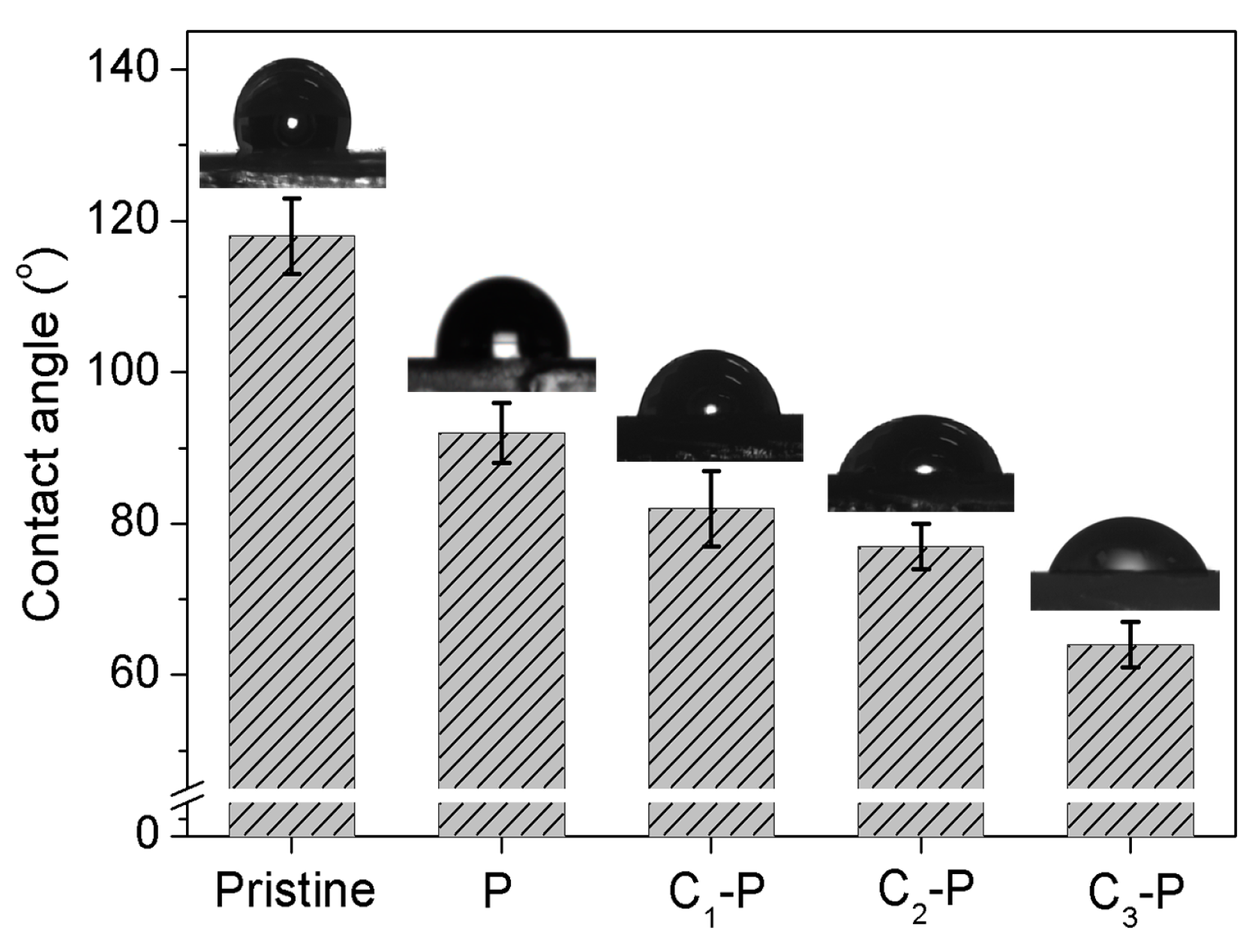
3.4. Contact Angle Tests

The surface wettability of PDMS before and after treatment was characterized by water contact angle tests, and the specific results are shown in Figure 6. The surface of the pristine PDMS exhibits obvious hydrophobicity with a water contact angle of 118°. After directly modifying dopamine without any treatment, the contact angle of the surface was measured to be about 92°. The surface of the sample showed weak hydrophobicity even though its contact angle decreased significantly, revealing that only a small amount of dopamine was grafted. For samples modified with dopamine assisted by CO2 plasma treatment (C1-P, C2-P, and C3-P), the contact angles of their surfaces are 82°, 77°, and 64°, respectively. Their hydrophilicity and surface energy are all significantly improved, which is the result of being completely covered by a poly(dopamine) layer with different hydrophilic groups (proved by Section 3.3). CO2 plasma treatment introduces many active sites for amide bonding interaction with dopamine, while only non-covalent interaction exists between PDMS and poly(dopamine) layer without any treatment [30]. Moreover, the larger the power and the longer the time in the plasma treatment process, the smaller the contact angle and the better the hydrophilicity of the final modified sample surface.

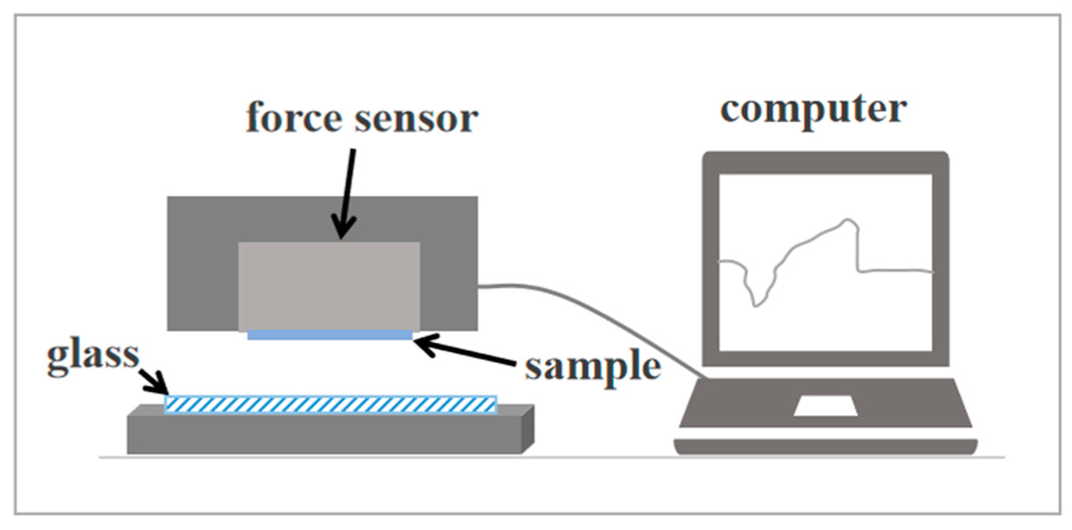
3.5. Adhesion Strength Measurements

The shearing adhesion strength and normal adhesion strength of different samples are calculated and illustrated in Figure 7a,b. The three-dimensional AFM images gave more precise information about the morphology of PDMS on a smaller scale (Figure 7c–f). It can be shown that the shearing adhesion strength of all modified samples is improved, while the normal adhesion strength is decreased. For pristine samples, the shearing adhesion strength is about 2.22 N/cm2, and the normal adhesion strength is about 0.64 N/cm2. After grafting dopamine without plasma treatment, the shearing adhesion strength slightly increased to 2.77 N/cm2, while the normal adhesion strength decreased to 0.25 N/cm2. This is because many particles and aggregates were deposited onto the surface after dopamine modification, and the surface changes from relatively smooth to rough (Figure 7c,d). So, the shearing adhesion is slightly improved due to increased friction caused by high roughness, while normal adhesion decreases due to a reduction of the contact area [40]. For samples modified with dopamine assisted by CO2 plasma treatment (C1-P, C2-P, and C3-P), the shearing adhesion strength is increased to 4.16 N/cm2, 6.02 N/cm2, and 4.48 N/cm2, respectively, while the normal adhesion strength is decreased to 0.17 N/cm2, 0.42 N/cm2, and 0.06 N/cm2, respectively. With the assistance of plasma treatment, the grafting amount of dopamine was significantly increased; thus, the shearing adhesion strength was significantly improved. Besides, the dopamine particles and aggregates were distributed more densely to form a uniform poly(dopamine) coating. Therefore, the surface roughness changed little compared to the untreated sample (Figure 7e,f), and a certain amount of normal adhesion was retained. In particular, after CO2 plasma treatment at 30 W for 10 min (C2-P), the shearing adhesion strength of the poly(dopamine) coating PDMS was nearly three times higher than that of the pristine sample. However, when the plasma power was too high (60 W), many cracks appeared on the surface, which seriously reduced the contact area, thus showing the worst shearing and normal adhesion strength.

4. Conclusions

In this paper, it is proposed for the first time that CO2 plasma-assisted treatment can significantly improve the grafting efficiency of dopamine. The results showed that many polar groups were introduced into the surface of PDMS without cracks, which will promote the formation of poly(dopamine) coating through amidation reaction. The test results of the contact angle and adhesion revealed that the wettability and shearing adhesion of PDMS prepared by this method were significantly improved. In particular, after plasma-assisted treatment for 10 min at 30 W, dopamine-modified PDMS displayed the highest shearing adhesion strength, which was almost three times that of the original sample (from 2.22 N/cm2 to 6.02 N/cm2), and its hydrophilicity was also significantly improved (from 118° to 77°). Furthermore, this simple and effective method can also be extended to the preparation of other types of elastic materials with good hydrophilicity and adhesion, which can be used in bionics, medicine, and other fields.

Author Contributions

Conceptualization, M.L. and L.D.; methodology, M.L.; formal analysis, M.L. and T.Z.; investigation, M.L.; data curation, M.L. and T.Z.; writing—original draft preparation, M.L.; writing—review and editing, M.L., L.D. and T.Z.; supervision, M.L. and L.D.; project administration, M.L. and Z.D.; and funding acquisition, M.L. and Z.D. All authors have read and agreed to the published version of the manuscript.

Funding

Funding for this research was provided through the National Nature Science Foundation of China (NSFC) (No. 52205311, 52005231) and the Technology Project of Changzhou (Nos. CJ20220054, CE20215050).

Institutional Review Board Statement

Not applicable.

Informed Consent Statement

Not applicable.

Data Availability Statement

Not applicable.

Conflicts of Interest

The authors declare no conflict of interest.

References

  1. Xia, S.; Chen, Y.; Tian, J.; Shi, J.; Geng, C.; Zou, H.; Liang, M.; Li, Z. Superior low-temperature reversible adhesion based on bio-inspired microfibrillar adhesives fabricated by phenyl containing polydimethylsiloxane elastomers. Adv. Funct. Mater. 2021, 31, 2101143. [Google Scholar] [CrossRef]
  2. Tsujioka, K.; Matsuo, Y.; Shimomura, M.; Hirai, Y. A new concept for an adhesive material inspired by clingfish sucker nanofilaments. Langmuir 2022, 38, 1215–1222. [Google Scholar] [CrossRef] [PubMed]
  3. Gao, P.; Pu, W.; Wei, P.; Kong, M. Molecular dynamics simulations on adhesion energy of PDMS-silica interface caused by molecular structures and temperature. Appl. Surf. Sci. 2022, 577, 151930. [Google Scholar] [CrossRef]
  4. Ibáñez-Ibáñez, P.F.; Ruiz-Cabello, F.J.M.; Cabrerizo-Vílchez, M.A.; Rodríguez-Valverde, M.A. Ice adhesion of PDMS surfaces with balanced elastic and water-repellent properties. J. Colloid Interf. Sci. 2022, 608, 792–799. [Google Scholar] [CrossRef]
  5. Liu, X.; Chen, K.; Zhang, D.; Guo, Z. One-step methods to fabricate durable superhydrophobic coatings for flexible electronic sensors. Coatings 2021, 11, 95. [Google Scholar] [CrossRef]
  6. Dirany, M.; Dies, L.; Restagno, F.; Léger, L.; Miquelard-Garnier, G. Chemical modification of PDMS surface without impacting the viscoelasticity: Model systems for a better understanding of elastomer/elastomer adhesion and friction. Colloids Surf. A 2015, 468, 174–183. [Google Scholar] [CrossRef]
  7. Jiang, B.; Guo, H.; Chen, D.; Zhou, M. Microscale investigation on the wettability and bonding mechanism of oxygen plasma-treated PDMS microfluidic chip. Appl. Surf. Sci. 2022, 574, 151704. [Google Scholar] [CrossRef]
  8. Yan, Y.; Qi, Y.; Chen, W. Strategies to hydrophilize silicones via spontaneous adsorption of poly(vinyl alcohol) from aqueous solution. Colloids Surf. A 2018, 546, 186–193. [Google Scholar] [CrossRef]
  9. Majhy, B.; Priyadarshini, P.; Sen, A.K. Effect of surface energy and roughness on cell adhesion and growth–facile surface modification for enhanced cell culture. RSC Adv. 2021, 11, 15467–15476. [Google Scholar] [CrossRef]
  10. Chang, K.Y.; Chou, Y.N.; Chen, W.Y.; Chen, C.Y.; Lin, H.R. Mussel-inspired adhesive and self-healing hydrogel as an injectable wound dressing. Polymers 2022, 14, 3346. [Google Scholar] [CrossRef]
  11. Yi, Z.; Zhao, B.; Liao, M.; Qin, Z. Fabrication of superhydrophobic wood surface by etching polydopamine coating with sodium hydroxide. Coatings 2020, 10, 847. [Google Scholar] [CrossRef]
  12. Tran, N.T.; Flanagan, D.P.; Orlicki, J.A.; Lenhart, J.L.; Proctor, K.L.; Knorr, D.B. Polydopamine and polydopamine-silane hybrid surface treatments in structural adhesive applications. Langmuir 2018, 34, 1274–1286. [Google Scholar] [CrossRef] [PubMed]
  13. Li, L.; Tirrell, M.; Korba, G.A.; Poclus, A. Surface energy and adhesion studies on acrylic pressure sensitive adhesives. J. Adhes. 2001, 76, 307–334. [Google Scholar] [CrossRef]
  14. Dabaghi, M.; Shahriari, S.; Saraei, N.; Da, K.; Chandiramohan, A.; Selvaganapathy, P.R.; Hirota, J.A. Surface modification of PDMS-based microfluidic devices with collagen using polydopamine as a spacer to enhance primary human bronchial epithelial cell adhesion. Micromachines 2021, 12, 132. [Google Scholar] [CrossRef]
  15. Perikamana, S.K.M.; Shin, Y.M.; Lee, J.K.; Lee, Y.B.; Heo, Y.; Ahmad, T.; Park, S.Y.; Shin, J.; Park, K.M.; Jung, H.S.; et al. Graded functionalization of biomaterial surfaces using mussel-inspired adhesive coating of polydopamine. Colloids Surf. B 2017, 159, 546–556. [Google Scholar] [CrossRef]
  16. Zhang, F.T.; Xu, L.; Chen, J.H.; Zhao, B.; Fu, X.Z.; Sun, R.; Chen, Q.; Wong, C.P. Electroless deposition metals on poly(dimethylsiloxane) with strong adhesion as flexible and stretchable conductive materials. Acs. Appl. Mater. Inter. 2017, 10, 2075–2082. [Google Scholar] [CrossRef]
  17. Huang, Y.; Yu, Q.; Li, M.; Jin, S.; Fan, J.; Zhao, L.; Yao, Z. Surface modification of activated carbon fiber by low-temperature oxygen plasma: Textural property, surface chemistry, and the effect of water vapor adsorption. Chem. Eng. J. 2021, 418, 129474. [Google Scholar] [CrossRef]
  18. Compoint, F.; Fall, D.; Piombini, H.; Belleville, P.; Montouillout, Y.; Duquennoy, M.; Jenot, F.; Piwakowski, B.; Sanchez, C. Sol-gel-processed hybrid silica-PDMS layers for the optics of high-power laser flux systems. J. Mater. Sci. 2016, 51, 5031–5045. [Google Scholar] [CrossRef]
  19. Aymes-Chodur, C.; Salmi-Mani, H.; Dragoe, D.; Aubry-Barroca, N.; Buchotte, M.; Roger, P. Optimization of microwave plasma treatment conditions on polydimethylsiloxane films for further surface functionalization. Eur. Polym. J. 2021, 150, 110416. [Google Scholar] [CrossRef]
  20. Farrell, M.; Beaudoin, S. Surface forces and protein adsorption on dextran- and polyethylene glycol-modified polydimethylsiloxane. Colloids Surf. B 2010, 81, 468–475. [Google Scholar] [CrossRef]
  21. Xiong, L.; Chen, P.; Zhou, Q. Adhesion promotion between PDMS and glass by oxygen plasma pre-treatment. J. Adhes. Sci. Technol. 2014, 28, 1046–1054. [Google Scholar] [CrossRef]
  22. Meincken, M.; Berhane, T.A.; Mallon, P.E. Tracking the hydrophobicity recovery of PDMS compounds using the adhesive force determined by AFM force distance measurements. Polymer 2005, 46, 203–208. [Google Scholar] [CrossRef]
  23. Bodas, D. Surface modification and aging studies of addition-curing silicone rubbers by oxygen plasma. Eur. Polym. J. 2008, 44, 2130–2139. [Google Scholar] [CrossRef]
  24. Eddington, D.T.; Puccinelli, J.P.; Beebe, D.J. Thermal aging and reduced hydrophobic recovery of polydimethylsiloxane. Sensor. Actuators B Chem. 2006, 114, 170–172. [Google Scholar] [CrossRef]
  25. Brock, S.L.; Marquez, M.; Suib, S.L.; Hayashi, Y.; Matsumoto, H. Plasma decomposition of CO2 in the presence of metal catalysts. J. Catal. 1998, 180, 225–233. [Google Scholar] [CrossRef]
  26. Babu, D.J.; Yadav, S.; Heinlein, T.; Cherkashinin, G.; Schneider, J.J. Carbon dioxide plasma as a versatile medium for purification and functionalization of vertically aligned carbon nanotubes. J. Phys. Chem. C 2014, 118, 12028–12034. [Google Scholar] [CrossRef]
  27. Chen, L.; Ren, X.; Li, Y.; Hu, D.; Feng, X.; Li, W. Enhancing interface compatibility of UiO-66-NH2 and polyamide by incorporating dopamine into thin film nanocomposite membranes. J. Membr. Sci. 2022, 654, 120565. [Google Scholar] [CrossRef]
  28. Zhang, W.; Yang, F.K.; Han, Y.; Gaikwad, R.; Leonenko, Z.; Zhao, B. Surface and tribological behaviors of the bioinspired polydopamine thin films under dry and wet conditions. Biomacromolecules 2013, 14, 394–405. [Google Scholar] [CrossRef]
  29. Wang, J.; Guo, H.; Shi, X.; Yao, Z.; Qing, W.; Liu, F.; Tang, C.Y. Fast polydopamine coating on reverse osmosis membrane: Process investigation and membrane performance study. J. Colloid Interface Sci. 2019, 535, 239–244. [Google Scholar] [CrossRef]
  30. Orishchin, N.; Crane, C.C.; Brownell, M.; Wang, T.; Jenkins, S.; Zou, M.; Nair, A.; Chen, J. Rapid deposition of uniform polydopamine coatings on nanoparticle surfaces with controllable thickness. Langmuir 2017, 33, 6046–6053. [Google Scholar] [CrossRef]
  31. Fang, M.; Zhang, H.; Chen, J.; Wang, T.; Liu, J.; Li, X.; Li, J.; Cao, X. A facile approach to construct hierarchical dense membranes via polydopamine for enhanced propylene/nitrogen separation. J. Membr. Sci. 2016, 499, 290–300. [Google Scholar] [CrossRef]
  32. Aerts, S.; Vanhulsel, A.; Buekenhoudt, A.; Weyten, H.; Kuypers, S.; Chen, H.; Bryjak, M.; Gevers, L.E.M.; Vankelecom, I.F.J.; Jacobs, P.A. Plasma-treated PDMS-membranes in solvent resistant nanofiltration: Characterization and study of transport mechanism. J. Membr. Sci. 2006, 275, 212–219. [Google Scholar] [CrossRef]
  33. Sharma, V.; Dhayal, M.; Shivaprasad, S.M.; Jain, S.C. Surface characterization of plasma-treated and PEG-grafted PDMS for micro fluidic applications. Vacuum 2007, 81, 1094–1100. [Google Scholar] [CrossRef]
  34. Al-Gamal, A.Q.; Falath, W.S.; Saleh, T.A. Enhanced efficiency of polyamide membranes by incorporating TiO2-Graphene oxide for water purification. J. Mol. Liq. 2021, 323, 114922. [Google Scholar] [CrossRef]
  35. Kim, H.; Park, S.S.; Seo, J.; Ha, C.S.; Moon, C.; Kim, Y. Stable protein device platform based on pyridine dicarboxylic acid-bound cubic-nanostructured mesoporous titania films. Acs. Appl. Mater. Interfaces 2013, 5, 6873–6878. [Google Scholar] [CrossRef] [PubMed]
  36. Hoppe, C.; Mitschker, F.; Awakowicz, P.; Kirchheim, D.; Dahlmann, R.; de los Arcos, T.; Grundmeier, G. Adhesion of plasma-deposited silicon oxide barrier layers on PDMS containing polypropylene. Surf. Coat. Technol. 2018, 335, 25–31. [Google Scholar] [CrossRef]
  37. ButrÓN-GarcÍA, M.I.; Jofre-Reche, J.A. Use of statistical design of experiments in the optimization of Ar-O2 low-pressure plasma treatment conditions of polydimethylsiloxane (PDMS) for increasing polarity and adhesion, and inhibiting hydrophobic recovery. Appl. Surf. Sci. 2015, 332, 1–11. [Google Scholar] [CrossRef] [Green Version]
  38. Jun, D.R.; Moon, S.K.; Choi, S.W. Uniform polydimethylsiloxane beads coated with polydopamine and their potential biomedical applications. Colloids Surf. B 2014, 121, 395–399. [Google Scholar] [CrossRef]
  39. Zhu, L.P.; Jiang, J.H.; Zhu, B.K.; Xu, Y.Y. Immobilization of bovine serum albumin onto porous polyethylene membranes using strongly attached polydopamine as a spacer. Colloids Surf. B 2011, 86, 111–118. [Google Scholar] [CrossRef]
  40. Liu, Y.; Fang, Y.; Liu, X.; Wang, X.; Yang, B. Mussel-inspired modification of carbon fiber via polyethyleneimine/ polydopamine co-deposition for the improved interfacial adhesion. Compos. Sci. Technol. 2017, 151, 164–173. [Google Scholar] [CrossRef]
Figure 1. Schematic diagram of experimental reaction process.
Coatings 13 00126 g001
Figure 2. Schematic diagram of adhesion testing platform.
Coatings 13 00126 g002
Figure 3. SEM images of the surface of the pristine PDMS (a) and the dopamine-modified PDMS assisted by CO2 plasma treatment (bd). (b) C1-P, (c) C2-P, and (d) C3-P. (e,f) are cross-sectional views of pristine and dopamine-modified PDMS (C2-P), respectively.
Coatings 13 00126 g003
Figure 4. (a) General XPS spectra of samples before and after CO2 plasma treatment. High-resolution C1s XPS spectra of samples before (b) and after CO2 plasma treatment (c). High-resolution Si 2p XPS spectra of samples before (d) and after plasma treating for 10 min at 30 W (e) and 60 W (f).
Coatings 13 00126 g004aCoatings 13 00126 g004b
Figure 5. FTIR (ATR) spectra of (a) pristine PDMS and (b) PDA-coated PDMS (C2-P).
Coatings 13 00126 g005
Figure 6. Contact angle test results for different sample.
Coatings 13 00126 g006
Figure 7. Shearing (a) and normal (b) adhesion strength of different samples. AFM images of different samples. (c) Pristine, (d) P, (e) C2-P, and (f) C3-P.
Coatings 13 00126 g007aCoatings 13 00126 g007b
Table 1. Representative experimental parameters and corresponding samples.
SamplesPower (W)Exposure Time (min)PDA-Coating Time (h)
Pristine000
C13050
C230100
C360100
P0018
C1-P30518
C2-P301018
C3-P601018
Table 2. Atomic content of surface N element for different samples.
SamplesPristinePC1-PC2-PC3-P
N (at.%)05.048.0310.0410.26
Table 3. Surface elemental composition of pristine PDMS and CO2 plasma treated PDMS.
SamplesC (at.%)O (at.%)Si (at.%)O/CO/Si
pristine45.6428.9325.430.631.13
C144.6629.5825.760.661.15
C233.4240.3926.191.21.54
C325.0946.4328.481.851.63
Table 4. Composition of Si 2p peaks on the surface of each samples.
ComponentsSi-Cx (%)O-Si-O (%)Si-Ox (%)
pristine3.3064.2532.45
C1062.3437.66
C2044.5855.42
C3023.1876.82
Disclaimer/Publisher’s Note: The statements, opinions and data contained in all publications are solely those of the individual author(s) and contributor(s) and not of MDPI and/or the editor(s). MDPI and/or the editor(s) disclaim responsibility for any injury to people or property resulting from any ideas, methods, instructions or products referred to in the content.

