Review

Surface Modification Techniques for Polyetheretherketone as Spinal Interbody Fusion Cage Material to Stimulate Biological Response: A Review

1
Department of Spine Surgery, Changhai Hospital, Navy Medical University, Shanghai 200433, China
2
Department of Orthopedics, Changhai Hospital, Navy Medical University, Shanghai 200433, China
3
School of Medical Instrument and Food Engineering, University of Shanghai for Science and Technology, Shanghai 200093, China
4
School of Exercise and Health, Shanghai University of Sport, Shanghai 200433, China
5
Department of Orthopedic Surgery, Spine Center, Changzheng Hospital, Navy Medical University, Shanghai 200003, China
*
Authors to whom correspondence should be addressed.
These authors contributed equally to this work.
Coatings 2023, 13(6), 977; https://doi.org/10.3390/coatings13060977
Submission received: 24 April 2023 / Revised: 19 May 2023 / Accepted: 21 May 2023 / Published: 24 May 2023
(This article belongs to the Special Issue Application of Coatings on Implants Surfaces)

Abstract

:
Currently, spinal interbody cages are crucial for spinal fusion surgeries. Due to the mechanical and imaging characteristics of polyetheretherketone (PEEK), it is a widely used material for cages. However, the bioinert PEEK has poor osseointegration, thereby preventing the ideal fusion of PEEK cages. Therefore, efforts have been made for improving biological activity using surface modification techniques, including physical as well as chemical modifications and surface coating. In this study, we reviewed and analyzed recent studies on PEEK surface modification techniques to enhance our understanding for future studies.

1. Introduction

Spine fusion, as introduced first by Albee and Hibbs in 1911, is a widely used technique in clinical settings to treat degenerative disc disease, spinal stenosis, spondylolisthesis, scoliosis, trauma, infection, and tumor. The concept of interbody fusion was proposed by Cloward [1] and Smith [2] in the 1950s. Initially, a tri-corticate autograft was implanted as the spacer between two adjacent vertebral bodies to achieve bony fusion and spine realignment. Trilateral corticate autograft iliac crest graft was once considered the “gold standard” for spinal fusion. However, it still has disadvantages, such as postoperative hematoma, pain, blood loss, infection, and intervertebral altitude loss [3].
The advent of the synthetic interbody fusion device, commonly known as a “cage,” was a milestone for spinal surgery [4]. In 1979, Bagby designed a stainless-steel hollow bone graft device named the “Bagby cage” for treating cervical spondylosis in horses. Based on this, in 1988, Bagby [5] and Kuslich [6] designed a titanium (Ti) BAK cage for the surgical treatment of patients with lumbar disc herniation. Next, various shapes of Ti cages with good biocompatibility and resistance to corrosion were designed and implemented. However, Ti cages have relatively higher elastic modulus (110 GPa) and a stress shielding effect; hence, there is an increased risk of these cages subsiding post surgery. Further, the radiopacity of these cages makes it difficult for surgeons to determine postoperative fusion [7,8]. With the advancement in material science, polyetheretherketone (PEEK) cages are extensively used for spinal fusion procedures [9].
PEEK is a semi-crystalline thermoplastic. It is radiolucent and has outstanding mechanical properties. The elastic modulus of PEEK is between cortical and cancellous bones (Table 1), thereby reducing the risk of subsiding and the stress shielding effect [10,11]. However, PEEK is biologically inert and hydrophobic; hence, it has unfavorable osteogenic efficiency and can form fibrous encapsulation, thereby causing fusions to fail and eventually leading to pseudoarthrosis [12,13,14].
Therefore, creating biologically active PEEK has gained widespread attention in material science. Studies have focused on improving the biocompatibility, osteointegration, and antibacterial ability of PEEK [15]. In this review, we have analyzed techniques for biologically modifying PEEK, including physical modification as well as chemical modification, and surface coating.

2. Surface Modification

2.1. Physical Modification

Modification techniques, such as physical treatment, allows modifying the structure of the surface at micro/nano levels or depositing active substances on the surface without altering the chemical properties of some materials. This technique is easy to use and cost effective.

2.1.1. Physical Vapor Deposition (PVD)

PVD is a surface modification technique, wherein gaseous atoms, molecules, or ions vaporized from a particular material are deposited onto substrates at a low gas or plasma pressure under vacuum conditions [16]. Various biocompatible films, such as metal, ceramic, and polymer, can be deposited using PVD on substrate surfaces [17]. Yu et al. (2018) [18] coated PEEK with high purity Mg using the PVD method at an optimal substrate temperature of 230 °C. Further, Mg-coated PEEK was immersed in Hank’s solution, and the Mg coating lasted for over 14 days. Additionally, the modified PEEK demonstrated an excellent antibacterial effect against Staphylococcus aureus (S. aureus) (Figure 1). Kratochvíl et al. [19] deposited a 10 nm thick plasma polymerized C: F film to embed Cu nanoparticles on a PEEK substrate using the PVD method. PEEK mediated the antibacterial effect against Escherichia coli (E. coli) by secreting Cu2+ from the embedded nanoparticles. Moreover, these modified PEEKs demonstrated negligible cytotoxic effects on MG63 cells. Together, these results indicate that PEEK could be biocompatible and exert antibacterial effects simultaneously. Compared to other surface modification techniques, PVD is relatively simple, pollution-free, cost effective, and forms a uniform and compact film with high bonding strength [20].

2.1.2. Plasma Treatment

Plasma is an ionized gas generated by mixing low-pressure gases via electromagnetic wave excitation [21]. Plasma treatment was initially used as a surface-cleaning technique in the electronics industry [22]. However, plasma treatment can trigger chain scission reactions; hence, it is used as a surface modification method. This allows functional groups, such as amino, hydroxyl, and carboxyl, to be introduced to the substrate, thereby improving their wettability and biocompatibility [23].
Waser-Althaus et al. [24] used oxygen and ammonia plasma to modify a PEEK surface, thereby enhancing the hydrophilicity and protein adsorption capacity of PEEK. Further, modifying the PEEK surface using oxygen and ammonia plasma could significantly improve the adhesive and proliferative abilities as well as osteogenic differentiation of adMSCs. Novotna et al. [25] significantly altered the wettability, surface morphology, and roughness of PEEK by treating the PEEK surface with Ar plasma for 120, 240, and 480 s. In vitro experiments demonstrated significant improvement in the metabolic activity and adhesive as well as proliferative abilities of mouse fibroblasts (L929) and human osteoblast (U-2 OS) treated with PEEK modified with Ar plasma. Figure 2 shows that prolonged treatment of the PEEK surface with plasma could induce filopodia formation.
Apart from in vitro studies, various studies have performed several in vivo experiments for validation. Poulsson et al. [26] implanted oxygen plasma-treated PEEK in the cortical and cancellous bones of sheep, and osseointegration was evaluated after 4, 12, and 26 weeks. Modified PEEK showed greater push-out force and higher bone-implant contact values at each time point compared to unmodified PEEK. Zhao et al. [27] modified PEEK surface with water and ammonia plasma immersion ion implantation method. In vivo results demonstrated enhanced osseointegration at the bone-implant interface. Further, in vitro studies demonstrated that this treatment could improve osteogenic differentiation and the adhesive and proliferative abilities of cells. Next, implantation of modified PEEK into the distal femur of Sprague−Dawley rats could significantly enhance osseointegration.

2.1.3. Accelerating Neutral Atomic Beam (ANAB) Technology

Plasma treatment is cost effective, reproducible, easy to use, and has versatile applications. However, it is difficult to achieve long-term surface modification with plasma treatment [28]. Therefore, new techniques for long-term surface modifications are required. ANAB technology allows controlled surface modification at the nanoscale with a 2~3 nm range [29,30].
ANAB technology uses intense directed neutral gas atom beams with an average energy per atom controlled between 10 eV to >100 eV to modify the surface. Hence, there is minimal influence on the surface [30,31].
Khoury et al., 2019 [32] introduced the ANAB treatment. PEEK treated with Ar gas during ANAB treatment could enhance the attachment and proliferation of osteoblasts, thereby partially alleviating bioinert PEEK. In fact, the improved attachment and proliferative ability of osteoblasts were comparable to Ti. Also, ANAB-treated PEEK enhanced osseointegration in a calvarial defect rat model. Further, adding -OH and -COOH functional groups onto the PEEK surface using ANAB treatment could improve the hydrophilicity and roughness of PEEK [33]. Further, increased adhesive and proliferative ability, osteogenic-related gene expression, and mineralization were observed in human osteoblast-like cells seeded on ANAB-modified PEEK. In addition, an increased push-out strength and bone ingrowth was observed at 4 and 12 weeks in ovine hind limbs implanted with ANAB-treated PEEK compared to untreated PEEK. Figure 3 shows the locations of epiphyseal and mid-diaphyseal PEEK bone implants stained using Goldner’s Trichrome. However, the ANAB technique is relatively expensive and difficult to perform, which may limit its clinical application in spinal implant surface treatment. It should be noted that most ANAB technology has its limitations. As spinal cages are geometrically complex, such modification technologies need further improvement.

2.2. Chemical Modification

Surface treatment using chemical reaction involves adding different molecular chains or surfactant groups onto substrate surfaces by forming chemical bonds [34]. The binding strength of the chemical bonds is higher compared to physical treatment [35].

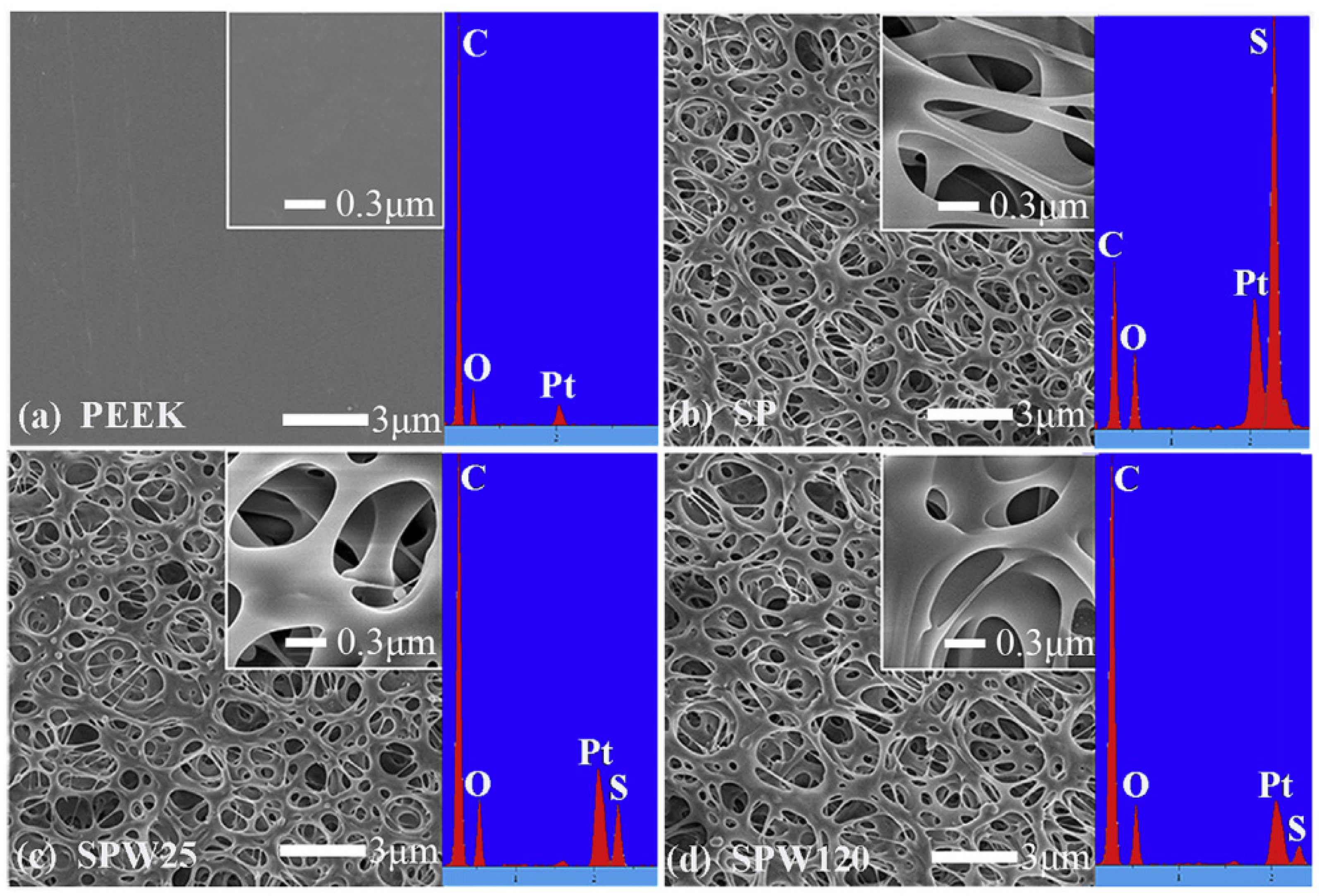
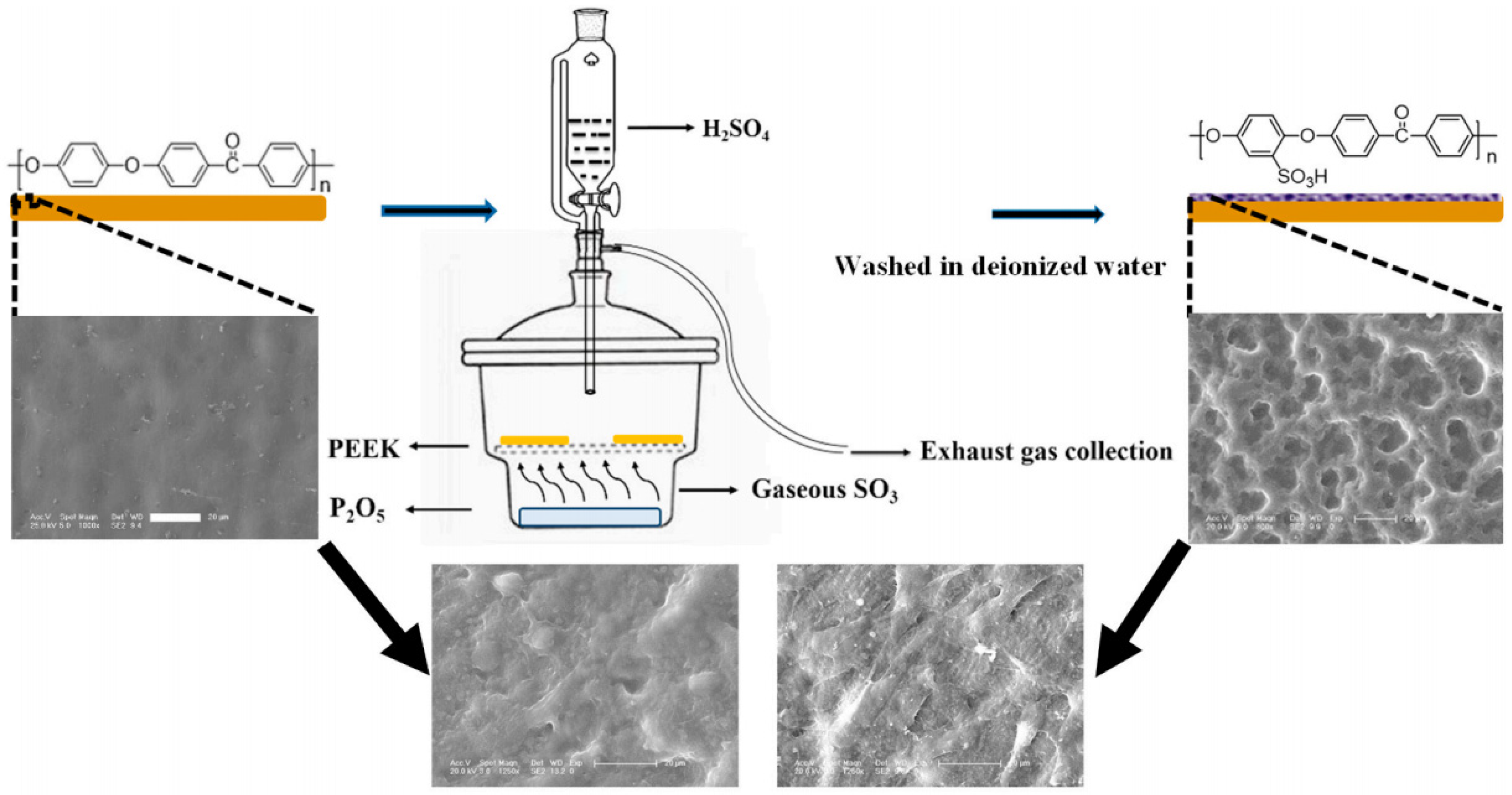
2.2.1. Sulfonation and Other Acid Treatment

Sulfonation is an electrophilic aromatic substitution reaction to introduce the sulfonate group to the aromatic ring in PEEK using concentrated sulfuric acid [36,37]. Figure 4 shows that sulfonation allows the formation of micro/nanoscale porous structures on the PEEK surface by adjusting the modification condition [38]. Previous studies have shown that the porous surface enhances the adhesive and proliferative abilities of cells and osteogenic differentiation, thereby enhancing osteointegration in vivo [39,40]. Despite the bactericidal effects, the residual sulfonic acid group post treatment could negatively affect cells and tissues in humans [41,42].
Recent studies have improved the sulfonation procedure to achieve better surface modification. Wan et al. [43] developed a new controllable sulfonation method using gaseous sulfur trioxide (SO3) fumigation to form a micro-topological porous structure with –SO3H component incorporated simultaneously. The schematic diagram is shown in Figure 5. Moreover, different degrees of modification could be achieved with a 6–14 mm pore diameter by controlling fumigation time. SO3-fumigated PEEK has enhanced hydrophilicity, cytocompatibility, protein absorption ability, and mineral apatite deposition capacity. Further, SO3-fumigated PEEK could significantly improve the adhesive and proliferation abilities of cells and extracellular matrix (ECM) secretion. Compared to the traditional sulfonation method, this technique introduces fewer changes in the structure. It could retain a certain degree of the mechanical properties of PEEK material since the pores formed by fumigation were significantly smaller and more uniform. Hence, the antibacterial and biological activity of this modified technique should be investigated in vivo.
Studies have investigated other acid treatments for modifying the PEEK surface. Huo et al. [44] modified the PEEK surface by treating PEEK with hydrofluoric acid and nitric acid simultaneously. Modified PEEK surface could enhance the adhesion, spreading, proliferative ability, and alkaline phosphatase (ALP) activity of osteoblasts, M2 macrophage polarization, and decrease pro-inflammatory factor secretion by attenuating the NF-κB signaling pathway. Further, a modified PEEK surface could enhance HUVEC endothelialization. Moreover, Li et al. [45] showed that modifying the PEEK surface using concentrated nitric acid, followed by hydrothermal treatment at high temperature, could enhance in vitro and in vivo bioactivity.

2.2.2. Ultraviolet Radiation Grafting

Surface grafting is used to modify the material surface by generating radicals on the material surface via several methods, followed by a reaction with a monomer or functional group [29]. Diphenylketone in the backbone of PEEK generates active radicals on UV exposure to induce functional monomer polymerization and formation of modified PEEK using functional groups [46,47].
Liu et al. [48] grafted poly (sodium vinylsulfonate) using UV radiation, and an increase in the amount of homogeneously distributed poly (sodium vinylsulfonate) grafted on the surface was observed with an increase in exposure time. Further, no changes in the inherent mechanical properties, enhanced hydrophilicity, and in vitro mineralizing ability of PEEK were observed after grafting. Additionally, an enhancement in the adhesive and proliferative ability, ECM secretion, and osteo-differentiation of MC3T3-E1 cells treated with increasing amounts of grafts was observed. Zhao et al. [49] grafted acrylic acid (AA) on PEEK using UV irradiation and demonstrated similar results. In addition, the hydration layer reduced the direct contact between two sliding surfaces and decreased the friction coefficient to 0.021, thereby improving bearing capacity and reducing the abrasion of PEEK. Figure 6 shows the mechanism.
In vitro studies fail to demonstrate a complete picture of the osseointegration of implants. Thus, several animal experiments have been performed. Zheng et al. [50] modified the PEEK surface with a phosphate group using single-step UV-initiated vinyl phosphonic acid graft polymerization. Further, the bioactivity of modified PEEK was determined in a rabbit proximal tibia defect model. Figure 7 shows an increase in the formation of new bones around modified PEEK (PEPA) after 12 weeks of implantation compared to unmodified PEEK.
These results indicate that grafting using UV radiation is a simple and effective method to improve PEEK bioactivity. However, this method has disadvantages, such as poor penetrability; hence, it cannot be used for complicated materials [51].

2.2.3. Biological Molecules

Several biological molecules, including proteins and growth factors, play an important role during osteogenesis [52]. Based on this, several studies have focused on immobilizing organic phases on the PEEK surface.
Bone morphogenetic protein 2 (BMP-2) is a potent osteoinductive factor, promotes spinal interbody fusion, and has been approved by FDA for its application in clinical settings [53]. Thus, several studies have explored methods to immobilize BMP-2 on the PEEK surface. Wu et al. [54] utilized phosphorylated gelatin covalent coating and sulfonation to modify the PEEK surface. Sun et al. [55] used lyophilization technology to modify the PEEK surface and obtained similar results. However, high BMP-2 concentration could lead to several side effects such as ectopic ossification, osteolysis, complications associated with inflammation, and abnormal adipogenesis [56]. Guillot et al. [57] loaded 9.3 µg BMP-2 on the PEEK surface, which was implanted into the femoral condyles of white rabbits from New Zealand. The results revealed that a high BMP-2 concertation could lead to poor osseointegration at an early stage. Hence, the BMP-2 concentration secreted by the modified surface should be carefully optimized. Therefore, studies should focus on optimizing the concentration of BMP-2 to be loaded on the PEEK surface and explore surface modification methods to achieve sustained release. Tropoelastin is an ECM protein that is involved in several signaling pathways [58,59]. Previous studies have shown that tropoelastin enhanced the adhesive and proliferative abilities of cells and the interactions of bone cells and certain materials [60,61]. Wakelin et al. [62] preliminarily treated PEEK using plasma immersion ion implantation (PIII), and tropoelastin was covalently immobilized. The modified PEEK was stable and could promote the adhesion, proliferation, and differentiation of osteoblasts as well as in vitro bone nodule formation. Further, immobilizing bone-forming peptides on the PEEK surface via the spin-coating process and peptide decoration could improve the osteogenic activity of PEEK [63]. Moreover, the functionalization of PEEK using adiponectin, an adipocyte-secreted adipokine, could improve the osteogenic and bacteriostatic ability of PEEK in vitro and in vivo [64].

2.3. Surface Coating

Surface coating is a simple, straightforward, and promising method to modify PEEK. PEEK bioactivity can be improved by coating its surface with certain substances using different methods, thereby enhancing the interaction between implants and tissues.

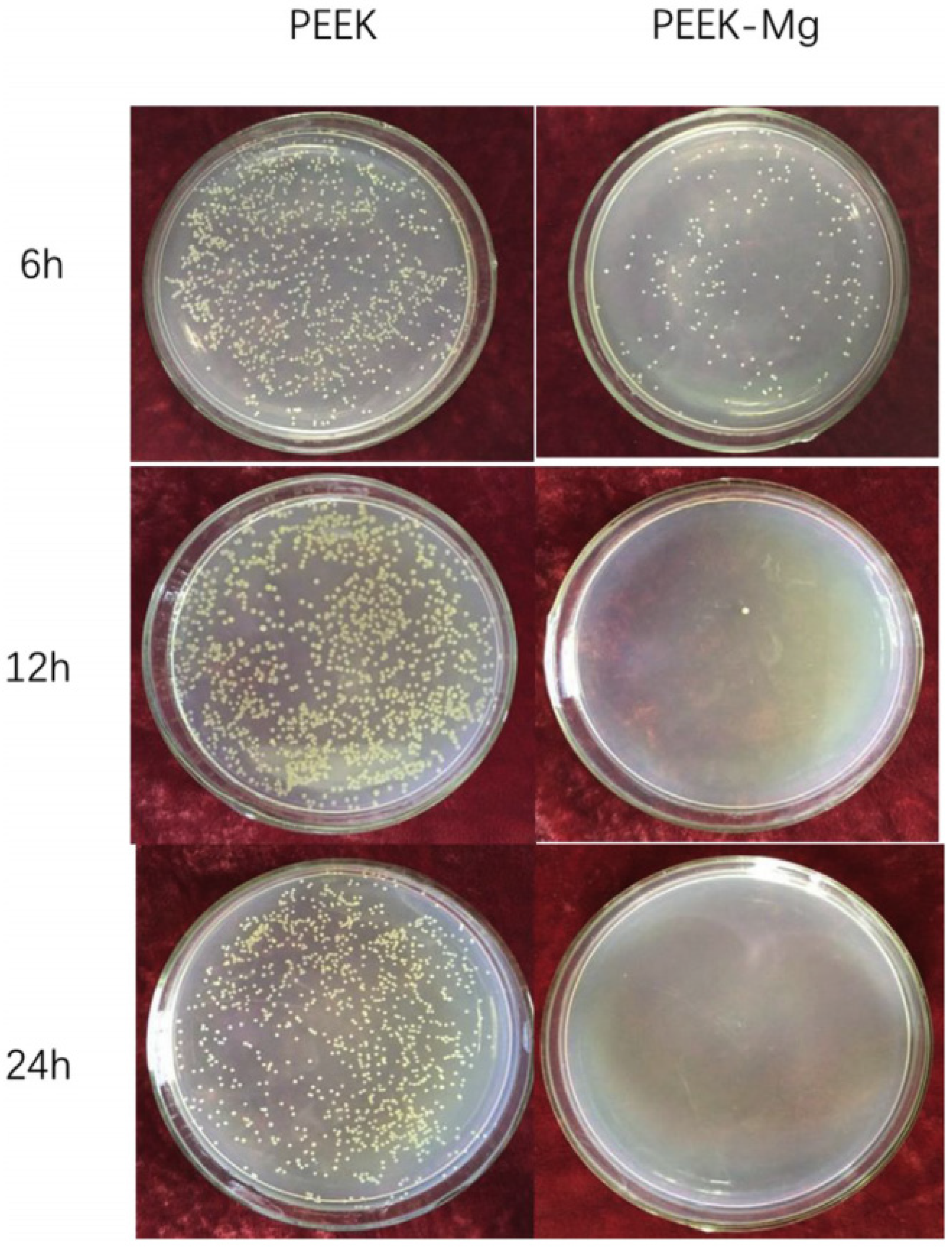
2.3.1. Metal Coating

Metals are good coating materials for the spinal interbody cage. The metal coating layer directly interacts with the end plate of vertebrae and improves osseointegration. A metal-coated cage can maintain favorable elastic modulus and radiopacity of PEEK.
Of those metals, Ti has mechanical and biological properties and is a suitable PEEK coating material. Techniques, including plasma spray, ionic plasma deposition, and electron beam deposition have previously been reported. Boonpok et al. [65] showed that titanium nitride coating of the PEEK surface was rough and porous compared to unmodified PEEK. In another study [66], Ti-coated PEEK enhanced the adhesive and proliferative abilities of osteoblasts and increased ALP and BMP-2 expression. Ti-coated PEEK implanted into the hind leg of sheep showed enhanced osseointegration, and the pullout strength was high after 12 and 24 weeks of implantation. Liu et al. [67] utilized a vacuum plasma spray technique to prepare a Ti-coated PEEK cage and obtained similar in vitro results. Hence, these Ti-coated PEEK cages were implanted into C3-4 intervertebral space. Results shows better fusion, and newer bones were formed. In clinical practice, patients who underwent lumbar interbody fusion surgery were followed up. Figure 8 shows the results of another study which demonstrated that a Ti-coated PEEK cage could increase fast and stable fixation at the bone-implant interface, significantly alleviating pain; fewer subsidence incidences were observed and the fusion rate was higher [68].
Despite the widespread use of plasma spray, it has a few disadvantages. High temperatures during spraying could damage the structure of PEEK, and the coating could spill in some situations. Hence, other methods to prepare Ti-coated PEEK were studied. Han et al. [69] utilized a low-temperature electron beam deposition method. Walsh et al. [70] used a novel high-energy and low-temperature ionic plasma deposition method to coat a sub-micron layer of Ti on PEEK. Satisfactory osseointegration in vivo was observed in both studies. Additionally, titanium dioxide (TiO2) could be used as a coating material for optimizing Ti coating. In an aqueous environment, TiO2 reacts with H2O2 molecules to form hydroxyl groups, thereby inducing bone-like calcium phosphate (CaP, apatite) in a simulated body fluid [71]. Further, calcium and phosphate ions binding to the hydroxylated TiO2 surface aids in osseointegration [72]. Yang et al. [73] demonstrated significant in vitro PEEK bioactivity by coating PEEK with a dense and stable TiO2 layer using a high-power impulse magnetron sputtering technique. Compared to uncoated PEEK, TiO2-coated PEEK using an arc ion plating technique demonstrated a 2.5 times higher bone-implant interface shear strength in the push-out test and better bone bonding performance in vivo [74]. Furthermore, a sol-gel-derived TiO2 layer-coated PEEK cage (Figure 9) demonstrated high bone-implant bonding and significant intervertebral fusion without bone grafts in the anterior cervical fusion canine model [75].
Several other metals have been used as coating materials for PEEK. Like Ti, tantalum (Ta) is a chemically stable and biocompatible metal. Lu et al. [76] deposited tantalum ions onto PEEK using PIII to form Ta2O5 nanoparticles, which enhanced osseointegration in a femoral implant of a rodent model, and the elastic modulus of the modified PEEK was similar to human cortical bone. Further, most metal ions could kill bacteria; hence, some studies explored metal coatings with antiseptic activity. Some studies used the magnetron sputtering technology to coat PEEK with metal nanoparticles, including nano silver and nano copper. These modified PEEKs have good biocompatibility, could activate macrophages to phagocytize pathogens, and achieved high antibacterial activity in vitro and in vivo [77,78].

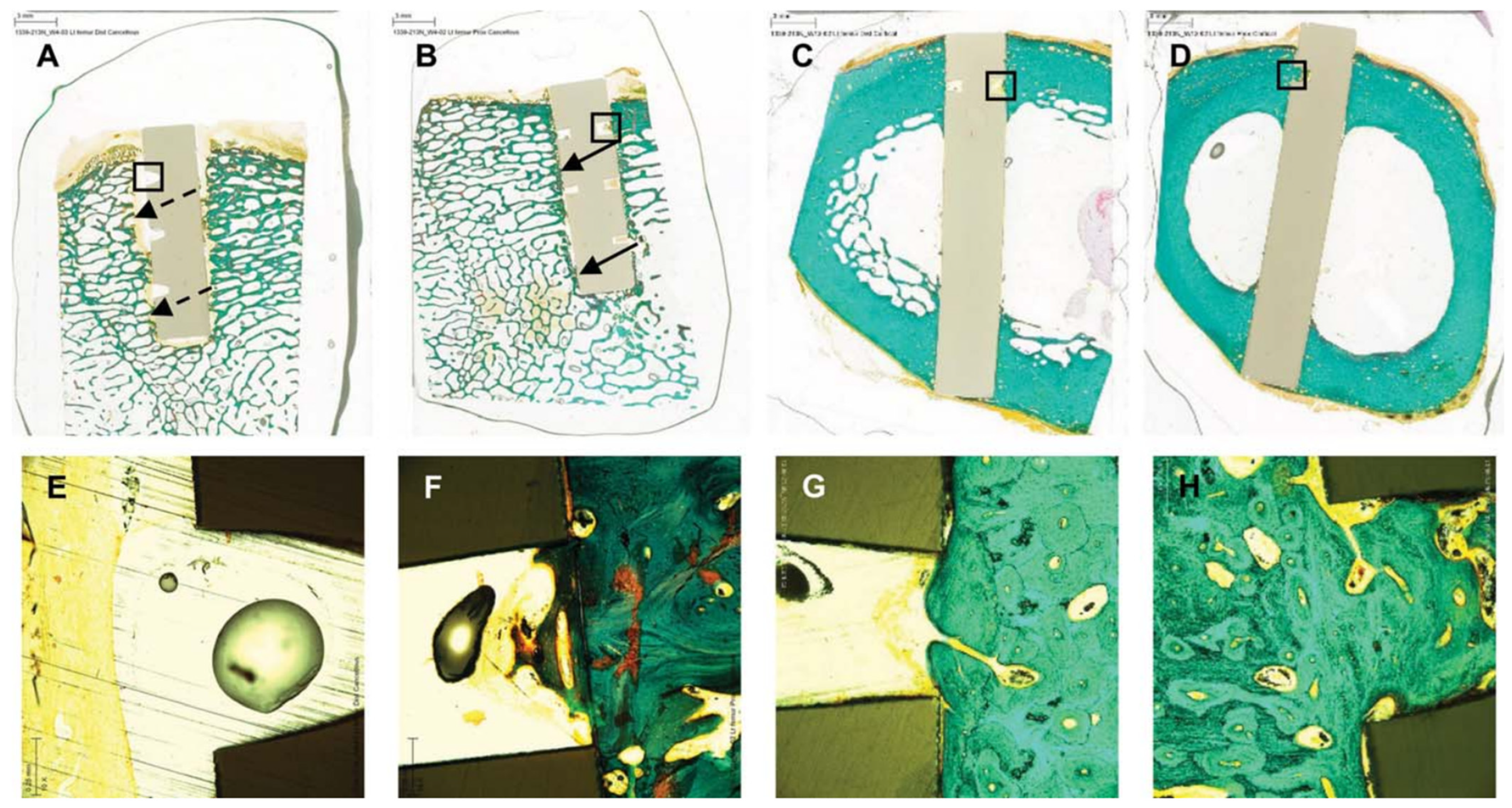
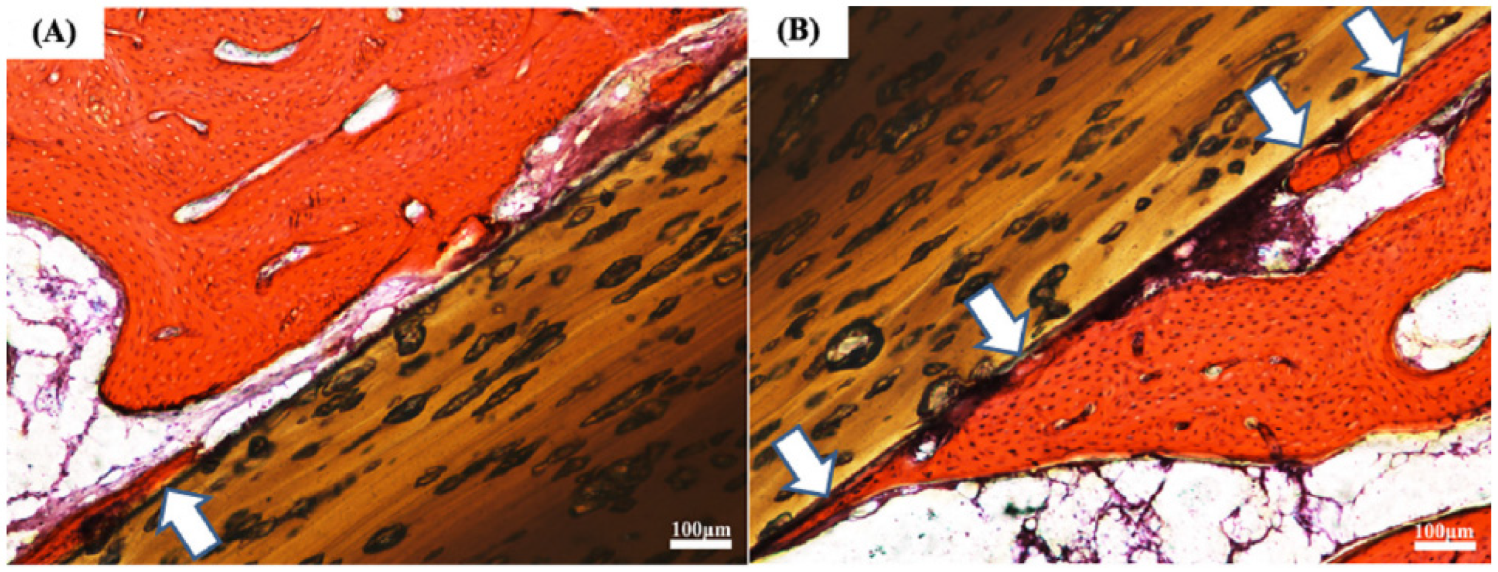
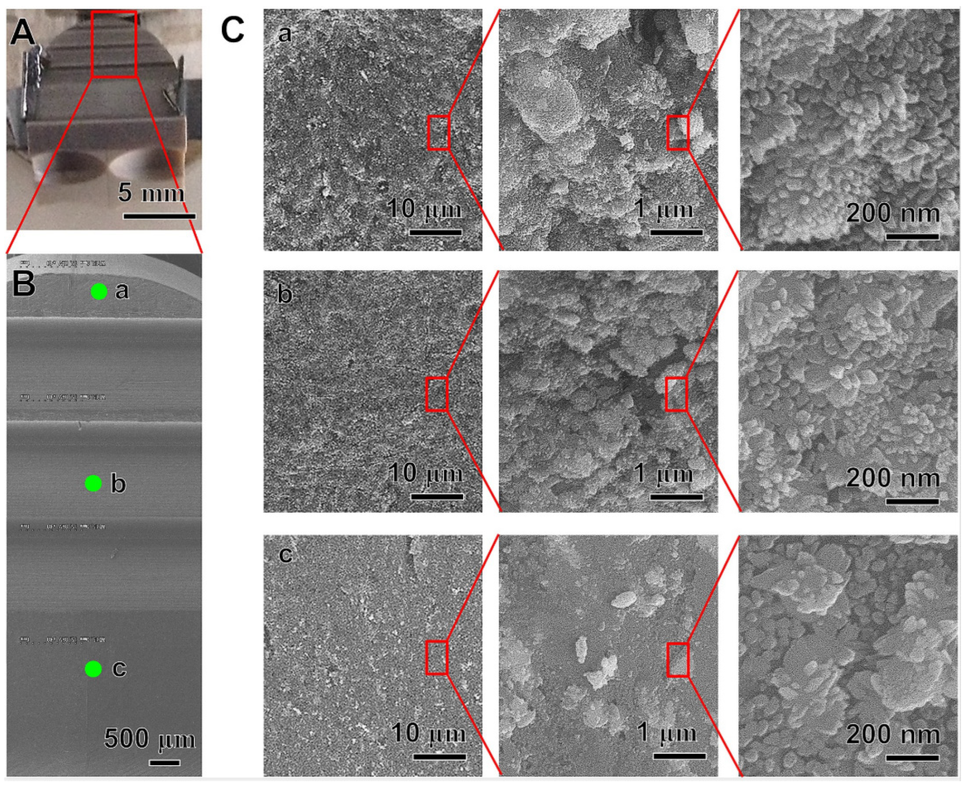
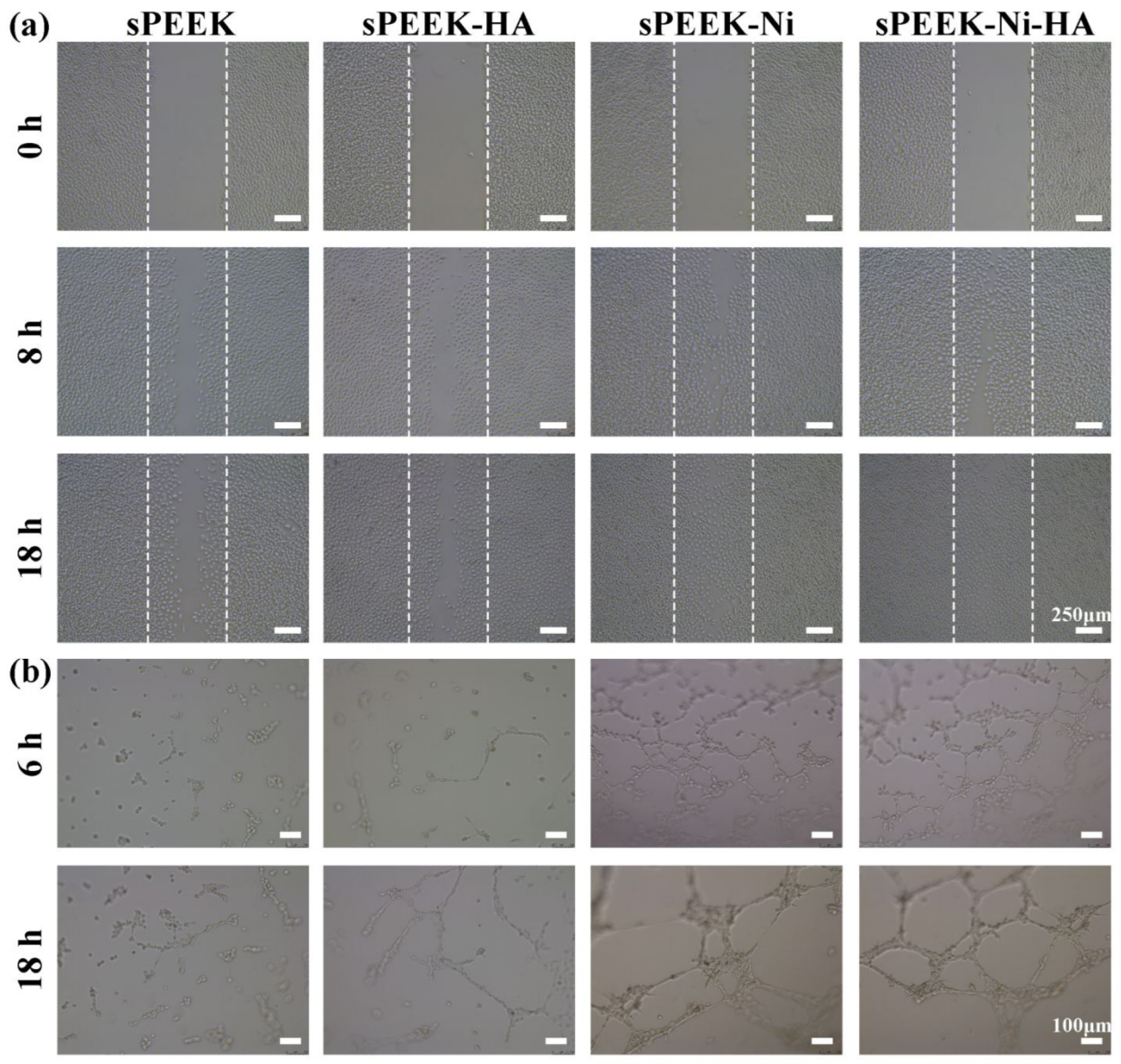
2.3.2. Hydroxyapatite Coating

The Ca/P ratio of the most stable and least soluble CaP ceramic and hydroxyapatite (HA, chemical formula Ca10(PO4)6(OH)2) is nearly 1.67. Moreover, it is osteoconductive, promotes new bone formation without toxicity, and induces inflammatory or antigen-antibody reactions [79,80]. Hence, HA is a good coating material and can provide an ideal scaffold for osteogenesis on a bioinert PEEK surface [81,82].
Despite the disadvantages of the plasma spray technique, it is still a classic coating method and has demonstrated good clinical outcomes in spinal fusion procedures. Zhu et al. [83] conducted a prospective and non-randomized study to compare clinical outcomes of patients who underwent single-level anterior cervical decompression and fusion surgery treated with Ti and HA-coated PEEK cages using plasma spray uncoated PEEK cages. The coated PEEK cages demonstrated a higher fusion rate three months after surgery. Both cages could achieve solid osseous fusion and had comparable clinical outcomes during the last follow-up. Furthermore, Ti and HA-coated cages demonstrated similar imaging and clinical outcomes in single-level transforaminal lumbar interbody fusion surgery [84]. Additionally, other coating methods have also been explored. Lee et al. [85] prepared HA-coated PEEK using the cold spray technique, and no chemical deformation of PEEK was observed due to the relatively low working temperature of the technique. Moreover, more interfacial contact with neighboring tissues was obtained due to the rough PEEK surface after coating. In vitro studies and the block-type ilium model demonstrated improved osseointegration. Additionally, intervertebral cage-type HA-coated PEEK was implanted in the mini-pig model, and the result demonstrated enhanced fusion rate and bone tissue regeneration. Dong et al. [86] prepared a micro-/nanostructured PEEK surface with nickel hydroxide nanoparticles and HA nanoflowers using a two-step hydrothermal synthesis approach. The modified PEEK demonstrated robust vascularization, vascular regeneration, and new bone-forming abilities, thereby significantly improving the osseointegration of a bioinert implant (Figure 10).

2.3.3. Dopamine Composite Coating

Dopamine can automatically polymerize in an alkaline environment and form a uniform polydopamine (PDA) layer on the material. The PDA layer is rich in bioactive groups and is often used as a secondary grafting platform to load other bioactive molecules or substances, thereby achieving a multifunctional modification effect.
Lyu et al. [87] coated PEEK with mussel-inspired dopamine-CuII. The results revealed that secretion of Cu ions and other surface characteristics (roughness, hydrophilicity, etc.) could enhance osteogenic, angiogenic, and antibacterial abilities. Furthermore, the modified PEEK initiated an early and mild M1 pro-inflammatory microenvironment to enhance osteogenesis and antibacterial ability.
Wang et al. [88] developed a surface modification technique which attaching basic fibroblast growth factor (bFGF) to a PDA-coated PEEK. Basic fibroblast growth factor secreted by the modified PEEK surface could promote in vitro osteogenic differentiation of human mesenchymal stem cells and demonstrate an antibacterial effect in vivo. Further antigen-antibody reaction and canine femur implantation model showed remarkable anti-inflammatory and osseointegration capacity of the modified PEEK.
Recently, Xiao et al. [89] fabricated a multifunctional PEEK. Figure 11 shows a PEEK coated with the dual-metal-organic framework (Zn-Mg-MOF74) loaded with DEX using PDA as an interlayer. This coating can secrete Mg2+, Zn2+, and DHTA and creates an alkaline microenvironment. The results showed significant osteogenic differentiation, angiogenic ability, and antibacterial properties against E. coli and S. aureus in vitro and in vivo. This indicates the significant clinical application potential of this coating as a spinal implant material.

2.3.4. Other Kinds of Coatings

Studies have used various other materials to coat the PEEK surface via different methods, apart from the materials mentioned previously.
PEEK coated using graphene (GF) as coating material demonstrated significant bioactivity [13,90]. In addition, several studies have shown that modifying the PEEK surface with GF and its derivatives could enhance the osseointegration and antibacterial abilities of PEEK [91,92,93].
Moreover, amorphous phosphate is also a good choice of coating material. Ren et al. [94] used the microwave-assisted coating technique to prepare homogeneous and amorphous magnesium phosphate (AMP) layers on PEEK. Cells cultured on AMP-coated PEEK showed no cytotoxicity, could promote cell adhesion at an early stage, induce apatite deposition, and increase ALP expression and osteocalcin in cells. This indicates that AMP-coated PEEK could trigger new bone formation and enhance osseointegration. In another study, precipitation of amorphous calcium phosphate on carbon fiber reinforced PEEK obtained similar in vitro and in vivo results [95].
Furthermore, Gao et al. [96] used a layer-by-layer self-assembly method to coat adherent polyacrylic acid and polyallylamine hydrochloride films with tunable structures on PEEK. Figure 12 shows that this modification could improve the immunomodulatory ability of PEEK and inhibit the acute inflammatory response of macrophages, thereby creating a favorable microenvironment for osteogenesis. Moreover, Dai et al. [97] coated PEEK with silicon nitride (Si3N4, SN) with micro/nanostructures, and the results demonstrated great osteogenic capacity both in vitro and in vivo.

3. Conclusions and Outlook

For spinal surgeons, an ideal spinal interbody fusion device should fulfill the following criteria. First, the cage should provide enough support to the axial load and have biomechanical properties close to human bone tissue. Second, it should be biocompatible, chemically stable, and promote bone fusion. Last, for ease of imaging, the cage should be radiolucent. Most cages are made of PEEK, and several advancements have been made to achieve optimal PEEK surface modification, including physical as well as chemical modification and surface coating. However, several obstacles associated with PEEK surface modification have prevented its clinical application, such as abrasion during a surgical procedure, long-term surface stability, and sustainable bioactivity. Therefore, extensive studies, especially long-term in vivo studies, are still required. Future studies should also focus on novel modification techniques including controlled release of drugs, 3D printing, blending new materials with PEEK, etc. In addition, the complexity associated with process and cost effectiveness should be considered to determine the optimal solution, even though it could be a long and winding process.

Author Contributions

S.L.: Conceptualization, investigation, methodology, writing—original draft preparation; writing—review and editing; J.S.: Conceptualization, investigation, methodology, writing—original draft preparation; writing—review and editing; K.C.: Conceptualization, investigation, methodology, writing—original draft preparation; Y.D.: investigation, data curation; X.C.: investigation, data curation; Y.H.: investigation, data curation; L.Z.: investigation, data curation; X.M.: investigation, data curation; Z.X.: investigation, data curation; L.M.: investigation, data curation; S.H.: conceptualization, methodology, supervision; G.W.: conceptualization, methodology, supervision; Z.S.: conceptualization, methodology, supervision. All authors have read and agreed to the published version of the manuscript.

Funding

This work was supported by the National Natural Science Foundation of China (Grant No. 81702666), the National Natural Science Foundation of China (Grant no. 81872171), and Shanghai Science and Technology Innovation Action Plan (Grant no. 22S31900400).

Institutional Review Board Statement

Not applicable.

Informed Consent Statement

Not applicable.

Data Availability Statement

All data that support the findings of this study are included within the article.

Acknowledgments

Shu Liu, Junhao Sui, and Kai Chen contributed equally to this work.

Conflicts of Interest

The authors declare no conflict of interest.

References

  1. Cloward, R.B. The treatment of ruptured lumbar intervertebral discs by vertebral body fusion. I. Indications, operative technique, after care. J. Neurosurg. 1953, 10, 154–168. [Google Scholar] [CrossRef] [PubMed]
  2. Smith, G.W.; Robinson, R.A. The treatment of certain cervical-spine disorders by anterior removal of the intervertebral disc and interbody fusion. J. Bone Jt. Surg. Am. 1958, 40-A, 607–624. [Google Scholar] [CrossRef]
  3. Abou-Madawi, A.M.; Ali, S.H.; Abdelmonem, A.M. Local Autograft Versus Iliac Crest Bone Graft PSF-Augmented TLIF in Low-Grade Isthmic and Degenerative Lumbar Spondylolisthesis. Glob. Spine J. 2022, 12, 70–78. [Google Scholar] [CrossRef]
  4. Park, K.H.; Chung, H.W.; Lee, H.D.; Jeon, C.H.; Koh, J.H.; Chung, N.S. Cage Obliquity and Radiological Outcomes in Oblique Lateral Interbody Fusion. Spine 2022. ahead of print. [Google Scholar] [CrossRef]
  5. Bagby, G.W. Arthrodesis by the distraction-compression method using a stainless steel implant. Orthopedics 1988, 11, 931–934. [Google Scholar] [CrossRef] [PubMed]
  6. Kuslich, S. Two-year follow up results of the BAK interbody fusion device. In Proceedings of the Meeting of the North American Spine Society, Minneapolis, MN, USA, 19–22 October 1994. [Google Scholar]
  7. Epstein, N.E.; Agulnick, M.A. Review of anterior cervical diskectomy/fusion (ACDF) using different polyetheretherketone (PEEK) cages. Surg. Neurol. Int. 2022, 13, 556. [Google Scholar] [CrossRef] [PubMed]
  8. Segi, N.; Nakashima, H.; Shinjo, R.; Kagami, Y.; Machino, M.; Ito, S.; Ouchida, J.; Morishita, K.; Oishi, R.; Yamauchi, I.; et al. Vertebral Endplate Concavity in Lateral Lumbar Interbody Fusion: Tapered 3D-Printed Porous Titanium Cage versus Squared PEEK Cage. Medicina 2023, 59, 327. [Google Scholar] [CrossRef] [PubMed]
  9. Zhou, E.; Huang, H.; Zhao, Y.; Wang, L.; Fan, Y. The effects of titanium mesh cage size on the biomechanical responses of cervical spine after anterior cervical corpectomy and fusion: A finite element study. Clin. Biomech. 2022, 91, 105547. [Google Scholar] [CrossRef]
  10. Muthiah, N.; Yolcu, Y.U.; Alan, N.; Agarwal, N.; Hamilton, D.K.; Ozpinar, A. Evolution of polyetheretherketone (PEEK) and titanium interbody devices for spinal procedures: A comprehensive review of the literature. Eur. Spine J. 2022, 31, 2547–2556. [Google Scholar] [CrossRef]
  11. Erjavec, A.K.; Črešnar, K.P.; Švab, I.; Vuherer, T.; Žigon, M.; Brunčko, M. Determination of Shear Bond Strength between PEEK Composites and Veneering Composites for the Production of Dental Restorations. Materials 2023, 16, 3286. [Google Scholar] [CrossRef]
  12. Wiessner, A.; Wassmann, T.; Wiessner, J.M.; Schubert, A.; Wiechens, B.; Hampe, T.; Bürgers, R. In Vivo Biofilm Formation on Novel PEEK, Titanium, and Zirconia Implant Abutment Materials. Int. J. Mol. Sci. 2023, 24, 1779. [Google Scholar] [CrossRef] [PubMed]
  13. Dondani, J.R.; Iyer, J.; Tran, S.D. Surface Treatments of PEEK for Osseointegration to Bone. Biomolecules 2023, 13, 464. [Google Scholar] [CrossRef] [PubMed]
  14. Veronesi, F.; Sartori, M.; Griffoni, C.; Valacco, M.; Tedesco, G.; Davassi, P.F.; Gasbarrini, A.; Fini, M.; Barbanti Brodano, G. Complications in Spinal Fusion Surgery: A Systematic Review of Clinically Used Cages. J. Clin. Med. 2022, 11, 6279. [Google Scholar] [CrossRef] [PubMed]
  15. Zhang, H.; Duan, M.; Qin, S.; Zhang, Z. Preparation and Modification of Porous Polyetheretherketone (PEEK) Cage Material Based on Fused Deposition Modeling (FDM). Polymers 2022, 14, 5403. [Google Scholar] [CrossRef] [PubMed]
  16. Münstedt, H.; Grossmann, J. Surface Modifications of Polyetheretherketone (PEEK): Results from the Literature and Special Studies of Copper-Coated Films. Polymers 2022, 14, 4797. [Google Scholar] [CrossRef]
  17. Lisoń, J.; Taratuta, A.; Paszenda, Z.; Szindler, M.; Basiaga, M. Perspectives in Prevention of Biofilm for Medical Applications. Coatings 2022, 12, 197. [Google Scholar] [CrossRef]
  18. Yu, X.; Ibrahim, M.; Liu, Z.; Yang, H.; Tan, L.; Yang, K. Biofunctional Mg coating on PEEK for improving bioactivity. Bioact. Mater. 2018, 3, 139–143. [Google Scholar] [CrossRef]
  19. Kratochvíl, J.; Štěrba, J.; Lieskovská, J.; Langhansová, H.; Kuzminova, A.; Khalakhan, I.; Kylián, O.; Straňák, V. Antibacterial effect of Cu/C:F nanocomposites deposited on PEEK substrates. Mater. Lett. 2018, 230, 96–99. [Google Scholar] [CrossRef]
  20. Durham, J.W.; Allen, M.J.; Rabiei, A. Preparation, characterization and in vitro response of bioactive coatings on polyether ether ketone. J. Biomed. Mater. Res. Part B Appl. Biomater. 2017, 105, 560–567. [Google Scholar] [CrossRef]
  21. Briem, D.; Strametz, S.; Schröoder, K.; Meenen, N.M.; Lehmann, W.; Linhart, W.; Ohl, A.; Rueger, J.M. Response of primary fibroblasts and osteoblasts to plasma treated polyetheretherketone (PEEK) surfaces. J. Mater. Sci. Mater. Med. 2005, 16, 671–677. [Google Scholar] [CrossRef]
  22. Recek, N. Biocompatibility of Plasma-Treated Polymeric Implants. Materials 2019, 12, 240. [Google Scholar] [CrossRef] [PubMed]
  23. Chen, J.-P.; Su, C.-H. Surface modification of electrospun PLLA nanofibers by plasma treatment and cationized gelatin immobilization for cartilage tissue engineering. Acta Biomater. 2011, 7, 234–243. [Google Scholar] [CrossRef] [PubMed]
  24. Waser-Althaus, J.; Salamon, A.; Waser, M.; Padeste, C.; Kreutzer, M.; Pieles, U.; Müller, B.; Peters, K. Differentiation of human mesenchymal stem cells on plasma-treated polyetheretherketone. J. Mater. Sci. Mater. Med. 2014, 25, 515–525. [Google Scholar] [CrossRef] [PubMed]
  25. Novotna, Z.; Reznickova, A.; Rimpelova, S.; Vesely, M.; Kolska, Z.; Svorcik, V. Tailoring of PEEK bioactivity for improved cell interaction: Plasma treatment in action. RSC Adv. 2015, 5, 41428–41436. [Google Scholar] [CrossRef]
  26. Poulsson, A.H.; Eglin, D.; Zeiter, S.; Camenisch, K.; Sprecher, C.; Agarwal, Y.; Nehrbass, D.; Wilson, J.; Richards, R.G. Osseointegration of machined, injection moulded and oxygen plasma modified PEEK implants in a sheep model. Biomaterials 2014, 35, 3717–3728. [Google Scholar] [CrossRef] [PubMed]
  27. Zhao, Y.; Wong, H.M.; Lui, S.C.; Chong, E.Y.W.; Wu, G.; Zhao, X.; Wang, C.; Pan, H.; Cheung, K.M.C.; Wu, S.; et al. Plasma Surface Functionalized Polyetheretherketone for Enhanced Osseo-Integration at Bone-Implant Interface. ACS Appl. Mater. Interfaces 2016, 8, 3901–3911. [Google Scholar] [CrossRef] [PubMed]
  28. Petlin, D.; Tverdokhlebov, S.; Anissimov, Y. Plasma treatment as an efficient tool for controlled drug release from polymeric materials: A review. J. Control. Release 2017, 266, 57–74. [Google Scholar] [CrossRef]
  29. Pidhatika, B.; Widyaya, V.T.; Nalam, P.C.; Swasono, Y.A.; Ardhani, R. Surface Modifications of High-Performance Polymer Polyetheretherketone (PEEK) to Improve Its Biological Performance in Dentistry. Polymers 2022, 14, 5526. [Google Scholar] [CrossRef]
  30. Liu, L.; Khoury, J.; Webster, T.J. Accelerated Neutral Atom Beam (ANAB) Modified Polypropylene for Reducing Bacteria Colonization Without Antibiotics. J. Biomed. Nanotechnol. 2022, 18, 868–874. [Google Scholar] [CrossRef]
  31. Ajami, S.; Coathup, M.J.; Khoury, J.; Blunn, G.W. Augmenting the bioactivity of polyetheretherketone using a novel accelerated neutral atom beam technique. J. Biomed. Mater. Res. Part B Appl. Biomater. 2017, 105, 1438–1446. [Google Scholar] [CrossRef]
  32. Khoury, J.; Selezneva, I.; Pestov, S.; Tarassov, V.; Ermakov, A.; Mikheev, A.; Lazov, M.; Kirkpatrick, S.R.; Shashkov, D.; Smolkov, A. Surface bioactivation of PEEK by neutral atom beam technology. Bioact. Mater. 2019, 4, 132–141. [Google Scholar] [CrossRef] [PubMed]
  33. Khoury, J.; Maxwell, M.; Cherian, R.E.; Bachand, J.; Kurz, A.C.; Walsh, M.; Assad, M.; Svrluga, R.C. Enhanced bioactivity and osseointegration of PEEK with accelerated neutral atom beam technology. J. Biomed. Mater. Res. Part B Appl. Biomater. 2017, 105, 531–543. [Google Scholar] [CrossRef] [PubMed]
  34. Buck, E.; Lee, S.; Gao, Q.; Tran, S.D.; Tamimi, F.; Stone, L.S.; Cerruti, M. The Role of Surface Chemistry in the Osseointegration of PEEK Implants. ACS Biomater. Sci. Eng. 2022, 8, 1506–1521. [Google Scholar] [CrossRef] [PubMed]
  35. Zheng, Z.; Liu, P.; Zhang, X.; Xin, J.; Wang, Y.; Zou, X.; Mei, X.; Zhang, S.; Zhang, S. Strategies to improve bioactive and antibacterial properties of polyetheretherketone (PEEK) for use as orthopedic implants. Mater. Today Bio 2022, 16, 100402. [Google Scholar] [CrossRef]
  36. Huang, R.Y.M.; Shao, P.; Burns, C.M.; Feng, X. Sulfonation of poly(ether ether ketone)(PEEK): Kinetic study and characterization. J. Appl. Polym. Sci. 2001, 82, 2651–2660. [Google Scholar] [CrossRef]
  37. Shi, Y.; Deng, T.; Peng, Y.; Qin, Z.; Ramalingam, M.; Pan, Y.; Chen, C.; Zhao, F.; Cheng, L.; Liu, J. Effect of Surface Modification of PEEK Artificial Phalanx by 3D Printing on its Biological Activity. Coatings 2023, 13, 400. [Google Scholar] [CrossRef]
  38. Ouyang, L.; Zhao, Y.; Jin, G.; Lu, T.; Li, J.; Qiao, Y.; Ning, C.; Zhang, X.; Chu, P.K.; Liu, X. Influence of sulfur content on bone formation and antibacterial ability of sulfonated PEEK. Biomaterials 2016, 83, 115–126. [Google Scholar] [CrossRef]
  39. Wang, C.; Wang, J.; Zeng, L.; Qiao, Z.; Liu, X.; Liu, H.; Zhang, J.; Ding, J. Fabrication of Electrospun Polymer Nanofibers with Diverse Morphologies. Molecules 2019, 24, 834. [Google Scholar] [CrossRef]
  40. Xin, H.; Shi, Q.; Ning, X.; Chen, Y.; Jia, X.; Zhang, Z.; Zhu, S.; Li, Y.; Liu, F.; Kong, L. Biomimetic Mineralized Fiber Bundle-Inspired Scaffolding Surface on Polyetheretherketone Implants Promotes Osseointegration. Macromol. Biosci. 2023, 23, e2200436. [Google Scholar] [CrossRef]
  41. Zhao, Y.; Wong, H.M.; Wang, W.; Li, P.; Xu, Z.; Chong, E.Y.; Yan, C.H.; Yeung, K.W.; Chu, P.K. Cytocompatibility, osseointegration, and bioactivity of three-dimensional porous and nanostructured network on polyetheretherketone. Biomaterials 2013, 34, 9264–9277. [Google Scholar] [CrossRef]
  42. Baltag, L.; Cojocaru, C.; Enache, A.-C.; Samoila, P.; Harabagiu, V. Ultrasonic-Assisted Rapid Preparation of Sulfonated Polyether Ether Ketone (PEEK) and Its Testing in Adsorption of Cationic Species from Aqueous Solutions. Materials 2022, 15, 7558. [Google Scholar] [CrossRef] [PubMed]
  43. Wan, T.; Jiao, Z.; Guo, M.; Wang, Z.; Wan, Y.; Lin, K.; Liu, Q.; Zhang, P. Gaseous sulfur trioxide induced controllable sulfonation promoting biomineralization and osseointegration of polyetheretherketone implants. Bioact. Mater. 2020, 5, 1004–1017. [Google Scholar] [CrossRef] [PubMed]
  44. Huo, S.; Meng, X.; Zhang, S.; Yue, B.; Zhao, Y.; Long, T.; Nie, B.; Wang, Y. Hydrofluoric acid and nitric acid cotreatment for biofunctionalization of polyetheretherketone in M2 macrophage polarization and osteogenesis. J. Biomed. Mater. Res. Part A 2021, 109, 879–892. [Google Scholar] [CrossRef] [PubMed]
  45. Li, Y.; Wang, J.; He, D.; Zhu, G.; Wu, G.; Chen, L. Surface sulfonation and nitrification enhance the biological activity and osteogenesis of polyetheretherketone by forming an irregular nano-porous monolayer. J. Mater. Sci. Mater. Med. 2019, 31, 11. [Google Scholar] [CrossRef]
  46. Flejszar, M.; Chmielarz, P. Surface Modifications of Poly(Ether Ether Ketone) via Polymerization Methods—Current Status and Future Prospects. Materials 2020, 13, 999. [Google Scholar] [CrossRef] [PubMed]
  47. Nakano, H.; Noguchi, Y.; Kakinoki, S.; Yamakawa, M.; Osaka, I.; Iwasaki, Y. Highly Durable Lubricity of Photo-Cross-Linked Zwitterionic Polymer Brushes Supported by Poly(ether ether ketone) Substrate. ACS Appl. Bio Mater. 2020, 3, 1071–1078. [Google Scholar] [CrossRef]
  48. Liu, L.; Dong, J.; Zhang, W.; He, C.; Liu, Y.; Zheng, Y. The Single-Step Fabrication of a Poly (Sodium Vinylsulfonate)-Grafted Polyetheretherketone Surface to Ameliorate Its Osteogenic Activity. Coatings 2022, 12, 868. [Google Scholar] [CrossRef]
  49. Zhao, X.; Xiong, D.; Wang, K.; Wang, N. Improved biotribological properties of PEEK by photo-induced graft polymerization of acrylic acid. Mater. Sci. Eng. C 2017, 75, 777–783. [Google Scholar] [CrossRef]
  50. Zheng, Y.; Liu, L.; Xiao, L.; Zhang, Q.; Liu, Y. Enhanced osteogenic activity of phosphorylated polyetheretherketone via surface-initiated grafting polymerization of vinylphosphonic acid. Colloids Surf. B Biointerfaces 2019, 173, 591–598. [Google Scholar] [CrossRef]
  51. Liu, S.; Zhu, Y.; Gao, H.; Ge, P.; Ren, K.; Gao, J.; Cao, Y.; Han, D.; Zhang, J. One-step fabrication of functionalized poly(etheretherketone) surfaces with enhanced biocompatibility and osteogenic activity. Mater. Sci. Eng. C 2018, 88, 70–78. [Google Scholar] [CrossRef]
  52. Saad, A.; Arias, C.P.; Wang, M.; ElKashty, O.; Brambilla, D.; Tamimi, F.; Cerruti, M. Biomimetic Strategy to Enhance Epithelial Cell Viability and Spreading on PEEK Implants. ACS Biomater. Sci. Eng. 2022, 8, 5129–5144. [Google Scholar] [CrossRef] [PubMed]
  53. Wetzell, B.; McLean, J.B.; Moore, M.A.; Kondragunta, V.; Dorsch, K. A large database study of hospitalization charges and follow-up re-admissions in US lumbar fusion surgeries using a cellular bone allograft (CBA) versus recombinant human bone morphogenetic protein-2 (rhBMP-2). J. Orthop. Surg. Res. 2020, 15, 544. [Google Scholar] [CrossRef] [PubMed]
  54. Wu, J.; Li, L.; Fu, C.; Yang, F.; Jiao, Z.; Shi, X.; Ito, Y.; Wang, Z.; Liu, Q.; Zhang, P. Micro-porous polyetheretherketone implants decorated with BMP-2 via phosphorylated gelatin coating for enhancing cell adhesion and osteogenic differentiation. Colloids Surf. B Biointerfaces 2018, 169, 233–241. [Google Scholar] [CrossRef] [PubMed]
  55. Sun, Z.; Ouyang, L.; Ma, X.; Qiao, Y.; Liu, X. Controllable and durable release of BMP-2-loaded 3D porous sulfonated polyetheretherketone (PEEK) for osteogenic activity enhancement. Colloids Surf. B Biointerfaces 2018, 171, 668–674. [Google Scholar] [CrossRef] [PubMed]
  56. James, A.W.; LaChaud, G.; Shen, J.; Asatrian, G.; Nguyen, V.; Zhang, X.; Ting, K.; Soo, C. A Review of the Clinical Side Effects of Bone Morphogenetic Protein-2. Tissue Eng. Part B Rev. 2016, 22, 284–297. [Google Scholar] [CrossRef] [PubMed]
  57. Guillot, R.; Pignot-Paintrand, I.; Lavaud, J.; Decambron, A.; Bourgeois, E.; Josserand, V.; Logeart-Avramoglou, D.; Viguier, E.; Picart, C. Assessment of a polyelectrolyte multilayer film coating loaded with BMP-2 on titanium and PEEK implants in the rabbit femoral condyle. Acta Biomater. 2016, 36, 310–322. [Google Scholar] [CrossRef] [PubMed]
  58. Hume, R.D.; Kanagalingam, S.; Deshmukh, T.; Chen, S.; Mithieux, S.M.; Rashid, F.N.; Roohani, I.; Lu, J.; Doan, T.; Graham, D.; et al. Tropoelastin Improves Post-Infarct Cardiac Function. Circ. Res. 2023, 132, 72–86. [Google Scholar] [CrossRef]
  59. Meng, S.; Tang, C.; Deng, M.; Yuan, J.; Fan, Y.; Gao, S.; Feng, Y.; Yang, J.; Chen, C. Tropoelastin-Pretreated Exosomes from Adipose-Derived Stem Cells Improve the Synthesis of Cartilage Matrix and Alleviate Osteoarthritis. J. Funct. Biomater. 2023, 14, 203. [Google Scholar] [CrossRef]
  60. Yeo, G.C.; Santos, M.; Kondyurin, A.; Liskova, J.; Weiss, A.S.; Bilek, M.M.M. Plasma-Activated Tropoelastin Functionalization of Zirconium for Improved Bone Cell Response. ACS Biomater. Sci. Eng. 2016, 2, 662–676. [Google Scholar] [CrossRef]
  61. Al Halawani, A.; Abdulkhalek, L.; Mithieux, S.M.; Weiss, A.S. Tropoelastin Promotes the Formation of Dense, Interconnected Endothelial Networks. Biomolecules 2021, 11, 1318. [Google Scholar] [CrossRef]
  62. Wakelin, E.A.; Yeo, G.C.; McKenzie, D.R.; Bilek, M.M.M.; Weiss, A.S. Plasma ion implantation enabled bio-functionalization of PEEK improves osteoblastic activity. APL Bioeng. 2018, 2, 026109. [Google Scholar] [CrossRef] [PubMed]
  63. Wang, C.; Wang, S.; Yang, Y.; Jiang, Z.; Deng, Y.; Song, S.; Yang, W.; Chen, Z.-G. Bioinspired, biocompatible and peptide-decorated silk fibroin coatings for enhanced osteogenesis of bioinert implant. J. Biomater. Sci. Polym. Ed. 2018, 29, 1595–1611. [Google Scholar] [CrossRef]
  64. Wang, S.; Yang, Y.; Li, Y.; Shi, J.; Zhou, J.; Zhang, L.; Deng, Y.; Yang, W. Strontium/adiponectin co-decoration modulates the osteogenic activity of nano-morphologic polyetheretherketone implant. Colloids Surf. B Biointerfaces 2019, 176, 38–46. [Google Scholar] [CrossRef] [PubMed]
  65. Boonpok, S.; Koonrungsrisomboon, K.; Suttiat, K.; Yavirach, P.; Boonyawan, D. Dissolution Behavior of Hydrothermally Treated Hydroxyapatite–Titanium Nitride Films Coated on PEEK: In Vitro Study. J. Funct. Biomater. 2022, 13, 99. [Google Scholar] [CrossRef]
  66. Cheng, B.C.; Koduri, S.; A Wing, C.; Woolery, N.; Cook, D.J.; Spiro, R.C. Porous titanium-coated polyetheretherketone implants exhibit an improved bone–implant interface: An in vitro and in vivo biochemical, biomechanical, and histological study. Med. Devices EÉvid. Res. 2018, 11, 391–402. [Google Scholar] [CrossRef]
  67. Liu, C.; Zhang, Y.; Xiao, L.; Ge, X.; Öner, F.C.; Xu, H. Vacuum plasma sprayed porous titanium coating on polyetheretherketone for ACDF improves the osteogenic ability: An in vitro and in vivo study. Biomed. Microdevices 2021, 23, 21. [Google Scholar] [CrossRef]
  68. Kashii, M.; Kitaguchi, K.; Makino, T.; Kaito, T. Comparison in the same intervertebral space between titanium-coated and uncoated PEEK cages in lumbar interbody fusion surgery. J. Orthop. Sci. 2020, 25, 565–570. [Google Scholar] [CrossRef] [PubMed]
  69. Han, C.-M.; Lee, E.-J.; Kim, H.-E.; Koh, Y.-H.; Kim, K.N.; Ha, Y.; Kuh, S.-U. The electron beam deposition of titanium on polyetheretherketone (PEEK) and the resulting enhanced biological properties. Biomaterials 2010, 31, 3465–3470. [Google Scholar] [CrossRef] [PubMed]
  70. Walsh, W.R.; Pelletier, M.H.; Christou, C.; He, J.; Vizesi, F.; Boden, S.D. The in vivo response to a novel Ti coating compared with polyether ether ketone: Evaluation of the periphery and inner surfaces of an implant. Spine J. 2018, 18, 1231–1240. [Google Scholar] [CrossRef]
  71. Wu, C.-Y.; Tu, K.-J.; Deng, J.-P.; Lo, Y.-S.; Wu, C.-H. Markedly Enhanced Surface Hydroxyl Groups of TiO2 Nanoparticles with Superior Water-Dispersibility for Photocatalysis. Materials 2017, 10, 566. [Google Scholar] [CrossRef]
  72. Zheng, T.; Wu, C.; Zhang, Y.; Chen, M.; Cummings, P.T. Molecular Investigation of the Initial Nucleation of Calcium Phosphate on TiO2 Substrate: The Effects of Surface Nanotopographies. Cryst. Growth Des. 2018, 18, 3283–3290. [Google Scholar] [CrossRef]
  73. Yang, Y.-J.; Tsou, H.-K.; Chen, Y.-H.; Chung, C.-J.; He, J.-L. Enhancement of bioactivity on medical polymer surface using high power impulse magnetron sputtered titanium dioxide film. Mater. Sci. Eng. C 2015, 57, 58–66. [Google Scholar] [CrossRef]
  74. Tsou, H.-K.; Chi, M.-H.; Hung, Y.-W.; Chung, C.-J.; He, J.-L. In Vivo Osseointegration Performance of Titanium Dioxide Coating Modified Polyetheretherketone Using Arc Ion Plating for Spinal Implant Application. BioMed Res. Int. 2015, 2015, 328943. [Google Scholar] [CrossRef]
  75. Shimizu, T.; Fujibayashi, S.; Yamaguchi, S.; Otsuki, B.; Okuzu, Y.; Matsushita, T.; Kokubo, T.; Matsuda, S. In vivo experimental study of anterior cervical fusion using bioactive polyetheretherketone in a canine model. PLoS ONE 2017, 12, e0184495. [Google Scholar] [CrossRef] [PubMed]
  76. Lu, T.; Wen, J.; Qian, S.; Cao, H.; Ning, C.; Pan, X.; Jiang, X.; Liu, X.; Chu, P.K. Enhanced osteointegration on tantalum-implanted polyetheretherketone surface with bone-like elastic modulus. Biomaterials 2015, 51, 173–183. [Google Scholar] [CrossRef] [PubMed]
  77. Liu, X.; Gan, K.; Liu, H.; Song, X.; Chen, T.; Liu, C. Antibacterial properties of nano-silver coated PEEK prepared through magnetron sputtering. Dent. Mater. 2017, 33, e348–e360. [Google Scholar] [CrossRef] [PubMed]
  78. Liu, W.; Li, J.; Cheng, M.; Wang, Q.; Qian, Y.; Yeung, K.W.; Chu, P.K.; Zhang, X. A surface-engineered polyetheretherketone biomaterial implant with direct and immunoregulatory antibacterial activity against methicillin-resistant Staphylococcus aureus. Biomaterials 2019, 208, 8–20. [Google Scholar] [CrossRef] [PubMed]
  79. Ajami, E.; Fu, C.; Wen, H.B.; Bassett, J.; Park, S.J.; Pollard, M. Early Bone Healing on Hydroxyapatite-Coated and Chemically-Modified Hydrophilic Implant Surfaces in an Ovine Model. Int. J. Mol. Sci. 2021, 22, 9361. [Google Scholar] [CrossRef]
  80. George, S.M.; Nayak, C.; Singh, I.; Balani, K. Multifunctional Hydroxyapatite Composites for Orthopedic Applications: A Review. ACS Biomater. Sci. Eng. 2022, 8, 3162–3186. [Google Scholar] [CrossRef]
  81. Hu, F.; Fan, X.; Peng, F.; Yan, X.; Song, J.; Deng, C.; Liu, M.; Zeng, D.; Ning, C. Characterization of Porous Titanium-Hydroxyapatite Composite Biological Coating on Polyetheretherketone (PEEK) by Vacuum Plasma Spraying. Coatings 2022, 12, 433. [Google Scholar] [CrossRef]
  82. Kang, J.; Zheng, J.; Hui, Y.; Li, D. Mechanical Properties of 3D-Printed PEEK/HA Composite Filaments. Polymers 2022, 14, 4293. [Google Scholar] [CrossRef] [PubMed]
  83. Zhu, C.; He, M.; Mao, L.; Li, T.; Zhang, L.; Liu, L.; Feng, G.; Song, Y. Titanium-interlayer mediated hydroxyapatite coating on polyetheretherketone: A prospective study in patients with single-level cervical degenerative disc disease. J. Transl. Med. 2021, 19, 14. [Google Scholar] [CrossRef] [PubMed]
  84. Zhu, C.; He, M.; Mao, L.; Yang, H.; Hu, B.; Zhang, L.; Feng, G.; Liu, L.; Song, Y. Titanium interlayer-mediated hydroxyapatite-coated polyetheretherketone cage in transforaminal lumbar interbody fusion surgery. BMC Musculoskelet. Disord. 2021, 22, 918. [Google Scholar] [CrossRef] [PubMed]
  85. Lee, J.H.; Jang, H.L.; Lee, K.M.; Baek, H.-R.; Jin, K.; Noh, J.H. Cold-spray coating of hydroxyapatite on a three-dimensional polyetheretherketone implant and its biocompatibility evaluated by in vitro and in vivo minipig model. J. Biomed. Mater. Res. Part B Appl. Biomater. 2017, 105, 647–657. [Google Scholar] [CrossRef] [PubMed]
  86. Dong, T.; Duan, C.; Wang, S.; Gao, X.; Yang, Q.; Yang, W.; Deng, Y. Multifunctional Surface with Enhanced Angiogenesis for Improving Long-Term Osteogenic Fixation of Poly(ether ether ketone) Implants. ACS Appl. Mater. Interfaces 2020, 12, 14971–14982. [Google Scholar] [CrossRef]
  87. Lyu, Z.; Zhao, Y.; Huo, S.; Wang, F.; Meng, X.; Yuan, Z.; Long, T.; Wang, Y. Mussel-inspired dopamine-CuII coated polyetheretherketone surface with direct and immunomodulatory effect to facilitate osteogenesis, angiogenesis, and antibacterial ability. Mater. Des. 2022, 222, 111069. [Google Scholar] [CrossRef]
  88. Wang, X.; Ma, N.; Feng, L.; Shen, M.; Zhou, Y.; Zhang, X.; Huang, R.; Zhou, L.; Ji, S.; Lou, Y.; et al. Fabrication of bFGF/polydopamine-loaded PEEK implants for improving soft tissue integration by upregulating Wnt/β-catenin signaling. Heliyon 2023, 9, e14800. [Google Scholar] [CrossRef]
  89. Xiao, T.; Fan, L.; Liu, R.; Huang, X.; Wang, S.; Xiao, L.; Pang, Y.; Li, D.; Liu, J.; Min, Y. Fabrication of Dexamethasone-Loaded Dual-Metal–Organic Frameworks on Polyetheretherketone Implants with Bacteriostasis and Angiogenesis Properties for Promoting Bone Regeneration. ACS Appl. Mater. Interfaces 2021, 13, 50836–50850. [Google Scholar] [CrossRef]
  90. Qiu, J.; Geng, H.; Wang, D.; Qian, S.; Zhu, H.; Qiao, Y.; Qian, W.; Liu, X. Layer-Number Dependent Antibacterial and Osteogenic Behaviors of Graphene Oxide Electrophoretic Deposited on Titanium. ACS Appl. Mater. Interfaces 2017, 9, 12253–12263. [Google Scholar] [CrossRef]
  91. Yan, J.-H.; Wang, C.-H.; Li, K.-W.; Zhang, Q.; Yang, M.; Di-Wu, W.-L.; Yan, M.; Song, Y.; Ba, J.-J.; Bi, L.; et al. Enhancement of surface bioactivity on carbon fiber-reinforced polyether ether ketone via graphene modification. Int. J. Nanomed. 2018, 13, 3425–3440. [Google Scholar] [CrossRef]
  92. Ouyang, L.; Deng, Y.; Yang, L.; Shi, X.; Dong, T.; Tai, Y.; Yang, W.; Chen, Z.-G. Graphene-Oxide-Decorated Microporous Polyetheretherketone with Superior Antibacterial Capability and In Vitro Osteogenesis for Orthopedic Implant. Macromol. Biosci. 2018, 18, e1800036. [Google Scholar] [CrossRef] [PubMed]
  93. Oladapo, B.I.; Ismail, S.O.; Ikumapayi, O.M.; Karagiannidis, P.G. Impact of rGO-coated PEEK and lattice on bone implant. Colloids Surf. B Biointerfaces 2022, 216, 112583. [Google Scholar] [CrossRef] [PubMed]
  94. Ren, Y.; Sikder, P.; Lin, B.; Bhaduri, S.B. Microwave assisted coating of bioactive amorphous magnesium phosphate (AMP) on polyetheretherketone (PEEK). Mater. Sci. Eng. C 2018, 85, 107–113. [Google Scholar] [CrossRef]
  95. Takaoka, Y.; Fujibayashi, S.; Yabutsuka, T.; Yamane, Y.; Ishizaki, C.; Goto, K.; Otsuki, B.; Kawai, T.; Shimizu, T.; Okuzu, Y.; et al. Synergistic effect of sulfonation followed by precipitation of amorphous calcium phosphate on the bone-bonding strength of carbon fiber reinforced polyetheretherketone. Sci. Rep. 2023, 13, 1443. [Google Scholar] [CrossRef] [PubMed]
  96. Gao, A.; Liao, Q.; Xie, L.; Wang, G.; Zhang, W.; Wu, Y.; Li, P.; Guan, M.; Pan, H.; Tong, L.; et al. Tuning the surface immunomodulatory functions of polyetheretherketone for enhanced osseointegration. Biomaterials 2020, 230, 119642. [Google Scholar] [CrossRef]
  97. Dai, Y.; Guo, H.; Chu, L.; He, Z.; Wang, M.; Zhang, S.; Shang, X. Promoting osteoblasts responses in vitro and improving osteointegration in vivo through bioactive coating of nanosilicon nitride on polyetheretherketone. J. Orthop. Transl. 2019, 24, 198–208. [Google Scholar] [CrossRef]
Figure 1. Culturing S. aureus and PEEK coated with and without Mg at 37 °C for 6, 12, and 24 h to demonstrate the antibacterial effects of PEEK. Reprinted with permission from ref. [18]. Copyright 2018 Elsevier.
Coatings 13 00977 g001
Figure 2. Scanning electron microscopy (SEM) images show L929 cells culture for 72 h on PEEK matrices treated with treated for 120, 240, and 480 s and glass microscopic slides. SEM images at three different magnifications. Reprinted with permission from ref. [25]. Copyright 2015 RSC.
Coatings 13 00977 g002
Figure 3. The tissue growth around untreated PEEK (A,E), ANAB-treated PEEK (B,F) in the cancellous bone at 4 weeks and untreated PEEK (C,G), and ANAB-treated PEEK (D,H) in the cortical bone at 12 weeks. Reprinted with permission from ref. [33]. Copyright 2017 Wiley.
Coatings 13 00977 g003
Figure 4. SEM and EDS images of PEEK treated with different concentrations of sulfuric acid. Reprinted with permission from ref. [38]. Copyright 2016 Elsevier.
Coatings 13 00977 g004
Figure 5. Schematic diagram of using gaseous SO3 fumigation to modify PEEK implant. Reprinted with permission from ref. [43]. Copyright 2020 Elsevier.
Coatings 13 00977 g005
Figure 6. Schematic representation of the tribological model of AA grafted PEEK under water lubrication. Reprinted with permission from ref. [49]. Copyright 2017 Elsevier.
Coatings 13 00977 g006
Figure 7. Toluidine blue fuchsine staining shows new bone tissue formation around PEEK (A) and PEPA (B). Reprinted with permission from ref. [50]. Copyright 2019 Elsevier.
Coatings 13 00977 g007
Figure 8. Micro-CT analysis of bone formation in the vertebral fusion area after 12 weeks of implantation. Images of the PEEK cage and Ti-coated PEEK cage are shown. (a,b) Case 1; a 43-year-old male with L4-5 lumbar disc hernia. (c,d) Case 2; A 71-year-old female with lumbar degenerative spondylolisthesis. Black arrows indicate cancellous condensation. Left panel, immediately postoperatively (0M). Right panel, 3 months postoperatively (3M). Reprinted with permission from ref. [68]. Copyright 2020 Elsevier.
Coatings 13 00977 g008
Figure 9. Macroscopic and SEM images show a TiO2-coated PEEK cage. (A) Macroscopic view. (B) SEM image (C) Magnified SEM images on each point (a, b, and c). A uniform nanoscale TiO2 layer on the microscale-rough surface was observed on each point. Reprinted with permission from ref. [75]. Copyright 2017 PLOS.
Coatings 13 00977 g009
Figure 10. Vascular regeneration and vascularization abilities of different samples. (a) The migration ability of HUVECs in different extracts after wounding. (b) The tube formation ability of HUVECs in different extracts cultured on Matrigel. Reprinted with permission from ref. [86]. Copyright 2020 ACS.
Coatings 13 00977 g010
Figure 11. Preparation process (a) and physical pictures (b) of functionalized PEEK samples. Reprinted with permission from ref. [89]. Copyright 2021 ACS.
Coatings 13 00977 g011
Figure 12. Evaluating the acute inflammatory response of RAW264.7 on different samples. (A) Heat map depicting the expression of the M1- and M2-related genes based on the RT-PCR results after incubation for 1, 3, and 5 days; (B) TNF-α level in the supernatant after culturing for 3 days; (C) TRAP activity after culturing for 8 days. * p < 0.05, ** p < 0.01. Reprinted with permission from ref. [96]. Copyright 2021 Elsevier.
Coatings 13 00977 g012
Table 1. The elastic modulus of different materials.
MaterialsElastic Modulus (GPa)Yield Strength (MPa)
Human Cortical bone11.5–1851–33
Human Trabecular Bone0.3–3.22–17
Titanium (Ti)100–110250–320
PEEK3–490–100
Disclaimer/Publisher’s Note: The statements, opinions and data contained in all publications are solely those of the individual author(s) and contributor(s) and not of MDPI and/or the editor(s). MDPI and/or the editor(s) disclaim responsibility for any injury to people or property resulting from any ideas, methods, instructions or products referred to in the content.

