
Physical and Mechanical Methods for the Removal of Lithobionts—A Review

Chemistry Department, University of Bologna, via Guaccimanni 42, 48121 Ravenna, Italy
Coatings 2024, 14(3), 272; https://doi.org/10.3390/coatings14030272
Submission received: 21 December 2023
/
Revised: 2 February 2024
/
Accepted: 13 February 2024
/
Published: 23 February 2024
(This article belongs to the Special Issue Biological Colonization of Cultural Heritage. Emerging Trends in the Protection of Cultural Properties—Novel Control Strategies and Preventive Approaches)
Abstract
:This paper describes and discusses the results of scientific experiences of the physical and mechanical methods used to control and inhibit the growth of lichens and biofilms that grow on indoor and outdoor historical stone artworks. It provides an extensive selection and examination of international papers published in the last two decades on the issue. The great advantage of physical and mechanical methods lies in the lack of potential risks associated with the irreversible application of microbicides. Indeed, they do not introduce any harmful chemicals to humans, to the environment, or to heritage objects. This review focuses on the application of (i) electromagnetic radiation, (ii) high temperatures, (iii) lasers, and (iv) mechanical tools, and includes the main achievements, limitations, and potential applications of the examined studies.
1. Introduction
The importance of the biodeterioration of stone artworks caused by lichens and biofilm-forming microorganisms (lithobionts) has received growing attention from professionals in charge of the conservation of cultural heritage. Many studies have increasingly documented and discussed the interaction between lithobionts and cultural heritage. Their growth depends on the nature of the substrates, as well as on the characteristics of the surrounding environment. Both natural and artificial stones differ in surface texture, hardness, porosity, pH, and chemical composition, characteristics that make them favorable or unfavorable to microbial colonization. The susceptibility of these materials to hold organisms and to biodeterioration is called bioreceptivity [1].
Generally, materials with a high porosity are more “vulnerable” to biocolonization because of their capacity to absorb more water for a long period. Although porosity is believed to be the most important factor to determine bioreceptivity, some studies have shown that stones’ chemical composition is highly relevant as well, in some cases even more important (see [2]). Also, the environment surrounding the monument and the monument itself act as limiting factors. Local microclimate, macroclimate, wind-driven rain, geographical location, pollution, architectural design, and the details of monuments or sculptures remarkably influence biocolonization [3].
Biofilms are highly structured aggregations of microorganisms and their extracellular polymeric substances (EPS) attached to a surface [4]. This lifestyle allows microorganisms an increased tolerance to different stresses, including UV radiation, desiccation, extreme temperature, poor nutrients, etc., [5,6]. Heritage objects are often colonized by biofilms composed of phototrophic and heterotrophic microorganisms.
Lichens are mutualistic symbiotic associations between fungi (mycobiont) and unicellular green algae and/or cyanobacteria (photobiont). The properties of the mutual life form differ from those of its component organisms growing separately. The main function of a photobiont is to provide the nutrients needed by the entire organism. The mycobiont produces large numbers of secondary metabolites, the lichen acids, which play a role in the biogeochemical weathering of rocks and in soil formation. There are three main growth forms: crustose, foliose, and fruticose.
Biofilms and lichens interact with stones in several ways. The damaging effects can be mechanical, chemical, and aesthetical. Subjects related with colonization by lithobionts are biodiversity, deteriorative or negligible effects, esthetical disfigurements, chromatic alterations, control, and preventive actions. Understanding of the effects of lichen and microbial development on stones is essential to plan appropriate conservation procedures [7].
Current approaches aim at removing lithobionts whenever they cause objective damage and/or structural impairments to the substrate. This procedure can also be necessary for reasons of safety or esthetics.
The operations for the elimination of lithobionts, where not possible by altering the environmental conditions (such as reduction in humidity and/or light), generally encompass three methods—mechanical removal, physical eradication, and chemical treatments with biocide formulations. Although the application of these control methods is part of common restoration praxis, issues related to the development and establishment of useful and successful treatment techniques are still to be solved [7].
The great advantage of mechanical and physical control methods lies in the lack of potential risks associated with the irreversible application of microbicides. Indeed, they do not introduce any harmful chemicals to humans, to the environment, or to heritage objects.
This review aims to outline, compare and evaluate the results of case studies and experiments published so far about physical and mechanical methods used for the removal of lithobionts that grow on indoor and outdoor historical stone artworks, including wall paintings.
2. Review Aim
This state-of-the-art review focuses on the application on stone artworks of (i) electromagnetic radiation, (ii) high temperatures, (iii) lasers, and (iv) mechanical tools. It includes the main achievements and limitations of the examined studies to develop a sustainable path for managing the biological colonization. Moreover, it indicates trends to foster research about the application and optimization of physical and mechanical methods in conservation.
The paper provides a comprehensive selection and examination of international papers published in the last two decades on the issue. Science research databases—JSTOR, SCOPUS, Google Scholars, ProQuest, Web of Science—were used for finding and accessing the collected articles.
The following databases were used (Figure 1):
- -
- Jstor, ADVANCED SEARCH, TERMS cultural heritage AND biodeterioration AND control methods, FIELDS all fields;
- -
- Scopus, TITLE-ABS-KEY biodeterioration, stone cultural heritage, control methods;
- -
- Google Scholars and ProQuest, keywords biodeterioration, stone cultural heritage, control methods.
Combinations of keywords were used in a later phase. Further keywords were physical methods, mechanical methods, and laser cleaning.
Additional papers were found by consulting the reference lists of the selected articles.
3. Electromagnetic Radiation
Various wavelengths (visible, ultraviolet, and gamma rays) have been used with the aim of inhibiting or eradicating microbial growth on stones.
Ultraviolet (UV) rays comprise the wavelength range of 100–400 nm and are divided into three regions: UV-A (315–400 nm), UV-B (280–315 nm), UV-C (100–280 nm). Short-wavelength UV-C is the most damaging type of UV radiation. It is germicidal, which means it deactivates the DNA of microorganisms disrupting their ability to multiply. However, UV-C rays are completely filtered by the atmosphere and do not reach the Earth’s surface.
UV radiation has been used to remove algae, cyanobacteria, and fungi especially in caves, crypts, tombs, and churches, all places where the control of microbial growth is very challenging [8] (Figure 2); recently, it has also been applied outdoors (Table 1). The method is easy to carry out and relatively inexpensive but has some drawbacks, as it poorly penetrates inside substrates and in very thick biofilms [9]. Moreover, it can induce photooxidation in organic materials and interacts with some pigments, effects that limit its use on wall paintings.
UV-C radiation was used on an algal colonization composed mainly of Chlorella minutissima growing on some surfaces of the Moidons Cave (Jura, France) [10]. Greening on some surfaces disappeared 1 month after the treatment and recolonization occurred after 16 months. As UV-C rays are not able to diffuse into thick layers of biofilms, a single treatment, even though quite long (14 h), was not enough and a second one was needed [10,11]. Therefore, the efficiency of UV-C treatment is primarily influenced by multiple layers of cells forming a thick biofilm.
Algae and cyanobacteria on the surfaces of La Glaciere Cave (France) were irradiated with UV-C rays. The treatment was fully successful, and no recolonization occurred for two years [12].
Generally, the studies report the use of lamps placed about 20 cm from the biofilms and emitting at 254 nm for variable times—30 min [13], 39 min [11], 4 h [14], intervals of 30 and 15 min corresponding to 8 h of exposure [10], and 48 h [15].
The performance of artificial daylight (350–1100 nm) and UV A-B-C rays alone and in conjunction with a biocide was evaluated on granite samples colonized by green algae and filamentous cyanobacteria [15]. The samples were irradiated with diverse wavelengths and then treated with 3% benzalkonium chloride. While daylight did not produce any cleaning effects, UV-B irradiation enhanced the biocide action and showed high efficiency, comparable to that of UV-C rays. Extending this experiment, the effects of UV-A or UV-B radiation together with red LED light were examined treating granite samples inoculated with two different cultures of algae and cyanobacteria [14]. Interestingly, the study evidenced that the wavelengths affected biofilms’ growth and diversity depending on the type and ratio of phototrophs. Red LED favored the growth of one biofilm while blocking the other. The higher content of cyanobacteria in the first biofilm can explain the result because phycobiliproteins (present only in cyanobacteria and red algae) are able to capture light energy [14]. UV-A rays and red LED stimulated the proliferation of the cyanobacterium Isocystis sp. and of the green alga Stichococcus bacillaris but blocked the development of the green alga Bracteacoccus minor. UV-B rays and red LED instead produced good results as they inhibited the growth of both biofilms.
The effects of UV-C rays on fungi are contrasting. In a Japanese cave, UV-C radiation killed most biofilms but a black basidiomycete (Burgoa sp.) [13]. Spores and colonies of fungi sampled at two caves and a church in France were irradiated with UV-C rays in a laboratory test [11]. While most spores and mycelia were eliminated at low intensity (2 kJ/m2, 160 s), the spores and mycelia of Ochroconis lascauxensis and Penicillium bilaiae showed a high resistance, likely caused by melanin and mycosporin-like amino acids. Melanins protect fungal species from UV and make them stress-tolerant and resistant to treatments. The mycosporin-like amino acids are natural UV-absorbing sunscreens, having evolved for protection against chronic UV rays’ exposure in a wide variety of organisms such as cyanobacteria, microalgae, fungi, seaweeds, corals, and lichens, as well as in freshwater and marine animals [16]. However, four treatments at 30 kJ/m2 (39 min irradiation) definitively eliminated all fungal strains [11].
A portable xenon flash-lamp system that emitted high-intensity pulsed UV and visible radiations at a fluence (energy per pulse per unit area, e.g., J/cm2) of 10 J/cm2 was employed to remove lichens from marble statues located in the Seattle Art Museum garden [17]. The treatment proved successful in removing the lichens without damaging the stone.
The safe and simple use of monochromatic visible light (blue, red, green, white) has been experimented with to reduce or prevent the colonization of photosynthetic microorganisms [18] (Table 1). Blue light (470–490 nm) was used in situ to damage and eventually inhibit the growth of cyanobacteria in caves [19]. The installation of lamps emitting blue light (emission peak around 490 nm) in the Roman Catacombs of St. Callistus and Domitilla, Rome (Italy), had good results because cyanobacteria cannot use this spectral emission for photosynthesis. After five months, no photosynthetic activity by cyanobacteria was detected, and after ten years there was a drastic reduction in the phototrophic community. However, bacteria belonging to the phyla Proteobacteria and Firmicutes, not affected by the presence of the blue light, increased in number after the treatment. Another study [20] showed that a moderate intensity of monochromatic light inactivates cyanobacteria, causing photoinhibition of photosynthesis, photobleaching of pigments, and photodamage to the cells. In that case, the blue light was not efficient (150 μmol photons/m2/s), while the same intensity of red (680–700 nm), green (500–530 nm), or white irradiation for 14 days severely damaged the cyanobacteria by the formation of reactive oxygen species (ROS) that are highly toxic to living cells. The efficiency of the irradiation depended on the composition and pigments of the cyanobacteria. Red light was the most effective for species rich in phycocyanin and allophycocyanin (Leptolyngbya sp. and Scytonema julianum). Green light inhibited species rich in phycoerythrin, like Oculatella subterranean, and white light showed a good performance on grayish and black cyanobacteria, such as Symphyonemopsis sp. and Eucapsis sp. Moreover, phycobilisomes and chlorophyll a produce ROS upon irradiation with strong red light.
Similarly, a 2-year-long experiment performed in the Shunling Mausoleum (China) showed that blue light did not affect the biofilms mainly composed of cyanobacteria covering the monument’s surfaces at all, while red and, primarily, green rays reduced the biofilms [21].
Other studies have shown that blue, green, and red wavelengths are effective on some microorganisms only if applied with photosensitizers [22,23]. These compounds can exchange electrons or protons with adjacent molecules to generate ROS. However, the literature reports just two laboratory studies focused on the issue. The antimicrobial properties of blue light LED (470 nm) alone and with the photosensitizer erythrosine (ERY) on bacteria (Leuconostoc mesenteroides, Bacillus atrophaeus, and Pseudomonas aeruginosa) and fungi (Penicillium digitatum and Fusarium graminearum) were experimented with [23]. Blue light alone significantly reduced bacteria and F. graminearum viability and, together with ERY, it considerably affected P. digitatum viability. However, its effectiveness depended on light purity, energy levels, and microbial species. The other study examined the use of red light (620–650 nm) together with the photosensitizer d-aminolevulinic acid (d-ALA) on cyanobacteria and biofilms collected from hypogea and catacombs in Rome (Italy) [22]. The photosensitizer showed the ability to enhance the light treatment because it was transformed into protochlorophyllide that, excited by red light, generated ROS in the cells.
In addition to the damage and elimination of microorganisms, these studies also aimed at suggesting new designs for the setting up of illumination systems in caves to prevent the growth of autotrophic microorganisms. Worth mentioning is an LED system that emits wavelengths in the blue, green, and red regions of the visible spectrum and the light appears white to the human eye [18]. This LED successfully inhibited microbial growth when applied in Roman Catacombs and, according to the authors, it is a good candidate for the development of new illumination systems in confined environments to prevent biodeterioration [18].
One study even experimented with different doses of gamma radiation (5 kGy, 10 kGy, 15 kGy, 20 kGy, and 25 kGy) that eliminated Streptomyces on wall paintings of ancient Egyptian tombs [24]. The applied doses did not cause any observable alterations or color changes to pigments and binding media (Arabic gum, animal glue, and egg-yolk) of the paintings. Gamma radiation consists of the shortest wavelength electromagnetic waves, typically shorter than those of X rays, and has the highest photon energy. Thus, unsurprisingly it killed microorganisms, but its use is not recommendable because it is likewise detrimental and harmful to humans. Important devices for protection must be set up, and this can be very difficult to adopt in the conservative practice of cultural heritage.

Table 1.
Data on electromagnetic radiation applied to remove microorganisms. (+) the treatment was efficient in removing the microorganisms; (±) the treatment does not completely remove the microorganisms; (-) the treatment was not efficient in removing the microorganisms.
Table 1.
Data on electromagnetic radiation applied to remove microorganisms. (+) the treatment was efficient in removing the microorganisms; (±) the treatment does not completely remove the microorganisms; (-) the treatment was not efficient in removing the microorganisms.
| Electromagnetic Radiation | Target Organisms | Site and Technical Data | Efficiency | Recolonization | Reference |
|---|---|---|---|---|---|
| UV-C radiation | Green alga Chlorella minutissima | Cave, 180 kJ/m2 of radiation, double treatment | + | After 16 months | [10] |
| UV-C radiation | Algae and cyanobacteria | Cave | + | After 2 years | [12] |
| Artificial daylight (350–1100 nm) | Algae and cyanobacteria | Granite samples | - | [15] | |
| UV-C radiation | Fungi | Cave | ± | [13] | |
| UV-C radiation | Fungi | Laboratory test, four treatments at 30 kJ/m2 | + | [11] | |
| Blue radiation | Cyanobacteria | Caves | ± | Drastic reduction of the phototrophic community after 10 years | [19] |
| Blue radiation | Cyanobacteria | Laboratory test | - | [20] | |
| Blue radiation | Biofilm mainly composed of cyanobacteria | Mausoleum. Continuous exposure for two years | - | [21] | |
| Red, green and white radiation | Cyanobacteria | Laboratory test. Treatment lasted 14 days | + | [20] | |
| Red and green radiation | Biofilm mainly composed of cyanobacteria | Mausoleum. Continuous exposure for two years | + | [21] | |
| Combination of UV-B rays and biocide | Algae and cyanobacteria | + | [15] | ||
| Combination of UV-A rays and red LED light | Two different cultures of algae and cyanobacteria | Granite samples | - | [14] | |
| Combination of UV-B rays and red LED light | Two different cultures of algae and cyanobacteria | Granite samples | + | [14] | |
| Combination of blue LED light and the photosensitizer erythrosine | Bacteria and fungi | Laboratory test | + | [23] | |
| Combination of red light and the photosensitizer d-aminolevulinic acid | Cyanobacteria | Laboratory test | + | [22] | |
| Gamma radiation | Fungi | Ancient Egyptian tombs | + | [24] |
4. Thermal Treatments
The use of high temperatures is a promising innovative approach in terms of feasibility, low costs, eco-compatibility, and low impact on the substrates.
Thermal treatments showed a high efficacy when applied to hydrated and metabolically active epilithic and endolithic lichens and bryophytes [25,26] (Table 2). These are poikilohydrous organisms; that is, they do not have active mechanisms to regulate their water content, and thus the water in the surrounding environment determines their water content [27]. They are desiccation-tolerant organisms capable of complete physiological recovery upon rehydration. They are heat-resistant when desiccated but thermosensitive when wet. In fully hydrated poikilohydrous organisms, heat-shock exposure determines the loss of membrane permeability, membrane damage, and the denaturation of proteins. The above-mentioned studies took advantage of this property to kill fully hydrated lichens and bryophytes applying heat-shock treatment at 55–60 °C [25,26].
The thermal treatment was also used on epilithic green algae [28]. Despite it causing damage, it did not kill the whole populations as, according to the authors, it might favor the growth of some resistant surviving cells.
A major drawback of the mentioned experiments was the long duration of the treatment, which is incompatible with most usual restoration methods. Considering this, an innovative portable instrument that produces microwave (MW) heating has been proposed [29]. Preliminary trials were carried out varying the temperature and duration of MWs to devitalize black fungal strains, cyanobacteria, and lichens that colonized some marble fragments [30,31]. Black fungi colonies of Sarcinomyces sp., Pithomyces sp., and Scolecobasidium sp. (mycelia and spores) in agar plates were fully devitalized by a 3 min treatment at 65 °C [30]. Lichens and cyanobacteria needed 1–3 min at 70 °C to be deactivated [30]. Notably, the MW treatment was also efficient at eliminating endolithic cells down to 700 μm.
When used in the field, the MW system was effective on fully hydrated biofilms and lichens growing on gravestones in the monumental English Cemetery of Firenze (Italy) and also eliminated cells present in the bulk of the substrata [32] (Figure 3). The induced thermal shock was very strong as the temperature increased up to 70 °C in 60–100 s. The temperature on the surface was maintained at 70 °C for 3 min. The same results were achieved applying MWs on fully hydrated foliose and crustose lichens and a cyanobacteria-dominated biofilm that covered the rock engravings of Valle Camonica (Italy) [33]. Temperatures higher than 50 °C for 240 sec and equal or higher than 70 °C for 170 sec caused the devitalization of the lithobionts (Table 2). When compared to biocides (2% Preventol RI80®, 3% Biotin T®, and 3% Biotin R® in deionized water, applied with cellulose poultice), the MWs provided the same successful devitalization, avoiding any dispersal of toxic residues in the environment.
Table 2.
Thermal treatments, also in conjunction with biocides, applied to remove lithobionts. (+) the treatment was efficient in removing the lithobionts; (-) the treatment was not efficient in removing the lithobionts.
Table 2.
Thermal treatments, also in conjunction with biocides, applied to remove lithobionts. (+) the treatment was efficient in removing the lithobionts; (-) the treatment was not efficient in removing the lithobionts.
| Thermal Treatments | Target Organisms | Site and Other Data | Efficiency | Recolonization | Reference |
|---|---|---|---|---|---|
| Heat shock | Endolithic lichens and bryophytes | Samples from rock outcrops. 6–12-h-long treatment at 55–60 °C | + | [25,26] | |
| Green algae | Laboratory test. Six-h-long treatment at 20 °C, 40 °C, 60 °C for 6 h. | - | [28] | ||
| Microwave heating | Biofilms and lichens | English Cemetery of Firenze | + | After 15 months | [32] |
| Microwave heating | Foliose and crustose lichens and a cyanobacteria-dominated biofilm | Rock engravings of Valle Camonica | + | [33] | |
| Combination of heat shock and biocides | Epilithic and endolithic lichens and bryophytes | Six hour-long treatment at 40 °C | + | [25] |
These in situ studies used a portable MW heating device operating at 2.45 GHz and at a constant power of 1 kW, equipped with an applicator designed to be contiguous to the surface. The geometry of the applicator allows users to localize the MW field distribution in a semi-ellipsoidal volume of the treated material, with an elliptical surface footprint 4 cm × 3 cm in size and a depth of approx. 1.5 cm [29] (Figure 3). The device can control the emitted power, maintaining the monitored surface temperature approximately constant at the desired value. Furthermore, the heating time was minimized to avoid thermal stresses to the substrates.
A key aspect is that MW radiation heats only targets containing water, thus selectively treating living cells that contain a higher water content than that of the materials hosting them. When exposed to the oscillating microwave field, the water molecules of both lithobionts and the stone move (ionic conduction) and rotate (dipolar rotation), with their friction resulting in heat generation and increase in temperature. After removing the microwave applicator, the molecules stop moving and vibrating, and the temperature comes back rapidly by emitting blackbody radiation in the IR range (9–12 μm) or dissipating the heat by conduction [34]. Accordingly, microwave irradiation does not impact rock surfaces with temperature shifts similar to those used in pulsed laser irradiation (see Section 5), which instead may cause thermal stress and the melting of rock-forming minerals [33].
5. Laser Cleaning
In the field of cultural heritage, laser cleaning is a well-established technique for cleaning highly valuable and fragile substrates, and a great solution for many conservation projects because it provides the controllable and selective removal of superficial deposits and encrustations. It is a non-contact, non-abrasive method that works on the principle of laser ablation, by which individual molecular bonds are broken down and released from the surface [35]. Laser ablation is a precise, time-efficient, and environmentally friendly alternative to conventional cleaning methods. Most studies have used nanosecond (ns) pulse lasers that generate thermal processes for the ejection of material. Pulses of a few nanoseconds heat the material that evaporates or is broken up into small particles [36]. On the other hand, lasers can cause thermal-related damage and shockwaves propagating through the substrate, which must be avoided [37]. Femtosecond (fs) laser pulses are able to remove materials without causing thermal damage because the pulse duration is so short that there is not sufficient time for the electrons to transfer energy to ions, which remain cold [38].
The suitable choice of irradiation parameters through optimization studies carried out on a set of specific conservation problems led to the effectiveness and selectivity of the ablation processes [36]. Laser cleaning has also shown potential for cleaning deteriorated substrates such as very friable stones that need consolidation.
Laser cleaning has found application in the removal of lithobionts. The Q-switched neodymium laser (Nd:YAG) emitting at 1064 nm (infrared region of the light spectrum, fundamental frequency) and its harmonics (532 nm, 355 nm, and 266 nm) is the most commonly used for conservation purposes, and many studies have applied it to remove lithobionts (Table 3). Lasers affect cells of lithobionts with various mechanisms, including thermal damage and photochemical and photomechanical actions associated with micro cavitation, and the propagation of acoustic shock waves [39]. The pre-wetting of lithobionts favors their deeper and more homogeneous ablation because they are thermosensitive when wet (see Section 4). Moreover, pre-wetting the surfaces usually resulted in a higher cleaning efficiency and less intense side-effects [38].
5.1. Efficiency on Lichens Removal
Two studies focused on the removal of the epilithic crustose lichen Verrucaria nigrescens on marble and dolomite, respectively, by using a 1064 nm Nd:YAG laser with a fluence of 2 J/cm2 [40,41]. The results were quite different because the first paper reported the almost complete removal of the lichen, while the second revealed that the laser was not effective because of the lichen’s low optical absorption. According to Osticioli and coauthors [41], the applied fluence has a peak intensity of 200 MW/cm2, an upper limit in laser application to avoid the plasma-mediated ablation of marble. The application of the same laser with a fluence of 5 J/cm2 on lichens on marble seemed to confirm these results as it removed lichens but also calcite grains [17].
Similar partial results were given by the same laser (fluences ranging from 1 to 25 J/cm2) on the crustose lichen Circinaria hoffmanniana colonizing schist, as there were isolated but dense residues of the lichen medulla after the treatment [42,43]. Other experimental attempts applied different Nd:YAG laser wavelengths. The second harmonic at 532 nm (fluence 1–1.4 J/cm2) successfully removed the crustose lichen Verrucaria nigrescens [41]. The third harmonic at 355 nm (fluence 0.35 J/cm2) eradicated crustose lichens on basalt [44], while it only partially affected Caloplaca sp. and Verrucaria nigrescens on dolomite (fluence 0.5 J/cm2) [39]. This wavelength was also utilized with neodymium-doped yttrium orthovanadate (Nd:YVO4) laser (fluencies 0.14 J/cm2 and 0.21 J/cm2) on the crustose epilithic lichen Pertusaria amara on granite. The lichen was removed, but some residues remained on the surface [45]. The influence of lichens’ color was also demonstrated by the same study treating the lichen Pertusaria pseudocorallina. After laser application, the stone surface showed an intense orange coloration around crystals that was due to the lichen’s remains—the algal layer and the medulla. On the contrary, these remains were scarcely observed after the treatment of P. amara. According to the authors, the results related either to the lesser coverage extent of P. amara in comparison to P. pseudocorallina or to its darker color that has a higher optical absorption in the wavelength 355 nm.
More accurate and consistent experimental set-ups were designed in a study by Sanz and coauthors [46], where the crustose lichens Candelariella vitellina, Aspicilia viridescens, Rhizocarpon disporum, and Protoparmeliopsis muralis on sandstone samples, and P. cf. bolcana and A. cf. contorta on granite samples were treated with a Nd:YAG laser emitting at 1064 nm, 355, and 266 nm, and sequences of IR-UV pulses (1064 + 355 nm or 266 nm). The fluence values (Table 3) were just below the ablation threshold of both bare sandstone and granite. The fundamental radiation 1064 nm was unsuccessful on some lichens, while an optimal cleaning was obtained at 266 nm for lichens with high absorption in the UV region, as Candelariella vitellina. Protoparmeliopsis muralis, and P. cf. bolcana were treated with a sequential IR-UV laser irradiation that partially removed the thalli upper cortex in the vegetative parts of the lichens but did not damage the apothecia, possibly due to the highly efficient protective role of their sterile elements [46]. The same treatment instead had quite good results on Aspicilia viridescens and Rhizocarpon disporum as it led to the exposure of medullar fungal cells and, in some cases, to the complete removal of lichen fragments. According to the authors, the presence of calcium oxalate crystals in the thalli of Aspicilia cf. contorta and Protoparmeliopsis cf. bolcana hindered the effects of laser irradiation. The sequential IR-UV laser irradiation (1064 nm + 355 nm) was effective at removing the lichens Caloplaca sp. and Verrucaria nigrescens on dolomite [39].
Some studies dealt with the use of the erbium laser (Er:YAG) that emits radiation at 2940 nm, a wavelength readily absorbed by hydroxyl groups [47]. While at fluences ranging from 0.38 J/cm2 to 12.74 J/cm2 it completely eradicated the lichen Diploschistes scruposus with a strong reduction or complete loss of polysaccharides and secondary products in the material that remained after ablation [35], at fluences between 1 J/cm2 and 10 J/cm2 it did not have the same effect on the lichen Circinaria hoffmanniana on schists as dark organic little spots were observed on stone surfaces after the treatment. Anyway, they were not as dense as the ones observed after the application of the 1064 nm Nd:YAG laser [42].
Painted terracotta figurines from ancient Cyprus preserved at the British Museum, London, were stained with dry, ingrained microbial remains, likely a past fungal colonization. Preliminary testing with a Nd:YAG laser resulted in discoloration and overcleaning [35]. The use of an Er:YAG laser allowed instead for the safe and efficient removal of the biological staining.
Other studies have focused on the use of lasers in conjunction with different cleaning methods (Table 3). They aimed at experimenting with a more effective and efficient treatment when lasers alone were unable to provide results. The lichens Pertusaria amara and P. pseudocorallina growing on granite were treated by mechanical cleaning with a scalpel followed by a 355 nm Nd:YVO4 laser [45]. Both lichens were removed in a much more effective way than with the laser or scalpel alone.
The application on lichens of a 532 nm Nd:YAG laser in conjunction with microwaves showed promising results [30].
Sequential laser irradiation at two wavelengths (1064 nm and 266 nm, fluences 1.8 J/cm2 and 0.2 J/cm2, respectively) followed by a biocide was effective at removing the lichens Verrucaria nigrescens, Calogaya decipiens, and Pyrenodesmia teicholyta from roofing tiles. The laser damaged the thalli and thus facilitated the biocide’s ingress [48].
The effects of three cleaning methods (a commercial biocide applied by brush, a scalpel, and a 355 nm Nd:YVO4 laser) on the epilithic lichen Diploschistes scruposus and the endolithic Polysporina simplex with an associated biofilm (algae and cyanobacteria) growing on granite were assessed [49]. The biocide was a water-soluble blend of n-octyl-isothiazolone and a quaternary ammonium salt. The Nd:YVO4 laser at 1064 nm was not chosen because it causes more intense damage to the granite minerals (e.g., mineral grains melting) than that observed on surfaces treated with 355 nm or 266 nm wavelengths. The use of a biocide followed by laser (two scans at 0.4 J/cm2 and two scans at 0.2 J/cm2) was the most effective combination and, regardless of the lithobionts, it enhanced their removal in comparison to the single methods. However, the endolithic growth form of Polysporina simplex influenced the effectiveness of the procedure because residues of the thallus remained on the surfaces.
5.2. Efficiency of Laser on Biofilms’ Removal
Information about the use of lasers on biofilms is less numerous than that on lichens. There are again divergences in the results obtained by using the 1064 nm Nd:YAG laser. While two studies [40,50] reported the successful removal of fungi and algae developing in the bulk of dolomite and the satisfactory cleaning of a sub-aerial biofilm composed of filamentous green algae (Trebouxia sp.) and cyanobacteria (Gloeocapsa and Chroococcus spp.) on granite, the same laser did not show positive results on algae, cyanobacteria, and black fungi on Carrara marble [51] (Table 3). These microorganisms were instead eliminated by a 532 nm Nd:YAG laser because of the major absorption of this wavelength by photosynthetic pigments and melanin. The same laser showed efficacy on biofilms growing on sandstone monuments at Angkor Wat [52]. A study applied three wavelengths—355 nm, 532 nm, and 1064 nm—of a Nd:YAG laser on a sub-aerial biofilm composed of filamentous green algae (Trebouxia sp.) with cyanobacteria (Gleocapsa sp. and Choococcus sp.) growing on granite, and different removal levels were obtained depending on the wavelength used [53]. However, the 532 nm wavelength at a fluence of 5 J/cm2 showed the best results, confirming what the above cited papers experimentally determined. The same sub-aerial biofilm [50] was also treated with a 2940 nm Er:YAG laser (fluences 2.0 J/cm2 and 5 J/cm2) that was unsuccessful because a considerable amount of organic residues remained on the surfaces regardless of the fluence used, leaving a dark coloration.
Microorganisms (Bacillus sp., Rhodotorula sp. and Penicillium sp.) inoculated on samples of basalt and scoriaceous basalt were not affected by a Nd:YAG laser emitting at 355 nm (fluences 0.08 J/cm2, 0.18 J/cm2 and 0.34 J/cm2) [44]. Differently, a 355 nm Nd:YVO4 laser (fluence ≥ 0.5 J/cm2) successfully removed black biofilms (Trebouxia sp. and cyanobacteria) growing on granite [54,55].
Another method of laser cleaning has been recently proposed. Femtosecond (fs) pulse lasers can be an alternative to nanosecond pulse lasers, widely applied in recent times. They minimize the heating on surfaces, a great advantage for heritage materials or for highly demanding industries such as aerospace [38]. In the past several years, this method was used once on biofilms developing on granite. They were successfully removed by using 120 fs and 130 fs at 790 nm and 395 nm, respectively [49]. Brand and coauthors [38] used a fs laser at its fundamental wavelength (1029 nm) on black and green biofilms (Sydney Harbour Bridge granite pylons) that were totally removed even at cracks and grain boundaries without laser focusing problems on the stone surfaces. The same authors also proved the second and third harmonics (515 nm and 343 nm) on sandstone covered in biofilm (Museum of Contemporary Art of Sydney), but discoloration occurred even at low fluences. According to the authors [31], these results show the potential of femtosecond pulse laser cleaning, but also some challenges for the conservation of cultural heritage.
5.3. Effects on Substrates of Laser Treatments
Several studies have indicated that the laser cleaning of lithobionts left undesirable signs on stones (Table 3). A Nd:YAG laser emitting at 1064 nm damaged schist rocks by forming visible streaks that increased the surface roughness [43], and when used at high fluences (around 12.3 J/cm2), it caused strong chromatic alterations on scoriaceous basalt that became darker, and on basalt that became lighter [44]. Rutile and hematite present in these stones were completely melted, while other minerals like calcite and aluminosilicates were not affected. The alteration was caused by the different light absorption of the minerals. Those with high iron contents can be chemically modified, namely iron changes from reddish (hematite) to blackish color hues [44]. However, a lower fluence (2.3 J/cm2) did not melt the basalt crystals. The same laser, applied at high fluences, induced the removal of calcite grains from marble [17] and caused the plasma-mediated ablation of this stone when used at a peak intensity higher than 200 MW/cm2 [41].
A Nd:YVO4 laser at 355 nm modified biotite and potassium feldspar grains of granite that appeared slightly molten even at low fluences (0.14 J/cm2 and 0.21 J/cm2) [45,54,55]. However, a 1064 nm Nd:YVO4 laser melted the granite minerals more intensely than Nd:YVO4 lasers at 355 nm or 266 nm [49]. Unlike an IR laser, the interaction between the UV laser and the materials is mostly chemical and the thermal component decreases; thus, less melting occurs [44]. A Nd:YAG laser at 532 nm, regardless of the fluence, induced a strong chromatic alteration on granite because it removed the kaolinite crackled layer and the Fe-rich segregations [53]. The ΔE* value of the treated stone was over 3 CIELAB units. The threshold value considered not perceptible to the human eye is ΔE* 3.5 [56]. Similar modifications also occurred on surfaces treated with 1064 Nd:YAG laser by using high fluence (5 J/cm2) but they were lower than those caused by the 532 nm wavelength. In addition to the biotite melting found in all of the treated surfaces regardless of wavelength and fluence, the 532 nm wavelength also caused a slight melting and fracturing of the muscovite exfoliation planes [53].
The worst effects caused by laser action were observed on biotite minerals of granite because of their low melting point [49].
The comparison between 1064 nm Nd:YAG and 2940 nm Er:YAG lasers showed that, despite a considerably higher fluence (around 10 -25 J/cm2), the Nd:YAG laser caused less intense morphological changes on schists than the Er:YAG one [35]. In fact, the latter induced chromatic modifications visible to the naked eye, as well as the melting of biotite that showed an amorphous-like texture. Differently, the Nd:YAG laser slightly affected the biotite grains with sporadic melting spots evident only under high microscopic magnifications [42]. According to the authors, the melting degree produced by lasers depended on the pulse duration that was quite short for the Nd:YAG laser (6 ns) while it was longer for the Er:YAG one (250 μs). A pulse duration of around 200 μs induced on marble thermal side effects similar to those caused by a continuous-wave laser [57].
Another study showed that a 2940 nm Er:YAG laser with a pulse duration of 250,000 ns melted the biotite grains of granite and produced a voluminous crust on the crystals [50]. The shorter pulse femtosecond laser showed far less alteration of the granite surface in comparison to the nanosecond laser [58]. According to Brand and coauthors [38], the heat produced by the laser accumulates inside the stone, melts some components, and causes crack formation, the exfoliation of flakes from the surface, and the softening of thin parts of the material. Nanosecond pulses can produce rapid and intense heating that leads to a pressure gradient that, in turn, generates thermoelastic waves propagating through the substrate [59]. If the pressure wave is higher than the tensile strength of the substrate, the ejection of materials may occur [38]. However, the laser fluence plays a very important role as well. A study [60] suggested 1.5 J/cm2 as the upper limit for Nd:YAG laser fluences on granite because, above this value, morphological and textural changes occur.
Table 3.
Types of lasers, fluences and combination of lasers in conjunction with different cleaning methods for the removal of lithobionts. (+) the treatment was efficient in removing the lithobionts; (±) the treatment causes substantial damage but does not completely remove the lithobionts; (-) the treatment was not efficient in removing the lithobionts.
| Laser | Target Organisms | Fluence (J/cm2) | Efficiency | Side Effects | Reference |
|---|---|---|---|---|---|
| Nd:YAG laser at 1064 nm | Lichen Verrucaria nigrescens | 2 | + | [40] | |
| Lichen Verrucaria nigrescens | 2 | - | Peak intensity higher than 200 MW/cm2 causes the plasma-mediated ablation of marble. | [41] | |
| Lichens | 5 | + | Removal of calcite grains. | [17] | |
| Lichen Circinaria hoffmanniana | 1–25 | ± | Formation of visible streaks. | [42,43] | |
| Lichens Aspicilia viridescens, Rhizocarpon disporum | 1.8 | - | [46] | ||
| Lichens | 1.06–12.3 | - | At high fluences (around 12.3 J/cm2), it caused strong chromatic alterations on basalt. | [44] | |
| Past microbial colonization | - | Discoloration and overcleaning of terracotta | [35] | ||
| Biofilms (cyanobacteria, green algae, fungi) | 2 | + | [40,50] | ||
| Biofilms (cyanobacteria, green algae, black fungi) | 1.5, 2.5, and 3.5 | - | [51] | ||
| Nd:YAG laser at 266 nm | Lichen Candelariella vitellina | 0.2 | + | [46] | |
| Lichen Rhizocarpon disporum | 0.2 | - | [46] | ||
| Nd:YAG laser at 532 nm | Lichen Verrucaria nigrescens | 1–1.4 | + | [41] | |
| Biofilms (cyanobacteria, green algae, black fungi) | 0.7–1 | + | Removal of the kaolinite crackled layer and the Fe-rich segregations. Slight melting and fracturing of the muscovite exfoliation planes. | [53] | |
| Biofilms | + | [52] | |||
| Green algae and cyanobacteria | 5 | + | [53] | ||
| Nd:YAG laser at 355 nm | Lichens | 0.35 | + | [44] | |
| Lichens Caloplaca sp. and Verrucaria nigrescens | 0.5 | ± | [39] | ||
| Nd:YVO4 laser at 355 nm | Lichen Pertusaria amara | 0.14 and 0.21 | + | Slight melting of biotite and potassium feldspar grains of granite. | [45] |
| Lichen Pertusaria pseudocorallina | 0.14 and 0.21 | ± | [45] | ||
| Lichen Protoparmeliopsis muralis | 0.4 | ± | [46] | ||
| Black biofilms (Trebouxia sp. and cyanobacteria) | ≥0.5 | + | Slight melting of biotite and potassium feldspar grains of granite. | [54,55] | |
| Sequences of IR-UV pulses (1064 + 266 nm) | Lichens Protoparmeliopsis cf. bolcana, P. muralis | ± | [46] | ||
| Lichen Aspicilia contorta | + | [46] | |||
| Sequences of IR-UV pulses (1064 + 355 nm) | Lichens Caloplaca sp. and Verrucaria nigrescens | + | [39] | ||
| Bacillus sp., Rhodotorula sp. and Penicillium sp. | 0.35 | - | [44] | ||
| Er:YAG laser at 2940 nm | Lichen Diploschistes scruposus | 0.38–12.74 | + | [35] | |
| Lichen Circinaria hoffmanniana | 1–10 | ± | Chromatic modifications visible to the naked eye and melting of biotite. | [42] | |
| Past microbial colonization | ≤2 | + | [34] | ||
| Biofilm | 2 and 5 | _ | Dark coloration of granite | [50] | |
| Mechanical cleaning with scalpel followed by 355 nm Nd: YVO4 laser | Lichens Pertusaria amara and P. pseudocorallina | + | [45] | ||
| Combination of 532 nm Nd:YAG laser and microwaves | Lichens | + | [30] | ||
| Nd:YAG laser at 1064 and 266 nm followed by a biocide | Lichens Verrucaria nigrescens, Calogaya decipiens and Pyrenodesmia teicholyta | 1.8 | + | [48] | |
| Nd:YAG laser at 266 nm followed by a biocide | Lichens Verrucaria nigrescens, Calogaya decipiens and Pyrenodesmia teicholyta | 0.2 | + | [48] | |
| Biocide followed by 355 nm Nd:YVO4 laser | Lichens Diploschistes scruposus and Polysporina simplex, and biofilm | Two scans at 0.4 and two scans at 0.2 | + | [58] | |
| Femtosecond laser at 790 and 395 nm | Biofilm | + | [58] | ||
| Femtosecond laser at 1029 nm | Black and green biofilms | 1 | + | [38] | |
| Femtosecond laser at 515 nm and 343 nm | Biofilm | + | Discoloration of sandstone | [38] |
6. Mechanical Methods
A variety of mechanical tools and measures for the treatment of biofilms and lichens have been applied such as brushing, scalpels, sand blasting, air abrasive, low-pressure washing, and vacuuming. Despite showing efficiency in some cases (for example, see [61]), these methods have proved poorly effective in the long-term for their superficial action [32] (Table 4). They detach biological layers from the surfaces, but lithobionts are often located inside the material, being able to penetrate pores, fissures, and cracks. Therefore, it is not possible to reach them mechanically and residues in the form of single viable cells or whole colonies are a source for the rapid reoccurring of colonization. Many crustose lichens show a close interaction with the stones, and their mechanical removal is not a good choice because the substrates can be severely damaged, and lichens’ fragments can remain on and inside the stone [62]. The mechanical treatment of lichens covering a sandstone tombstone (English Cemetery, Florence, Italy) seemed successful, but it actually did not remove the whole thalli (Figure 4). In fact, recolonization started after one year and, after about five years, the same lichens grew again (Figure 4). However, a gentle mechanical brushing that partially removes lithobionts when the layers they form are thick and large is suitable prior to a biocide application.
The mechanical removal of the dead biomass after a treatment, usually biocide-based, is a common practice undertaken by washing with deionized water and then scrubbing [63]. In some cases, conservators remove the dead biomass immediately after the treatment, while in others they leave the objects untouched for several months, and then lightly brush them to remove any detaching residues, reducing the intervention to a minimum [63]. A discordant “voice” suggested not scrubbing or removing the treated lichens [64]. The authors did not consider that, when the dead biomass stays in place, it provides nutrients for spores and microorganisms to develop.
In indoor environments, a microbiological attack on objects can occur when elevated air contamination and favorable climate conditions are present. Velvety or powdery fungal colonies contain high amounts of spores and therefore are sources of contamination for other objects. In such cases, mechanical cleaning using a vacuum cleaner equipped with high efficiency particulate arrestants (HEPA filters) [65] or brush-vacuuming and swabbing with solvents [35] are suitable procedures to remove most hyphae and spores.
Mechanical methods are often used in conjunction with other treatments. In most cases, the partial mechanical removal of lithobionts is followed by the application of biocides. The mechanical removal of crustose lichens ensures deeper diffusion of the biocide inside the substrate where endolithic fungal hyphae are located [43].
Mechanical cleaning with a scalpel followed by the laser removal of crustose lichens is a much more effective method than laser or scalpel alone (see Section 5.1).
Low and high-pressure water washing or heated jets are methods that are known to be aggressive for many types of stone [38]. Conservators should not apply them on porous materials because not only they do not remove the lithobionts but they also push them deep into the material. Consequently, microbial recolonization will be faster afterwards and will take place deeper inside the material.
7. Concluding Remarks and Perspectives
Despite the positive aspects related to the application of physical methods in the removal of lithobionts, there are several issues that need to be addressed and retard their application on a large scale on stone heritage objects (Table 4).
Reviewed studies have shown that the permanent installation of monochromatic visible light (blue, green, and red) in caves can inhibit, reduce, or eliminate phototrophic microorganisms. However, these microorganisms have accessory pigments, and the monochromatic light is able to block the photosynthetic process but cannot affect other pigments [66]. For this reason, different wavelengths are required alone or together with photosensitizers. Moreover, microorganisms can contain other substances that capture light. Red LED light stimulated the development of cyanobacteria likely because phycobiliproteins (present only in cyanobacteria and red algae) are able to capture light energy [14]. Therefore, monochromatic light has either positive or negative effects on phototrophic biofilms and further accurate studies are needed. A site-specific study along with the characterization of the microorganisms are very important diagnostic steps to make a suitable decision about the control of microbial colonizations in caves and other indoor sites [18].
The thermal treatment with MW irradiation produced very good results (similar to those of biocides) in a limited time, overcame any lithobiontic stress resistance, and avoided the useless or excessive spread of biocides [33]. It proved to be effective on biofilms and lichens, also eliminating cells embedded in the stones. It is a promising sustainable method to control lithobionts on outdoor artworks and has a low impact on the environment. Repeated applications of MW heating can be performed frequently and are thus a suitable alternative to biocides to treat recolonizations. Nonetheless, a subject that requires additional research regards its efficiency against unicellular green algae. Heating shock not only did not kill them at 60 °C, but it might favor the growth of some resistant surviving cells [28]. Biofilms mainly composed of cyanobacteria did not show any resistance, indicating different behaviors of green algae and cyanobacteria when heated [33].
A drawback of the MW portable equipment is that it only allows the irradiation of small surfaces (4 cm × 3 cm), and thus multiple adjacent applications are needed. Therefore, the device takes a long time to treat lithobionts (approx. 6–7 h to cover 1 m2), a treatment rate suitable to cover just small areas [33]. It would indeed be relevant to develop something more portable and practical, given the effectiveness of the technique. This implementation may help to publicize the potential of MW irradiation as a safe cleaning process. Moreover, further examinations focused on the collateral damage to the treated materials and the evaluation of lithobionts’ lethal doses would contribute to improving the procedure [67].
Although there are accurate studies on the application of lasers to remove lithobionts, the research is still in its exploratory stage, and the interaction between lithobionts and lasers still needs further examination. Some authors [45,53] reasonably believe that there are still few scientific studies on a limited number of species. In addition, there are discordant results on the efficiency of the same laser, even at low fluences, on the removal of lichens and biofilms. Nonetheless, some indications for future developments of the technique emerge from the reviewed literature. Many factors contribute to the removal of lithobionts. The thickness of the crustose lichens can be an obstacle to a successful laser performance [42], but the endolithic growth, the chemical composition, and the state of conservation of stone objects can also play a role. Additionally, the presence of calcium oxalate crystals in the thalli of crustose lichens and the lichen coverage extent have an effect as well.
Laser efficiency strongly depends on wavelength and fluence, as well as on the optical properties of the target material, such as the absorption of light and heat diffusion [46].
Several studies have shown that the 1064 nm Nd:YAG laser was unsuccessful on lichen removal even at low fluences [41,42,45,46], while the 266 nm wavelength provided optimal cleaning conditions of lichens that absorb in the UV region [46]. The same laser showed very different results on biofilms.
Similarly discordant was the performance of an erbium laser on lichens and biofilms and of sequences of IR-UV wavelengths (1064 + 355 nm or 266 nm) on lichens.
The latter were partially effective on some lichens and, instead, very effective on others with the complete removal of lichen fragments [39,46].
The Nd:YAG laser emitting at 532 nm is worth mentioning here. It was successful in eliminating black fungi, filamentous green algae, and cyanobacteria because of the major absorption of this wavelength by photosynthetic pigments and melanin [51,52,53]. Applications of this laser warrant further research.
Summing up, the different laser irradiations lead to different degrees of lithobionts’ removal, highly depending on the species characteristics. Indeed, the presence of dark melanins can positively affect laser ablation because they absorb higher amounts of radiation than other molecules [41]. On the other hand, attention should be paid to the laser cleaning of dark-pigmented lichens and fungal patinas because the pigments can enter the crystal matrix causing black stains, even more difficult to remove [68]. Moreover, controversial issues can drive future research to develop new experimental approaches.
The new proposed method that uses femtosecond (fs) pulse lasers for biofilm removal [38] has had some good results but also failures because discoloration occurred on sandstones, and even at lower fluences. Thus, there are still challenges for the safe and adequate applicability of these lasers.
Appreciable and promising results were provided by studies that associated lasers at low fluences with other methods to remove lichens, which made for a more effective and efficient cleaning than if lasers alone were used. The literature reports (i) mechanical cleaning with a scalpel followed by a 355 nm Nd:YVO4 laser [45]; (ii) a 532 nm Nd:YAG laser with microwaves [31]; (iii) a laser emitting at three wavelengths (1064 nm, 355 nm, and 266 nm) and a biocide [48,49]. In contrast, a 1064 nm Nd:YAG laser in conjunction with a scalpel was not able to completely remove lichen residues from granite micro fissures [45].
A very negative side-effect of the laser used to eradicate lithobionts is the damage generated on granite, marble, schist, basalt, and sandstone.
Lastly, the results clearly demonstrate that other systematic studies are warranted to evaluate the efficiency of types of lasers and laser parameters (fluences, pulses, etc.) in removing lithobionts from stone substrates. Moreover, the interaction of laser with stones needs further examination in order to minimize undesirable changes or damage.
An aspect that pertains to all physical methods regards possible recolonization. When surfaces are cleaned, free of biofilms and lichens, they can be prone to new microbial growth, whose occurrence varies depending on numerous factors [62]. Unfortunately, the reviewed literature reports just a few pieces of information about this subject. In a favorable environment like caves, recolonization occurred 16–24 months after treatment with UV-C rays [10,12], which can be considered a quite good result for such challenging places. Blue light (470–490 nm) was successful in drastically reducing the cyanobacterial communities in Roman Catacombs of St. Callistus and Domitilla, Rome (Italy) for ten years. Regarding the microwave treatment, recolonization was observed after 15 months from its application on tombstones of the English Cemetery (Firenze, Italy) [32]. As the control of lithobionts could benefit from the systematic implementation of a long-term follow-up to cleaning procedures, it would be advisable that the monitoring of physical treatments’ efficacy be the subject of forthcoming studies on the conservation of stone heritage.
Funding
This research received no external funding.
Institutional Review Board Statement
Not applicable.
Informed Consent Statement
Not applicable.
Data Availability Statement
No new data was created.
Conflicts of Interest
The authors declare no conflict of interest.
References
- Guillitte, O. Bioreceptivity: A new concept for building ecological studies. Sci. Total Environ. 1995, 167, 215–220. [Google Scholar] [CrossRef]
- Pinna, D. Coping with Biological Growth On Stone Heritage Objects. Methods, Products, Applications, and Perspectives, Apple; Academic Press: Cambridge, MA, USA, 2017. [Google Scholar]
- Pinna, D. Microbial growth and its effects on inorganic heritage materials. Chapter 1. In Microorganisms in the Deterioration and Preservation of Cultural Heritage; Joseph, E., Ed.; Springer: Berlin/Heidelberg, Germany, 2021; pp. 3–36. [Google Scholar]
- López, D.; Vlamakis, H.; Kolte, R. Biofilms. Cold Spring Harb. Perspect. Biol. 2010, 2, a00398. [Google Scholar] [CrossRef]
- Flemming, H.C.; Wingender, J.; Szewzyk, U.; Steinberg, P.; Rice, S.A.; Kjelleberg, S. Biofilms: An emergent form of bacterial life. Nat. Rev. Microbiol. 2016, 14, 563–575. [Google Scholar] [CrossRef]
- Jacob, J.M.; Schmull, M.; Villa, F. Biofilms and lichens on eroded marble monuments. APT Bull. J. Preserv. Technol. 2018, 49, 55–60. [Google Scholar]
- Favero-Longo, S.E.; Viles, H.A. A review of the nature, role and control of lithobionts on stone cultural heritage: Weighing-up and managing biodete-rioration and bioprotection. World J. Microbiol. Biotechnol. 2020, 36, 100. [Google Scholar] [CrossRef]
- Cameron, S.; Urquhart, D.C.M.; Young, M.E. Biological Growths on Sandstone Buildings: Control and Treatment; Historic: Edinburgh, Scotland, 1997. [Google Scholar]
- Borderie, F.; Tête, N.; Cailhol, D.; Alaoui-Sehmer, L.; Bousta, F.; Rieffel, D.; Aleya, L.; Alaoui-Sossé, B. Factors driving epilithic algal colonization in show caves and new insights into combating biofilm development with UV-C treatments. Sci. Total Environ. 2014, 484, 43–52. [Google Scholar] [CrossRef]
- Borderie, F.; Alaoui-Sehmer, L.; Bousta, F.; Alaoui-Sossé, B.; Aleya, L. Cellular and molecular damage caused by high UV-C irradiation of the cave-harvested green alga Chlorella minutissima: Implications for cave management. Int. Biodeterior. Biodegrad. 2014, 93, 118–130. [Google Scholar] [CrossRef]
- Pfendler, S.; Karimi, B.; Alaoui-Sosse, L.; Bousta, F.; Alaoui-Sossé, B.; Abdel-Daim, M.M.; Aleya, L. Assessment of fungi proliferation and diversity in cultural heritage: Reactions to UV-C treatment. Sci. Total Environ. 2019, 647, 905–913. [Google Scholar] [CrossRef]
- Pfendler, S.; Einhorn, O.; Karimi, B.; Bousta, F.; Cailhol, D.; Alaoui-Sosse, L.; Alaoui-Sosse, B.; Aleya, L. UV-C as an efficient means to combat biofilm formation in show caves: Evidence from in situ and laboratory experiments. Environ. Sci. Pollut. Res. 2017, 24, 24611–24623. [Google Scholar] [CrossRef]
- Kigawa, R.; Sano, C.; Kiyuna, T.; Tazato, N.; Sugiyama, J.; Takatori, K.; Kumeda, Y.; Morii, M.; Hayakawa, N.; Kawanobe, W. New measure to control microorganisms in Kitora Tumulus: Effects of intermittent UV irradiation. Sci. Conserv. 2010, 49, 253–264. [Google Scholar]
- Sanmartín, P.; Méndez, A.; Carballeira, R.; López, E. New insights into the growth and diversity of subaerial biofilms colonizing granite-built heritage exposed to UV-A or UV-B radiation plus red LED light. Int. Biodeterior. Biodegrad. 2021, 161, 105225. [Google Scholar] [CrossRef]
- Pozo-Antonio, J.S.; Sanmartín, P. Exposure to artificial daylight or UV irradiation (A, B or C) prior to chemical cleaning: An effective combination for removing phototrophs from granite. Biofouling 2018, 34, 851–869. [Google Scholar] [CrossRef]
- Geraldes, V.; Pinto, E. Mycosporine-like amino acids (MAAs): Biology, chemistry and identification features. Pharmaceuticals 2021, 14, 63. [Google Scholar] [CrossRef]
- Leavengood, P.; Twilley, J.; Asmus, J.F. Lichen removal from Chinese Spirit Path figures of marble. J. Cult. Herit. 2000, 1, S71–S74. [Google Scholar] [CrossRef]
- Bruno, L.; Valle, V. Effect of white and monochromatic lights on cyanobacteria and biofilms from Roman catacombs. Int. Biodeterior. Biodegrad. 2017, 123, 286–295. [Google Scholar] [CrossRef]
- Bruno, L.; Bellezza, S.; Urzì, C.; De Leo, F. A study for monitoring and conservation in the Roman catacombs of St. Callistus and Domitilla, Rome (Italy). In The Conservation of Subterranean Cultural Heritage; Jimenez, C.S., Ed.; CRC Press/Balkema: Leiden, The Netherlands, 2014; pp. 37–44. [Google Scholar]
- Hsieh, P.; Pedersen, J.Z.; Bruno, L. Photoinhibition of cyanobacteria and its application in cultural heritage conservation. Photochem. Photobiol. 2014, 90, 533–543. [Google Scholar] [CrossRef]
- Bao, Y.; Ma, Y.; Liu, W.; Li, X.; Li, Y.; Zhou, P.; Feng, Y.; Delgado-Baquerizo, M. Innovative strategy for the conservation of a millennial mausoleum from biodeterioration through artificial light management. Biofilms Microbiomes 2023, 9, 69. [Google Scholar] [CrossRef]
- Hsieh, P.; Pedersen, J.Z.; Albertano, P. Generation of reactive oxygen species upon red light exposure of cyanobacteria from Roman hypogea. Int. Biodeterior. Biodegrad. 2013, 84, 258–265. [Google Scholar] [CrossRef]
- De Lucca, A.J.; Carter-Wientjes, C.; Williams, K.A.; Bhatnagar, D. Blue light (470 nm) effectively inhibits bacterial and fungal growth. Lett. Appl. Microbiol. 2012, 55, 460–466. [Google Scholar] [CrossRef]
- Abdel-Haliem, M.E.F.; Ali, M.F.; Ghaly, M.F.; Sakr, A. Efficiency of antibiotics and gamma irradiation in eliminating Streptomyces strains Isolated from paintings of ancient Egyptian tombs. J. Cult. Herit. 2013, 14, 45–50. [Google Scholar] [CrossRef]
- Tretiach, M.; Bertuzzi, S.; Candotto Carniel, F. Heat shock treatments: A new safe approach against lichen growth on outdoor stone surfaces. Environ. Sci. Technol. 2012, 46, 6851–6859. [Google Scholar] [CrossRef]
- Bertuzzi, S.; Candotto Carniel, F.; Pipan, G.; Tretiach, M. Devitalization of poikilohydric lithobionts of open-air monuments by heat shock treatments: A new case study centred on bryophytes. Int. Biodeterior. Biodegrad. 2013, 84, 44–53. [Google Scholar] [CrossRef]
- Gasulla, F.; Del Campo, E.M.; Casano, L.M.; Guéra, A. Advances in understanding of desiccation tolerance of lichens and lichen-forming algae. Plants 2021, 10, 807. [Google Scholar] [CrossRef]
- Bertuzzi, S.; Gustavs, L.; Pandolfini, G.; Tretiach, M. Heat shock treatments for the control of lithobionts: A case study with epilithic green microalgae. Int. Biodeterior. Biodegrad. 2017, 123, 236–243. [Google Scholar] [CrossRef]
- Riminesi, C.; Olmi, R. Localized microwave heating for controlling biodeteriogens on cultural heritage assets. Int. J. Conserv. Sci. 2016, 7, 281–294. [Google Scholar]
- Cuzman, O.A.; Olmi, R.; Riminesi, C.; Tiano, P. Preliminary study on controlling black fungi dwelling on stone monuments by using a microwave heating system. Int. J. Conserv. Sci. 2013, 4, 133–144. [Google Scholar]
- Mascalchi, M.; Osticioli, I.; Riminesi, C.; Cuzman, O.A.; Salvadori, B.; Siano, S. Preliminary investigation of combined laser and microwave treatment for stone biodeterioration. Stud. Conserv. 2015, 60 (Suppl. S1), S19–S27. [Google Scholar] [CrossRef]
- Mascalchi, M.; Orsini, C.; Pinna, D.; Salvadori, B.; Siano, S.; Riminesi, C. Assessment of different methods for the removal of biofilms and lichens on gravestones of the English Cemetery in Florence. Int. Biodeterior. Biodegrad. 2020, 154, 105041. [Google Scholar] [CrossRef]
- Favero-Longo, S.E.; Matteucci, E.; Pinna, D.; Ruggiero, M.G.; Riminesi, C. Efficacy of the environmentally sustainable microwave heating compared to biocide applications in the devitalization of phototrophic communities colonizing rock engravings of Valle Camonica, UNESCO world heritage site, Italy. Int. Biodeterior. Biodegrad. 2021, 165, 105327. [Google Scholar] [CrossRef]
- Metaxas, A.C.; Meredith, R.J. Industrial Microwave Heating; The Institution of Engineering and Technology: London, UK, 1988. [Google Scholar]
- Pereira-Pardo, L.; Camurcuoglu, D.; Orsini, M.; Vasiliou, S.; Weglowska, K.; Kiely, T.; Korenberg, C. The use of an Er:YAG laser in the removal of biological growth from polychrome archaeological terracotta figurines from Cyprus. In Proceedings of the Recent Advances in Glass and Ceramics Conservation, Interim Meeting of the ICOM-CC Working Group, London, UK, 5–7 September 2019. [Google Scholar]
- DeCruz, A.; Wolbarsht, M.L.; Andreotti, A.; Colombini, M.P.; Pinna, D.; Culberson, C.F. Investigation of the Er:YAG Laser at 2.94 μm to Remove Lichens Growing on Stone. Stud. Conserv. 2009, 54, 268–277. [Google Scholar] [CrossRef]
- Rode, A.V.; Baldwin, K.G.H.; Wain, A.; Madsen, N.R.; Freeman, D.; Delaporte, P.; Luther-Davies, B. Ultrafast laser ablation for restoration of heritage objects. Appl. Surf. Sci. 2008, 254, 3137–3146. [Google Scholar] [CrossRef]
- Brand, J.; Wain, A.; Rode, A.V.; Madden, S.; Rapp, L. Towards safe and effective femtosecond laser cleaning for the preservation of historic monuments. Appl. Phys. A 2023, 129, 246. [Google Scholar] [CrossRef]
- Sanz, M.; Oujja, M.; Ascaso, C.; de los Ríos, A.; Pérez-Ortega, S.; Souza-Egipsy, V.; Wierzchos, J.; Speranza, M.; Vega Cañamares, M.; Castillejo, M. Infrared and ultraviolet laser removal of crustose lichens on dolomite heritage stone. Appl. Surf. Sci. 2015, 346, 248–255. [Google Scholar] [CrossRef]
- Speranza, M.; Sanz, M.; Oujja, M.; de los Rios, A.; Wierzchos, J.; Pérez-Ortega, S.; Castillejo, M.; Ascaso, C. Nd-YAG laser irradiation damages to Verrucaria nigrescens. Int. Biodeterior. Biodegrad. 2013, 84, 281–290. [Google Scholar] [CrossRef]
- Osticioli, I.; Mascalchi, M.; Pinna, D.; Siano, S. Removal of Verrucaria nigrescens from Carrara marble artefacts using Nd:YAG Lasers: Comparison among different pulse durations and wavelengths. Appl. Phys. A 2015, 118, 1517–1526. [Google Scholar] [CrossRef]
- Pozo-Antonio, J.S.; Barreiro, P.; Gonzalez, P.; Paz-Bermudez, G. Nd:YAG and Er:YAG laser cleaning to remove Circinaria hoffmanniana (Lichenes, Ascomycota) from schist located in the Coa Valley Archaeological Park. Int. Biodeterior. Biodegrad. 2019, 144, 104748. [Google Scholar] [CrossRef]
- Sanmartín, P.; Fuentes, E.; Montojo, C.; Barreiro, P.; Paz-Bermúdez, G.; Prieto, B. Tertiary bioreceptivity of schists from prehistoric rock art sites in the Côa Valley (Portugal) and Siega Verde (Spain) archaeological parks: Effects of cleaning treatments. Int. Biodeterior. Biodegrad. 2019, 142, 151–159. [Google Scholar] [CrossRef]
- Gemeda, B.T.; Lahoz, R.; Caldeira, A.T.; Schiavon, N. Efficacy of laser cleaning in the removal of biological patina on the volcanic scoria of the rock-hewn churches of Lalibela, Ethiopia. Environ. Earth Sci. 2018, 77, 36. [Google Scholar] [CrossRef]
- Rivas, T.; Pozo-Antonio, J.S.; López de Silanes, M.E.; Ramil, A.; López, A.J. Laser versus scalpel cleaning of crustose lichens on granite. Appl. Surf. Sci. 2018, 440, 467–476. [Google Scholar] [CrossRef]
- Sanz, M.; Oujja, M.; Ascaso, C.; Pérez-Ortega, S.; Souza-Egipsy, V.; Fort, R.; de los Ríos, A.; Wierzchos, J.; Canamares, M.V.; Castillejo, M. Influence of wavelength on the laser removal of lichens colonizing heritage stone. Appl. Surf. Sci. 2017, 399, 758–768. [Google Scholar] [CrossRef]
- Pereira-Pardo, L.; Korenberg, C. The use of erbium lasers for the conservation of cultural heritage. A review. J. Cult. Herit. 2018, 31, 236–247. [Google Scholar] [CrossRef]
- Pena-Poza, J.; Ascaso, C.; Sanz, M.; Pérez-Ortega, S.; Oujja, M.; Wierzchos, J.; Souza-Egipsy, V.; Cañamares, M.V.; Urizal, M.; Castillejo, M.; et al. Effect of biological colonization on ceramic roofing tiles by lichens and a combined laser and biocide procedure for its removal. Int. Biodeterior. Biodegrad. 2018, 126, 86–94. [Google Scholar] [CrossRef]
- Pozo-Antonio, J.S.; Rivas, T.; López de Silanes, M.E.; Ramil, A.; López, A.J. Dual combination of cleaning methods (scalpel, biocide, laser) to enhance lichen removal from granite. Int. Biodeterior. Biodegrad. 2022, 168, 105373. [Google Scholar] [CrossRef]
- Barreiro, P.; González, P.; Pozo-Antonio, J.S. IR irradiation to remove a sub-aerial biofilm from granitic stones using two different laser systems: An Nd: YAG (1064 nm) and an Er:YAG (2940 nm). Sci. Total Environ. 2019, 688, 632–641. [Google Scholar] [CrossRef]
- Mascalchi, M.; Osticioli, I.; Cuzman, O.A.; Mugnaini, S.; Giamello, M.; Siano, S. Laser removal of biofilm from Carrara marble using 532 nm: The first validation study. Measurement 2018, 130, 255–263. [Google Scholar] [CrossRef]
- Warscheid, T.; Leisen, H. Microbiological studies on stone deterioration and development of conservation measures at Angkor Wat. In Biocolonization of Stone: Control and Preventive Methods; Charola, A.E., McNamara, C., Koestler, R.J., Eds.; Smithsonian Institute Scholarly Press: Washington, DC, USA, 2011; pp. 1–18. [Google Scholar]
- Barreiro, P.; Andreotti, A.; Colombini, M.P.; González, P.; Pozo-Antonio, J.S. Influence of the laser wavelength on harmful effects on granite due to biofilm removal. Coatings 2020, 10, 196. [Google Scholar] [CrossRef]
- López, A.J.; Rivas, T.; Lamas, J.; Ramil, A.; Yáñez, A. Optimisation of laser removal of biological crusts in granites. Appl. Phys. A: Mater. Sci. Process. 2010, 100, 733–739. [Google Scholar] [CrossRef]
- Pozo, S.; Montojo, C.; Rivas, T.; López-Díaz, A.J.; Fiorucci, M.P.; López De Silanes, M.E. Comparison between methods of biological crust removal on granite. Key Eng. Mater. 2013, 548, 317–325. [Google Scholar] [CrossRef]
- Mokrzycki, W.S.; Tatol, M. Color difference Delta E—A survey. Mach. Graph. Vis. 2011, 20, 383–411. [Google Scholar]
- Siano, S.; Margheri, F.; Pini, R.; Mazzinghi, P.; Salimbeni, R. Cleaning processes of encrusted marbles by Nd:YAG lasers operating in free-running and Q-switching regimes. Appl. Opt. 1997, 36, 7073–7079. [Google Scholar] [CrossRef]
- Rivas, T.; Lopez, A.J.; Ramil, A.; Pozo, S.; Fiorucci, M.P.; López de Silanes, M.E.; Garcia, A.; Vazquez de Aldana, J.R.; Romero, C.; Moreno, P. Comparative study of ornamental granite cleaning using femtosecond and nanosecond pulsed lasers. Appl. Surf. Sci. 2013, 278, 226–233. [Google Scholar] [CrossRef]
- Fotakis, C.; Anglos, D.; Zafiropoulos, V.; Georgiou, S.; Tornari, V. Lasers in the Preservation of Cultural Heritage, Principles and Applications; CRC Press: Boca Raton, FL, USA, 2006. [Google Scholar]
- Delgado Rodrigues, J.; Costa, D.; Mascalchi, M.; Osticioli, I.; Siano, S. Laser ablation of iron-rich black films from exposed granite surfaces. Appl. Phys. A Mater. Sci. Process. 2014, 117, 365–370. [Google Scholar] [CrossRef]
- Sanmartín, P.; Rodríguez, A.; Aguiar, U. Medium-term field evaluation of several widely used cleaning-restoration techniques applied to algal biofilm formed on a granite-built historical monument. Int. Biodeterior. Biodegr. 2020, 147, 104870. [Google Scholar] [CrossRef]
- Pinna, D. Microbial recolonization of artificial and natural stone artworks after cleaning and coating treatments. J. Cult. Herit. 2023, 61, 217–228. [Google Scholar] [CrossRef]
- Delgado Rodrigues, J.; Vale Anjos, M.; Charola, A.E. Recolonization of marble sculptures in a garden environment. In Biocolonization of Stone: Control and Preventive Methods: Proceedings from the MCI Workshop Series; Charola, A.E., McNamara, C.J., Koestler, R.J., Eds.; Smithsonian Institution Scholarly Press: Washington, DC, USA, 2011; pp. 71–85. [Google Scholar]
- DePriest, P.T.; Beaubien, H.F. Case study: Deer stones of Mongolia after three millennia. In Biocolonization of Stone: Control and Preventive Methods: Proceedings from the MCI Workshop Series; Charola, A.E., McNamara, C.J., Koestler, R.J., Eds.; Smithsonian Institution Scholarly Press: Washington, DC, USA, 2011; pp. 103–108. [Google Scholar]
- Sterflinger, K.; Sert, H. Biodeterioration of buildings and works of art—Practical implications on restoration practice. In Heritage, Weathering and Conservation; Fort, R., Alvarez de Buergo, M., Gomez-Heras, M., Vazquez-Calvo, C., Eds.; Taylor & Francis Group: London, UK, 2006; Volume 1, pp. 299–304. [Google Scholar]
- Lo Schiavo, S.; De Leo, F.; Urzì, C. Present and future perspectives for biocides and antifouling products for stone-built cultural heritage: Ionic liquids as a challenging alternative. Appl. Sci. 2020, 10, 6568. [Google Scholar] [CrossRef]
- Romeo, S.; Zeni, O. Microwave heating for the conservation of cultural heritage assets: A review of main approaches and challenges. IEEE J. Electromagn. 2023, 7, 110–121. [Google Scholar] [CrossRef]
- Sterflinger, K. Fungi: Their role in deterioration of cultural heritage. Fungal Biol. Rev. 2010, 24, 47–55. [Google Scholar] [CrossRef]
Figure 1.
Data collection methods.
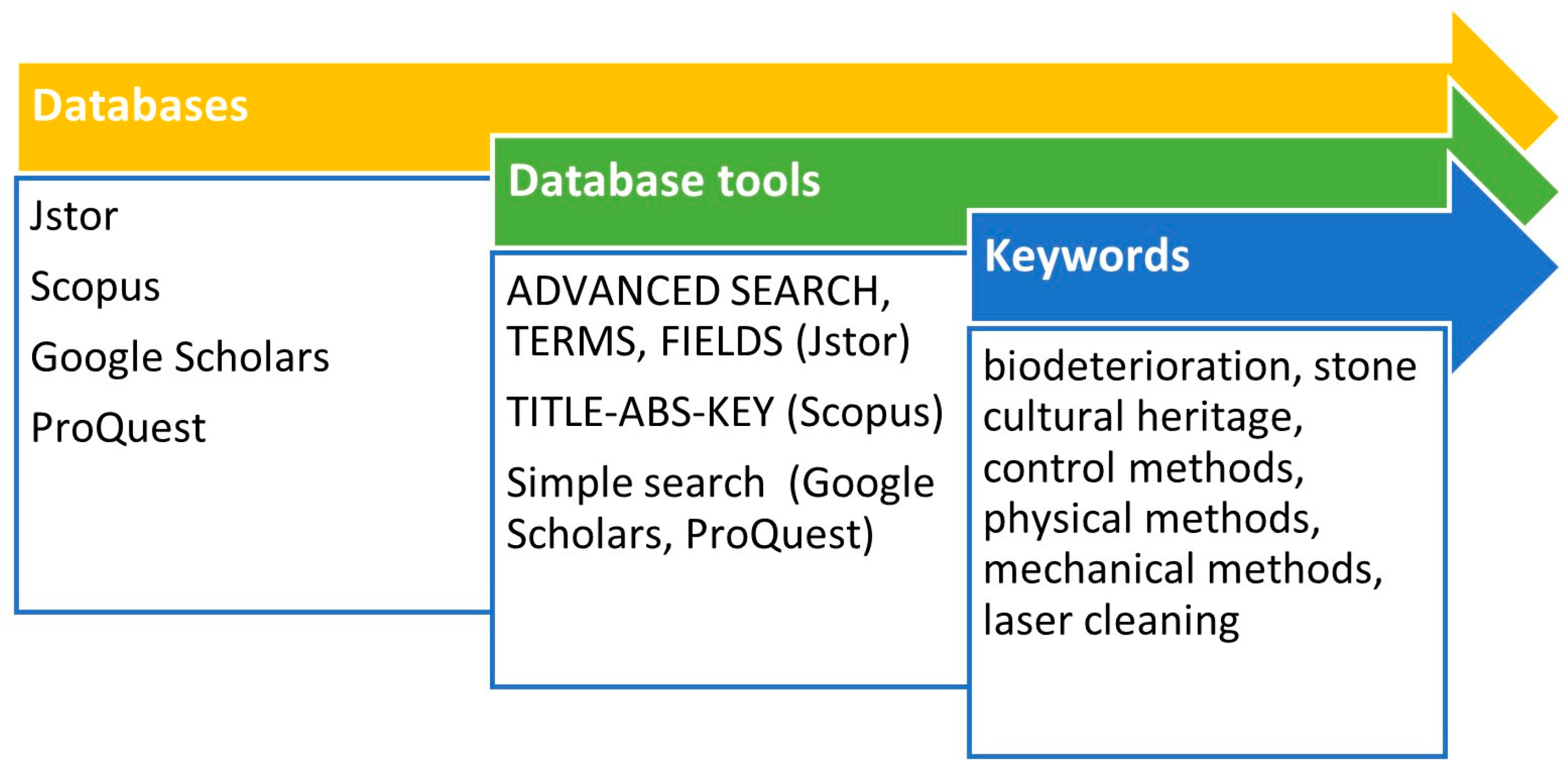
Figure 2.
Different patterns of the widespread colonization of phototrophic microorganisms on surfaces of Cueva del Indio (Cuba) (a,b), and Domus Aurea (Rome, Italy) (c).
Figure 2.
Different patterns of the widespread colonization of phototrophic microorganisms on surfaces of Cueva del Indio (Cuba) (a,b), and Domus Aurea (Rome, Italy) (c).
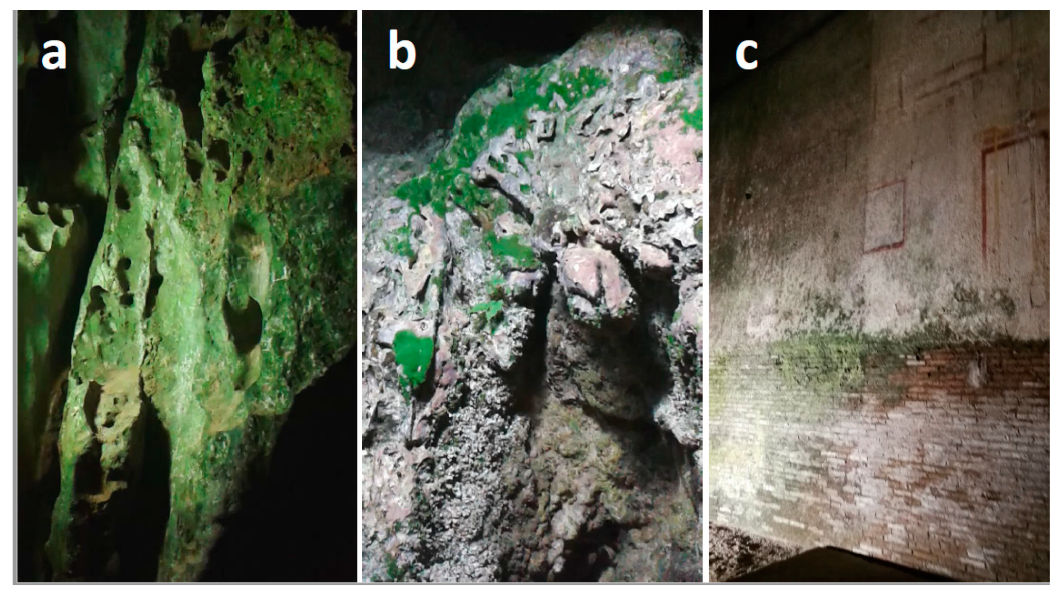
Figure 3.
Microwave treatment on two tombstones at the monumental English Cemetery (Firenze, Italy). (A) Crustose lichens and biofilms covered the marble tombstone compromising its legibility and obscuring the stone color. (B) Treatment of the lithobionts of the same tombstone with the microwave applicator (1) of the portable equipment that is connected to a device (2) for monitoring the surface temperature. (C) A marble tombstone partially covered by crustose lichens and biofilms. (D) Surface area (red circle) where the MW treatment was applied. (E) Macro photographs of the treated area before the treatment (1), immediately (2), and about five years (3) after the treatment. Bar = 1 cm.
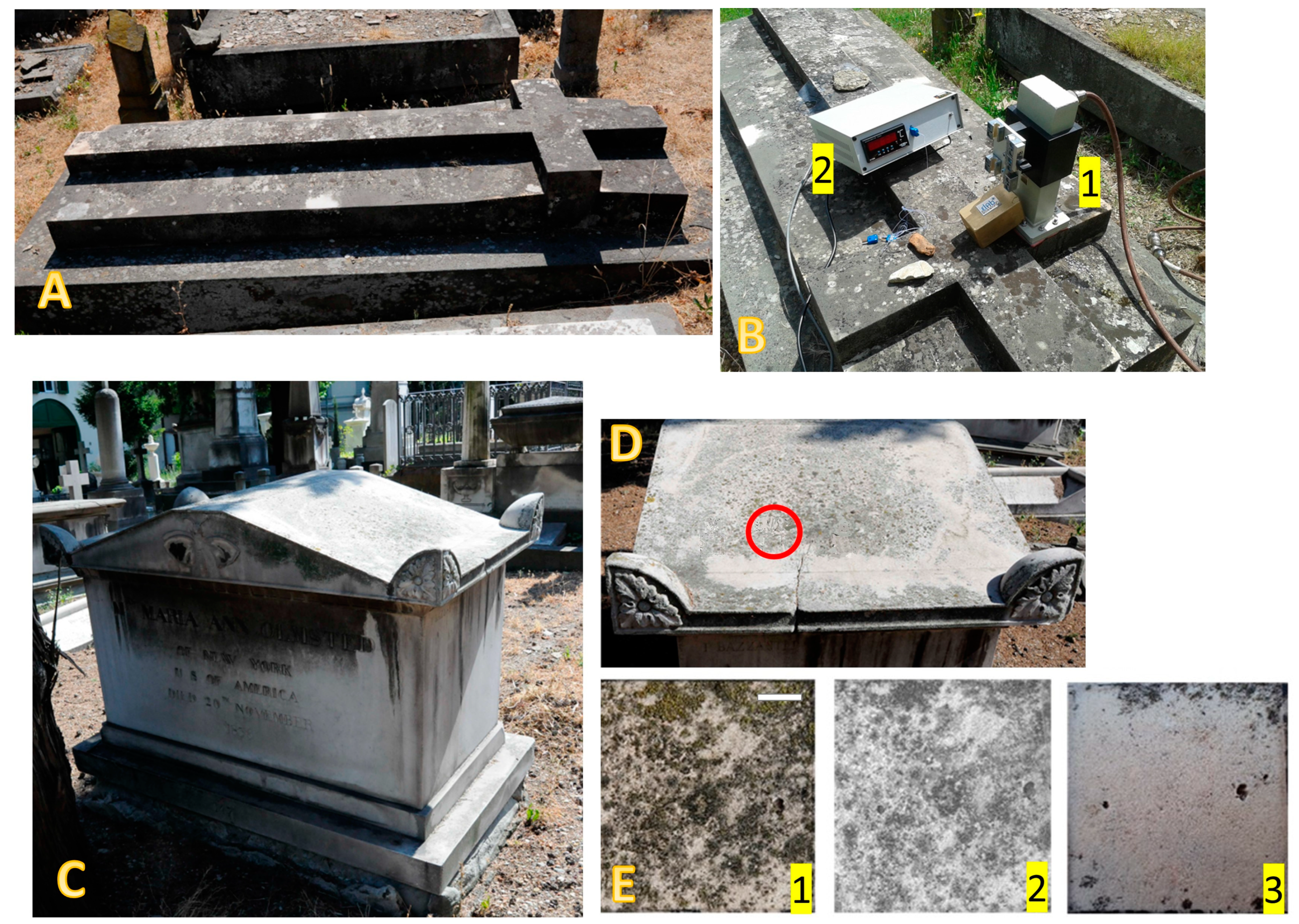
Figure 4.
A marble tombstone of the monumental English Cemetery in Florence (Italy). Close-up of an area with the lichen Circinaria caesiocinerea (a). The same area, three months after the lichen’s removal by using a soft scraper and deionized water (b). The same area, four and a half years after the mechanical treatment (c). The picture clearly shows that it was unsuccessful as the lichen regrowth pattern is identical to that before the treatment. Bar = 1 cm.
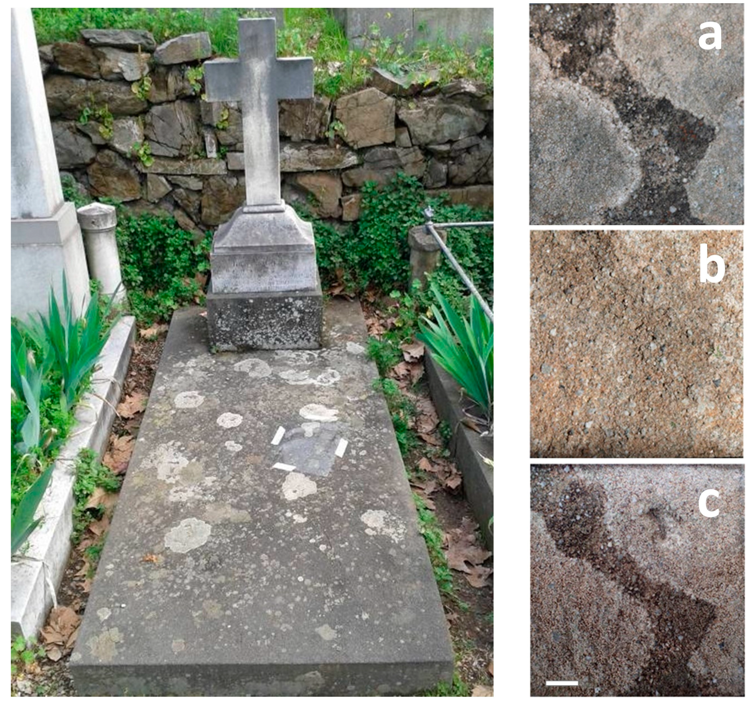
Table 4.
Some specific advantages and drawbacks of physical and mechanical methods.
| Pros | Cons | |
|---|---|---|
| UV-C radiation | The method is easy to carry out and relatively inexpensive. | It poorly penetrates inside substrates and in very thick biofilms. It can induce photooxidation in organic materials and it interacts with some pigments. Difficult to use in remote areas. |
| Monochromatic visible light | illumination systems in caves for the prevention of the growth of autotrophic microorganisms. | Controversial results on its efficacy. A site-specific study along with the characterization of the microorganisms is needed. |
| Microwaves | Eco-compatible. Low impact on the substrates. Localized effects. Low costs. | The equipment has limitations for large-scale applications. It requires access to energy supply. It only allows the irradiation of small surfaces (4 cm × 3 cm), and thus multiple adjacent applications are needed. |
| Laser cleaning | Selective, time-efficient, contactless, and environmentally friendly. Localized effects. | Discordant results on the efficiency in eliminating lithobionts. It can cause thermal-related damage and shockwaves propagating through the substrate. Costly. |
| Mechanical methods | Efficient in removing superficial layers. Efficient when combined with other cleaning methods. | Substrates can be severely damaged. Microbial fragments can remain on and inside the stone. The surfaces must be in a good state of conservation. Poorly effective in the long-term. |
Disclaimer/Publisher’s Note: The statements, opinions and data contained in all publications are solely those of the individual author(s) and contributor(s) and not of MDPI and/or the editor(s). MDPI and/or the editor(s) disclaim responsibility for any injury to people or property resulting from any ideas, methods, instructions or products referred to in the content. |
© 2024 by the author. Licensee MDPI, Basel, Switzerland. This article is an open access article distributed under the terms and conditions of the Creative Commons Attribution (CC BY) license (https://creativecommons.org/licenses/by/4.0/).
Share and Cite
MDPI and ACS Style
Pinna, D. Physical and Mechanical Methods for the Removal of Lithobionts—A Review. Coatings 2024, 14, 272. https://doi.org/10.3390/coatings14030272
AMA Style
Pinna D. Physical and Mechanical Methods for the Removal of Lithobionts—A Review. Coatings. 2024; 14(3):272. https://doi.org/10.3390/coatings14030272
Chicago/Turabian StylePinna, Daniela. 2024. "Physical and Mechanical Methods for the Removal of Lithobionts—A Review" Coatings 14, no. 3: 272. https://doi.org/10.3390/coatings14030272
Note that from the first issue of 2016, this journal uses article numbers instead of page numbers. See further details here.