Article

A Multifunctional Magnetic Fluorescent Nanoprobe for Copper(II) Using ZnS-DL-Mercaptosuccinic Acid-Modified Fe3O4 Nanocomposites

1
School of Chemical and Pharmaceutical Engineering, Jilin Institute of Chemical Technology, Jilin 132013, China
2
Center of Characterization and Analysis, Jilin Institute of Chemical Technology, Jilin 132013, China
*
Author to whom correspondence should be addressed.
These authors contributed equally to this work.
Coatings 2024, 14(6), 685; https://doi.org/10.3390/coatings14060685
Submission received: 13 May 2024 / Revised: 24 May 2024 / Accepted: 27 May 2024 / Published: 1 June 2024

Abstract

:
Cu2+ has increasingly become a great threat to the natural environment and human health due to its abundant content and wide application in various industries. DL-Mercaptosuccinic acid and ZnS-modified Fe3O4 nanocomposites were designed, synthesized, and applied in the determination of Cu2+. The prepared nanocomposites were characterized by scanning electron microscopy (SEM), transmission electron microscopes (TEM), Fourier transform infrared spectroscopy (FTIR), X-ray diffractometer (XRD), X-ray photoelectron spectroscopy (XPS), vibrating sample magnetometer (VSM), and thermogravimetric analyzer (TG). The magnetic fluorescent nanoprobe exhibited highly selective and sensitive fluorescence-quenching characteristics with Cu2+ ions. The fluorescence detection linear range was 0–400 μM, with the detection limit being 0.489 μM. In addition, the magnetic fluorescent nanoprobe exhibited a high adsorption and removal rate for Cu2+. It had been successfully applied to detect Cu2+ in real water samples with a satisfactory recovery rate. The magnetic fluorescent nanoprobe could simultaneously realize the functions of enrichment, quantitative detection, and separation, reduce the pollution of copper ions and probes, and establish an environment-friendly detection method. Consequently, the magnetic fluorescent nanoprobe offered a new pathway for the removal and detection of not only Cu2+ but also other heavy metal ions in water.

1. Introduction

Copper is an essential trace element and active component in the human body and plays an important role in signal pathways, multiple cell matrices, and other varieties of chemical, biological, and environmental systems [1,2,3]. It is an important catalytic cofactor for many metalloenzymes, including tyrosinase, ceruloplasmin, cytochrome, superoxide dismutase c-oxidase, and carbon monoxide dehydrogenases [4]. At the same time, low concentrations of copper absorption are necessary factors for the typical growth of active systems. Another biological importance of copper is that it is used randomly for agricultural, industrial, medicinal, and domestic purposes. Despite their importance, the wide use of Cu2+ in industry, which leads to excessive Cu2+ ions in the body, causes mild toxicity to the living organism, organ damage, and disruption of the body’s metabolism. It can cause various detrimental effects, including Alzheimer’s disease, asthma, pneumonitis, and central nervous system disorders, as well as Wilson’s disease, lung cancer, and Parkinson’s disease [5]. Therefore, the contamination of water bodies by copper ions (Cu2+) has become a significant component of environmental pollutants and an increasing threat to ecological systems and human health. The acceptable concentration of Cu2+ ions in portable water, recommended by the World Health Organization (WHO), should be below 31.5 μM [6]. Therefore, research on the detection and determination of Cu2+ ions has attracted more and more attention. The development of a fast and convenient method for the identification of Cu2+ has become a particularly important demand.
Though some traditional analytical methods with high sensitivity, such as graphite furnace atomic absorption spectrometry (GFAAS) [7], inductively coupled plasma emission spectroscopy (ICP-ES) [8], atomic absorption spectroscopy (AAS) [9], and chromatography, have been introduced to detect Cu2+ ions, they are time-consuming, have high repairing costs of tools, require expensive instruments and trained operators, and have complicated sample-making processes. These significant drawbacks make them not very convenient to use and apply.
In this situation, the selective and sensitive detection of Cu2+ has gradually become an important environmental chemistry research area. The fluorometric probes for recognizing Cu2+ ions have gained more and more attention in recent years; they have become a powerful sensing tool due to their easy operation, fast response time, high sensitivity, excellent selectivity, and technical simplicitywith an obvious signal that can be seen with the naked eyeand potential applications in biological and environmental systems [10,11]. Various small molecule fluorophores have been designed as probes to detect heavy metal ions based on fluorescence change; for example, single organic fluorophore molecules [12,13], fluorescent polymers [14], quantum dots [15], and covalent organic frameworks [2]. However, most of these fluorescent methodologies might be disturbed by changes in environmental conditions like the pH and other paramagnetic metal ions and cannot eliminate interference from other metal ions effectively, which greatly limits their potential applications. More importantly, these fluorescent probes cannot be separated from the detection system, which introduces further pollution to the environment. In this regard, developing versatile fluorescent probes to selectively detect and effectively remove heavy metal ions would be highly necessary.
As the Fe3O4 particles are especially reactive and their magnetic forces are reduced, the Fe3O4 particles are usually coated with a solid layer and thus build up a core–shell structure to prevent this reduction. Core–shell-nanostructure Fe3O4 nanoparticles (Fe3O4NPs) have been extensively studied and widely utilized in biosensors or adsorbents to remove dyes, heavy metals ions, and pharmaceuticals because of their facial preparation, superparamagnetic property, high facial magnetic recovery, high surface area, size regulation, high adsorption ability, biocompatibility, and easy preparation [16,17,18]. Thus far, more and more surface-coating Fe3O4 nanoparticles have been synthesized to improve the biocompatibility and multi-function. Zinc sulfide (ZnS) is one of the most commonly used as a solid coat due to its good thermal stability, high electric mobility, excellent transport properties, and the presence of polar surfaces [19,20]. Many new nanomaterials combined ZnS with various nanostructures and have been used in a wide range of applications because of their unique physical properties. At the same time, ZnS has been widely used in the design of fluorescence sensors because of its excellent thermal stability, adjustable fluorescence characteristics, high fluorescence quantum yield, as well as its non-toxic, stable chemical structure [21,22,23].
Considering that most fluorescent probes will bring secondary pollution to the environment when recognizing copper, we applied Fe3O4 as the core structure of the probe for the magnetic separation property and ZnS as the shell structure of the probe, and then a fluorescence probe with multiple functions of enrichment, separation, and detection was designed to solve the problem (Scheme 1). This magnetic fluorescent nanoprobe showed high sensitivity and selectivity to Cu2+, and there was a good linear relationship in the concentration range from 0 to 400 μM, with the limit of detection (LOD) being 0.489 μM. This magnetic fluorescent nanoprobe was successfully used to detect Cu2+ in actual water samples with a satisfactory recovery rate. The magnetic fluorescent nanoprobe could enrich, detect, and separate the heavy metal ions at the same time and reduce secondary pollution caused by the probe in an environmentally friendly way with high selectivity. It also provided a new way to detect and treat other heavy metal ions in water.

2. Materials and Methods

2.1. Materials

Ferric chloride (FeCl3, ≥99%), trihydrate sodium acetate (CH3COONa·3H2O, ≥99%), ethylene glycol(EG, ≥99%), zinc acetate (Zn(ac)2, 99%), sodium sulfide (Na2S, 99%), polyethylene glycol 2000, and DL-Mercaptosuccinic acid were obtained from Shanghai Aladdin Bio-Chem Technology Co., Ltd. (Shanghai, China).

2.2. Adsorbent Characterization

Fe3O4@ZnS@DL-Mercaptosuccinic acid nanoparticles were analyzed by SEM (Quanta 200, FEI, Waltham, MA, USA) and TEM (JEM 2100, JEOL, Tokyo, Japan) to characterize their morphological structures. The FTIR (Thermo Scientific Nicolet IS50, Thermofisher, Waltham, MA, USA), XRD (D8FOCUS, Bruker, Berlin, Germany), and XPS (Krayos AXIS Ultra DLDX, Shimadzu, Kyoto, Japan) analyses were used to determine the crystal structures and chemical compositions of the Fe3O4@ZnS-COOH nanoparticles. A VSM (7404 vibrating sample magnetometer, LakeShore, Carson, CA, USA) was used to acquire the magnetic characterization of the nanoparticles. TG (Discovery SDT650 synchronous TGA-DTA instrument, TA, New Castle, DE, USA) was used to determine the groups of DL-Mercaptosuccinic acid groups on the surface of the materials. A fluorescence spectrometer was used to measure the fluorescence intensity of Fe3O4@ZnS-COOH. AAS (ICP-OES:Thermo Fisher iCAP 7400, Thermofisher, Waltham, MA, USA) was used to measure the concentrations of Cu2+ in the adsorption solution.

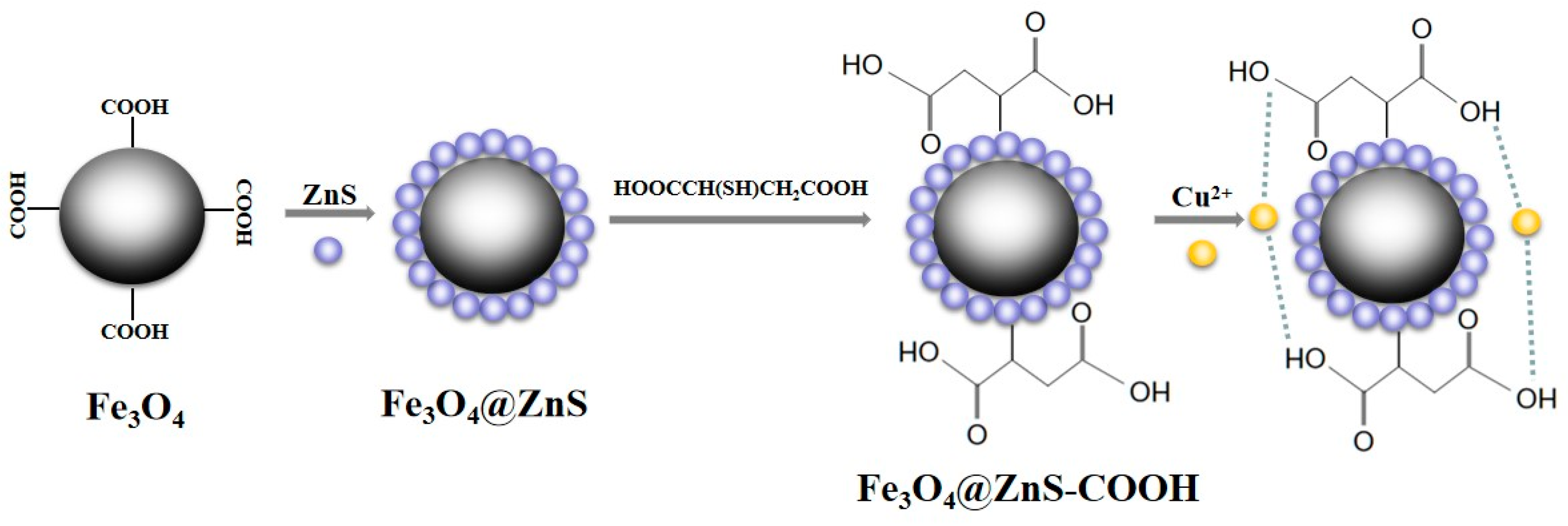
2.3. Synthesis of Fe3O4 Nanoparticles

FeCl3 (2.7 g) and CH3COONa·3H2O (7.2 g) were dispersed in EG (60 mL). Magnetic stirring (30 min) and sonication (30 min) were performed to promote dispersion. Then, the solution mixture was transferred to a 100 mL autoclave and heated at 200 °C for 12 h. After the solution was left to cool to 25 °C in the autoclave, the Fe3O4 NPs were obtained by magnetic separation, washed with distilled water and ethanol, and then oven-dried at 80 °C for 6 h for further modification.

2.4. Synthesis of Fe3O4@ZnS Nanoparticles

Fe3O4 and ZnS, with a certain weight ratio, were dissolved in deionized water, and then the two solutions were mixed. The reaction was continuously stirred at 80 °C for 60 min and then ultrasonically treated at 50 °C for 90 min. Subsequently, the resulting product was washed at least once by centrifugation at 1500 rpm for 12 min using deionized water and ethanol. Finally, the collected black precipitate was dried at 60 °C for 12 h, and then the product was ground into fine powder to prepare the Fe3O4@ZnS magnetic fluorescent nanoparticles.

2.5. Synthesis of Fe3O4@ZnS-COOH Nanoparticles

A certain amount of Fe3O4@ZnS was taken to make it into an ethanol dispersion. A total of 5 mL of the Fe3O4@ZnS ethanol solution was placed into a round-bottom flask. Then, 5 mL of the DL-Mercaptosuccinic acid (75.074 mg, 0.1 mmol/L) ethanol solution was added. Then, the mixture was stirred in a water bath at 40 °C for 4 h in the dark. After the item was attracted by a magnetic force, it then underwent a thorough cleansing process with ultrapure water and ethanol. The prepared Fe3O4@ZnS-COOH microspheres were added to water to make a dispersion solution.

2.6. The Selective and Competitive Experiments

The interactions between Fe3O4@ZnS-COOH (0.2 mg/mL) and different metal ions (Cu2+, Co2+, Pb2+, Ni2+, Hg2+, Al3+, Cd2+, Zn2+, Fe2+, Fe3+, K+, Ca2+, and Na+) were investigated by designing selective experiments. In addition, the influence of different types of metal ions (Cu2+, Co2+, Pb2+, Ni2+, Hg2+, Al3+, Cd2+, Zn2+, Fe2+, Fe3+, K+, Ca2+, and Na+) on the interaction of Fe3O4@ZnS-COOH with Cu2+ was also explored. The concentrations of Cu2+ and different metal ions were 400 μM.

2.7. Adsorption Experiments

First, a Fe3O4@ZnS-COOH aqueous suspension was prepared using sonication with the pH regulated to 7.0. For the adsorption experiment, the Fe3O4@ZnS-COOH (0.2 mg/mL) suspension and different Cu2+ ion solutions were added to a conical flask and placed in a thermostatic shaker (200 rpm) for 12 h at room temperature. After the adsorption process was complete, the Fe3O4@ZnS-COOH was separated by an external magnet. The ion adsorption amount (Q, mg/g) was determined and calculated using the equation Q = (C0 − C) × V/m. Then, the remove rate (R, %) was calculated by the equation: R = (C0 − C)/C0, where m (g) defines the mass of Fe3O4@ZnS-COOH, C0 (mg/L) is the initial concentration of Cu2+ ions in the solution, C (mg/L) is the final Cu2+ ion concentration in the solution, and V (L) is the volume of the solution.

3. Results and Discussion

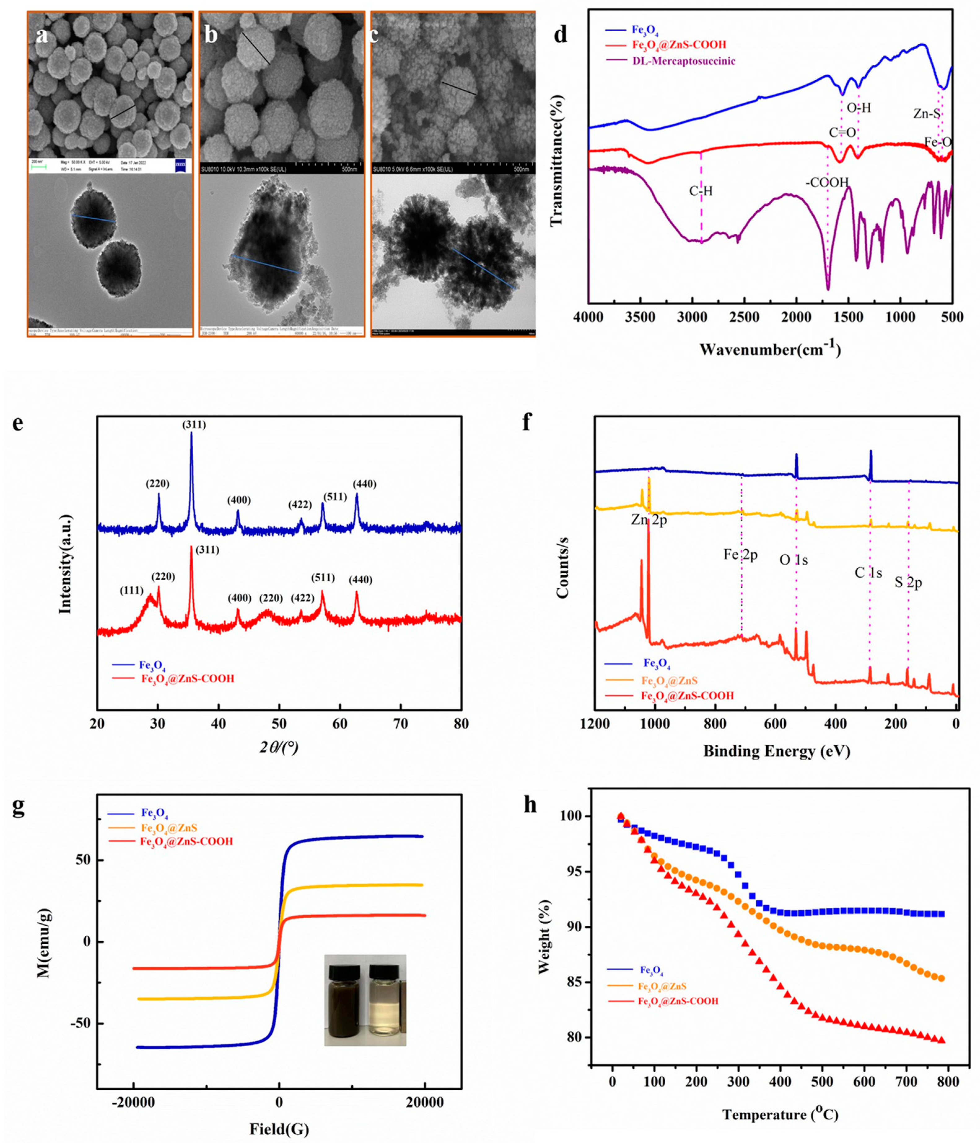
3.1. Characterization of Fe3O4, Fe3O4@ZnS, and Fe3O4@ZnS@ DL-Mercaptosuccinic

The morphologies of Fe3O4, Fe3O4@ZnS, and Fe3O4@ZnS@DL-Mercaptosuccinic acid were analyzed by SEM (first line) and TEM (second line). As shown in Figure 1a–c, with the number of layers increased, all materials were still spherical. The commercial Fe3O4, with a diameter distribution from 150 to 250 nm, reveals that Fe3O4 MNPs have a regular and uniform distribution. The particle diameter of the three samples increased, accompanied by more rougher surfaces and more irregular fine particles. In addition, a TEM image (Figure 1c second line) of Fe3O4@ZnS-COOH, with a diameter distribution from 200 to 300 nm, was achieved. It can be concluded that the introduction of ZnS and DL-Mercaptosuccinic acid improved the crystal growth process of Fe3O4, leading to the changes in morphologies observed in materials.
FTIR spectroscopy was applied to identify the functional groups of these nanoparticles, and the results are illustrated in Figure 1d. It can be seen in Figure 1d that the FTIR spectra of the bare ZnS showed characteristic peaks at 640 and 716 cm−1. The spectra of the Fe3O4@ZnS-COOH composites were highly similar to ZnS at these two peaks. The typical peaks around 584 cm−1, referring to the Fe-O bond vibration, could also be found in Fe3O4 and Fe3O4@ZnS-COOH. Finally, the peaks at around 1550 cm−1 were attributed to the stretching vibration of the C=O bonds that may originate from the residual Fe3O4 on the surface of the particles [24]. As can be seen from the comparison of Fe3O4@ZnS-COOH and DL-Mercaptosuccinic acid, two typical peaks at about 1728 cm−1 and about 2930 cm−1 could be found. The peaks at about 1728 cm−1 and 2930 cm−1 were assigned to the vibration of the -COOH and C-H bonds, which are supposed to stem from the residue of DL-Mercaptosuccinic acid. The results proved that the magnetic nanoparticles Fe3O4@ZnS@ DL-Mercaptosuccinic acid had been successfully synthesized.
To determine the crystal structure of the synthesized material, XRD analysis was used to characterize Fe3O4 and Fe3O4@ZnS. The results are illustrated in Figure 1e. The results showed that the peaks at about 30.1°, 35.6°, 43.1°, 53.7°, 57.1°, and 62.8° were found in all diffraction patterns, which could be attributed to the (220), (311), (400), (422), (511) and (440) planes of Fe3O4, respectively. The peaks at about 28.9°, 47.7°, and 57.0° were attributed to the characteristic peaks of (111), (220), and (311) of ZnS [25]. The peaks of the Fe3O4@ZnS (in Figure 1e) could be seen with the slightly decreased intensity of typical peaks, and the positions of the peaks were unchanged. The results suggested that the coating of the ZnS and DL-Mercaptosuccinic acid layers did not disrupt the crystalline structure of Fe3O4.
Figure 1f shows the XPS full spectrum of the Fe3O4, Fe3O4@ZnS, and Fe3O4@ZnS@ DL-Mercaptosuccinic acid nanoparticles, indicating the presence of five elements (Fe, O, Zn, S, and C). The XPS spectrum of the Fe 2p characteristic peak was at 709.8 eV. The XPS spectrum of O1s, with a peak at 529.6 eV, is attributed to the O and Fe-O bonds. The binding energy peaks at 1019.2 eV are attributed to the spin–orbital splitting of Zn2+ [26]. The binding energy peaks at 159.2 eV correspond to S 2p3/2 and S 2p1/2, indicating S2- in the samples. The peaks at the binding energies of 282.7 eV come from the carbon and carboxyl functional groups of DL-Mercaptosuccinic acid.
The response of the materials to an external applied magnetic field is decided by saturation magnetization, which significantly affects the efficiency of magnetic separation in aqueous solution. Therefore, these three types of magnetic nanoparticles were analyzed by VSM, and the results are shown in Figure 1g. With the externally applied magnetic field changing from −20 kOe to 20 kOe, the saturation magnetization of the samples decreased from 64.5 ± 0.1 emu/g to 34.7 ± 0.1 emu/g and 16.3 ± 0.1 emu/g, along with the core–shell structure thickening gradually. They all showed typical hysteresis loops and superparamagnetic properties. Though the enlarged particle diameter hampered the magnetic performance of Fe3O4, Fe3O4@ZnS@DL-Mercaptosuccinic still possessed excellent magnetism, making this magnetic nanomaterial a promising detection and separation tool.
These three types of magnetic nanoparticles were estimated by TGA, and the results are shown in Figure 1h. The initial weight loss at 20–140 °C was ascribed to the evaporation of water molecules in the polymer matrix. The second weight loss that occurred between 120 and 800 °C was about 14%, which could be attributed to polymer chain degradation. From Fe3O4 to Fe3O4@ZnS@DL-Mercaptosuccinic, the total weight loss was about 8.0%, 14.0%, and 21%, respectively.
In combination with the above experimental results, it is proved that the magnetic fluorescence nanoparticle Fe3O4@ZnS@DL-Mercaptosuccinic (Fe3O4@ZnS-COOH) had been successfully synthesized.

3.2. Spectroscopic Characteristics of the Magnetic Fluorescence Nanoprobe

The spectroscopic characteristics of the magnetic fluorescence nanoprobe with Cu2+ were investigated first (Figure 2). When excited at 370 nm, the maximal strong fluorescence emission peak of the magnetic fluorescence nanoprobe at 425 nm could be observed. When Cu2+ is added, the fluorescence intensity is greatly quenched. The result indicated that the probe could be used as a fluorescent probe to detect Cu2+.
Based on the result that the Cu2+ ion can react with this magnetic fluorescence nanoprobe and quench its fluorescence intensity, the fluorescence intensity before and after adding different concentrations of Cu2+ was measured and calculated as a function of the Cu2+ concentration. In Figure 3, the fluorescence intensity value shows an excellent linear relationship with the Cu2+ concentration in 0–400 μM (R2 = 0.9906), and the LOD was calculated to be 0.489 μM. The LOD value was much lower than the current metal concentration limits in drinking water (~20 μmol/L), which is regulated by the U.S. Environmental Protection Agency [14]. As summarized in Table 1, based on the detection range and LOD, the modified magnetic fluorescence nanoprobe exhibits a wide detection range and excellent LOD.

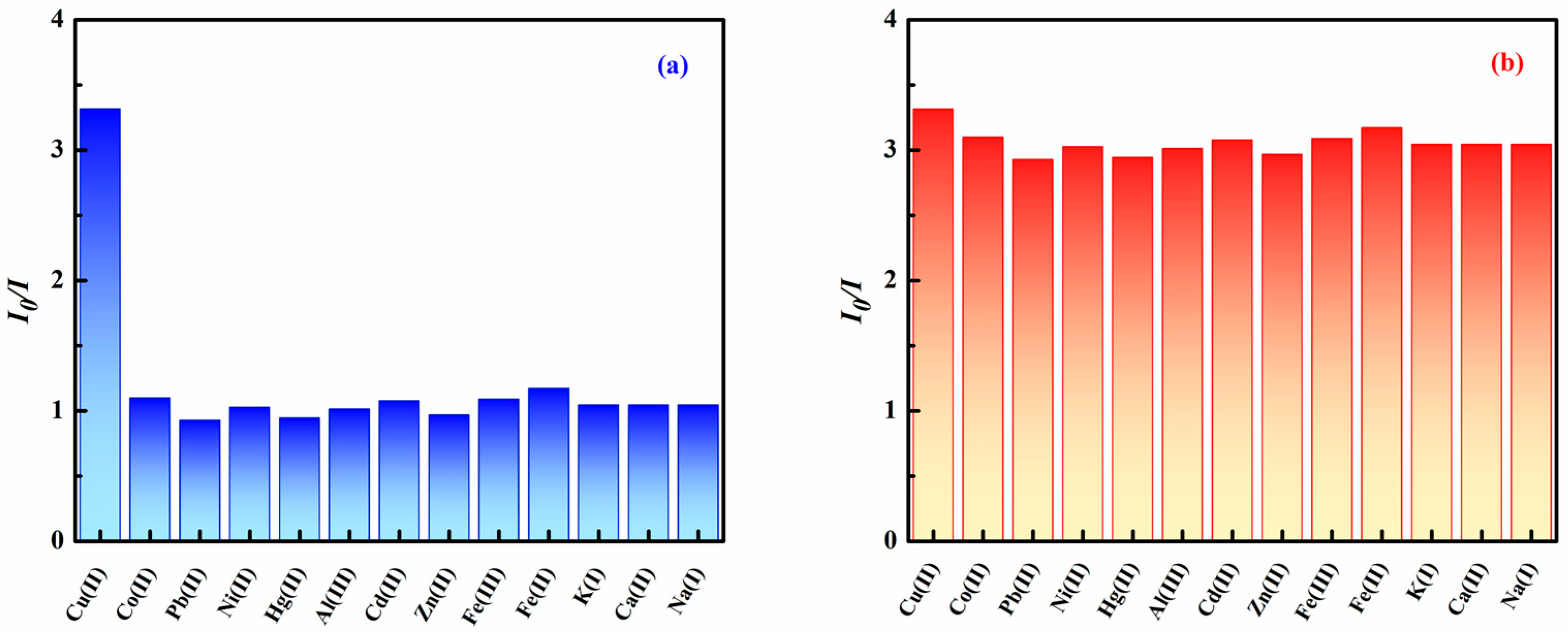
3.3. Specificity and Anti-Interference Capability of the Magnetic Fluorescent Nanoprobe

A new type of magnetic fluorescent nanoprobe was developed, which recognizes Cu2+ through the fluorescence quench of the probe. The response effect of the magnetic fluorescent nanoprobes among a variety of metal ions (Cu2+, Co2+, Pb2+, Ni2+, Hg2+, Al3+, Cd2+, Zn2+, Fe2+, Fe3+, K+, Ca2+, and Na+) were measured using a fluorescence spectrophotometer, respectively (Figure 4a). With the addition of metal ions, other metal ions except Cu2+ weakly quenched the fluorescent intensity of the magnetic fluorescent nanoprobe. When added with Cu2+, the fluorescent intensity of the magnetic fluorescent nanoprobe decreased significantly. Consequently, the recognition of Cu2+ by the magnetic fluorescent nanoprobe can be fulfilled quickly and effectively. It is a competitive fluorescent nanoprobe for the sensitive and selective determination of Cu2+.
Considering that when the magnetic fluorescent nanoprobes were used to detect Cu2+ in water, it may be interfered with by other metal ions; thus, the interference experiment needs to be evaluated. The common metal ions (Cu2+, Co2+, Pb2+, Ni2+, Hg2+, Al3+, Cd2+, Zn2+, Fe2+, Fe3+, K+, Ca2+, and Na+) in water were selected for the test as follows: the interfering ions (400 μM) and Cu2+ (400 μM) were added to the solution of magnetic fluorescent nanoprobes solution (0.2 mg/mL), and the fluorescence spectra were tested. The obtained results are shown in Figure 4b. The results showed that these metal ions did not introduce too much interference to the detection and did not affect the selectivity of the probe. These results demonstrated the high selectivity of the magnetic fluorescent nanoprobe to Cu2+ against metal ions. It possesses many potential applications in detecting Cu2+, both in the biological and physiological fields.

3.4. Adsorption Capacity and Removal Rate Study

The adsorption capacity is one of the most important properties of nanomaterials, so adsorption tests were performed in a centrifuge tube (10 mL) to provide the adsorption amount of the magnetic fluorescent nanoprobe. As the results listed in Table 2 show, the magnetic fluorescent nanoprobe showed excellent adsorption capacity (310.239 mg/g) and a high removal rate (97.64%).

3.5. The Practicability of Magnetic Fluorescent Nanoprobe on the Detection of Cu2+

To validate the applicability of the magnetic fluorescent nanoprobe for determining Cu2+, three actual water samples (tap water, bottled water, and electrolysis wastewater) were analyzed. River water samples were obtained from a local Songhua river (Jilin), and the tap water samples were collected from Jilin Water Group Co., Ltd. (Jilin, China). All the water samples needed to be filtered with a 0.45 mm water-phase microporous membrane and were analyzed at least three times. In these actual samples, the Cu2+ could be detected directly, and the results are summarized in Table 3. The recoveries for real Cu2+ detection are in the range of 83.00%–108.00% with relative standard deviation (RSD) values below 4.53%. The results confirm the good performance of the magnetic fluorescent nanoprobe for real sample detection.

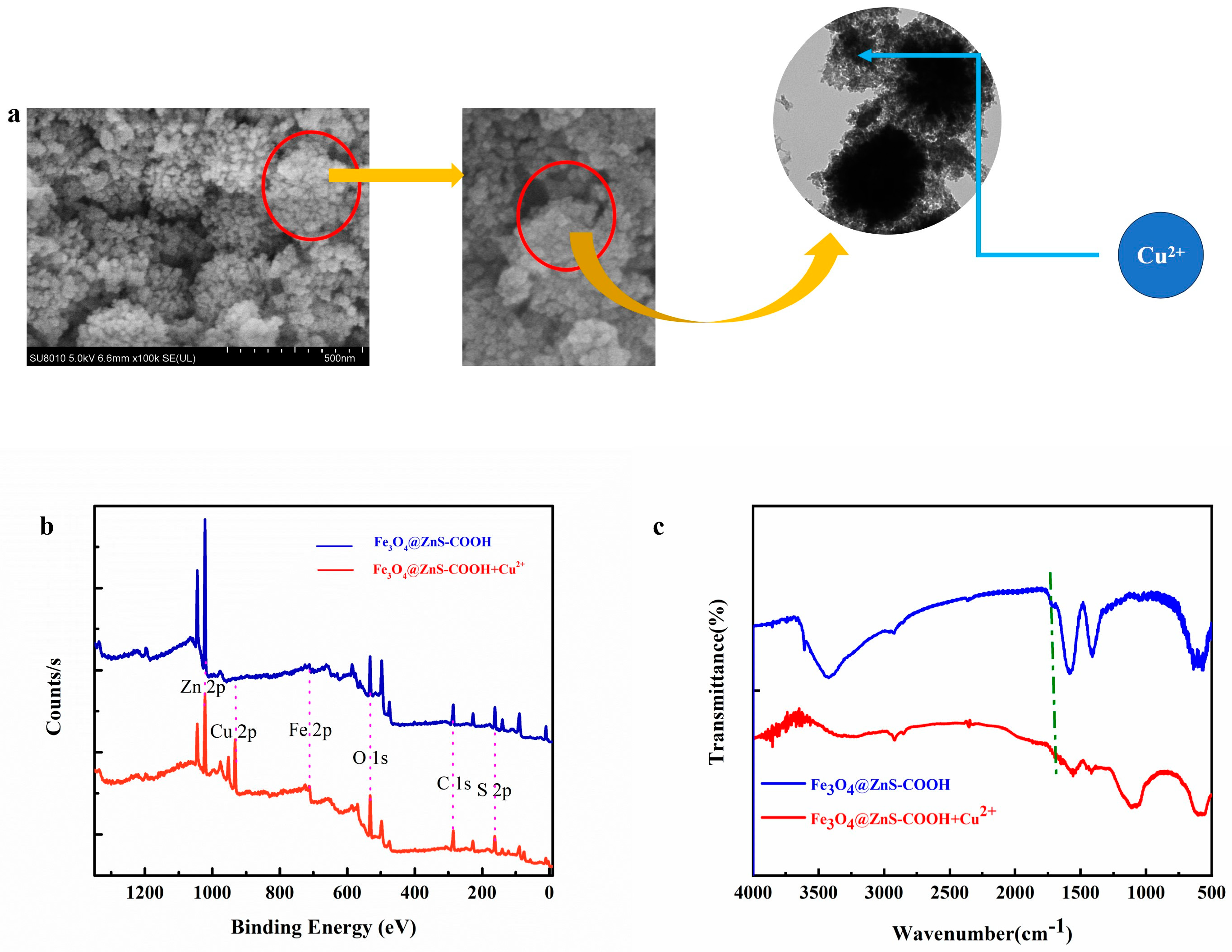
3.6. Detection Mechanism

The Cu2+-binding mechanism was schematically illustrated in Scheme 2. The analyses of the functional groups and morphological features were examined using SEM, FTIR, and XPS spectra to confirm how Cu2+ ions were detected by the magnetic fluorescent nanoprobe.
After interacting with Cu2+, metal particle deposition was found on the surface of the magnetic fluorescent nanoprobe, suggesting that Cu2+ ions have adhered successfully to the surface of the magnetic fluorescent nanoprobe (Scheme 2a). We analyzed the changes in the XPS spectra of the magnetic fluorescent nanoprobe before and after adding Cu2+ to further prove the detection mechanism of the magnetic fluorescent nanoprobe (Scheme 2b). The XPS full-scanning spectra of the magnetic fluorescent nanoprobe and the magnetic fluorescent nanoprobe after the detection of Cu2+ exhibited a new peak (932.6 eV), which was assigned to Cu 2p [37], confirming that Cu2+ had been adsorbed on the surface of the magnetic fluorescent nanoprobe. Finally, as can be seen in Scheme 2c, the position of -COOH-containing groups in the Fe3O4@ZnS-COOH is blue-shifted from 1728 cm−1 to 1683 cm−1 following Cu2+ adsorption. The reason for the change in the infrared absorption peak of the carboxyl group may be that the oxygen atoms in the carboxyl group cooperate with Cu2+ to form a synergistic complex. The oxygen atom of the carboxyl group is negatively charged, indicating that the -COOH group can adsorb Cu2+ through an electrostatic interaction. It is speculated that the oxygen atom of the -COOH group provides electrons to Cu2+, resulting in a charge transfer, thus resulting in changes in the infrared spectrum [34].

4. Conclusions

In general, DL-Mercaptosuccinic acid-modified Fe3O4@ZnS was prepared by the solvothermal method and was designed as a multi-functional magnetic fluorescent nanoprobe for the sensitive and selective detection of Cu2+. The results of SEM, TEM, FRIT, XRD, and XPS proved that the magnetic fluorescent nanoprobe had been successfully synthesized. The fluorescence assay showed outstanding detection performance, with a wide linear range (0–400 μM), high linearity (R2 = 0.9906), and low LOD (0.489 μM). The probe has been successfully applied to the determination of Cu2+ in real water samples and has realized three functions: enrichment, detection, and separation. Herein, this magnetic fluorescent nanoprobe developed a three-in-one functional detection system, aiming at providing an environmentally friendly probe by modifying the recognized probe for other heavy metal ions.

Author Contributions

Data curation, P.X., X.C. and X.Z.; Writing—original draft, X.C.; Writing—review & editing, X.Z.; Project administration, Z.L.; Funding acquisition, J.C. and S.Y. All authors have read and agreed to the published version of the manuscript.

Funding

This research was funded by the Jilin Provincial Department of Science and Technology (20220203020SF) and the National Natural Science Foundation of China (51902125, 22106051).

Institutional Review Board Statement

Not applicable.

Informed Consent Statement

Not applicable.

Data Availability Statement

Data are contained within the article.

Conflicts of Interest

The authors declare no conflict of interest.

References

  1. Amoah, C.; Obuah, C.; Ainooson, M.K.; Hamenu, L.; Oppong, A. A new sulfonamide-based chemosensor for potential fluorescent detection of Cu2+ and Zn2+ ions. Tetrahedron 2023, 133, 133276. [Google Scholar] [CrossRef]
  2. Li, S.X.; Zhao, B.; Kan, W.; Song, T.S.; Zheng, W.; Qi, X.; Wang, L.Y.; Song, B. A fluorescent covalent organic framework with a brick-wall topology for the detection of Cu2+ promoted by the associated mechanism. Dye. Pigment. 2023, 214, 111178. [Google Scholar] [CrossRef]
  3. Choudhury, N.; Saha, B.; De, P. Recent progress in polymer-based optical chemosensors for Cu2+ and Hg2+ Ions: A comprehensive review. Eur. Polym. J. 2021, 145, 110233. [Google Scholar] [CrossRef]
  4. Segura, C.; Yañez, O.; Galdámez, A.; Tapia, V.; Núñez, M.T.; Osorio-Román, I.; García, C.; García-Beltrán, O.J. Synthesis and characterization of a novel colorimetric and fluorometric probe “Turn-on” for the detection of Cu2+ of derivatives rhodamine. Photoch. Photobio. A 2023, 434, 114278. [Google Scholar] [CrossRef]
  5. Liu, L.; Cui, Y.Y.; Yang, Y.X.; Zhu, W.J.; Li, C.; Fang, M. A novel lipid droplets/lysosomes-targeting colorimetric and ratiometric fluorescent probe for Cu2+ and its application. Spectrochim. Acta A 2023, 291, 122333. [Google Scholar] [CrossRef] [PubMed]
  6. Jia, Y.; Sun, T.; Jiang, Y.; Sun, W.; Zhao, Y.; Xin, J.; Hou, Y.; Yang, W. Green, fast, and large-scale synthesis of highly fluorescent Au nanoclusters for Cu2+ detection and temperature sensing. Analyst 2018, 143, 5145–5150. [Google Scholar] [CrossRef] [PubMed]
  7. Kumamaru, T.; Okamoto, Y.; Hara, S.; Matsuo, H.; Kiboku, M. Simultaneous determination of copper and lead by graphite-furnace atomic absorption spectrometry after liquid-liquid extraction of the ion pair with zephiramine. Anal. Chim. Acta 1989, 218, 173–178. [Google Scholar] [CrossRef]
  8. Ebrahimi-Najafabadi, H.; Pasdaran, A.; Bezenjani, R.R.; Bozorgzadeh, E. Determination of toxic heavy metals in rice samples using ultrasound assisted emulsification microextraction combined with inductively coupled plasma optical emission spectroscopy. Food Chem. 2019, 289, 26–32. [Google Scholar] [CrossRef] [PubMed]
  9. Es’haghi, Z.; Azmoodeh, R. Hollow fiber supported liquid membrane microextraction of Cu2+ followed by flame atomic absorption spectroscopy determination. Arab J. Chem. 2010, 3, 21–26. [Google Scholar] [CrossRef]
  10. Zhao, L.X.; Chen, K.Y.; Xie, K.B.; Hu, J.J.; Deng, M.Y.; Zou, Y.L.; Gao, S.; Fu, Y.; Ye, F. A benzothiazole-based “on-off” fluorescence probe for the specific detection of Cu2+ and its application in solution and living cells. Dye. Pigment. 2023, 210, 110943. [Google Scholar] [CrossRef]
  11. Loya, M.; Ghosh, S.; Atta, A.K. A review on dual detection of Cu2+ and Ni2+ ions by using single fluorometric and colorimetric organic molecular probes. J. Mol. Struct. 2023, 1278, 134949. [Google Scholar] [CrossRef]
  12. Zhang, J.Q.; Yao, G.X.; La, Y.T.; Dong, W.K. Structural insights into three novel Cu(II), Ni(II) and Zn(II) complexes derived of a pyridine-containing N4-cavity bisoxime ligand. Inorg. Chim. Acta 2022, 533, 120775. [Google Scholar] [CrossRef]
  13. Meng, Z.Y.; Wang, Z.L.; Liang, Y.Y.; Zhou, G.C.; Li, X.Y.; Xu, X.; Yang, Y.Q.; Wang, S.F. A naphthalimide functionalized chitosan-based fluorescent probe for specific detection and efficient adsorption of Cu2. Int. J. Biol. Macromol. 2023, 239, 124261. [Google Scholar] [CrossRef] [PubMed]
  14. Zhang, Y.Y.; Zhu, T.; Xu, Y.; Yang, Y.; Sheng, D.L.; Ma, Q.L. Quaternized salicylaldehyde Schiff base side-chain polymer-grafted magnetic Fe3O4 nanoparticles for the removal and detection of Cu2+ ions in water. Appl. Surf. Sci. 2023, 611, 155632. [Google Scholar] [CrossRef]
  15. Liu, Y.Q.; Seidi, F.; Deng, C.; Li, R.Y.; Xu, T.T.; Xiao, H.N. Porphyrin derived dual-emissive carbon quantum dots: Customizable synthesis and application for intracellular Cu2+ quantification. Sensor Actuat. B 2021, 343, 130072. [Google Scholar] [CrossRef]
  16. Hubetska, T.S.; Kobylinska, N.G.; Menendez, J.R.G. Application of Hydrophobic Magnetic Nanoparticles as Cleanup Adsorbents for Pesticide Residue Analysis in Fruit, Vegetable, and Various Soil Samples. J. Agric. Food Chem. 2020, 68, 13550–13561. [Google Scholar] [CrossRef]
  17. Naini, N.; Kalal, H.S.; Almasian, M.R.; Niknafs, D.; Taghiof, M.; Hoveidi, H. Phosphine-functionalized Fe3O4/SiO2/composites as efficient magnetic nanoadsorbents for the removal of palladium ions from aqueous solution: Kinetic, thermodynamic and isotherm studies. Mater. Chem. Phys. 2022, 287, 126242. [Google Scholar] [CrossRef]
  18. Liu, Z.G.; Lei, M.; Zeng, W.; Li, Y.; Li, B.L.; Liu, D.X.; Liu, C. Synthesis of magnetic Fe3O4@SiO2-(-NH2/-COOH) nanoparticles and their application for the removal of heavy metals from wastewater. Ceram. Int. 2023, 49, 20470–20479. [Google Scholar] [CrossRef]
  19. Goswami, Y.C.; Bisauriya, R.; Hlaing, A.A.; Moe, T.T.; Aryanto, D.; Yudianti, R. Highly fluorescent ZnS encapsulated flexible nanocellulose grown by two-step hydrothermal route and their optical, structural and morphological characterization. Mat. Sci. Eng. B 2023, 296, 116708. [Google Scholar]
  20. Mili, K.; Hsine, Z.; Chevalier, Y.; Ledou, G.; Mlika, R. Application of thiol capped ZnS quantum dots as a fluorescence probe for determination of tetracycline residues. Solid. State Commun. 2023, 360, 115040. [Google Scholar] [CrossRef]
  21. Zhang, J.R.; Wang, Y.Z.; Xu, Z.Y.; Shi, C.; Yang, X.T. A sensitive fluorescence-visualized sensor based on an InP/ZnS quantum dots-sodium rhodizonate system for monitoring fish freshness. Food Chem. 2022, 384, 132521. [Google Scholar] [CrossRef] [PubMed]
  22. Chen, X.P.; Lin, J.W.; Zhuang, Y.F.; Huang, S.Q.; Chen, J.H.; Han, Z.Z. Facile one-step fabrication of Cu-doped carbon dots as a dual-selective biosensor for detection of pyrophosphate ions and measurement of pH. Spectrochim. Acta A 2022, 270, 120851. [Google Scholar] [CrossRef] [PubMed]
  23. Mahata, T.; Das, G.M.; Dantham, V.R. Study of surface enhanced fluorescence of CdSeS/ZnS alloyed quantum dots using cytop layer as a dielectric spacer. Spectrochim. Acta A 2022, 283, 121739. [Google Scholar] [CrossRef]
  24. Yang, C.K.; Pang, Y.; Han, Y.; Zhan, X.H.; Wang, H.; Liu, J.Y.; Gao, R.; Liu, H.R.; Shi, H.X. Removal of trace concentration Sb(V) in textile wastewater by Mn-doped Fe3O4: The mechanisms of Mn affect adsorption performance. Micropor. Mesopor. Mat. 2022, 343, 112150. [Google Scholar] [CrossRef]
  25. Beketov, I.V.; Safronov, A.P.; Medvedev, A.I.; Alonso, J.; Kurlyandskaya, G.V.; Bhagat, S.M. Iron oxide nanoparticles fabricated by electric explosion of wire: Focus on magnetic nanofluids. AIP Adv. 2012, 2, 022154. [Google Scholar] [CrossRef]
  26. Zhang, K.; Zhang, J.J.; He, X.; Zhao, Y.; Zada, A.; Peng, A.Z.; Qi, K.Z. Fe3O4@MIL-100(Fe) modified ZnS nanoparticles with enhanced sonocatalytic degradation of tetracycline antibiotic in water. Ultrason. Sonochem. 2023, 95, 106409. [Google Scholar] [CrossRef] [PubMed]
  27. Wu, X.; Gong, X.; Dong, W.; Ma, J.; Chao, J.; Li, C.; Wang, L.; Dong, C. A novel fluorescein-based colorimetric probe for Cu2+ detection. RSC Adv. 2016, 6, 59677–59683. [Google Scholar] [CrossRef]
  28. Huang, A.; Yang, X.; Xia, T.; He, D.; Zhang, R.; Li, Z.; Yang, S.; Liu, Y.; Wen, X. A fluorescence probe of sulfur quantum dots for sensitive detection of copper ions in Paris polyphylla var. yunnanensis. Microchem. J. 2022, 179, 107639. [Google Scholar] [CrossRef]
  29. Liu, Y.; Jiang, B.; Zhao, L.; Zhao, L.; Wang, Q.; Wang, C.; Xu, B. A dansyl-based fluorescent probe for sensing Cu2+ in aqueous solution. Spectrochim. Acta A 2021, 261, 120009. [Google Scholar] [CrossRef]
  30. Lian, J.J.; Liu, P.; Liu, Q.Y. Nano-scale minerals in-situ supporting CeO2 nanoparticles for off-on colorimetric detection of L–penicillamine and Cu2+ ion. J. Hazard Mater. 2022, 433, 128766. [Google Scholar] [CrossRef]
  31. Trung, L.G.; Subedi, S.; Dahal, B.; Truong, P.L.; Gwag, J.S.; Tran, N.T.; Nguyen, M.K. Highly efficient fluorescent probes from chitosan-based amino-functional carbon dots for the selective detection of Cu2+ traces. Mater. Chem. Phys. 2022, 291, 126772. [Google Scholar] [CrossRef]
  32. Wang, X.; Wang, J.W. Adsorption of Pb2+ and Cu2+ in wastewater by lignosulfonate adsorbent prepared from corn straw. Int. J. Biol. Macromol. 2023, 247, 125820. [Google Scholar] [CrossRef] [PubMed]
  33. Rahmatpour, A.; Alijani, N. An all-biopolymer self-assembling hydrogel film consisting of chitosan and carboxymethyl guar gum: A novel bio-based composite adsorbent for Cu2+ adsorption from aqueous solution. Int. J. Biol. Macromol. 2023, 242, 124878. [Google Scholar] [CrossRef] [PubMed]
  34. Kong, Q.P.; Zhang, H.Z.; Wang, P.G.; Lan, Y.L.; Ma, W.W.; Shi, X.Q. NiCo bimetallic and the corresponding monometallic organic frameworks loaded CMC aerogels for adsorbing Cu2+: Adsorption behavior and mechanism. Int. J. Biol. Macromol. 2023, 244, 125169. [Google Scholar] [CrossRef]
  35. Xie, L.Q.; Peng, S.; Xin, Y.N.; Liu, B.; Jiang, X.Y.; Yu, J.G. Preparation of three-dimensional 2-mercaptothiazoline modified GO aerogel for selective adsorption of Cu2+ in aqueous solution. J. Environ. Chem. Eng. 2023, 11, 110332. [Google Scholar] [CrossRef]
  36. Lv, X.Y.; Hu, H.Y.; Yao, L.F.; Deng, L.L.; Liu, X.H.; Yu, L.D.; He, H.F. Surface-Enhanced Raman scattering (SERS) filter paper substrates decorated with silver nanoparticles for the detection of molecular vibrations of Acyclovir drug. Spectrochim. Acta A 2023, 298, 122723. [Google Scholar] [CrossRef]
  37. Fan, X.; Wang, X.; Cai, Y.; Xie, H.; Han, S.; Hao, C.J. Functionalized cotton charcoal/chitosan biomass-based hydrogel for capturing Pb2+, Cu2+ and MB. Hazard. Mater. 2022, 423, 127191. [Google Scholar] [CrossRef]
Scheme 1. Fabrication of Fe3O4@ZnS-COOH.
Coatings 14 00685 sch001
Figure 1. (a) SEM and TEM images of Fe3O4; (b) SEM and TEM images of Fe3O4@ZnS; (c) SEM and TEM images of Fe3O4@ZnS@DL-Mercaptosuccinic; (d) FTIR spectra of Fe3O4, Fe3O4@ZnS, and Fe3O4@ZnS@DL-Mercaptosuccinic; (e) XRD patterns of Fe3O4 and Fe3O4@ZnS; (f) XPS spectra of Fe3O4, Fe3O4@ZnS, and Fe3O4@ZnS@DL-Mercaptosuccinic; (g) hysteresis loops of Fe3O4, Fe3O4@ZnS, and Fe3O4@ZnS@DL-Mercaptosuccinic; (h) thermogravimetric analysis curves of Fe3O4, Fe3O4@ZnS, and Fe3O4@ZnS@DL-Mercaptosuccinic.
Coatings 14 00685 g001
Figure 2. The fluorescence spectra of the magnetic fluorescent nanoprobe (0.2 mg/mL) before and after the addition of Cu2+ (400 μM).
Coatings 14 00685 g002
Figure 3. Cu2+ detection limit tests with concentration of Cu2+ varying from 0 to 400 μM.
Coatings 14 00685 g003
Figure 4. The fluorescence selectivity response of magnetic fluorescent nanoprobe with different surveys: (a) individual metal ions (400 μM for each ion); (b) Cu2+ blend with other metal ions (400 μM).
Coatings 14 00685 g004
Scheme 2. (ac) Illustration of plausible Cu2+-binding mechanism with the magnetic fluorescent nanoprobe.
Coatings 14 00685 sch002
Table 1. The work of other articles was compared with the detection of Cu2+ in this work.
ProbeLiner RangeLODReferences
CSFH0–14 μM0.25 μM[27]
SQDs20–200 μM6.78 μM[28]
D-GT0–20 μM0.69 μM[29]
CeO2 nanoparticles20–80 μM9.8 μM[30]
Carbon dots0–100 μM0.106 μM[31]
Fe3O4@ZnS-COOH0–400 μM0.489 μMthis work
Table 2. Comparison of copper adsorption by different nanomaterials.
NanomaterialsQ (mg/g)References
Lignosulfonate adsorbent 450.3 [32]
CS/CMGG composite hydrogel151.51[33]
Ni-MOF-CMC 216.95[34]
3D GO-MT aerogels33.91[35]
Cu2+ ion-imprinted polymers
(RH-CIIP)
87.8[36]
Fe3O4@ZnS-COOH310.239This work
Table 3. The performance of the magnetic fluorescent nanoprobe in real samples.
SampleAdded (μM)Measured (μM)Recovery (%)RSD (%)
Tap water10.9393.002.00
5.04.9298.402.52
10.010.50105.003.21
Bottled water11.08108.002.93
5.05.13102.603.26
10.010.45104.502.85
Electrolysis wastewater10.8383.004.53
5.04.2084.003.93
10.08.7587.504.62
Disclaimer/Publisher’s Note: The statements, opinions and data contained in all publications are solely those of the individual author(s) and contributor(s) and not of MDPI and/or the editor(s). MDPI and/or the editor(s) disclaim responsibility for any injury to people or property resulting from any ideas, methods, instructions or products referred to in the content.

