Article

Biochemical Evaluation and Structural Characteristics of Copper Coating Cellulose Nonwovens Prepared by Magnetron Sputtering Technology

by
Małgorzata Świerczyńska
1,2,
Zdzisława Mrozińska
1,
Agnieszka Lisiak-Kucińska
1,
Anetta Walawska
1 and
Marcin H. Kudzin
1,*
1
Łukasiewicz Research Network-Łódź Institute of Technology, Marii Sklodowskiej-Curie 19/27, 90-570 Lodz, Poland
2
Institute of Polymer and Dye Technology, Faculty of Chemistry, Lodz University of Technology, Stefanowskiego 16, 90-537 Lodz, Poland
*
Author to whom correspondence should be addressed.
Coatings 2024, 14(7), 843; https://doi.org/10.3390/coatings14070843
Submission received: 11 June 2024 / Revised: 25 June 2024 / Accepted: 2 July 2024 / Published: 5 July 2024
(This article belongs to the Special Issue Advances in Functional Bio-Coatings)

Abstract

:
The research aimed to enhance the aqua-jet/spunlace cellulose nonwoven fabric by deposition of copper coating by magnetron sputtering technology. Plasma technology facilitated the efficient distribution of copper particles on the surface of the cellulose nonwoven fabric, while maintaining free airflow and eliminating the need for additional layers. New cellulose-copper composites exhibit potential in biomedical applications, while minimizing their impact on biological processes such as blood plasma coagulation. Consequently, they can be utilized in the production of dressings, bandages, and other medical products requiring effective protection against bacterial infections. The cellulose-copper composite material was subjected to the physiochemical and biological investigations. The physiochemical analysis included the elemental analysis of composites, their microscopic analysis and the surface properties analysis (specific surface area and total pore volume). The biological investigations consisted of biochemical-hematological tests including the evaluation of the activated partial thromboplastin time and pro-thrombin time. Biodegradable materials based on cellulose nonwoven fabrics with the addition of copper offer a promising alternative to conventional materials. Their innovative properties, coupled with environmental friendliness and minimal impact on biological processes, offer vast application possibilities in healthcare and the production of hygiene products.

1. Introduction

Cellulose, one of the most abundant types of biomass on Earth, has gained popularity in the production of nonwovens due to its ecological advantages: renewability, biodegradability, and low toxicity. This natural polymer, found in plants, is readily available, safe for health, and environmentally friendly [1,2,3,4]. The physicochemical properties of cellulose fibers are determined by various factors, including chemical composition, internal structure, microfibril angle, cell size, and the presence of structural defects. These parameters vary depending on the part of the plant from which the fibers originate, as well as between different plant species [5,6]. It is characterized by excellent air permeability in a dry state and remarkable water absorption capacity in a humid environment, attributed to the presence of numerous hydroxyl groups in its chemical structure. These properties make cellulose widely utilized in various fields [7,8,9,10].
Antibacterial agents containing metals or their oxides, such as silver (Ag), copper (Cu), silver oxide (Ag2O), copper oxide (CuO, Cu2O), titanium oxide (TiO2), and zinc oxide (ZnO), are commonly utilized in the production of cellulose fabrics with antibacterial properties. Nanoparticles of these materials exhibit antimicrobial properties owing to their large specific surface area and nanoscale dimensions, which facilitate close interaction and binding with the cell membranes of microorganisms, thereby disrupting their homeostasis. These nanoparticles have demonstrated effectiveness against a variety of bacteria and fungi [11,12,13,14,15].
Copper is considered a natural source of protection against microorganisms. Its enduring antibacterial effect stems from electron transfer, disrupting microbial homeostasis. Effectiveness hinges on concentration, dictating growth inhibition. Copper holds promise for enhancing hygiene and safety in public spaces and industries [16,17,18,19,20,21,22,23,24,25,26,27,28,29,30,31,32,33,34,35,36]. Blood clotting is a sophisticated process, known as the coagulation cascade, involving numerous proteins and enzymes. The introduction of metal ions into whole blood can influence clotting time by either promoting several clotting factors to accelerate the process or conversely [37]. Therefore, it is essential to investigate and assess the impact of copper ions on the blood clotting process.
Studies on copper-coated cellulose, particularly regarding its biological effects, encounter several significant limitations. Firstly, copper at high concentrations can be toxic, necessitating precise control of copper ion release to ensure cellular safety [17,38,39,40]. The impact on blood plasma coagulation requires comprehensive testing, as copper possesses anticoagulant properties [41]; however, its interaction with cellulose may alter its efficacy. Additionally, the durability and uniformity of the copper coating are essential to maintain long-term antimicrobial properties. Excess copper can result in undesirable changes to the mechanical properties of cellulose, such as increased brittleness or decreased elasticity, potentially limiting its application in certain fields. Although there are promising applications in antibacterial dressings and hemostatic agents, further studies are needed to optimize biocompatibility and efficacy
The publication introduces a straightforward and efficient method for the single-step modification of cellulose nonwoven fabrics, designed to meet stringent hygienic standards. It outlines an innovative approach to producing cellulose nonwovens, utilizing the spunlace/aqua-jet method. Employing magnetron sputtering, we applied a stable layer of metallic copper onto the cellulose nonwoven fabric surface. Furthermore, we evaluated the materials’ impact on the blood plasma coagulation process, crucial in early wound treatment, through diagnostic tests such as aPTT (active partial thromboplastin time) and PT (prothrombin time). These findings hold significant promise for their potential application in medical material production.

2. Materials and Methods

2.1. Materials

Cotton nonwoven fabric made of 100% raw fibers, grammage 75 g/m2, was used. The fabric was made to order at the Harper Hygienics S.A. plant in Poland (Harper Hygienics S.A., Warsaw, Poland).
The 99.99% pure copper used in the tests was purchased from Testbourne Ltd. from Hampshire, UK (Testbourne Ltd., Hampshire, UK). The copper shield had dimensions of 798 mm × 122 mm × 6 mm.
Dia-PTT, Dia-PT and 0.025 M calcium chloride (CaCl2) reagents were purchased from Diagon Kft, Budapest (Diagon Kft, Budapest, Hungary).
Bacterial and fungal cultures were acquired from Microbiologics located in St. Cloud, Minnesota, USA (Microbiologics, St. Cloud, MN, USA). The bacterial strains obtained comprised Escherichia coli (ATCC 25922) and Staphylococcus aureus (ATCC 6538), while the fungal strains included Chaetomium globosum (ATCC 6205) and Aspergillus niger (ATCC 6275).

2.2. Methods

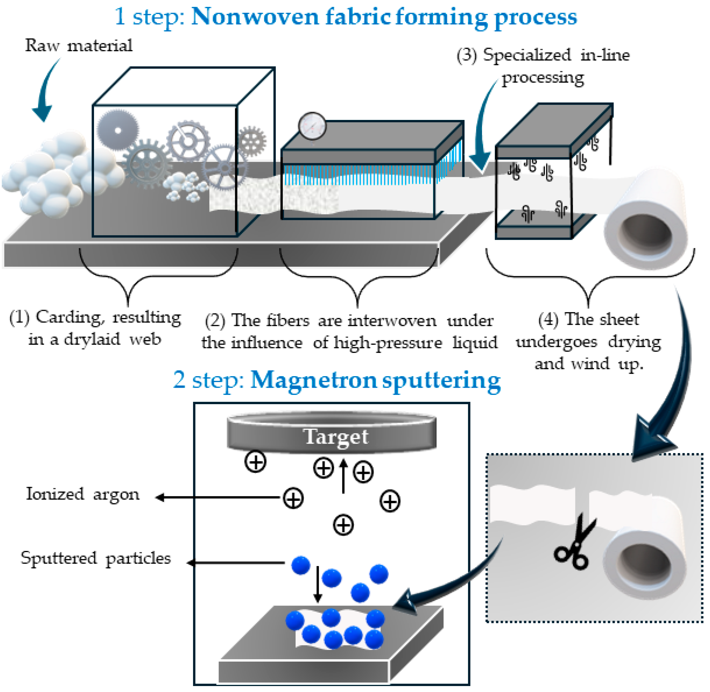
2.2.1. Aqua-Jet/Spunlace Technique

The fabric with web weight 75 g/m2 was made using the spunlace/aqua-jet method at Harper Hygienics S.A., Poland (Harper Hygienics S.A., Warsaw, Poland) using a Trützschler (Trützschler Group SE, Mönchengladbach, Deutschland) cotton nonwoven production line consisting of: (1) three cotton bale openers; (2) blending bin; (3) fine opener; (4) two random card; (5) aqua-jet (nozzle diameter 0.14 mm; 5000 nozzles per row); (6) multi-drum dryers; (7) disc winder. Average line speed was 50 m/min.

2.2.2. Magnetron Sputtering

The cellulose nonwoven material was subjected to modification employing a DC magnetron sputtering system (P.P.H. Jolex sc., Częstochowa, Poland), utilizing a copper disc target (Testbourne Ltd., Hampshire, UK). The coating deposition was conducted at a distance of 15 cm from the target within an argon atmosphere. Variation in disc discharge power ranging from 0.4 to 0.8 kW facilitated process optimization. Two distinct deposition durations, namely 8 min (designated as samples NW-Cel-Cu(0.4 kW/8) and NW-Cel-Cu(0.8 kW/8)) and 32 min (designated as samples NW-Cel-Cu(0.4 kW/32) and NW-Cel-Cu(0.8 kW/32)), were used.

2.2.3. Microscopy Analysis

The morphology of the cellulose nonwoven fabrics was assessed using a Keyence VHX-7000N digital microscope (Keyence, Osaka, Japan); the applied magnification was equal to 100× and 1000×.

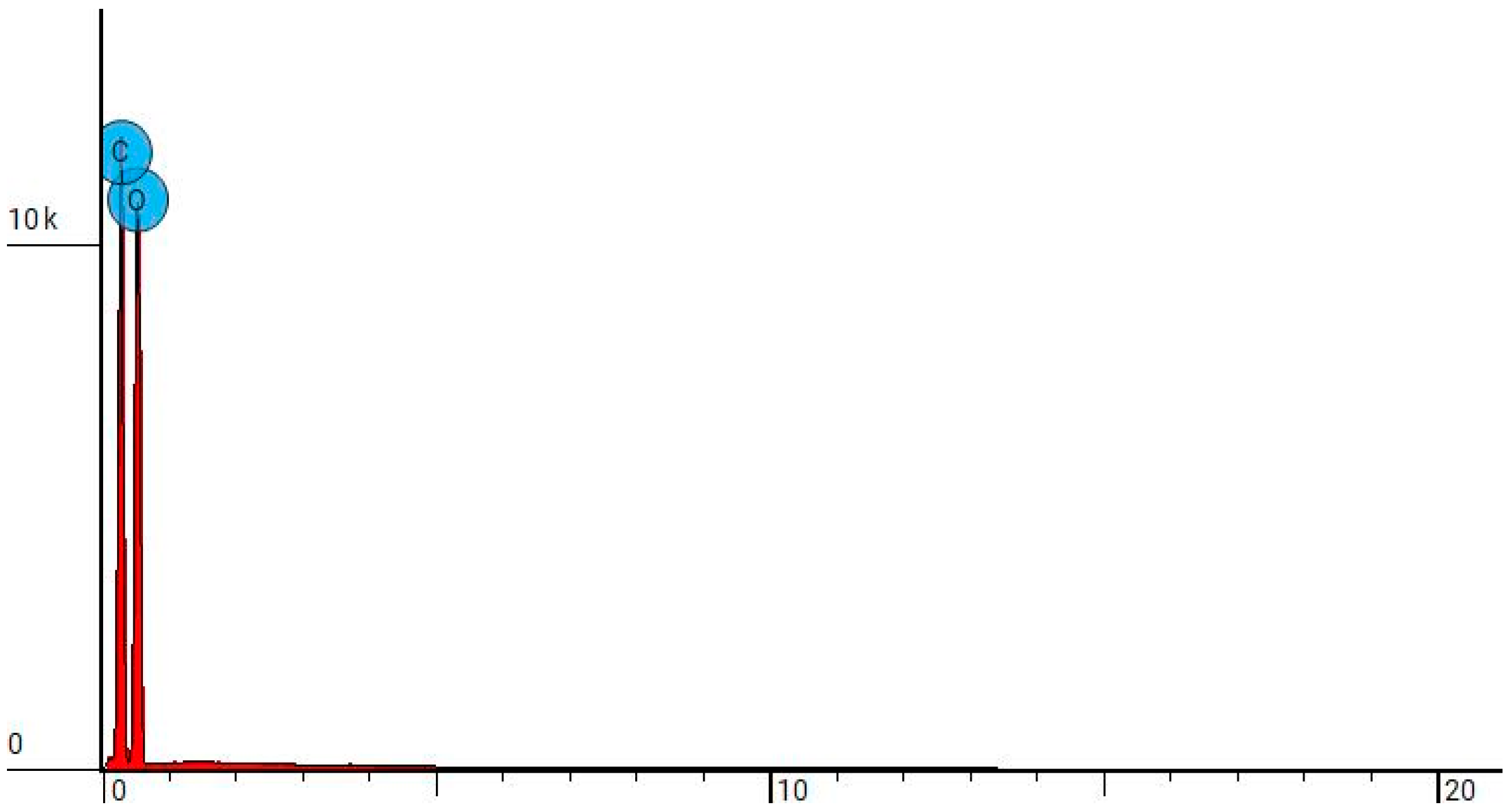
2.2.4. Scanning Electron Microscopy (SEM) and Energy-Dispersive X-ray Spectroscopy (EDS)

A Tescan Vega 3 scanning electron microscope (SEM) was employed to conduct microscopic analyses of cellulose nonwoven fabrics. This instrument, manufactured by Tescan Analytics (Tescan Analytics, Brno, Czech Republic), was equipped with an EDS X-ray microanalyzer from Oxford Instruments (Oxford Instruments, Abingdon, UK). Surface topography investigations using SEM were conducted under high vacuum conditions, with the probe beam energy set at 15 keV. Microscopic observations were performed at magnifications ranging from 5000× to 10,000×.

2.2.5. UV-Vis Analysis

The structure of the modified samples was examined by measuring the light transmission (%T) in the range λ = 200–800 nm, using a Jasco V-550 dual-beam UV/Vis spectrophotometer (Jasco, Tokyo, Japan), equipped with an integrating sphere attachment.

2.2.6. Flame Atomic Absorption Spectrometry (FAAS)-Copper Content Assessment

For the quantification of copper content in NW-Cel-Cu samples, mineralization was conducted utilizing the Ertec Magnum II single-module microwave digester, headquartered in Wrocław, Poland. Following this, copper(II) ions were quantitatively analyzed employing flame atomic absorption spectrometry (FAAS) with a Thermo Scientific Thermo Solar M6 spectrometer (Thermo Fisher Scientific Inc., Midland, Canada). The spectrometer was outfitted with a 100 mm diameter titanium burner and coded single-element hollow cathode lamps. Background correction was executed utilizing a deuterium D2 lamp.

2.2.7. Measurement of Blood Clotting Factors, including aPTT and PT

Human plasma, post freezing and lyophilization, was dissolved in deionized water. Samples weighing 1 mg were added to 200 µL of plasma and, following centrifugation, incubated for 15 min at a controlled temperature of 37 °C. The aPTT evaluation employed Dia-PTT reagent comprising kaolin, cephalin, and a 0.025 M calcium chloride (CaCl2) solution. aPTT measurements were conducted using a K-3002 OPTIC coagulometer, with 50 µL of plasma and 50 µL of Dia-PTT suspension collected for analysis. Subsequently, 50 µL of 0.025 M CaCl2 solution was added after 3 min of incubation. For PT assessment, 100 µL of plasma sample was incubated for 2 min at 37 °C, then 100 µL of Dia-PTT suspension was added and the measurement was started. Dia-PTT suspension contained thromboplastin from rabbit brain tissue, calcium ions and a preservative. The Dia-PTT suspension was thoroughly mixed before use.

2.2.8. Antibacterial and Antifungal Tests

The assessment of the antimicrobial properties of the developed materials was conducted in accordance with the PN-EN ISO 20645:2006 standard [42]. This involved targeting both Gram-negative (E. coli, ATCC 25922) and Gram-positive (S. aureus, ATCC 6538) bacteria using the agar diffusion method on Muller Hinton agar plates. Sterilized agar was poured into Petri dishes and inoculated with bacterial broth cultures, followed by incubation before placing the material samples. After 24 h at 37 °C, the diameter of the inhibition zone was measured to evaluate the antimicrobial activity. Parallel tests were conducted using unmodified cotton as controls for comparison.
The evaluation of antifungal characteristics followed the guidelines outlined in the PN-EN 14119:2005 standard [43]. In this procedure, C. globosum (ATCC 6205) and A. niger (ATCC 6275) fungi were employed. The samples were positioned on agar plates previously inoculated with the fungi, then maintained at a temperature of 29 °C for a duration of 14 days. Subsequently, the degree of fungal proliferation at the interface between the agar and the sample surfaces was scrutinized, while any discernible inhibition zones were documented. Each experiment was conducted in duplicate, with parallel trials utilizing unaltered cotton specimens serving as controls.

3. Results

3.1. Preparation of Nonwoven Cellulose-Copper Composites

The fabrication process employing the spunlace/aqua-jet method (refer to Figure 1) is intricate and exerts a substantial influence on the ultimate properties of the material. Initially, raw fibers traverse through a sequence of rollers with fine teeth, meticulously aligning the fibers in parallel (1). This initial phase is pivotal in determining the material’s structural formation and subsequent characteristics. Subsequently, the aligned fiber webs undergo high-pressure water jet treatment, exerting significant force on the fibers (2). This phase governs the interconnection of fibers and the formation of a cohesive nonwoven structure (3). Water jets induce fiber rolling and intertwining, facilitating the development of a cohesive nonwoven structure. This phase is pivotal for achieving targeted properties such as mechanical robustness, flexibility, and deformation resistance. Subsequently, the fabric undergoes hydroneedling, wherein it is traversed by specialized needles, further enhancing its density and strength (4). The final stages of production entail specialized on-line processing, during which the fabric may undergo additional treatments, such as the application of special coatings or chemical treatments to enhance specific properties. Subsequently, the fabric is dried and wound (5).
Overall, the spunlace/aqua-jet method represents an advanced fabric manufacturing technique enabling precise control over material properties. This capability is pivotal for products with stringent technical specifications, such as fabrics incorporating copper nanocomposites.
Subsequent to the cellulose nonwoven fabric formation via the aqua-jet/spunlace method, the sputtering process was initiated using a magnetron sputtering system. The magnetron sputtering system, consisting of a copper sputtering disk placed on a cotton substrate and an inert gas (Ar+), started the process by introducing argon into the process chamber. When an electrical voltage was applied, the argon was ionized, creating plasma with ions and electrons. An atomic stream of copper, suspended in the process chamber, was deposited on a prepared cellulose nonwoven substrate, enabling the construction of atom-by-atom layers for precise control of the thickness and structure of the coating. The parameters of the magnetron sputtering process are detailed in Table 1.

3.2. Determination of Copper Content

The concentration of copper in the cellulose-copper nonwoven samples was quantified using flame atomic absorption spectrometry (FAAS), with the results presented in Table 2. The power of the magnetron during the sputtering process is a critical factor influencing the deposition of copper onto the cellulose nonwoven fabric. Higher sputtering power facilitates the maximum incorporation of copper ions, which is vital for ensuring the durability of the resulting coating. Moreover, the duration of the sputtering process is equally significant, as extended sputtering times result in higher concentrations of copper in the nonwoven samples. These metal coatings hold substantial potential for application in the manufacture of medical dressings and devices.

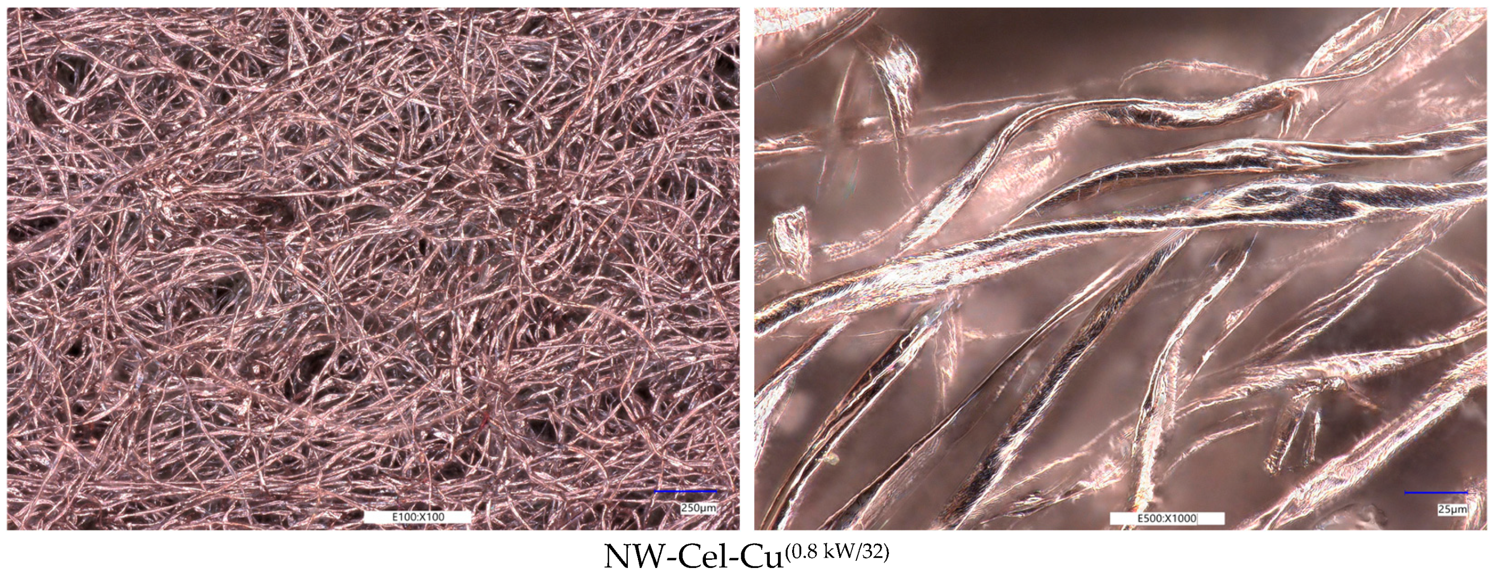
Optical Microscopy Analysis

The digital optical microscopic images of the investigated samples are presented in Figure 2. The optical microscopy images show the visual difference between modified (NW-Cel-Cu) and unmodified samples (NW-Cel). It may be observed that the obtained copper coatings cover the surface of the fibers, and the Cu coating is uniform. The uniformity of the layer of Cu coating was confirmed by the determination of copper content tests (FAAS). The modification does not damage the fibrous structure of the samples, even with higher power and longer process duration (NW-Cel-Cu(0.8 kW/32)).

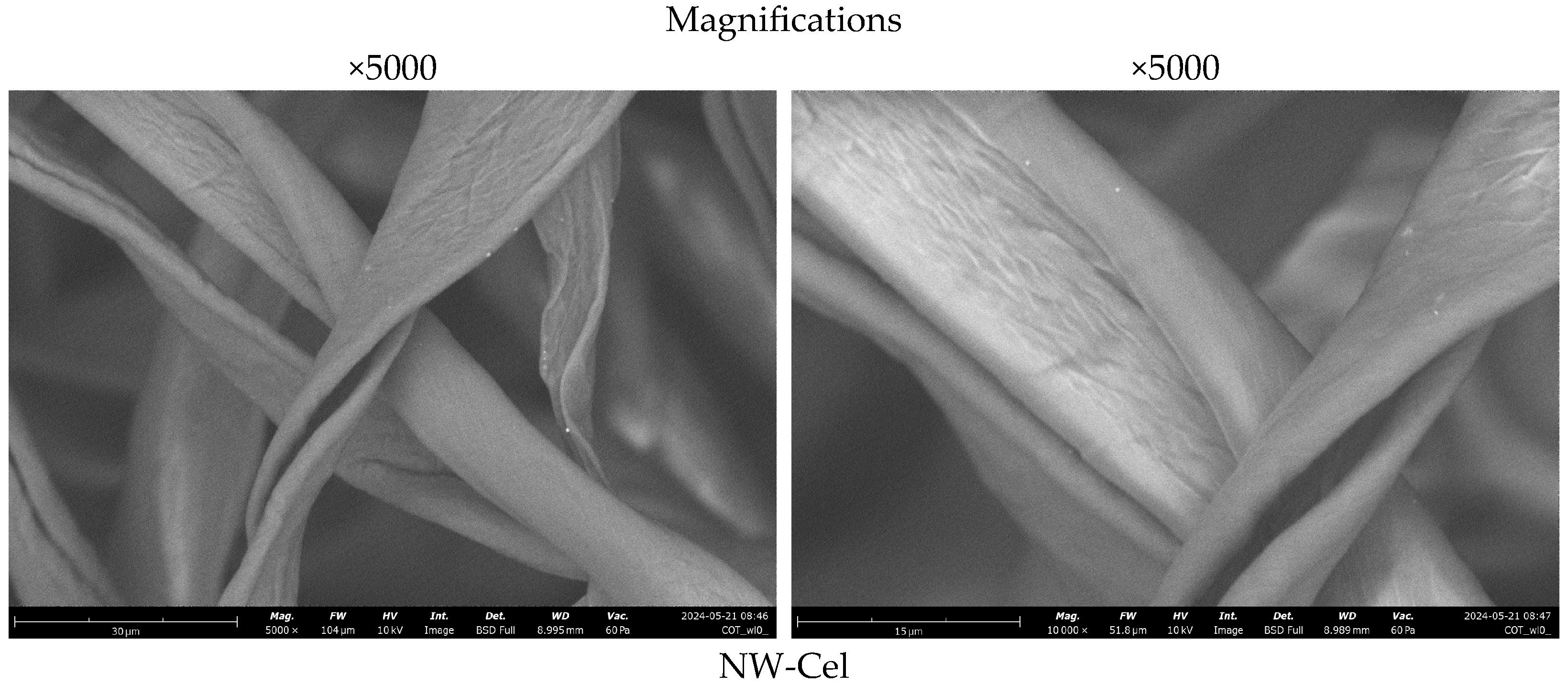
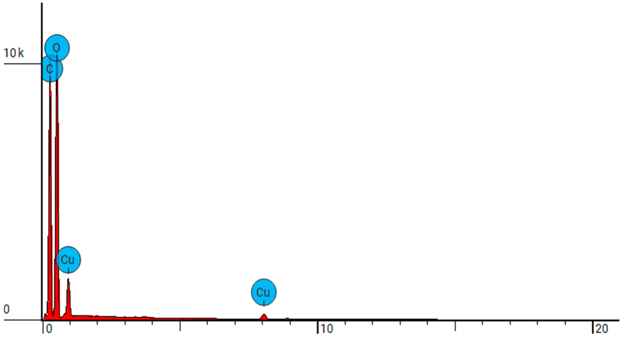
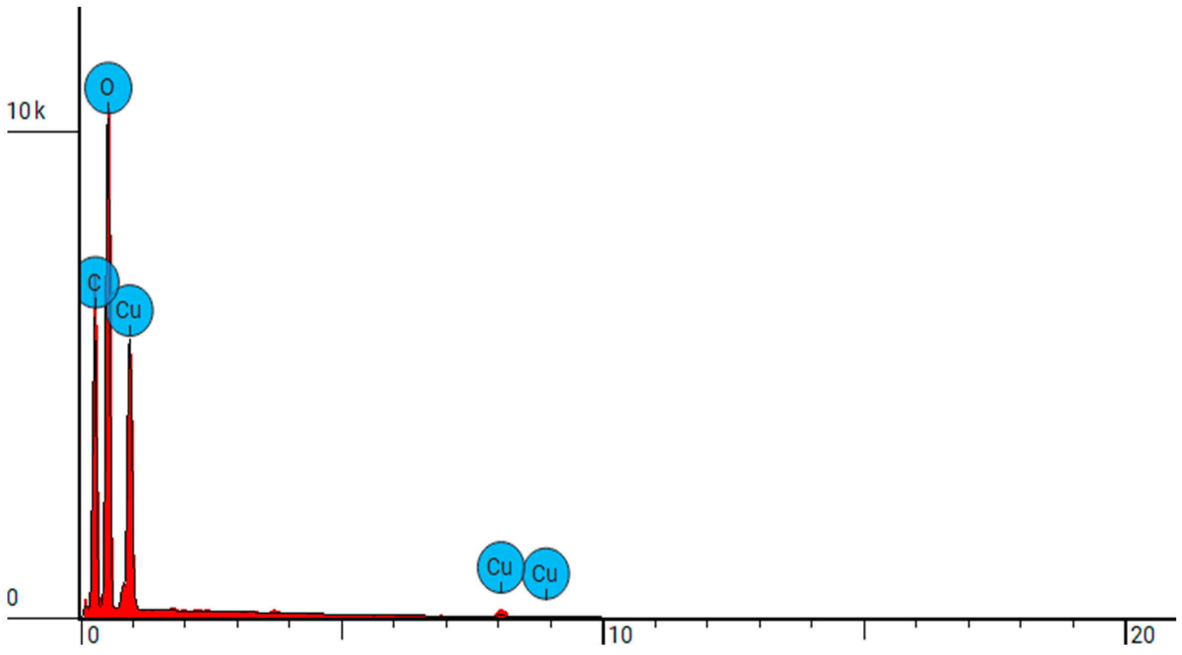
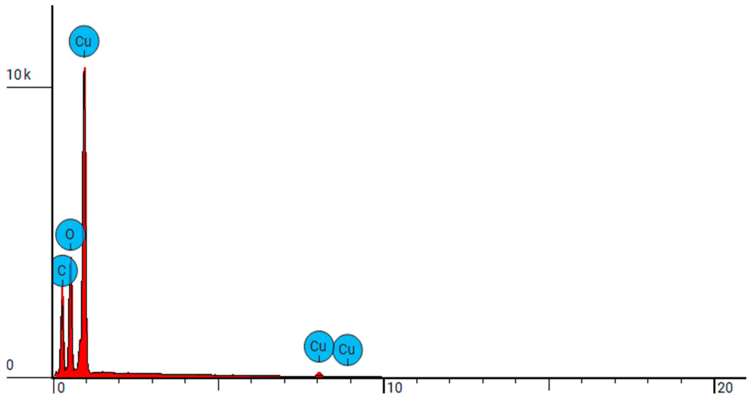
3.3. Scanning Electron Microscopy

Examination of the sample surfaces before and after modification is crucial for assessing the effectiveness of applying the copper coating to the cellulose nonwoven fabric. Microscopic observations reveal significant changes in the surface structure (Figure 3). Morphological analysis further allows for the identification of potential changes in the fiber structure resulting from the modification process. Detailed microscopic examinations enable the assessment of the coating’s uniformity and the detection of possible agglomerates of copper particles. The results confirm that the obtained copper coating is uniform and durable, which is essential for the effectiveness of the material in antibacterial and antimicrobial applications.
Scanning electron microscopy (SEM) images of cellulose nonwoven fabric before modification (NW-Cel) depict regular, parallel fibers with a relatively smooth surface. SEM images of NW-Cel-Cu materials obtained by magnetron sputtering illustrate changes in the surface structure of the samples. Following the application of a copper coating to cellulose fibers, surface roughness becomes apparent. These irregularities primarily arise during the sputtering process, where copper particles are irregularly deposited onto the fiber surface, resulting in the formation of microscopic irregularities. Importantly, no fiber damage or cracks were observed in the NW-Cel-Cu samples.
The samples were exposed to an electron beam within a scanning electron microscope, facilitating precise energy-dispersive X-ray spectroscopy (EDS). This technique not only enabled a thorough examination of the materials’ chemical composition but also provided an accurate assessment of their elemental makeup. With the EDS results, it was possible to precisely identify the elements within the sample by analyzing the position of peaks in the X-ray spectrum, where signal intensity correlated directly with the concentration of each element. The conducted analyses are comprehensively summarized in Table 3 and visually depicted in Figure 4, Figure 5, Figure 6, Figure 7 and Figure 8.
The findings from our EDS analysis align with the results obtained through flame atomic absorption spectrometry (FAAS). The correlation between the sputtering power and copper concentration is evident, indicating that higher sputtering power results in elevated levels of copper in the samples. Similarly, extending the sputtering duration also leads to an increase in copper concentration. This suggests that the duration and intensity of the sputtering process significantly influence the deposition of copper onto the cellulose nonwoven fabric, consequently affecting its copper content. Such insights are crucial for optimizing the fabrication process and enhancing the efficacy of copper-coated materials, particularly in medical applications where durability and antimicrobial properties are paramount.

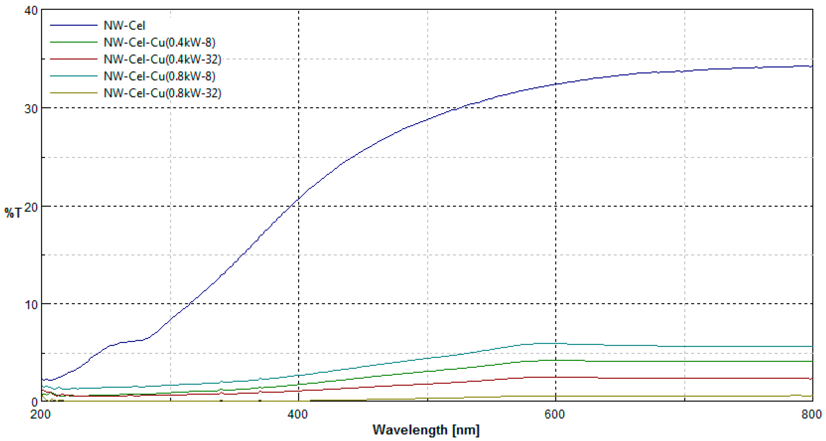
3.4. UV/Vis Transmittance

Comparison of transmittance spectra (%T) in the range λ = 200–800 nm of NW-Cel/NW-Cel-Cu samples is given in Figure 9.
Recorded UV/Vis transmittance spectra (%T) of samples after sputter modification (NW-Cel-Cu) revealed changes in the macrostructure of nonwoven samples after modification expressed by the depress of transmittance ability in the range λ = 200–800 nm. All modified nonwoven samples with Cu coatings had similar spectral characteristics and transmittance. There is a relationship between the sputter process time/copper content in the sample and the reduction in light transmission in the in the range λ = 200–800 nm area. A copper layer on the surface of the nonwoven material reduces light transmission (NW-Cel-Cu).

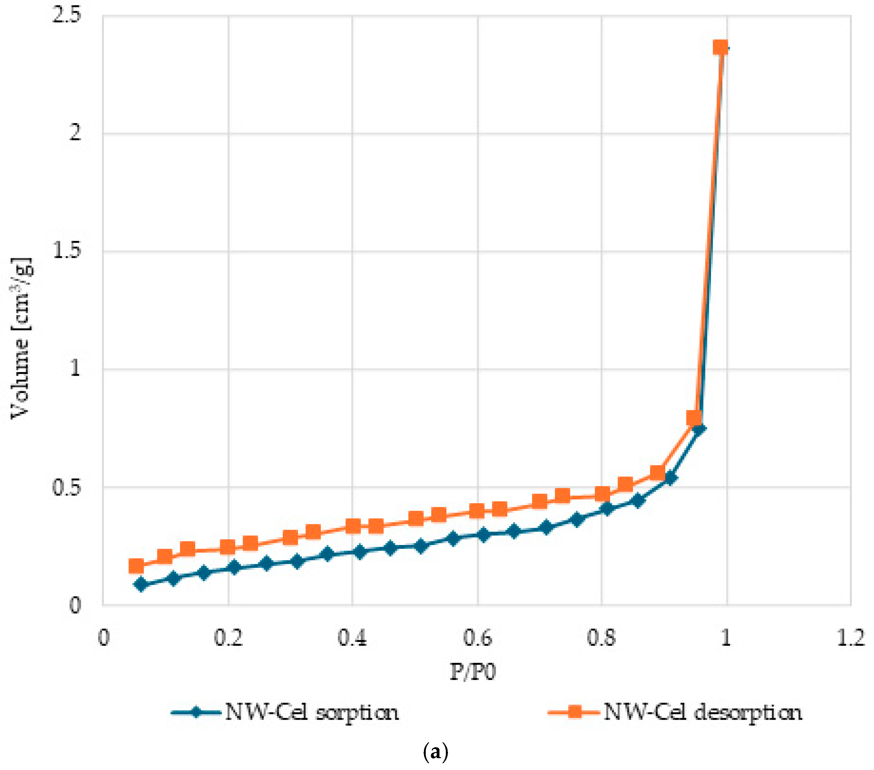
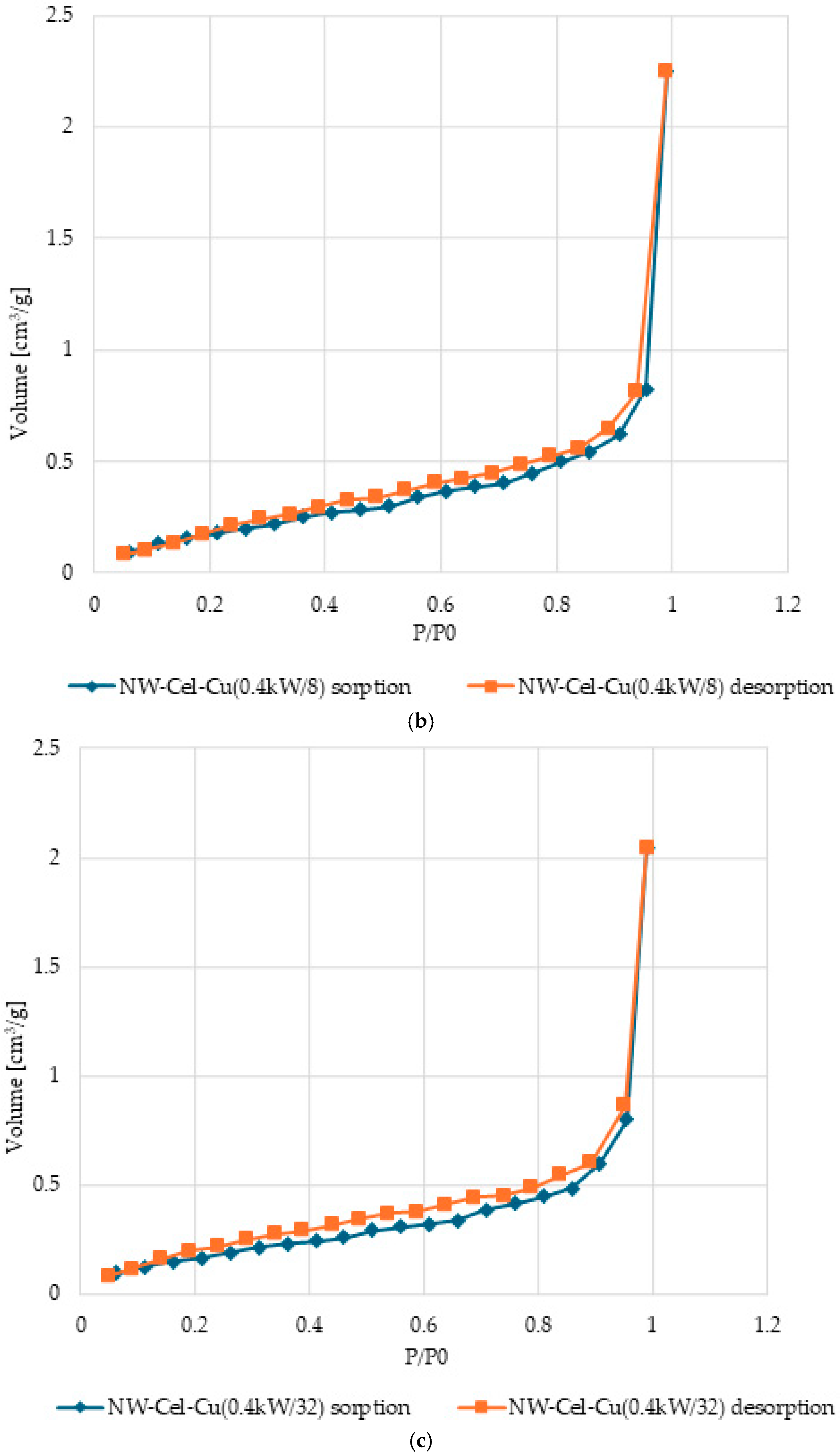
3.5. Surface Characteristics and Pore Volume in Cel-Cu Samples

Table 4 below provides a detailed overview of the specific surface area (SSA) and total pore volume (TPV) for various copper-modified cellulose nonwoven samples under different conditions. These parameters include different magnetron powers (0.4 kW and 0.8 kW) and different sputtering durations (8 min and 32 min).
The specific surface area (SSA) and total pore volume (TPV) of copper-modified cellulose nonwoven samples were analyzed, revealing significant effects of sputtering power and duration. The SSA values, which ranged from 3.172 × 10⁻³ m2/g to 3.660 × 10⁻³ m2/g, showed a tendency to decrease with increasing sputtering power and duration, indicating denser copper deposition. For example, the NW-Cel-Cu sample which sputtered at 0.4 kW for 8 min had an SSA of 3.483 × 10⁻³ m2/g, while increasing the duration to 32 min reduced the SSA to 3.172 × 10⁻³ m2/g. Similarly, increasing the sputtering power to 0.8 kW further reduced the SSA. In terms of TPV, the values ranged from 0.6021 cm3/g to 0.7985 cm3/g. Initially, TPV increased with sputtering power and duration due to more vigorous sputtering creating larger pore volumes, followed by a slight decrease as the deposition became denser. These findings indicate that optimizing sputtering conditions is essential for applications such as medical dressings, where specific surface area and pore volume are critical for antibacterial efficacy. Achieving an optimal balance is necessary to ensure adequate copper deposition for antimicrobial activity without significantly diminishing the porosity and surface area required for effective functionality.
Figure 10 shows nitrogen (N2) adsorption and desorption isotherms for samples of the cellulose nonwoven fabric before modification and samples following sputtering of NW-Cel-Cu.
The characteristic shape of the type III isotherm, consistent with the IUPAC classification, indicates weak interactions between the adsorbent and the adsorbate, with the adsorbed molecules concentrating mainly in optimal places on the surface of non-porous or macroporous solid materials [44,45,46,47]. The quantity of adsorbed adsorbate upon reaching saturation pressure is limited (p/p0 = 1), indicating a multilayer adsorption mechanism across the entire pressure spectrum. The emergence of a type III isotherm is frequently linked to the existence of non-porous or macroporous configurations [48,49]. Examination of the plots reveals an exponential rise in the adsorbate quantity as pressure increases. Initially, the rate of this increase may be constrained even at low relative pressure levels, but a substantial upsurge in the adsorbed adsorbate amount is evident at values nearing p/p0 = 1 [50]. The emergence of H3-type hysteresis loops indicates the existence of materials with slit-like pores, which tend to retain some of the adsorbate even at lower pressures. Nonetheless, as pressure rises, there is a rapid increase in the adsorbate amount, typical of slit-like pores [51,52,53].

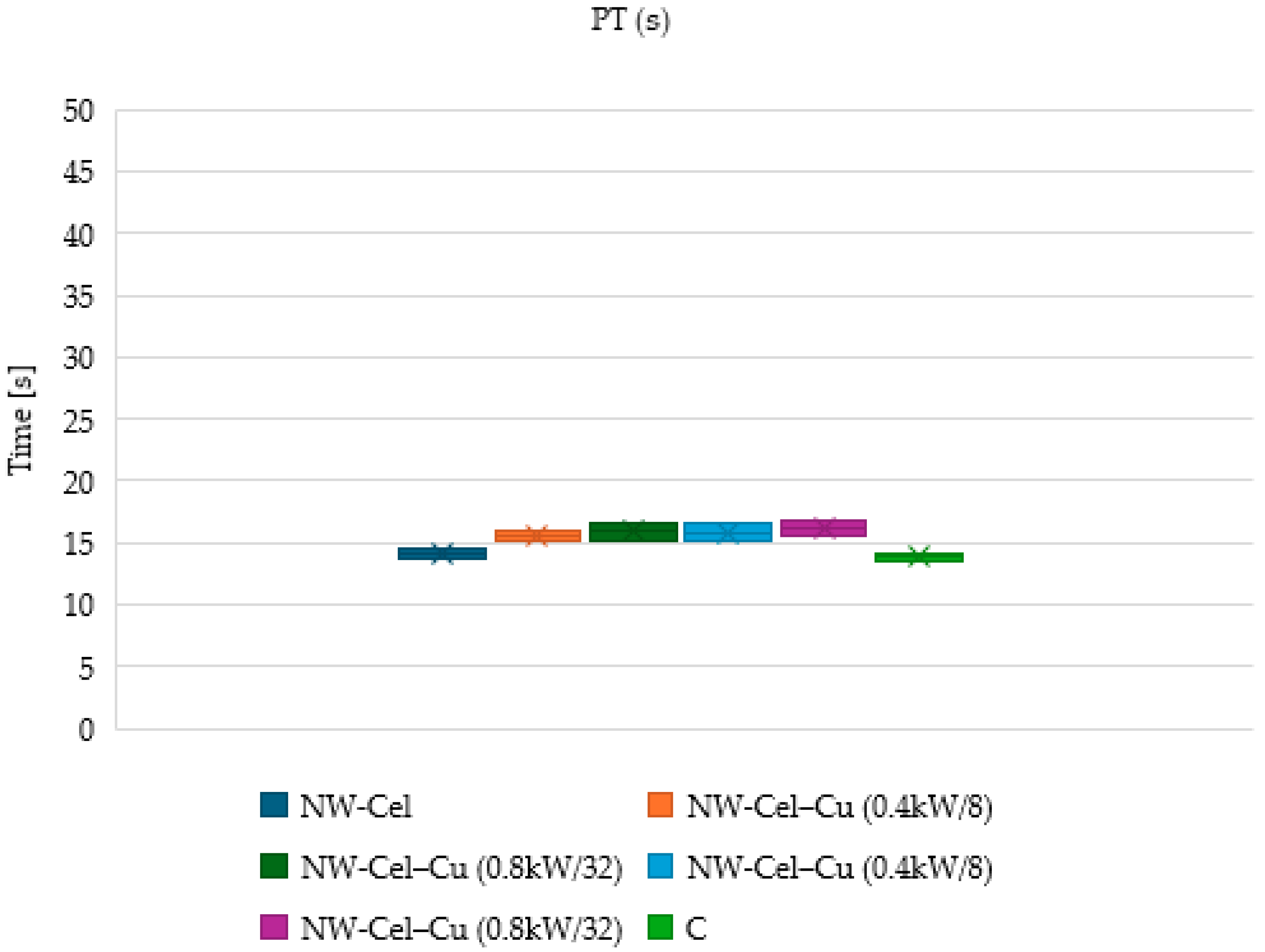
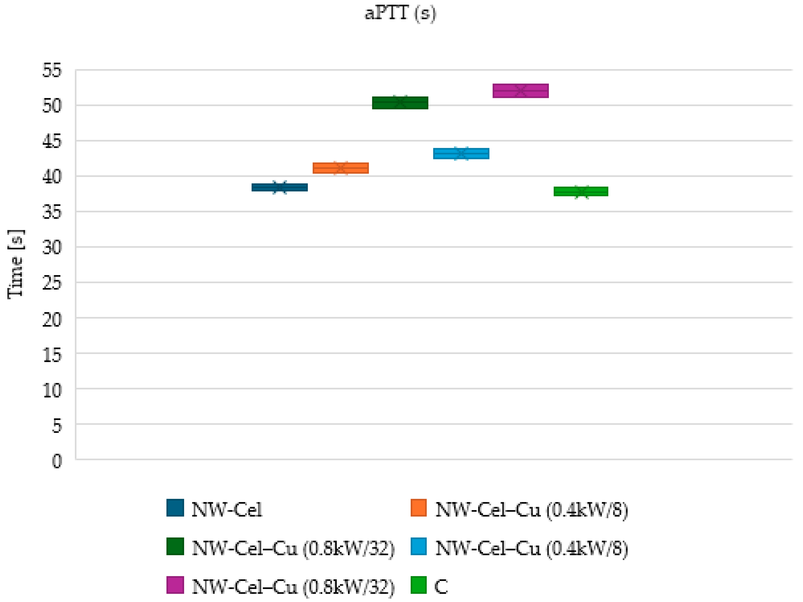
3.6. Measurements of aPTT and PT Times

Figure 11 and Figure 12 present the prothrombin time (PT) and activated partial thromboplastin time (aPTT) for different samples, including copper-modified cellulose nonwoven (NW-Cel) samples under various sputtering conditions (0.4 kW and 0.8 kW) and durations (8 and 32 min), as well as a control sample.
The observations indicate that PT and aPTT times generally increase with the copper modification of cellulose nonwoven samples, with variations depending on the sputtering power and duration. Higher sputtering power (0.8 kW) results in slightly longer PT and aPTT times compared to lower power (0.4 kW), while extended sputtering duration (32 min) correlates with the further extension of PT and aPTT times compared to a shorter duration (8 min). The studies on prothrombin time (PT) showed a marginal prolongation after copper modification, indicating a negligible effect on the extrinsic coagulation pathway. However, more pronounced differences in aPTT suggest a potential effect of copper on the intrinsic coagulation pathway. This is based on the hypothesis that copper may interact with contact factors (XI, XII, HK) that are crucial in initiating this pathway [54]. The binding of these factors by copper may lead to a decrease in their concentration in plasma, consequently resulting in aPTT prolongation. The effect of copper on the extrinsic coagulation pathway may be minimal due to the presence of various coagulation mechanisms capable of compensating for extrinsic changes. The control sample shows relatively lower PT and aPTT times compared to the copper-modified samples. These results suggest that copper modification introduces subtle changes in surface properties, potentially affecting blood-material interactions. Increased sputtering power and longer duration may lead to greater copper deposition on the material surface, altering its physicochemical properties, which in turn affects blood clotting parameters.

Antibacterial Effect

To assess the antimicrobial and antifungal efficacy of NW-Cel-Cu materials, experiments were carried out using Gram-negative bacteria (E. coli), Gram-positive bacteria (S. aureus), and fungi (C. globosum, A. niger), as per the guidelines specified in EN-ISO 20645:2006 [42] and EN-ISO 14119:2005 [43] standards. This approach enabled the assessment of the antibacterial and antifungal activity of the obtained materials and their potential effectiveness against these microorganisms. By analyzing these data, we can evaluate the efficacy of NW-Cel-Cu materials and their potential impact on Gram-negative and Gram-positive bacteria, which is crucial for ensuring the safety and hygiene of these materials.
The analysis of Table 5 results shows the different performances of NW-Cel-Cu samples depending on the sputtering power (0.4 kW or 0.8 kW) and the process time (8 or 32 min). The variations in the diameter of the inhibition zones for different test microorganisms are readily apparent. Samples processed with a sputtering power of 0.8 kW generally exhibited larger inhibition zone diameters compared to those processed with 0.4 kW. Moreover, in most cases, an extended process duration (32 min) led to larger inhibition zone diameters than a shorter duration (8 min). The analysis suggests that both sputtering power and process duration significantly influence the antibacterial and antifungal activity of NW-Cel-Cu. Specifically, samples processed with a sputtering power of 0.8 kW and a longer duration (32 min) appeared more effective in inhibiting microbial growth. This implies that higher copper content was attained under these conditions, enhancing the materials’ efficacy against both bacteria and fungi. These observations support earlier research on the antimicrobial effects of copper compounds, indicating their potential to strengthen biosecurity measures and reduce pathogen growth [50,55].

4. Conclusions

In this study, we employed the aqua-jet/spunlace-forming process to manufacture cellulose nonwoven fabric, followed by Cu magnetron sputtering deposition onto these fabrics. Through a combination of quantitative copper analysis, morphological characterization using SEM imaging, and elemental analysis via EDS, we thoroughly examined the properties of the resulting materials. Specifically, we investigated the specific surface area and pore volume of the fabricated materials. Additionally, we explored the effects of these materials on the blood coagulation process, assessing parameters such as activated partial thromboplastin time (aPTT) and prothrombin time (PT). Our findings revealed promising outcomes. Despite the copper modification, the cellulose nonwoven fabrics exhibited negligible interference with the blood coagulation process, suggesting their compatibility with biological systems. This underscores the potential of these materials for various biomedical applications, including wound dressings. Overall, our study underscores the efficacy of magnetron sputtering deposition on cellulose nonwoven fabrics and highlights their potential for biomedical applications. Further research in this direction could lead to the development of advanced biomaterials with enhanced functionality and performance.

Author Contributions

M.Ś. developed the concept and designed experiments, performed experiments, analyzed data, and wrote the paper; Z.M. performed experiments and analyzed data; A.W. analyzed data; A.L.-K. performed experiments; M.H.K. developed the concept and designed experiments, analyzed data and wrote the paper. All authors have read and agreed to the published version of the manuscript.

Funding

This research was partly carried out within the National Science Centre (Poland), project M-ERA.NET 2022, No. 2022/04/Y/ST4/00157.

Institutional Review Board Statement

Not applicable.

Informed Consent Statement

Not applicable.

Data Availability Statement

Data are contained within the article.

Acknowledgments

The authors would like to thank Piotr Kaczmarek for their technical contributions.

Conflicts of Interest

The authors declare no conflicts of interest.

References

  1. Cheng, W.; Zhu, Y.; Jiang, G.; Cao, K.; Zeng, S.; Chen, W.; Zhao, D.; Yu, H. Sustainable Cellulose and Its Derivatives for Promising Biomedical Applications. Prog. Mater. Sci. 2023, 138, 101152. [Google Scholar] [CrossRef]
  2. Li, T.; Chen, C.; Brozena, A.H.; Zhu, J.Y.; Xu, L.; Driemeier, C.; Dai, J.; Rojas, O.J.; Isogai, A.; Wågberg, L.; et al. Developing Fibrillated Cellulose as a Sustainable Technological Material. Nature 2021, 590, 47–56. [Google Scholar] [CrossRef] [PubMed]
  3. Gupta, P.K.; Raghunath, S.S.; Prasanna, D.V.; Venkat, P.; Shree, V.; Chithananthan, C.; Choudhary, S.; Surender, K.; Geetha, K.; Gupta, P.K.; et al. An Update on Overview of Cellulose, Its Structure and Applications. In Cellulose; IntechOpen: Houston, TX, USA, 2019; ISBN 978-1-83968-057-1. [Google Scholar]
  4. Carolin, C.F.; Kamalesh, T.; Kumar, P.S.; Hemavathy, R.V.; Rangasamy, G. A Critical Review on Sustainable Cellulose Materials and Its Multifaceted Applications. Ind. Crops Prod. 2023, 203, 117221. [Google Scholar] [CrossRef]
  5. Dufresne, A. Chapter 19—Cellulose-Based Composites and Nanocomposites. In Monomers, Polymers and Composites from Renewable Resources; Belgacem, M.N., Gandini, A., Eds.; Elsevier: Amsterdam, The Netherlands, 2008; pp. 401–418. ISBN 978-0-08-045316-3. [Google Scholar]
  6. Siqueira, G.; Bras, J.; Dufresne, A. Cellulosic Bionanocomposites: A Review of Preparation, Properties and Applications. Polymers 2010, 2, 728–765. [Google Scholar] [CrossRef]
  7. Li, X.; Wan, C.; Tao, T.; Chai, H.; Huang, Q.; Chai, Y.; Wu, Y. An Overview of the Development Status and Applications of Cellulose–Based Functional Materials. Cellulose 2024, 31, 61–99. [Google Scholar] [CrossRef]
  8. Qiu, X.; Hu, S. “Smart” Materials Based on Cellulose: A Review of the Preparations, Properties, and Applications. Materials 2013, 6, 738–781. [Google Scholar] [CrossRef] [PubMed]
  9. Liu, M.; Ma, C.; Zhou, D.; Chen, S.; Zou, L.; Wang, H.; Wu, J. Hydrophobic, Breathable Cellulose Nonwoven Fabrics for Disposable Hygiene Applications. Carbohydr. Polym. 2022, 288, 119367. [Google Scholar] [CrossRef] [PubMed]
  10. Aziz, T.; Farid, A.; Haq, F.; Kiran, M.; Ullah, A.; Zhang, K.; Li, C.; Ghazanfar, S.; Sun, H.; Ullah, R.; et al. A Review on the Modification of Cellulose and Its Applications. Polymers 2022, 14, 3206. [Google Scholar] [CrossRef] [PubMed]
  11. Sánchez–López, E.; Gomes, D.; Esteruelas, G.; Bonilla, L.; Lopez-Machado, A.L.; Galindo, R.; Cano, A.; Espina, M.; Ettcheto, M.; Camins, A.; et al. Metal-Based Nanoparticles as Antimicrobial Agents: An Overview. Nanomaterials 2020, 10, 292. [Google Scholar] [CrossRef]
  12. Calabrese, C.; La Parola, V.; Testa, M.L.; Liotta, L.F. Antifouling and Antimicrobial Activity of Ag, Cu and Fe Nanoparticles Supported on Silica and Titania. Inorganica Chim. Acta 2022, 529, 120636. [Google Scholar] [CrossRef]
  13. Brandelli, A.; Ritter, A.C.; Veras, F.F. Antimicrobial Activities of Metal Nanoparticles. In Metal Nanoparticles in Pharma; Rai, M., Shegokar, R., Eds.; Springer International Publishing: Cham, Switzerland, 2017; pp. 337–363. ISBN 978-3-319-63790-7. [Google Scholar]
  14. Zhang, E.; Zhao, X.; Hu, J.; Wang, R.; Fu, S.; Qin, G. Antibacterial Metals and Alloys for Potential Biomedical Implants. Bioact. Mater. 2021, 6, 2569–2612. [Google Scholar] [CrossRef] [PubMed]
  15. Hossein Mohammadi, A.; Sobhani-Nasab, A.; Nejati, M.; Hadi, S.; Behjati, M.; Mirzaii-Dizgah, I.; Moradi Hasan-Abad, A.; Karami, M. Preparation and Characterization of CuO, Ag2O and ZnO Nanoparticles and Investigation of Their Antibacterial and Anticancer Properties on HCT-116 and C26 Cells. Inorg. Chem. Commun. 2023, 149, 110404. [Google Scholar] [CrossRef]
  16. Vincent, M.; Duval, R.E.; Hartemann, P.; Engels-Deutsch, M. Contact Killing and Antimicrobial Properties of Copper. J. Appl. Microbiol. 2018, 124, 1032–1046. [Google Scholar] [CrossRef] [PubMed]
  17. Ramos-Zúñiga, J.; Bruna, N.; Pérez-Donoso, J.M. Toxicity Mechanisms of Copper Nanoparticles and Copper Surfaces on Bacterial Cells and Viruses. Int. J. Mol. Sci. 2023, 24, 10503. [Google Scholar] [CrossRef] [PubMed]
  18. Alselami, A.; Drummond, R.A. How Metals Fuel Fungal Virulence, yet Promote Anti-Fungal Immunity. Dis. Model. Mech. 2023, 16, dmm050393. [Google Scholar] [CrossRef] [PubMed]
  19. Borkow, G.; Gabbay, J. Copper as a Biocidal Tool. Curr. Med. Chem. 2005, 12, 2163–2175. [Google Scholar] [CrossRef] [PubMed]
  20. Borkow, G.; Gabbay, J. Copper, An Ancient Remedy Returning to Fight Microbial, Fungal and Viral Infections. Curr. Chem. Biol. 2009, 3, 272–278. [Google Scholar] [CrossRef]
  21. Grass, G.; Rensing, C.; Solioz, M. Metallic Copper as an Antimicrobial Surface. Appl. Environ. Microbiol. 2011, 77, 1541–1547. [Google Scholar] [CrossRef] [PubMed]
  22. Chyderiotis, S.; Legeay, C.; Verjat-Trannoy, D.; Le Gallou, F.; Astagneau, P.; Lepelletier, D. New Insights on Antimicrobial Efficacy of Copper Surfaces in the Healthcare Environment: A Systematic Review. Clin. Microbiol. Infect. 2018, 24, 1130–1138. [Google Scholar] [CrossRef]
  23. Ermini, M.L.; Voliani, V. Antimicrobial Nano-Agents: The Copper Age. ACS Nano 2021, 15, 6008–6029. [Google Scholar] [CrossRef]
  24. Crisan, M.C.; Teodora, M.; Lucian, M. Copper Nanoparticles: Synthesis and Characterization, Physiology, Toxicity and Antimicrobial Applications. Appl. Sci. 2022, 12, 141. [Google Scholar] [CrossRef]
  25. Maliki, M.; Ifijen, I.H.; Ikhuoria, E.U.; Jonathan, E.M.; Onaiwu, G.E.; Archibong, U.D.; Ighodaro, A. Copper Nanoparticles and Their Oxides: Optical, Anticancer and Antibacterial Properties. Int. Nano Lett. 2022, 12, 379–398. [Google Scholar] [CrossRef]
  26. Li, X.; Cong, Y.; Ovais, M.; Cardoso, M.B.; Hameed, S.; Chen, R.; Chen, M.; Wang, L. Copper-Based Nanoparticles against Microbial Infections. Wiley Interdiscip. Rev. Nanomed. Nanobiotechnol. 2023, 15, e1888. [Google Scholar] [CrossRef] [PubMed]
  27. Su, Y.; Xu, H.; Liao, Z.; Liu, K.; Chaima, M.; Zhang, X.; Hu, Y.; Wei, Y.; Huang, D. Copper-Based Nanoparticles as Antibacterial Agents. Eur. J. Inorg. Chem. 2023, 26, e202200614. [Google Scholar] [CrossRef]
  28. Cortes, A.A.; Zuñiga, J.M. The Use of Copper to Help Prevent Transmission of SARS-Coronavirus and Influenza Viruses. A General Review. Diagn. Microbiol. Infect. Dis. 2020, 98, 115176. [Google Scholar] [CrossRef] [PubMed]
  29. Puchkova, L.V.; Kiseleva, I.V.; Polishchuk, E.V.; Broggini, M.; Ilyechova, E.Y. The Crossroads between Host Copper Metabolism and Influenza Infection. Int. J. Mol. Sci. 2021, 22, 5498. [Google Scholar] [CrossRef] [PubMed]
  30. Rani, I.; Goyal, A.; Bhatnagar, M.; Manhas, S.; Goel, P.; Pal, A.; Prasad, R. Potential Molecular Mechanisms of Zinc- and Copper-Mediated Antiviral Activity on COVID-19. Nutr. Res. 2021, 92, 109–128. [Google Scholar] [CrossRef] [PubMed]
  31. Tortella, G.R.; Pieretti, J.C.; Rubilar, O.; Fernández-Baldo, M.; Benavides-Mendoza, A.; Diez, M.C.; Seabra, A.B. Silver, Copper and Copper Oxide Nanoparticles in the Fight against Human Viruses: Progress and Perspectives. Crit. Rev. Biotechnol. 2022, 42, 431–449. [Google Scholar] [CrossRef] [PubMed]
  32. Zakharova, O.V.; Vasyukova, I.A.; Gusev, A.A. Metal-Based Nanoparticles for the Diagnostics, Therapy, and Prevention of Viral Infections. Nanotechnol. Russia 2023, 18, 165–188. [Google Scholar] [CrossRef]
  33. Gerwien, F.; Skrahina, V.; Kasper, L.; Hube, B.; Brunke, S. Metals in Fungal Virulence. FEMS Microbiol. Rev. 2018, 42, fux050. [Google Scholar] [CrossRef]
  34. Robinson, J.R.; Isikhuemhen, O.S.; Anike, F.N. Fungal-Metal Interactions: A Review of Toxicity and Homeostasis. J. Fungi 2021, 7, 225. [Google Scholar] [CrossRef] [PubMed]
  35. Pereira, D.; Carreira, T.S.; Alves, N.; Sousa, Â.; Valente, J.F.A. Metallic Structures: Effective Agents to Fight Pathogenic Microorganisms. Int. J. Mol. Sci. 2022, 23, 1165. [Google Scholar] [CrossRef] [PubMed]
  36. Madkhali, O.A. A Comprehensive Review on Potential Applications of Metallic Nanoparticles as Antifungal Therapies to Combat Human Fungal Diseases. Saudi Pharm. J. 2023, 31, 101733. [Google Scholar] [CrossRef] [PubMed]
  37. Jelis, E.; Kristol, D.; Arora, R.R.; Spillert, C.R. The Effect of Copper Ion on Blood Coagulation. In Proceedings of the IEEE 30th Annual Northeast Bioengineering Conference, Springfield, MA, USA, 17–18 April 2004; p. 127. [Google Scholar]
  38. Muthulakshmi, L.; Varada Rajalu, A.; Kaliaraj, G.S.; Siengchin, S.; Parameswaranpillai, J.; Saraswathi, R. Preparation of Cellulose/Copper Nanoparticles Bionanocomposite Films Using a Bioflocculant Polymer as Reducing Agent for Antibacterial and Anticorrosion Applications. Compos. Part B Eng. 2019, 175, 107177. [Google Scholar] [CrossRef]
  39. Royer, A.; Sharman, T. Copper Toxicity. In StatPearls; StatPearls Publishing: Treasure Island, FL, USA, 2024. [Google Scholar]
  40. Alkhanjaf, A.A.M.; Sharma, S.; Sharma, M.; Kumar, R.; Arora, N.K.; Kumar, B.; Umar, A.; Baskoutas, S.; Mukherjee, T.K. Microbial Strategies for Copper Pollution Remediation: Mechanistic Insights and Recent Advances. Environ. Pollut. 2024, 346, 123588. [Google Scholar] [CrossRef] [PubMed]
  41. Wang, L.; Erişen, D.E.; Yang, K.; Zhang, B.; Guan, H.; Chen, S. Anticoagulation and Antibacterial Functional Coating on Vascular Implant Interventional Medical Catheter. J. Biomed. Mater. Res. Part B Appl. Biomater. 2020, 108, 2868–2877. [Google Scholar] [CrossRef]
  42. EN ISO 20645:2006; Textile Fabrics. Determination of Antibacterial Activity—Agar Diffusion Plate Test. International Organization for Standardization: Geneva, Switzerland, 2006.
  43. EN 14119: 2005 Point 10.5 (B2); Testing of Textiles. Evaluation of the Action of Microfungi. Visual Method. International Organization for Standardization: Geneva, Switzerland, 2005.
  44. Mathews, S.; Hans, M.; Mücklich, F.; Solioz, M. Contact Killing of Bacteria on Copper Is Suppressed If Bacterial-Metal Contact Is Prevented and Is Induced on Iron by Copper Ions. Appl. Environ. Microbiol. 2013, 79, 2605–2611. [Google Scholar] [CrossRef] [PubMed]
  45. Thommes, M.; Kaneko, K.; Neimark, A.V.; Olivier, J.P.; Rodriguez-Reinoso, F.; Rouquerol, J.; Sing, K.S.W. Physisorption of Gases, with Special Reference to the Evaluation of Surface Area and Pore Size Distribution (IUPAC Technical Report). Pure Appl. Chem. 2015, 87, 1051–1069. [Google Scholar] [CrossRef]
  46. Qi, L.; Tang, X.; Wang, Z.; Peng, X. Pore Characterization of Different Types of Coal from Coal and Gas Outburst Disaster Sites Using Low Temperature Nitrogen Adsorption Approach. Int. J. Min. Sci. Technol. 2017, 27, 371–377. [Google Scholar] [CrossRef]
  47. Xu, L.; Zhang, J.; Ding, J.; Liu, T.; Shi, G.; Li, X.; Dang, W.; Cheng, Y.; Guo, R. Pore Structure and Fractal Characteristics of Different Shale Lithofacies in the Dalong Formation in the Western Area of the Lower Yangtze Platform. Minerals 2020, 10, 72. [Google Scholar] [CrossRef]
  48. Sing, K.S.W. Reporting Physisorption Data for Gas/Solid Systems with Special Reference to the Determination of Surface Area and Porosity (Recommendations 1984). Pure Appl. Chem. 1985, 57, 603–619. [Google Scholar] [CrossRef]
  49. ALOthman, Z.A. A Review: Fundamental Aspects of Silicate Mesoporous Materials. Materials 2012, 5, 2874–2902. [Google Scholar] [CrossRef]
  50. Mrozińska, Z.; Kudzin, M.H.; Ponczek, M.B.; Kaczmarek, A.; Król, P.; Lisiak-Kucińska, A.; Żyłła, R.; Walawska, A. Biochemical Approach to Poly(Lactide)-Copper Composite—Impact on Blood Coagulation Processes. Materials 2024, 17, 608. [Google Scholar] [CrossRef] [PubMed]
  51. Lowell, S.; Shields, J.E.; Thomas, M.A.; Thommes, M. Characterization of Porous Solids and Powders: Surface Area, Pore Size and Density; Springer Science & Business Media: Berlin/Heidelberg, Germany, 2012; ISBN 978-1-4020-2303-3. [Google Scholar]
  52. Gregg, S.J.; Sing, K.S.W.; Salzberg, H.W. Adsorption Surface Area and Porosity. J. Electrochem. Soc. 1967, 114, 279Ca. [Google Scholar] [CrossRef]
  53. Medrano, V.G.B.; Celis, V.N.; Giraldo, R.I. Systematic Analysis of the Nitrogen Adsorption-Desorption Isotherms Recorded for a Series of Microporous-Mesoporous Amorphous Aluminosilicates Using Classical Methods. J. Chem. Eng. Data 2023, 68, 2512–2528. [Google Scholar] [CrossRef]
  54. Mutch, N.J.; Waters, E.K.; Morrissey, J.H. Immobilized Transition Metal Ions Stimulate Contact Activation and Drive Factor XII-mediated Coagulation. J. Thromb. Haemost. 2012, 10, 2108–2115. [Google Scholar] [CrossRef]
  55. Mrozińska, Z.; Ponczek, M.; Kaczmarek, A.; Boguń, M.; Sulak, E.; Kudzin, M.H. Blood Coagulation Activities of Cotton-Alginate-Copper Composites. Mar. Drugs 2023, 21, 625. [Google Scholar] [CrossRef]
Figure 1. Scheme of aqua-jet/spunlace nonwoven forming and magnetron sputtering process.
Coatings 14 00843 g001
Figure 2. Optical microscopy images of the samples before and after the sputter deposition processes (NW-Cel/NW-Cel-Cu). Magnification: 100×; 1000×.
Coatings 14 00843 g002aCoatings 14 00843 g002b
Figure 3. Scanning electron microscope (SEM) images of NW-Cel and NW-Cel-Cu. Magnification: 5000×; 10,000×.
Coatings 14 00843 g003aCoatings 14 00843 g003b
Figure 4. Energy-dispersive X-ray spectroscopy (EDS): NW-Cel (8 310 c/s).
Coatings 14 00843 g004
Figure 5. Energy-dispersive X-ray spectroscopy (EDS): NW-Cel-Cu(0.4 kW/8) (7 611 c/s).
Coatings 14 00843 g005
Figure 6. Energy-dispersive X-ray spectroscopy (EDS): NW-Cel-Cu(0.4 kW/32) (3 792 c/s).
Coatings 14 00843 g006
Figure 7. Energy-dispersive X-ray spectroscopy (EDS): NW-Cel-Cu(0.8 kW/8) (4 243 c/s).
Coatings 14 00843 g007
Figure 8. Energy-dispersive X-ray spectroscopy (EDS): NW-Cel-Cu(0.8 kW/32) (6 640 c/s).
Coatings 14 00843 g008
Figure 9. Comparison of transmittance spectra (%T) in the range λ = 200–800 nm of NW-Cel/NW-Cel-Cu samples.
Coatings 14 00843 g009
Figure 10. Obtained N2 adsorption-desorption isotherms for (a) NW-Cel, (b) NW-Cel-Cu(0.4 kW/8), (c) NW-Cel-Cu(0.4 kW/32), (d) NW-Cel-Cu(0.8 kW/8), and (e) NW-Cel-Cu(0.8 kW/32) samples.
Coatings 14 00843 g010aCoatings 14 00843 g010bCoatings 14 00843 g010c
Figure 11. The influence of the tested copper-coated cellulose nonwoven fabrics on PT.
Coatings 14 00843 g011
Figure 12. The influence of the tested copper-coated cellulose nonwoven fabrics on aPTT.
Coatings 14 00843 g012
Table 1. Key parameters of the sputtering magnetron process.
ParameterRange
Gas Pressure2.3 × 10−3 mbar
Magnetron Power0.4 kW and 0.8 kW
Sample NameProcess Duration (Magnetron Power)
NW-Cel-Cu(0.4 kW/8)8 min (0.4 kW)
NW-Cel-Cu(0.4 kW/32)32 min (0.4 kW)
NW-Cel-Cu(0.8 kW/8)8 min (0.8 kW)
NW-Cel-Cu(0.8 kW/32)32 min (0.8 kW)
Table 2. Determination of copper content in the analyzed samples.
Sample NameCu Concentration [g/kg]
NW-Cel0
NW-Cel-Cu(0.4 kW/8)9.59
NW-Cel-Cu(0.4 kW/32)26.13
NW-Cel-Cu(0.8 kW/8)14.35
NW-Cel-Cu(0.8 kW/32)28.11
The results have been measured in triplicate and are presented
as a mean value with ± deviation equal to approximately 2%.
Table 3. Energy-dispersive X-ray spectroscopy (EDS) experimental data.
Sample NameElement
Symbol
Element
Name
Atomic
Conc.
Weight
Conc.
NW-CelCCarbon45.26538.300
OOxygen54.73561.700
NW-Cel-Cu(0.4 kW/8)CCarbon45.27435.500
OOxygen52.36454.700
CuCopper2.3629.800
NW-Cel-Cu(0.4 kW/32)CCarbon41.08626.374
OOxygen49.76542.557
CuCopper9.14831.069
NW-Cel-Cu(0.8 kW/8)CCarbon39.13724.200
OOxygen50.37841.500
CuCopper10.48534.300
NW-Cel-Cu(0.8 kW/32)CCarbon37.27715.415
OOxygen32.16017.718
CuCopper30.56366.867
Table 4. Specific surface area and total pore volume for a sample of unmodified cellulose nonwoven fabric and Cel-Cu materials.
Sample NameTotal Pore Volume (TPV)Specific Surface Area (SSA)
cm3/gm2/g
NW-Cel0.60213.660 × 10−3
NW-Cel-Cu(0.4 kW/8)0.79853.483 × 10−3
NW-Cel-Cu(0.4 kW/32)0.72563.172 × 10−3
NW-Cel-Cu(0.8 kW/8)0.77173.508 × 10−3
NW-Cel-Cu(0.8 kW/32)0.74253.243 × 10−3
Table 5. Antibacterial activity results for NW-Cel-Cu materials.
Sample NameAverage Inhibition Zone (mm)
E. ColiS. aureusA. NigerC. Globosum
NW-Cel0000
NW-Cel-Cu(0.4 kW/8)2112
NW-Cel-Cu(0.4 kW/32)3232
NW-Cel-Cu(0.8 kW/8)2122
NW-Cel-Cu(0.8 kW/32)3232
Concentration of inoculum [CFU/mL]:
E. coli: = 1.5 × 10⁸; S. aureus: = 1.3 × 10⁸; A.Niger: 1.8 × 10⁶; C. Globosum: 2.1 × 10⁶.
Disclaimer/Publisher’s Note: The statements, opinions and data contained in all publications are solely those of the individual author(s) and contributor(s) and not of MDPI and/or the editor(s). MDPI and/or the editor(s) disclaim responsibility for any injury to people or property resulting from any ideas, methods, instructions or products referred to in the content.

