Article

Revealing the Mechanical Impact of Biomimetic Nanostructures on Bacterial Behavior

by
Xin Wu
1,
Xianrui Zou
1,
Donghui Wang
2,
Mingjun Li
2,
Bo Zhao
1,
Yi Xia
2,*,
Hongshui Wang
2,* and
Chunyong Liang
1,*
1
Tianjin Key Laboratory of Materials Laminating Fabrication and Interface Control Technology, School of Materials Science and Engineering, Hebei University of Technology, Tianjin 300130, China
2
Center for Health Science and Engineering, Hebei Key Laboratory of Biomaterials and Smart Theranostics, School of Health Sciences and Biomedical Engineering, Hebei University of Technology, Tianjin 300131, China
*
Authors to whom correspondence should be addressed.
Coatings 2024, 14(7), 860; https://doi.org/10.3390/coatings14070860
Submission received: 5 June 2024 / Revised: 6 July 2024 / Accepted: 7 July 2024 / Published: 9 July 2024

Abstract

:
Naturally inspired nanostructured surfaces, by mechanically inhibiting bacterial adhesion or killing bacteria, effectively prevent the emergence of antibiotic resistance, making them a promising strategy against healthcare-associated infections. However, the current mechanical antibacterial mechanism of nanostructures is not clear, thus limiting their potential application in medical devices. This work mainly investigates the mechanical influence mechanism of biomimetic nanostructure parameters on bacterial adhesion and growth status. The results of 12 h bacterial culture showed that compared to flat surfaces, nanostructures reduced the adhesion of both E. coli and S. aureus bacteria by 49%~82%. The bactericidal efficiency against E. coli increased by 5.5%~31%, depending on the shape of the nanostructures. Nanostructures with smaller tip diameters exhibited the best anti-bacterial adhesion effects. Nanostructures with sharp tips and larger interspaces showed greater bactericidal effects against E. coli. Surfaces with larger tip diameters had the poorest antibacterial effects. Subsequently, a finite element model was established to quantitatively analyze the mechanical interactions between bacteria and nanostructures. It was found that different nanostructures affect bacterial adhesion and growth by altering the contact area with bacteria and inducing stress and deformation on the cell wall. Nanostructures with smaller tip diameters reduced the attachment area to bacteria, thereby reducing bacterial adhesion strength. Nanostructures with larger interspaces induced greater stress and deformation on the cell wall, thereby enhancing bactericidal efficiency. Finally, experimental verification with L929 cells confirmed that nanostructures do not cause mechanical damage to the cells. These studies deepen our understanding of the antibacterial mechanism of biomimetic nanostructures and provide new insights for the design of optimal nanostructures.

1. Introduction

Bacterial adhesion, colonization, and biofilm formation leading to infection are the primary reasons for the failure of orthopedic implants. Currently, the main strategies to address this issue involve inhibiting bacterial adhesion to surfaces or killing bacteria attached to surfaces [1]. Common methods for preparing antibacterial surfaces include surface modification using antibiotics, metal ions, quaternary ammonium compounds, and other antibacterial chemicals [2,3,4,5]. However, these methods often struggle to ensure sustained effective release at sufficient concentrations [6]. Additionally, these antibacterial chemicals typically lead to increased bacterial resistance and cytotoxicity [7,8]. Therefore, killing bacteria or inhibiting bacterial adhesion without the use of antibiotics or other chemical agents would be an attractive approach for infection prevention.
In recent years, an increasing number of studies have shown that insects such as cicadas and dragonflies rely on nanostructures present on the surface of their wings to kill bacteria, and this is the result of a physical action, unrelated to surface chemistry [9,10]. Inspired by these natural surfaces, people have used various nanofabrication techniques to prepare nanostructured surfaces with antibacterial functionality in different materials, including polymers, black silicon, stainless steel, pure titanium, and titanium alloys [11,12,13,14,15]. It has been found that the antibacterial efficiency of nanostructures depends on various factors, including the shape, size, spacing, stiffness, and other characteristics of the nanostructures [16,17,18,19]. Nanostructures prepared on surfaces of inorganic non-metallic materials such as silicon and metallic materials like titanium using methods such as femtosecond laser, hydrothermal treatment, and anodization are mostly randomly distributed, with inconsistent sizes and shapes [20,21,22]. This poses a challenge for studying the antibacterial properties of nanostructures. In contrast, polymer surfaces can achieve regularly arranged nanostructures more easily through methods such as template methods [23,24], nanoimprinting [25], and plasma etching [13,26]. Among these, template methods are widely used for their advantages of simple manufacturing processes, low cost, and ability to achieve large-area and uniform preparation of nanostructures on polymer surfaces.
Due to the multitude of factors influencing the interaction between bacteria and nanostructures, there is still controversy regarding the mechanisms by which various factors affect bacterial attachment and survival capabilities [27,28,29,30]. Currently, the mechanical antibacterial mechanisms of nanostructures can mainly be divided into two categories: stretching and rupture of the cell membrane/wall during bacterial adhesion to nanostructured surfaces [19,31,32,33], and direct puncturing of the cell membrane/wall by high-aspect-ratio nanostructures [34,35,36]. These mechanisms ultimately lead to the same outcome: physical damage to the bacterial cell membrane/wall, resulting in cytoplasm leakage and cell death. Additionally, according to adhesion theory, nanoscale features can significantly affect the strength of bacterial adhesion, which depends on the number and area of contact points [37]. Therefore, the mechanical antibacterial performance of nanostructures needs to be assessed based on bacterial adhesion and survival on the surface.
Although the precise antibacterial mechanism of surface nanostructures remains unclear, it is widely believed to be the result of mechanical action [10,38,39,40]. Currently, there is still limited evidence regarding bacterial adhesion and deformation on nanostructured surfaces. This is mainly due to the complex and dynamic nature of the interaction between bacteria and surfaces, making characterization and quantification challenging. Mathematical models have been established to describe this process, but these analytical models may not intuitively reflect the adhesion and deformation of bacteria on surfaces [41,42,43]. Therefore, numerical simulations have been widely used to explore this process. Finite element simulations or molecular dynamics simulations have been employed to track bacterial adhesion and deformation on nanostructured surfaces, providing tools for quantitative analysis of the interaction between bacteria and nanostructures [25,44,45,46].
In this study, we employed a template method to prepare nanostructures on polymer surfaces with different tip diameters and spacings. We evaluated the adhesion and growth status of Escherichia coli (E. coli) and Staphylococcus aureus (S. aureus) on surfaces with different structures. To better understand the antibacterial mechanism of nanostructures, we reconstructed the real nanostructure surfaces in three dimensions and established finite element models to quantitatively analyze the mechanical interactions between bacteria and nanostructures. We analyzed the mechanical impact of nanostructures on bacterial adhesion and growth behavior. Finally, the biocompatibility of the nanostructures was validated through cell experiments. This study contributes to a better understanding of how nanostructure parameters influence the mechanisms of mechanical bactericidal and adhesion efficiency, thereby providing more beneficial guidance for the design of long-term antibacterial surface structures.

2. Materials and Methods

2.1. Materials

Diglycidyl ether bisphenol A (DGEBA) was purchased from Macklin Reagent (Shanghai, China). Polyetheramine D230 was purchased from Ron Reagent (Shanghai, China). Porous anodic aluminum oxide (AAO) template obtained from Shenzhen Topfoison Membrane Technology Co., Ltd. (Shenzhen, China). Copper chloride, hydrochloric acid, sodium hydroxide, ethyl acetate, and anhydrous ethanol were purchased from Fuchen Chemical (Tianjin, China). S. aureus (ATCC6538) and E. coli (CMCC44102) were purchased from Beijing Baiouweibo Biotechnology Co. (Beijing, China). Bacterial live-dead staining kit (SYTO9/PI) was purchased from Thermo Fisher Reagent (Shanghai, China). L929 mouse fibroblasts were purchased from Prosperity Biologicals (Wuhan, China). Cell proliferation and toxicity assay kit (CCK-8 kit) and Calcein acetylmethyl acetate/propidium iodide (Calcein-AM/PI) double stain were purchased from Biyuntian Biologicals (Shanghai, China).

2.2. Bionanostructure Preparation Process

The article employed a template-assisted method to prepare three different featured nanostructures mimicking cicada wings (Scheme 1a), with a smooth plane as a control. The preparation process is illustrated in Scheme 1b. The approach referenced Liu et al.’s method [47]. Initially, 2 g of DGEBA and 1 g of polyether amine D230 were dissolved in 2.5 mL of ethyl acetate, followed by thorough stirring to obtain a homogeneous precursor solution. The AAO template surface was treated using a plasma cleaner for 2 min to increase surface hydrophilicity. The AAO template was then cut into a 10 mm × 10 mm size and placed in a mold with grooves. Subsequently, the precursor solution was poured onto the surface of the AAO template, followed by repeated vacuum deaeration and air release treatments at room temperature. Vacuum deaeration aims to fully remove air from the pores, while air release treatment helps the solution to better penetrate into the template pores by utilizing pressure differences. The sample was then precured at 60 °C for 6 h to avoid the generation of surface bubbles, followed by complete curing at 80 °C for 8 h. The template was then etched by immersing the sample in a mixed solution of 10 wt% CuCl2 and 0.1 M HCl, a process during which hydrogen gas is generated vigorously. The etching process continued until no bubbles were produced, indicating the complete removal of the aluminum substrate. Subsequently, the sample was immersed in a 10 wt% NaOH solution for 1 h to etch the porous aluminum oxide layer, exposing the nanostructures completely. Finally, the sample was thoroughly rinsed with deionized water and then dried in a freeze dryer. To exclude the influence of chemical processes on subsequent experiments, the plane control samples underwent the same chemical treatment process.

2.3. Surface Characterization

Surface morphology images of the samples were obtained using a scanning electron microscope (SEM, S-4800, Tokyo, Japan) at an accelerating voltage of 15 kV. The three-dimensional morphology and roughness of the samples were measured using an atomic force microscope (AFM, Bruker Dimension ICON, Karlsruhe, Germany) in contact mode. All AFM images had dimensions of 5 × 5 μm2 and were analyzed using NanoScope Analysis 3.0 software. The water contact angle of the sample surface was measured using a contact angle goniometer (JC2000DM, Shanghai, China) with a droplet volume of 2 μL.

2.4. Cu2+ Release Test (ICP)

The ion release of Cu2+ from the prepared nanostructured samples and the flat samples subjected to the same treatment were tested. Each sample was immersed in 5 mL of PBS and incubated at 37 °C for 1 day. The concentration of Cu2+ in the solution was measured using inductively coupled plasma mass spectrometry (ICP, Agilent 7700, Santa Clara, CA, USA).

2.5. In Vitro Antibacterial Test

Bacterial culture: Two representative bacterial strains, S. aureus and E. coli, were selected to test bacterial growth and adhesion on the samples. The strains were cultured in LB and incubated at 37 °C with shaking at 160 rpm until they reached the exponential growth phase for subsequent use.
Sample preparation: The samples were cut into 5 mm × 5 mm sizes. Due to the possibility of dissolution and deformation of the polymer nanostructures in some organic solvents, a thin layer of gold film was sputtered onto their surfaces prior to the experiment.
Inoculation: The prepared samples were sterilized under ultraviolet (UV) irradiation and placed in a 48-well plate. Bacterial cultures in the exponential growth phase were diluted to an OD600 value of 0.1 using LB medium, and then further diluted 10-fold with PBS. Subsequently, 400 μL of the diluted bacterial suspension was inoculated into each well containing the samples. The plate was then incubated at 37 °C under static conditions for 12 h.
Bacterial Morphology Observation: The samples were analyzed using SEM to observe the adhesion quantity and growth status of bacteria on the sample surface. After static cultivation for 12 h, the samples were gently rinsed with PBS buffer to remove residual bacterial liquid and unattached bacteria from the sample surface. The samples were then immersed in 2.5% glutaraldehyde solution for fixation at 4 °C for 30 min, followed by rinsing with PBS buffer. Subsequently, the samples were dehydrated using a series of ethanol gradients (30%, 50%, 70%, 80%, 90%, and 100%) for 15 min each. After drying, a thin layer of gold film was sputtered onto the samples using a high-vacuum ion sputtering instrument. Random regions were captured using SEM (acceleration voltage of 15 kV), and the bacterial coverage area in each region was quantitatively analyzed using Image J 1.45 software.
Bacterial Viability Analysis: The quantity and viability of adhered bacteria on the sample surface were assessed using the live/dead bacterial staining method. After static cultivation for 12 h, the samples were gently rinsed with PBS buffer to remove residual bacterial liquid and unattached bacteria from the sample surface. Then, 100 μL of SYTO9/PI fluorescent staining reagent was added to the surface of the samples. SYTO 9 stains all bacteria green, while PI only stains bacteria with damaged membranes/walls red. After avoiding light for 30 min for staining, the sample surface was gently rinsed with 0.85% NaCl solution. Observation and analysis were conducted using a fluorescence microscope (EVOS M7000, Waltham, MA, USA). The bacterial coverage area in each region was analyzed using Image J software.

2.6. Finite Element Model (FEM)

The interaction process between bacteria and nanostructured surfaces was simulated using a finite element model as an approximation of partial differential equations. The Abaqus 2022 nonlinear implicit solver was employed for solving.
E. coli was modeled as a capsule-shaped geometry with a diameter of 800 nm and a length of 2000 nm, with a cell wall thickness of 6 nm (Figure S7a) [48,49,50]. S. aureus was modeled as a spherical geometry with a diameter of 600 nm and a cell wall thickness of 20 nm (Figure S7b) [50,51,52]. To more accurately replicate the interaction interface between bacteria and nanostructured surfaces, the AFM images of the nanostructured surface were reconstructed in three dimensions for subsequent simulations (Figure S8).
In the simulation, bacteria were modeled as comprising two parts: the cytoplasm and the cell wall. The cell wall used a linear orthotropic elastic constitutive model, as peptidoglycan exhibits an approximately linear response in many cases [52,53]. Therefore, assuming linearity was a reasonable approximation. Additionally, orthotropy was employed to account for the directional differences in the stiffness of peptidoglycan [46]. The cytoplasm adopted a neo-Hookean hyperelastic constitutive model and was set as an incompressible material to simulate the characteristics of the cytoplasm (Equation (1)) [54]. In the equation, μ is the initial shear modulus. D1 is the incompressibility parameter of the material. For incompressible materials, J = 1 , making the second term (D1) zero. In Abaqus,   C 10 = μ 2 . The stiffness of the nanostructures used in the simulation was much greater than that of the bacterial cell wall, so they were set as discrete rigid bodies. The specific settings for the constitutive model parameters in the simulation are shown in Table S1. In the simulation, the nanostructures were set as fixed. Bacteria came into contact with the surface and deform under the traction force of the surface. The boundary conditions were set as shown in Figure S9. To simulate the adhesive effect between bacteria and the surface, the tangential behavior between the bacterial surface and the structured surface was set as rough, while the normal behavior was set as hard contact [46]. To obtain more accurate deformation results in the contact area, the mesh in the contact region was locally refined (Figure S10).
W = μ 2 ( I ¯ 1 3 ) + 1 D 1 ( J 1 ) 2

2.7. In Vitro Cytocompatibility Assay

Cell culture: L929 mouse fibroblast cells were used to evaluate the biocompatibility of different surfaces. The L929 cells were cultured in DMEM cell culture medium containing 10% fetal bovine serum, 100 U/mL streptomycin, and 100 U/mL penicillin. The cells were maintained in a sterile environment in a 37 °C humidified CO2 incubator with a CO2 concentration of 5%. Passaging was performed every 3 days.
Cell proliferation assay: Cell adhesion and proliferation on sample surfaces were quantitatively assessed using the CCK-8 method. Sterilized samples of 5 mm × 5 mm were placed in 48-well plates, with 5000 cells seeded onto the sample surfaces. After 4 h of wall attachment in the incubator, 500 μL of culture medium was added and cells were further cultured. Cell viability was tested at 1 day and 7 days. After culturing, cells were washed with PBS solution and subsequently treated with complete culture medium containing 10% CCK-8. The samples were then further incubated at 37 °C in a cell culture incubator for 2 h, and the absorbance at OD450 was measured using a microplate reader.
Cell viability staining: Cell adhesion and spreading on sample surfaces at 1 day and 7 days were analyzed using live/dead cell staining. The seeding process was as described previously, and after culturing, samples were washed with PBS. Samples were transferred to new wells in a plate, and 200 µL of a mixture of propidium iodide (PI) and calcein-AM diluted in buffer solution was added. Calcein-AM stains live cells green, while PI stains the nuclei of dead cells red. The samples were incubated at 37 °C in a cell culture incubator for 30 min, after which the staining solution was aspirated and an appropriate amount of PBS was added. Random areas were photographed and observed under a fluorescence microscope.
Cell morphology observation: Cell morphology in the samples was observed using SEM. Following the same seeding process as described above, after the completion of culture, the samples were washed with PBS. The cells were then fixed with a 4% paraformaldehyde solution for 30 min and subsequently subjected to freeze-drying. Prior to imaging, a thin layer of gold coating was sputtered onto the sample surface to enhance sample conductivity. Random regions were selected for photography and observation.

2.8. Statistical Analysis

All experimental data were collected from at least three repeated experiments and are expressed as mean ± standard deviation (S.D). The measured experimental results were compared with those from a nonparametric ANOVA test. p < 0.05 was considered significantly different.

3. Results

3.1. Preparation and Characterization of Biomimetic Cicada-Wing Nanostructures on Polymer Surface

The cicada wing (Figure S1a) possesses certain superhydrophobic (Figure S1d), self-cleaning, and antibacterial properties attributed to the nanostructures present on its surface (Figure S1b,c,e,f). To comprehensively investigate the influence of nanostructure size distribution on bacterial growth and adhesion behaviors, we prepared three types of periodic array nano-columns on the surface of epoxy resin using the template-assisted method. The preparation process is illustrated in the Scheme 1, with a smooth epoxy resin surface serving as the control group for subsequent antibacterial experiments.
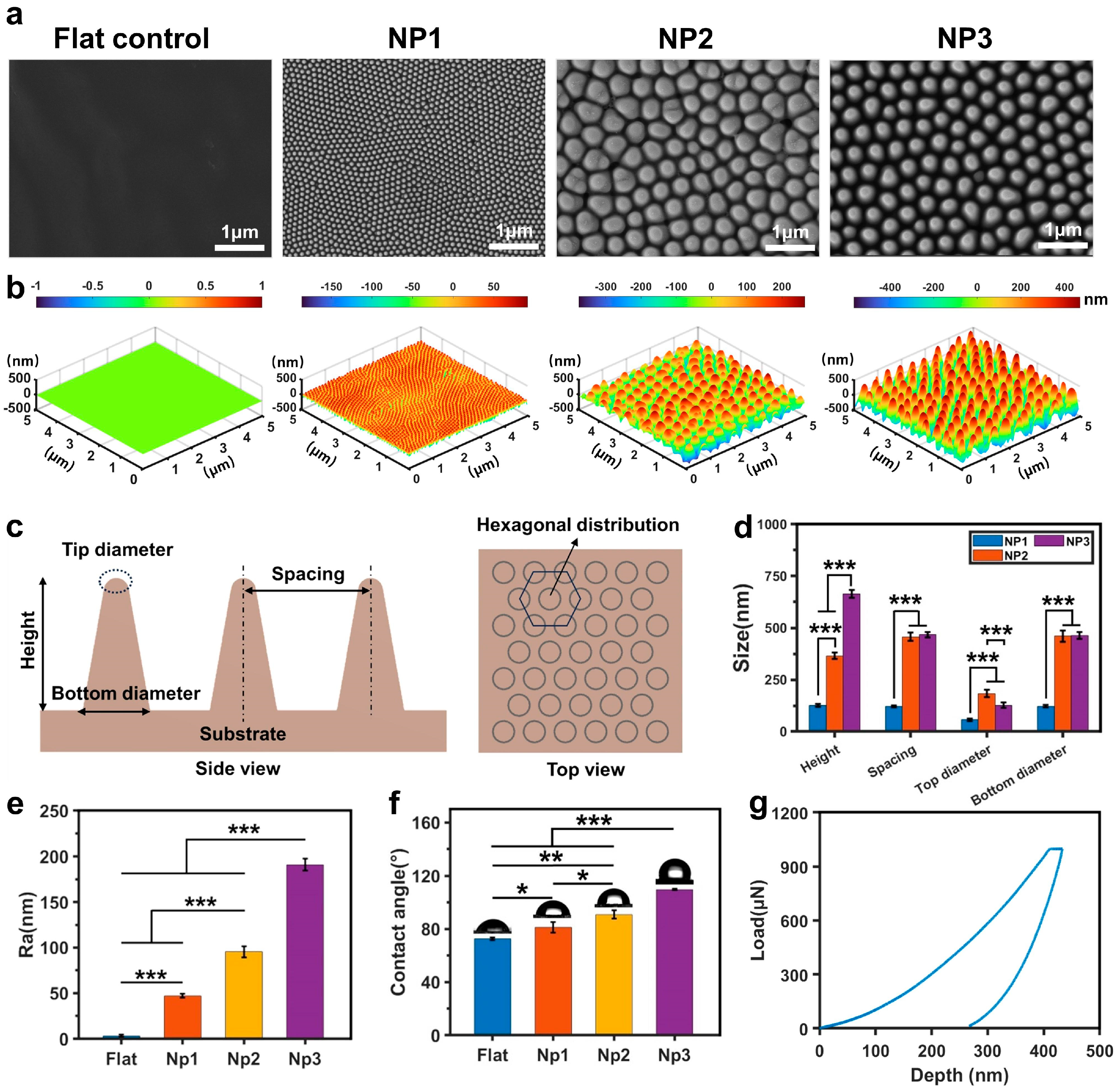
The SEM results in Figure 1a show that the structures on the AAO template (Figure S2) were successfully replicated, with the nanostructures arranged in a periodic hexagonal pattern. Low-magnification SEM observations indicate that this method produced uniformly distributed cicada wing-inspired nanostructures over a large area (Figure S3).
The results from AFM in Figure 1b indicate that the three types of prepared nanostructures were conical in shape. NP1 nano-pillars were small and densely packed, with dimensions and distribution of heights similar to nano-pillars on cicada wing surfaces. NP2 were taller but less sharp, with greater spacing compared to NP1. NP3 nano-pillars had spacing similar to NP2, with heights and sharpness approximately twice that of NP2. The cross-sectional images of the samples in Figure S4 also show that the shapes, sizes, and distributions of the prepared nanostructures were highly uniform, with noticeable differences in the dimensions of the three types of nano-pillars. Based on the features indicated in Figure 1c, statistical analysis was conducted on the characteristic dimensions of the prepared nanostructures. Specific parameters from the statistical results in Figure 1d show that NP1 had a height of 126 ± 7 nm, a spacing of 121 ± 5 nm, a top diameter of 57 ± 6 nm, and a bottom diameter of 122 ± 5 nm. NP2 had a height of 365 ± 15 nm, a spacing of 457 ± 20 nm, a top diameter of 184 ± 18 nm, and a bottom diameter of 460 ± 27 nm. NP3 had a height of 663 ± 18 nm, a spacing of 468 ± 13 nm, a top diameter of 127 ± 13 nm, and a bottom diameter of 464 ± 17 nm. Additionally, the roughness values of the four surfaces showed significant differences, with Ra values for NP1, NP2, and NP3 being 47 ± 2 nm, 95 ± 6 nm, and 191 ± 7 nm, respectively, whereas the roughness value for the flat surface was nearly zero (Figure 1e).
Figure 1f shows the static contact angles of water droplets on the three types of nanostructures and the smooth surface presented. The smooth surface had the smallest contact angle at 73.9°, while NP1 had a contact angle of 81.2°. This was because the air trapped in the gaps between the surface nanostructures inhibited the penetration of water droplets, reducing the contact area between the solid and liquid phases and thus decreasing the spreading of the water droplets. The contact angle of NP2 was 90.9°, higher than that of NP1, indicating increased hydrophobicity. This was because NP2 structures had larger spacing and a certain degree of taper, resulting in increased gaps between the nanostructures and a larger contact area between the solid and liquid phases compared to NP1. The contact angle of NP3 was 109.7°, indicating higher hydrophobicity compared to NP1 and NP2. This was because NP3 had the highest sharpness, with the largest gaps between the nanostructures and the smallest contact area between the solid and liquid phases, resulting in the highest hydrophobicity.
Due to the series of chemical etching processes involved in the sample preparation, even after thorough washing and soaking, residual Cu2+ may have still existed on the surface, which could have significantly affected the activity of bacteria and cells, thereby influencing the accuracy of subsequent experiments. Therefore, Cu2+ release testing was conducted on the four surfaces subjected to the same treatment. Figure S5 shows the Cu2+ release amounts of the samples immersed in PBS for 1 day. Data statistics reveal that the Cu2+ release amount was the lowest on the flat surface, while nanostructures NP1 and NP3 exhibited the highest Cu2+ release amounts, with NP2 showing lower release than NP1 but higher than that of the flat surface. This is attributed to the difference in specific surface area among the different surfaces, as depicted in Figure S6, which shows the surface area values detected by AFM. Higher specific surface areas adsorb more ions, leading to greater release amounts. Studies have shown that copper ion concentrations below 9 mg/L do not cause damage to osteoblasts and fibroblast cells, nor do they exhibit bactericidal effects [55]. The Cu2+ concentrations released from the four surface types were far below this threshold, thus having almost no impact on the activity of bacteria and cells. Finally, we tested the elastic modulus of the flat sample (Figure 1g), finding it to be approximately 5.06 ± 0.10 GPa, which was significantly higher than the modulus of the bacterial cell wall. This high modulus reduces the deformation of the nanostructures under the influence of bacteria.

3.2. Bacteriostatic Properties of Different Structured Surfaces

To investigate the effects of the flat surface and three different nanostructure surfaces (NP1, NP2, NP3) on bacterial adhesion and growth, two types of bacteria, S. aureus and E. coli, were selected as the research subjects. The antibacterial performance of different sample surfaces was tested for 12 h.
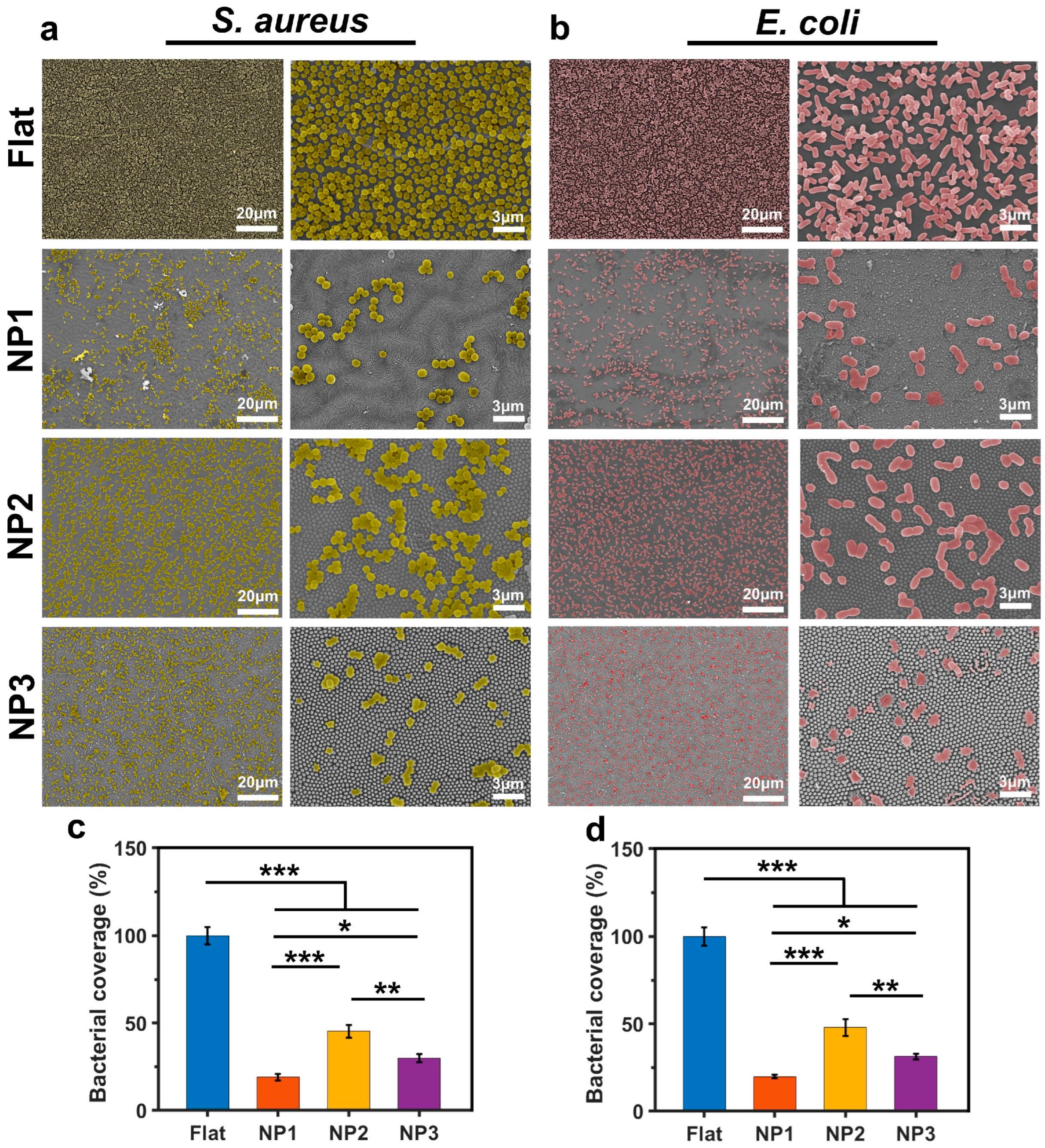
Figure 2 depicts the adhesion and growth of two types of bacteria on surfaces with different structures. Firstly, the adhesion of bacteria on different surfaces was analyzed. As shown in Figure 2a, the flat surface was completely covered by S. aureus, with closely connected secretion of adhesins. In contrast, the number of S. aureus adhering to the surfaces of NP1, NP2, and NP3 nanostructures was significantly reduced. Additionally, there were differences in bacterial adhesion among the three nanostructured surfaces, with NP2 showing the highest S. aureus adhesion, followed by NP3, and NP1 exhibiting the least adhesion (Figure 2c). Similar trends were observed for the adhesion of E. coli to different surfaces (Figure 2b), with the flat surface having the highest bacterial adhesion and all three nanostructured surfaces showing significant reductions. The quantity of bacteria on NP1, NP3, and NP2 surfaces increased sequentially (Figure 2d). The adhesion strength between bacteria and surfaces can be attributed to the size of their contact area [56]. The flat surface provided a larger adhesion area for bacteria, thereby facilitating bacterial adhesion. The presence of nanostructures reduced the contact area between bacteria and surfaces, resulting in decreased bacterial adhesion. The differences in bacterial adhesion among nanostructures can be attributed to the size, shape, and distribution of the nanostructures. The NP2 surface had larger and smoother structures, leading to higher bacterial adhesion. Compared to NP1, NP2 had a slightly reduced adhesion due to its sharper features. NP1 surface, being finer and sharper, exhibited the lowest adhesion.
From the growth state perspective, S. aureus on the flat surface (Figure 2a) appeared as regularly spherical, with full and rounded morphology. On the NP1 and NP2 nanostructured surfaces, S. aureus tended to aggregate and grow in chains. However, the degree of aggregation of S. aureus on the NP3 surface was lower, and the size of bacterial growth was smaller. Nevertheless, no evidence of nanostructures causing S. aureus elimination was found. Regarding E. coli (Figure 2b), the adhered E. coli on the flat surface exhibited regular rod shapes without any dead bacteria and maintained full morphology. Few E. coli cell wall ruptures were observed on the NP1 surface, while the status of E. coli on the NP2 surface was good, without any observed damaged cell walls. Conversely, a large number of cell wall ruptures was observed on the NP3 surface. The growth state of bacteria on the surface depends on the stress stimulation exerted by the structures on the bacteria. Smooth and flat surfaces hardly damage bacterial cell walls. Nanostructures smaller than bacterial sizes usually cause varying degrees of mechanical damage to bacterial cell walls, which is related to the shape and distribution of nanostructures. The NP3 surface with higher sharpness and larger spacing increased the stress exerted on individual nanostructures on bacteria, thereby inhibiting bacterial growth or even killing bacteria. The NP2 surface was relatively smoother, resulting in less stress stimulation on cell walls. The NP1 surface was sharper but more densely distributed, thus reducing the damage caused by individual nanostructures to cell walls due to the “nail bed effect.” The reason why the NP3 surface killed E. coli without killing S. aureus may be that S. aureus has a thicker cell wall. These patterns may require better explanation through simulation.
To further investigate the growth and adhesion of the two bacteria on different surfaces, bacterial staining was performed after 12 h of surface cultivation, followed by fluorescence microscopy imaging and quantitative analysis. Figure 3a shows the fluorescence staining results. From the perspective of bacterial adhesion quantity, the smooth flat surface had the highest adhesion of both bacteria, with a bacterial coverage rate of 52%. In contrast, the bacterial adhesion on nanostructured surfaces decreased by 49%–82%, with adhesion quantities in the following order: NP2 > NP3 > NP1 (Figure 3b). Regarding bacterial growth state (Figure 3c), for S. aureus, both the smooth surface and nanostructured surfaces had very few dead bacteria, indicating almost no killing effect. However, for E. coli, 14% of dead bacteria were observed on the NP1 surface, while the NP3 surface had 31% dead bacteria. NP2 and flat surfaces did not exhibit bactericidal effects. These results also validated the SEM findings mentioned above.

3.3. Simulation of Bacteria-Surface Mechanical Interaction Process

To achieve a deeper understanding of the mechanical interaction between different types of nanostructures and bacteria, we developed a three-dimensional finite element model of bacteria–nanostructure mechanical interaction. Currently, most finite element models abstract the structural characteristics obtained from experiments into simple geometric models [22,25,57], which cannot effectively reflect the actual interaction process between structures and bacteria. To address this issue, we reconstructed three-dimensional AFM images of several surfaces and employed them for finite element simulations, a practice that has been rarely adopted previously. Using the Abaqus/Standard nonlinear implicit solver, we simulated the adhesion and deformation of E. coli and S. aureus on different surfaces. In the simulation calculations, the adhesion strength of bacteria was represented by the contact area, while the growth status of bacteria was assessed based on the stress and strain of the cell wall. Based on previous studies, we selected a rupture strength of 13 MPa and a fracture strain of 35% for the cell wall. In the simulation process, if both criteria were met simultaneously, we considered the bacteria dead due to cell wall rupture [58,59,60,61].
Figure 4 presents the simulation results of mechanical deformation of E. coli and S. aureus on both smooth and nanostructured surfaces. Figure 4a,b show the strain maps of the deformation of the two types of bacteria on the surface. Here, we can clearly observe the contact status and deformation between the cell walls of the two bacteria and different surfaces. First, we analyzed the adhesion areas between bacteria and different surfaces to reflect the degree of bacterial adhesion. Figure 4e shows that the adhesion areas of E. coli and S. aureus on the smooth surface were 0.37 μm2 and 0.049 μm2, respectively, while the adhesion areas between E. coli and S. aureus and nanostructured surfaces were below 0.13 μm2 and 0.018 μm2, respectively. It can be observed that nanostructures smaller than the size of bacteria significantly reduced the adhesion area between bacteria and surfaces. Notably, among the three nanostructures, the adhesion area of E. coli and S. aureus on the NP2 surface was the largest, at 0.13 μm2 and 0.018 μm2, respectively, while the adhesion area on the NP1 surface was the smallest, at 0.051 μm2 and 0.0084 μm2, respectively. The adhesion areas on the NP3 surface were 0.086 μm2 and 0.013 μm2, respectively, falling between NP1 and NP2. This indicates that the adhesion area between bacteria and nanostructures was closely related to the size and distribution of nanostructures. The nanostructures on the NP1 surface were smaller with smaller tip diameters, resulting in smaller adhesion areas with bacteria. The nanostructures on the NP2 surface were taller and smoother compared to NP1, thereby increasing the adhesion area on the bacteria surface. Although the nanostructures on the NP3 surface were also taller, they had higher sharpness compared to NP2, leading to a reduction in adhesion area. Thus, smaller and sharper nanostructures more effectively reduced the adhesion area with bacteria, thereby achieving the effect of inhibiting bacterial adhesion.
Next, we analyzed the stress and deformation caused to the cell walls by different surfaces to reflect the bactericidal efficiency of the nanostructures. As shown in Figure 4c,d, the statistical results indicate that the cell walls of both bacteria experienced the lowest stress and strain on the smooth surface. The maximum von Mises stress and maximum principal strain of the E. coli cell wall were 2.26 MPa and 3.2%, respectively, while those of S. aureus were 2.54 MPa and 3.2%, respectively. The maximum stress and strain occurred at the edge of the cell wall in contact with the surface, where the bending curvature of the cell wall was also the highest. However, these values were much lower than the rupture stress and strain of the cell wall and were unlikely to cause damage. The presence of nanostructures led to greater mechanical deformation of the cell wall, which depended on the size and distribution of the nanostructures. Among the three nanostructures, the cell wall deformation was minimal on the NP2 surface, with the maximum von Mises stress and maximum principal strain of the E. coli cell wall being 8.77 MPa and 19%, respectively, and those of S. aureus being 8.17 MPa and 17%, respectively. This was mainly due to the smoother nanostructures on the NP2 surface. The cell wall deformation was greatest on the NP3 surface, with the maximum von Mises stress and maximum principal strain of the E. coli cell wall reaching 13.48 MPa and 39%, respectively, and those of S. aureus being 12 MPa and 32%, respectively. Compared to NP2, NP3 had increased sharpness, resulting in deeper penetration into the cell wall. It is noteworthy that the nanostructures on the NP1 surface were sharper than those on NP3, yet the maximum von Mises stress and maximum principal strain of the E. coli cell wall on the NP1 surface were 10.24 MPa and 24%, respectively, and those of S. aureus were 10.09 MPa and 23%, respectively, which were lower than those on the NP3 surface. This was mainly because NP1 had smaller spacing, leading to the “bed of nails” effect, which reduced the stress exerted by individual nanostructures on the cell wall.
In summary, compared to the flat surface, nanostructures effectively reduced bacterial adhesion and significantly influenced bacterial growth on surfaces, which was correlated with the size and distribution of the nanostructures. Regarding bacterial adhesion, the NP1 structure, being too small, provided fewer adhesion sites for bacteria, resulting in the least bacterial adhesion. The NP2 structure, being larger and smoother, increased the available adhesion area for bacteria, leading to the highest bacterial adhesion. The NP3 structure, being larger and sharper, reduced the available adhesion area for bacteria compared to NP2, resulting in a decrease in bacterial adhesion. As for bacterial growth, the NP3 structure, with larger spacing and sharper edges, imposed the highest stress on bacteria, exhibiting the strongest inhibition of bacterial growth and killing ability. The NP2 structure, with larger spacing and smoother surfaces, reduced stress on bacteria, resulting in the best bacterial growth state. The NP1 structure, although sharp, was dense, leading to reduced stress on bacteria compared to NP3 but higher than NP2 (Figure 5).

3.4. In Vitro Cytocompatibility Assay

The above results indicate that nanostructures effectively inhibited bacterial adhesion and growth through mechanical action. However, for many antibacterial surfaces, efficient bactericidal ability often accompanies cytotoxicity [62,63]. Whether nanostructures also cause mechanical damage to cells requires further verification. Therefore, the biocompatibility of the samples was tested using L929 mouse fibroblast cells. As shown in Figure 6c, the results of the CCK-8 assay after 1 and 7 days of culture indicated that there was no significant difference in cell viability between the flat samples and the three types of nanostructured groups. These results suggest that the presence of nanostructures did not induce significant mechanical cytotoxicity effects on cells, and variations in nanostructure size also did not affect the cell growth status.
Figure 6a shows the live/dead staining results of cells grown on the four types of samples for 1 day and 7 days. Cells on the smooth surface and NP1/NP2/NP3 nanostructured surfaces were stained green, indicating that the cells were healthy, well-spread, and exhibited no significant differences in quantity or morphology. SEM images in Figure 6b also demonstrate that cells on both smooth and nanostructured surfaces were able to spread and adhere normally. These results are consistent with the CCK-8 assay, indicating that the presence of nanostructures promoted neither cell proliferation nor adhesion significantly, nor did it cause mechanical damage to cells. This was mainly due to inherent differences between bacteria and cells (Figure 6d). Firstly, there was a significant difference in size between the two; bacteria typically range from 0.5~2 μm, while cells are usually larger than 10 μm [64,65,66]. Therefore, bacteria are more sensitive to nanostructured surfaces, while cells may require larger microscale structures to stimulate growth. Additionally, cells can adapt and alter their growth morphology in response to structural changes, whereas bacterial cell walls, composed of relatively rigid peptidoglycan layers, lack the ability to adapt to structural changes, making it difficult for them to survive on such surfaces [67,68].

4. Conclusions

In this study, we evaluated the comprehensive impact of different structural features on bacterial growth and adhesion by preparing three nanoarray structures with distinct characteristics. We found that the presence of nanostructures significantly influenced bacterial growth and adhesion states, which correlated closely with the sharpness and spacing of the nanostructures. Nanostructures with sharp and small features exhibited higher antibacterial and anti-adhesive effects. As the sharpness decreased and the size increased, the antibacterial and anti-adhesive effects of the nanostructures decreased. Additionally, reducing the spacing of the nanostructures lowered their antibacterial efficiency. Through the establishment of a 3D finite element model of bacterial-nanostructure adhesion, we accurately simulated the interaction process. We discovered that sharp and small nanostructures exerted higher mechanical stress and deformation on bacterial cell walls, while providing smaller adhesion areas, thus hindering bacterial growth and adhesion. Conversely, smoother nanostructures reduced stress on the cell wall and increased adhesion areas, thereby relatively favoring bacterial growth and adhesion. Spacing was also an important factor; nanostructures with overly dense distribution tended to produce a “bed of nails” effect, reducing the stress exerted by individual nanostructures on the cell wall, while overly sparse distribution made it easier for bacteria to fall into the gaps between structures, leading to structural failure. Furthermore, the height of nanostructures was a neglected factor; under the premise of determining sharpness and spacing, maintaining a height that prevents bacteria from contacting the flat area of the base surface can achieve antibacterial effects. Finally, cell experiments verified that nanostructures did not cause mechanical harm to cells. This study aimed to analyze the interaction mechanism between bacteria and nanostructures, providing new insights for the design of antibacterial structures and realizing their potential biomedical applications.

Supplementary Materials

The following supporting information can be downloaded at: https://www.mdpi.com/article/10.3390/coatings14070860/s1, included in the supporting information are physical images of the cycads, SEM and AFM images of the cycad surface nanostructures, wettability measurements of the cycad surface, SEM images of the AAO template, surface area measurements of the same range of the flat, NP1, NP2, NP3, schematic geometric modeling of E. coli and S. aureus for finite element simulations, schematic reconstruction of geometric modeling of the nanostructures, and finite modeling of the material properties (PDF).

Author Contributions

Conceptualization, C.L. and Y.X.; methodology, D.W.; data curation, X.W., X.Z. and B.Z.; formal analysis, X.W., X.Z. and B.Z.; funding acquisition, C.L., Y.X. and M.L.; investigation, X.W. and X.Z.; project administration, D.W., M.L. and Y.X.; resources, D.W. and Y.X.; supervision, D.W. and H.W.; writing—original draft, X.W.; writing—review and editing, X.W., D.W. and Y.X. All authors have read and agreed to the published version of the manuscript.

Funding

This work was supported by “National Natural Science Foundation of China (U21A2055, 82025025)”; “Natural Science Foundation of Tianjin of China: (No. 21JCYBJC01380, No. 21JCQNJC01590)”.

Institutional Review Board Statement

Not applicable.

Informed Consent Statement

Not applicable.

Data Availability Statement

The data presented in this study are available on request from the corresponding author. The data are not publicly available due to privacy.

Conflicts of Interest

The authors declare no conflicts of interest.

References

  1. Linklater, D.P.; Baulin, V.A.; Juodkazis, S.; Crawford, R.J.; Stoodley, P.; Ivanova, E.P. Mechano-bactericidal actions of nanostructured surfaces. Nat. Rev. Microbiol. 2021, 19, 8–22. [Google Scholar] [CrossRef] [PubMed]
  2. Asri, L.A.; Crismaru, M.; Roest, S.; Chen, Y.; Ivashenko, O.; Rudolf, P.; Tiller, J.C.; van der Mei, H.C.; Loontjens, T.J.; Busscher, H.J. A shape-adaptive, antibacterial-coating of immobilized quaternary-ammonium compounds tethered on hyperbranched polyurea and its mechanism of action. Adv. Funct. Mater. 2014, 24, 346–355. [Google Scholar] [CrossRef]
  3. Chernousova, S.; Epple, M. Silver as antibacterial agent: Ion, nanoparticle, and metal. Angew. Chem. Int. Ed. 2013, 52, 1636–1653. [Google Scholar] [CrossRef] [PubMed]
  4. Arciola, C.R.; Campoccia, D.; Montanaro, L. Implant infections: Adhesion, biofilm formation and immune evasion. Nat. Rev. Microbiol. 2018, 16, 397–409. [Google Scholar] [CrossRef] [PubMed]
  5. Glinel, K.; Thebault, P.; Humblot, V.; Pradier, C.-M.; Jouenne, T. Antibacterial surfaces developed from bio-inspired approaches. Acta Biomater. 2012, 8, 1670–1684. [Google Scholar] [CrossRef] [PubMed]
  6. Bagherifard, S. Mediating bone regeneration by means of drug eluting implants: From passive to smart strategies. Mater. Sci. Eng. C 2017, 71, 1241–1252. [Google Scholar] [CrossRef]
  7. Liao, H.; Zhao, Q.; Cui, P.; Chen, Z.; Yu, Z.; Geisen, S.; Friman, V.-P.; Zhou, S. Efficient reduction of antibiotic residues and associated resistance genes in tylosin antibiotic fermentation waste using hyperthermophilic composting. Environ. Int. 2019, 133, 105203. [Google Scholar] [CrossRef]
  8. Gallardo-Godoy, A.; Muldoon, C.; Becker, B.; Elliott, A.G.; Lash, L.H.; Huang, J.X.; Butler, M.S.; Pelingon, R.; Kavanagh, A.M.; Ramu, S. Activity and predicted nephrotoxicity of synthetic antibiotics based on polymyxin B. J. Med. Chem. 2016, 59, 1068–1077. [Google Scholar] [CrossRef]
  9. Bandara, C.D.; Singh, S.; Afara, I.O.; Wolff, A.; Tesfamichael, T.; Ostrikov, K.; Oloyede, A. Bactericidal effects of natural nanotopography of dragonfly wing on Escherichia coli. ACS Appl. Mater. Interfaces 2017, 9, 6746–6760. [Google Scholar] [CrossRef] [PubMed]
  10. Ivanova, E.P.; Hasan, J.; Webb, H.K.; Truong, V.K.; Watson, G.S.; Watson, J.A.; Baulin, V.A.; Pogodin, S.; Wang, J.Y.; Tobin, M.J. Natural bactericidal surfaces: Mechanical rupture of Pseudomonas aeruginosa cells by cicada wings. Small 2012, 8, 2489. [Google Scholar] [CrossRef]
  11. Nouri-Goushki, M.; Mirzaali, M.; Angeloni, L.; Fan, D.; Minneboo, M.; Ghatkesar, M.; Staufer, U.; Fratila-Apachitei, L.; Zadpoor, A. 3D printing of large areas of highly ordered submicron patterns for modulating cell behavior. ACS Appl. Mater. Interfaces 2019, 12, 200–208. [Google Scholar] [CrossRef]
  12. Bagherifard, S.; Hickey, D.J.; de Luca, A.C.; Malheiro, V.N.; Markaki, A.E.; Guagliano, M.; Webster, T.J. The influence of nanostructured features on bacterial adhesion and bone cell functions on severely shot peened 316L stainless steel. Biomaterials 2015, 73, 185–197. [Google Scholar] [CrossRef]
  13. Hazell, G.; Fisher, L.E.; Murray, W.A.; Nobbs, A.H.; Su, B. Bioinspired bactericidal surfaces with polymer nanocone arrays. J. Colloid Interface Sci. 2018, 528, 389–399. [Google Scholar] [CrossRef]
  14. Sjöström, T.; Nobbs, A.H.; Su, B. Bactericidal nanospike surfaces via thermal oxidation of Ti alloy substrates. Mater. Lett. 2016, 167, 22–26. [Google Scholar] [CrossRef]
  15. Ivanova, E.P.; Hasan, J.; Webb, H.K.; Gervinskas, G.; Juodkazis, S.; Truong, V.K.; Wu, A.H.; Lamb, R.N.; Baulin, V.A.; Watson, G.S. Bactericidal activity of black silicon. Nat. Commun. 2013, 4, 2838. [Google Scholar] [CrossRef]
  16. Cui, Q.; Liu, T.; Li, X.; Song, K.; Ge, D. Nanopillared polycarbonate surfaces having variable feature parameters as bactericidal coatings. ACS Appl. Nano Mater. 2020, 3, 4599–4609. [Google Scholar] [CrossRef]
  17. Bhadra, C.M.; Werner, M.; Baulin, V.A.; Truong, V.K.; Kobaisi, M.A.; Nguyen, S.H.; Balcytis, A.; Juodkazis, S.; Wang, J.Y.; Mainwaring, D.E. Subtle variations in surface properties of black silicon surfaces influence the degree of bactericidal efficiency. Nano-Micro Lett. 2018, 10, 36. [Google Scholar] [CrossRef] [PubMed]
  18. Wu, S.; Zuber, F.; Maniura-Weber, K.; Brugger, J.; Ren, Q. Nanostructured surface topographies have an effect on bactericidal activity. J. Nanobiotechnol. 2018, 16, 20. [Google Scholar] [CrossRef]
  19. Ivanova, E.P.; Linklater, D.P.; Werner, M.; Baulin, V.A.; Xu, X.; Vrancken, N.; Rubanov, S.; Hanssen, E.; Wandiyanto, J.; Truong, V.K. The multi-faceted mechano-bactericidal mechanism of nanostructured surfaces. Proc. Natl. Acad. Sci. USA 2020, 117, 12598–12605. [Google Scholar] [CrossRef] [PubMed]
  20. Jenkins, J.; Ishak, M.I.; Eales, M.; Gholinia, A.; Kulkarni, S.; Keller, T.F.; May, P.W.; Nobbs, A.H.; Su, B. Resolving physical interactions between bacteria and nanotopographies with focused ion beam scanning electron microscopy. iScience 2021, 24, 102818. [Google Scholar] [CrossRef]
  21. Ye, J.; Li, B.; Zheng, Y.; Wu, S.; Chen, D.; Han, Y. Eco-friendly bacteria-killing by nanorods through mechano-puncture with top selectivity. Bioact. Mater. 2022, 15, 173–184. [Google Scholar] [CrossRef] [PubMed]
  22. Valiei, A.; Lin, N.; Bryche, J.-F.; McKay, G.; Canva, M.; Charette, P.G.; Nguyen, D.; Moraes, C.; Tufenkji, N. Hydrophilic mechano-bactericidal nanopillars require external forces to rapidly kill bacteria. Nano Lett. 2020, 20, 5720–5727. [Google Scholar] [CrossRef] [PubMed]
  23. Zhao, X.; Xu, Z.; Wei, Z.; Sun, Y.; Zhou, Q. Nature-inspired mechano-bactericidal nanostructured surfaces with photothermally enhanced antibacterial performances. Prog. Org. Coat. 2023, 182, 107599. [Google Scholar] [CrossRef]
  24. Liu, Z.; Yi, Y.; Wang, S.; Dou, H.; Fan, Y.; Tian, L.; Zhao, J.; Ren, L. Bio-inspired self-adaptive nanocomposite array: From non-antibiotic antibacterial actions to cell proliferation. ACS Nano 2022, 16, 16549–16562. [Google Scholar] [CrossRef]
  25. Alameda, M.T.; Osorio, M.R.; Pedraz, P.; Rodríguez, I. Mechano-Dynamic Analysis of the Bactericidal Activity of Bioinspired Moth-Eye Nanopatterned Surfaces. Adv. Mater. Interfaces 2022, 9, 2200608. [Google Scholar] [CrossRef]
  26. Oehrlein, G.S.; Phaneuf, R.J.; Graves, D.B. Plasma-polymer interactions: A review of progress in understanding polymer resist mask durability during plasma etching for nanoscale fabrication. J. Vac. Sci. Technol. B 2011, 29, 010801. [Google Scholar] [CrossRef]
  27. Bierbaum, S.; Mulansky, S.; Bognár, E.; Kientzl, I.; Nagy, P.; Vrana, N.E.; Weszl, M.; Boschke, E.; Scharnweber, D.; Wolf-Brandstetter, C. Osteogenic nanostructured titanium surfaces with antibacterial properties under conditions that mimic the dynamic situation in the oral cavity. Biomater. Sci. 2018, 6, 1390–1402. [Google Scholar] [CrossRef]
  28. Truong, V.K.; Pham, V.T.; Medvedev, A.; Lapovok, R.; Estrin, Y.; Lowe, T.C.; Baulin, V.; Boshkovikj, V.; Fluke, C.J.; Crawford, R.J. Self-organised nanoarchitecture of titanium surfaces influences the attachment of Staphylococcus aureus and Pseudomonas aeruginosa bacteria. Appl. Microbiol. Biotechnol. 2015, 99, 6831–6840. [Google Scholar] [CrossRef]
  29. Stolzoff, M.; Burns, J.E.; Aslani, A.; Tobin, E.J.; Nguyen, C.; De La Torre, N.; Golshan, N.H.; Ziemer, K.S.; Webster, T.J. Decreased bacterial growth on titanium nanoscale topographies created by ion beam assisted evaporation. Int. J. Nanomed. 2017, 12, 1161–1169. [Google Scholar] [CrossRef]
  30. Sengstock, C.; Lopian, M.; Motemani, Y.; Borgmann, A.; Khare, C.; Buenconsejo, P.J.S.; Schildhauer, T.A.; Ludwig, A.; Köller, M. Structure-related antibacterial activity of a titanium nanostructured surface fabricated by glancing angle sputter deposition. Nanotechnology 2014, 25, 195101. [Google Scholar] [CrossRef]
  31. Linklater, D.P.; De Volder, M.; Baulin, V.A.; Werner, M.; Jessl, S.; Golozar, M.; Maggini, L.; Rubanov, S.; Hanssen, E.; Juodkazis, S. High aspect ratio nanostructures kill bacteria via storage and release of mechanical energy. Acs Nano 2018, 12, 6657–6667. [Google Scholar] [CrossRef] [PubMed]
  32. Zahir, T.; Pesek, J.; Franke, S.; Van Pee, J.; Rathore, A.; Smeets, B.; Ramon, H.; Xu, X.; Fauvart, M.; Michiels, J. Model-driven controlled alteration of Nanopillar Cap Architecture reveals its effects on bactericidal activity. Microorganisms 2020, 8, 186. [Google Scholar] [CrossRef] [PubMed]
  33. Michalska, M.; Gambacorta, F.; Divan, R.; Aranson, I.S.; Sokolov, A.; Noirot, P.; Laible, P.D. Tuning antimicrobial properties of biomimetic nanopatterned surfaces. Nanoscale 2018, 10, 6639–6650. [Google Scholar] [CrossRef] [PubMed]
  34. Ghosh, S.; Niu, S.; Yankova, M.; Mecklenburg, M.; King, S.M.; Ravichandran, J.; Kalia, R.K.; Nakano, A.; Vashishta, P.; Setlow, P. Analysis of killing of growing cells and dormant and germinated spores of Bacillus species by black silicon nanopillars. Sci. Rep. 2017, 7, 17768. [Google Scholar] [CrossRef] [PubMed]
  35. Modaresifar, K.; Azizian, S.; Ganjian, M.; Fratila-Apachitei, L.E.; Zadpoor, A.A. Bactericidal effects of nanopatterns: A systematic review. Acta Biomater. 2019, 83, 29–36. [Google Scholar] [CrossRef] [PubMed]
  36. Modaresifar, K.; Kunkels, L.B.; Ganjian, M.; Tümer, N.; Hagen, C.W.; Otten, L.G.; Hagedoorn, P.-L.; Angeloni, L.; Ghatkesar, M.K.; Fratila-Apachitei, L.E. Deciphering the roles of interspace and controlled disorder in the bactericidal properties of nanopatterns against Staphylococcus aureus. Nanomaterials 2020, 10, 347. [Google Scholar] [CrossRef] [PubMed]
  37. Yang, K.; Shi, J.; Wang, L.; Chen, Y.; Liang, C.; Yang, L.; Wang, L.-N. Bacterial anti-adhesion surface design: Surface patterning, roughness and wettability: A review. J. Mater. Sci. Technol. 2022, 99, 82–100. [Google Scholar] [CrossRef]
  38. Mainwaring, D.E.; Nguyen, S.H.; Webb, H.; Jakubov, T.; Tobin, M.; Lamb, R.N.; Wu, A.H.-F.; Marchant, R.; Crawford, R.J.; Ivanova, E.P. The nature of inherent bactericidal activity: Insights from the nanotopology of three species of dragonfly. Nanoscale 2016, 8, 6527–6534. [Google Scholar] [CrossRef]
  39. Kelleher, S.M.; Habimana, O.; Lawler, J.; O’reilly, B.; Daniels, S.; Casey, E.; Cowley, A. Cicada wing surface topography: An investigation into the bactericidal properties of nanostructural features. ACS Appl. Mater. Interfaces 2016, 8, 14966–14974. [Google Scholar] [CrossRef]
  40. Ivanova, E.; Crawford, R. Antibacterial Surfaces; Springer: Berlin/Heidelberg, Germany, 2015. [Google Scholar]
  41. Li, X.; Chen, T. Enhancement and suppression effects of a nanopatterned surface on bacterial adhesion. Phys. Rev. E 2016, 93, 052419. [Google Scholar] [CrossRef]
  42. Xue, F.; Liu, J.; Guo, L.; Zhang, L.; Li, Q. Theoretical study on the bactericidal nature of nanopatterned surfaces. J. Theor. Biol. 2015, 385, 1–7. [Google Scholar] [CrossRef]
  43. Pogodin, S.; Hasan, J.; Baulin, V.A.; Webb, H.K.; Truong, V.K.; Boshkovikj, V.; Fluke, C.J.; Watson, G.S.; Watson, J.A.; Crawford, R.J. Biophysical model of bacterial cell interactions with nanopatterned cicada wing surfaces. Biophys. J. 2013, 104, 835–840. [Google Scholar] [CrossRef]
  44. Lazzini, G.; Lutey, A.; Romoli, L.; Fuso, F. Molecular dynamics model for the antibactericity of textured surfaces. Colloids Surf. B Biointerfaces 2021, 199, 111504. [Google Scholar] [CrossRef]
  45. Cui, Q.; Liu, T.; Li, X.; Zhao, L.; Wu, Q.; Wang, X.; Song, K.; Ge, D. Validation of the mechano-bactericidal mechanism of nanostructured surfaces with finite element simulation. Colloids Surf. B Biointerfaces 2021, 206, 111929. [Google Scholar] [CrossRef]
  46. Velic, A.; Hasan, J.; Li, Z.; Yarlagadda, P.K. Mechanics of bacterial interaction and death on nanopatterned surfaces. Biophys. J. 2021, 120, 217–231. [Google Scholar] [CrossRef]
  47. Liu, Z.; Yi, Y.; Song, L.; Chen, Y.; Tian, L.; Zhao, J.; Ren, L. Biocompatible mechano-bactericidal nanopatterned surfaces with salt-responsive bacterial release. Acta Biomater. 2022, 141, 198–208. [Google Scholar] [CrossRef]
  48. Touhami, A.; Jericho, M.H.; Beveridge, T.J. Atomic force microscopy of cell growth and division in Staphylococcus aureus. J. Bacteriol. 2004, 186, 3286–3295. [Google Scholar] [CrossRef]
  49. Harris, L.G.; Foster, S.; Richards, R.G. An introduction to Staphylococcus aureus, and techniques for identifying and quantifying S. aureus adhesins in relation to adhesion to biomaterials: Review. Eur. Cell Mater. 2002, 4, 100–120. [Google Scholar] [CrossRef]
  50. Maleki, E.; Mirzaali, M.J.; Guagliano, M.; Bagherifard, S. Analyzing the mechano-bactericidal effect of nano-patterned surfaces on different bacteria species. Surf. Coat. Technol. 2021, 408, 126782. [Google Scholar] [CrossRef]
  51. Tuson, H.H.; Auer, G.K.; Renner, L.D.; Hasebe, M.; Tropini, C.; Salick, M.; Crone, W.C.; Gopinathan, A.; Huang, K.C.; Weibel, D.B. Measuring the stiffness of bacterial cells from growth rates in hydrogels of tunable elasticity. Mol. Microbiol. 2012, 84, 874–891. [Google Scholar] [CrossRef]
  52. Yao, X.; Jericho, M.; Pink, D.; Beveridge, T. Thickness and elasticity of gram-negative murein sacculi measured by atomic force microscopy. J. Bacteriol. 1999, 181, 6865–6875. [Google Scholar] [CrossRef]
  53. Amir, A.; Babaeipour, F.; McIntosh, D.B.; Nelson, D.R.; Jun, S. Bending forces plastically deform growing bacterial cell walls. Proc. Natl. Acad. Sci. USA 2014, 111, 5778–5783. [Google Scholar] [CrossRef]
  54. Bavi, N.; Nakayama, Y.; Bavi, O.; Cox, C.D.; Qin, Q.-H.; Martinac, B. Biophysical implications of lipid bilayer rheometry for mechanosensitive channels. Proc. Natl. Acad. Sci. USA 2014, 111, 13864–13869. [Google Scholar] [CrossRef]
  55. Fowler, L.; Engqvist, H.; Öhman-Mägi, C. Effect of copper ion concentration on bacteria and cells. Materials 2019, 12, 3798. [Google Scholar] [CrossRef]
  56. Yang, K.; Wang, L.; Zou, X.; Wang, H.; Liang, C.; Zhang, D.; Wang, L.-N. Modeling bacterial adhesion on the nanopatterned surface by varying contact area. J. Mater. Sci. Technol. 2024, 196, 137–147. [Google Scholar] [CrossRef]
  57. Valiei, A.; Lin, N.; McKay, G.; Nguyen, D.; Moraes, C.; Hill, R.J.; Tufenkji, N. Surface wettability is a key feature in the mechano-bactericidal activity of nanopillars. ACS Appl. Mater. Interfaces 2022, 14, 27564–27574. [Google Scholar] [CrossRef]
  58. Thwaites, J.; Surana, U.; Jones, A. Mechanical properties of Bacillus subtilis cell walls: Effects of ions and lysozyme. J. Bacteriol. 1991, 173, 204–210. [Google Scholar] [CrossRef]
  59. Stocks, S.; Thomas, C. Viability, strength, and fragmentation of Saccharopolyspora erythraea in submerged fermentation. Biotechnol. Bioeng. 2001, 75, 702–709. [Google Scholar] [CrossRef]
  60. Stocks, S.M.; Thomas, C.R. Strength of mid-logarithmic and stationary phase Saccharopolyspora erythraea hyphae during a batch fermentation in defined nitrate-limited medium. Biotechnol. Bioeng. 2001, 73, 370–378. [Google Scholar] [CrossRef]
  61. Thwaites, J.; Surana, U. Mechanical properties of Bacillus subtilis cell walls: Effects of removing residual culture medium. J. Bacteriol. 1991, 173, 197–203. [Google Scholar] [CrossRef]
  62. Zeng, J.; Wang, Y.; Sun, Z.; Chang, H.; Cao, M.; Zhao, J.; Lin, K.; Xie, Y. A novel biocompatible PDA/IR820/DAP coating for antibiotic/photodynamic/photothermal triple therapy to inhibit and eliminate Staphylococcus aureus biofilm. Chem. Eng. J. 2020, 394, 125017. [Google Scholar] [CrossRef]
  63. Lara, H.H.; Ixtepan-Turrent, L.; Jose Yacaman, M.; Lopez-Ribot, J. Inhibition of Candida auris biofilm formation on medical and environmental surfaces by silver nanoparticles. ACS Appl. Mater. Interfaces 2020, 12, 21183–21191. [Google Scholar] [CrossRef]
  64. Higgins, S.G.; Becce, M.; Belessiotis-Richards, A.; Seong, H.; Sero, J.E.; Stevens, M.M. High-aspect-ratio nanostructured surfaces as biological metamaterials. Adv. Mater. 2020, 32, 1903862. [Google Scholar] [CrossRef]
  65. Tay, A. The benefits of going small: Nanostructures for mammalian cell transfection. ACS Nano 2020, 14, 7714–7721. [Google Scholar] [CrossRef] [PubMed]
  66. Liu, T.; Cui, Q.; Wu, Q.; Li, X.; Song, K.; Ge, D.; Guan, S. Mechanism study of bacteria killed on nanostructures. J. Phys. Chem. B 2019, 123, 8686–8696. [Google Scholar] [CrossRef] [PubMed]
  67. Lee, S.W.; Phillips, K.S.; Gu, H.; Kazemzadeh-Narbat, M.; Ren, D. How microbes read the map: Effects of implant topography on bacterial adhesion and biofilm formation. Biomaterials 2021, 268, 120595. [Google Scholar] [CrossRef]
  68. Elbourne, A.; Crawford, R.J.; Ivanova, E.P. Nano-structured antimicrobial surfaces: From nature to synthetic analogues. J. Colloid Interface Sci. 2017, 508, 603–616. [Google Scholar] [CrossRef]
Scheme 1. (a) Schematic diagram of flat and three differently characterized cicada wing-inspired nanostructures; (b) the preparation process of cicada wing-inspired nanostructures.
Coatings 14 00860 sch001
Figure 1. Characterization of Flat, NP1, NP2, and NP3 surfaces. (a) SEM images of the four surfaces; (b) AFM 3D morphology of the four surfaces; (c) characteristic parameters of the nanostructures; (d) statistical analysis of the characteristic parameters of NP1, NP2, and NP3 nanostructures; (e) roughness values of the four surfaces; (f) static water contact angle tests on different structured surfaces; (g) nanoindentation curve of the flat sample. Significant difference: * p < 0.05, ** p < 0.01, and *** p < 0.001.
Coatings 14 00860 g001
Figure 2. SEM images of (a) S. aureus and (b) E. coli after 12 h of incubation on Flat, NP1, NP2, and NP3 surfaces; Bacterial coverage statistics (c) S. aureus and (d) E. coli. Significant difference: * p < 0.05, ** p < 0.01, and *** p < 0.001.
Coatings 14 00860 g002
Figure 3. (a) Fluorescence images of live/dead staining of S. aureus and E. coli cultured on flat, NP1, NP2, and NP3 surfaces for 12 h. (b) Total coverage of bacteria on different surfaces; (c) proportion of dead bacteria to the total number of bacteria on different surfaces. Significant difference: * p < 0.05, ** p < 0.01, and *** p < 0.001.
Coatings 14 00860 g003
Figure 4. Finite element simulation of mechanical deformation on flat, NP1, NP2, and NP3 surfaces for (a) E. coli and (b) S. aureus with strain contour maps; (c) statistical analysis of maximum von Mises stress on the cell wall; (d) statistical analysis of maximum principal strain on the cell wall; (e) statistical analysis of the contact area between bacteria and the surface. Significant difference: * p < 0.05, and *** p < 0.001.
Coatings 14 00860 g004
Figure 5. Schematic diagram of bacterial growth and adhesion on different surfaces.
Coatings 14 00860 g005
Figure 6. Analysis of L929 cell biocompatibility on different surfaces for 1 day and 7 days. (a) Fluorescence staining images of cell viability on different surfaces. (b) SEM images of cell adhesion on different surfaces. (c) CCK-8 results on different surfaces. (d) Schematic diagram of bacterial/cellular selective adhesion and growth.
Coatings 14 00860 g006
Disclaimer/Publisher’s Note: The statements, opinions and data contained in all publications are solely those of the individual author(s) and contributor(s) and not of MDPI and/or the editor(s). MDPI and/or the editor(s) disclaim responsibility for any injury to people or property resulting from any ideas, methods, instructions or products referred to in the content.