Article

Chemical Bonding of Nanorod Hydroxyapatite to the Surface of Calciumfluoroaluminosilicate Particles for Improving the Histocompatibility of Glass Ionomer Cement

1
Department of Dentistry, College of Medicine, Yeungnam University, Daegu 42415, Republic of Korea
2
Department of Polymer Science and Engineering, Kyungpook National University, Daegu 41566, Republic of Korea
3
Materials Science and Engineering, Yeungnam University, Gyeongsan-si 38541, Republic of Korea
*
Author to whom correspondence should be addressed.
Coatings 2024, 14(7), 893; https://doi.org/10.3390/coatings14070893
Submission received: 21 June 2024 / Revised: 13 July 2024 / Accepted: 15 July 2024 / Published: 17 July 2024
(This article belongs to the Special Issue Advanced Biomaterials and Coatings)

Abstract

:
Glass ionomer cement (GIC) is composed of anionic polyacrylic acid and a silica-based inorganic powder. GIC is used as a filling material in the decayed cavity of the tooth; therefore, compatibility with the tooth tissue is essential. In the present study, we aimed to improve the histocompatibility of GIC by introducing nano-hydroxyapatite (nHA), a component of teeth, into a silica-based inorganic powder. CFAS-nHA was prepared by chemically bonding nanorod hydroxyapatite (nHA) to the surface of calciumfluoroaluminosilicate (CFAS). The synthesis of CFAS-nHA was confirmed using Fourier transform infrared spectroscopy (FTIR) and scanning electron microscopy (SEM). The prepared CFAS-nHA was mixed with polyacrylic acid and cured to prepare GIC containing nHA (GIC-nHA). Cytocompatibility tests of GIC-nHA and GIC were performed using osteoblasts. Osteoblast activity and bone formation ability were superior after GIC-nHA treatment than after control GIC treatment. This enhanced histocompatibility is believed to be due to the improvement of the biological activity of osteoblasts induced by the HA introduced into the GIC. Therefore, to enhance its compatibility with dental tissues, GIC could be manufactured by chemically bonding nHA to the surface of GI inorganic powder.

1. Introduction

Glass ionomer cement (GIC) has various applications in dentistry, including use as a bonding agent for restorative materials, liners, fissure sealants, and orthodontic brackets for permanent and primary teeth [1]. Compared with previously used materials, dental restoration approaches using GIC have been developed. The GIC comprises a mixture of silica-based inorganic powders and anionic polyacrylic acid. GIC, which exhibits hydrophilicity owing to the characteristics of silica powder with many hydroxyl groups, can effectively absorb the liquid remaining at the bottom of the fissure compared to composite resins, thus adhering well to the tooth enamel [2]. Although GIC is actively utilized in dental restorations because of its high adhesion to teeth, there is a need for further improvement in terms of its brittleness, abrasion resistance, bending, and tensile strength [3]. It has been reported that the wear resistance or brittleness of GIC can be improved via the introduction of polyalkenoic acid [4] or nanosized bioceramics [5]. Among them, nanosized biomaterials have shown promising potential for improving the strength, gloss, and aesthetics of dental filling materials compared to conventional modifiers [6]. In recent years, there have been many reports focused on improving the mechanical strength of GICs through the introduction of hydroxyapatite (HA) [7,8]. HA has a chemical structure that closely resembles that of human teeth and skeletal systems [9]. Recent advances in HA synthesis technology has enabled the synthesis of HA of various sizes and shapes [10,11,12], facilitating their application as biocompatible fillers that resemble natural teeth. In addition to exhibiting unique radiopaque properties [13], HA plays an important role in orthopedic surgery because of its excellent biological activity and osteoconductivity. Furthermore, research has been conducted to enhance the antibacterial activity of GIC through the use of HA. Praveen et al. [14] added 8% HA powder to an existing glass ionomer (GC Fuji Type IX gold label, GC Corporation, Tokyo, Japan) and combined it with liquid polyacrylic acid to obtain cylindrical GIC test specimens. They reported that GIC with 8% HA demonstrated higher antibacterial activity than GIC without HA through antibacterial testing using Streptococcus mutans. Haider et al. [15] investigated the topographical effects of HA on the physiological activity of osteoblasts by preparing nanocomposites. They added spherical HA (sHA) and nanorod HA (nHA) to the poly(lactic-co-glycolic acid) scaffold. Based on the results of studying the interaction between the nanocomposite and osteoblasts, they concluded that the HA-containing nanorod scaffold further promoted osteoblast bioactivity. In this study, nHA was chemically bonded to the surface of calciumfluoroaluminosilicate (CFAS), a solid component of GIC, using the biocomponents L-glutamic acid (G) and albumin (Alb) as spacers. The progress of the surface reaction was confirmed by attenuated total reflectance Fourier transform infrared (ATR-FTIR) and scanning electron microscopy (SEM). The resulting CFAS-nHA powder was mixed with polyacrylic acid and UV-cured to prepare the disc-shaped GIC-nHA. The cytocompatibility and bone formation ability of the GIC and GIC-nHA discs were investigated using pre-osteoblasts.

2. Materials and Methods

2.1. The Preparation of Materials

The following materials were purchased from Sigma Aldrich Chemical Company (St. Louis, MO, USA): 3-Aminopropyltriethoxysilane (A), L-glutamic acid, N-(3-dimethylaminopropyl)-N′-ethylcarbodiimide hydrochloride (EDC), N-hydroxysuccinimide (NHS), fluorescein isothiocyanate (FITC), and bovine serum albumin. The nHA was synthesized according to previously described procedures [10,15]. The mouse MC3T3-E1 cell line was purchased from the Korea Cell Bank (Seoul, Republic of Korea) and stored in liquid nitrogen until cell seeding. The MC3T3-E1 cell line is a cultured mouse osteoblast cell line derived from mouse embryo pre-osteoblasts; it is capable of differentiating into osteoblasts. A phosphate-buffered saline (PBS) solution (pH 7.4) containing Na2HPO4, KH2PO4, NaCl, and KCl was acquired from Sigma-Aldrich. CFAS, a solid powder used in the preparation of GIC, was provided by Professor Sukyoung Kim of Yeungnam University, Republic of Korea. The XRD pattern of the CFAS was broad and showed poor crystallinity. The nHA was chemically bonded to the surface of the CFAS for use as a solid component of the GIC. The liquid component of GIC (acquired from GC International, Tokyo, Japan, GC Fuji II LC) was used as the polyacrylic liquid component to prepare the GIC. The MC3T3-E1 cells were cultured in α-minimum essential medium (α-MEM) supplemented with 10% fetal bovine serum and 1.0% penicillin G-streptomycin at a temperature of 37 °C. Under a 5% CO2 atmosphere, the culture medium was replaced every other day. 3-(4,5-Dimethylthiazol-2 yl)-2,5-diphenyltetrazolium bromide (MTT) was acquired from Sigma-Aldrich (United States). The presence of nHA on the surfaces of the solid particles was observed using SEM (Hitachi S-400, Tokyo, Japan).

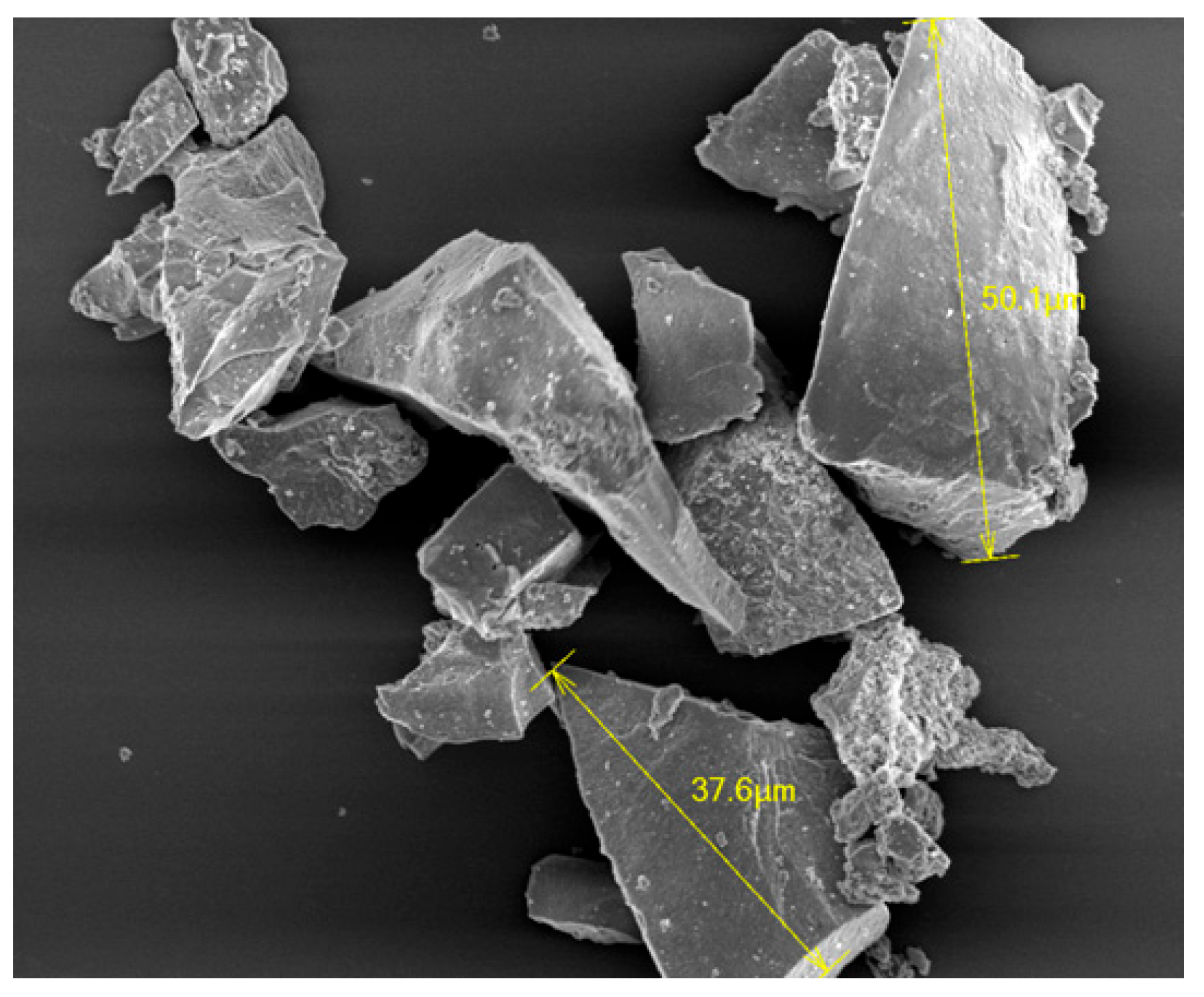
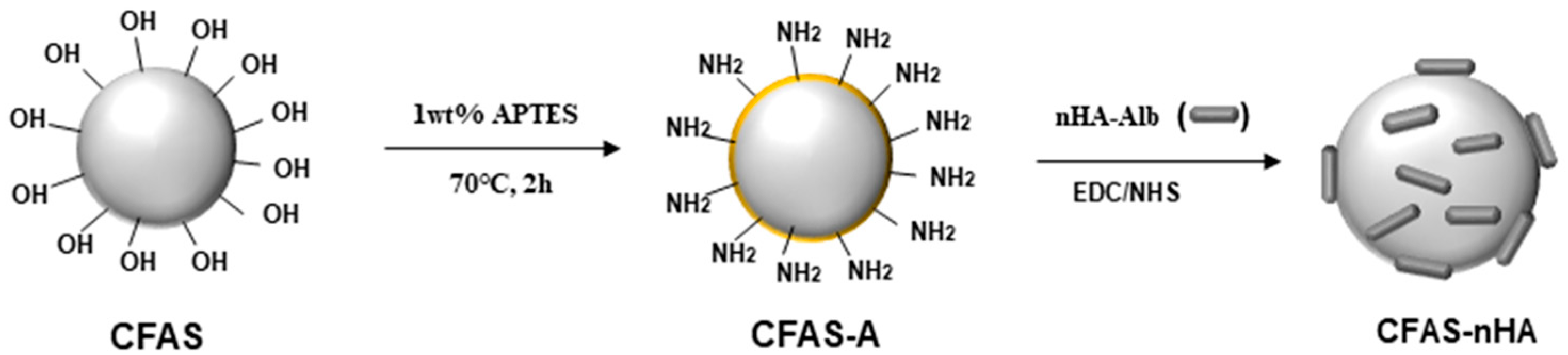
2.2. Synthesis and Surface Modification of CFAS

We used the sol–gel method described by Khiri et al. [16] to prepare the CFAS. The compositions of the ingredients used to prepare the CFAS are shown in Table 1. Initially, 23.89 mL of tetraethylorthosilicate was dissolved in 400 mL of ethanol. Subsequently, an aqueous solution (100 mL) containing aluminum nitrate, calcium nitrate, and ammonium dihydrogen phosphate was added dropwise. Finally, rapid addition of fluorosilicic acid followed by stirring the mixture at 80 °C for 4 h resulting in the formation of a gel-like product. The gel was then dried for 24 h at a temperature of 80 °C and then heat-treated for 10 min at a temperature of 750 °C. After heat treatment, the sample was quenched in water, powdered, and sieved to obtain a micrometre-sized sample (Figure 1).
To incorporate the primary amino groups on the surface of the CFAS particles, a mixture of aminopropyltriethoxysilane (A) and distilled water in a 1:9 ratio was prepared, and 0.06 g was added to the mixture. The mixture was subjected to ultrasonication for 30 min. Subsequently, acetic acid was used to set the pH of the reaction solution at 4.5–5.0. The reaction solution was maintained at 90 °C for 2 h under flowing nitrogen. The reactants were then transferred to distilled water and ultrasonicated for 5 min to remove unreacted A. The resulting CFAS with immobilized A (CFAS-A) was dried under reduced pressure for 12 h at a temperature of 25 °C.

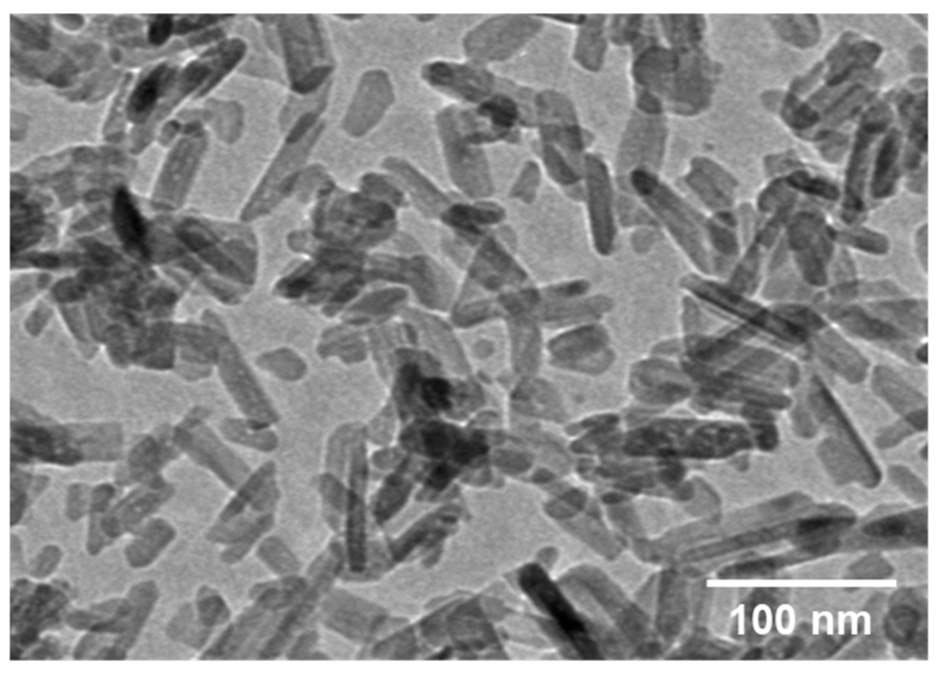
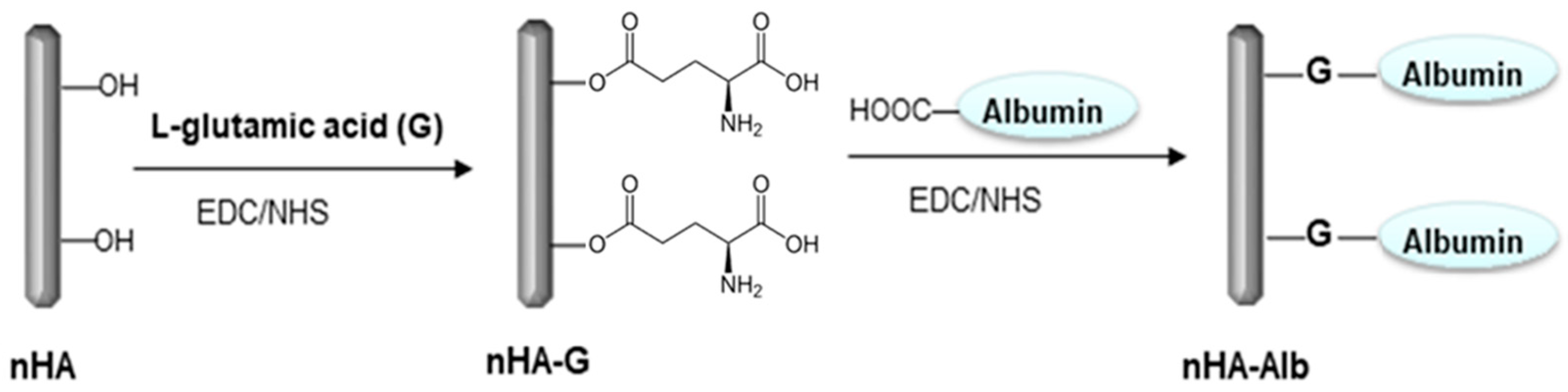
2.3. Surface Modification of nHA

Transmission electron microscopy (TEM) analysis of nHA synthesized according to a previously reported method [15] showed that it was rod-shaped with a length of 30–150 nm (Figure 2). nHA particles contain numerous hydroxyl groups on their surfaces [16,17]. In this study, we aimed to chemically bind nHA particles to the surfaces of CFAS microparticles via a chemical reaction. These solid–solid reactions have low reactivity because they occur in heterogeneous systems [18]. To increase the surface reactivity of the nHA particles and improve their dispersibility in aqueous solutions, albumin was chemically coupled to the nHA surface. The carboxyl group present in the side chain of the introduced albumin molecule was reacted with the CFAS particles (Figure 3). For this purpose, nHA-G (nHA into which L-glutamic acid was introduced) was first prepared by reacting L-glutamic acid with hydroxyl groups on the nHA surface [19]. L-glutamic acid was dissolved in an aqueous solution at pH 5. Then, 1-ethyl-3-(3-dimethylaminopropyl) carbodiimide hydrochloride (0.5 g, 0.25 wt%) and N-hydroxysuccinimide (0.5 g, 0.25 wt%) was added and stirred for 4 h at room temperature, resulting in the activation of the carboxyl group of L-glutamic acid. Next, nHA (0.5 g) was added and the mixture was stirred for 24 h. Finally, the reaction mixture was centrifuged to obtain the precipitate. After washing the precipitate three times with deionized water, it was lyophilized to obtain nHA-G [19].
The resulting nHA-G was mixed with distilled water, stirred for 1 min, and allowed to stand. No precipitate was observed with the naked eye when observing the solution. The nHA particles aggregated in an aqueous solution and formed precipitates. However, when L-glutamic acid was bound to the surface, nHA aggregation did not occur, and the dispersibility of the particles improved, indicating that precipitation did not occur. Despite the improved dispersibility of nHA, to increase its reactivity with microsized CFAS solid particles, the spacer introduced on the nHA surface must be sufficiently long and dynamically mobile in an aqueous solution [20]. Therefore, we attempted to bind water-soluble albumin to the surface of nHA-G as a second spacer [21]. We dissolved 0.06 g of albumin, acting as a chain extender, in 100 mL of deionized water. Thereafter, 0.1 g each of EDC and NHS was added and stirred for 2 h at a temperature of 25 °C, resulting in the activation of the carboxyl group of albumin. The nHA-G particles (0.5 g) were then added to the albumin solution, and the mixture was allowed to react for 24 h. The reaction solution was placed on a semi-permeable membrane (MWCO: 100,000) and dialyzed to separate the unreacted albumin. It was then washed thrice with deionized water. The resulting product was termed as albumin-immobilized nHA (nHA-Alb). Albumin immobilized on the nHA surface in an aqueous solution not only exhibited dynamic motion but also contained several carboxyl groups. Therefore, it is expected that the probability of a chemical bond forming between the primary amino group of A immobilized on the CFAS surface and albumin immobilized on nHA increases. The ATR-FTIR spectra of albumin chemically bound to the nHA particle surface and nHA-Alb chemically bound to the CFAS surface were obtained using a Galaxy 7020A device (Mattson, Fremont, CA, USA).

2.4. Chemical Bonding of nHA to the CFAS Surface

The chemical bonding of nHA-Alb to CFAS-A was performed as follows (Figure 4): nHA-Alb (0.5 g) was added to 100 mL of an aqueous solution containing 0.5 g of EDC (0.25 wt%) and 0.5 g of NHS (0.25 wt%). By stirring at 25 °C for 6 h, the carboxyl group of albumin was activated. CFAS-A was then added to the aqueous solution and stirred mechanically for 24 h. The reaction mixture was then centrifuged at 1200 rpm for 10 min to precipitate CFAS-nHA. Distilled water was added to the precipitate and centrifugation was repeated to remove any unreacted activator. The introduction of nHA into the CFAS was confirmed by ATR-FTIR spectroscopy. Additionally, the surface morphology of CFAS-nHA was examined using FE-SEM on a Hitachi 400 instrument (Tokyo, Japan).

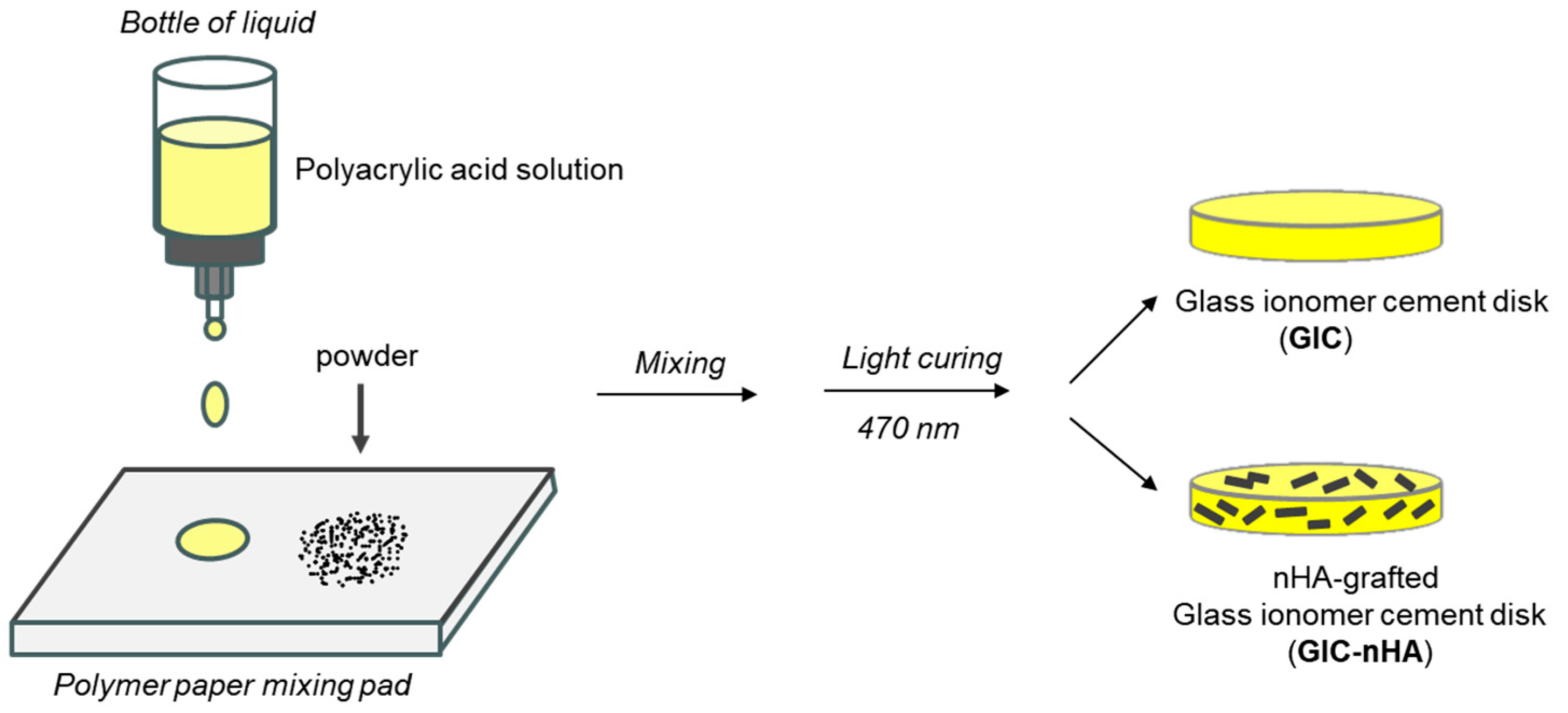
2.5. Preparation of GIC-nHA Discs

GIC-nHA discs were prepared as follows (Figure 5): 1 g of CFAS-nHA was placed on a paper mixing pad and a commercially available liquid component (0.5 g) of GIC (GC International, Japan, GC Fuji II LC) was added dropwise and mixed. After mixing for 30 s, the cells were transferred to a single well of a 24-well culture dish. After that, LED Spotlights (470 nm blue) were irradiated for 20 s to proceed with photopolymerization to manufacture a GIC-nHA disc. A control sample was prepared by photopolymerization in the same manner using CFAS (0.5 g) and a liquid component (0.5 g) from the GC Fuji II LC. After preparing the experimental specimens, we left them for 2 days under UV clean benches before the cell culture experiment.

2.6. Cytocompatibility of GIC-nHA

Using a standard protocol, we investigated the adhesion, proliferation, and osteogenic characteristics of MC3T3-E1 cells to confirm the effects of nHA incorporation into GIC.

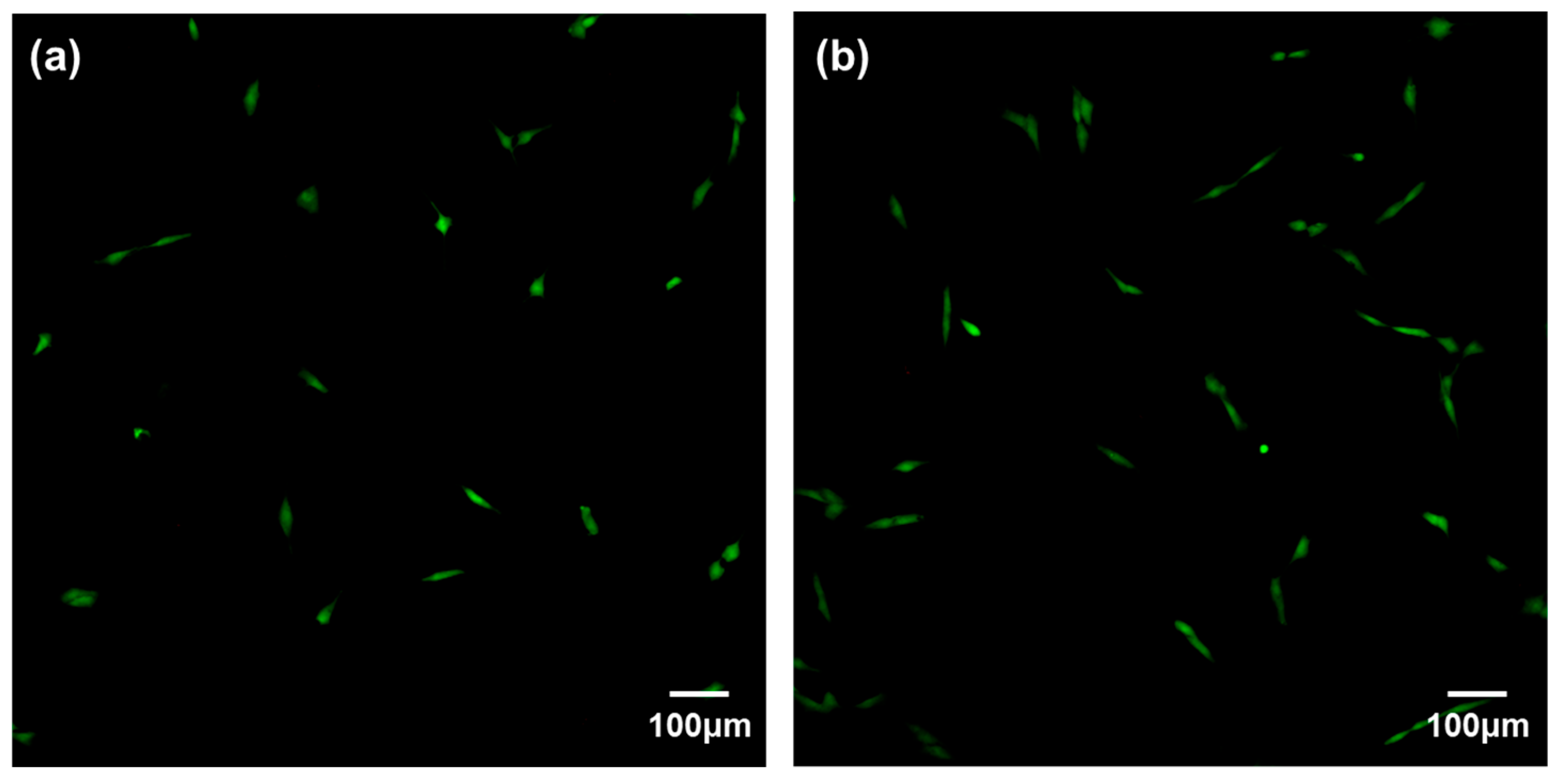
2.6.1. Cell Adhesion
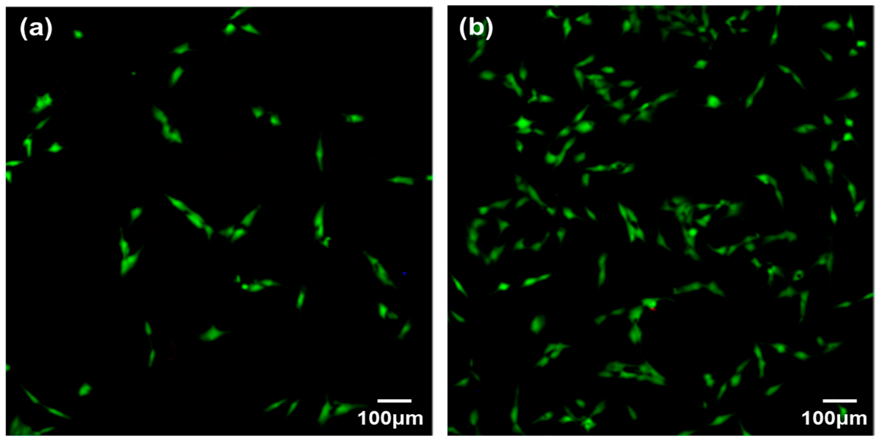
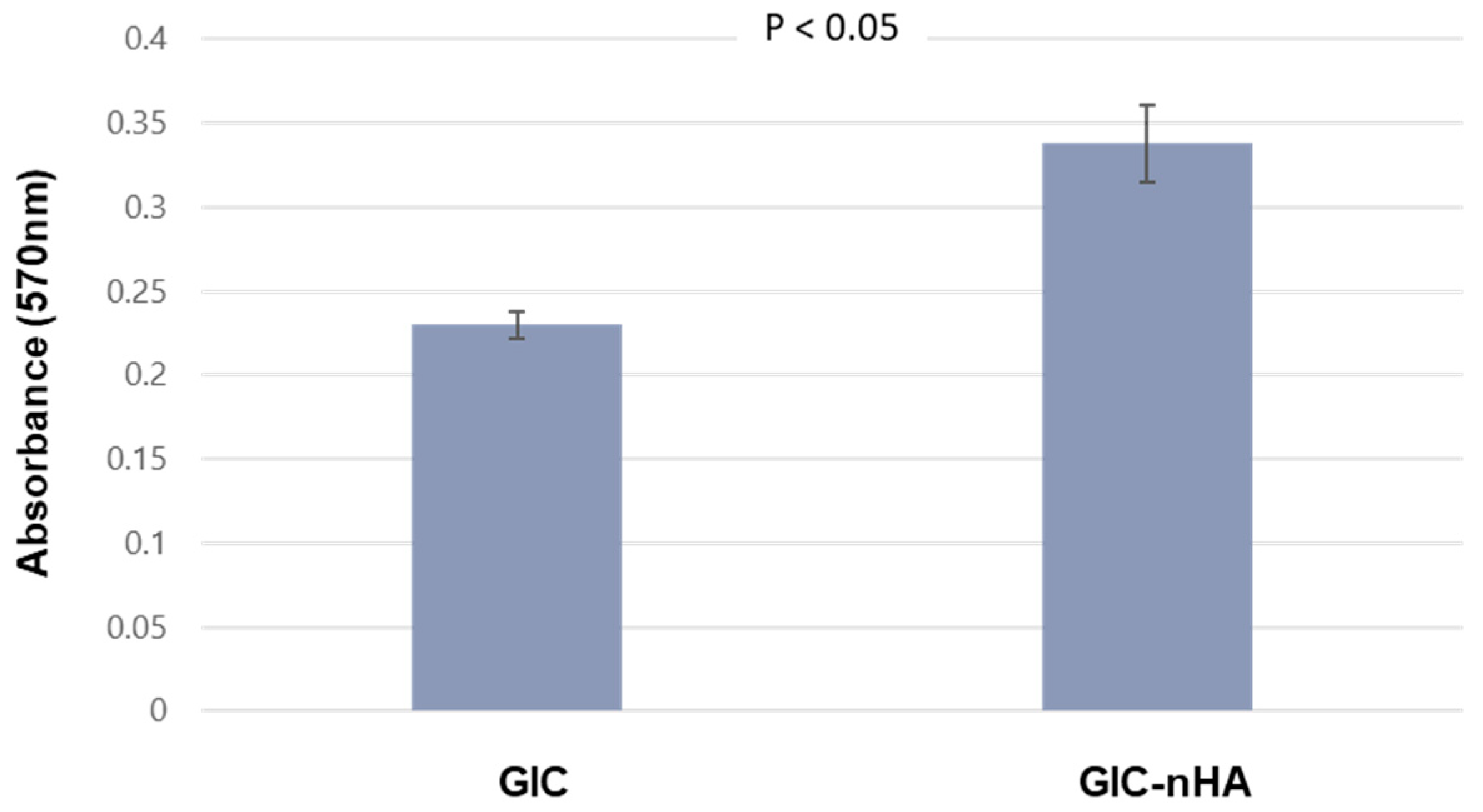
We evaluated the cellular response of MC3T3-E1 cells (4 × 10⁴ cells/mL) on disc surfaces by seeding them onto the GIC and GIC-nHA discs. The cells were cultured in a humidified atmosphere for 24 h using α-MEM. After removing the supernatant, the cells were washed with PBS and fixed with a 2.5% glutaraldehyde solution for 10 min. The samples were then dehydrated and dried using a critical-point dryer. Before capturing SEM images, the samples were sputter-coated with Au.

2.6.2. MTT Assay

MC3T3-E1 cells were cultured on the GIC and GIC-nHA discs for 3 days. Afterwards, the viability of the cells was measured through MTT assay. This assay relies on the reduction of MTT, a yellow water-soluble tetrazolium dye primarily produced by mitochondrial dehydrogenases, to purple formazan crystals [22]. For MTT analysis, sterilized disc samples were plated in 24-well dishes, 500 μL of non-osteogenic α-MEM was added, and MC3T3-E1 cells were seeded onto each disc at a density of 1.2 × 10⁴ cells/cm2. After three days of culture, the supernatant was removed and two subsequent washes of the scaffold with PBS solution were conducted sequentially. Cell-seeded scaffolds were cultured in 500 μL of MTT solution (500 μg/mL) at 37 °C for 4 h, followed by the removal of the supernatant. The generated purple formazan crystals were dissolved for 10 min using 250 μL of dimethyl sulfoxide. Wells injected with MC3T3-E1 cells without discs served as positive controls, whereas empty wells without cells served as negative controls. The absorbance of the extract was recorded at 570 nm based on 690 nm for the medium using a Synergy HT multidetection microplate reader (Synergy HT; BioTek, Shoreline, WA, USA). The amount of formazan generated was determined using a microplate reader. Data from negative controls were subtracted from the measurements. The number of viable cells was correlated with optical density, and cell viability was assessed by normalizing the values to those of the positive control wells.

2.6.3. Cytotoxicity

The cytotoxicity of GIC and GIC-nHA was evaluated by seeding cells at a density of 2.0 × 10⁴ cells/mL onto both discs. The discs were then incubated in Dulbecco’s Modified Eagle Medium (DMEM) at 37 °C in a 5% CO2 atmosphere for 1 or 2 days. Afterwards, 5 μL of calcein-AM (4 mM in anhydrous DMSO) and 20 μL of ethidium homodimer III (2 mM in DMSO/H2O) were added to 10 mL of PBS solution and thoroughly mixed to prepare the staining solution, which was then used to stain the cells [19,23]. After removing the DMEM from the culture wells, both the GIC and GIC-nHA discs were washed twice with PBS solution two times. A large amount of the staining solution was added to fully cover the cells. The discs were then incubated in the dark at room temperature for an additional period. After 30 min, the staining solution was removed and the cells were washed three times with PBS [19]. Finally, the cells were preserved in PBS until observation under a confocal laser scanning microscope. Live and dead cells were simultaneously assessed using calcein-AM (494 nm excitation wavelength) and ethidium homodimer III (530 nm excitation wavelength).

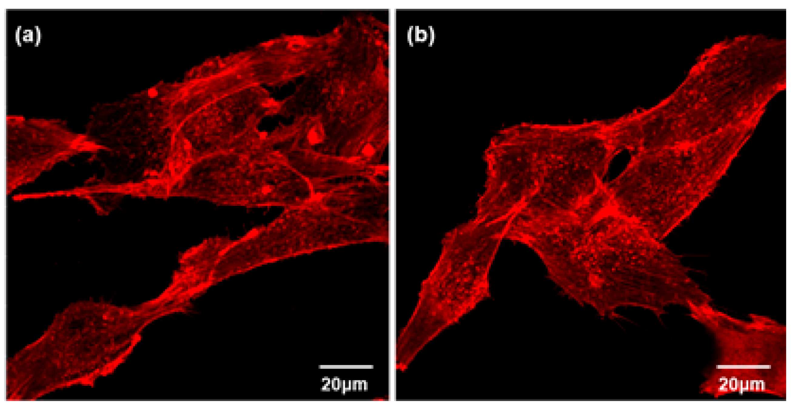
2.6.4. Actin Cytoskeleton Assay

Osteoblasts were seeded onto GIC discs at a concentration of 2 × 10⁴ cells/mL and cultured for 3 days. The cells were then fixed with 4% paraformaldehyde solution. The cells were then washed with PBS containing 0.05% Tween-20. Subsequently, the samples were permeabilized with 0.1% Triton X-100 in PBS solution for 15 min at 25 °C. After permeabilization, the samples were incubated in a mixture of 1% bovine serum albumin (BSA) and PBS for 30 min. After washing the cells 2–3 times with PBS, 5(6)-tetramethylrhodamine isothiocyanate (TRITC)-conjugated phalloidin (Millipore, Burlington, MA, USA, Cat. No. 90228) was added, and the cells were incubated at ambient temperature for 30 min. Finally, a confocal laser scanning microscope (Model 700, Carl Zeiss, Oberkochen, Germany) was used to capture fluorescence images [24].

2.6.5. Von Kossa Assay

Using the von Kossa assay, we assessed the calcium deposition response of MC3T3-E1 cells cultured on the GIC and GIC-nHA discs. MC3T3-E1 cells were seeded in 24-well culture plates at a density of 5 × 10⁴ cells/mL and cultured for 14 days in α-MEM at 37 °C in a humidified atmosphere of 5% CO2. After gently washing the discs three times with PBS for 5 min each, they were fixed in 10% formaldehyde for 30 min. Afterwards, each disc was washed thrice with distilled water for 10 min each, followed by treatment with 5% AgNO3 solution and exposure to UV irradiation for 5 min. The GIC discs were washed twice with PBS to remove the remaining AgNO3 and soaked in 5% Na2S2O3 solution for 5 min. After washing the GIC discs twice with distilled water, digital images of the stained cells were captured using an optical microscope equipped with a camera (Nikon E 4500, Minato, Japan) [22].

2.7. Statistical Analysis

The experiments were performed in triplicates, and all data were expressed as means ± standard deviation. Statistical analyses were conducted using Student’s two-tailed test and Scheffe’s test for multiple comparisons using IBM SPSS (version 20.0; IBM Corp., Armonk, NY, USA). Differences with p-values < 0.05 were considered statistically significant.

3. Result

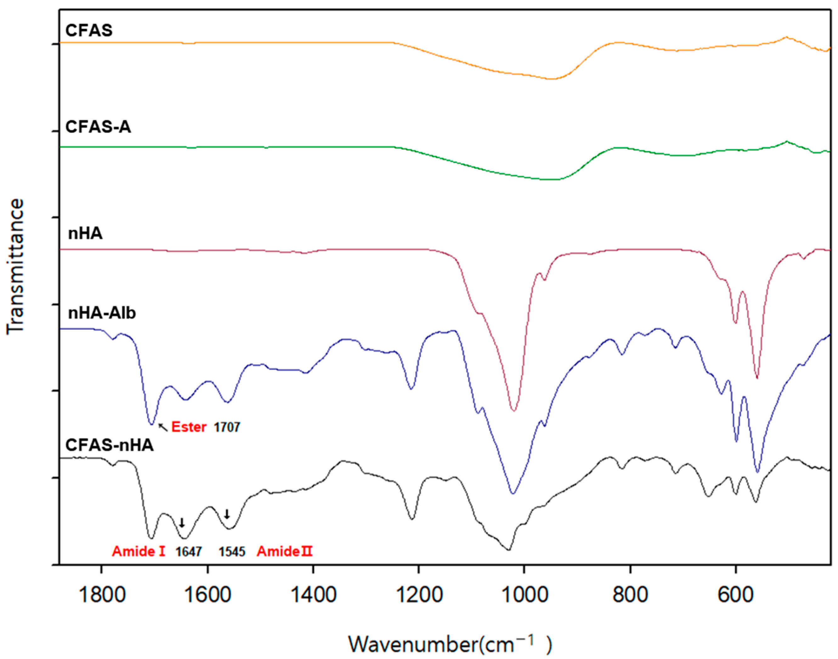
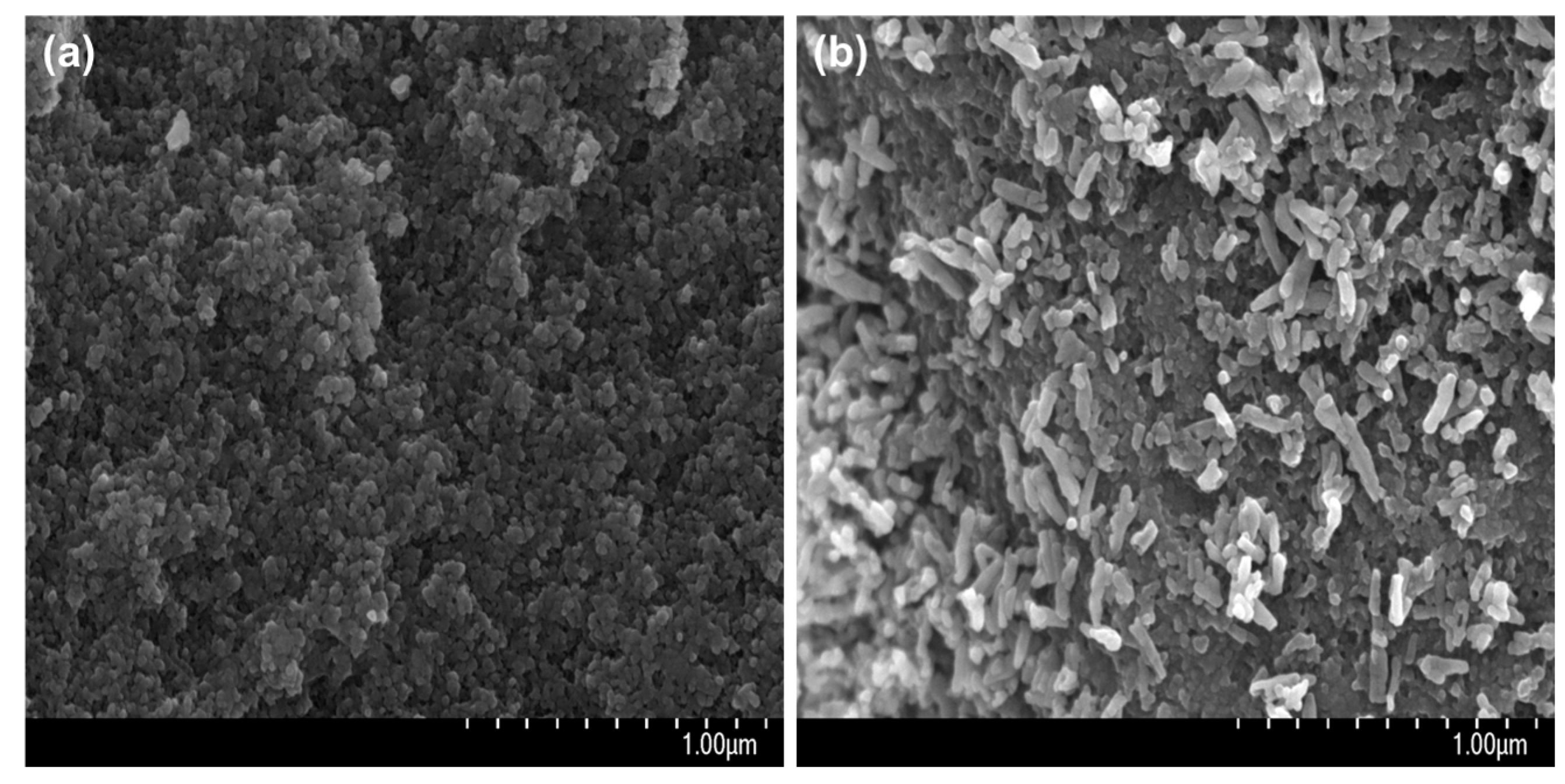
3.1. Immobilization of nHA on CFAS Particle Surface

ATR-FTIR is a recommended method for confirming whether biomolecules such as amino acids or proteins have been introduced to the surface of solid particles such as CFAS or nHA [25]. Figure 6 shows the results of the ATR-FTIR measurements after the surface modification of nHA and CFAS. The infrared spectrum of the nHA sample showed typical apatite modes near 1086, 1021, 961, 600, and 562 cm−1 [25]. In the ATR-FTIR spectrum of nHA-Alb, obtained by binding L-glutamic acid to nHA, followed by binding to albumin, the carbonyl stretching vibration of the ester formed by the reaction between the hydroxyl group of nHA and the carboxyl group of L-glutamic acid appeared at 1707 cm−1. Furthermore, peaks attributed to amides I and II of the introduced albumin appeared at 1647 cm−1 and 1545 cm−1, respectively [26]. The characteristic peak of nHA-Alb appeared in the IR spectrum of CFAS-nHA, indicating that nHA was chemically bonded to the CFAS surface (Figure 6).
Figure 7 shows SEM images of the CFAS surface before (a) and after (b) the chemical bonding of nHA. The CFAS surface showed a nanoporous structure (Figure 7a), whereas the CFAS-nHA surface had nanorod nHA bound to the surface (Figure 7b). As such, nHA bound to the CFAS surface is expected to play a major role in improving histocompatibility when used as a filling material for hard tooth tissues [27].

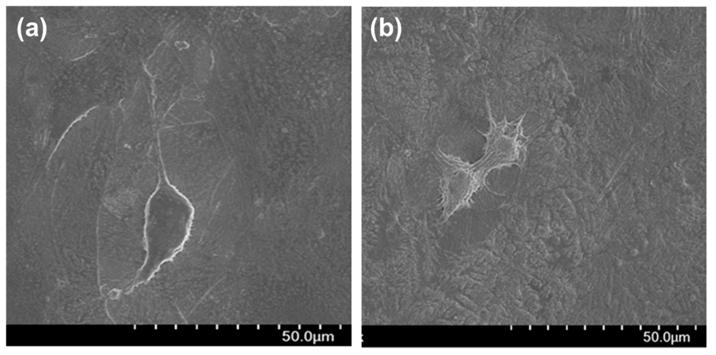
3.2. Cytocompatibility of GIC-nHA

The GIC was prepared using CFAS and polyacrylic acid. The GIC-nHA was prepared from CFAS-nHA and polyacrylic acid. After culturing the osteoblasts on the two samples for 24 h, they were observed by SEM. As shown in Figure 8, the osteoblasts were well spread on the GIC (Figure 8a) and GIC-nHA (Figure 8b) samples after 24 h.
Osteoblasts were cultured in GIC and GIC-nHA samples for 24 and 48 h and then stained using calcein-AM and ethidium homodimer III dye; the results are shown in Figure 9 and Figure 10. No red fluorescence was observed in the GIC or GIC-nHA samples. These results indicated that the GIC and GIC-nHA samples were not toxic. Additionally, as shown in Figure 10, the number of cells attached to GIC-nHA was much higher than that attached to GIC. This is believed to be because the introduction of CFAS-nHA into the GIC promotes cell proliferation.
MC3T3-E1 cell proliferation in GIC and GIC-nHA was assessed using an MTT assay [28]. According to the MTT analysis, the proliferation rate of MC3T3-E1 cells cultured on the GIC-nHA sample for 3 days was significantly higher than that on the GIC (Figure 11). This result indicates that the immobilization of nHA on the surface of CFAS not only enhances adhesion but also provides better opportunities for cell proliferation. This may be because nHA acts as an extracellular matrix.
Actin microfilaments, which constitute the cytoskeleton, play a crucial role in cellular processes, cell shape determination, and cell attachment patterns. When cells attach to the extracellular matrix, they form filopodia. They are guided into their position by actin and interact with the plasma membrane [29]. Figure 12 shows confocal microscopy images of osteoblasts on the GIC (a) and GIC-nHA (b) discs. These results suggested that osteoblasts in both GIC and GIC-nHA exhibited a highly organized cytoskeleton.
Figure 13 shows the results of the von Kossa analysis performed after culturing the osteoblasts in the GIC and GIC-nHA samples for 14 days. Bone nodules are considered as specific markers of osteoblast differentiation. In von Kossa analysis, calcified areas were stained as black spots. According to the von Kossa analysis, more bone nodule formation was observed in GIC-nHA (Figure 13b) than in GIC (Figure 13a). These results indicated that HA triggered and accelerated osteoblast differentiation [30].

4. Discussion

Glass ionomer cement (GIC) has been recognized as an ideal dentin substitute because of its ability to exhibit anti-cavity properties, form stable ionic bonds, and aid the remineralization process. Initially developed for grade 5 cavity restorations, GIC is now being expanded for use as a permanent adhesive, base, grade 1 and 3 filling, and as core and fissure sealants. To be effective as dental filling materials, GIC must withstand high loads. Light-cured GIC exhibits high mechanical strength and is suitable for areas where occlusal forces are applied. Additionally, light-cured GIC offers advantages such as reduced curing time, which significantly reduces the overall clinical chair time. However, formulations of light-cured material are associated with the cytotoxicity of resin monomers; this limits their clinical use near the dental pulp. In this case, traditional self-curing GICs have been used instead of light-cured GICs. Many studies have aimed at improving the mechanical properties of GIC composed of polyacrylic acid-based resins and silica-based inorganic particles [3,31,32]. However, while numerous studies have examined the mechanical properties of GIC in direct contact with the enamel or dentin, few have reported the tissue compatibility of GIC. As silica-based inorganic powder, a key component of GIC, differs chemically from enamel or dentin, long-term tissue compatibility between GIC and teeth is crucial.
Hydroxyapatite (HA) is an inorganic mineral found in bones and teeth that contributes to structural strength and bone regeneration. It has been reported that HA induces a favourable immune response [33], exerts an angiogenic effect on defective bone tissue [34], and activates osteoblasts and osteoclasts to accelerate the differentiation of these cells, resulting in bone tissue remodelling. [35]. A study was conducted to improve the histocompatibility of GIC by introducing HA into an inorganic solid powder of a glass ionomer. Noorani et al. [36] investigated the cytotoxicity of GICs containing 5% HA-SiO2, Fuji IX GP, and Fuji II LC. Fuji IX GP exhibited the lowest cytotoxicity, HA-SiO2-GIC showed medium toxicity, and Fuji II LC showed the highest toxicity, which was attributed to the residual unreacted monomers [37]. This study suggests that the introduction of 5% HA-SiO2 into GIC was insufficient to effectively suppress toxicity.
CFAS are inorganic particles tens of micrometres in size. nHA is an inorganic particle that is several tens of nanometres in size. These two types of high-density inorganic particles easily precipitate in aqueous solution; therefore, no reaction occurs between the two particles. First, we introduced primary amino groups by reacting 3-aminopropyltriethoxysilane on the CFAS surface. In addition, L-glutamic acid was chemically bonded to the nHA surface to introduce an organic spacer on the CFAS surface. By binding L-glutamic acid to nHA, the water dispersibility of nHA-G was slightly improved, but the water dispersibility of nHA-G was still low [38]; therefore, no reaction occurred between the two particles. After trial and error, albumin was selected as the second linker. When albumin was combined with nHA-G, the water dispersibility of nHA-Alb significantly improved. Thus, the reaction between CFAS-A and nHA-Alb was successful [21]. Chemical bonding between the CFAS surface and nHA was confirmed using ATR-FTIR spectroscopy, and the presence of nHA on the CFAS particle surface was directly confirmed using a scanning electron microscope [39].
GIC can dissolve upon exposure to acidic media. Jaiswal et al. [40] measured the weight of GIC after immersion in drinking water and distilled water, which is similar to the acidic environment of the oral cavity, for a certain period. It has been reported that solubility increases when immersed in acidic beverages. Further research is required on the hydrolysis and abrasion resistance of the GIC-nHA prepared in this study in an oral environment. Kumar et al. [41] investigated the interactions between various HA discs and osteoblasts. Cell adhesion, proliferation, and metabolic activity are significantly increased in HA with charges developed on its surface. Polarizing HA discs and generating positive and negative charges on their surfaces can affect the in vitro response and activity of osteoblasts [41]. It has been reported that when a charge is generated on the HA surface, cell adhesion increases within 4 h and cell proliferation increases over a period of 7 days. From this, it can be seen that adding charge to the HA surface resulted in a significant increase in metabolic activity. Simply physically mixing nHA with CFAS can cause phase separation; therefore, nanosized nHA cannot be uniformly sprayed onto the microsized CFAS surface. In this study, the activity of fibroblasts was improved by nHA-CFAS, which was introduced as the main component of GIC by chemically bonding nHA to the surface of CFAS, an artificial mineral.
Oliva et al. [42] compared the response of cultured human osteoblasts to five commercially used glass ionomer cements (Ketac-Fil Aplicap, Ionocem, GC Fuji II, GC Fuji II LC, and Vitremer 3M). Most glass ionomer cements tested showed biocompatibility, except for Vitremer 3M, and proliferated and expressed biochemical markers of the osteoblast phenotype. As in the above study, Figure 8 shows that the osteoblasts were well spread on both the GIC and GIC-nHA samples after 24 h of culture. After culturing osteoblasts on GIC and GIC-nHA discs for 24 and 48 h, dead and live cells were observed under a confocal laser microscope. Staining with calcein-AM and ethidium homodimer III revealed that neither GIC nor GIC-nHA discs were toxic, as only live (green-stained) cells were observed (Figure 9 and Figure 10). Additionally, more osteoblasts adhered to the GIC-nHA disc than to the GIC disc, likely because of the increased affinity of osteoblasts from the HA for binding to the inorganic powder component of GIC. The MTT assay, performed after culturing osteoblasts on GIC and GIC-nHA discs for 3 days, revealed greater cell proliferation on GIC-nHA than on pristine GIC; this was attributable to nHA enhancing osteoblastic cell growth [41].
Von Kossa staining, used to quantify calcium-like mineralization in cell cultures, showed darker black images for the GIC-nHA discs than for the GIC discs after 14 days of osteoblast culture (Figure 13). This suggests an increased osteoblast affinity for GIC-nHA due to nHA on the CFAS surface [43,44].
In the present study, the complete setting time of the light-cured GIC was not measured post-light curing. This is a limitation, as the setting process continues beyond the initial light curing phase through a chemical setting reaction. According to other studies, the complete chemical setting of light-cured glass ionomer cement typically takes 24 h to 7 days, depending on the specific formulation and environmental conditions [1,45]. For example, Sidhu et al. [1] report that while initial light curing substantially hardens the material, the chemical setting continues and typically completes within 24 h to several days. These findings suggest that researchers should consider a prolonged observation period post-light curing to fully understand the setting characteristics of GICs. Future studies should aim to measure the physical properties of the material at various intervals post-light curing to better understand the setting process. This will help provide clearer guidelines for clinical application and expected performance timelines.
The use of CFAS-nHA in GIC-nHA significantly enhanced its biocompatibility compared to conventional GIC. The introduction of nanohydroxyapatite (nHA) effectively supported cell compatibility and proliferation, as demonstrated by the MTT assays and von Kossa staining. The chemical bonding between CFAS and nHA improves the water dispersibility and ensures uniform particle distribution, indicating potential improvements in the mechanical properties. This study aimed to overcome the long working time and poor physical properties of conventional GIC, as well as the limited use of light-cured GIC due to its lower biocompatibility. Future research should explore the long-term tissue compatibility of GIC-nHA in vivo to validate these findings and optimize the properties of GIC by adjusting the nHA concentration and exploring other potential linkers. The development of GIC formulations tailored for specific dental applications may lead to broader clinical use and improved patient outcomes.

5. Conclusions

In conclusion, in the present study, we synthesized CFAS-nHA by chemically bonding nHA to the surface of CFAS, a silica-based solid particle. GIC-nHA was prepared by mixing CFAS-nHA with polyacrylic acid, a liquid component of Fuji II LC. Additionally, CFAS was mixed with the liquid components of Fuji II LC to prepare the GIC. If nano-HA is combined with the silicate-based inorganic powder of commercialized GIC using the spacer method proposed in this study, nHA-GICs with improved tooth tissue compatibility can be manufactured in large quantities. Culturing osteoblasts on GIC-nHA and GIC discs and examining cell activity and osteogenic potential revealed that GIC-nHA exhibited higher cell activity and osteogenic potential than GIC. This was attributed to the chemical bonded to the surface of GIC’s silica-based solid GIC powder, which promoted osteoblast activity. Chemically combining the surface of silica-based solid particles with nHA and using it as a GIC component could improve the histocompatibility of GIC.

Author Contributions

Conceptualization, S.K. (Sohee Kang); data curation, S.J.P. and I.-K.K.; formal analysis, S.J.P.; funding acquisition, S.K. (Sohee Kang); investigation, S.J.P. and S.K. (Sukyoung Kim); methodology, S.K. (Sohee Kang), S.J.P., S.K. (Sukyoung Kim) and I.-K.K.; resources, S.K. (Sukyoung Kim); supervision, I.-K.K.; visualization, S.K. (Sohee Kang) and I.-K.K.; writing—original draft, S.J.P. and I.-K.K.; writing—review and editing, S.K. (Sohee Kang). All authors have read and agreed to the published version of the manuscript.

Funding

This work was supported by the 2023 Yeungnam University research grant (223A580032).

Institutional Review Board Statement

Not applicable.

Informed Consent Statement

Not applicable.

Data Availability Statement

Data is contained within the article.

Conflicts of Interest

The authors declare no conflicts of interest. The funders had no role in the design of the study, such as the collection, analyses, or interpretation of data; writing of the manuscript; or the decision to publish the results.

References

  1. Sidhu, S.K.; Nicholson, J.W. A review of glass-ionomer cements for clinical dentistry. J. Funct. Biomater. 2016, 7, 16. [Google Scholar] [CrossRef] [PubMed]
  2. Farahnaz, S.; Ali, A.A.; Saba, S.; Mina, S. Comparison of shear bond strength of three types of glass ionomer cements containing hydroxyapatite nanoparticles to deep and superficial dentin. J. Dent. 2020, 21, 132–140. [Google Scholar]
  3. Bilić-Prcić, M.; Brzović Rajić, V.; Ivanišević, A.; Pilipović, A.; Gurgan, S.; Miletić, I. Mechanical properties of glass ionomer cements after incorporation of marine derived hydroxyapatite. Materials 2020, 13, 3542. [Google Scholar] [CrossRef] [PubMed]
  4. Lathikumari, S.S.; Saraswathy, M. Modifications of Polyalkenoic Acid and Its Effect on Glass Ionomer Cement. Mater. Adv. 2024, 5, 2719–2735. [Google Scholar] [CrossRef]
  5. Faraji, F.; Heshmat, H.; Banava, S. Effect of Protective Coating on Microhardness of a New Glass Ionomer Cement: Nanofilled Coating Versus Unfilled Resin. J. Conserv. Dent. 2017, 20, 260–263. [Google Scholar] [CrossRef] [PubMed]
  6. Amin, F.; Rahman, S.; Khurshid, Z.; Zafar, M.S.; Sefat, F.; Kumar, N. Effect of nanostructures on the properties of glass ionomer dental restoratives/cements: A comprehensive narrative review. Materials 2021, 14, 6260. [Google Scholar] [CrossRef] [PubMed]
  7. Arita, K.; Yamamoto, A.; Shinonaga, Y.; Harada, K.; Abe, Y.; Nakagawa, K.; Sugiyama, S. Hydroxyapatite particle characteristics influence the enhancement of the mechanical and chemical properties of conventional restorative glass ionomer cement. Dent. Mater. J. 2011, 30, 672–683. [Google Scholar] [CrossRef] [PubMed]
  8. Alatawi, R.A.S.; Elsayed, N.H.; Mohamed, W.S. Influence of Hydroxyapatite Nanoparticles on the Properties of Glass Ionomer Cement. J. Mater. Res. Technol. 2019, 8, 344–349. [Google Scholar] [CrossRef]
  9. Mohd Pu’ad, N.A.S.; Abdul Haq, R.H.; Mohd, N.; Abdullah, H.Z.; Idris, M.I.; Lee, T.C. Synthesis method of hydroxyapatite: A review. Mater. Today Proc. 2020, 29, 233–239. [Google Scholar] [CrossRef]
  10. Ramay, H.R.; Zhang, M. Biphasic Calcium Phosphate Nanocomposite Porous Scaffolds for Load-Bearing Bone Tissue Engineering. Biomaterials 2004, 25, 5171–5180. [Google Scholar] [CrossRef]
  11. Sadat-Shojai, M.; Khorasani, M.T.; Dinpanah-Khoshdargi, E.; Jamshidi, A. Synthesis methods for nanosized hydroxyapatite with diverse structures. Acta Biomater. 2013, 9, 7591–7621. [Google Scholar] [CrossRef] [PubMed]
  12. Mohandes, F.; Salavati-Niasari, M. Particle Size and Shape Modification of Hydroxyapatite Nanostructures Synthesized via a Complexing Agent-Assisted Route. Mater. Sci. Eng. C 2014, 40, 288–298. [Google Scholar] [CrossRef] [PubMed]
  13. Kang, I.G.; Park, C.I.; Lee, H.; Kim, H.E.; Lee, S.M. Hydroxyapatite microspheres as an additive to enhance radiopacity, biocompatibility and osteoconductivity of poly(methylmethacrylate) bone cement. Materials 2018, 11, 258. [Google Scholar] [CrossRef] [PubMed]
  14. Praveen, B.; Attiguppe, R.P.; Nadig, B. An in vitro comparative evaluation of compressive strength and antibacterial activity of conventional GIC and hydroxyapatite reinforced GIC in different storage media. J. Clin. Diagn. Res. 2015, 9, ZC51–ZC55. [Google Scholar]
  15. Haider, A.; Gupta, K.C.; Kang, I.K. Morphological effects of HA on the cell compatibility of electrospun HA/PLGA composite nanofiber scaffolds. Biomed. Res. Int. 2014, 2014, 11. [Google Scholar] [CrossRef]
  16. Khiri, M.Z.A.; Matori, K.A.; Zaid, M.H.M.; Abdullah, A.C.; Zainuddin, N.; Jusoh, W.N.W.; Jalil, R.A.; Rahman, N.A.A.; Kul, E.; Wahab, S.A.A.; et al. Soda lime silicate glass and clam shell act as precursor in synthesize calciumfluoroaluminosilicate glass to fabricate glass ionomer cement with different ageing time. J. Mater. Res. Technol. 2020, 9, 6125–6134. [Google Scholar] [CrossRef]
  17. Jia, S.; Liu, Y.; Ma, Z.; Liu, C.; Chai, J.; Li, Z.; Song, W.; Hu, K. A novel vertical aligned mesoporous silica coated nanohydroxyapatite particle as efficient dexamethasone carrier for potential application in osteogenesis. Biomed. Mater. 2021, 16, 035030. [Google Scholar] [CrossRef]
  18. Aykol, M.; Montoya, J.H.; Hummelshøj, J. Rational solid-state synthesis routes for inorganic materials. J. Am. Chem. Soc. 2021, 143, 9244–9259. [Google Scholar] [CrossRef]
  19. Haider, A.; Versace, D.; Gupta, K.C.; Kang, I.K. Pamidronic acid-grafted nHA/PLGA hybrid nanofiber scaffolds suppress osteoclastic cell viability and enhance osteoblastic cell activity. J. Mater. Chem. B 2016, 4, 7596. [Google Scholar] [CrossRef]
  20. Kamiya, H.; Iijima, M. Surface modification and characterization for dispersion stability of inorganic nanometer-scaled particles in liquid media. Sci. Technol. Adv. Mater. 2010, 11, 044304. [Google Scholar] [CrossRef]
  21. Park, S.J.; Gupta, K.C.; Kim, H.; Kim, S.; Kang, I.K. Osteoblast behaviours on nanorod hydroxyapatite-grafted glass surfaces. Biomater. Res. 2019, 23, 28. [Google Scholar] [CrossRef] [PubMed]
  22. Ghasemi, M.; Turnbull, T.; Sebastian, S.; Kempson, I. The MTT Assay: Utility, Limitations, Pitfalls, and Interpretation in Bulk and Single-Cell Analysis. Int. J. Mol. Sci. 2021, 22, 12827. [Google Scholar] [CrossRef] [PubMed]
  23. Xing, Z.C.; Chae, W.P.; Baek, J.Y.; Choi, M.J.; Jung, Y.; Kang, I.K. In Vitro Assessment of Antibacterial Activity and Cytocompatibility of Silver-Containing PHBV Nanofibrous Scaffolds for Tissue Engineering. Biomacromolecules 2010, 11, 1248–1253. [Google Scholar] [CrossRef] [PubMed]
  24. Przekora, A.; Benko, A.; Blazewicz, M.; Ginalska, G. Hybrid Chitosan/β-1,3-Glucan Matrix of Bone Scaffold Enhances Osteoblast Adhesion, Spreading and Proliferation via Promotion of Serum Protein Adsorption. Biomed. Mater. 2016, 11, 045001. [Google Scholar] [CrossRef]
  25. Tiernan, H.; Byrne, B.; Kazarian, S.G. ATR-FTIR spectroscopy and spectroscopic imaging for the analysis of biopharmaceuticals. Spectrochim. Acta Part A Mol. Biomol. Spectrosc. 2020, 241, 118636. [Google Scholar] [CrossRef] [PubMed]
  26. Alhazmi, H.A. FT-IR spectroscopy for the identification of binding sites and measurements of the binding interactions of important metal ions with bovine serum albumin. Sci. Pharm. 2019, 87, 5. [Google Scholar] [CrossRef]
  27. Wan Jusoh, W.N.; Matori, K.A.; Mohd Zaid, M.H.; Zainuddin, N.; Ahmad Khiri, M.Z.; Abdul Rahman, N.A.; Abdul Jalil, R.; Kul, E. Incorporation of Hydroxyapatite into Glass Ionomer Cement (GIC) Formulated Based on Alumino-Silicate-Fluoride Glass Ceramics from Waste Materials. Materials 2021, 14, 954. [Google Scholar] [CrossRef]
  28. Soesilawati, P.; Rizqiawan, A.; Roestamadji, R.I.; Arrosyad, A.R.; Firdauzy, M.A.; Abu Kasim, N.H. In Vitro Cell Proliferation Assay of Demineralized Dentin Material Membrane in Osteoblastic MC3T3-E1 Cells. Clin. Cosmet. Investig. Dent. 2021, 13, 443–449. [Google Scholar] [CrossRef]
  29. Fletcher, D.A.; Mullins, R.D. Cell mechanics and the cytoskeleton. Nature 2010, 28, 485–492. [Google Scholar] [CrossRef]
  30. Tayalia, P.; Mooney, D.J. Controlled growth factor delivery for tissue engineering. Adv. Mater. 2009, 21, 3269–3285. [Google Scholar] [CrossRef]
  31. Moshaverinia, A.; Ansari, S.; Moshaverinia, M.; Roohpour, N.; Darr, J.A.; Rehman, I. Effects of incorporation of hydroxyapatite and fluoroapatite nanobioceramics into conventional glass ionomer cements (GIC). Acta Biomater. 2008, 4, 432–440. [Google Scholar] [CrossRef] [PubMed]
  32. Barandehfard, F.; Kianpour Rad, M.; Hosseinnia, A.; Khoshroo, K.; Tahriri, M.; Jazayeri, H.E.; Moharamzadeh, K.; Tayebi, L. The addition of synthesized hydroxyapatite and fluorapatite nanoparticles to a glass-ionomer cement for dental restoration and its effects on mechanical properties. Ceram. Int. 2016, 42, 17866–17875. [Google Scholar] [CrossRef]
  33. Teng, N.C.; Nakamura, S.; Takagi, Y.; Yamashita, Y.; Ohgaki, M.; Yamashita, K. A New Approach to Enhancement of Bone Formation by Electrically Polarized Hydroxyapatite. J. Dent. Res. 2001, 80, 1925–1929. [Google Scholar] [CrossRef] [PubMed]
  34. Kobayashi, T.; Nakamura, S.; Yamashita, K. Enhanced Osteobonding by Negative Surface Charges of Electrically Polarized Hydroxyapatite. J. Biomed. Mater. Res. 2001, 57, 477–484. [Google Scholar] [CrossRef] [PubMed]
  35. Royce, B.S.H. Field Induced Transport Mechanism in Hydroxyapatite. Ann. N. Y. Acad. Sci. 1974, 238, 131–138. [Google Scholar] [CrossRef] [PubMed]
  36. Noorani, T.Y.; Luddin, N.; Ab Rahman, I.; Masudi, S.M. In vitro cytotoxicity evaluation of novel nano-hydroxyapatite-silica incorporated glass ionomer cement. J. Clin. Diagn. Res. 2017, 11, ZC105–ZC109. [Google Scholar] [CrossRef] [PubMed]
  37. Stanislawski, L.; Daniau, X.; Lautié, A.; Goldberg, M. Factors responsible for pulp cell cytotoxicity induced by resin-modified glass ionomer cements. J. Biomed. Mater. Res. 1999, 48, 277–288. [Google Scholar] [CrossRef]
  38. Kang, S.; Haider, A.; Gupta, K.C.; Kim, H.; Kang, I.K. Chemical bonding of biomolecules to the surface of nano-hydroxyapatite to enhance its bioactivity. Coatings 2022, 12, 999. [Google Scholar] [CrossRef]
  39. Kim, H.; Lee, Y.H.; Kim, N.K.; Kang, I.K. Bioactive surface of zirconia implant prepared by nano-hydroxyapatite and type I collagen. Coatings 2022, 12, 1335. [Google Scholar] [CrossRef]
  40. Jaiswal, S.; Kusumitha, P.; Alla, R.K.; Guduri, V.; AV, R.; Sajjan, S.M.C. Solubility of Glass Ionomer Cement in Various Acidic Beverages at Different Time Intervals: An In Vitro Study. Int. J. Dent. Mater. 2022, 4, 78–81. [Google Scholar] [CrossRef]
  41. Kumar, D.; Gittings, J.P.; Turner, I.G.; Bowen, C.R.; Bastida-Hidalgo, A.; Cartmell, S.H. Polarization of hydroxyapatite: Influence on osteoblast cell proliferation. Acta Biomater. 2010, 6, 1549–1554. [Google Scholar] [CrossRef] [PubMed]
  42. Oliva, A.; Della Ragione, F.; Salerno, A.; Riccio, V.; Tartaro, G.; Cozzolino, A.; D’Amato, S.; Pontoni, G.; Zappia, V. Biocompatibility studies on glass ionomer cements by primary cultures of human osteoblasts. Biomaterials 1996, 17, 1351–1356. [Google Scholar] [CrossRef]
  43. Schneider, M.R. Von Kossa and his staining technique. Histochem. Cell Biol. 2021, 156, 523–526. [Google Scholar] [CrossRef] [PubMed]
  44. Chen, C.S.; Chang, J.H.; Srimaneepong, V.; Wen, J.Y.; Tung, O.H.; Yang, C.H.; Lin, H.C.; Lee, T.H.; Han, Y.; Huang, H.H. Improving the in vitro cell differentiation and in vivo osseointegration of titanium dental implant through oxygen plasma immersion ion implantation treatment. Surf. Coat. Technol. 2020, 399, 126125. [Google Scholar] [CrossRef]
  45. Pearson, G.J.; Atkinson, A.S. Long-Term Flexural Strength of Glass Ionomer Cements. Biomaterials 1991, 12, 658–660. [Google Scholar] [CrossRef]
Figure 1. SEM image of calciumfluoroaluminosilicate (CFAS) microparticles prepared in the present study.
Coatings 14 00893 g001
Figure 2. TEM image of nHA used in the present study synthesized by the chemical precipitation method (reference [15] is cited for the picture).
Coatings 14 00893 g002
Figure 3. Schematic diagram showing the surface modification of nHA using L-glutamic acid and albumin as linkers.
Coatings 14 00893 g003
Figure 4. Schematic diagram showing the chemical bonding of nHA on the surface of CFAS microparticles.
Coatings 14 00893 g004
Figure 5. Schematic diagram showing the manufacturing process of glass ionomer disc using polyacrylic acid and surface-modified CFAS powder.
Coatings 14 00893 g005
Figure 6. ATR-FTIR spectra of albumin-bound nHA (nHA-Alb) and nHA-bound CFAS (CFAS-nHA).
Coatings 14 00893 g006
Figure 7. SEM images of CFAS microparticles before (a) and after (b) the chemical bonding of nHA.
Coatings 14 00893 g007
Figure 8. SEM images of osteoblasts cultured for 24 h in GIC (a) and GIC-nHA (b).
Coatings 14 00893 g008
Figure 9. Live and dead assay of MC3T3-E1 cells cultured for 24 h in the presence of GIC (a) and the GlC-nHA (b). The cells were stained with calcein AM and ethidium homodimer III. Initial cell number = 1.2 × 10⁴/mL.
Coatings 14 00893 g009
Figure 10. Live and dead assay of MC3T3-E1 cells cultured for 48 h in the presence of GIC (a) and the GlC-nHA (b). The cells were stained with calcein AM and ethidium homodimer III. Initial cell number = 1.2 × 10⁴/mL.
Coatings 14 00893 g010
Figure 11. MTT assay of MC3T3-E1 cells cultured for 3 days in the presence of GIC and GIC-nHA in α-MEM. Initial cell concentration: 1.2 × 10⁴/mL.
Coatings 14 00893 g011
Figure 12. Confocal microscopy images of the actin filaments of osteoblasts cultured for 3 days in the presence of GIC (a) and GIC-nHA (b) (magnification, ×400).
Coatings 14 00893 g012
Figure 13. Von Kossa staining of osteoblast cells cultured on GIC (a) and GIC-nHA (b) for 14 days. The calcium-containing areas are stained in black.
Coatings 14 00893 g013
Table 1. Feed ratio of calciumfluoroaluminosilicate microparticles.
IngredientChemical FormulaMolar Ratio50 mmol
(Amount Used in the Experiment)
TetraethylorthosilicateSi(OC2H5)42.13323.89 mL
Ammonium dihydrogen phosphateNH4H2PO40.1600.92 g
Fluorosilicic acidH2SiF60.1672.95 mL
Aluminum nitrateAl(NO3)32.20041.27 g
Calcium nitrateCa(NO3)31.00011.81 g
Disclaimer/Publisher’s Note: The statements, opinions and data contained in all publications are solely those of the individual author(s) and contributor(s) and not of MDPI and/or the editor(s). MDPI and/or the editor(s) disclaim responsibility for any injury to people or property resulting from any ideas, methods, instructions or products referred to in the content.

