Article

Natural Selection on Hydroxyapatite Fiber Orientations for Resisting Damage of Enamel

1
College of Mechanical Engineering, Jiangsu University of Science and Technology, Zhenjiang 212000, China
2
Department of Oral and Maxillofacial Surgery, Jinan Stamotological Hospital, Jinan 250001, China
*
Author to whom correspondence should be addressed.
These authors contributed equally to this work.
Coatings 2024, 14(9), 1122; https://doi.org/10.3390/coatings14091122
Submission received: 22 July 2024 / Revised: 22 August 2024 / Accepted: 26 August 2024 / Published: 2 September 2024

Abstract

:
Teeth have excellent mechanical properties, with high wear resistance and excellent fracture resistance. This is due to their well-organized multilevel hierarchical structure. While a number of studies in the last decades have revealed the relationship between tooth structure and mechanical properties, there is still no general agreement on how different orientations of hydroxyapatite (HAp) fibers affect the mechanical properties of enamel. With a scanning electron microscope and nanoindenter, the orientations of HAp fibers and their properties were investigated. HAp fibers have two different orientations: parallel and perpendicular to the surface. Fibers oriented parallel to the surface exhibited higher hardness, elastic modulus and wear resistance. Under applied force, fibers oriented perpendicular to the surface suffered deeper shearing in the protein along the long axis, resulting in lower mechanical properties. Teeth resist damaging fractures by combining hard and soft structures. This study may lead to new insights into how nature selects for tooth structure and provide a theoretical basis for the bioinspired design.

1. Introduction

The human tooth is a natural mineral substance that plays a very important role in human life. Teeth are among the hardest tissues in the human body, and are directly exposed to the mouth environment. Even after millions of chewing cycles, teeth disperse high chewing contact pressure with relatively limited damage rather than failure. The excellent mechanical properties of teeth have a close relationship with their complex chemical composition and orderly structure. The study of the relationship between tooth structure and properties is helpful for understanding the internal mechanism of natural materials to resist external damage, and provides a new idea for the construction and performance optimization of bionic materials.
From the overall structure, a tooth crown is mainly composed of the outer enamel and the inner dentin, with the dentin–enamel junction (DEJ) between them. It has been noted that enamel, dentin, and the DEJ play important roles in the resistance to fractures. Enamel is harder and more resistant to wear, while dentin is less hard and tougher, making teeth both strong and tough [1]. Borero-Lopez et al. [2] suggested that tooth enamel is a functionally graded material with anticracking properties. When cracks move from the outer enamel to the inner enamel, they are dissipated by the HAp column, deviating from their original path. Additionally, when faced with harder enamel at the boundary and softer underlying dentin, the cracks are further resisted. Also, research has shown the presence of numerous parallel microcrack structures in teeth at the boundary between enamel and dentin. These microcracks can self-repair through continuous absorption of organic matter. Enamel can alleviate stress concentration and act as a stress shield when impacted [3]. The role of the DEJ in tooth cracking cannot be ignored. The width and gradient structure of the DEJ vary at occlusal and cervical sites, with a wider and smoother transition at occlusal sites, promoting more efficient stress transfer between enamel and dentin without affecting the overall stiffness of the teeth [4]. In addition, the viscoelasticity of the DEJ functions as a loading attenuation mechanism between enamel and dentin [5]. Under a tensile load perpendicular to the DEJ, failure does not occur in the DEJ; this helps to stop the crack from extending into the dentin, thus preventing a potentially dangerous rupture [6,7].
Enamel is composed of keyhole-like enamel rods and inter-rods enamel [8]. The interfacial area between enamel rods and inter-rods enamel, termed sheaths, has a high organic content and is about 100–200 nm wide [9]. Enamel rods are arranged in different ways across the entire enamel. In the outer layer enamel, enamel rods almost arrange in parallel, revealing a radial pattern. In the inner enamel, enamel rods appear as bands of approximately cross-sectioned arrangement [10]. The varying mechanical properties from the outer layer of enamel to the inner layer have attracted a lot of attention. Cuy et al. [11] found a significant decrease in elastic modulus and hardness from the outer layer to the inner layer. However, the inner layer enamel demonstrated higher toughness and better ability to prevent crack propagation. In another study, it was found that the outer enamel displayed better wear resistance, whereas the inner enamel exhibited better creep and stress redistribution ability. The inner enamel also demonstrated better fracture toughness, dispersing stress from the outer enamel, and contributing to the overall structural integrity of the enamel in terms of wear and fracture resistance [12]. In the microscale, enamel rod and inter-rod consist of Hydroxyapatite (HAp) fibers with different orientations and compactness [13,14]. These fibers are roughly hexagonal and are bonded together by a protein layer [15]. The fibers reveal anisotropic mechanical properties in the occlusal surface and the axial-sectional plane, and it was found that the occlusal surfaces had better microscopic mechanical properties [16,17]. The change in the orientation of the HAp fibers enhanced the fracture toughness of enamel. Additionally, the transverse fibers effectively inhibit crack propagation [18,19]. The more the orientation and loading direction of HAp fibers converge, the better the bearing capacity and wear resistance of tooth enamel [17]. The exceptional properties of tooth enamel are not only due to its perfect structure, but also because of the effective binding of organic proteins and inorganic minerals. Proteins are mainly found in the sheath between the enamel rod and inter-rod enamel. They are considered the main reason for the visco-plastic and visco-elastic properties of enamel [20]. He et al. [15] found that the protein undergoes shear deformation when the enamel is subjected to an external load. During chewing, the enamel consumes energy through microdamage of proteins, thereby increasing the fracture toughness of enamel. An et al. [21] found that under the action of the indentation damage mode, the protein binding HAp particles undergo shear deformation and the macromolecular chains in the protein extend. With the increase in load, the degree of shear deformation of protein intensifies, and the macromolecular chains in the protein will break, leading to irreversible deformation of tooth enamel.
This study aims to investigate the effect of HAp fibers orientations on the mechanical properties of enamel. First, the orientation of HAp fibers in enamel rod and inter-rod enamel is observed. Then, the influence of HAp fiber orientations on hardness, elastic modulus, friction, and wear properties is analyzed. Finally, the factors contributing to these performance differences are analyzed.

2. Materials and Methods

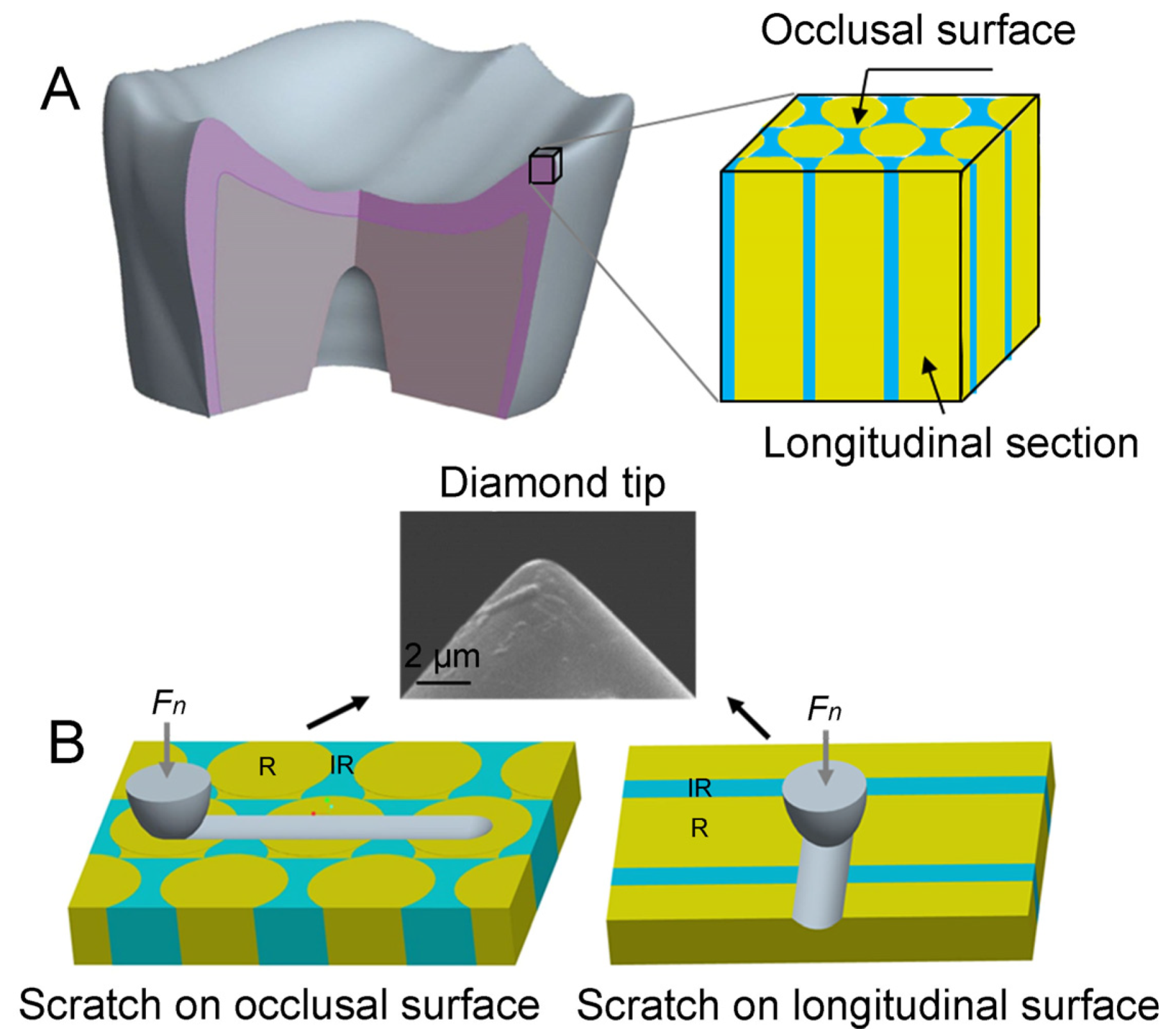
2.1. Specimen Preparation

The specimens used in the study were prepared from healthy human third molar teeth obtained with the consent of the subjects. These teeth were stored in deionized water at 4 °C before use. The tooth roots were cut with a saw at low speed under constant water irrigation, while the other eight teeth were embedded with the occlusal surface facing down in epoxy resin, and the other eight teeth were cut into two halves along the axial section and embedded into the epoxy resin with the longitudinal section facing down. Subsequently, all specimens were ground sequentially using 800, 1200, and 1500 grit sandpaper, followed by polishing with 0.5 µm diamond paste. The polished samples were then cleaned with deionized water before testing. The samples were etched for 1 min with a 0.001 M citric acid solution to reveal the orientations of HAp fibers.

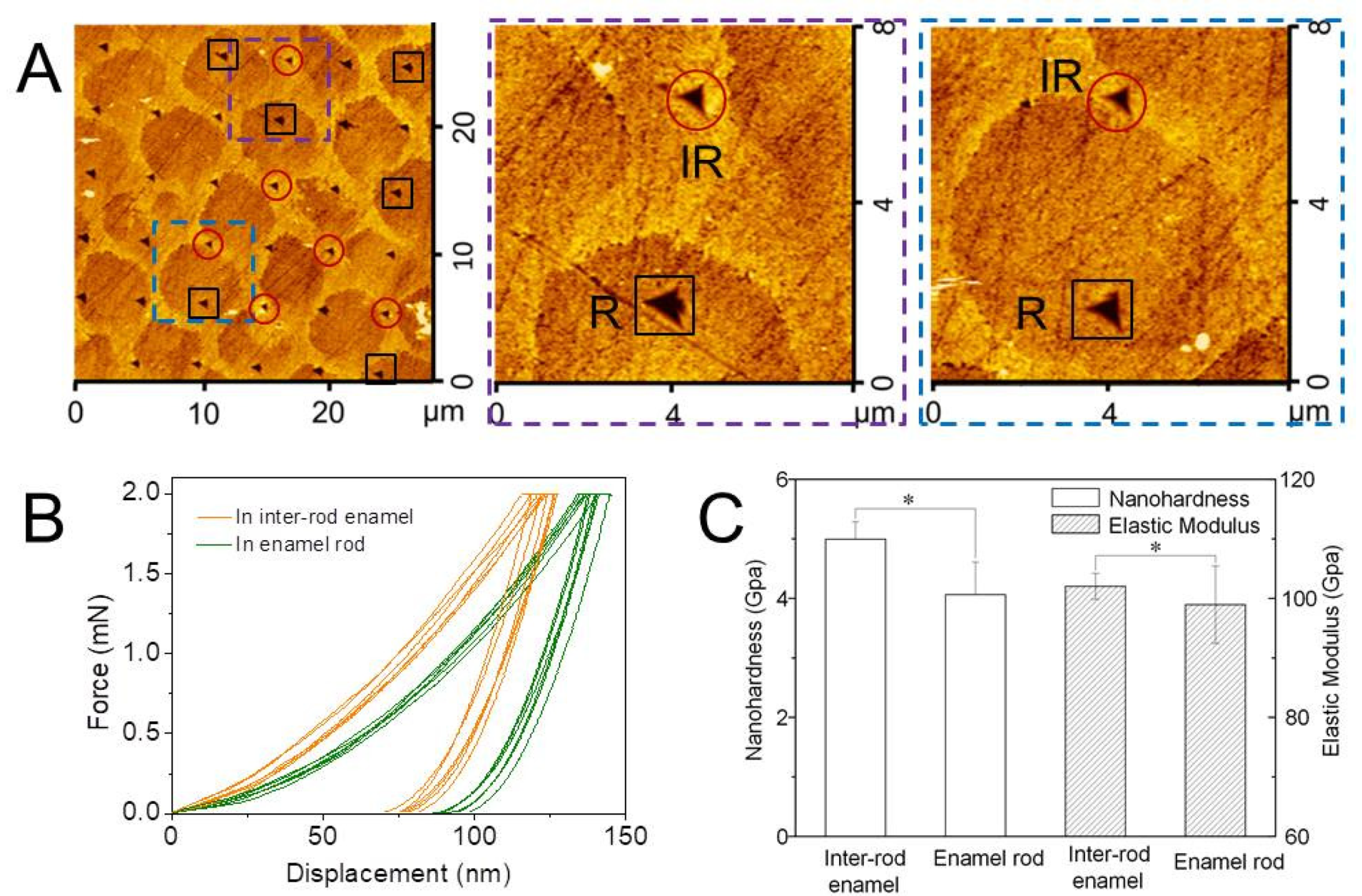
2.2. Nanoindentation Tests

To understand how the orientation of HAp fibers affects the mechanical properties of enamel, the mean elasticity modulus and hardness values of the different aligned HAp fibers were obtained using a nanoindenter (T750, Hysitron Inc., Minneapolis, MN, USA) with a Berkovich diamond tip. Four occlusal surface samples and four longitudinal section samples from different people were taken, respectively, for the nanoindentation tests. As shown in Figure 1A, the experiments were conducted on the outer enamel of the occlusal surface and longitudinal section samples, respectively. On the occlusal surface, approximately 7 × 7 indentations were placed with a spacing of about 5 µm under a normal load of 2 mN for each specimen. On the longitudinal section, about 10 × 5 indentations were placed with a spacing of about 2 µm under a normal load of 1 mN for each specimen. For each indentation, the loading function was adjusted to maintain 5 s of loading time, 2 s of delay at peak load, and 5 s of unloading time. The hardness and elastic modulus of the material are obtained by calculating the load–displacement curve drawn by the system in real time according to the Oliver–Pharr method [22]. For standard diamond tips, the determined elastic modulus is 1140 GPa and the hardness is 0.07. The Poisson’s ratio of enamel used in the analysis was assumed to be 0.28 based on the literature [23]. After the nanoindentation tests, the topographies of the indentations on the enamel surface were scanned using an atomic force microscope (AFM, SPI 3800N, Seiko, Tokyo, Japan) with a standard Si3N4 tip, having a nominal radius of 10 nm. The indentations on the sample that were completely located in the enamel rod or the inter-rod enamel were selected to be included in the calculation of the mean hardness and elasticity modulus, and the indentations located in the sheath of the enamel were abandoned to avoid errors.

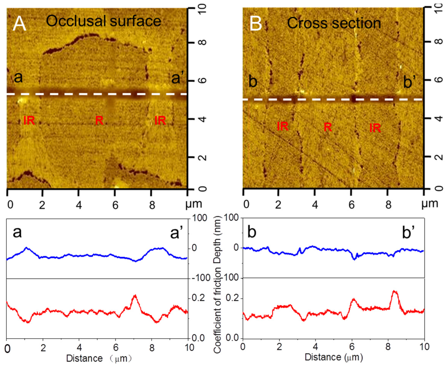
2.3. Nanoscratch Tests

To simulate the shearing movement of opposing teeth parallel to the occlusal surface and longitudinal section, scratch tests were conducted. The wear behavior and coefficient of friction were studied using a nanoindenter (T750, Hysitron Inc., Minneapolis, MN, USA) by a diamond tip with a radius of curvature of 1 µm. As depicted in Figure 1B, scratches were created on four specimens of the occlusal surface and four specimens of the longitudinal section. The scratches were made vertically to the inter-rod enamel in the occlusal surface, and perpendicular to the long axes of enamel rod in the longitudinal section. The scratches were made using normal loads of 0.5 mN, with a sliding velocity of 1 μm/s and sliding displacements of 15 μm. The friction coefficient of scratches was obtained in real time during the experiment. Following the nanoscratch tests, the topographies of the scratch-induced damage on the enamel were scanned using an Si3N4 tip in the AFM to indicate the depth of the scratch.

2.4. Statistical Analysis

The statistical software SPSS25 (International Business Machines Corporation, Ar-monk, NY, USA) was utilized to conduct a test aimed at identifying any significant differences between the samples. If p < 0.05, it signifies a difference between the samples, denoted by an asterisk (*). If p < 0.01, it indicates a significant difference between the samples, denoted by double asterisks (**).

3. Results and Discussion

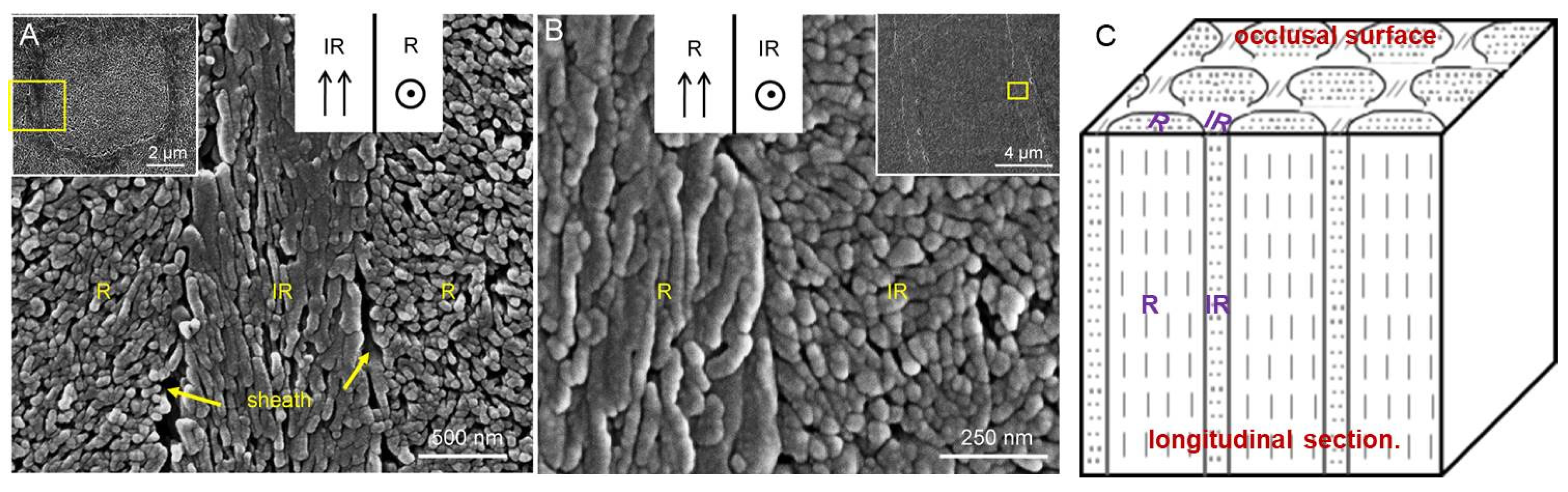
3.1. HAp Fiber Orientations in Occlusal Surface and Longitudinal Section

HAp fibers are long, roughly flat hexagonal structures, approximately 20–25 nm thick and 50–70 nm wide, with lengths ranging from about 200 to 500 nm [24]. Human enamel is composed of these HAp fibers, with their long axes aligned at different orientations within enamel rod and inter-rod enamel. Using a high-resolution scanning electron microscope (SEM, JSM-7001F, JEOL Ltd., Tokyo, Japan), the orientations of HAp fibers in enamel rod and inter-rod enamel on the occlusal surface and the longitudinal section of the outer layer enamel were clearly observed. As shown in Figure 2A, the inter-rod enamel surrounds the enamel rod to form a typical keyhole shape. Two different orientations of HAp fibers were found; the long axes of HAp fibers in enamel rod (R) were approximately perpendicular to the occlusal surface, while in the inter-rod enamel (IR), the hydroxyapatite fibers aligned with their long axes parallel to the occlusal surface. Sheaths were observed with a width of about 100–200 nm between enamel rod and inter-rod enamel. In the longitudinal section displayed in Figure 2B, these two different aligned fibers were also observed. The long axes of HAp fibers in enamel rod were parallel to the longitudinal section, while in the inter-rod enamel, they were approximately perpendicular to the surface. Figure 1A,B both confirm the orientations of HAp fibers in the outer enamel—parallel or perpendicular to the surface. Several studies have noted that HAp fibers in the enamel rod are perpendicular to the occlusal surface, while the fiber orientation in the inter-rod enamel is parallel to the occlusal surface [25,26], which aligns with the findings of this experiment. The enamel rods in the outer and inner layers are arranged in different directions. In the outer layer enamel, enamel rods are almost arranged in parallel, revealing a radial pattern. The inner enamel is made up of enamel rods with different orientations arranged in a Hunter–Schreger band structure. This structure consists of bundles of hydroxyapatite crystals arranged at different angles to the surface [10]. In this paper, we only studied two different fiber orientations of the outer enamel. Further research is needed to understand the impact of the angle of deflection of HAp fibers on their properties.

3.2. Effect of Fiber Orientation on Mechanical Properties of Enamel

As shown in Figure 3A, the topography of the enamel indentations in the occlusal surface was observed by AFM. The indentations completely located in the enamel rod (shown in the black block) and inter-rod enamel (shown in the red circle) were selected to calculate the mean hardness and elastic modulus. The enamel sheath between enamel rod and inter-rod enamel has much lower hardness and elastic modulus [27,28]. Therefore, the indentations located in the enamel sheath area were excluded to enhance the accuracy of the calculation. Based on the criteria mentioned above, 20 points in the enamel rod and inter-rod enamel were chosen from four samples to calculate the average hardness, elastic modulus, and standard deviation. The amplifying images in Figure 3A clearly show the differences in the indentations, with those in enamel rods displaying larger impression areas compared to those in inter-rod enamel. The load–displacement curves in Figure 3B demonstrate that the indentations in enamel rod exhibited greater depth displacement. The mean hardness and elastic modulus of the inter-rod enamel were found to be higher than those of the enamel rods, with indentations in the inter-rod enamel yielding a mean hardness of 4.99 ± 0.29 GPa and an elastic modulus of 102.01 ± 2.20 GPa. In comparison, the enamel rod exhibited a mean hardness of 4.06 ± 0.55 GPa and an elastic modulus of 98.94 ± 6.50 GPa. According to the statistical analysis, the hardness and elastic modulus of the indentations in the enamel rod and inter-rod enamel are different (p < 0.05). In combination with HAP fibers orientations as shown in Figure 2, HAp fibers with long axes parallel to the occlusal surface displayed superior mechanical behavior.
In Figure 4, the mechanical behaviors of enamel rod and inter-rod enamel in a longitudinal section are depicted. The mean hardness elastic modulus and standard deviation were calculated by 20 indentations completely located in the enamel rod (shown in the black block) and inter-rod enamel (shown in the red circle) on each of the four samples, respectively. As shown in Figure 4A, indentations located in enamel rods show lower impression areas and displacements compared to those located in the inter-rod enamel. The mean hardness and elastic modulus of the inter-rod enamel are lower than those of the enamel rods in the outer enamel. Specifically, the hardness and elastic modulus increase from H = 3.70 ± 0.45 GPa and E = 85.67 ± 0.19 GPa, where the indentations are located in the inter-rod enamel to H = 4.60 ± 0.27 GPa and E = 90.86 ± 5.63 GPa for enamel rods. According to the statistical analysis, the hardness and elastic modulus of the indentations in the enamel rod and inter-rod enamel are different (p < 0.05). Taking the HAp fiber orientations of enamel rod and inter-rod enamel in the longitudinal section into account, the results further demonstrate that HAp fibers with long axes parallel to the surface exhibit better mechanical behavior. The relationship between the orientation of HAp fibers and their mechanical properties is still a subject of debate. Staine et al. [29] discovered that when the force applied to the HAp fibers is perpendicular to the direction of its long axis, the measured hardness and elastic modulus are higher compared to when the force is applied parallel to the long axis. However, Sha and Katz et al. [30] obtained opposite results, suggesting that parallel loading leads to higher mechanical properties. These findings were based on comparing the hardness and elastic modulus of the occlusal surface and the longitudinal section. They suggested that the long axis direction of the HAp fibers on the occlusal surface is perpendicular to the surface, so the loading direction of the indentation tip is parallel to the long axis of the HAp fibers. On the longitudinal section, the long axis direction of the HAp fibers is parallel to the surface, and the loading direction of the indentation tip is perpendicular to the long axis of the HAp fibers. It is important to note that there are HAp fibers with different orientations in the enamel rod and inter-rod enamel in the occlusal surface and longitudinal section, leading to inconsistencies in the study process. Therefore, further exploration of the relationship between particle orientation and mechanical properties based on the specific arrangement orientation of hydroxyapatite particles is necessary.

3.3. Effect of Fiber Orientation on Microfriction Behavior of Enamel

Figure 5 illustrates the images of the scratches and the coefficient–distance curves. The friction curve corresponds to the scratch damage morphology and the cross-section profile curve of tooth enamel. It can be observed that on the occlusal surface, the coefficient of friction on the enamel rod is about 0.144, while the coefficient of friction on the inter-rod enamel is about 0.087. The coefficient of friction in the inter-rod enamel region is lower than that in the enamel rod region. On the longitudinal section, the coefficient of friction on the inter-rod enamel is about 0.158, while that on the enamel rod is about 0.108. The coefficient of friction in the inter-rod enamel region is higher than that in the enamel rod region. The enamel sheath shows the highest friction coefficient, which is consistent with its poor mechanical properties. Considering the HAp fiber orientations of the enamel rod and inter-rod enamel on these two surfaces, it can be concluded that HAp fibers with long axes parallel to the surface exhibited better microfriction behavior.

3.4. Effect of Fiber Orientation on the Wear Behavior of Enamel

In Figure 6, the wear behavior in the enamel rod and inter-rod enamel on the occlusal surface is compared. The scratches were made across the inter-rod enamel and enamel rod with a constant normal load of 0.5 mN. It was observed that wear in the enamel rod was more severe than in the inter-rod enamel. Based on the cross-sectional profile of the scratch in inter-rod enamel and enamel rod, shown in Figure 6D, the maximum wear depth in inter-rod enamel is about 15.15 nm, and in the enamel rod, it is 23.8 nm. The maximum wear depth decreased by more than 36.3% when moving from the inter-rod enamel to the enamel rod under the same load. Additionally, the inter-rod enamel showed a relatively smaller wear width compared with the enamel rod. When combined with the fiber orientations, it was evident that HAp fibers with long axes parallel to the surface exhibited better wear behavior.
The image in Figure 7 depicts the wear behavior in enamel rod and inter-rod enamel in a longitudinal section. Similar to the adverse fiber arrangements observed in the occlusal surface and longitudinal section, both surfaces exhibited adverse wear behavior of enamel rod and inter-rod enamel. The scratching was performed across the direction perpendicular to the long axes of inter-rod enamel and enamel rod with a constant normal load of 0.5 mN. Wear in the inter-rod enamel was found to be more severe compared to the enamel rod, with the maximum wear depth in the inter-rod enamel measuring about 21.4 nm, and 11.8 nm in the enamel rod. The maximum wear depth decreased by more than 44.8% when moving from the enamel rod to the inter-rod enamel under the same load. Considering the fiber orientations of enamel rod and inter-rod enamel in the longitudinal section, the results further demonstrate that HAp fibers with long axes parallel to the surface exhibited better wear behavior. In terms of structure, horizontally arranged HAp fibers have higher density, while there are more gaps between vertically arranged HAp fibers perpendicular to the occlusion surface. Studies have shown that the densification of fibers gives teeth good mechanical properties [31]. Horizontally arranged HAp fibers have better wear resistance than longitudinally arranged HAp fibers. Moreover, the change in the orientation of the fibers enhances the fracture toughness of enamel. Transverse fibers can effectively inhibit crack propagation [18,19]. It was suggested that the more similar the orientation of HAp fibers is to the direction of loading, the better the bearing capacity and wear resistance of tooth enamel [17]. HAp fibers align in various orientations to disperse energy and prevent stress concentration, thereby enhancing the overall toughness of teeth.

3.5. Influence Mechanism of Hydroxyapatite Alignment on Tooth Enamel Properties

The mechanical properties and microtribological behavior of HAp fibers vary with different orientations. Human enamel possesses a well-organized microstructure with a hierarchical assembly [10]; an analysis of how enamel reacts under load should consider its unique structure and inner bonding manner. HAp fibers are long, roughly flat hexagonal structures, approximately 20–25 nm thick and 50–70 nm wide, with lengths ranging from 200 to 500 nm [23]. These fibers are self-assembled from smaller nanoparticles, each about 20 nm in diameter, connected by rigid interfacial molecular bonds, including hydrogen bonds mediated by surface-hydrated water molecules [28]. The HAp fibers are bonded together by a thin layer of hydrated protein [15]. Proteins play a crucial role in the mechanical properties of teeth. Human tooth enamel, containing a small amount of protein, is about three times harder than dense HAp [8]. When the protein is removed, the fracture toughness of tooth enamel decreases by about 40% [32]. The protein “glue” layer between the HAp fibers experiences shear strain during enamel contact loading, which is 16 times higher than the contact strain. This facilitates the enamel to dissipate the contact deformation energy [15]. Additionally, the memory behavior of protein components makes tooth enamel exhibit a stress–strain relationship and creep behavior similar to metal [33,34,35]. In this paper, the theory of protein stress shear deformation is used to explain the difference between the two orientations of HAp fibers.
According to the structure and bonding manner of HAp fibers, the force analysis of the different aligned HAp fibers under indentation tests was analyzed, as shown in Figure 8. The protein matrix bonded to the HAp fibers has a yield stress of 0.4 GPa and an elastic modulus of 4.3 GPa, which are much lower than the mineral HAp fibers [36]. Therefore, when an indentation load is applied, it would initially result in a large shear strain in the protein layer. When the HAp fibers with long axes perpendicular to the surface experience force in a direction coaxial with the long axes of the fibers, the protein layer would be sheared along the long axes of the fiber (200–500 nm). The HAp fibers would be pressed into the underside due to sufficient space within the enamel matrix. On the other hand, when the HAp fibers with long axes parallel to the surface experience force in a direction perpendicular to the long axes of the fibers, the protein layer at the end of the fiber would be sheared (50–70 nm) first. However, further shear in the protein layer would be impeded by the fiber below. With an increase in the indentation load, the lower fiber would be bent, and during unloading, the bent fiber would be restored to some extent. The fiber elements are separated by continuous protein layers leading to a strong anisotropy between axial and transverse moduli, the latter being substantially lower due to the deformation of proteins [37,38]. As a result, HAp fibers with long axes parallel to the surface survived with lower indentation depth and had a better ability to resist elastic–plastic deformation, resulting in higher hardness and elastic modulus.
The different fiber orientations led to anisotropic microtribological behavior. Figure 9 explains the phenomenon through force analysis of the differently aligned HAp fibers under applied scratching load. The HAp fiber was made up of smaller nanoparticles with a diameter of approximately 20 nm and bonded by rigid interfacial molecular bonds, including H-bonds [28]. During scratch tests, the HAp fiber would be refined and fragmented into smaller ones. When the HAp fibers’ long axes were perpendicular to the surface, under applied load, the protein layer would first be sheared along the depth direction (200–500 nm), and then the fiber would be bent forward along the scratch direction and refined into smaller ones (20 nm). When the HAp fibers were aligned with their long axes parallel to the surface, the protein that glued the fibers below would first be sheared along the long axes, and the fiber would be bent forward along the scratch direction and refined into smaller ones. As a result, the shear in the protein and the refinement of the fiber occurred along the long axes. HAp fibers with long axes parallel to the surface would endure a lower wear depth.
In this study, only the differences in the two different aligned HAp fibers in the outer layer of enamel of different healthy third molars were observed, and mechanical analysis was conducted on the observed fiber orientations. The orientations of HAp fibers at different layers of enamel showed great differences. In addition, different diets, oral environment, and different trace elements also had potential differences in tooth structural properties. These differences have not been studied in detail and are worth further exploration in the future.
The human enamel has different orientations of HAp in the outer layer, resulting in anisotropic properties in enamel rod and inter-rod enamel. The soft layer of the enamel helps to dissipate energy by limiting abrasion or deformation under large contact stresses. The combination of soft and hard structure limits the growth of cracks and prevents catastrophic failure. Studying the relationship between HAp fiber orientation and properties of enamel has broad implications, ranging from materials science to evolutionary biology to clinical dentistry. Structural anisotropy could serve as a fundamental design principle for self-assembly materials. For example, embedding hard materials vertically into soft materials or arranging nanoparticles with different orientations can optimize the mechanical properties of materials. Zhao et al. [39] synthesized HAp nanowires similar to natural tooth enamel using the hydrothermal method. The uniform growth of amorphous zirconia ceramics was achieved on the surface of the HAp nanowires by regulating material growth and nucleation. This resulted in the composite of amorphous/crystal nanowire and polymer and allowed for controllable assembly at a macro scale. The artificial tooth enamel possesses good mechanical properties. Controlling the direction of growth of the hydroxyapatite nanowires based on the orientation of the hydroxyapatite crystals in teeth, and assembling the nano-HAp nanowires into composite hydroxyapatite nanowires at different angles, may further enhance the mechanical properties of artificial tooth enamel. Furthermore, researchers have drawn inspiration from the fracture resistance mechanism of natural mother of pearl to develop mother-of-pearl imitation structural ceramics, which significantly improves the fracture toughness of ceramics [40]. The complex multistage structure of teeth also demonstrates strong fracture resistance, and the relevant theories can provide a theoretical reference for the toughening mechanism of composite ceramics.

4. Conclusions

With the development of nanoindentation and microscope techniques, the experimental accuracy was greatly improved. The main conclusions of this study can be summarized as follows:
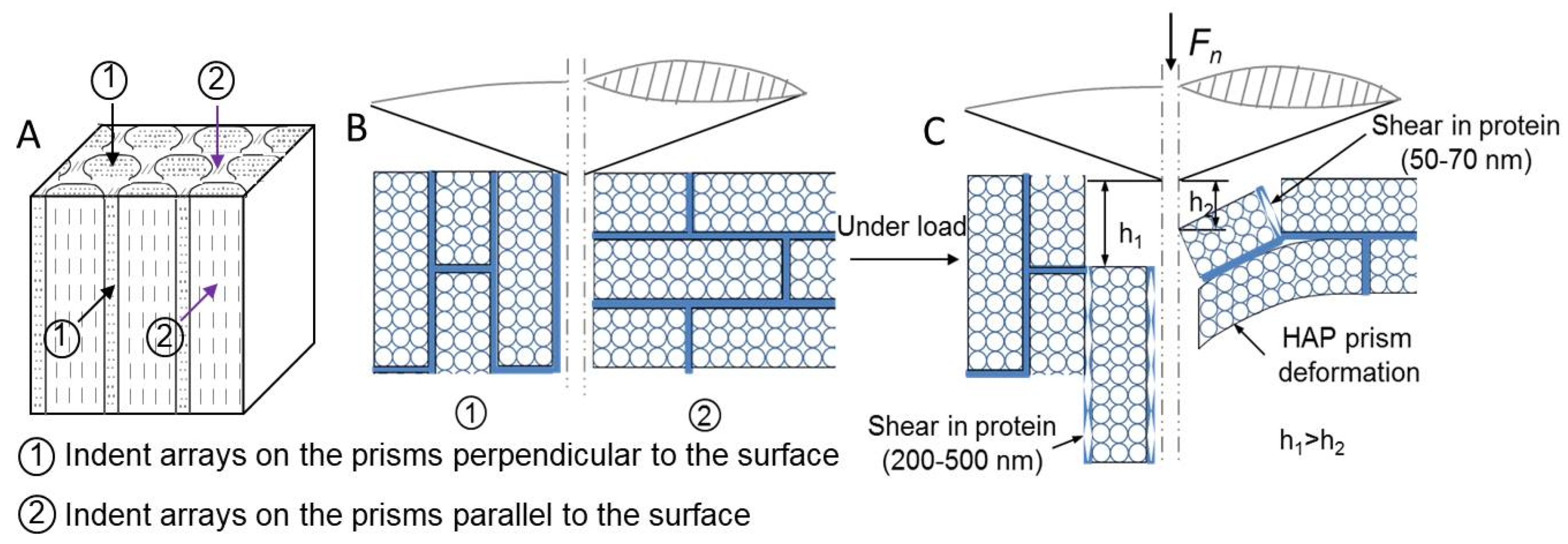
  • There are two types of orientation of HAp fibers in the outer layer of tooth enamel: parallel and perpendicular to the occlusal surface.
  • The arrangements of HAp fibers significantly influenced the mechanical and microtribological behavior of human tooth enamel. HAp fibers with long axes parallel to the surface had higher hardness and elastic modulus and showed better microtribological behavior.
  • Teeth resist damaging fractures by combining hard and soft structures.
This research provides new insights into how nature selects for tooth structure. Animals have evolved different HAp fiber orientations to resist tooth damage. Additionally, research on the wear behavior of enamel can provide a theoretical basis for clinical dental prevention. In future studies, exploring the dynamic response of enamel under different loading conditions or different microstructures and the molecular mechanisms behind the observed anisotropic properties is worth further investigation. In addition, biomimetic design or optimization of materials based on the structural properties of teeth will be a hot focus of research.

Author Contributions

Conceptualization, J.S., H.X. and J.X.; data curation, J.S., H.X., X.L., S.Z. and Y.K.; funding acquisition, H.X. and J.X.; methodology, J.S. and J.X.; supervision, J.S., Y.Z., Y.F. and J.X.; validation, J.S., H.X. and J.X.; writing—original draft, J.S. and H.X.; writing—review and editing, J.X., J.S. and H.X. J.S. and H.X. contributed equally to this work. All authors have read and agreed to the published version of the manuscript.

Funding

This research was funded by National Science Foundation of China (51805226), Postgraduate Research & Practice Innovation Program of Jiangsu Province (KYCX24-4081), Hospital level clinical research project of Zhenjiang First People’s Hospital in 2022, Y2022012, Jiangsu Provincial Health Commission’s 2023 Guiding Project, 307.

Institutional Review Board Statement

Not applicable.

Informed Consent Statement

Not applicable.

Data Availability Statement

The original contributions presented in the study are included in the article; further inquiries can be directed to the corresponding author.

Acknowledgments

The authors thank all reviewers for their remarkable guidance on this article.

Conflicts of Interest

The authors declare that they have no known competing financial interests or personal relationships that could have appeared to influence the work reported in this paper.

References

  1. Carreon, A.H.; Funkenbusch, P.D. Nanoscale properties and deformation of human enamel and dentin. J. Mech. Behav. Biomed. Mater. 2019, 97, 74–84. [Google Scholar] [CrossRef] [PubMed]
  2. Borrero-Lopez, O.; Constantino, P.J.; Bush, M.B.; Lawn, B.R. On the vital role of enamel prism interfaces and graded properties in human tooth survival. Biol. Lett. 2020, 16, 20200498. [Google Scholar] [CrossRef] [PubMed]
  3. Chai, H.; Lee, J.J.-W.; Constantino, P.J.; Lucas, P.W.; Lawn, B.R. Remarkable resilience of teeth. Proc. Natl. Acad. Sci. USA 2009, 106, 7289–7293. [Google Scholar] [CrossRef] [PubMed]
  4. Wang, Z.; Wang, K.; Xu, W.; Gong, X.; Zhang, F. Mapping the mechanical gradient of human dentin-enamel-junction at different intratooth locations. Dent. Mater. 2018, 34, 376–388. [Google Scholar] [CrossRef]
  5. Hasegawa, M.; Tanaka, R.; Zhong, J.; Kobayashi, M.; Manabe, A.; Shibata, Y. Deciphering load attenuation mechanisms of the dentin-enamel junction, Insights from a viscoelastic constitutive model. Acta Biomater. 2023, 171, 193–201. [Google Scholar] [CrossRef]
  6. Yamada, Y.; Inoue, T.; Saito, M.; Nishimura, F.; Miyazaki, T. Anisotropic ultimate strength and microscopic fracture patterns during tensile testing in the dentin–enamel junction region. Dent. Mater. J. 2020, 39, 272–277. [Google Scholar] [CrossRef]
  7. Zhu, X.; Wang, S.; Ye, J.; Guo, H.; Wang, R. Investigation of Fracture-resistance of Human Teeth at the Dentin-enamel Junction Using the J-integral Calculation of Finite Element Analysis. J. Mech. Med. Biol. 2022, 22, 2250069. [Google Scholar] [CrossRef]
  8. White, S.; Luo, W.; Paine, M.; Fong, H.; Sarikaya, M.; Snead, M. Biological organization of hydroxyapatite crystallites into a fibrous continuum toughens and controls anisotropy in human enamel. J. Dent. Res. 2001, 80, 321–326. [Google Scholar] [CrossRef]
  9. Xie, Z.-H.; Mahoney, E.; Kilpatrick, N.; Swain, M.; Hoffman, M. On the structure–property relationship of sound and hypomineralized enamel. Acta Biomater. 2007, 3, 865–872. [Google Scholar] [CrossRef]
  10. Cui, F.Z.; Ge, J. New observations of the hierarchical structure of human enamel, from nanoscale to microscale. J. Tissue Eng. Regen. Med. 2007, 1, 185–191. [Google Scholar] [CrossRef]
  11. Cuy, J.L.; Mann, A.B.; Livi, K.J.; Teaford, M.F.; Weihs, T.P. Nanoindentation mapping of the mechanical properties of human molar tooth enamel. Arch. Oral Biol. 2002, 47, 281–291. [Google Scholar] [CrossRef]
  12. He, L.H.; Swain, M.V. Enamel—A functionally graded natural coating. J. Dent. 2009, 37, 596–603. [Google Scholar] [CrossRef]
  13. Beniash, E.; Metzler, R.A.; Lam, R.S.; Gilbert, P. Transient amorphous calcium phosphate in forming enamel. J. Struct. Biol. 2009, 166, 133–143. [Google Scholar] [CrossRef] [PubMed]
  14. Habelitz, S.; Marshall, S.J.; Marshall Jr, G.W.; Balooch, M. Mechanical properties of human dental enamel on the nanometre scale. Arch. Oral Biol. 2001, 46, 173–183. [Google Scholar] [CrossRef] [PubMed]
  15. He, L.H.; Swain, M.V. Contact induced deformation of enamel. Appl. Phys. Lett. 2007, 90, 171916. [Google Scholar] [CrossRef]
  16. Jia, Y.F.; Xuan, F.Z. Anisotropic fatigue behavior of human enamel characterized by multi-cycling nanoindentation. J. Mech. Behav. Biomed. Mater. 2012, 16, 163–168. [Google Scholar] [CrossRef] [PubMed]
  17. Jeng, Y.R.; Lin, T.T.; Hsu, H.M.; Chang, H.J.; Shieh, D.B. Human enamel rod presents anisotropic nanotribological properties. J. Mech. Behav. Biomed. Mater. 2011, 4, 515–522. [Google Scholar] [CrossRef]
  18. Ang, S.F.; Schulz, A.; Fernandes, R.P.; Schneider, G.A. Sub-10-micrometer toughening and crack tip toughness of dental enamel. J. Mech. Behav. Biomed. Mater. 2011, 4, 423–432. [Google Scholar] [CrossRef]
  19. Xie, Z.; Swain, M.; Munroe, P.; Hoffman, M. On the critical parameters that regulate the deformation behaviour of tooth enamel. Biomaterials 2008, 29, 2697–2703. [Google Scholar] [CrossRef]
  20. Rathsam, C.; Farahani, R.M.; Hains, P.G.; Valova, V.A.; Charadram, N.; Zoellner, H.; Swain, M.; Hunter, N. Characterization of inter-crystallite peptides in human enamel rods reveals contribution by the Y allele of amelogenin. J. Struct. Biol. 2018, 204, 26–37. [Google Scholar] [CrossRef]
  21. An, B.; Wang, R.; Zhang, D. Role of crystal arrangement on the mechanical performance of enamel. Acta Biomater. 2012, 8, 3784–3793. [Google Scholar] [CrossRef] [PubMed]
  22. Oliver, W.C.; Pharr, G.M. An improved technique for determining hardness and elastic modulus using load and displacement sensing indentation experiments. J. Mater. Res. 1992, 7, 1564–1583. [Google Scholar] [CrossRef]
  23. Waters, N.E. Some mechanical and physical properties of teeth. Symp. Soc. Exp. Biol. 1980, 34, 99–135. [Google Scholar] [PubMed]
  24. Moradian-Oldak, J. Protein-mediated enamel mineralization. Front. Biosci. A J. Virtual Libr. 2012, 17, 1996. [Google Scholar] [CrossRef]
  25. Yilmaz, E.D.; Koldehoff, J.; Schneider, G.A. On the systematic documentation of the structural characteristics of bovine enamel, A critic to the protein sheath concept. Dent. Mater. 2018, 34, 1518–1530. [Google Scholar] [CrossRef] [PubMed]
  26. Wang, C.; Li, Y.; Wang, X.; Zhang, L.; Tiantang Fu, B. The enamel microstructures of bovine mandibular incisors. Anat.Rec. Adv. Integr. Anat. Evol. Biol. 2012, 295, 1698–1706. [Google Scholar] [CrossRef]
  27. Ge, J.C.F.Z.; Cui, F.Z.; Wang, X.M.; Feng, H.L. Property variations in the prism and the organic sheath within enamel by nanoindentation. Biomaterials 2005, 26, 3333–3339. [Google Scholar]
  28. Mann, S. The biomimetics of enamel: A paradigm for organized biomaterials synthesis. In Ciba Foundation Symposium 205-Dental Enamel: Dental Enamel: Ciba Foundation Symposium 205; John Wiley & Sons, Ltd.: Chichester, UK, 2007; pp. 261–274. [Google Scholar]
  29. Staines, M.; Robinson, W.H.; Hood, J.A.A. Spherical indentation of tooth enamel. J. Mater. Sci. 1981, 16, 2551–2556. [Google Scholar] [CrossRef]
  30. Sha, M.C.; Li, Z.; Bradt, R.C. Single-crystal elastic constants of fluorapatite, Ca5F (PO4)3. J. Appl. Phys. 1994, 75, 7784–7787. [Google Scholar] [CrossRef]
  31. Lacruz, R.S.; Habelitz, S.; Wright, J.T.; Paine, M.L. Dental enamel formation and implications for oral health and disease. Physiol. Rev. 2017, 97, 939–993. [Google Scholar] [CrossRef]
  32. Yahyazadehfar, M.; Arola, D. The role of organic proteins on the crack growth resistance of human enamel. Acta Biomater. 2015, 19, 33–45. [Google Scholar] [CrossRef]
  33. He, L.H.; Fujisawa, N.; Swain, M.V. Elastic modulus and stress–strain response of human enamel by nano-indentation. Biomaterials 2006, 27, 4388–4398. [Google Scholar] [CrossRef]
  34. He, L.H.; Swain, M.V. Enamel—A “metallic-like” deformable biocomposite. J. Dent. 2007, 35, 431–437. [Google Scholar] [CrossRef]
  35. He, L.H.; Swain, M.V. Nanoindentation creep behavior of human enamel. J. Biomed. Mater. Res. 2009, 91, 352–359. [Google Scholar] [CrossRef] [PubMed]
  36. Makrodimitris, K.; Masica, D.L.; Kim, E.T.; Gray, J.J. Structure prediction of protein–solid surface interactions reveals a molecular recognition motif of statherin for hydroxyapatite. J. Am. Chem. Soc. 2007, 129, 13713–13722. [Google Scholar] [CrossRef]
  37. Katz, J.L. Hard tissue as a composite material—I. Bounds on the elastic behavior. J. Biomech. 1971, 4, 455–473. [Google Scholar] [CrossRef] [PubMed]
  38. Spears, I.R. A three-dimensional finite element model of prismatic enamel: A re-appraisal of the data on the Young’s modulus of enamel. J. Dent. Res. 1997, 76, 1690–1697. [Google Scholar] [CrossRef] [PubMed]
  39. Zhao, H.; Liu, S.; Wei, Y.; Yue, Y.; Gao, M.; Li, Y.; Zeng, X.; Deng, X.; Kotov, N.A.; Guo, L.; et al. Multiscale engineered artificial tooth enamel. Science 2022, 375, 551–556. [Google Scholar] [CrossRef]
  40. Meng, Y.F.; Zhu, Y.B.; Zhou, L.C.; Meng, X.S.; Yang, Y.L.; Zhao, R.; Xia, J.; Yang, B.; Lu, Y.J.; Wu, H.A.; et al. Artificial Nacre with High Toughness Amplification Factor: Residual Stress-Engineering Sparks Enhanced Extrinsic Toughening Mechanisms. Adv. Mater. 2022, 34, 2108267. [Google Scholar] [CrossRef]
Figure 1. Schematic diagram of the test area and nanoscratch test. (A) Schematic diagram of the occlusal surface and longitudinal section of enamel. (B) Schematic diagram of the preparation direction of the scratch, with the upper illustration showing the SEM topography of the tip (IR: inter-rod enamel; R: enamel rod).
Coatings 14 01122 g001
Figure 2. The orientations of HAp fibers in the inter-rod enamel (IR) and the enamel rod (R) in the outer enamel. (A) Occlusal surface. (B) Longitudinal section. (C) Stereoscopic diagram of the orientations of HAp fibers.
Coatings 14 01122 g002
Figure 3. Observation of mechanical behaviors in enamel rod and inter-rod enamel on the occlusal surface under indentation tests. (A) Images of indentations in enamel surface. (B) Force–displacement curves of the indentations located in enamel rods and inter-rods enamel in (A). (C) Comparison of hardness and elastic modulus of the enamel rods and inter-rods enamel (* p < 0.05).
Coatings 14 01122 g003
Figure 4. Observation of mechanical behaviors in enamel rod and inter-rod enamel on the longitudinal section under indentation tests. (A) Images of indentations in enamel surface. (B) Force–displacement curves of the indentations located in enamel rod and inter-rod enamel in (A). (C) Comparison of hardness and elastic modulus of enamel rod and inter-rod enamel (* p < 0.05).
Coatings 14 01122 g004
Figure 5. Friction–displacement curve of tooth enamel corresponding to scratch damage. (A) Occlusal surface. (B) Longitudinal section.
Coatings 14 01122 g005
Figure 6. Observation of wear behavior in enamel rod and inter-rod enamel on the occlusal surface by scratch tests under normal load of 0.5 mN. (A) Image of scratch-induced damage in occlusal surface. (B,C) Details of the scratch in (A). (D) Cross-sectional profile of the scratches in (B,C).
Coatings 14 01122 g006
Figure 7. Observation of wear behaviors in enamel rod and inter-rod enamel on the longitudinal section by scratching tests under normal load of 0.5 mN. (A) Image of scratch-induced damage in longitudinal section. (B,C) Details of the scratch in (A). (D) Cross-sectional profile of the scratches in (B,C).
Coatings 14 01122 g007
Figure 8. The force analysis of the different aligned HAp fibers under indentation tests. (A) Stereoscopic diagram of indentation positions. (B) Indentation direction diagram. (C) HAp fibers deformation diagram.
Coatings 14 01122 g008
Figure 9. The force analysis of the different aligned HAp fibers under scratch tests. (A) Stereoscopic diagram of scratch positions. (B) Scratch direction diagram. (C) HAp fibers deformation diagram.
Coatings 14 01122 g009
Disclaimer/Publisher’s Note: The statements, opinions and data contained in all publications are solely those of the individual author(s) and contributor(s) and not of MDPI and/or the editor(s). MDPI and/or the editor(s) disclaim responsibility for any injury to people or property resulting from any ideas, methods, instructions or products referred to in the content.

