Advances in Surface Engineering and Biocompatible Coatings for Biomedical Applications, 2nd Edition

A special issue of Coatings (ISSN 2079-6412). This special issue belongs to the section "Surface Coatings for Biomedicine and Bioengineering".

Deadline for manuscript submissions: 1 September 2025 | Viewed by 9220

Special Issue Editors


E-Mail Website
Guest Editor
Department of Mechanical Engineering, Kocaeli University, Kocaeli 41001, Turkey
Interests: surface engineering; composites; materials science; biomaterials; mechanical behavior
Special Issues, Collections and Topics in MDPI journals
Department of Civil and Environmental Engineering, Environment and Resource Efficiency Cluster (EREC), Nazarbayev University, Nur-Sultan 010000, Kazakhstan
Interests: surface engineering; materials characterization; bioavailability; risk assessment; sustainability
Special Issues, Collections and Topics in MDPI journals

E-Mail Website
Guest Editor
Department of Mechanical Engineering, Kocaeli University, Kocaeli 41001, Turkey
Interests: mechanical surface treatment; tribology; materials science; biomaterials; coatings
Special Issues, Collections and Topics in MDPI journals

Special Issue Information

Dear Colleagues,

We are thrilled to announce the release of Volume II of our Special Issue, "Advances in Surface Engineering and Biocompatible Coatings for Biomedical Applications". The previous volume contained 14 published articles and garnered substantial interest, with 24056 views at the time of writing and 40 citations in less than a year. We are certain that the release of Volume II will advance the field by gathering critical reviews and cutting-edge research articles, with similar interest and success. Below is a summary of the goals, objectives, and scope of this Special Issue.

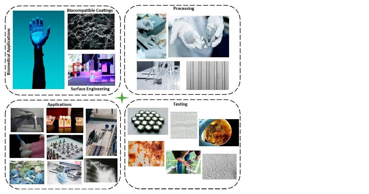
The surface characteristics of biomaterials, including roughness, wettability, antibacterial activity, chemical composition, electrical charge, crystallinity, modulus, and hardness, have a significant impact on their performance as they influence various factors, from biocompatibility to protein adsorption, anti-inflammatory properties, adhesion, and tribological and corrosion behaviours. The surface modification of bulk materials, such as metal alloys, polymers, and ceramics, is essential for addressing significant performance issues, such as a lack of osseointegration, infections, toxicity, low corrosion/wear resistance, and inadequate antibacterial activity. Modifying the surface properties of bulk materials and applying biocompatible coatings lead to significant improvements in biomaterials’ performance and longevity by altering their surface properties, showing potential for facilitating the formulation of effective strategies to tackle specific clinical requirements.

This Special Issue seeks to emphasize the recent progress in surface engineering and biocompatible coatings for biomedical applications. Our objective is to publish a minimum of ten articles of exceptional quality, at which point this Issue could be published in book format. We encourage the submission of original research articles and critical reviews on the following possible research areas (but not restricted to them):

  • Mechanical and physical surface treatment, including grit blasting, polishing, shot peening, water jet shot peening, surface mechanical attrition treatment, laser peening, sputtering, laser/electron beam patterning, and plasma electrolyte oxidation;
  • Chemical and electrochemical surface treatment, including etching, anodizing, sol–gel, nitriding, electrophoretic deposition, PVD, and CVD;
  • Biocompatible coatings, including hydroxyapatite-, bioactive glass-, and polymer-based coatings;
  • Understanding the tribological and corrosion behaviours of biomaterials.

We thank you for your interest and look forward to receiving your contributions.

Dr. Egemen Avcu
Dr. Mert Guney
Dr. Yasemin Yıldıran Avcu
Guest Editors

Dr. Mustafa Armağan
Dr. Eray Abakay
Dr. Berzah Yavuzyeğit
Guest Editor Assistants

Manuscript Submission Information

Manuscripts should be submitted online at www.mdpi.com by registering and logging in to this website. Once you are registered, click here to go to the submission form. Manuscripts can be submitted until the deadline. All submissions that pass pre-check are peer-reviewed. Accepted papers will be published continuously in the journal (as soon as accepted) and will be listed together on the special issue website. Research articles, review articles as well as short communications are invited. For planned papers, a title and short abstract (about 100 words) can be sent to the Editorial Office for announcement on this website.

Submitted manuscripts should not have been published previously, nor be under consideration for publication elsewhere (except conference proceedings papers). All manuscripts are thoroughly refereed through a single-blind peer-review process. A guide for authors and other relevant information for submission of manuscripts is available on the Instructions for Authors page. Coatings is an international peer-reviewed open access monthly journal published by MDPI.

Please visit the Instructions for Authors page before submitting a manuscript. The Article Processing Charge (APC) for publication in this open access journal is 2600 CHF (Swiss Francs). Submitted papers should be well formatted and use good English. Authors may use MDPI's English editing service prior to publication or during author revisions.

Keywords

  • adhesion
  • coating technology
  • corrosion
  • surface topography
  • biocompatibility
  • tribology

Benefits of Publishing in a Special Issue

  • Ease of navigation: Grouping papers by topic helps scholars navigate broad scope journals more efficiently.
  • Greater discoverability: Special Issues support the reach and impact of scientific research. Articles in Special Issues are more discoverable and cited more frequently.
  • Expansion of research network: Special Issues facilitate connections among authors, fostering scientific collaborations.
  • External promotion: Articles in Special Issues are often promoted through the journal's social media, increasing their visibility.
  • e-Book format: Special Issues with more than 10 articles can be published as dedicated e-books, ensuring wide and rapid dissemination.

Further information on MDPI's Special Issue policies can be found here.

Published Papers (8 papers)

Order results
Result details
Select all
Export citation of selected articles as:

Research

11 pages, 3354 KiB  
Article
Evaluation of Bone–Implant Interface: Effects of Angiotensin II Receptor Blockade in Hypertensive Rats
by Gabriel Mulinari-Santos, Jaqueline Silva dos Santos, Fábio Roberto de Souza Batista, Letícia Pitol-Palin, Ana Cláudia Ervolino da Silva, Paulo Roberto Botacin, Cristina Antoniali and Roberta Okamoto
Coatings 2025, 15(1), 73; https://doi.org/10.3390/coatings15010073 - 11 Jan 2025
Viewed by 631
Abstract
Hypertension is a global health concern not only correlated with cardiovascular complications, but also with impaired bone metabolism, potentially affecting healing at the bone–implant interface. Losartan, an angiotensin II receptor blocker (ARB) commonly prescribed for hypertension, has shown beneficial effects on bone healing [...] Read more.
Hypertension is a global health concern not only correlated with cardiovascular complications, but also with impaired bone metabolism, potentially affecting healing at the bone–implant interface. Losartan, an angiotensin II receptor blocker (ARB) commonly prescribed for hypertension, has shown beneficial effects on bone healing in spontaneously hypertensive rats (SHRs). However, the influence of hypertension and ARBs like losartan on the bone cellular response at the bone–implant interface remains underexplored. Methods: A total of 32 rats were included in this study: 16 SHRs, with 8 receiving losartan (30 mg/kg daily) and 8 receiving no treatment, and 16 normotensive Wistar rats, with 8 receiving losartan and 8 receiving no treatment. After one week of treatment, titanium implants were placed into the tibia of all the animals. The bone–implant interface was assessed 60 days post-implantation using micro-computed tomography (µCT) and an immunohistochemical analysis. Results: (i) The ARB treatment significantly increased the bone volume and bone–implant contact in the SHRs receiving losartan compared to the untreated SHRs. (ii) Consistent with the µCT findings, the immunohistochemistry further confirmed regular bone turnover and increased osteocalcin (OC) mineralization in the treated SHRs. In contrast, no alterations in the bone microarchitecture were noted in the Wistar rats treated with losartan. Conclusions: The results suggest that losartan, an ARB drug, improves bone volume and bone turnover at the bone–implant interface in SHRs. Full article
Show Figures

Figure 1

15 pages, 28312 KiB  
Article
Silver Nanoparticles–Chitosan Nanocomposites as Protective Coatings for Dental Remineralization Treatment: An In Vitro Study
by Katleen A. Aguirre de Rodríguez, Wendy Y. Escobar de González, Vianney Castañeda Monroy, Sean Murphy, Gabriel-Alejandro Martínez-Castañón, Horacio Bach and Nereyda Niño-Martínez
Coatings 2025, 15(1), 40; https://doi.org/10.3390/coatings15010040 - 3 Jan 2025
Viewed by 1095
Abstract
Research with nanoparticles for the treatment and prevention of dental caries is of special interest given the high prevalence of the disease worldwide. Several studies support the use of nanoparticles associated with materials given their antimicrobial properties and potential demineralization reduction. This study [...] Read more.
Research with nanoparticles for the treatment and prevention of dental caries is of special interest given the high prevalence of the disease worldwide. Several studies support the use of nanoparticles associated with materials given their antimicrobial properties and potential demineralization reduction. This study aimed to evaluate the impact of the application of silver nanoparticles (AgNPs) and chitosan gel in combination with commercial fluoride varnish on the remineralization of dental enamel. Ninety-six tooth blocks were macroscopically evaluated via stereomicroscopy, ICDAS II, and laser fluorescence. Enamel blocks were subjected to artificial demineralization and divided into four exposure groups (24, 48, 120, and 168 h), and five different remineralizing agents were applied, namely, FV (fluoride varnish), FV + CG (fluoride varnish + chitosan gel), FV + AgNPs (fluoride varnish + AgNPs), FV + AgNPs + CG (fluoride varnish + AgNPs + chitosan gel), and AgNPs + CG (AgNPs + chitosan gel). Enamel surface changes were evaluated via laser fluorescence, X-ray diffraction, scanning electron microscopy, and energy-dispersive X-ray spectroscopy. Laser fluorescence results obtained from demineralized blocks and subsequently exposed to remineralizing treatment indicate significant differences. After exposure to remineralizing agents, hydroxyapatite and modified apatite phases were identified mainly in the samples treated with FV + AgNPs + CG in the groups exposed for 24, 48, and 120 h. The FV + AgNPs + CG and AgNPs + CG indicate good performance in terms of the Ca/P ratio in in vitro demineralization compared to the group treated with fluorine varnish. Full article
Show Figures

Figure 1

13 pages, 1995 KiB  
Article
Potential of Cranberry to Stimulate Osteogenesis: An In Vitro Study
by Yasmim Guterres Bauer, Eduarda Blasi Magini, Ingrid Vicente Farias, José Della Pasqua Neto, Gislaine Fongaro, Flávio Henrique Reginatto, Izabella Thais Silva and Ariadne Cristiane Cabral Cruz
Coatings 2024, 14(11), 1352; https://doi.org/10.3390/coatings14111352 - 24 Oct 2024
Viewed by 971
Abstract
This study investigated the potential of Cranberry extract to stimulate osteogenesis in vitro. The total phenolic and monomeric anthocyanin contents in the Cranberry were determined. Liquid chromatography coupled with mass spectrometry was used to identify the Cranberry’s constituents. To assess the Cranberry’s cytotoxicity, [...] Read more.
This study investigated the potential of Cranberry extract to stimulate osteogenesis in vitro. The total phenolic and monomeric anthocyanin contents in the Cranberry were determined. Liquid chromatography coupled with mass spectrometry was used to identify the Cranberry’s constituents. To assess the Cranberry’s cytotoxicity, a thiazolyl blue tetrazolium bromide assay was employed. Concerning the osteogenesis potential of Cranberry, alkaline phosphatase (ALP) activity, bone morphogenetic protein 2 (BMP-2) expression, and extracellular matrix mineralization were evaluated. The total phenolic content was 522.72 ± 9.80 mg GAE g-1 ES and 364.95 ± 12.49 mg GAE/g detected by the Fast Blue BB and Folin–Ciocalteu method, respectively. For monomeric anthocyanin, the content was 460 ± 30 mg ECG g-1 ES. Moreover, Cranberry concentrations ranged from 62.5 to 500 mg/mL and were found to be biocompatible with osteoblasts and mesenchymal stromal cells. Regarding osteogenesis, 20 mg/mL of Cranberry promoted 2-fold more ALP activity and almost 1.5-fold more BMP-2 than compared to the positive control group. Additionally, 200 mg/mL of Cranberry stimulated a 1.7-fold increase in extracellular matrix mineralization compared to the positive control group. In conclusion, Cranberry displayed potential in stimulating early and late markers of osteogenesis. Its ability to promote osteogenesis and its biocompatibility at higher concentrations hold promise for future application into biomaterials for bone regeneration. Full article
Show Figures

Figure 1

21 pages, 11497 KiB  
Article
Enhancing Passivation Behaviors and Wear Resistance of Biomedical Ti-15Mo Alloy via {332} Twinning Pre-Tension and Aging
by Shanshan Zhang, Fazhan Yang, Wei Sun, Ning Cui and Tiewei Xu
Coatings 2024, 14(10), 1332; https://doi.org/10.3390/coatings14101332 - 17 Oct 2024
Viewed by 1025
Abstract
The purpose of this investigation was to determine the extent to which a combination of pre-deformation and aging procedures could enhance the corrosion and wear resistance of the Ti-15Mo alloy for biomedical materials. The initial grains were refined with {332} twins and other [...] Read more.
The purpose of this investigation was to determine the extent to which a combination of pre-deformation and aging procedures could enhance the corrosion and wear resistance of the Ti-15Mo alloy for biomedical materials. The initial grains were refined with {332} twins and other defects after the pre-deformation, increasing the amount of precipitation nucleation and forming finer α phases during aging. The finer precipitates with numerous boundaries are beneficial in forming a thicker passivation film in phosphate-buffered saline (PBS) solution, and the corrosion resistance of the 20% pre-deform plus aged alloy is improved by 4.23 times. The result indicates that the corrosion passivation behavior and film structure of the biomedical Ti-15Mo alloy are significantly influenced by its microstructure. The worn track and debris of the alloy after pre-tension and aging gradually decreases with the increase in pre-deformation rates, caused by the increase in the hardness and wear resistance. The mechanism of corrosion and wear resistance in PBS solution were revealed, which showed the pre-tension and aging processes that were performed on the Ti-15Mo alloy. This study proposes that pre-tension with {332} twins contributed to precipitation refinement, which would enhance the passivation behaviors and wear resistance of the biomedical β titanium alloy. Full article
Show Figures

Figure 1

13 pages, 5650 KiB  
Article
Corrosion Resistance and In Vitro Biological Properties of TiO2 on MAO-Coated AZ31 Magnesium Alloy via ALD
by Xiao Han, Yan Wang, Jianxiong Ma and Xinlong Ma
Coatings 2024, 14(9), 1198; https://doi.org/10.3390/coatings14091198 - 18 Sep 2024
Viewed by 1509
Abstract
The surface corrosion of magnesium alloys is effectively addressed currently by the creation of a micro-arc oxidation (MAO) ceramic layer. However, oxide film porousness restricts magnesium alloy use. Thus, this work used atomic layer deposition (ALD) to create a TiO2 coating on [...] Read more.
The surface corrosion of magnesium alloys is effectively addressed currently by the creation of a micro-arc oxidation (MAO) ceramic layer. However, oxide film porousness restricts magnesium alloy use. Thus, this work used atomic layer deposition (ALD) to create a TiO2 coating on MAO-coated AZ31B magnesium alloy to plug micropores and increase corrosion resistance and biological characteristics. The samples were analyzed using SEM, EDS, XPS, and XRD to determine their surface appearance, chemical content, and microstructure. Micro-arc oxidation produced a 20 μm oxide coating. The TiO2 film reached 47.41 nm after 400 atomic layer deposition cycles. All corroded samples were tested for corrosion resistance using electrochemical and hydrogen evolution methods and examined for surface morphology. In vitro cell experiments examined biocompatibility. The results indicate that the TiO2 layer sealed the MAO coating’s micro-pores and micro-cracks, enhanced corrosion resistance, and preserved surface morphology following corrosion. The TiO2/MAO composite coating is more biocompatible than the substrate and MAO coating. This research proposes coating AZ31B magnesium alloy for bio-remediation to increase corrosion resistance and biocompatibility. Full article
Show Figures

Figure 1

14 pages, 1255 KiB  
Article
Impact of Diamond-like Carbon Films on Reverse Torque: Superior Performance in Implant Abutments with Internal Conical Connections
by Arianne Vallim Pinto Coelho, Viviane Maria Gonçalves de Figueiredo, Leandro Lameirão Ferreira, Alecsandro de Moura Silva, Marcelo Gallo Oliani, José Renato Cavalcanti de Queiroz, Argemiro Soares da Silva Sobrinho, Lafayette Nogueira Junior and Renata Falchete do Prado
Coatings 2024, 14(9), 1168; https://doi.org/10.3390/coatings14091168 - 10 Sep 2024
Viewed by 928
Abstract
The loosening or fracture of the prosthetic abutment screw is the most frequently reported complication in implant dentistry. Thin diamond-like carbon (DLC) films offer a low friction coefficient and high wear resistance, functioning as a solid lubricant to prevent the weakening of the [...] Read more.
The loosening or fracture of the prosthetic abutment screw is the most frequently reported complication in implant dentistry. Thin diamond-like carbon (DLC) films offer a low friction coefficient and high wear resistance, functioning as a solid lubricant to prevent the weakening of the implant–abutment system. This study evaluated the effects of DLC nanofilms on the reverse torque of prosthetic abutments after simulated chewing. Abutments with 8° and 11° taper connections, with and without DLC or silver-doped DLC coatings, were tested. The films were deposited through the plasma enhanced chemical vapor deposition process. After two million cycles of mechanical loading, reverse torque was measured. Analyses with scanning electron microscopy were conducted on three samples of each group before and after mechanical cycling to verify the adaptation of the abutments. Tribology, Raman and energy-dispersive spectroscopy analyses were performed. All groups showed a reduction in insertion torque, except the DLC-coated 8° abutments, which demonstrated increased reverse torque. The 11° taper groups experienced the most torque loss. The nanofilm had no significant effect on maintaining insertion torque, except for the DLC8 group, which showed improved performance. Full article
Show Figures

Figure 1

21 pages, 4502 KiB  
Article
Ultrasound-Assisted Acellular Spinal Cord Scaffold for Spinal Cord Injury Treatment
by Xi Deng, Yun Liu, Zhongsheng Xu and Hong Yin
Coatings 2024, 14(9), 1137; https://doi.org/10.3390/coatings14091137 - 4 Sep 2024
Viewed by 1116
Abstract
Spinal cord injury (SCI) treatment remains challenging globally, with limited breakthroughs. Tissue engineering offers promise, particularly using acellular spinal cord scaffolds. This study developed a 1-ethyl-(3-dimethylaminopropyl) carbodiimide hydrochloride (EDC)-crosslinked vascular endothelial growth factor (VEGF)-modified acellular spinal cord scaffold for sustained VEGF release. The [...] Read more.
Spinal cord injury (SCI) treatment remains challenging globally, with limited breakthroughs. Tissue engineering offers promise, particularly using acellular spinal cord scaffolds. This study developed a 1-ethyl-(3-dimethylaminopropyl) carbodiimide hydrochloride (EDC)-crosslinked vascular endothelial growth factor (VEGF)-modified acellular spinal cord scaffold for sustained VEGF release. The results show sustained VEGF release over 20 days without altering the scaffold’s properties. Enhanced stability and mechanical properties were observed without increased cytotoxicity. In a rat SCI model, the system improved motor function, reduced glial scarring, and restored spinal cord morphology and histology, indicating potential for SCI therapy. Full article
Show Figures

Figure 1

13 pages, 4795 KiB  
Article
Natural Selection on Hydroxyapatite Fiber Orientations for Resisting Damage of Enamel
by Junfu Shen, Haiyan Xin, Xiaopan Li, Yiyun Kong, Siqi Zhu, Yuankai Zhou, Yujie Fan and Jing Xia
Coatings 2024, 14(9), 1122; https://doi.org/10.3390/coatings14091122 - 2 Sep 2024
Viewed by 1066
Abstract
Teeth have excellent mechanical properties, with high wear resistance and excellent fracture resistance. This is due to their well-organized multilevel hierarchical structure. While a number of studies in the last decades have revealed the relationship between tooth structure and mechanical properties, there is [...] Read more.
Teeth have excellent mechanical properties, with high wear resistance and excellent fracture resistance. This is due to their well-organized multilevel hierarchical structure. While a number of studies in the last decades have revealed the relationship between tooth structure and mechanical properties, there is still no general agreement on how different orientations of hydroxyapatite (HAp) fibers affect the mechanical properties of enamel. With a scanning electron microscope and nanoindenter, the orientations of HAp fibers and their properties were investigated. HAp fibers have two different orientations: parallel and perpendicular to the surface. Fibers oriented parallel to the surface exhibited higher hardness, elastic modulus and wear resistance. Under applied force, fibers oriented perpendicular to the surface suffered deeper shearing in the protein along the long axis, resulting in lower mechanical properties. Teeth resist damaging fractures by combining hard and soft structures. This study may lead to new insights into how nature selects for tooth structure and provide a theoretical basis for the bioinspired design. Full article
Show Figures

Figure 1

Back to TopTop