Article

Fabrication of a Hydrophobic Coating Using Acacia mearnsii De Wild Tannin Melamine Formaldehyde Microcapsules

by
Ana Paula Steigleder
1 and
Liane Roldo
2,*
1
Undergraduate Program in Design, Feevale University, Av. Dr. Maurício Cardoso, 510, Novo Hamburgo CEP 93510-235, RS, Brazil
2
Faculty of Maritime Studies, University of Split, Ruđera Boškovića 37, 21000 Split, Croatia
*
Author to whom correspondence should be addressed.
Coatings 2025, 15(4), 419; https://doi.org/10.3390/coatings15040419
Submission received: 17 February 2025 / Revised: 27 March 2025 / Accepted: 28 March 2025 / Published: 1 April 2025
(This article belongs to the Special Issue Synthesis and Application of Functional Polymer Coatings)

Abstract

:
The aim of this study is to develop and investigate the effect of applying microcapsules containing a melamine formaldehyde resin shell and a tannin extract as a core to form a hydrophobic coating. The microcapsules were obtained by in situ polymerization. Morphological analysis was performed by focused ion beam/scanning electron microscopy. The chemical composition of the tannin extract, the melamine formaldehyde resin, and the polymeric microcapsules with the core of the tannin extract was determined by Fourier transform infrared spectroscopy. The thermal stability of tannin, melamine formaldehyde resin, and tannin microcapsules was investigated by thermogravimetric analysis. In addition, the durability of the coating over time was tested in an environmental test chamber. The polymeric microcapsules containing tannin extract are quasi-spherical, rough, and dense, with a diameter ranging from 1 to 5 μm and a shell thickness of 50 nm. The coating exhibited a hierarchical structure with improved hydrophobic properties, resulting in a contact angle of up to 148°.

Graphical Abstract

1. Introduction

Polymeric microcapsules are renowned for their effective controlled release mechanism of the core substances, while simultaneously protecting the active ingredient from external influences through the use of a shell. The present research represents a novel approach to the development of a coating with hydrophobic or superhydrophobic properties based on the Cassie–Baxter model. This model has been previously studied in research on Salvinia molesta plant and the Salvinia Effect [1]. In the present study, the microcapsules function as microscopic pillars, while the topcoat of hexadecyltrimethoxysilane functions as nanopillars. In this study, microcapsules were synthesized via in situ polymerization using melamine formaldehyde as the shell material and an extract of Acacia mearnsii De Wild tannin as the core material, which was combined with a hexadecyltrimethoxysilane topcoat and applied to an Al alloy substrate. Subsequently, the wettability and average roughness of the coating were evaluated.
This study is part of a larger research project examining the use of micro- and nanocapsules and micro- and nanoparticles in a variety of materials and products. Since 2007, in situ polymerization synthesis as one of the encapsulation methods has been optimized over the years by studying the conditions, parameters, and reagents used. Over the course of the research project, a variety of core materials have been tested. In this study, the Acacia mearnsii De Wild tannin extract was selected as the material of choice due to its renewable nature, anti-corrosive, and anti-fouling properties, and potential as a natural preservative. It has become evident that green resource materials derived from plant extracts have the potential to enhance the quality and performance of modern coatings. The coatings industry has shown a keen interest in natural extracts as a means of improving the properties of polymer coatings [2].
This research project was meticulously executed in three distinct phases. The initial phase centered on the process of microencapsulation through in situ polymerization. The subsequent phase aimed to develop an effective method for the application of microcapsules, with the objective of obtaining a hydrophobic coating. Two application methods were investigated: microcapsule pulverization and sieving onto the aluminum plate. The sieved microcapsules exhibited the most favorable test outcomes for contact angle, contact angle hysteresis, coating durability, and average roughness. The findings of this study are presented herein.
In the interest of providing a comprehensive overview of the techniques and materials utilized, this article focused on a review of the key topics of microcapsules, tannin, and hydrophobic coatings.
Microcapsules are used in various industry segments, such as food, pharmaceuticals, textiles, coatings, chemicals, and cosmetics [3,4]. Their versatility and effectiveness make them a useful tool in product design. Microcapsules are a common inclusion in a variety of applications, providing a multitude of functions and properties. These include, but are not limited to, the sensing of fragrances, the attenuation of odors, the regulation of thermal comfort, the protection against corrosion, and the prevention of fouling, among others. Moreover, its versatility is evidenced by the fact that its morphology, contingent on the core material and the synthesis process employed to form the shell, can assume either a regular or irregular shape [3].
Tannins are antifouling agents that can be obtained from natural sources, plants, and animals, as they have antibacterial, antimicrobial, and corrosion inhibiting properties [5]. The tannin content extracted from the plant can be as high as 20%. The tannin derived from black wattle, as a pigment to develop an antifouling surface, contributes to certain characteristics, including color, astringency, and reactivity [6]. Condensed tannins are prevalent throughout nature and can be found in high concentrations in the wood and bark of different trees, including Acacia mearnsii De Wild (black mimosa bark) and Pinus species. In addition, condensed tannins facilitate the development of environmentally friendly tannin-based resins, adhesives, and leather tanning due to their reactivity with aldehydes and other reagents [7,8]. Thus, the coating has a wide range of applications in various fields, including architecture, construction, and shipbuilding. Additionally, it can be used on furniture, impregnated into fabrics, fibers, and paper, or incorporated into paints.
Previous research on the plant Salvinia molesta has yielded results related to the Salvinia Effect through biomimetics. Depending on the presence or absence of waxes, the top of plant’s the trichome exhibited hydrophilic properties in combination with hydrophobic surfaces [1]. This earlier study, along with others [9,10,11], provided the necessary experience for researching hydrophobic coatings using microcapsules. It also emphasized the relevance and wide application of bioinspired coatings, materials, and products.
Analogous to a cellular model, microcapsules are composed of an outer layer, typically constructed of polymeric or ceramic material. This external layer functions as a protective barrier, thereby isolating the substance contained within, whether gaseous, solid, or liquid [3,12]. The contained material is released when pressure, heat, light, or a solvent is applied to the microcapsule. Depending on the core material, the morphology of the microcapsules can be divided into three basic types: mononuclear, multinuclear, and matrix capsules [3].
In the field of chemical microencapsulation, in situ polymerization processes are the most commonly utilized technique [3,13,14]. With the development of chemical microencapsulation processes, melamine formaldehyde (MP) has proven to be an efficient shell material, with the core material being released by pressure or friction [15,16]. The microcapsules prepared by this method have a smooth and spherical morphology that is temperature sensitive [17].
The coating, in turn, can be classified according to its wettability. The classification of different types of coatings depends on the micro and nano-roughness of the substrate surface. That is, a dual structure of micro and nanopillars combined with surface energy exerts a significant influence on the repellency of liquids from surfaces. The roughness hierarchy and the surrounding air between the pillars stabilize the superhydrophobicity, so that the droplet can settle on the solid surface and the air provides an increase in the contact angle [18]. The classification of surfaces is based on their hydrophobicity. Hydrophobic surfaces exhibit a contact angle between 90° and 150°, while superhydrophobic surfaces demonstrate a contact angle of 150° or greater. The lotus leaf, with a static contact angle of 161°, is an example of a superhydrophobic surface [9,19,20]. The method by which a coating is applied can substantially alter the surface roughness and modify the contact angle [21].
With respect to the silane topcoat, Wiesener et al. [22] investigated a coating with PU-hybrid microcapsules containing triethoxy(methyl)silane that was applied to a steel substrate by tape casting. They reported that due to cracking during stretch forming on the coating, where the droplet has an advancing and a receding side, there is a decrease in contact angles, which can be attributed to an increase in surface roughness. Furthermore, the findings regarding the contact angle and contact angle hysteresis are consistent with those of Pantoja et al. [23], who achieved superhydrophobic surfaces through a series of pretreatments involving the use of a silane-based solution containing SiO2 nanoparticles on stainless steel. In a related study, Avossa et al. [24] obtained a high contact angle of 149° and a low hysteresis angle (CAH = 2°) on surfaces with hierarchical roughness, exhibiting nanometer- and micrometer-range structures through the use of SiO2 particles and dichlorodimethylsilane.
The development and behavior of tannin extract melamine formaldehyde microcapsules and hexadecyltrimethoxysilane solution coating applied on an aluminum substrate were investigated. To obtain relevant results, a series of analytical techniques were employed, including focused ion beam scanning electron microscopy morphology analysis, Fourier transform infrared spectroscopy, and thermogravimetric analysis. These techniques were utilized to gain insights into the microcapsule shell thickness and core characteristics, as well as the tannin and melamine formaldehyde resin. Furthermore, measurements of the contact angle, contact angle hysteresis, and average roughness were conducted before and after exposure to an environmental chamber to establish a correlation between the microcapsule coating and surface wettability.

2. Materials and Methods
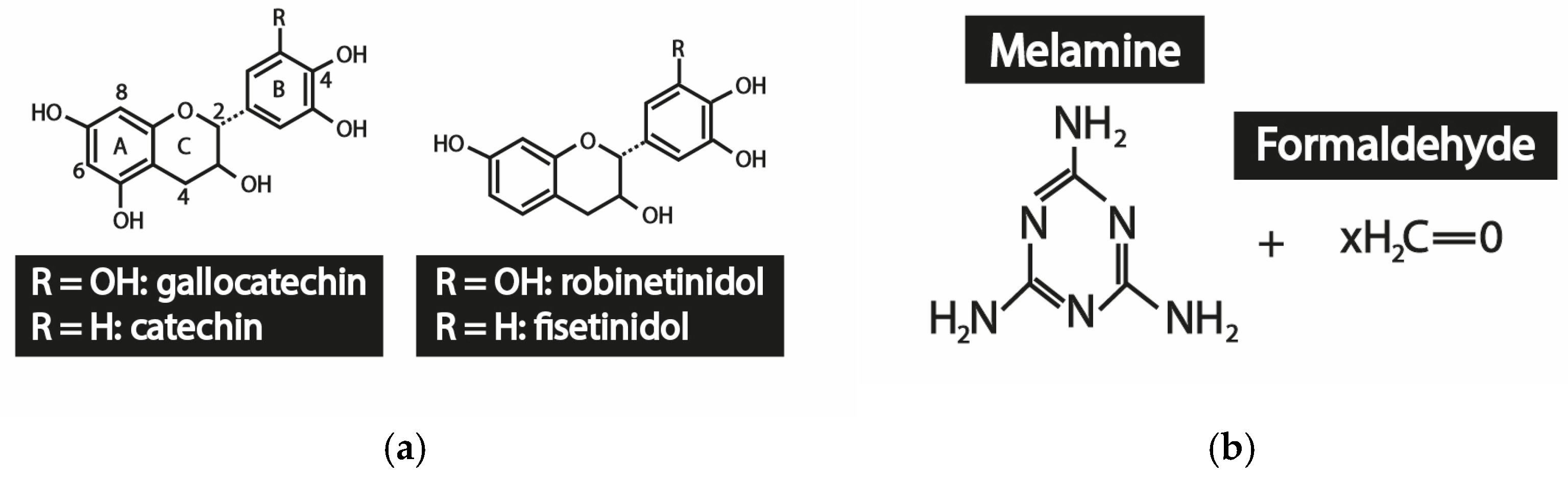
2.1. Materials

Tannin extracted from Acacia mearnsii De Wild, which was used as a core material for the production of polymeric microcapsules with tannin extract—PMiTE, was supplied by Tanac do Brasil (Montenegro, Brazil). Tannins, used as the core of microcapsules in the present study, are often used as pigments and are supplied in powder form (72.5% of tannin extract), have a beige color, are hygroscopic, and are dust-free. The product is water soluble, anionic and has a pH (aqueous solution 20% w/v) between 4 and 5.
Melamine 99% (white powder decomposes at >345 °C and water solubility of 3.23 mg/L) purchased from Sigma-Aldrich, São Paulo, Brazil and formaldehyde 37% (solution stabilized with about 10% methanol) purchased from FMaia, Cotia, Brazil, were used as shell materials. Cetyltrimethylammonium bromide (CTAB, white powder with a melting point from 248 °C to 251 °C and water solubility of 36 g/L), purchased from Anidrol, São Paulo, Brazil was used as an emulsifier and distilled water (DW) as solvent. Acetic acid 90% and triethanolamine p.a. (≥99% by GC) were purchased from Sigma-Aldrich (São Paulo, Brazil) and Cromoline (São Paulo, Brazil), respectively, and used as pH controllers. All reagents were used without any further purification.
Plates of Al-Mn (AA 3104-H24) alloy with a size of (50 × 20 × 7 mm) were used as the substrate for the coating. Varnish (BASF, São Bernardo do Campo, Brazil), a beige alkyd resin, was used as a coating on the aluminum plates and as an adhesive medium for the PMiTE. Hexadecyltrimethoxysilane 85% (by GC), purchased from Sigma-Aldrich (São Paulo, Brazil), was used to prepare the topcoat.

2.2. In Situ Polimerization

The microencapsulation was carried out at the Laboratory of Design and Materials Selection (LDSM) of the Federal University of Rio Grande do Sul (room temperature, 34% humidity).
The first step in the synthesis of PMiTE was the preparation of the emulsion and melamine formaldehyde prepolymer. The emulsion was prepared at a temperature of 70 °C with 200 mL of deionized water, 1 g of the surfactant CTAB, and 0.3 g of tannin extract. The emulsion was homogenized in an ultrasonic processor (Cole-Parmer CP 750 W, 230 VAC, Cole-Parmer Instrument Company, Vernon Hills, IL, USA) at 60% amplitude for 10 min. Subsequently, the pH of the emulsion was adjusted to 5 using a 90% acetic acid solution, and the emulsion was then set aside. In the second step, preparation of the prepolymer was achieved where deionized water was mixed with formaldehyde and stirred with a magnetic stirrer, adding melamine until the dispersion became clear. After this procedure, the pH was adjusted to 8.5 with triethanolamine. The prepolymer was gradually added to the emulsion with mechanical stirring set at 500 rpm. The emulsion was pH-adjusted with a 60 wt% triethanolamine solution to reach the correct pH of 9.0, thus completing the polymerization process.
After polymerization, the solution was filtered and the resulting microcapsules were washed twice with DW. The wet microcapsules (on filter paper) were then left to dry in a desiccator for at least 24 h or until their weight became constant.
The chemical structure of condensed tannin from the bark extract of black mimosa (Acacia mearnsii De Wild) is described in Scheme 1a according to Noreljaleel et al. [25]. The melamine and formaldehyde monomers, responsible for the poly(melamine-formaldehyde, MF polymerization reaction, can be seen in Scheme 1b.
The specific mechanism by which in situ polymerization leads to microcapsule formation has been previously described in previous research [26]. In principle, the methylolation reaction between melamine and formaldehyde comprises two steps. Initially, under alkaline conditions, nucleophilic addition reactions occur, resulting in the substitution of the amino groups, and the subsequent formation of water-soluble methylamines. Subsequently, the formation of oligomers is due to the formation of different types of bonds between triazine rings. The polymerization of melamine and formaldehyde occurs through the auto-condensation of two methylol groups, forming methylene–ether bridges. The condensation between a methylol group and an amine group results in methylene bonds. Thus, the reaction between melamine and formaldehyde forms melamine methylol groups. Furthermore, the polymerization process is carried out by the formation of cross-linked amino resin shells, which encapsulate the tannin extract, using the oligomers of trimethylol melamine as the building molecules [26,27,28].

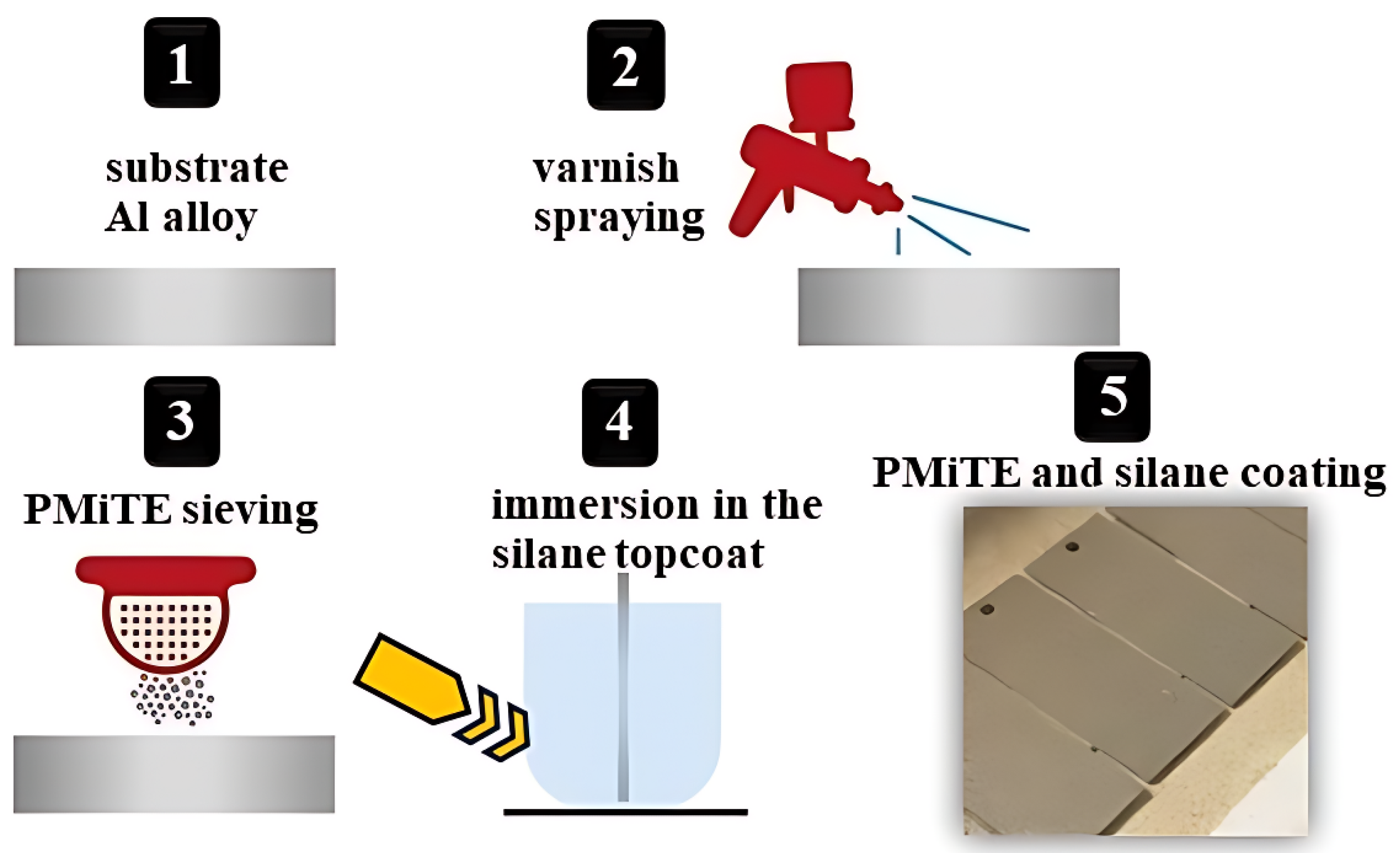
2.3. Coating Application

The coating was applied at room temperature at the Advanced Materials Studies Laboratory of Feevale University, Novo Hamburgo, Brazil. The Al samples were cut with a laser and subsequently degreased with a combination of acetone and deionized water [6]. Post-cleaning, the varnish was diluted with a 10% turpentine solution, according to the manufacturer’s guidelines, and then sprayed onto the Al samples using a 50 W Wagner W550 spray system (J. Wagner GmbH, Markdorf, Germany). This formed a film that served as an adhesive base for the PMiTE (tannin core and melamine formaldehyde shell microcapsules). After a drying period of 50 min at room temperature, the PMiTE were sifted onto the substrate using a 65-mesh sieve. The topcoat was prepared using a solution consisting 90% of ethanol p.a., 5% of deionized water, and 5% of hexadecyltrimethoxysilane. This solution was hydrolyzed for 24 h at room temperature. The pH was then adjusted to between 4 and 5. The Al samples, which had already been varnished and sieved with PMiTE, were submerged in the topcoat solution for 2 min and dried for 48 h in desiccator. After a subsequent drying phase, these samples, henceforth referred to as “coated Al samples”, were ready for further examination. The various stages of coating application are illustrated in Figure 1.

2.4. Characterization

Zeiss FEG-SEM Auriga scanning electron microscope (Zeiss AG, Oberkochen, Germany) with Focused ion beam (FIB) was used for morphological analysis of PMiTE. The microcapsules were metallized by sputtering with a gold layer between 15 nm and 20 nm (Desk V, Denton Vacuum, Moorestown, NJ, USA).
Fourier Transform Infrared (FT–IR) spectra of the core (tannin extract), MF resin, and PMiTE microcapsules were obtained at room temperature using a PerkinElmer Spectrum 100 spectrophotometer (wavenumber range 650 to 4000 cm−1) purchased from PerkinElmer Life and Analytical Sciences, Shelton, CT, USA.
The thermal properties, of PMiTE, tannin extract, and resin MF were determined using a Perkin Elmer TGA 4000 (PerkinElmer, Inc., Shelton, CT, USA). Thermogravimetric analysis—TGA was performed in air at a heating rate of 10 °C/min in a temperature range between 30 °C and 800 °C.
To evaluate the hydrophobic properties, including contact angle and contact angle hysteresis, as well as the durability and average roughness of the coated substrates, 12 aluminum samples were prepared as described in Section 2.3 and exposed in an environmental chamber (Marconi MA835/UR, Piracicaba, Brazil). To compare the results, one of the coated Al samples was not tested in the environmental test chamber. The coated Al samples were exposed to 90% humidity at 37 °C for 1008 h (equivalent to 41 days). The tests were performed according to the ASTM D2247 standard [29]. After a fixed time of 24, 48, 72, 96, 168, 240, 336, 432, 576, 744, and 1008 h, the coated Al samples were kept in a desiccator to remove moisture and then analyzed by optical microscopy.
A drop shape analysis system (DSA100, Krüss GmbH, Hamburg, Germany) was used to measure the contact angle and contact angle hysteresis of the coating. To carry out the measurements, the instrument is calibrated to drop a volume of 10 μL of deionized water. These measurements were conducted at room temperature on a total of 24 coated Al samples. The contact angle was determined using an arithmetic mean of five drops at different locations on the coated Al samples.
For the hysteresis angle, Surftens open-source software and ImageJ software version 1.51 were used to determine the angle of the moving droplet. The hysteresis contact angle measurement was determined by calculating the difference between the advancing (θA) and receding (θR) contact angles of the droplet on the inclined surface [30,31]. The Mitutoyo Surftest SJ-210 measured the arithmetic average roughness, Ra, which represents the average surface roughness over the length of the measurement [32]. The measurements were performed in accordance with the standard ABNT NBR ISO 4287/2002 [33].

3. Results and Discussion

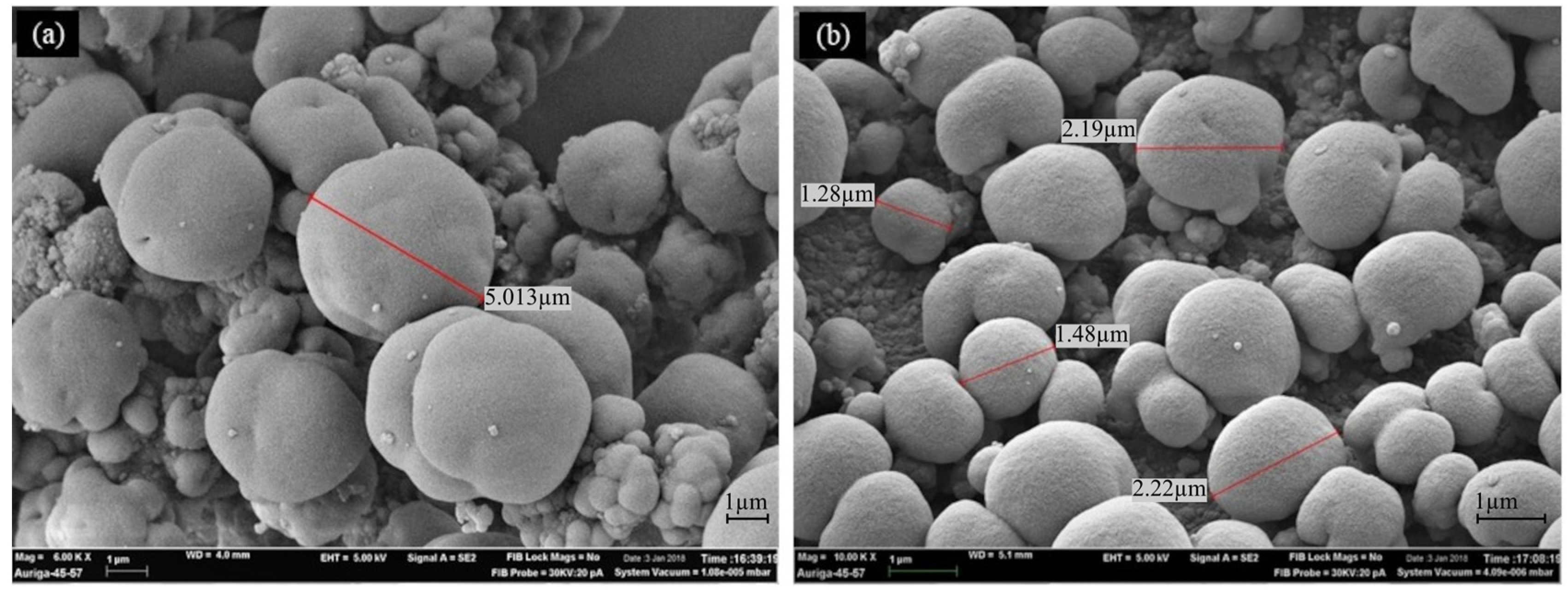
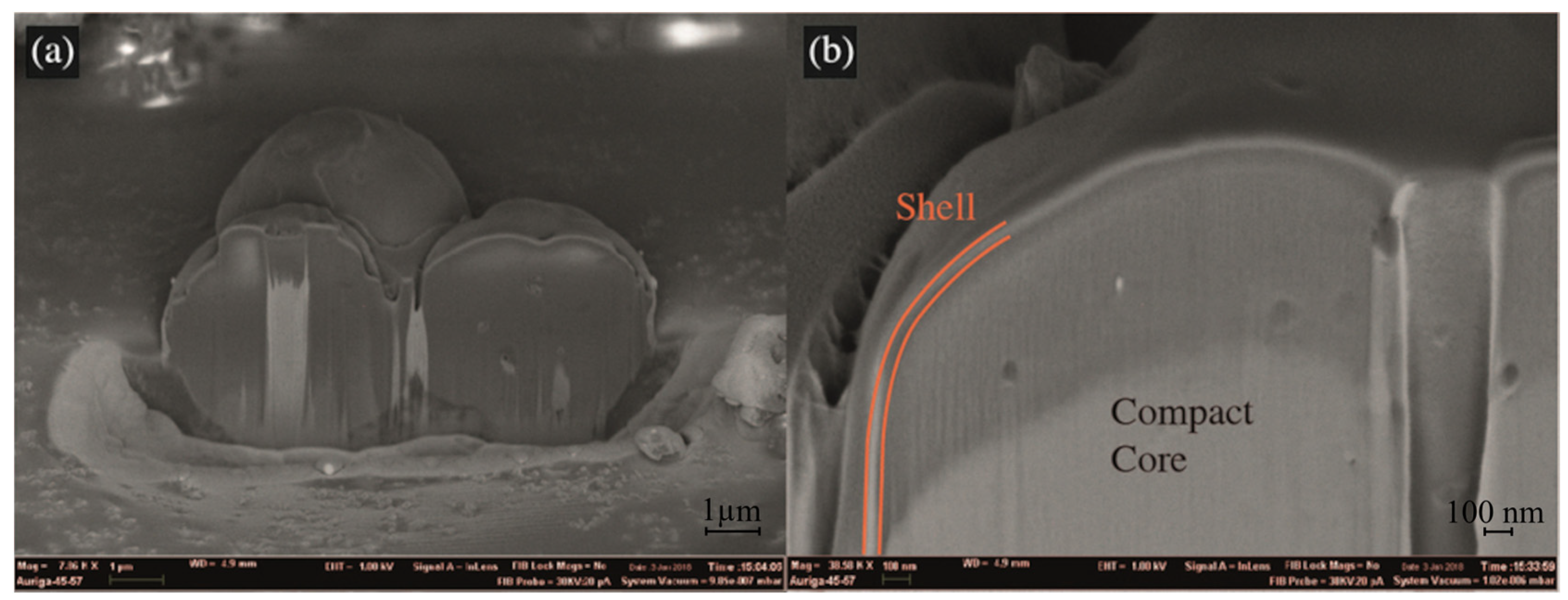
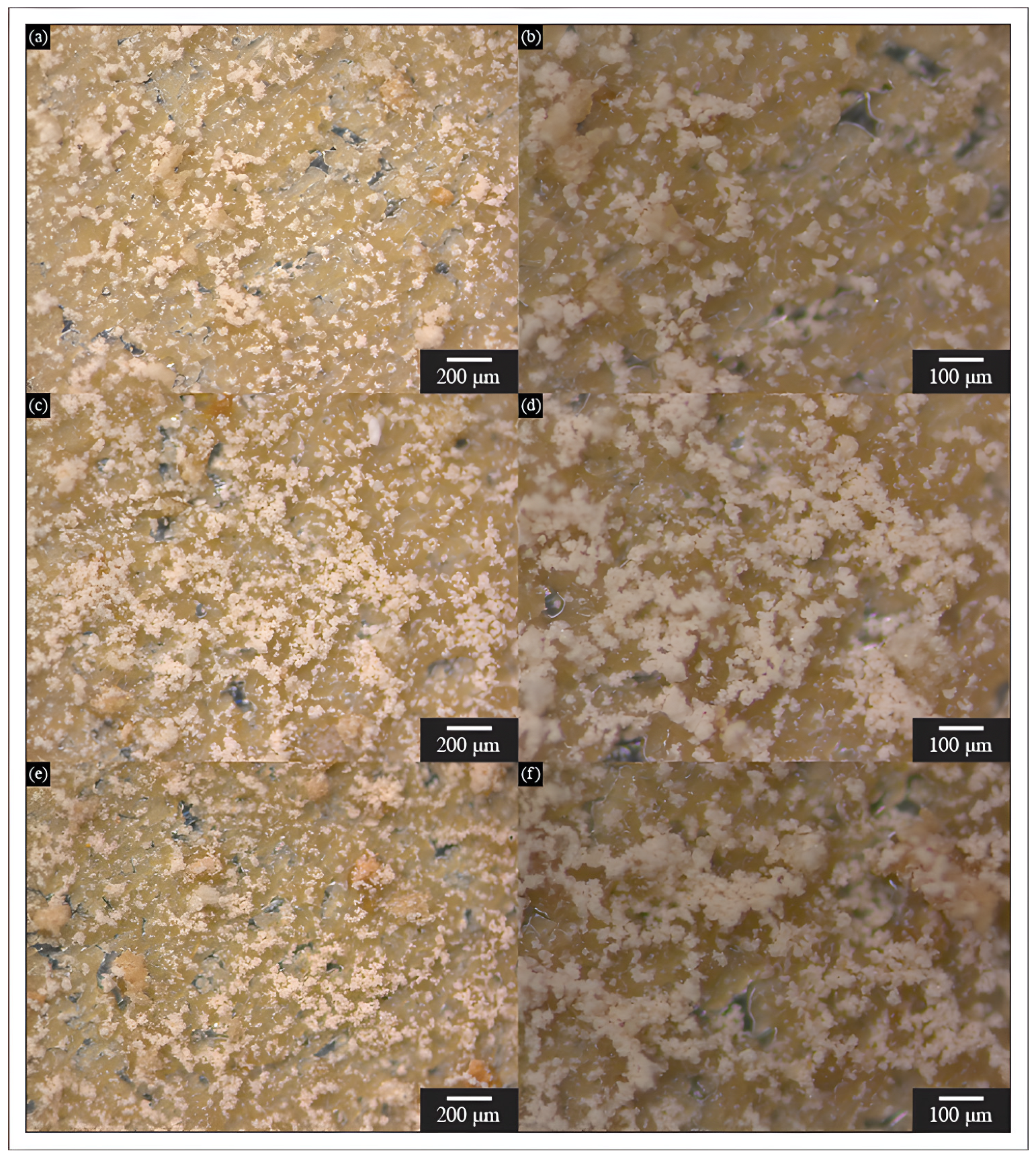
3.1. Microcapsules Morphology

Figure 2 shows the microstructure of PMiTE obtained by SEM. It shows regular quasi-spherical microcapsules with apparent roughness between (1 and 5 μm) in diameter. Clusters of agglomerated melamine formaldehyde microcapsules with possible nanocapsules were also observed by Zhu et al. [13] and Escobar et al. [26].
The internal structure of PMiTE was analyzed using a focused ion beam (FIB) coupled with scanning electron microscopy (SEM). Figure 3a presents two spherical microcapsules sliced by FIB, showing that the microencapsulation occurred and that the microcapsules have a dense core and a shell thickness of about 50 nm, as shown in Figure 3b. The microcapsules in question are compact. Furthermore, the present microcapsules are classified as a type of matrix microcapsule [3,34].

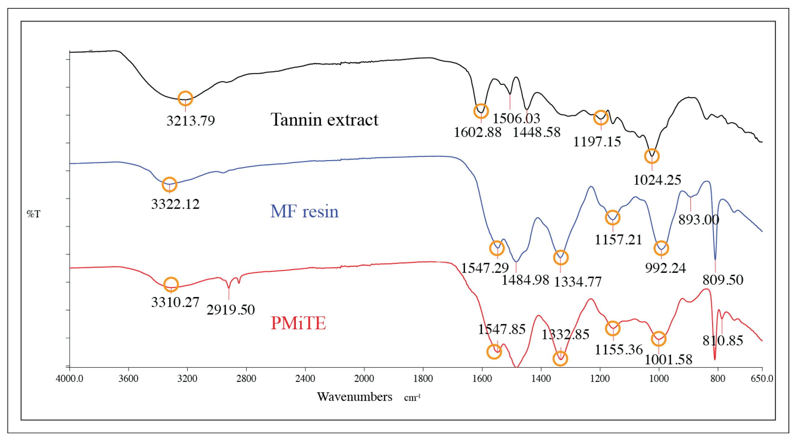
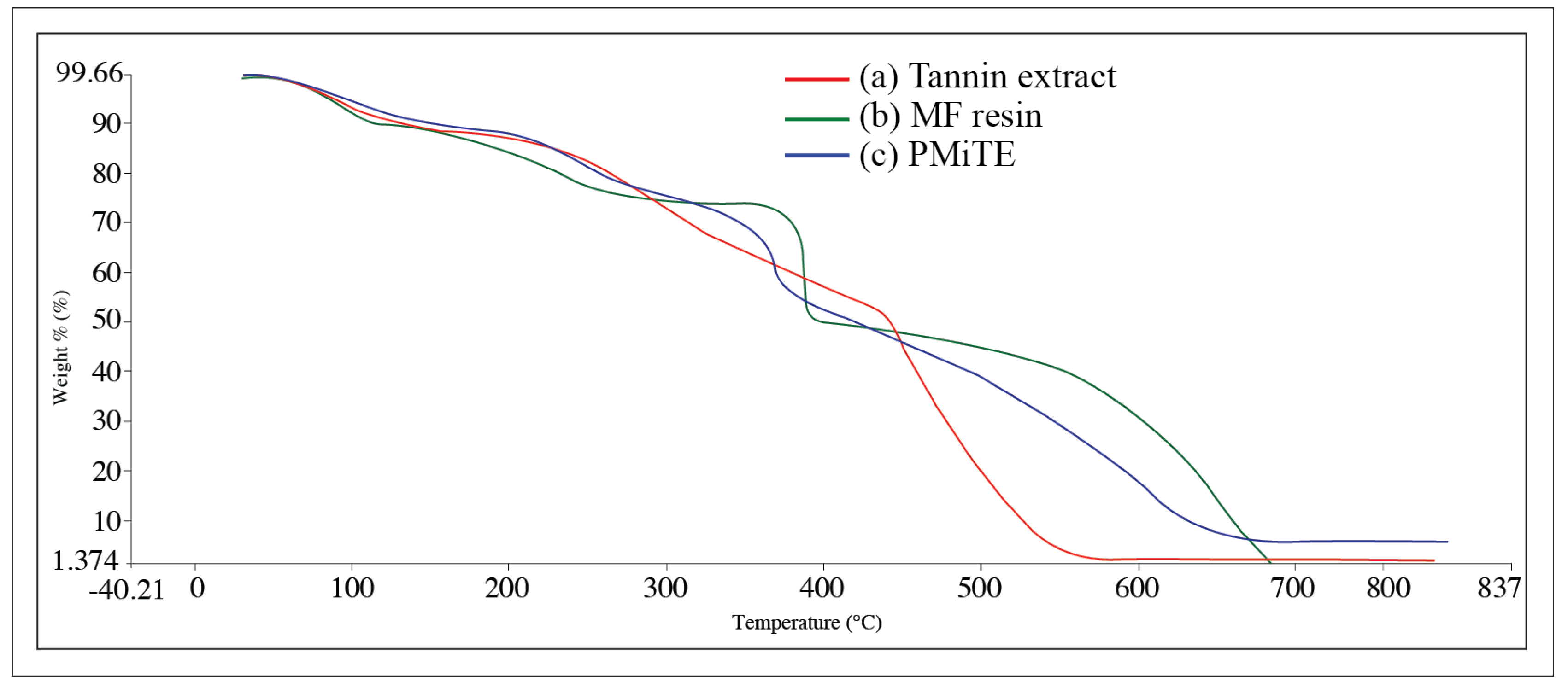
3.2. Chemical Structure and Thermal Stability

The infrared spectra of PMiTE, MF resin, and tannin extract are shown in Figure 4.
The absorption bands at 3322 cm−1 are associated with the hydroxyl group and phenolic hydroxides O-H in both spectra [35,36,37]. In the study of Noor Idora et al. [5], the same picks were observed in the range of 3240 cm−1 and 3640 cm−1 for two different types of tannins; they correspond to the stretching of the -OH hydroxyl group. The bands for the MF resin and PMiTe methylene group were detected at 1547 cm−1 and 1484 cm−1, respectively [38]. The bands between 1600 cm−1 and 1547 cm−1 in the spectra of PMiTE, MF resin, as well as the tannin extract correspond to C=C, which refers to the aromatic groups [35,36,37]. Oscillations arise from the deformation of the C-C bonds in the phenolic groups absorbed in the range of 1506 cm−1 and 1448 cm−1 [36]. According to Monteiro et al. [7], the hydrolyzable tannins consist of esters, and according to Mangrich et al. [39], tannins have multiple hydroxyl groups attached to the aromatic rings. Band 1024 cm−1, in the spectrum of the tannin extract, is associated with the absorption of the C-O ester group [38].
The thermogram of the TGA analysis of tannin extract, MF resin, and PMiTE in the temperature range of 30 °C and 800 °C is shown in Figure 5. The TGA curve of the tannin extract (Figure 5, curve a) indicates an initial loss of mass due to water loss and some low molecular weight and easily degradable substances such as organic acid, phenol, and pigment. With additional heating above 100 °C, the decomposition of tannin stearates becomes apparent. A substantial mass reduction takes place within the temperature range of 200 °C to 450 °C, with pyrolytic degradation occurring at about 450 °C [36,37,40].
The TGA thermogram of the MF resin (Figure 5, curve b) and the PMiTE (Figure 5, curve c) was divided into four intervals, respectively. The MF resin (Figure 5, curve b) and PMiTe (Figure 5, curve c) thermograms show an initial mass between 30 °C and 120 °C due to the evaporation of absorbed water [13,38]. In the second interval, from 120 °C to about 370 °C, the MF resin showed a constant mass loss. However, between 190 °C and 370 °C, a faster decay in mass loss of about 13% was observed. In the PMiTE TGA curve (Figure 5, curve c), a constant mass loss of about 20% was observed between 120 °C and 345 °C. These mass losses are mainly attributed to the elimination of formaldehyde from the MF resin [38]. In the third interval, between 370 °C and 400 °C, there is a significant mass loss of about 21% of the MF resin and 12% of the PMiTE between 345 °C and 380 °C. In this case, the mass loss is attributed to the deterioration of the methylene bonds [16,38]. The thermal stability of the MF resin and PMiTE differs slightly from each other. Specifically, the former exhibits stability up to a temperature of 370 °C, while the latter demonstrates stability up to 345 °C. The mass loss continues until the degradation of the PMiTE and MF resin is complete at approximately 650 °C and 690 °C, respectively. The final degradation and the fourth interval may be attributable to a rupture of the triazine bond, which can result in the breakdown of the shell [26,38]. Thermogravimetric analysis was performed up to 800 °C to ensure complete mass loss and to verify that there was no further reaction or degradation of the MF resin, tannin extract or microcapsules.

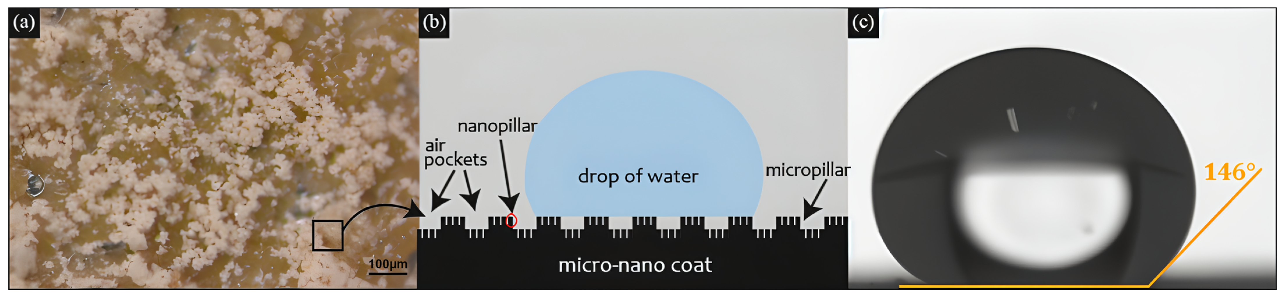
3.3. Coating Wettability and Average Roughness

The coating developed is primarily characterized as hydrophobic, with a high contact angle and low contact angle hysteresis angle. Figure 6 displays the coated Al samples, which have a relatively homogeneous distribution of microcapsules onto the wet varnish, although some agglomeration is observed in certain areas. The light beige/brown coloration of the substrate can be attributed to the tannin remaining from the microencapsulation, as well as from the varnish.
The coating resulted in a significant 57° increase in contact angle compared to the initial angle of the aluminum substrate. The tests showed that the aluminum plate achieved a contact angle of 76°, which increased to 79° after the paint was sprayed onto it. When the PMiTE coating was sieved over the varnish applied to the aluminum plate, the contact angle increased to 91°. The aluminum sample, which was sprayed with varnish, screened with PMiTE, and coated with a hexadecyltrimethoxysilane solution, achieved an overall contact angle of 133° (Table 1).
After exposing the coated Al samples to an environmental chamber for a specified period of time (24, 48, 72, 96, 168, 240, 336, 432, 576, 744, and 1008 h) and comparing them with the coated Al sample without exposure (0 h), it was found that there was no significant deviation in the measurement of the contact angle within the proposed time. A difference of up to 13° in the contact angle is attributed to the different PMiTE concentrations at certain locations of the coated Al samples. Thus, these are not absolute values, but acceptable variations for a hydrophobic coating that has a contact angle of 135° or more. As a function of exposure time, it was observed that the coating reached an angle of 148°, which is close to superhydrophobicity (≥150°) [20].
As with the contact angle (Table 1), it is assumed that the small variations in the contact angle hysteresis are related to the PMiTE concentrations at specific locations on the coated Al samples. Aiming at surface coating on a metallic surface, PMiTE produces a heterogeneous surface resembling the protuberances and undulations of the plant Nelumbo nucifera (lotus leaf) [41,42].
The exposure of the coated Al samples in the environmental test chamber also affected the surface roughness of PMiTE. The Ra values, representing the average roughness, which is the average height difference between bands and valleys from the surface centerline, ranged from 9.4 µm to 11.1 μm for the 11 coated Al samples exposed to the environmental chamber. This resulted in a variation in roughness, with the range indicating an approximate 2 μm difference across the samples. The observed result is likely attributed to the homogeneous dispersion of the microcapsules within the coating. Depending on the surface finish, more specifically the roughness, it can affect the hydrophobicity, corrosion resistance, and wear or fatigue resistance of a particular component [37].
After exposure of the coating to the conditions within the environmental chamber, it was observed that the behavior of the PMiTE coating remained largely unchanged even after 1008 h, indicating a durable coating with minimal alterations in morphology and thermal and chemical stability. The findings regarding the average roughness, which fell between 9.4 μm and 11.3 μm, demonstrated a minimal influence on the measured contact angle of the coating. However, they did have an impact on the shape of the drop.
The findings concerning hydrophobicity are in accordance with those of prior studies in this domain. As such, the developed coating exhibits a maximum contact angle of 148° and a contact angle hysteresis ranging from 6° to 15° (Table 1). This indicates a hydrophobic surface with a high contact angle and a low contact angle hysteresis.
The schematic drawing in Figure 7 shows the fabricated hierarchical structured coating. It provides information regarding the hydrophobic mechanism observed in the context of the present investigation. In this instance, the micro- and nanopillars, in conjunction with the presence of air pockets, are observed on hydrophobic and superhydrophobic surfaces—Figure 7b [43].
According to the Cassie–Baxter model, the droplet shape on a hydrophobic surface can vary depending on factors such as surface texture and surface energy [30,44,45]. This phenomenon can be attributed to the presence of air pockets beneath the surface, which serve to support the droplet, thereby resulting in a spherical or nearly spherical droplet and, consequently, a hydrophobic or nearly superhydrophobic surface, as observed by the 146° (maximum 148°) contact angle obtained by the present coating (Figure 7b,c). The contact angle can be highly influenced by the type of coating material and the surface properties. The water repellency of a material refers to the combination of the chemical composition and the surface roughness it exhibits [46].

4. Conclusions

It was found that the polymeric microcapsules with a core of tannin extract are relatively agglomerated, have a quasi-spherical morphology, are rough, and have a diameter between (1 to 5 μm). In addition, FIB/SEM analysis reveals that microcapsules containing tannin have a dense core enveloped by a 50 nm thick shell. These findings confirm the effectiveness of in situ polymerization as a technique to microencapsulate tannin extracts. Furthermore, the thin melamine formaldehyde shell is expected to facilitate the release of tannin onto the metal surface in response to light friction, thereby enabling the formation of an additional anticorrosive coating. Future research should investigate the anticorrosive and antifouling properties of the PMiTE coating.
Based on the TGA curves, it was observed that the microcapsules (PMiTE), which are composed of a melamine formaldehyde shell with a core containing tannin extract, are stable up to 345 °C.
It was also found that the exposure time of aluminum plates coated with PMiTE microcapsules under chamber conditions (90% humidity at 37 °C) had little effect on the change in contact angle, confirming the durability and thermal, chemical, and morphological stability of the microcapsules. The microcapsule’s thermal stability and the environmental chamber results indicate that the coating can be used in outdoor areas exposed to weather conditions.
The coating with water-repellent properties was achieved by combining the roughness of the microcapsules and their sieving over a thin layer of varnish followed by a silane topcoat. This resulted in a contact angle of more than 135°. The contact angle is significantly affected by the method of coating application, particularly the distribution (degree of agglomeration) of the microcapsules on the substrate.
As for the application method of microcapsules, industrial sieving is a low-cost, practical, and efficient option. It allows quick sieve replacement, produces low noise, has low energy consumption, and is a practical installation and handling method.

Author Contributions

A.P.S.: methodology, investigation, writing—original draft, formal analysis. L.R.: conceptualization, supervision, methodology, formal analysis, project administration, writing—review and editing. All authors have read and agreed to the published version of the manuscript.

Funding

This research was funded by the Brazilian National Council for Technological and Scientific Development—CNPq under Grant No. 301373/2016-7.

Institutional Review Board Statement

Not applicable.

Informed Consent Statement

Not applicable.

Data Availability Statement

Data are contained within the article.

Acknowledgments

Thanks to Tanac Brazil Industry, to the staff of Feevale University, Novo Hamburgo, Brazil, to the colleagues of the Laboratory of Design and Materials Selection (LDSM), the Center for Microscopy and Microanalysis (CMM), the Multi-user Surface Analysis Laboratory (LAMAS), all from the Federal University of Rio Grande do Sul, Porto Alegre, Brazil. The authors also thank the Faculty of Maritime Studies, University of Split, Croatia.

Conflicts of Interest

The authors declare that they have no known competing financial interests or personal relationships that could appear to influence the work reported in this paper. There are no conflicts to declare or disclose.

References

  1. Steigleder, A.P.; Roldo, L. Surface morphology and repellency mechanism investigation of Salvinia molesta plant. Rev. Mater. 2019, 24, e12480. [Google Scholar] [CrossRef]
  2. Hamidi, N.A.S.M.; Kamaruzzaman, W.M.I.W.M.; Nasir, N.A.M.; Shaifudin, M.S.; Ghazali, M.S.M. Potential Application of Plant-Based Derivatives as Green Components in Functional Coatings: A Review. Clean. Mater. 2022, 4, 100097. [Google Scholar] [CrossRef]
  3. Ghosh, S.K. Functional Coatings and Microencapsulation: A General Perspective. In Functional Coatings: By Polymer Microencapsulation, 1st ed.; Ghosh, S.K., Ed.; Wiley-VCH Verlag GmbH & Co. KGaA: Weinheim, Germany, 2006; pp. 1–28. [Google Scholar]
  4. Shrimali, K.; Dedhia, E.M. Microencapsulation for Textile Finishing. J. Polym. Text. Eng. 2015, 2, 1–4. [Google Scholar] [CrossRef]
  5. Noor Idora, M.S.; Ferry, M.; Wan Nik, W.B.; Jasnizat, S. Evaluation of tannin from Rhizophora apiculata as natural antifouling agents in epoxy paint for marine application. Prog. Org. Coat. 2015, 81, 125–131. [Google Scholar] [CrossRef]
  6. Peres, R.S.; Armelin, E.; Alemán, C.; Ferreira, C.A. Modified tannin extracted from black wattle tree as an environmentally friendly antifouling pigment. Ind. Crop. Prod. 2015, 65, 506–514. [Google Scholar] [CrossRef]
  7. Monteiro, J.M.; de Albuquerque, U.P.; de Lima Araújo, E.; de Amorim, E.L.C. Taninos: Uma abordagem da química à ecologia. Quim. Nova. 2005, 28, 892–896. [Google Scholar] [CrossRef]
  8. Aristri, M.A.; Sari, R.K.; Lubis, M.A.R.; Laksana, R.P.B.; Antov, P.; Iswanto, A.H.; Mardawati, E.; Lee, S.H.; Savov, V.; Kristak, L.; et al. Eco-Friendly Tannin-Based Non-Isocyanate Polyurethane Resins for the Modification of Ramie (Boehmeria nivea L.). Fibers Polym. 2023, 15, 1492. [Google Scholar] [CrossRef]
  9. Barthlott, W.; Mail, M.; Neinhuis, C. Superhydrophobic hierarchically structured surfaces in biology: Evolution, structural principles and biomimetic applications. Phil. Trans. R. Soc. A 2016, 374, 20160191. [Google Scholar] [CrossRef]
  10. Jin, H.; Tian, L.; Bing, W.; Zao, J.; Ren, L. Bioinspired marine antifouling coatings: Status, prospects, and future. Prog. Mater. Sci. 2022, 124, 100889. [Google Scholar] [CrossRef]
  11. Lamprakou, Z.; Bi, H.; Weinell, C.E.; Dam-Johansen, K. Tannin-based inhibitive pigment for sustainable epoxy coatings formulation. Prog. Org. Coat. 2022, 167, 106841. [Google Scholar] [CrossRef]
  12. Eqbal, D.; Gundabala, V. Controlled fabrication of multi-core alginate microcapsules. J. Colloid. Interf. Sci. 2017, 507, 27–34. [Google Scholar] [CrossRef]
  13. Zhu, K.; Qi, H.; Wang, S.; Zhou, J.; Zhao, Y.; Su, J.; Yuan, X. Preparation and Characterization of Melamine-Formaldehyde Resin Micro- and Nanocapsules Filled with n-Dodecane. J. Macromol. Sci. B 2012, 51, 1976–1990. [Google Scholar] [CrossRef]
  14. Pires, F.Q.; Gross, I.P.; Sa-Barreto, L.L.; Gratieri, T.; Gelfuso, G.M.; Bao, S.N.; Cunha-Filho, M. In-situ formation of nanoparticles from drug-loaded 3D polymeric matrices. Eur. J. Pharm. Sci. 2023, 188, 106517. [Google Scholar] [CrossRef]
  15. Ollier, R.P.; Alvarez, V.A. Synthesis of epoxy-loaded poly(melamine-formaldehyde) microcapsules: Effect of pH regulation method and emulsifier selection. Colloids Surf. A Physicochem. Eng. Asp. 2017, 520, 872–882. [Google Scholar] [CrossRef]
  16. Urdl, K.; Weiss, S.; Karpa, A.; Perić, M.; Zikulnig-Rusch, E.; Brecht, M.; Kandelbauer, A.; Müller, U.; Kern, W. Furan-functionalised melamine-formaldehyde particles performing Diels-Alder reactions. Eur. Polym. J. 2018, 108, 225–234. [Google Scholar] [CrossRef]
  17. Yataganbaba, A.; Ozkahraman, B.; Kurtbas, I. Worldwide trends on encapsulation of phase change materials: A bibliometric analysis (1990–2015). Appl. Energ. 2017, 185, 720–731. [Google Scholar] [CrossRef]
  18. Sturzenegger, P.N.; Gonzenbach, U.T.; Koltzenburg, S.; Martynczuk, J.; Gauckler, L.J. Particle-stabilized gel-core microcapsules: Synthesis and properties. Colloids Surf. A Physicochem. Eng. Asp. 2014, 447, 44–50. [Google Scholar] [CrossRef]
  19. Rajput, A.; Mishra, A.; Ali, A.; Goswami, R.; Bhatt, N. Transparent superhydrophobic coating for copper metal and study of surface morphology and antimicrobial characteristics. Colloid Surf. A Physicochem. Eng. Asp. 2024, 684, 133067. [Google Scholar] [CrossRef]
  20. Koch, K.; Bhushan, B.; Barthlott, W. Diversity of structure, morphology and wetting of plant surfaces. Soft Matter. 2008, 4, 1943–1963. [Google Scholar] [CrossRef]
  21. Madadi, F.; Rezaeian, A.; Edris, H.; Zhiani, M. Influence of surface roughness and hydrophobicity of bipolar plates on PEM performance. Surf. Coat Tech. 2020, 389, 125676. [Google Scholar] [CrossRef]
  22. Wiesener, M.; Regenspurger, R.; Pilz, M.; Shchukin, D.; Latnikova, A.; Yang, J.; Grundmeier, G. In-situ contact angle studies of the release of water displacing agents from capsule filled organic coatings. Surf. Coat Tech. 2012, 206, 4481–4487. [Google Scholar] [CrossRef]
  23. Pantoja, M.; Velasco, F.; Abenojar, J.; Martinez, M.A. Development of superhydrophobic coatings on AISI 304 austenitic stainless steel with different surface pretreatments. Thin Solid Films 2019, 671, 22–30. [Google Scholar] [CrossRef]
  24. Avossa, J.; Bifulco, A.; Amendola, E.; Gesuele, F.; Oscurato, S.L.; Gizaw, Y.; Mensitieri, G.; Branda, F. Forming nanostructured surfaces through Janus colloidal silica particles with nanowrinkles: A new strategy to superhydrophobicity. Appl. Surf. Sci. 2019, 465, 73–81. [Google Scholar] [CrossRef]
  25. Noreljaleel, A.E.M.; Wilhelm, A.; Bonnet, S.L. Analysis of commercial proanthocyanidins. part 6: Sulfitation of flavan-3-ols catechin and epicatechin, and procyanidin B-3. Molecules 2020, 25, 4980. [Google Scholar] [CrossRef]
  26. Escobar, C.F.; Roldo, L.; Rocha, T.L.A.; Kindlein, W., Jr. Nanoencapsulation mechanism of fragrant oil: Effect of formaldehyde−melamine molar ratio. Polym. Int. 2018, 67, 220–226. [Google Scholar] [CrossRef]
  27. Cheong, I.W.; Shin, J.S.; Kim, J.H.; Lee, S.J. Preparation of Monodisperse Melamine-Formaldehyde Microspheres via Dispersed Polycondensation. Macromol. Res. 2004, 12, 225–232. [Google Scholar] [CrossRef]
  28. Leskovšek, M.; Kortnik, J.; Elesini, U.S.; Šumiga, B. Characterisation of melamine formaldehyde microspheres synthesised with prolonged microencapsulated reaction time. J. Polym. Eng. 2022, 42, 288–297. [Google Scholar] [CrossRef]
  29. ASTM D2247; Standard Practice for Testing Water Resistance of Coatings in 100 % Relative Humidity. American Society for Testing and Materials: Montgomery County, PA, USA, 2015.
  30. Latthe, S.S.; Gurav, A.B.; Maruti, C.S.; Vhatkar, R.S. Recent Progress in Preparation of Superhydrophobic Surfaces: A Review. J. Surf. Eng. Mat. Adv. Tech. 2012, 2, 76–94. [Google Scholar] [CrossRef]
  31. Verplanck, N.; Coffinier, Y.; Thomy, V.; Boukherroub, R. Wettability Switching Techniques on Superhydrophobic Surfaces. Nanoscale Res. Lett. 2007, 2, 577–596. [Google Scholar] [CrossRef]
  32. Bandeira, R.M.; van Drunen, J.; Garcia, A.C.; Tremiliosi-Filho, G. Influence of the thickness and roughness of polyaniline coatings on corrosion protection of AA7075 aluminum alloy. Electrochim. Acta 2017, 240, 215–224. [Google Scholar] [CrossRef]
  33. ABNT NBR ISO 4287:2002; Especificações Geométricas do Produto (GPS)—Rugosidade: Método do Perfil—Termos, Definições e Parâmetros da Rugosidade. Associação Brasileira de Normas Técnicas: Rio de Janeiro, Brazil, 2002.
  34. Porras-Saavedra, J.; Alamilla-Beltrán, L.; Lartundo-Rojas, L.; Perea-Flores, M.J.; Yáñez-Fernandez, J.; Palacios-González, E.; Gutierrez-López, G.F. Chemical components distribution and morphology of microcapsules of paprika oleoresin by microscopy and spectroscopy. Food Hydrocolloid. 2018, 81, 6–14. [Google Scholar] [CrossRef]
  35. bin Hussein, M.Z.; Ghotbi, M.Y.; Yahaya, A.H.; Rahman, M.Z.A. Synthesis and characterization of (zinc-layered-gallate) nanohybrid using structural memory effect. Mater. Chem. Phys. 2009, 113, 491–496. [Google Scholar] [CrossRef]
  36. Viswanath, V.; Leo, V.V.; Sabna Prabha, S.; Prabhakumari, C.; Potty, V.P.; Jisha, M.S. Thermal properties of tannin extracted from Anacardium occidentale L. using TGA and FT-IR spectroscopy. Nat. Prod. Res. 2015, 30, 223–227. [Google Scholar] [CrossRef] [PubMed]
  37. Baaka, N.; Ammar, M.; Saad, M.K.; Khiari, R. Properties of tannin-glyoxal resins prepared from lyophilized and condensed tannin. J. Text. Eng. Fashion Technol. 2017, 4, 705–711. [Google Scholar] [CrossRef]
  38. Merline, D.J.; Vukusic, S.; Abdala, A.A. Melamine formaldehyde: Curing studies and reaction mechanism. Polym. J. 2013, 45, 413–419. [Google Scholar] [CrossRef]
  39. Mangrich, A.S.; Doumer, M.E.; Mallmann, A.S.; Wolf, C.R. Química verde no tratamento de águas: Uso de coagulante derivado de tanino de Acacia mearnsii. Rev. Virtual Quim. 2014, 6, 2–15. [Google Scholar] [CrossRef]
  40. Luo, C.; Grigsby, W.; Edmonds, N.; Easteal, A.; Al-Hakkak, J. Synthesis, characterization, and thermal behaviors of tannin stearates prepared from quebracho and pine bark extracts. J. Appl. Polym. Sci. 2010, 117, 352–360. [Google Scholar] [CrossRef]
  41. Yang, F.; Guo, Z. Characterization of micro-morphology and wettability of lotus leaf, waterlily leaf and biomimetic ZnO surface. J. Bionic Eng. 2015, 12, 88–97. [Google Scholar] [CrossRef]
  42. Guo, Z.; Liu, W. Biomimic from the superhydrophobic plant leaves in mature: Binary structure and unitary structure. Plant Sci. 2007, 172, 1103–1112. [Google Scholar] [CrossRef]
  43. Sajadinia, S.H.; Sharif, F. Thermodynamic Analysis of the Wetting Behavior of Dual Scale Patterned Hydrophobic Surfaces. J. Colloid Interface Sci. 2010, 344, 575–583. [Google Scholar] [CrossRef]
  44. Kwon, T.W.; Jang, J.; Ambrosia, M.S.; Ha, M.Y. Molecular Dynamics Study on the Hydrophobicity of a Surface Patterned with Hierarchical Nanotextures. Colloids Surf. A Physicochem. Eng. Asp. 2018, 559, 209–217. [Google Scholar] [CrossRef]
  45. Bormashenko, E.; Bormashenko, Y.; Whyman, G.; Pogreb, R.; Stanevsky, O. Micrometrically scaled textured metallic hydrophobic interfaces validate the Cassie–Baxter wetting hypothesis. J. Colloid Interf. Sci. 2006, 302, 308–311. [Google Scholar] [CrossRef]
  46. Vanithakumari, S.C.; George, R.P.; Kamachi Mudali, U. Influence of silanes on the wettability of anodized titanium. Appl. Surf. Sci. 2014, 292, 650–657. [Google Scholar] [CrossRef]
Scheme 1. Representation of (a) condensed tannin monomer from black mimosa bark extract and (b) melamine and formaldehyde monomers responsible for the cross-linked condensation polymerization reaction.
Coatings 15 00419 sch001
Figure 1. Steps of the coating application.
Coatings 15 00419 g001
Figure 2. SEM morphology of polymeric microcapsules containing tannin extract (PMiTE): (a) Microcapsule agglomerates; (b) Microcapsules average diameter (1–5 μm).
Coatings 15 00419 g002
Figure 3. FIB–SEM images of polymeric microcapsules containing tannin extract. (a) Cross section of two microcapsules showing the dense and compact core. (b) Detail of a section of a microcapsule indicating the shell thickness of about 50 nm.
Coatings 15 00419 g003
Figure 4. FT–IR spectra of the tannin extract, the MF resin and the PMiTE.
Coatings 15 00419 g004
Figure 5. TGA curves: (a) Tannin extract; (b) MF resin; (c) PMiTE.
Coatings 15 00419 g005
Figure 6. Optical microscopy showing the coated Al samples that were exposed to different environmental conditions in an environmental chamber. Plates (a,b) were exposed for 24 h, while plates (c,d) were exposed for 168 h. Plates (e,f) were exposed for 1008 h.
Coatings 15 00419 g006
Figure 7. (a) Optical microscopy of a coated Al sample; (b) Schematic illustration of the hydrophobic and superhydrophobic behavior of a droplet; (c) Drop shows a contact angle of 146°.
Coatings 15 00419 g007
Table 1. Contact angles and average roughness of the coated Al samples after exposure in the environmental chamber at 90% humidity and 37 °C.
Coated Samples Exposure Time (h)Contact AngleContact Angle
Hysteresis
Average Roughness (μm)
0135°10°10.9
24146°9.9
48142°10.2
72143°11°11.3
96146°11°9.4
168146°15°9.7
240137°10°10.2
336138°13°10.4
432148°12°11.1
576146°13°10
744139°9.6
1008136°9.4
Sample Mean141.9°10.8°10.2
Standard deviation4.6°2.7°0.7
RangeFrom 135° to 148°From 65° to 15°From 9.4 to 11.3
Disclaimer/Publisher’s Note: The statements, opinions and data contained in all publications are solely those of the individual author(s) and contributor(s) and not of MDPI and/or the editor(s). MDPI and/or the editor(s) disclaim responsibility for any injury to people or property resulting from any ideas, methods, instructions or products referred to in the content.

