Article

Biocompatibility of Al2O3-Doped Diamond-like Carbon Laparoscope Coatings

1
Department of Mechanical, Aerospace, and Biomedical Engineering, University of Tennessee Space Institute, Tullahoma, TN 37388, USA
2
Center for Laser Applications, University of Tennessee Space Institute, Tullahoma, TN 37388, USA
3
Department of Biomedical Engineering, Vanderbilt University, Nashville, TN 37235, USA
4
Department of Civil and Environmental Engineering, Vanderbilt University, Nashville, TN 37235, USA
*
Author to whom correspondence should be addressed.
Coatings 2025, 15(4), 437; https://doi.org/10.3390/coatings15040437
Submission received: 28 January 2025 / Revised: 19 March 2025 / Accepted: 31 March 2025 / Published: 7 April 2025

Abstract

:
Laparoscopic lens fogging and contamination pose significant challenges, leading to a reduced surgical field of view. Intraoperative cleaning to address these issues extends the surgical duration and elevates the risk of surgical site infections. The authors propose that a hydrophilic diamond-like carbon (DLC) coating would effectively mitigate fogging and fouling, thereby eliminating the requirement for intraoperative cleaning, while the scratch-resistant nature of DLC would provide additional benefits. The present study investigates the efficacy of aluminum oxide (Al2O3) as a dopant in diamond-like carbon (DLC) films for antifogging applications. The authors hypothesized that adding oxygen to the DLC matrix would increase surface energy by increased hydrogen bonding, resulting in a highly hydrophilic coating. Varying dopant concentrations were tested to observe their effects on hydrophilicity, transparency, biocompatibility, and wear properties. The doped films displayed a notable improvement in transparency throughout the visible spectrum. Plasma-cleaned samples demonstrated a substantial reduction in contact angles, achieving values less than 8°. The biocompatibility of these films was analyzed with CellTiter-Glo assays; the films demonstrated statistically similar levels of cell viability when compared to the control media. The absence of adenosine triphosphate released by blood platelets in contact with the DLC coatings suggests in vivo hemocompatibility. These films, characterized by high transparency, biocompatibility, and biostability, could be valuable for biomedical applications necessitating transparent coatings.

1. Introduction

The widespread adoption of laparoscopic surgery is attributed to its numerous advantages, including shorter surgical durations, improved patient comfort, and lower mortality rates relative to traditional open surgery [1,2,3]. However, fogging of the laparoscopic lens presents a significant concern during surgery, resulting in a diminished surgical field of view, and requiring intraoperative cleaning. The cleaning procedure, which entails removing and reinserting the lens, extends the surgical duration and increases the potential for procedural errors and surgical site infections [4,5]. Current antifogging methods, including the use of surfactants, insufflation, water baths, and laparoscopes modified with heating elements, have been shown to be ineffective [6,7,8]. More recently, attention has been given to the development of coatings to prevent fogging [6,9]. The authors propose a hydrophilic coating as an advanced laparoscopic antifogging technique: moisture will spread evenly across the lens surface, reducing light scattering associated with water droplets. The authors have previously explored this approach using DLC films doped with oxygen and silicon or titanium [10,11].
The desirable properties of diamond-like carbon films—chemical inertness, bioinertness, high wear resistance, and low friction—have driven extensive research into their use as protective coatings for biomedical devices [12,13]. Diamond-like carbon belongs to a class of amorphous carbon materials whose diamond-like properties arise from a high proportion of sp³-hybridized bonds [13]. The inclusion of dopants in DLC films enables the enhancement of desired properties and the reduction in residual internal stresses, leading to improved interfacial adhesion with the substrate [14]. The incorporation of dopants has been demonstrated to improve the antibacterial, hydrophilic, and mechanical characteristics of DLC films [14,15].
This study reports, for the first time, the synthesis of aluminum oxide-doped DLC films by pulsed laser deposition (PLD), and it investigates their suitability as biocompatible antifogging coatings for biomedical applications [16]. The films’ physical and optical characteristics were determined using Raman spectroscopy, atomic force microscopy, contact angle measurements, and spectrophotometry. Time-dependent studies were conducted to evaluate the hydrophilicity’s longevity. Biostability and biocompatibility were assessed through simulated body fluid tests, cellular assays, and blood compatibility testing.
This study explores the potential of aluminum oxide doping to improve the antifogging properties of DLC films. The authors propose that incorporating oxygen into the DLC structure will lead to increased surface energy due to enhanced hydrogen bonding, ultimately creating a more hydrophilic coating. Aluminum oxide, commonly known as alumina, is the most abundant oxide of aluminum. Due to its many favorable characteristics, such as high hardness, wear resistance, chemical stability, transparency, and favorable dielectric properties, this material has found a multitude of applications, such as lasers, microelectronics, optical windows, and nuclear technology [17,18,19,20]. The biocompatibility of Al2O3 has prompted research into its application as a protective coating for biomedical devices and joint replacements [21,22,23].
The incorporation of oxygen into DLC films for the purpose of promoting increased hydrogen bonding, therefore increasing hydrophilicity, has been explored previously with SiO and TiO2 dopants [10,11]. Titanium dioxide doping proved ineffective due to its large water contact angle and poor transparency. Doping with silicon monoxide produced more favorable results in terms of transparency and water contact angle; however, a longer-lasting hydrophilic effect would be desirable, as after two days, the contact angle measurements of the films increased to over 20°. The incorporation of oxygen into DLC has been shown to affect other characteristics, including transparency and hardness [24,25,26].
The incorporation of aluminum into DLC films has been shown to affect their structure and properties, including mechanical, tribological, and optical properties [27,28,29,30].
The diamond-like carbon (DLC) films in this study are intended for future use in laparoscopes. Given that these instruments are employed in minimally invasive surgeries and come into contact with blood, hemocompatibility is a paramount translational consideration. Hemocompatibility testing protocols are standardized by the International Organization for Standardization (ISO 10993-4) [31]. Although the blood contact time differs across laparoscopic procedures, 60 min serves as a reasonable average for common applications. Among hemocompatibility assessments, platelet activation and subsequent thrombosis are considered to be the most critical risks associated with the relatively brief, extravascular blood exposure encountered during these procedures [32].
The development of antifogging coatings, the purpose of this work, is of importance extending far beyond laparoscopes. Antifogging coatings hold the potential to significantly improve the functionality of a wide array of medical devices, including laryngoscopes, bronchoscopes, cystoscopes, endoscopes, and dental mirrors, as well as consumer products like eyewear and camera lenses.

2. Experimental Details

Films were deposited onto 500 µm thick UV-grade Corning 7980 fused silica substrates (Corning, Corning, NY, USA). Three 25 mm × 25 mm substrates per deposition were designated for physical and optical characterization, while a 38 mm × 38 mm substrate was reserved for biocompatibility assessment. To ensure optimal surface cleanliness, the substrates underwent sequential ultrasonic cleaning in acetone (VWR Chemicals, Radnor, PA, USA) and methanol (VWR Chemicals, Radnor, PA, USA) for 10 min each, rinsing with ultrapure water, and nitrogen drying. This was followed by a 2 min immersion in a 1:1 (v/v) piranha solution (H2SO4 (99.99%))(Thermo Fisher Scientific, Waltham, MA, USA):H2O2 (30%)(Thermo Fisher Scientific, Waltham, MA, USA), a 1 min ultrapure water soak, rinsing, and final nitrogen drying prior to mounting on the sample holder.
Pulsed laser deposition (PLD) was employed to synthesize thin film samples using an ExciStar XS 200 193 nm ArF excimer laser (Coherent, Saxonburg, PA, USA) with 5 ns pulse duration, FWHM. The laser beam, incident at 45°, was focused by a 31 cm lens, achieving a fluence of 4.1 J/cm2 (0.06 mm2 spot size), accounting for 49% beam attenuation by the optical components. The laser pulse repetition rate was maintained at 100 Hz. The target-to-substrate distance was set at 66 mm, ensuring parallel target and substrate faces with concentric rotation. The deposition chamber pressure was consistently held below 4.0 × 10⁻⁴ Pa. A multicomponent PLD target, composed of semiconductor-grade graphite (Poco Graphite, Inc., Decatur, TX, USA) and Al2O3 (Kurt J. Lesker Co., Ltd., Jefferson Hills, PA, USA), underwent axial rotation controlled by a programmable stepper motor, enabling precise manipulation of the laser interaction point on the target surface.
A series of six DLC samples were fabricated to systematically explore the influence of Al2O3 dopant concentration, while keeping the total number of laser pulses constant at 400,000. The Al2O3 dopant percentage was varied from 0% to 25% of the total pulses. Increments of 5% (10%, 15%, 20%, and 25%) were initially used to assess the impact of increasing dopant levels on the film properties. To further investigate a notable surface energy transition observed between 15% and 20% doping, an additional sample with 17.5% Al2O3 pulses was synthesized. Table 1 provides a comprehensive overview of the laser pulse allocation for each sample.
The samples’ optical transmission, as a function of the Al2O3 dopant concentration, was evaluated by spectrophotometry. Transmission spectra were recorded across the 400–800 nm spectral range, with 1.0 nm resolution, using a GenTech Scientific TU-1901 spectrophotometer and UVWin5.0 software for data acquisition. Each measurement was referenced to an uncoated fused silica substrate.
Following ASTM D3359, film adhesion to the substrate was assessed via cross-hatch tape testing using Elcometer 99 tape, with subsequent visual examination of the samples and applied tape for signs of delamination [33].
The contact angles of the films were determined using a Krüss DSA20E Easy Drop Standard, employing a software-controlled dosing system. The probe liquids, ultrapure water (resistivity ≥12.6 MΩ∙cm) and benzyl alcohol (Sigma-Aldrich, St. Louis, MO, USA), were dispensed in 2 µL volumes. For each sample, five measurements were performed with both liquids. Water contact angle measurements were carried out on both as-made and plasma-cleaned samples [34]. Equation (1) presents the Fowkes method, which calculates surface energy as the sum of dispersive and polar components derived from contact angle measurements of two probe liquids [35]:
γ s = γ s d + γ s p
Film thickness was measured by using white-light interferometry to measure the step height between the films and an uncoated portion of the substrates (witness marks resulting from the mounting hardware masking a small portion of the substrates during deposition). The data were acquired using a VK-X3050 profilometer (Keyence Corporation, Itasca, IL, USA).
To assess the films’ biostability, immersion experiments were conducted in simulated body fluid (SBF). A 38 mm × 38 mm substrate-deposited film was diced into twelve 8.45 mm × 8.45 mm pieces. This ensured a proper fit within the well plate cells and removal of masked regions, providing uniform film–substrate interface exposure for all tests. Prior to SBF immersion, samples were cleaned through sequential sonication in methanol and ultrapure water. The SBF solution was formulated following the protocol established by Cho et al. [36]. Simulated body fluid (SBF) immersion was performed at 37 °C, maintained by a Lauda Ecocline E100 immersion thermostat. The samples were monitored using optical microscopy every 4 weeks over a 40-week period. Prior to immersion, the films were scored to enable the observation of any interfacial changes between the film and the fused silica substrate during the soaking process.
CellTiter-Glo assays (Promega, Madison, WI, USA) were used to evaluate the impact of the DLC films on cell viability. Prior to cell viability assessment, the samples were cleaned by sonication in ultrapure water. NIH/3T3 cells (ATCC stock number CRL-1658) were cultured in Dulbecco’s Modified Eagle’s Medium, supplemented with 10% iron-supplemented bovine calf serum and penicillin/streptomycin, according to the ATCC guidelines. NIH/3T3 cells, maintained in T-75 flasks (Thermo Fisher Scientific, Waltham, MA, USA) and grown to a maximum of 80% confluence, were seeded into 24-well plates at a density of 25,000 cells/well. The cells were cultured for 24 h in 1 mL of medium containing the DLC films. After fabrication, the films were sterilized using 70% ethanol, washed with 1x PBS to eliminate residual ethanol, and positioned coated-side-up in 24-well plates. Subsequently, NIH/3T3 cells were seeded, and the samples were maintained under sterile cell culture conditions for 24 h. To assess cell viability following the 24 h incubation, a modified commercial CellTiter-Glo assay was performed, and the resulting luminescence was quantified using an in vivo imaging system (IVIS). Prior to the assay, 24-well plates were equilibrated to room temperature for 30 min. To perform the modified CellTiter-Glo assay, 600 µL of medium was aspirated from each well, and 400 µL of CellTiter-Glo reagent was subsequently added. Following 2 min of orbital shaking, the 24-well plates were incubated at room temperature for 10 min. Then, 100 µL aliquots of the resulting solution were transferred to black-walled 96-well plates for IVIS imaging, thereby mitigating potential interference from the DLC films. To assess the impact of the DLC films on cell viability, the NIH/3T3 cells’ viability was normalized against media controls. A 1% Triton t-x-100 treatment of NIH/3T3 cells was used as a positive control for complete cytotoxicity.
To evaluate blood compatibility, adenosine triphosphate (ATP) release from platelet-rich plasma (PRP) was quantified following exposure to the samples. Uncoated fused silica substrates served as control samples. Each measurement utilized PRP from a single donor, and the study incorporated three independent blood donations. The samples, with the DLC coating facing upwards, were placed individually into the wells of 24-well cell culture plates (Kemtech America, Inc., Pleasant Prairie, WI, USA). Then, 0.75 mL of platelet-rich plasma (PRP) was added to each well, ensuring complete sample submersion. The plates were then incubated at 37 °C for 60 min, after which ATP release was quantified using a Model 700 Lumi-Aggregometer (Chrono-Log Corporation, Havertown, PA, USA). Positive controls were established using γ-thrombin, a platelet-aggregating agent that induces ATP release. The maximum ATP release was determined by exposing PRP to ultrasound using a Cole-Palmer EW-59989-29 ultrasonic cleaner (Cole-Parmer Instrument Company, Vernon Hills, IL, USA) for 60 s at room temperature to lyse the platelets and their ATP-containing granules. The ATP release for each sample was determined according to the manufacturer’s instructions, using the following formula: (test luminescence intensity/test luminescence gain) × (ATP standard gain/ATP standard luminescence intensity) × 2 nmol.
To assess the effects of argon plasma cleaning on the films’ hydrophilicity, a Harrick Plasma PDC-32G benchtop cleaner was utilized. The films were treated for 3 min at medium radio-frequency (RF) power, and contact angle measurements were performed within 60 min of treatment. Two samples containing 17.5% Al2O3 doping were evaluated.
To evaluate the temporal stability of the contact angles for both as-made and plasma-cleaned samples, measurements were performed over a 14-day period. The measurements were conducted at 24 h intervals, with more frequent readings during the initial stages of the experiment.
Atomic force microscopy (AFM) was employed to characterize the films’ morphology and roughness for both as-made and plasma-cleaned samples. The plasma-cleaned film was analyzed within 12 h of treatment. Measurements were obtained using a Dimension Icon AFM, operating in contact mode using the ScanAsyst feature of Nanoscope V1.2.0 software (Bruker Corporation, Billerica, MA, USA), over a 2 µm × 2 µm scan area at a scan speed of 10 µm/s and a resolution of 256 × 256 pixels. The data were subsequently analyzed using NanoScope Analysis V1.2.0 software.
Wear testing of the undoped and doped films was conducted using a Hysitron TI 980 Triboindenter (Bruker Corporation, Billerica, MA, USA), with a diamond Berkovich tip. An automated wear-testing load function was created, which brought the tip into contact with the sample at a load of 100 µN and then scanned over a 15 µm × 15 µm area; this load, as confirmed by optical microscopy, penetrated the films but did not puncture them. The load function then collected a scanning probe image over the test area (20 µm × 20 µm scan size, 2 µN load) to determine the average wear depth into the film. A scan rate of 1 Hz, a scan resolution of 256 × 256, and an integral gain of 240 were used for both the wear test and the scanning probe image. Three locations were tested for each film. The wear test conditions, including the scan rate, were selected to mimic an abrupt blunt-impact point load on the films.
Raman spectroscopic analysis was conducted using a XploRA PLUS Raman microscope and LabSpec 6 Spectroscopy Suite software (HORIBA Scientific, Irvine, CA, USA). A 532 nm laser, focused through a 50× objective lens, was employed. Each measurement consisted of 30 accumulations with a 10 s integration time.

3. Results and Discussion

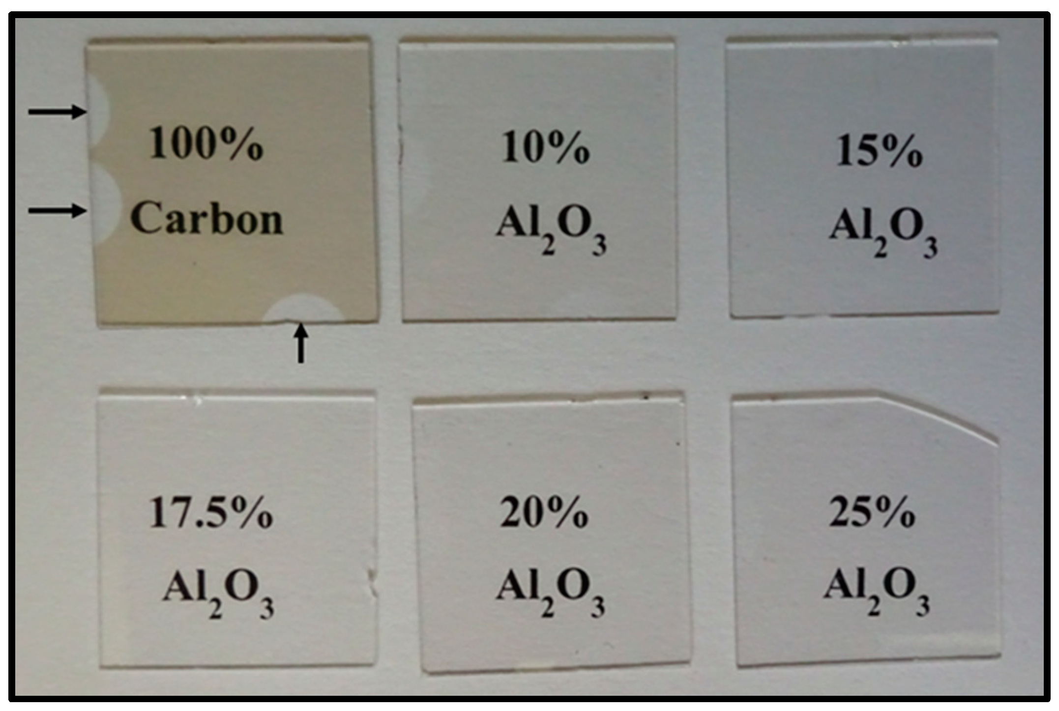
3.1. Visual Inspection

Figure 1 displays representative images of the as-made Al2O3-doped films. Visual inspection revealed that the films were transparent and free of delamination and other defects. Tape tests further substantiated the robust adhesion of these films to the substrate. The undoped carbon sample exhibited a visibly darker appearance compared to the Al2O3-doped films. While a faint brown tint was apparent in the Al2O3-doped films with the lowest dopant concentration, at greater dopant levels this coloration was almost imperceptible.

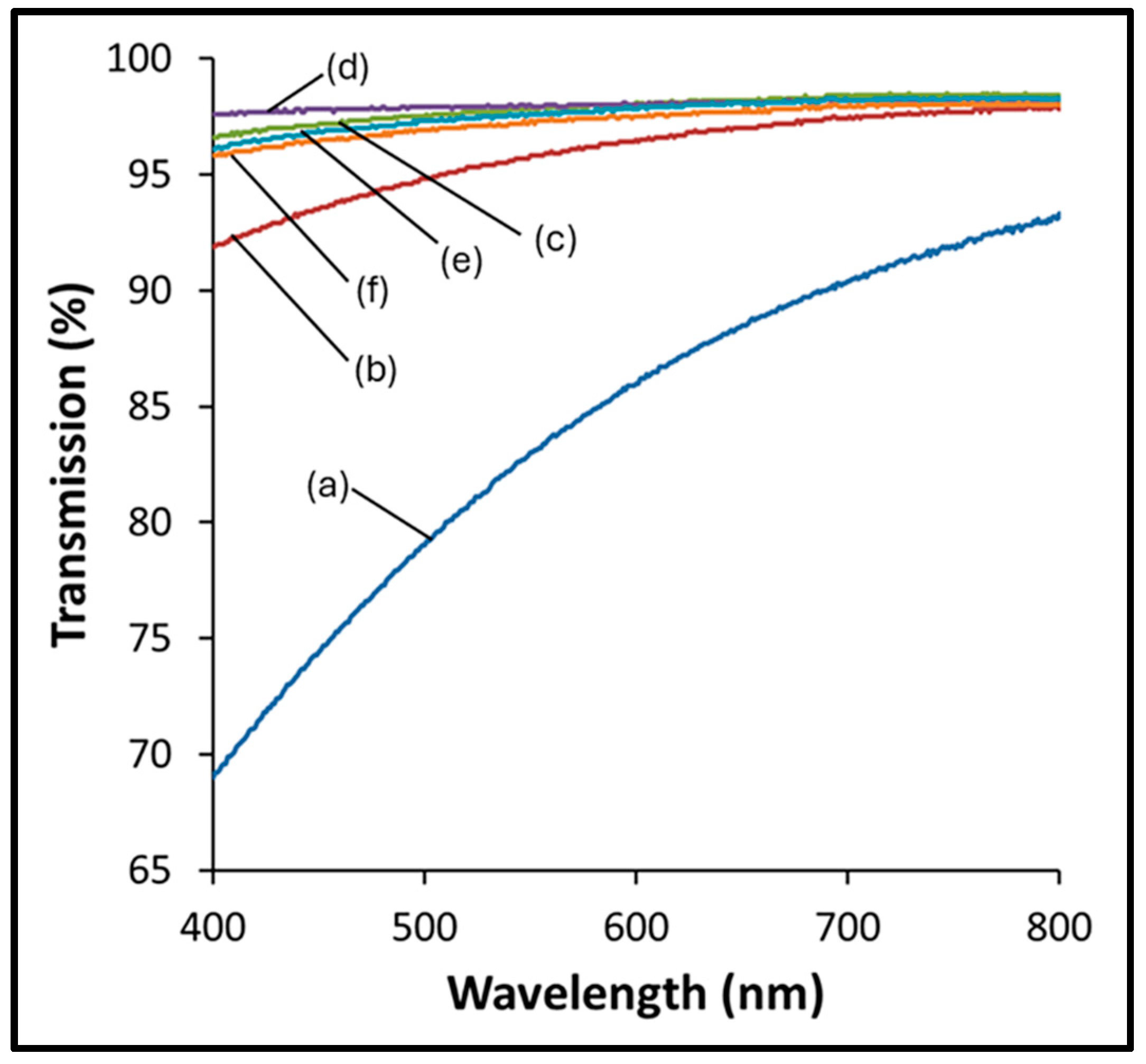
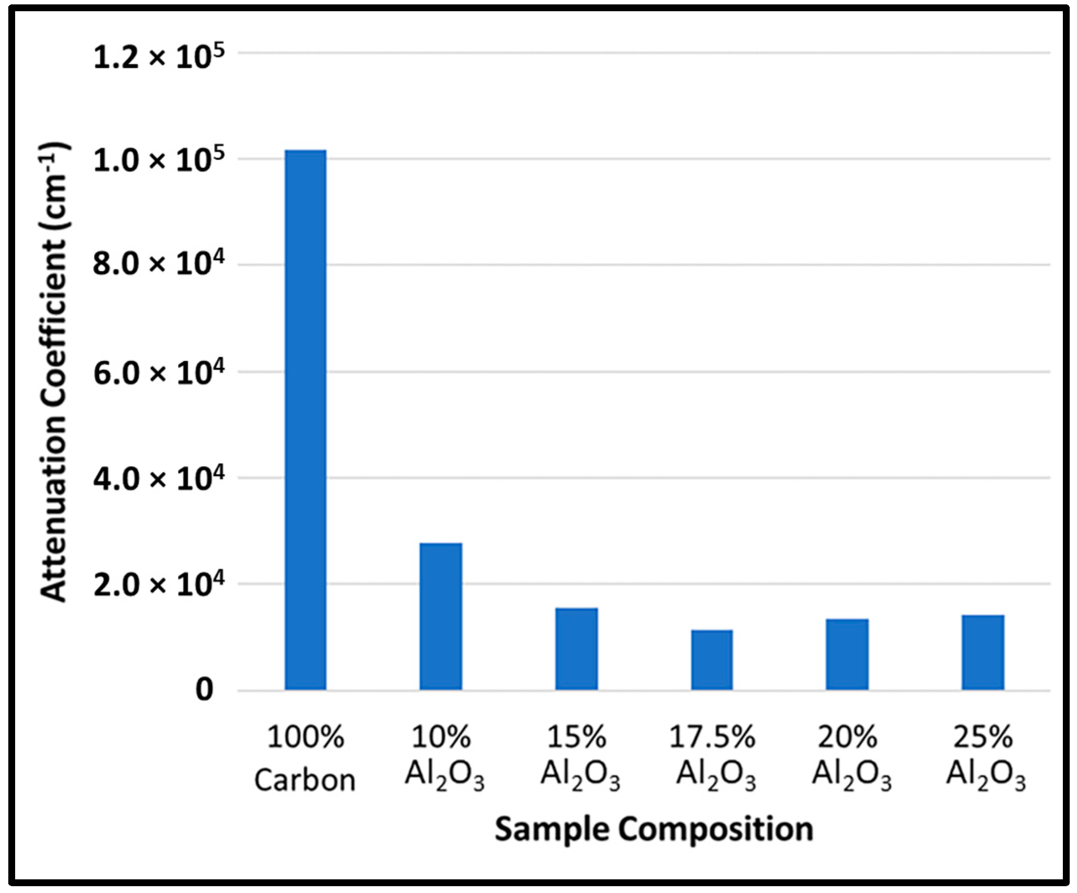
3.2. Film Thickness and Transparency

Figure 2 illustrates the transmission spectra for each film. To facilitate a direct comparison of the films, given their varying thicknesses, attenuation coefficients were calculated at a wavelength of 450 nm. The thicknesses of the doped films, measured using white-light interferometry, were found to range from approximately 30 to 45 nm, as detailed in Table 2. For each of the films, the attenuation coefficients, as shown in Figure 3, were calculated following the methods of Manjunatha and Paul, using the Beer–Lambert law [37]:
I = I0e−αt
where I is the transmitted intensity of light, I0 is the incident intensity, α is the attenuation coefficient, and t is the thickness of the material.
Compared to the Al2O3-doped samples, the undoped DLC sample showed a marked reduction in transparency. Within the doped films, transparency increased significantly as the dopant levels rose, particularly up to 15% Al2O3. Above this level, further increases in dopant concentration resulted in negligible changes in transparency. The changes in transparency indicate that the dopant significantly alters the film structure, as investigated in Section 3.7.

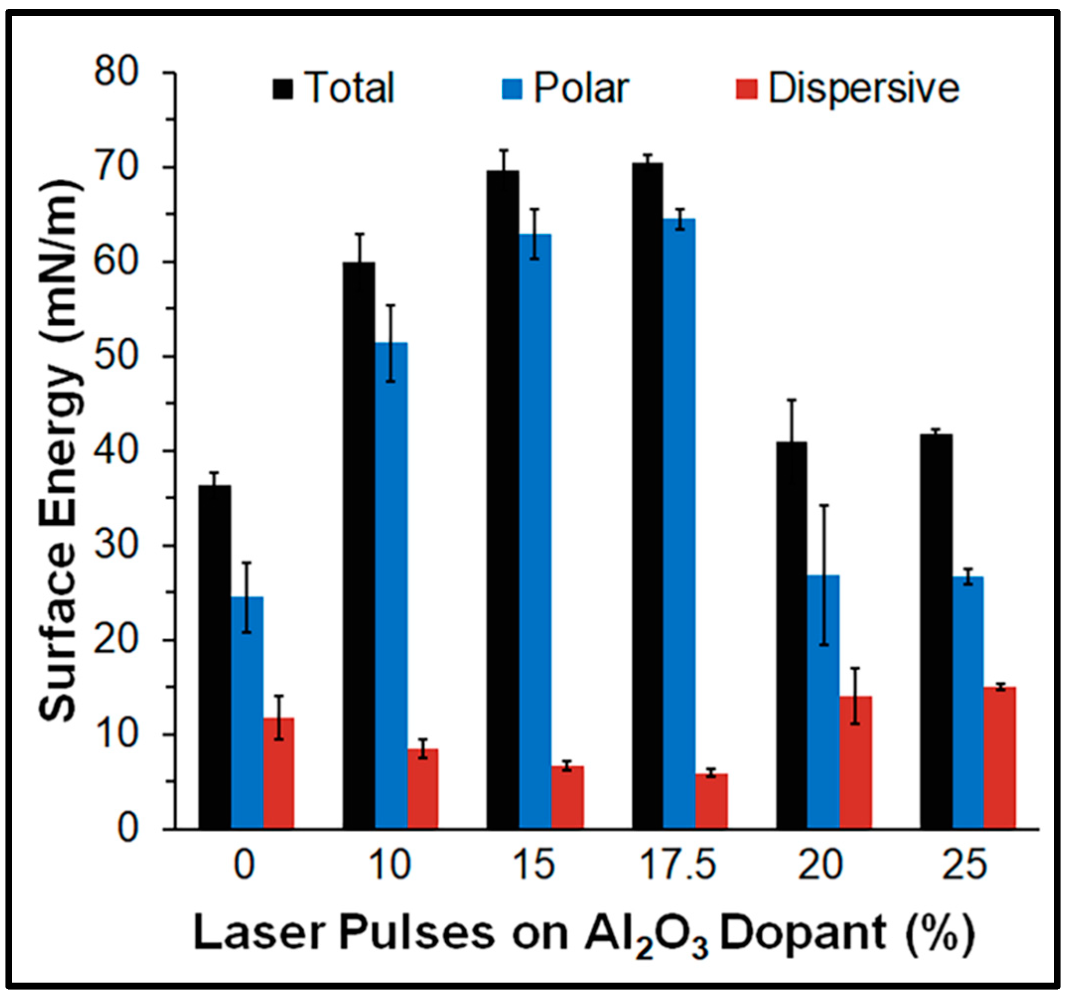
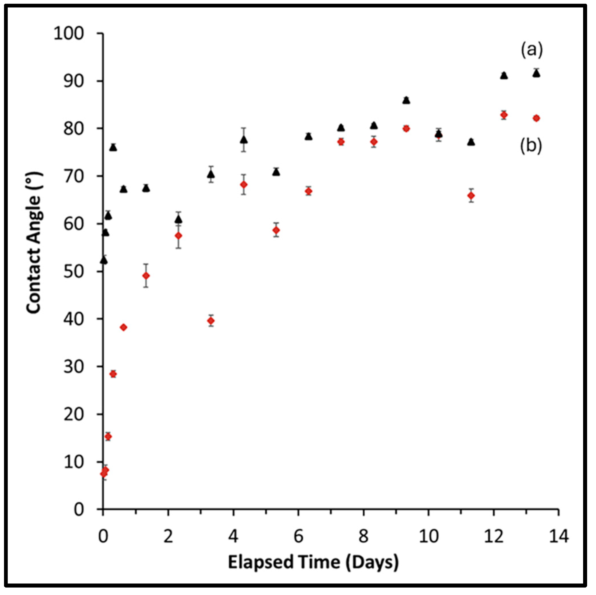
3.3. Water Contact Angle and Surface Energy

A strong correlation was observed between the aluminum oxide dopant concentration and contact angle. As depicted in Figure 4, the water contact angle initially decreased with increasing dopant levels. However, a sharp reversal occurred at 20% dopant, where the contact angle increased from 32° to above 60°. This suggests that 17.5% dopant represents an approximate upper limit for contact angle reduction. The potential influence of film thickness variations on surface morphology, and consequently on the contact angle, is explored in Section 3.5. To gain a deeper understanding of the observed results, surface energy measurements were performed.
The surface energy of the Al2O3-doped films, along with their polar and dispersive components, is shown in Figure 5. Surface energy measurements of the doped films ranged from a maximum of 70 mN/m for the 17.5% Al2O3-doped sample to a minimum of 41 mN/m for the 25% Al2O3-doped sample. With increased dopant levels, up to 17.5% Al2O3, the films’ surface energy increased, primarily due to the polar component, driving the increase in hydrophilicity. However, with doping beyond 17.5% Al2O3, both the overall surface energy and the polar component were greatly reduced, resulting in decreased hydrophilicity.
A comparison of contact angles between the as-made and plasma-cleaned 17.5% Al2O3-doped samples is presented in Figure 6. Plasma cleaning, with measurements taken within 60 min of treatment, resulted in a contact angle reduction from 32° to 8°. This suggests that plasma cleaning can be an effective pre-surgical technique for enhancing the antifogging characteristics of the films.
Figure 7 illustrates the temporal evolution of the contact angles. The plasma-cleaned film displayed an initial average contact angle of 8° within 2 h post-treatment, but this value increased to above 50° within 48 h. Throughout the remaining observation period, both the treated and as-made samples exhibited a significant increase in contact angle, converging to approximately 90° by the study’s conclusion. The initial decrease in contact angle was likely due to the formation of hydroxyl free radicals on the film’s surface, consistent with the findings of Subedi et al. [38]. The observed substantial increase in contact angle for both films was likely due to the adsorption of airborne hydrocarbons and other contaminants onto the film surfaces from the ambient atmosphere [38]. These findings suggest that argon plasma cleaning is not a viable method for achieving durable hydrophilicity in these films, given the observed time-dependent loss of the hydrophilic effect.

3.4. Stability and Biocompatibility

3.4.1. Simulated Body Fluid Studies

Following 40 weeks of immersion in SBF, no delamination or significant structural alterations were observed in either the doped or undoped films. Visual evidence of this stability is presented in Figure 8, which depicts a 10% doped sample before (Figure 8a) and after (Figure 8b) immersion, demonstrating the absence of delamination. This lack of change substantiates the films’ long-term stability.

3.4.2. Assessment of Biocompatibility Using CellTiter-Glo Assays

Figure 9 presents the ex vivo acute toxicity results of Al2O3-doped DLC films, evaluated 24 h following NIH/3T3 cell incubation. No comparisons among the tested samples generated p values equal to or below 0.05. Statistical analysis of the cell viability assay results revealed no significant differences between the experimental films and control media, demonstrating the films’ biocompatibility.

3.4.3. Blood Compatibility Testing

Samples with Al2O3 coatings or the uncoated control substrate did not induce measurable ATP release by blood platelets. The estimated limit of instrument sensitivity for ATP detection under the conditions of our tests was 0.01 nmol, and the measured total ATP in the PRP samples was 2.60 ± 1.92 nmols (n = 3, standard deviation). Thus, the maximum potential ATP release induced by any of the six DLC coatings or the uncoated substrate was less than 0.01 nmol, or 0.4% of the total ATP available for release.
Stimulation of PRP with 100 nM γ-thrombin demonstrated moderate, bi-phasic aggregation and released 1.23 ± 0.21 nmol of ATP [10]. Increasing the γ-thrombin dose to 300 nM induced the release of 1.70 ± 0.47 nmols of ATP and was associated with strong platelet aggregation, demonstrating positive dose–response behavior [10].
ATP release is a characteristic of platelet activation and is an early step in the progression to platelet aggregation and the formation of thrombi. The lack of measurable ATP release induced by the DLC coatings implies that these coatings are unlikely to support platelet activation and aggregation during 60 min of exposure to blood.
The platelet aggregation and ATP release results are consistent with similarly treated controls and SiO-coated materials reported previously [10,39]. The absence of measurable ATP release from platelets in contact with Al2O3 coatings for 60 min suggests that these DLC coatings pose a low risk of triggering coagulation-related adverse events in their intended use as laparoscopic components. The test conditions used in this study promoted platelet activation and aggregation relative to the blood exposure of the DLC coatings in use for a laparoscope. The experimental setup, utilizing a closed system with small PRP volumes, extended the potential contact time between individual platelets and the DLC coating. This contrasts with the intended use in dynamic laparoscopic instruments, which operate with larger blood volumes. Thus, the lack of measurable ATP release under the study conditions provides an additional degree of confidence that Al2O3 coatings will be hemocompatible under the conditions anticipated during laparoscope use.
The biocompatibility results parallel the findings of previous work conducted by the authors investigating SiO-doped DLC films [10].
Based on our cell viability and hemocompatibility results, these coatings are unlikely to induce worrying biocompatibility when used during laparoscopic procedures. Cell viability was tested following 24 h contact with the coatings, and hemocompatibility was tested following 1 h contact with a small volume of blood plasma. Contact of any particular cell or particular platelet with coated lenses will be momentary during laparoscopic procedures and unlikely to induce even the low levels of cytotoxicity/activation observed in vitro.
Our results suggest that antifogging efficacy and biocompatibility are not correlated. The small degree of platelet aggregation and cytotoxicity observed following contact with these coatings (a positive characteristic) complicates the detection of this type of correlation. As a result, the selection of coatings for laparoscopic lenses should be primarily based on antifogging function, transparency, and wear characteristics.

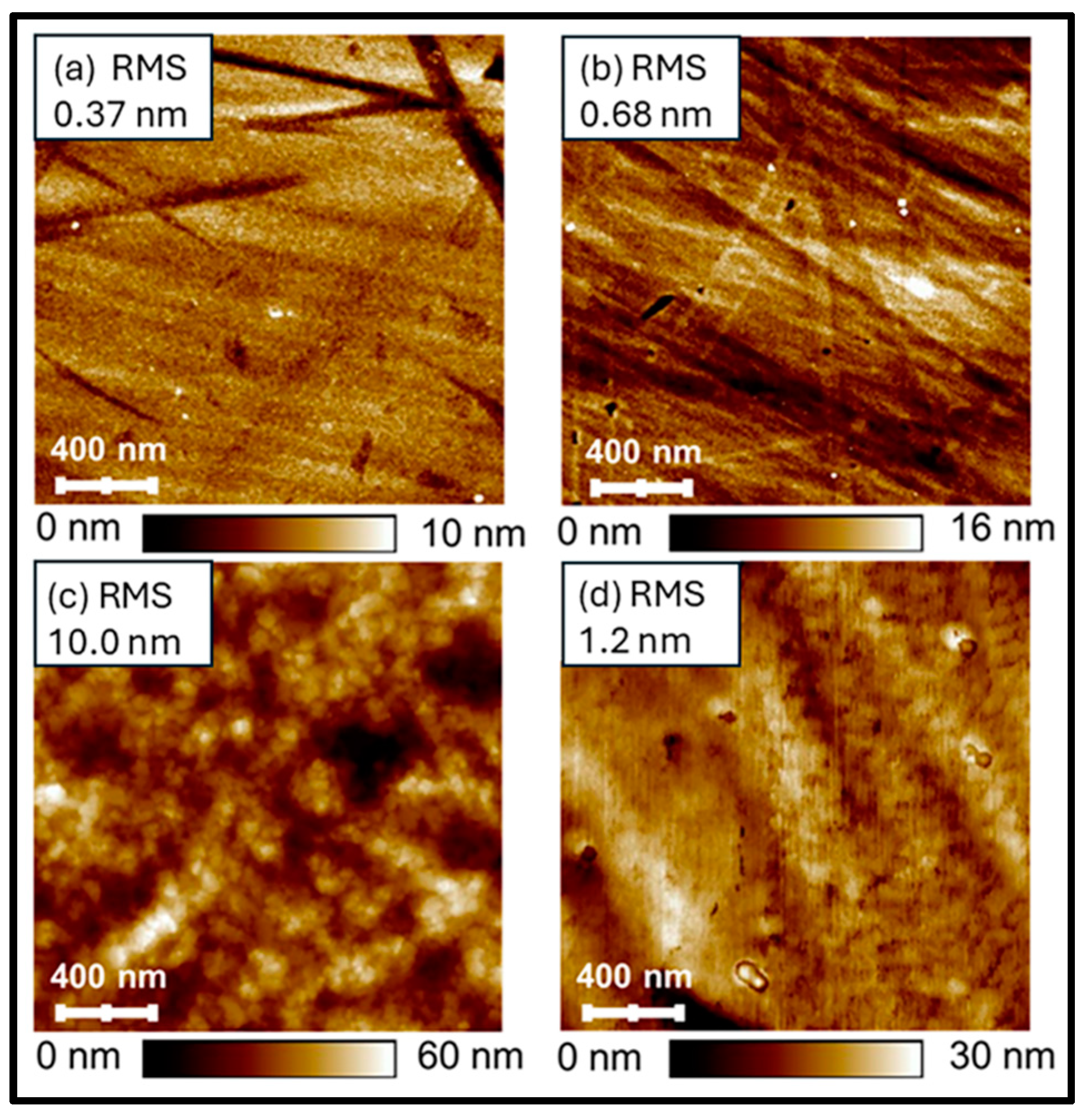
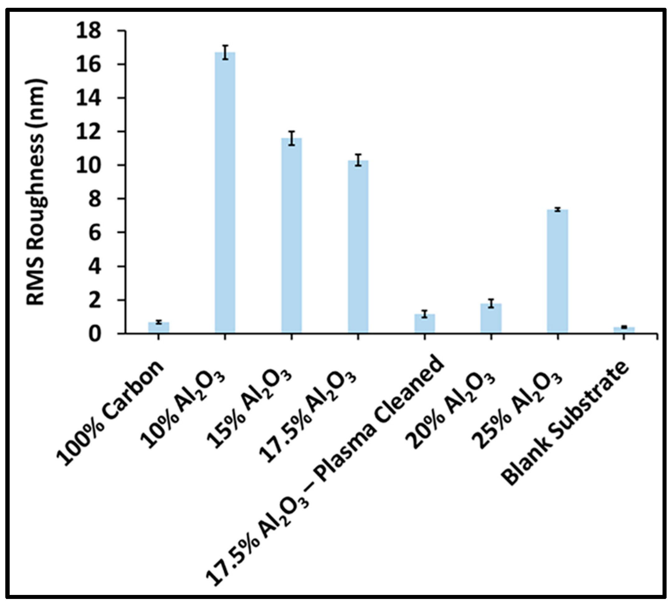
3.5. Surface Roughness and Morphology

Surface roughness is known to influence contact angles, with the contact angle of hydrophilic surfaces decreasing with increased roughness [40]. The surface morphology of the doped and undoped samples is visualized in Figure 10. The blank substrate (Figure 10a) reveals a smooth surface with manufacturing-induced striations, which are also present in the undoped sample (Figure 10b), along with additional surface particulates. The 17.5%-doped sample (Figure 10c) exhibits small, smooth particulates, resulting in a more textured surface. These particulates, with sizes dependent on the dopant levels, were a common feature in all doped films. Plasma cleaning of the 17.5%-doped film (Figure 10d) led to a decrease in the observed small particulates and significantly altered the surface morphology, which may account for the reduced contact angle.
The root-mean-square (RMS) roughness of the films is presented in Figure 11. Surface roughness exhibited minimal variation between the undoped DLC film and the uncoated substrate. In contrast, a substantial increase in surface roughness was observed upon dopant incorporation. While a gradual reduction in surface roughness was generally correlated with increasing dopant concentration, the 20% doped sample deviated from this trend. A change in the PLD target’s condition may have contributed to the variation in roughness. These variations in surface roughness were not correlated with the films’ contact angle, indicating that surface chemistry is the primary influencer of this characteristic.

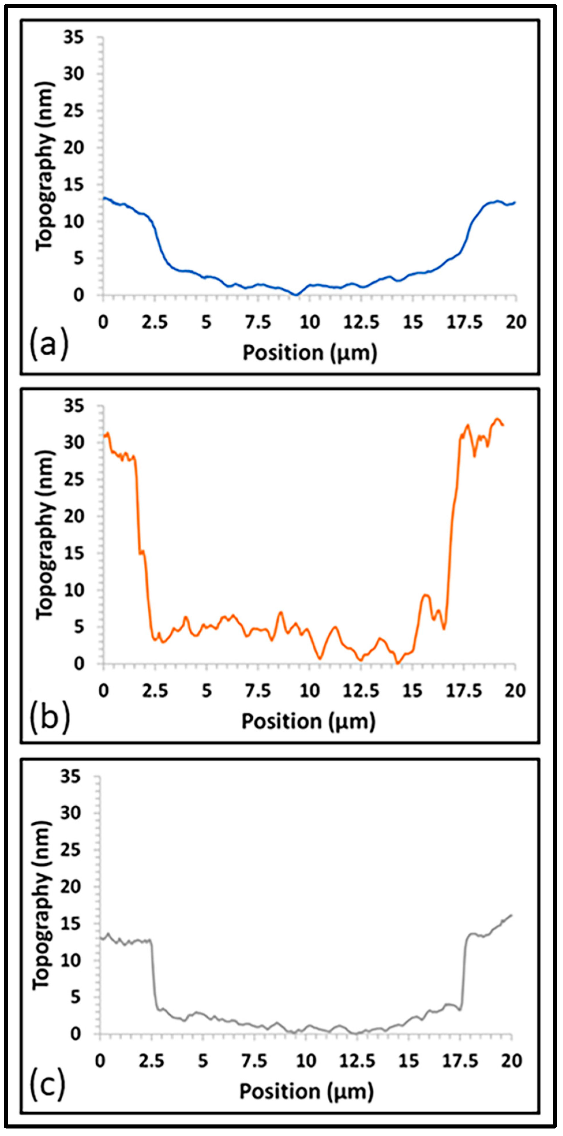
3.6. Wear Properties

The addition of Al2O3 had a content-dependent effect on the wear depth of the films (see Figure 12). For laser pulse percentages on the target of less than 20%, the addition of Al2O3 resulted in deeper average wear depths compared to the non-doped film (17–23 nm vs. 7 nm, respectively). The shape of the wear depth cross-section was also different than that of the non-doped film; films created with 20% alumina pulses or less presented a more square-bottomed cross-sectional profile compared to the non-doped film, which presented a more rounded cross-sectional profile (see Figure 13a,b). The square-bottomed cross-sectional profile, combined with the deeper wear depth, is indicative of the test load moving through the majority of the film’s thickness. However, once the number of laser pulses on the Al2O3 target was at least 20%, the wear depth began to approach that of the non-doped film (13 nm at 20% dopant and 10 nm at 25% dopant), and a more rounded cross-sectional depth profile was seen (see Figure 13c). This suggests that there may be a minimum amount of Al2O3 needed in order to mitigate the influence of the dopant on the mechanical properties of the film. Further investigation is needed into whether an optimal dosage can be determined, with the expectation that the optimal dosage(s) may be application-dependent, given the need to prioritize multiple material properties.

3.7. Raman Spectroscopy

The Raman spectra for the samples are shown in Figure 14. The undoped and 10%-doped samples display typical overlapping D and G peaks for diamond-like carbon deposited at room temperature under vacuum [41,42]. The D peak, typically found at ~1360 cm−1, results from contractions and stretching of carbon–carbon bonds; the G peak, typically occurring at ~1580 cm−1, is caused by carbon–carbon bonds’ in-plane stretching [43]. At 15% dopant inclusion, these D and G peaks narrow and separate [44]. Interestingly, the peaks associated with DLC are not present with Al2O3 doping greater than 15%, demonstrating that a fundamental change in bonding occurs with greater levels of Al2O3 incorporation [29]; this change affects the transparency of the films, as shown in Figure 3.
The relative intensity of the D and G peaks and the full width at half-maximum and position of the G peak can provide insight into the DLC films’ sp3/sp2 ratio of hybrid bonding [45,46,47]. The parameters derived from the deconvoluted D spectra of the undoped and 10%-doped samples are shown in Table 3. With Al2O3 doping, the relative intensity of the D and G peaks increases, the position of the G peak increases, and the full width at half-maximum decreases, indicating decreased sp3 content in the films.
The authors posit that the small feature near 1450 cm−1 in the 17.5%-doped sample was due to increased disorder related to the incorporation of Al2O3 into the film and was associated with the interactions of the Al-C and C=C chain stretching [11]; this feature was less visible in the 15%-doped sample. Amorphous and crystalline Al2O3 bands would be expected below 800 cm−1, which unfortunately would be obscured by the background signal from the fused silica substrate [48,49,50]. In future work, the coatings will be deposited on a substrate more conducive to detecting the amorphous and crystalline Al2O3 bands.

4. Conclusions

For the first time, aluminum oxide-doped diamond-like carbon thin films were synthesized by pulsed laser deposition. The films were produced for the purpose of evaluating their suitability as a transparent, antifogging film for laparoscopes. The doped films demonstrated exceptional transparency over the visible spectrum, achieving an average of up to 98% transmission. Additionally, doping the films increased the hydrophilicity and surface energy of the DLC at lower dopant amounts. A composition-dependent effect on the wear depth of the films was seen, with the wear depths improving with increasing Al2O3 dopant amounts. In future work, synthesis of films with greater Al2O3 content may yield films with the desired transparency and wear properties equal or superior to those of undoped DLC. Introduction of Al2O3 into the DLC films resulted in the contact angle reducing to 33° at its lowest and the surface energy increasing up to 70 mN/m. The contact angles after plasma-cleaning treatments were reduced to 8°; however, time studies demonstrated that the effects of this method of surface treatment are shortlived and diminish rapidly within a few hours. CellTiter-Glo assays and simulated body fluid soaks demonstrated the biocompatibility and biostability of the films, respectively. The amount of ATP released by blood platelets in contact with the DLC coatings was very small, not greater than the limit of instrument detectability, suggesting in vivo hemocompatibility.
Films synthesized with 17.5% Al2O3 doping had the best combination of desirable characteristics, including biocompatibility, contact angle, surface energy, transparency, and wear depth. However, due to their relatively high contact angles, which did not show long-term improvement after plasma treatment, further optimization is required for their use in antifogging applications. Nonetheless, because of their satisfactory initial biocompatibility and biostability results, they do show promise for biomedical applications where a transparent coating is desired.
The authors suggest that, in future work, improvements in hydrophilicity may be explored through various methods, including photochemical oxidation through ultraviolet light exposure and plasma treatment with oxygen [40,51]. Another possibility for improvements in hydrophilicity is the use of a dopant material with a greater ratio of oxygen anions to cations than aluminum oxide (3:2).

Author Contributions

Conceptualization, R.L.L., T.D.G. and J.A.J.; methodology, R.L.L., C.W.B., L.R.B. and T.D.G.; formal analysis, R.L.L., A.B.B., F.X., C.P.H., L.R.B. and T.D.G.; investigation, A.B.B., F.X., C.P.H., S.K.G., P.E.B. and L.R.B.; data curation, R.L.L.; writing—original draft preparation, R.L.L., A.B.B., F.X., C.P.H., L.R.B. and J.A.J.; writing—review and editing, R.L.L., A.B.B., S.K.G., C.W.B., P.E.B., L.R.B., T.D.G. and J.A.J.; project administration, T.D.G. and J.A.J.; funding acquisition, R.L.L., T.D.G. and J.A.J. All authors have read and agreed to the published version of the manuscript.

Funding

This work was funded by the National Institute of Biomedical Imaging and Bioengineering of the National Institutes of Health under award number 1R15EB027449.

Institutional Review Board Statement

The study was conducted in accordance with the Declaration of Helsinki and approved by the Institutional Review Board (or Ethics Committee) of Vanderbilt University (protocol number 111251, dated 28 November 2024).

Informed Consent Statement

Informed consent was obtained from all subjects involved in the study.

Data Availability Statement

The original contributions presented in this study are included in the article. Further inquiries can be directed to the corresponding author.

Acknowledgments

The authors recognize the support from Heidi Hamm, Pharmacology, Ophthalmology and Visual Sciences, and Orthopedics at Vanderbilt University for access to the Chrono-Log Lumi-Aggregometers, consumable supplies and associated laboratory equipment. We also recognize the assistance of Kevin Erreger, Molecular Physiology and Biophysics, and Jackson Cassada, Pharmacology, both at Vanderbilt University, for their invaluable, professional help in carrying out the platelet studies.

Conflicts of Interest

The authors declare no conflict of interest.

References

  1. Dua, A.; Aziz, A.; Desai, S.S.; McMaster, J.; Kuy, S. National Trends in the Adoption of Laparoscopic Cholecystectomy over 7 Years in the United States and Impact of Laparoscopic Approaches Stratified by Age. Minim. Invasive Surg. 2014, 2014, 635461. [Google Scholar] [CrossRef] [PubMed]
  2. Mattingly, A.S.; Chen, M.M.; Divi, V.; Holsinger, F.C.; Saraswathula, A. Minimally Invasive Surgery in the United States, 2022: Understanding Its Value Using New Datasets. J. Surg. Res. 2023, 281, 33–36. [Google Scholar] [CrossRef] [PubMed]
  3. Spaner, S.J.; Warnock, G.L. A brief history of endoscopy, laparoscopy, and laparoscopic surgery. J. Laparoendosc. Adv. Surg. Tech. 1997, 7 Pt A, 369–373. [Google Scholar] [CrossRef]
  4. Drysch, A.; Schmitt, K.; Uribe, B.; Yoon, R.; Okhunov, Z.; Landman, J. Comparative analysis of techniques to prevent laparoscopic fogging. Minim. Invasive Ther. Allied Technol. 2016, 25, 319–322. [Google Scholar] [CrossRef] [PubMed]
  5. Richards, C.; Edwards, J.; Culver, D.; Emori, T.G.; Tolson, J.; Gaynes, R.; National Nosocomial Infections Surveillance (NNIS) System, Centers for Disease Control and Prevention. Does using a laparoscopic approach to cholecystectomy decrease the risk of surgical site infection? Ann. Surg. 2003, 237, 358–362. [Google Scholar] [CrossRef] [PubMed]
  6. Kumar, R.; Jallu, S.; Pasricha, K.; Basumatary, B.; Parmar, B.P.S.; Sahani, A.K. Laparoscopic Lens Defogging: A Review of Methods to Maintain a Clear Operating Field. Indian J. Surg. 2022, 84, 698–706. [Google Scholar] [CrossRef]
  7. Manning, T.G.; Perera, M.; Christidis, D.; Kinnear, N.; McGrath, S.; O’Beirne, R.; Zotov, P.; Bolton, D.; Lawrentschuk, N. Visual Occlusion During Minimally Invasive Surgery: A Contemporary Review of Methods to Reduce Laparoscopic and Robotic Lens Fogging and Other Sources of Optical Loss. J. Endourol. 2017, 31, 327–333. [Google Scholar] [CrossRef]
  8. Nabeel, A.; Al-Sabah, S.K.; Ashrafian, H. Effective cleaning of endoscopic lenses to achieve visual clarity for minimally invasive abdominopelvic surgery: A systematic review. Surg. Endosc. 2022, 36, 2382–2392. [Google Scholar] [CrossRef]
  9. Naguib, S.M.; Abdelaal, Y.A.E.-A.; Khodary, A.R.; Fahmi, K.S.; Abdalla, W.M. Nano-Ceramic Coating: A Novel Idea to Reduce Laparoscope Lens Fogging—Experimental Study. Silicon 2022, 14, 7693–7700. [Google Scholar] [CrossRef]
  10. Leonard, R.L.; Bull, A.B.; Xue, F.; Haycook, C.P.; Gray, S.K.; Bond, C.W.; Bond, P.E.; McDearman, J.C.; Woods, D.P.; Mayfield, J.; et al. Biocompatibility of antifogging SiO-doped Diamond-Like carbon laparoscope coatings. Appl. Surf. Sci. 2023, 634, 157606. [Google Scholar] [CrossRef]
  11. Leonard, R.L.; Terekhov, A.Y.; Thompson, C.; Erck, R.A.; Johnson, J.A. Antifog coating for bronchoscope lens. Surf. Eng. 2012, 28, 468–472. [Google Scholar] [CrossRef]
  12. Leonard, R.L.; Hasan, S.A.; Terekhov, A.Y.; Thompson, C.; Erck, R.A.; Dickerson, J.H.; Johnson, J.A. Protective coatings for enhanced performance in biomedical applications. Surf. Eng. 2012, 28, 473–479. [Google Scholar] [CrossRef]
  13. Ohtake, N.; Hiratsuka, M.; Kanda, K.; Akasaka, H.; Tsujioka, M.; Hirakuri, K.; Hirata, A.; Ohana, T.; Inaba, H.; Kano, M.; et al. Properties and Classification of Diamond-Like Carbon Films. Materials 2021, 14, 315. [Google Scholar] [CrossRef] [PubMed]
  14. Sánchez-López, J.C.; Fernández, A. Doping and Alloying Effects on DLC Coatings. In Tribology of Diamond-Like Carbon Films: Fundamentals and Applications; Donnet, C., Erdemir, A., Eds.; Springer: Boston, MA, USA, 2008; pp. 311–338. [Google Scholar]
  15. Zhang, M.; Xie, T.; Qian, X.; Zhu, Y.; Liu, X. Mechanical Properties and Biocompatibility of Ti-doped Diamond-like Carbon Films. ACS Omega 2020, 5, 22772–22777. [Google Scholar] [CrossRef] [PubMed]
  16. Lu, Y.M.; Huang, G.J.; Wang, S.; Mi, C.W.; Wei, S.F.; Tian, F.T.; Li, W.; Cao, H.Y.; Cheng, Y. A review on diamond-like carbon films grown by pulsed laser deposition. Appl. Surf. Sci. 2021, 541, 12. [Google Scholar] [CrossRef]
  17. Antuzevics, A.; Elsts, E.; Kemere, M.; Lushchik, A.; Moskina, A.; Scherer, T.A.; Popov, A.I. Thermal annealing of neutron irradiation generated paramagnetic defects in transparent Al2O3 ceramics. Opt. Mater. 2023, 135, 6. [Google Scholar] [CrossRef]
  18. Deng, N.M.; Zhao, J.; Yang, L.L.; Zheng, Z.Q. Effects of Brazing Technology on Hermeticity of Alumina Ceramic-Metal Joint Used in Nuclear Power Plants. Front. Mater. 2021, 7, 7. [Google Scholar] [CrossRef]
  19. Houska, J.; Blazek, J.; Rezek, J.; Proksova, S. Overview of optical properties of Al2O3 films prepared by various techniques. Thin Solid Film. 2012, 520, 5405–5408. [Google Scholar] [CrossRef]
  20. Molla, J.; Heidinger, R.; Ibarra, A. Alumina ceramics for heating-systems. J. Nucl. Mater. 1994, 212, 1029–1034. [Google Scholar] [CrossRef]
  21. Merola, M.; Affatato, S. Materials for Hip Prostheses: A Review of Wear and Loading Considerations. Materials 2019, 12, 495. [Google Scholar] [CrossRef]
  22. Rahmati, M.; Mozafari, M. Biocompatibility of alumina-based biomaterials–A review. J. Cell. Physiol. 2019, 234, 3321–3335. [Google Scholar] [CrossRef] [PubMed]
  23. Spajic, I.; Rodi, P.; Sekularac, G.; Lekka, M.; Fedrizzi, L.; Milosev, I. The effect of surface preparation on the protective properties of Al2O3 and HfO2 thin films deposited on cp-titanium by atomic layer deposition. Electrochim. Acta 2021, 366, 16. [Google Scholar] [CrossRef]
  24. Cheng, Y.; Lu, Y.; Huang, G.; Guo, Y.; Xi, L.; Wang, S.; Tian, F. Infrared and mechanical properties of the oxygen-doped non-hydrogenated diamond-like carbon film prepared by pulsed laser deposition. Infrared Phys. Technol. 2019, 102, 102983. [Google Scholar] [CrossRef]
  25. Safaie, P.; Eshaghi, A.; Bakhshi, S.R. Structure and mechanical properties of oxygen doped diamond-like carbon thin films. Diam. Relat. Mater. 2016, 70, 91–97. [Google Scholar] [CrossRef]
  26. Yoshitake, T.; Nishiyama, T.; Nagayama, K. The role of hydrogen and oxygen gas in the growth of carbon thin films by pulsed laser deposition. Diam. Relat. Mater. 2000, 9, 689–692. [Google Scholar] [CrossRef]
  27. Bouabibsa, I.; Lamri, S.; Sanchette, F. Structure, Mechanical and Tribological Properties of Me-Doped Diamond-Like Carbon (DLC) (Me = Al, Ti, or Nb) Hydrogenated Amorphous Carbon Coatings. Coatings 2018, 8, 370. [Google Scholar] [CrossRef]
  28. Ghadai, R.K.; Shanmugasundar, G.; Cepova, L.; Das, S.; Kumar Mahto, P.; Kalita, K. A temperature-based synthesis and characterization study of aluminum-incorporated diamond-like carbon thin films. Front. Mech. Eng. 2023, 9, 1325040. [Google Scholar] [CrossRef]
  29. Peckus, D.; Meškinis, Š.; Vasiliauskas, A.; Rajackaitė, E.; Andrulevičius, M.; Kopustinskas, V.; Tamulevičius, S. Structure and optical properties of diamond like carbon films containing aluminium and alumina. Appl. Surf. Sci. 2020, 529, 147040. [Google Scholar] [CrossRef]
  30. Voevodin, A.A.; O’Neill, J.P.; Zabinski, J.S. Nanocomposite tribological coatings for aerospace applications. Surf. Coat. Technol. 1999, 116–119, 36–45. [Google Scholar] [CrossRef]
  31. ISO 10993-4:2017; Biological Evaluation of Medical Devices, in: Part 4: Selection of Tests for Interactions with Blood. International Organization for Standardization: Geneva, Switzerland, 2017.
  32. Jung, F.; Braune, S.; Lendlein, A. Haemocompatibility testing of biomaterials using human platelets. Clin. Hemorheol. Microcirc. 2013, 53, 97–115. [Google Scholar] [CrossRef]
  33. ASTM D3359-23; Standard Test Methods for Rating Adhesion by Tape Test. ASTM International: West Conshohocken, PA, USA, 2023.
  34. Panzer, J. Components of solid surface free energy from wetting measurements. J. Colloid Interface Sci. 1973, 44, 142–161. [Google Scholar] [CrossRef]
  35. Fernández, V.; Khayet, M. Evaluation of the surface free energy of plant surfaces: Toward standardizing the procedure. Front. Plant Sci. 2015, 6, 510. [Google Scholar] [CrossRef]
  36. Cho, S.-B.; Nakanishi, K.; Kokubo, T.; Soga, N.; Ohtsuki, C.; Nakamura, T.; Kitsugi, T.; Yamamuro, T. Dependence of Apatite Formation on Silica Gel on Its Structure: Effect of Heat Treatment. J. Am. Ceram. Soc. 2005, 78, 1769–1774. [Google Scholar] [CrossRef]
  37. Nama Manjunatha, K.; Paul, S. Investigation of optical properties of nickel oxide thin films deposited on different substrates. Appl. Surf. Sci. 2015, 352, 10–15. [Google Scholar] [CrossRef]
  38. Yi, J.W.; Moon, M.-W.; Ahmed, S.F.; Kim, H.; Cha, T.-G.; Kim, H.-Y.; Kim, S.-S.; Lee, K.-R. Long-Lasting Hydrophilicity on Nanostructured Si-Incorporated Diamond-Like Carbon Films. Langmuir 2010, 26, 17203–17209. [Google Scholar] [CrossRef] [PubMed]
  39. Miller, C.H.; Rice, A.S.; Garrett, K.; Stein, S.F. Gender, race and diet affect platelet function tests in normal subjects, contributing to a high rate of abnormal results. Br. J. Haematol. 2014, 165, 842–853. [Google Scholar] [CrossRef] [PubMed]
  40. Nagashima, S.; Hasebe, T.; Kamijo, A.; Yoshimoto, Y.; Hotta, A.; Morita, H.; Terada, H.; Tanaka, M.; Takahashi, K.; Suzuki, T. Effect of oxygen plasma treatment on non-thrombogenicity of diamond-like carbon films. Diam. Relat. Mater. 2010, 19, 861–865. [Google Scholar] [CrossRef]
  41. Cappelli, E.; Scilletta, C.; Mattei, G.; Valentini, V.; Orlando, S.; Servidori, M. Critical role of laser wavelength on carbon films grown by PLD of graphite. Appl. Phys. A 2008, 93, 751–758. [Google Scholar] [CrossRef]
  42. Jadhav, H.; Singh, A.K.; Sinha, S. Pulsed laser deposition of graphite in air and in vacuum for field emission studies. Mater. Chem. Phys. 2015, 162, 279–285. [Google Scholar] [CrossRef]
  43. Ferrari, A.C. Determination of bonding in diamond-like carbon by Raman spectroscopy. Diam. Relat. Mater. 2002, 11, 1053–1061. [Google Scholar] [CrossRef]
  44. Pang, H.; Wang, X.; Zhang, G.; Chen, H.; Lv, G.; Yang, S. Characterization of diamond-like carbon films by SEM, XRD and Raman spectroscopy. Appl. Surf. Sci. 2010, 256, 6403–6407. [Google Scholar] [CrossRef]
  45. Balon, F.; Stolojan, V.; Silva, S.R.P.; Michalka, M.; Kromka, A. Diamond-like carbon thin films for high-temperature applications prepared by filtered pulsed laser deposition. Vacuum 2005, 80, 163–167. [Google Scholar] [CrossRef]
  46. Chhowalla, M.; Ferrari, A.C.; Robertson, J.; Amaratunga, G.A.J. Evolution of sp2 bonding with deposition temperature in tetrahedral amorphous carbon studied by Raman spectroscopy. Appl. Phys. Lett. 2000, 76, 1419–1421. [Google Scholar] [CrossRef]
  47. Ding, X.; Li, Q.; Kong, X. Effect of repetition rates of laser pulses on microstructure and optical properties of diamond-like carbon films. Int. J. Mod. Phys. B—IJMPB 2009, 23, 5671–5681. [Google Scholar]
  48. Lan, Y.; Zou, Y.; Ma, X.; Xu, L.; Shi, L.; Zhang, J. Fabrication of amorphous Al2O3 optical film with various refractive index and low surface roughness. Mater. Res. Express 2020, 7, 086405. [Google Scholar] [CrossRef]
  49. Solonenko, D.; Žukauskaitė, A.; Pilz, J.; Moridi, M.; Risquez, S. Raman Spectroscopy and Spectral Signatures of AlScN/Al2O3. Micromachines 2022, 13, 1961. [Google Scholar] [CrossRef] [PubMed]
  50. Ullah, I.; Tamseel, M.; Amami, M.; Javed, M.R.; Javaid, K.; Mahmood, K.; Ikram, S.; Ali, A.; Amin, N.; Hussain, M.S.; et al. Growth and characterization of Ag–Al2O3 composites thin films for thermoelectric power generation applications. Ceram. Int. 2022, 48, 3647–3651. [Google Scholar] [CrossRef]
  51. Jalvo, B.; Santiago-Morales, J.; Romero, P.; Guzman de Villoria, R.; Rosal, R. Microbial colonisation of transparent glass-like carbon films triggered by a reversible radiation-induced hydrophobic to hydrophilic transition. RSC Adv. 2016, 6, 50278–50287. [Google Scholar] [CrossRef]
Figure 1. Undoped and Al2O3-doped DLC samples. Note: The black arrows highlight witness marks—areas of uncoated substrate—on the 100% carbon sample. These marks, caused by mounting hardware during deposition, were observed on all samples. The transparency of the films is illustrated by the underlying text.
Coatings 15 00437 g001
Figure 2. Transmission spectra of the DLC films as a function of laser pulses applied to the Al2O3 dopant material: (a) 100% carbon, (b) 10% dopant, (c) 15% dopant, (d) 17.5% dopant, (e) 20% dopant, and (f) 25% dopant.
Coatings 15 00437 g002
Figure 3. Attenuation coefficients of Al2O3-doped DLC films calculated at the 450 nm wavelength.
Coatings 15 00437 g003
Figure 4. The effect of Al2O3 dopant on average water contact angle. Note: Error bars are included for each data point, but their small size makes them difficult to observe.
Coatings 15 00437 g004
Figure 5. The effect of Al2O3 dopant on total surface energy, polar component, and dispersive component.
Coatings 15 00437 g005
Figure 6. Representative water droplet profiles on 17.5% Al2O3-doped films: (a) An as-made film and (b) a plasma-cleaned film, imaged 60 min after treatment. Note: The yellow dashed line indicates the baseline for each measurement.
Coatings 15 00437 g006
Figure 7. Temporal evolution of average contact angles for plasma-cleaned and as-made 17.5% Al2O3-doped films over 14 days: (a) As-made and (b) argon plasma-cleaned. Note: Error bars are included for each data point, but their small size makes them difficult to observe.
Coatings 15 00437 g007
Figure 8. Photographs of a 10% Al2O3-doped DLC film: (a) before soaking and (b) after forty weeks of soaking in SBF.
Coatings 15 00437 g008
Figure 9. Average NIH 3T3 cell viability, normalized to media controls, following 24 h incubation with the DLC films (n = 5 technical replicates). The data’s significance was evaluated via ordinary one-way ANOVA with Dunnett’s multiple comparison test.
Coatings 15 00437 g009
Figure 10. AFM results for Al2O3-doped films: (a) uncoated substrate, (b) 0% dopant, (c) 17.5% dopant, and (d) plasma-cleaned 17.5% dopant.
Coatings 15 00437 g010
Figure 11. Root-mean-square roughness of Al2O3-doped films from AFM measurements.
Coatings 15 00437 g011
Figure 12. Average wear depth as a function of laser pulses on the Al2O3 dopant.
Coatings 15 00437 g012
Figure 13. Cross-sectional profile of wear depth for the (a) undoped film, (b) film with 15% Al2O3 dopant, and (c) film with 25% Al2O3 dopant.
Coatings 15 00437 g013
Figure 14. Raman spectra of DLC films as a function of laser pulses applied to the dopant: (a) 100% carbon, (b) 10% Al2O3, (c) 15% Al2O3, and (d) 17.5% Al2O3. Note: The spectra are normalized. For the samples with 20% and 25% pulses on the Al2O3 target, no appreciable signal was detected above the amorphous fused silica background.
Coatings 15 00437 g014
Table 1. DLC sample variations as a function of laser pulses and target.
Sample NamePulses on Dopant (%)Pulses on CarbonPulses on Al2O3
ABB025N/A400,000N/A
ABB01910360,00040,000
ABB02015340,00060,000
ABB02317.5330,00070,000
ABB02120320,00080,000
ABB02225300,000100,000
Table 2. Effect of the variation in the proportion of laser pulses on the Al2O3 target portion on sample thickness.
Sample NameTotal PulsesPulses on Dopant (%)Thickness (nm)
ABB025400,000None45.0
ABB019400,0001029.6
ABB020400,0001541.0
ABB023400,00017.537.4
ABB021400,0002033.0
ABB022400,0002531.8
Table 3. Parameters derived from the deconvoluted Raman spectra of the undoped and 10%-doped samples.
Pulses on Dopant (%)ID/IGG-Peak PositionFWHM G-Peak
00.45 ± 0.041555 ± 1217 ± 2
10 0.89 ± 0.031562 ± 1150 ± 1
Disclaimer/Publisher’s Note: The statements, opinions and data contained in all publications are solely those of the individual author(s) and contributor(s) and not of MDPI and/or the editor(s). MDPI and/or the editor(s) disclaim responsibility for any injury to people or property resulting from any ideas, methods, instructions or products referred to in the content.

