Article

Bioactive Hydroxyapatite–Carboplatin–Quercetin Coatings for Enhanced Osteointegration and Antitumoral Protection in Hip Endoprostheses

by
Gheorghe Iosub
1,
Dana-Ionela Tudorache (Trifa)
2,
Ionuț Marinel Iova
2,
Liviu Duta
3,
Valentina Grumezescu
3,
Alexandra Cătălina Bîrcă
2,
Adelina-Gabriela Niculescu
2,4,
Paul Cătălin Balaure
5,*,
Ionela Cristina Voinea
6,
Miruna S. Stan
6,
Dragoș Mihai Rădulescu
1,
Adrian Emilian Bădilă
1,
Bogdan Ștefan Vasile
2,
Alexandru Mihai Grumezescu
2,4 and
Adrian Radu Rădulescu
1
1
Faculty of Medicine, Carol Davila University of Medicine and Pharmacy, 8 Eroii Sanitari Street, 050474 Bucharest, Romania
2
Department of Science and Engineering of Oxide Materials and Nanomaterials, University Politehnica of Bucharest, 1-7 Gh. Polizu Street, 060042 Bucharest, Romania
3
Lasers Department, National Institute for Laser, Plasma and Radiation Physics, 077125 Magurele, Romania
4
Research Institute of the University of Bucharest—ICUB, University of Bucharest, 90-92 Panduri, 050663 Bucharest, Romania
5
Organic Chemistry Department, University Politehnica of Bucharest, 1-7 Gh. Polizu Street, 060042 Bucharest, Romania
6
Department of Biochemistry and Molecular Biology, Faculty of Biology, University of Bucharest, 91-95 Splaiul Independentei, 050095 Bucharest, Romania
*
Author to whom correspondence should be addressed.
Coatings 2025, 15(4), 489; https://doi.org/10.3390/coatings15040489
Submission received: 15 March 2025 / Revised: 17 April 2025 / Accepted: 17 April 2025 / Published: 20 April 2025
(This article belongs to the Special Issue Synthesis and Applications of Bioactive Coatings)

Abstract

:
The recurrence of bone cancer poses severe complications, particularly after orthopedic surgery, necessitating advanced biomaterials with dual functionality. This study develops nanostructured coatings composed of hydroxyapatite, carboplatin, and quercetin, designed to enhance bone regeneration while delivering localized cancer therapy. These coatings present a promising solution for hip endoprostheses, addressing osteointegration and tumor recurrence prevention simultaneously. Hydroxyapatite was synthesized and characterized using XRD, TEM, SAED, FTIR, and SEM to assess crystallinity, surface morphology, and functional groups. The coatings were obtained by MAPLE. In vitro biocompatibility tests showed that HAp@CPT and HAp@CPT/QUE coatings supported osteoblast viability and adhesion while exhibiting selective cytotoxic effects on osteosarcoma cells. The Griess assay indicated that nitric oxide (NO) levels remained unchanged in hFOB osteoblasts, confirming that neither coating induced inflammatory responses in healthy cells. In contrast, MG63 osteosarcoma cells exhibited significantly elevated NO levels (p < 0.05) in response to HAp@CPT/QUE, suggesting increased oxidative stress. MTT assay results showed a 12% and 28% reduction in osteosarcoma cell viability for HAp@CPT and HAp@CPT/QUE, respectively. Phase-contrast microscopy further confirmed strong osteoblast adhesion and reduced osteosarcoma attachment, particularly on HAp@CPT/QUE surfaces. These findings highlight the dual functionality of hydroxyapatite–carboplatin–quercetin coatings, promoting osteointegration while exerting localized anticancer effects. Their bone-regenerative and selective cytotoxic properties make them a promising material for hip endoprostheses in oncological orthopedic applications.

1. Introduction

Osteosarcoma is a rare form of cancer that evolves in bone and is characterized by the production of malignant osteoids by cancer cells, which can expand into adjacent soft tissue. The tumor grade determines the treatment option and might also be a combination of surgery, radiation therapy, and multi-agent chemotherapy [1,2,3,4].
Concerning the clinical symptoms of bone cancer, there are various types of hip pain, usually in the inguinal part or the greater iliac area and also reduced mobility in the patients. Regarding this, the principles of surgery are to bring back functions and reduce pain. Orthopedic surgery heavily relies on the use of medical implants, which are made of biopolymers, bioceramics, and composite biomaterials, but especially from metal, because they have the mechanical strength necessary to enable support [5,6,7,8]. While there is postoperative success in most cases, there are some situations where side effects can appear. Aggravation factors can be enumerated as excessive bleeding, the formation of post-surgical clots, postoperative infection, and osteoconductive characteristics [9,10,11,12,13].
An additional challenge that can arise a long time after the surgical procedure is the regression of the bone tumor, which is presented as a calcified soft tissue nodule placed in or on the proximal operative bed. To diminish these two additional challenges—namely, regression and osteointegration issues—additional coated layers on the surfaces of metallic implants may serve as a potential solution [12,13,14,15,16,17].
Hydroxyapatite (HAp), with the chemical formula Ca10(PO4)6(OH)2, represents a calcium phosphate compound, exhibiting both chemical and structural similarities to phosphate systems found in bone [18,19,20]. Considering this significant characteristic, the additional HAp coatings on the metal implants play an important role in the osteointegration process due to biocompatibility, bioactivity, and osteoconductive properties and promote implant fixation [21,22,23,24,25]. Recently, researchers started tailoring hydroxyapatite-coated implant materials’ structural, physicochemical, and adhesive properties by functionalizing the conventional HAp [26]. The material’s mechanical strength and biological activity can be enhanced by incorporating an optimal proportion of HAp in magnesium phosphate cement. Magnesium phosphate cement has gained much interest due to its good mechanical resistance, high dissolution rates, good fixation of bone fragments, and bone ingrowth acceleration [24,27,28].
Besides their multiple properties, calcium phosphates have the potential to be used as an antitumoral drug carrier. Carboplatin, a key platinum-based chemotherapeutic agent, is known for its efficacy in various cancer-type treatments, including osteosarcoma, but it can have a negative impact by decreasing antioxidant protection, which causes skeletal muscle dysfunction and increases the susceptibility of bones to metastatic lesions [29,30,31,32,33,34].
Quercetin (3,3′,4′,5,7-pentahydroxyflavone) is a polyphenolic flavonoid known for its antioxidant, anticancer, antibacterial/antifungal, and anti-inflammatory activities. Regarding the application of quercetin in bone cancer, this flavonoid positively influences bone generation by promoting bone formation, reducing oxidative stress and inflammation, and supporting blood vessel growth. Also, this compound possesses chemo-preventive properties, an advantage that can be used to treat cancer either independently or synergically [35,36,37,38,39,40,41,42].
Because the principal objective of bioactive surface coating on metallic implants is to achieve the desirable characteristics mentioned above, various techniques are available. Conventional techniques, such as sol-gel, electrospinning, or thermal spray techniques, present several disadvantages, such as a slow process speed [43,44,45], the limitation of complicated objects [46], high operating temperature [47], brittleness [48], low production effectiveness [49], and uniformity [50]. In contrast, matrix-assisted pulsed laser evaporation (MAPLE) is an efficient method for thin film coats of organic materials, like pharmaceuticals, biomolecules, or polymers [51,52,53].
The aim of this study was to develop nanostructured coatings that enhance osteointegration, prevent bone cancer recurrence, and provide protective effects against potential cellular damage. To achieve this, hydroxyapatite was combined with carboplatin and quercetin and deposited using the MAPLE technique, which is well-known for the stoichiometric transfer of very sensitive, organic molecules [54]. This ensured the integration of osteoconductive, antitumoral, and antioxidant properties. These coatings are designed for hip endoprostheses, offering a dual therapeutic approach by simultaneously promoting bone regeneration and delivering localized cancer therapy.

2. Materials and Methods

2.1. Materials

All reagents employed in the experiments were acquired from Sigma-Aldrich/Merck (Darmstadt, Germany), were of analytical grade, and did not require further purification before use.

2.2. Methods

2.2.1. Synthesis of Hydroxyapatite Nanoparticles

Nano-hydroxyapatite was obtained using a classical synthesis method. The co-precipitation technique was utilized to obtain nano-hydroxyapatite. Namely, two solutions were prepared; solution A was obtained by dissolving 11 g CaCl2 in a total volume of 200 mL. Solution B was prepared by adding 10.6 g of Na2HPO4 2 H2O in 200 mL ultrapure water. Both solutions were stirred on a magnetic plate for 10 and 15 min at room temperature. After this, solution B was dripped into the CaCl2 solution. To obtain a stable HAp, a 3 M NaOH solution was added until the pH value was adjusted to 9.5 intermittently for 12 h. The resulting white precipitate was filtered, washed several times with ultrapure water, dried at room temperature for 48 h, and then milled.

2.2.2. MAPLE Deposition of Hydroxyapatite-Based Coatings

The focused radiation of a KrF* (λ = 248 nm, τFWHM = 25 ns) excimer laser source (COMPexPro 205, Lambda Physics-Coherent, Göttingen, Germany) impinged the frozen targets at an angle of 45°. The layers were grown by applying 90,000 subsequent laser pulses at varying laser fluences of 200, 300, and 400 mJ/cm2 and a repetition rate of 20 Hz. The laser spot area was set at 30 mm2. The depositions were carried out at room temperature with a background pressure of 0.1 Pa and a target substrate separation distance of 4 cm. For each HAp system, two different solutions (i.e., pristine HAp@CPT, 3:1 wt% and HAp@CPT/QUE, 3:1:1 wt%) with a 2.5% concentration were prepared by mixing the suspensions in DMSO. Subsequently, solid targets were obtained by freezing the HAp mixtures at liquid nitrogen temperature. All coatings were synthesized onto both Si (100) 10 × 10 mm2 and titanium substrates (Φ = 12 mm, thickness 1.5 mm). Prior to introduction into the deposition chamber, the substrates were successively cleaned following a three-step protocol implemented in our laboratory: acetone, ethylic alcohol, and deionized water, for 15 min each.

2.3. Powder and Coatings Investigations

The hydroxyapatite powder’s crystallinity and phase properties were analyzed using an X-ray diffraction (XRD) system using a Shimadzu XRD 6000 diffractometer model (Duisburg, Germany). The experimental determinations were performed at room temperature with 2θ Bragg angle intervals values between 10°–80°, using Cu Kα radiation with λ = 1.5406 Å, at a current of 15 mA and a voltage of 30 kV.
The TEM images were obtained using a TecnaiTM G2 G2 F30 S-TWIN, equipped with a Selected Area Electron Diffraction (SAED), purchased from FEI (Hillsboro, OR, USA). The microscope’s transmission mode operated at a voltage of 300 kV, with guaranteed point and line resolutions of 2 Å and 1 Å, respectively. A small amount of the powdered sample was dispersed in pure ethanol and subjected to ultrasound treatment for 15 min to obtain transmission electron microscopy (TEM) images. After dispersion, the sample was placed on a carbon-coated copper grid and allowed to dry at room temperature.
The integrity of the functional groups was analyzed by means of a ZnSe crystal in a FT-IR Nicolet 6700 spectrometer, purchased from Thermo Fisher Scientific (Waltham, MA, USA). The experimental determination was performed at room temperature, with 32 scans of the sample between 4000 and 400 cm−1 and a resolution value of 4 cm−1. The acquisition of recorded data was accomplished by interfacing the spectrometer with a data acquisition and processing unit, utilizing Omnic software (version 8.2, Thermo Fisher Scientific, Waltham, MA, USA) for data handling and analysis.
Infrared Mapping (IRM) was performed using a Nicolet iN10 MX FT-IR Microscope (Thermo Fisher Scientific) equipped with an MCT liquid-nitrogen-cooled detector, covering a spectral range of 4000 to 1000 cm−1. Spectral data were collected in reflection mode with a 4 cm−1 resolution. A total of 32 scans per sample were acquired, processed using Omnic Picta 8.2 software (Thermo Fisher Scientific) and converted to absorbance. For each sample, approximately 250 spectra were analyzed.
Regarding the investigation of the morphology of the coatings, the images were captured using the secondary electron beam at an energy of 30 keV using a Scanning Electron Microscope (FEI, Hillsboro, OR, USA). All samples were capped with a thin gold layer (to diminish the accumulation of electric charges)

2.4. In Vitro Cell-Based Assays—Biocompatibility and Oxidative Stress Production Assessment
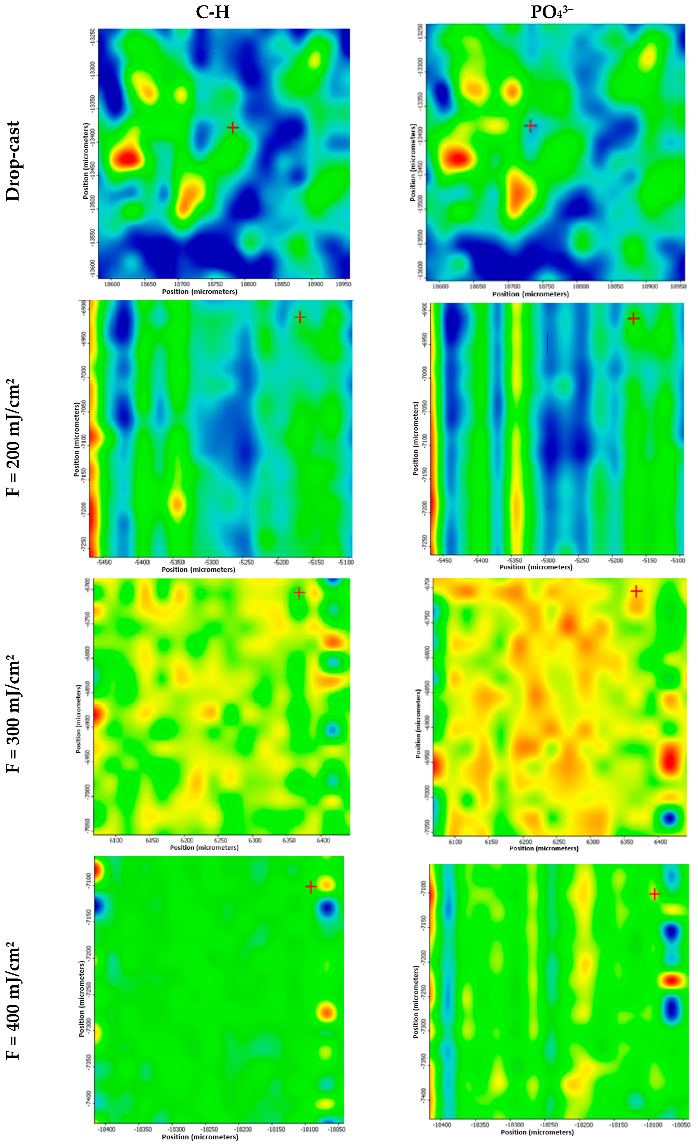
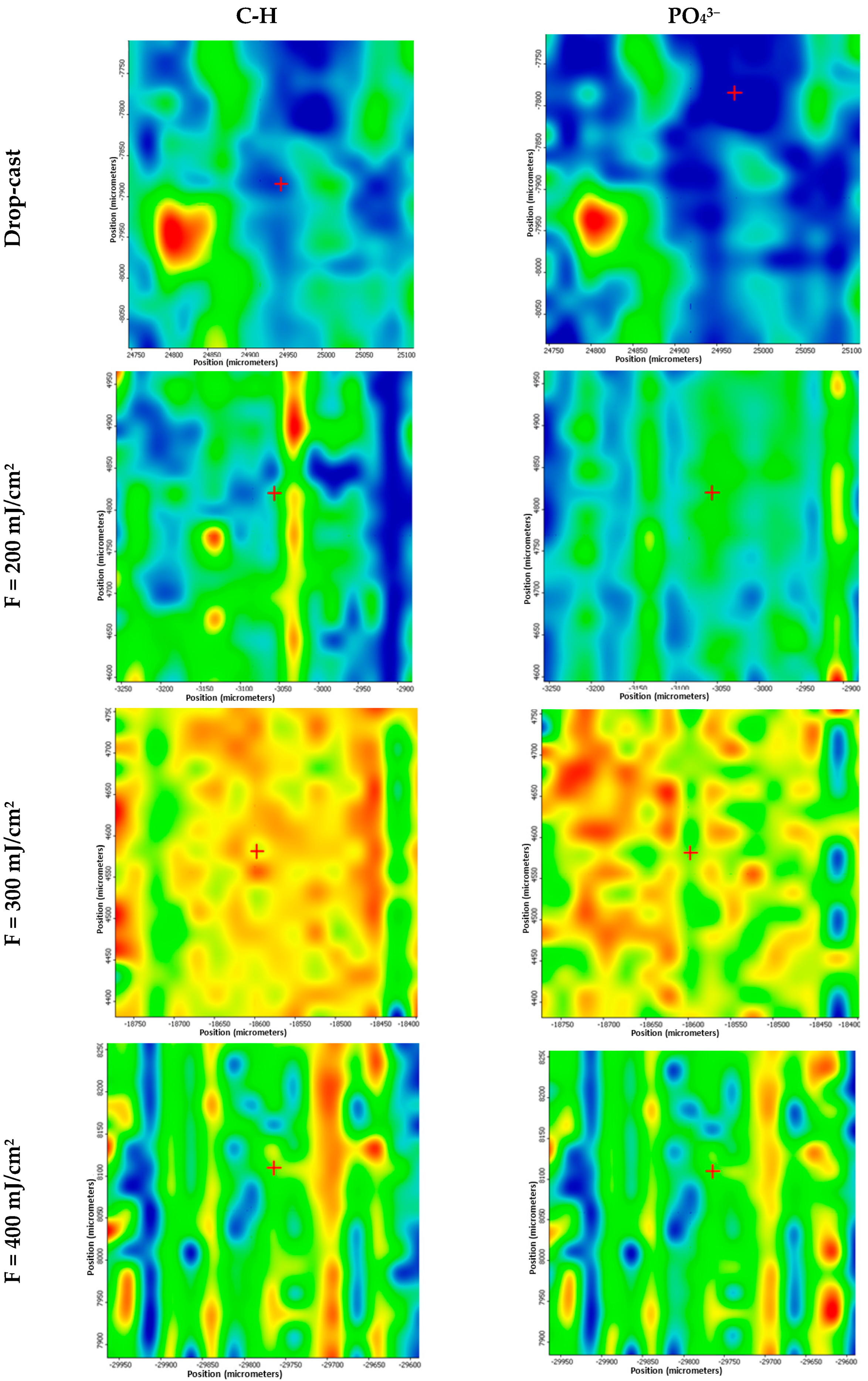
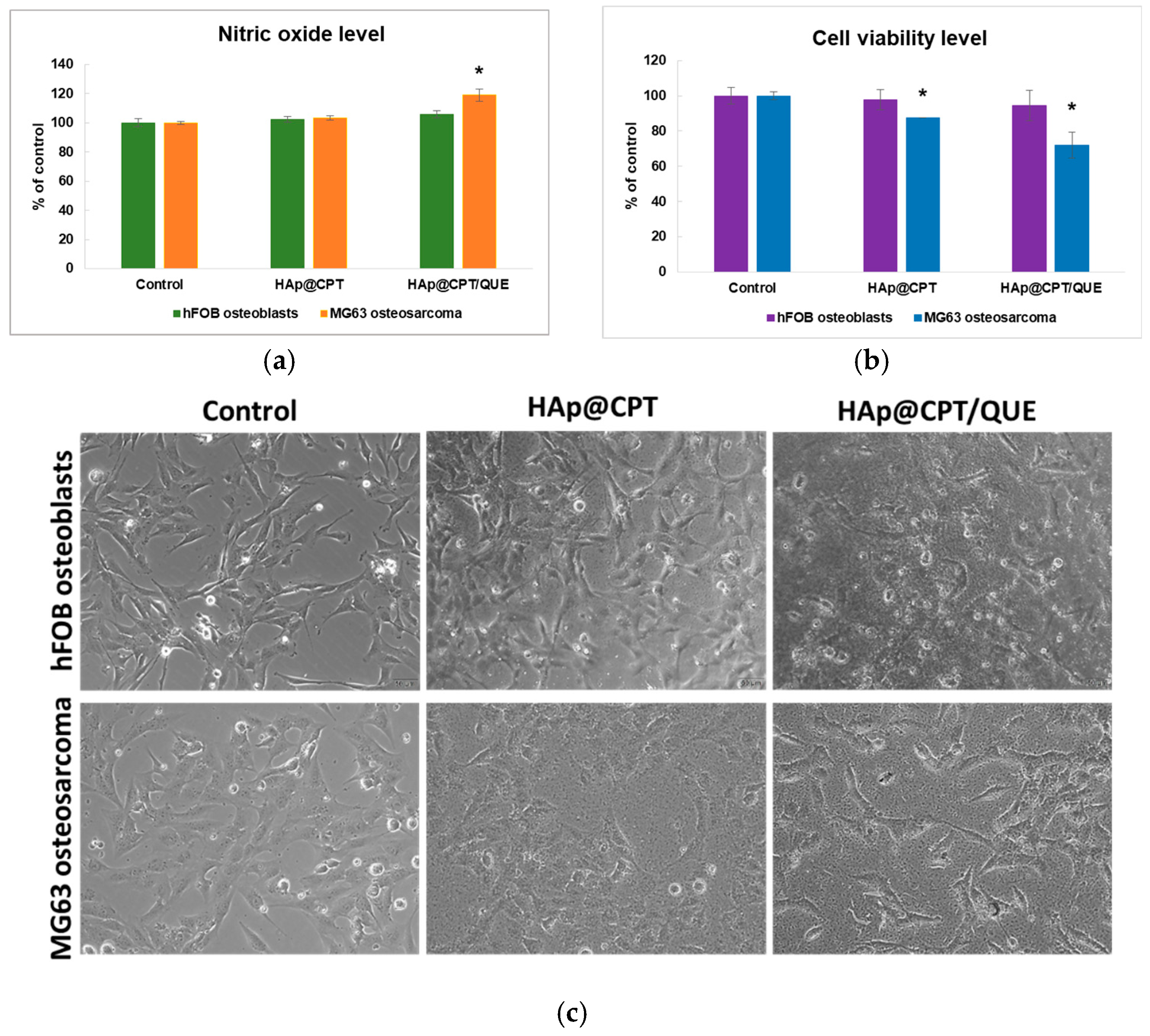
Human fetal osteoblasts hFOB 1.19, obtained from the American Type Culture Collection (ATCC CRL-3602, Rockville, MD, USA), were cultured in Dulbecco’s Modified Eagle’s Medium (DMEM), Ham’s F12 (1:1) with 2.5 mM L-glutamine (without phenol red) (Sigma-Aldrich/Merck, Darmstadt, Germany), 10% fetal bovine serum (FBS, Gibco/Life Technologies, Waltham, OR, USA), and 0.3 mg/mL G418 at 34 °C, 5% CO2 humidified atmosphere. The medium was changed every three days until reaching 80% confluency. The human osteosarcoma cell line MG63 (ATCC CRL-1427) was maintained in high-glucose DMEM supplemented with 10% FBS and penicillin-streptomycin antibiotics at 37 °C in a humified 5% CO2 incubator. Both cell lines were detached with 0.25% trypsin, 0.53 mM EDTA, and seeded on coated (HAp@CPT and HAp@CPT) or uncoated Ti (which served as controls) at a density of 2 × 10⁵ cells/cm2. All slide samples were UV-sterilized previously for one hour. After 24 h of cell growth, the osteoblasts’ morphology was examined using an Olympus IX71 microscope (Olympus, Tokyo, Japan).
At 24 h, the culture medium was collected and mixed with an equal volume of Griess reagent (0.1% naphthylethylenediamine dihydrochloride, 1% sulfanilamide in 5% H3PO4). Absorbance was measured at 550 nm (FlexStation, Molecular Device, San Jose, CA, USA), and the results were extrapolated on a NaNO2 standard curve. The cells cultured on slides were incubated with 1 mg/mL MTT solution for 2 h, and after, the formazan crystals were dissolved in isopropanol; the absorbance was measured at 595 nm (FlexStation, Molecular Device, San Jose, CA, USA).
Experiments were performed in triplicate, with results expressed as mean ± SD. Statistical significance was determined using a one-way ANOVA with the Bonferroni post hoc test (GraphPad Prism 5), a value of p < 0.05 being considered significant.

3. Results

Figure 1 presents the X-ray diffraction pattern of the synthesized hydroxyapatite (HA) powder. The diffraction profile exhibited indicates a high degree of crystallinity. Prominent peaks were observed at 2θ values of approximately 25.8°, 28.9°, 31.8°, 32.8°, 34.0°, 40.1°, 46.7°, 49.5°, and 53.2°, corresponding to the crystallographic planes (002), (210), (211), (112), (202), (310), (222), (213), and (004), respectively.
These reflections are in good agreement with the standard JCPDS card no. 09-0432, which is commonly used as a reference for stoichiometric hydroxyapatite. The presence of these specific planes confirms the formation of a hexagonal crystal structure characteristic of HA, with no detectable secondary phases or crystalline impurities. These results were consistent with previous reports on hydrothermally or chemically synthesized hydroxyapatite and further validate the structural purity of the prepared material [28,55,56,57]. No additional diffraction peaks associated with common secondary calcium phosphate phases, such as β-tricalcium phosphate or amorphous calcium phosphate, were detected.
The bright-field TEM image (Figure 2a) reveals that the particles exhibit a needle-shaped morphology, which is typical for nanoscale HAp. Due to this elongated shape, only the particle widths were measured for the size distribution analysis (Figure 2c). Moreover, the tendency to agglomerate is another morphological aspect that can be observed in the micrograph. The SAED pattern shown in Figure 2b exhibits a well-defined polycrystalline ring structure, confirming the crystalline nature of the HAp nanoparticles. The diffraction rings correspond to various crystallographic planes and are in good agreement with the XRD results, further validating the formation of phase-pure hydroxyapatite.
The FT-IR spectrum of the synthesized HAp (Figure 3) presented characteristic vibrational modes corresponding to phosphate and hydroxyl functional groups. The prominent absorption peak at 1024.50 cm−1 corresponded to the asymmetric stretching modes ν3 of phosphate, while the strong band at 962.66 cm−1 represented the symmetric stretching mode ν1, confirming the presence of HAp. The phosphate bending modes ν4 appeared at 600.83 cm−1 and 561.50 cm−1. Additionally, the vibrational mode of hydroxyl was identified at 628.67 cm−1.
In Figure 4, the FTIR spectra of the HAp@CPT and HAp@CPT/QUE coatings obtained at 300 mJ/cm2 are presented, highlighting the characteristic vibrational modes of hydroxyapatite, carboplatin, and quercetin.
In the HAp@CPT spectrum, the characteristic phosphate stretching vibrations of HAp are evident at 961.59 cm−11, symmetric stretching) and 1115.23 cm−13, asymmetric stretching), confirming the preservation of the HAp crystalline phase. The presence of carboplatin is supported by the band at 1642.38 cm−1, attributed to the C=O stretching vibration, and by a band at 1372.19 cm−1, corresponding to the C–H bending of methyl groups. Additionally, a well-defined peak at 3570.60 cm−1 is observed, assigned to O–H stretching, which likely reflects the hydroxyl groups present in both the HAp lattice and possibly coordinated water in carboplatin.
Absorptions in the 2990–2800 cm−1 range further indicate C–H stretching vibrations, originating from the organic framework of carboplatin. The absence of major band shifts or new peaks suggests a physical interaction between carboplatin and hydroxyapatite, with no significant chemical bonding or degradation in the individual components. These findings are consistent with previous literature reports [58].
The HAp@CPT/QUE spectrum shows additional bands assigned to quercetin. A prominent band at 1496.89 cm−1 corresponds to aromatic C=C stretching within the flavonoid ring system. An additional peak at 3019.11 cm−1 indicates aromatic C–H stretching. Vibrations at 1319.87 cm−1 and 1274.01 cm−1 are attributed to C–O stretching and asymmetric C–O–C stretching, respectively, while the band at 1194.70 cm−1 is characteristic of phenolic C–O stretching [59].
The retention of the 3570.60 cm−1 O–H band and the C–H stretching region (2990–2800 cm−1) in the HAp@CPT/QUE spectrum confirms that the structural features of organic components are preserved upon laser processing.
For the IR microscopy analysis, the absorbance intensity of the collected infrared spectra is directly related to the color changes within the resulting IR maps. The colors, ranging from blue to red, correspond to the lowest and highest intensities, respectively. To identify the optimal laser fluence for processing the HAp-based materials, a parametric study was performed. Thus, the laser fluences were varied between 200 and 400 mJ/cm2. The pristine material was used as reference (i.e., drop-cast).
Similar to the case of drop-cast (Figure 5), the intensity distribution was relatively low at 200 mJ/cm2, corresponding to the predominance of the blue color for the distribution of C-H and PO43− groups.
From the compositional point of view, the laser fluence of 300 mJ/cm2 was identified as optimal for the synthesis of HAp@CPT coatings, the yellow and green colors being representative in this case for the distribution of C-H and PO43− groups. This corresponds to a minimum chemical alteration and adequate material transfer.
In the case of the highest fluence (i.e., 400 mJ/cm2), the green color is prevalent in the IR map. Even though the functional groups are present on the surface, their distribution is less intense than in the case of the 300 mJ/cm2 samples, due to the increased fluence.
The drop-cast corresponding to the HAp@CPT/QUE samples (Figure 6) presented structural similarities with the one of HAp@CPT (Figure 5). At both the fluences of 200 and 400 mJ/cm2, the intensity distribution of the functional groups is decreased, which corresponds to a low quantity of transferred material (200 mJ/cm2) and to a non-stoichiometry of the transferred material (400 mJ/cm2). In the case of the 300 mJ/cm2 fluence, the presence of green, yellow, and orange colors confirmed an increased amount of synthesized HAp@CPT/QUE material.
Based on the IRM mapping results, the coatings obtained at 300 mJ/cm2 were considered the optimal version and were further characterized through additional techniques.
The SEM analysis of the HAp@CPT (Figure 7) and HAp@CPT/QUE (Figure 8) coatings at different magnifications revealed their structural differences and biomedical potential.
At 2000× magnification, both coatings showed a continuous but rough structure, with HAp@CPT appearing more uniform, while HAp@CPT/QUE exhibited increased surface roughness, likely due to quercetin incorporation. At 20,000× magnification, an interconnected network of nanoparticles became evident, with HAp@CPT maintaining compact morphology, while HAp@CPT/QUE presented enhanced porosity, suggesting better drug diffusion and controlled release. At higher magnifications (50,000× and 100,000×), nanostructures and rough features confirmed the coatings’ potential for bioactivity and sustained drug release. The cross-sectional SEM images revealed thickness values ranging from 30 to 100 nm (HAp@CPT samples, Figure 7d) and from 30 to 150 nm (HAp@CPT/QUE samples, Figure 8d).
From a biological perspective, the rough nanostructure plays a crucial role in osteoblast adhesion, proliferation, and bone integration. The porosity enhances cell attachment by increasing the surface area available for interactions with proteins and signaling molecules, mimicking the natural bone extracellular matrix. Additionally, the interconnected pores facilitate nutrient and oxygen diffusion, promoting osteogenesis and new bone tissue formation around the implant. The nanoscale roughness also encourages mechanical interlocking with the surrounding bone, improving implant stability and reducing the risk of loosening over time.
The in vitro biocompatibility testing (Figure 9) of HAp@CPT and HAp@CPT/QUE coatings demonstrated differential effects on human non-tumoral osteoblast and osteosarcoma cell viability and inflammatory responses. The NO release quantification by the Griess assay (Figure 9a) in the case of hFOB osteoblasts showed similar values to those of the control for both types of coatings, indicating that these compositions did not alter the inflammatory status of normal cells. In contrast, the MG63 osteosarcoma cells exhibited higher NO levels (p < 0.05) after incubating with the HAp@CPT/QUE sample compared to the control or HAp@CPT coating, which may correlate with increased oxidative stress and inflammatory signaling.
The MTT assay results (Figure 9b) showed almost similar cell viability for hFOB osteoblasts cultured on control or coated samples, indicating a favorable environment for osteoblast proliferation. On the other hand, the MG63 osteosarcoma cell viability was reduced by 12% and 28% from the control on HAp@CPT and HAp@CPT/QUE coatings, respectively, suggesting a selective cytotoxic effect of these formulations. Phase-contrast microscopy observations (Figure 9c) confirmed that hFOB osteoblasts adhered well to both coated surfaces, maintaining their typical morphology. In contrast, MG63 osteosarcoma cells displayed reduced attachment, particularly on HAp@CPT/QUE-coated surfaces.

4. Discussion

Endoprostheses are generally utilized in orthopedic surgery after the tumor is removed. Even if this type of surgery is a way of cancer treatment, some complications, such as prosthetic joint infection, structural implant failure, soft-tissue attachment failure, or aseptic loosening, may appear, and together with a recurrence of the bone cancer, it can further decrease the patient’s survival rate [14,60]. These clinical challenges underline the need for multifunctional implant coatings that not only promote osseointegration but also provide local therapeutic benefits to reduce tumor recurrence and infection.
In this study, HAp was chosen based on its increasingly gained interest due to its excellent bioactivity, biocompatibility, and osteoconductivity, which are desirable properties for combating the aseptic loosening of endoprostheses [61,62,63].
HAp-based coatings incorporating natural derived agents (i.e., flavonoids) exhibit therapeutic potential for hard tissue applications by boosting the osseointegration of metallic surfaces and exerting local antibiotherapy [64,65]. In this context, quercetin, a plant-derived flavonoid, has demonstrated pro-apoptotic and ROS-inducing effects in cancer cells, while supporting osteoblast proliferation.
Thus, in the case of organic compounds, such as active pharmaceutical ingredients, the ability to achieve uniform deposition on surfaces was demonstrated for a range of laser fluence values [66,67,68], supporting the feasibility of using the MAPLE technique to fabricate composite coatings containing carboplatin and quercetin.
The role of in vitro assays was both to demonstrate the antitumoral effect of carboplatin on osteosarcoma cells and the capacity of this innovative coating to have no cytotoxic effect on human non-tumoral osteoblast cells. Notably, the HAp@CPT/QUE coating did not have a negative impact on hFOB osteoblasts and promoted bone cell proliferation. These findings confirm the biocompatibility of the dual-functionalized coating with healthy bone tissue.
In contrast, the MG63 osteosarcoma cells were shown to not adhere to the HAp@CPT/QUE-coated surface. This selectivity can be partially explained by the known ability of carboplatin to cause G2/M cell cycle arrest after 24 h, as previously demonstrated [69]. Moreover, quercetin-mediated apoptosis could explain the higher percent of MG63 cell viability decrease for the HAp@CPT/QUE surface, most probably by the mitochondrial-dependent pathway [70].
From the Griess test, it can be concluded that the HAp@CPT/QUE coating exhibits good results by increasing oxidative stress and inflammatory signaling.This effect may contribute to enhanced antitumor immunity. It has been documented that the induction of acute inflammatory reactions often stimulates antitumor immune responses, thereby efficiently contributing to cancer cell death [71]. As reported [72,73], acute ‘therapeutic inflammation’ is typically initiated by cellular stress and is able to activate dendritic cell-mediated antitumor T-cell response. As a consequence, the apoptosis of cancer cells is induced. Moreover, the disturbing reactive oxygen species homeostasis in osteosarcoma cells by quercetin [73] could correlate very well with the increased NO level, activating the autophagy process in MG63 cells.

5. Conclusions

This study reports on the fabrication by Matrix-Assisted Pulsed Laser Evaporation of hydroxyapatite-based coatings blended with carboplatin (HAp@CPT) and quercetin (HAp@CPT/QUE) for hip endoprostheses, using three different laser fluences (i.e., 200, 300, and 400 mJ/cm2). After physicochemical investigations, the 300 mJ/cm2 fluence was advanced as optimum for the unaltered material transfer. In vitro tests demonstrated that the HAp@CPT/QUE coatings proved biocompatibility with osteoblasts and selective cytotoxicity toward osteosarcoma cells. When compared to the control, the MG63 osteosarcoma cell viability tests indicated a reduction by 12% and 28% on HAp@CPT and HAp@CPT/QUE coatings, respectively, suggesting a selective cytotoxic effect of these composite structures. Moreover, the coatings’ rough nanostructure further supported osteointegration by promoting cell attachment and nutrient diffusion. These findings suggest that these novel HAp-based coatings are qualified to provide a multifunctional surface for orthopedic implants, offering structural support, enhanced bioactivity, and targeted cancer protection. The dual regenerative and therapeutic potential of these MAPLE coatings further advances them as promising candidates for hip endoprostheses in oncological orthopedic applications.

Author Contributions

Conceptualization, P.C.B.; data curation, L.D., A.-G.N., M.S.S., D.M.R., A.E.B., and B.Ș.V.; formal analysis, G.I., D.-I.T., I.M.I., L.D., V.G., A.C.B., A.-G.N., P.C.B., I.C.V., M.S.S., D.M.R., A.E.B., B.Ș.V., A.M.G., and A.R.R.; investigation, G.I., D.-I.T., I.M.I., L.D., V.G., A.C.B., A.-G.N., P.C.B., I.C.V., M.S.S., D.M.R., A.E.B., B.Ș.V., A.M.G., and A.R.R.; methodology, P.C.B. and A.R.R.; writing—original draft, G.I., D.-I.T., I.M.I., L.D., V.G., A.C.B., A.-G.N., P.C.B., I.C.V., M.S.S., D.M.R., A.E.B., B.Ș.V., A.M.G., and A.R.R.; writing—review and editing, A.-G.N., P.C.B., M.S.S., A.M.G., and A.R.R. All authors have read and agreed to the published version of the manuscript.

Funding

This research was supported by the Romanian Ministry of Research, Innovation, and Digitization under Romanian National Core Program LAPLAS VII—contract no. 30N/2023.

Institutional Review Board Statement

Not applicable.

Informed Consent Statement

Not applicable.

Data Availability Statement

At authors, by request.

Conflicts of Interest

The authors declare no conflicts of interest.

References

  1. Levett, J.J.; Turcotte, R.E.; Jung, S.; Antoniou, J.; Huk, O.L. Osteosarcoma Around a Ceramic-on-Ceramic Total Hip Arthroplasty. Arthroplast. Today 2023, 19, 101094. [Google Scholar] [CrossRef] [PubMed]
  2. Jin, Q.-L.; Su, H.-B.; Du, S.-H.; Hou, C.-H.; Lu, M.; Dai, S.-W.; Lei, Z.-X.; Chen, W.; Li, H.-M. Revision surgery for periprosthetic fracture of distal femur after endoprosthetic replacement of knee joint following resection of osteosarcoma. Front. Oncol. 2024, 14, 1328703. [Google Scholar] [CrossRef] [PubMed]
  3. Callan, A.K.; Alexander, J.H.; Montgomery, N.I.; Lindberg, A.W.; Scharschmidt, T.J.; Binitie, O. Contemporary surgical management of osteosarcoma and Ewing sarcoma. Pediatr. Blood Cancer 2025, 72, e31374. [Google Scholar] [CrossRef]
  4. Khakzad, T.; Putzier, M.; Paksoy, A.; Rau, D.; Thielscher, L.; Taheri, N.; Wittenberg, S.; Märdian, S. Outcome of Endoprosthetic Hip Reconstruction Following Resection of Malignant Bone Tumors. Cancers 2024, 16, 2890. [Google Scholar] [CrossRef]
  5. Latała, B.; Kamiński, P.; Jagielski, P.; Kłosiński, M.; Wilk-Frańczuk, M. Evaluation of functional capacity, pain level and quality of life in patients following hip arthroplasty due to degenerative changes depending on the type of implanted endoprosthesis. Acta Neuropsychol. 2024, 22, 417–433. [Google Scholar] [CrossRef]
  6. Tepper, S.C.; Lee, L.; Kasson, L.B.; Herbst, L.R.; Vijayakumar, G.; Colman, M.W.; Gitelis, S.; Blank, A.T. Hip Arthroplasty Outcomes in Patients with Metastatic Bone Disease. Orthop. Rev. 2024, 16, 94568. [Google Scholar] [CrossRef]
  7. Radunović, A.; Radunović, O.; Vulović, M.; Aksić, M. Bioceramics in Orthopedic Surgery. In Bioceramics, Biomimetic and Other Compatible Materials Features for Medical Applications; Najman, S., Mitić, V., Groth, T., Barbeck, M., Chen, P.-Y., Sun, Z., Randjelović, B., Eds.; Springer International Publishing: Cham, Switzerland, 2023; pp. 53–65. [Google Scholar]
  8. Meng, M.; Wang, J.; Huang, H.; Liu, X.; Zhang, J.; Li, Z. 3D printing metal implants in orthopedic surgery: Methods, applications and future prospects. J. Orthop. Transl. 2023, 42, 94–112. [Google Scholar] [CrossRef]
  9. Fenske, F.; Krause, L.; Meyer, S.; Kujat, B.; Repmann, J.; Neuhaus, M.; Zimmerer, R.; Roth, A.; Lethaus, B.; Ziebolz, D.; et al. Oral Health Screening for Risk Reduction for Early Periprosthetic Joint Infections of Hip and Knee Endoprostheses—Results of a Prospective Cohort Study. J. Clin. Med. 2023, 12, 4451. [Google Scholar] [CrossRef]
  10. Liang, W.; Zhou, C.; Bai, J.; Zhang, H.; Jiang, B.; Wang, J.; Fu, L.; Long, H.; Huang, X.; Zhao, J.; et al. Current advancements in therapeutic approaches in orthopedic surgery: A review of recent trends. Front. Bioeng. Biotechnol. 2024, 12, 1328997. [Google Scholar] [CrossRef]
  11. Hariharan, K.; Selvakumar, M.; Ramkumar, T.; Chandramohan, P. Cell viability and bioactive coating on additive manufactured polycaprolactone substrate for osteointegration applications. Chem. Phys. Impact 2024, 8, 100409. [Google Scholar] [CrossRef]
  12. Uklejewski, R.; Winiecki, M.; Dąbrowski, M.; Rogala, P. Towards the First Generation of Biomimetic Fixation for Resurfacing Arthroplasty Endoprostheses. Biomimetics 2024, 9, 99. [Google Scholar] [CrossRef] [PubMed]
  13. Deng, Y.; Zhou, C.; Fu, L.; Huang, X.; Liu, Z.; Zhao, J.; Liang, W.; Shao, H. A mini-review on the emerging role of nanotechnology in revolutionizing orthopedic surgery: Challenges and the road ahead. Front. Bioeng. Biotechnol. 2023, 11, 1191509. [Google Scholar] [CrossRef] [PubMed]
  14. Shah, A.; Cardoso, F.N.; Souza, F.; Montreuil, J.; Pretell-Mazzini, J.; Temple, H.T.; Hornicek, F.; Crawford, B.; Subhawong, T.K. Failure Modes in Orthopedic Oncologic Reconstructive Surgery: A Review of Imaging Findings and Failure Rates. Curr. Oncol. 2024, 31, 6245–6266. [Google Scholar] [CrossRef] [PubMed]
  15. Uklejewski, R.; Uklejewski, R.; Rogala, P.; Winiecki, M.; Winiecki, M. Structural and Hydroxyapatite-like Surface Functionalization of Advanced Biomimetic Prototype Interface for RA Endoprostheses to Enhance Osteoconduction and Osteointegration. Adv. Surf. Eng. Mater. 2016, 175–240. [Google Scholar]
  16. Rios-Pimentel, F.F.; Méndez-González, M.M.; García-Rocha, M. A short review: Hydroxyapatite coatings for metallic implants. Heat. Treat. Surf. Eng. 2023, 5, 2202002. [Google Scholar] [CrossRef]
  17. Ioniță-Radu, F.; Nicolau, I.-N.; Petrache, O.-G.; Groșeanu, M.-L.; Bojincă, V.-C.; Negru, M.-M.; Bucurică, S.; Anghel, D. Correlation Between Trabecular Bone Score and Homocysteine Level in Rheumatoid Arthritis Patients on Anti-TNF Inhibitors. Life 2024, 14, 463. [Google Scholar] [CrossRef]
  18. Salahuddin, N.; Ibrahim, E.M.; El-Kemary, M. Different methods for preparation of hydroxyapatite nanostructures. Biointerface Res. Appl. Chem. 2023, 13, 236. [Google Scholar]
  19. Alberts, A.; Bratu, A.G.; Niculescu, A.-G.; Grumezescu, A.M. Collagen-Based Wound Dressings: Innovations, Mechanisms, and Clinical Applications. Gels 2025, 11, 271. [Google Scholar] [CrossRef]
  20. Buradagunta, R.S.; Madiga, J.; Dumpala, R. Producing Calcium Deficient Nano-hydroxyapatite from Silver pomfret Fish Bones for Biomedical Applications. Lett. Appl. NanoBioScience 2024, 13, 130. [Google Scholar] [CrossRef]
  21. Liu, C.; Xu, M.; Wang, Y.; Yin, Q.; Hu, J.; Chen, H.; Sun, Z.; Liu, C.; Li, X.; Zhou, W.; et al. Exploring the potential of hydroxyapatite-based materials in biomedicine: A comprehensive review. Mater. Sci. Eng. R Rep. 2024, 161, 100870. [Google Scholar] [CrossRef]
  22. Fendi, F.; Abdullah, B.; Suryani, S.; Usman, A.N.; Tahir, D. Development and application of hydroxyapatite-based scaffolds for bone tissue regeneration: A systematic literature review. Bone 2024, 183, 117075. [Google Scholar] [CrossRef] [PubMed]
  23. Murphy, B.; Morris, M.A.; Baez, J. Development of Hydroxyapatite Coatings for Orthopaedic Implants from Colloidal Solutions: Part 2—Detailed Characterisation of the Coatings and Their Growth Mechanism. Nanomaterials 2023, 13, 2606. [Google Scholar] [CrossRef] [PubMed]
  24. Shoeib, M.A.; Abdel-Gawad, S.A. High performance nano hydroxyapatite coating on zinc for biomedical applications. J. Mater. Sci. 2023, 58, 740–756. [Google Scholar] [CrossRef]
  25. Murugesan, V.; Govindarasu, M.; Manoharadas, S.; Pandiaraj, S.; Thiruvengadam, M.; Govindasamy, R.; Vaiyapuri, M. Combinatorial anticancer effects of multi metal ion and drug substitute with hydroxyapatite coatings on surgical grade 316LSS stainless steel alloys towards biomedical applications. J. Mater. Res. Technol. 2023, 27, 7244–7258. [Google Scholar] [CrossRef]
  26. Youness, R.A.; Saleh, H.A.; Taha, M.A. Microstructure and elastic properties of hydroxyapatite/alumina nanocomposites prepared by mechanical alloying technique for biomedical applications. Biointerface Res. Appl. Chem. 2023, 13, 395. [Google Scholar]
  27. Awasthi, S.; Pandey, S.K.; Arunan, E.; Srivastava, C. A review on hydroxyapatite coatings for the biomedical applications: Experimental and theoretical perspectives. J. Mater. Chem. B 2021, 9, 228–249. [Google Scholar] [CrossRef]
  28. Florea, D.A.; Grumezescu, V.; Bîrcă, A.C.; Vasile, B.Ș.; Iosif, A.; Chircov, C.; Stan, M.S.; Grumezescu, A.M.; Andronescu, E.; Chifiriuc, M.C. Bioactive Hydroxyapatite-Magnesium Phosphate Coatings Deposited by MAPLE for Preventing Infection and Promoting Orthopedic Implants Osteointegration. Materials 2022, 15, 7337. [Google Scholar] [CrossRef]
  29. Komarova, E.G.; Senkina, E.I.; Lozhkomoev, A.S.; Kazantseva, E.A.; Prosolov, K.A.; Kazantsev, S.O.; Akimova, E.B.; Tolkacheva, T.V.; Khimich, M.A.; Sharkeev, Y.P. Controlled anticancer 5-Fluorouracil release from functionalized 5-FU/PLGA/CaP coating on titanium implants: Characterization, in vitro drug delivery and cytotoxicity. Mater. Today Commun. 2024, 39, 109332. [Google Scholar] [CrossRef]
  30. Pourmadadi, M.; Eshaghi, M.M.; Shaghaghi, M.; Das, S.S.; Arshad, R.; Ghotekar, S.; Rahdar, A.; Manicum, A.-L.E.; Pandey, S. Nano-scale drug delivery systems for carboplatin: A comprehensive review. OpenNano 2023, 13, 100175. [Google Scholar] [CrossRef]
  31. Choeyprasert, W.; Natesirinilkul, R.; Charoenkwan, P.; Sittipreechacharn, S. Carboplatin and doxorubicin in treatment of pediatric osteosarcoma: A 9-year single institute experience in the Northern Region of Thailand. Asian Pac. J. Cancer Prev. 2013, 14, 1101–1106. [Google Scholar] [CrossRef]
  32. Skverchinskaya, E.; Levdarovich, N.; Ivanov, A.; Mindukshev, I.; Bukatin, A. Anticancer Drugs Paclitaxel, Carboplatin, Doxorubicin, and Cyclophosphamide Alter the Biophysical Characteristics of Red Blood Cells, In Vitro. Biology 2023, 12, 230. [Google Scholar] [CrossRef] [PubMed]
  33. Hain, B.A.; Xu, H.; Wilcox, J.R.; Mutua, D.; Waning, D.L. Chemotherapy-induced loss of bone and muscle mass in a mouse model of breast cancer bone metastases and cachexia. JCSM Rapid Commun. 2019, 2, 1–12. [Google Scholar] [CrossRef]
  34. Szefler, B.; Makarewicz, A.; Obońska, E.; Rutkowska, D.; Czeleń, P. Docking of carboplatin towards chosen nanostructures. Biointerface Res. Appl. Chem. 2022, 13, 109. [Google Scholar]
  35. Mirza, M.A.; Mahmood, S.; Hilles, A.R.; Ali, A.; Khan, M.Z.; Zaidi, S.A.; Iqbal, Z.; Ge, Y. Quercetin as a Therapeutic Product: Evaluation of Its Pharmacological Action and Clinical Applications—A Review. Pharmaceuticals 2023, 16, 1631. [Google Scholar] [CrossRef]
  36. Aghababaei, F.; Hadidi, M. Recent Advances in Potential Health Benefits of Quercetin. Pharmaceuticals 2023, 16, 1020. [Google Scholar] [CrossRef]
  37. Lotfi, M.-S.; Sheibani, M.; Jafari-Sabet, M. Quercetin-based biomaterials for enhanced bone regeneration and tissue engineering. Tissue Cell 2024, 91, 102626. [Google Scholar] [CrossRef]
  38. Durmaz, B.; Gunes, N.; Koparal, M.; Gul, M.; Dundar, S.; Bingul, M.B. Investigation of the effects of quercetin and xenograft on the healing of bone defects: An experimental study. J. Oral. Biol. Craniofacial Res. 2023, 13, 22–27. [Google Scholar] [CrossRef]
  39. Sharma, S.; Cwiklinski, K.; Mahajan, S.D.; Schwartz, S.A.; Aalinkeel, R. Combination Modality Using Quercetin to Enhance the Efficacy of Docetaxel in Prostate Cancer Cells. Cancers 2023, 15, 902. [Google Scholar] [CrossRef]
  40. El-Far, M.; Essam, A.; Fardous, F.; Abd El-Azim, A.O.; Yahia, S.; El-Sherbiny, I.M. Newly Modified Nanoformulation of Quercetin as Promising Chemotherapeutic Anticancer Agent. Biointerface Res. Appl. Chem. 2023, 13, 387. [Google Scholar]
  41. Alberts, A.; Moldoveanu, E.-T.; Niculescu, A.-G.; Grumezescu, A.M. Nigella sativa: A Comprehensive Review of Its Therapeutic Potential, Pharmacological Properties, and Clinical Applications. Int. J. Mol. Sci. 2024, 25, 13410. [Google Scholar] [CrossRef]
  42. Pati, S.; Antara, N.S.; Gunam, I.B.W.; Sarkar, T.; Lahiri, D. Elucidating the Quorum Sensing Inhibitory Mechanism of Flavonoid Quercetin by Molecular Docking, Molecular Dynamics Simulation, and MM-GBSA Study. Lett. Appl. NanoBioScience 2024, 13, 152. [Google Scholar] [CrossRef]
  43. Dehghanghadikolaei, A.; Ansary, J.; Ghoreishi, R. Sol-gel process applications: A mini-review. Proc. Nat. Res. Soc. 2018, 2, 02008–02029. [Google Scholar] [CrossRef]
  44. Grămadă, A.M.; Stoica, A.-E.; Niculescu, A.-G.; Bîrcă, A.C.; Vasile, B.Ș.; Holban, A.M.; Mihaiescu, T.; Șerban, A.I.; Ciceu, A.; Balta, C.; et al. Zinc Oxide-Loaded Recycled PET Nanofibers for Applications in Healthcare and Biomedical Devices. Polymers 2025, 17, 45. [Google Scholar] [CrossRef]
  45. Bîrcă, A.C.; Minculescu, M.A.; Niculescu, A.-G.; Hudiță, A.; Holban, A.M.; Alberts, A.; Grumezescu, A.M. Nanoparticle-Enhanced Collagen Hydrogels for Chronic Wound Management. J. Funct. Biomater. 2025, 16, 91. [Google Scholar] [CrossRef]
  46. Barrino, F. Hybrid Organic–Inorganic Materials Prepared by Sol–Gel and Sol–Gel-Coating Method for Biomedical Use: Study and Synthetic Review of Synthesis and Properties. Coatings 2024, 14, 425. [Google Scholar] [CrossRef]
  47. Shankar, D.; Jayaganesh, K.; Gowda, N.; Lakshmi, K.S.; Jayanthi, K.J.; Jambagi, S.C. Thermal spray processes influencing surface chemistry and in-vitro hemocompatibility of hydroxyapatite-based orthopedic implants. Biomater. Adv. 2024, 158, 213791. [Google Scholar] [CrossRef] [PubMed]
  48. Shankar, D.; Jambagi, S.C. Improvements in bioactivity, blood compatibility, and wear resistance of thermally sprayed carbon nanotube reinforced hydroxyapatite-based orthopedic implants. Tribol. Int. 2024, 197, 109809. [Google Scholar] [CrossRef]
  49. Constantinescu, S.; Niculescu, A.-G.; Hudiță, A.; Grumezescu, V.; Rădulescu, D.; Bîrcă, A.C.; Dorcioman, G.; Gherasim, O.; Holban, A.M.; Gălățeanu, B.; et al. Nanostructured Coatings Based on Graphene Oxide for the Management of Periprosthetic Infections. Int. J. Mol. Sci. 2024, 25, 2389. [Google Scholar] [CrossRef]
  50. Paneer Selvam, S.; Ayyappan, S.; I Jamir, S.; Sellappan, L.K.; Manoharan, S. Recent advancements of hydroxyapatite and polyethylene glycol (PEG) composites for tissue engineering applications—A comprehensive review. Eur. Polym. J. 2024, 215, 113226. [Google Scholar] [CrossRef]
  51. Kravanja, K.A.; Finšgar, M. A review of techniques for the application of bioactive coatings on metal-based implants to achieve controlled release of active ingredients. Mater. Des. 2022, 217, 110653. [Google Scholar] [CrossRef]
  52. Liao, T.-Y.; Biesiekierski, A.; Berndt, C.C.; King, P.C.; Ivanova, E.P.; Thissen, H.; Kingshott, P. Multifunctional cold spray coatings for biological and biomedical applications: A review. Prog. Surf. Sci. 2022, 97, 100654. [Google Scholar] [CrossRef]
  53. Zureigat, O.A.; NeamŢU, J.; Duta, L.; Icriverzi, M.; Florian, P.; Sima, L.-e.; Dorcioman, G.; Grumezescu, V.; BĂLĂŞOiu, R.; ButeicĂ, S.A. Development and characterisation of implantable sandwich structures made of lithium-doped biological-derived hydroxyapatite and ciprofloxacin. Farmacia 2024, 72, 6. [Google Scholar] [CrossRef]
  54. Jelinek, M. 23—Hybrid laser technology for biomaterials. In Lasers for Medical Applications; Jelínková, H., Ed.; Woodhead Publishing: Sawston, UK, 2013; pp. 704–724. [Google Scholar]
  55. Grumezescu, V.; Grumezescu, A.M.; Ficai, A.; Negut, I.; Vasile, B.Ș.; Gălățeanu, B.; Hudiță, A. Composite Coatings for Osteoblast Growth Attachment Fabricated by Matrix-Assisted Pulsed Laser Evaporation. Polymers 2022, 14, 2934. [Google Scholar] [CrossRef] [PubMed]
  56. Rabiei, M.; Palevicius, A.; Monshi, A.; Nasiri, S.; Vilkauskas, A.; Janusas, G. Comparing Methods for Calculating Nano Crystal Size of Natural Hydroxyapatite Using X-Ray Diffraction. Nanomaterials 2020, 10, 1627. [Google Scholar] [CrossRef]
  57. Shah, S.; Joshi, R.; Rai, N.; Adhikari, R.; Pandit, R. Microstructural analysis of biowaste-derived hydroxyapatite-chitosan nanocomposites. Micro Nano Lett. 2022, 17, 369–376. [Google Scholar] [CrossRef]
  58. Iliescu, R.I.; Andronescu, E.; Ghitulica, C.D.; Berger, D.; Ficai, A. Montmorillonite-alginate nanocomposite beads as drug carrier for oral administration of carboplatin-preparation and characterization. UPB Sci. Bull. Ser. B 2011, 73, 3–16. [Google Scholar]
  59. Wang, Q.; Wei, H.; Deng, C.; Xie, C.; Huang, M.; Zheng, F. Improving Stability and Accessibility of Quercetin in Olive Oil-in-Soy Protein Isolate/Pectin Stabilized O/W Emulsion. Foods 2020, 9, 123. [Google Scholar] [CrossRef]
  60. Stevenson, J.; Siddiqi, M.A.; Sheehy, V.; Kendrick, B.; Whitwell, D.; Taylor, A.; Blunn, G.; Mohammad, H.R.; Kamath, A.F.; Thoma, S. Early radiological outcomes of a fully porous bridging collar in lower-limb endoprosthetic reconstructions: A case-matched retrospective series to assess osseointegration. Arthroplasty 2024, 6, 17. [Google Scholar] [CrossRef]
  61. Kubiak-Mihkelsoo, Z.; Kostrzębska, A.; Błaszczyszyn, A.; Pitułaj, A.; Dominiak, M.; Gedrange, T.; Nawrot-Hadzik, I.; Matys, J.; Hadzik, J. Ionic Doping of Hydroxyapatite for Bone Regeneration: Advances in Structure and Properties over Two Decades—A Narrative Review. Appl. Sci. 2025, 15, 1108. [Google Scholar] [CrossRef]
  62. Tang, G.; Yang, H.; Zhao, B.; Wang, D.; Zeng, F.; Liu, Q. Microstructure, electrical properties, bioactivity, biocompatibility and osteogenic differentiation ability of bio-piezocomposite fabricated by hydroxyapatite and (Ba,Ca)(Ti,Sn)O3-based ceramics. J. Mater. Res. Technol. 2025, 35, 4482–4495. [Google Scholar] [CrossRef]
  63. Es-saddik, M.; Laasri, S.; Bensemlali, M.; Hariti, N.; Laghzizil, A.; Taha, M. Experimental and Finite Element Study of the Mechanical Behavior of Hydroxyapatite/Tricalcium Phosphate/Alumina Biocomposite. Biointerface Res. Appl. Chem. 2024, 14, 1–14. [Google Scholar]
  64. Yang, X.; Huang, P.; Wang, H.; Cai, S.; Liao, Y.; Mo, Z.; Xu, X.; Ding, C.; Zhao, C.; Li, J. Antibacterial and anti-biofouling coating on hydroxyapatite surface based on peptide-modified tannic acid. Colloids Surf. B Biointerfaces 2017, 160, 136–143. [Google Scholar] [CrossRef] [PubMed]
  65. Palierse, E.; Hélary, C.; Krafft, J.-M.; Génois, I.; Masse, S.; Laurent, G.; Alvarez Echazu, M.I.; Selmane, M.; Casale, S.; Valentin, L.; et al. Baicalein-modified hydroxyapatite nanoparticles and coatings with antibacterial and antioxidant properties. Mater. Sci. Eng. C 2021, 118, 111537. [Google Scholar] [CrossRef]
  66. Dorcioman, G.; Hudiță, A.; Gălățeanu, B.; Craciun, D.; Mercioniu, I.; Oprea, O.C.; Neguț, I.; Grumezescu, V.; Grumezescu, A.M.; Dițu, L.M.; et al. Magnetite-Based Nanostructured Coatings Functionalized with Nigella sativa and Dicloxacillin for Improved Wound Dressings. Antibiotics 2023, 12, 59. [Google Scholar] [CrossRef]
  67. Pirușcă, I.A.; Balaure, P.C.; Grumezescu, V.; Irimiciuc, S.-A.; Oprea, O.-C.; Bîrcă, A.C.; Vasile, B.; Holban, A.M.; Voinea, I.C.; Stan, M.S.; et al. New Fe3O4-Based Coatings with Enhanced Anti-Biofilm Activity for Medical Devices. Antibiotics 2024, 13, 631. [Google Scholar] [CrossRef]
  68. Alfe, M.; Minopoli, G.; Tartaglia, M.; Gargiulo, V.; Ausanio, G. Biocompatible Hybrid Graphenic Thin Coatings on Flexible Substrates Through Matrix-Assisted Pulsed Laser Evaporation (MAPLE). ACS Appl. Mater. Interfaces 2024, 16, 38956–38967. [Google Scholar] [CrossRef]
  69. Robson, H.; Meyer, S.; Shalet, S.M.; Anderson, E.; Roberts, S.; Eden, O.B. Platinum agents in the treatment of osteosarcoma: Efficacy of cisplatin vs. carboplatin in human osteosarcoma cell lines. Med. Pediatr. Oncol. 2002, 39, 573–580. [Google Scholar] [CrossRef]
  70. Liang, W.; Li, X.; Li, C.; Liao, L.; Gao, B.; Gan, H.; Yang, Z.; Liao, L.; Chen, X. Quercetin-mediated apoptosis via activation of the mitochondrial-dependent pathway in MG-63 osteosarcoma cells. Mol. Med. Rep. 2011, 4, 1017–1023. [Google Scholar] [CrossRef]
  71. Zhao, H.; Wu, L.; Yan, G.; Chen, Y.; Zhou, M.; Wu, Y.; Li, Y. Inflammation and tumor progression: Signaling pathways and targeted intervention. Signal Transduct. Target. Ther. 2021, 6, 263. [Google Scholar] [CrossRef]
  72. Liu, X.; Yin, L.; Shen, S.; Hou, Y. Inflammation and cancer: Paradoxical roles in tumorigenesis and implications in immunotherapies. Genes. Dis. 2023, 10, 151–164. [Google Scholar] [CrossRef]
  73. Nigam, M.; Mishra, A.P.; Deb, V.K.; Dimri, D.B.; Tiwari, V.; Bungau, S.G.; Bungau, A.F.; Radu, A.F. Evaluation of the association of chronic inflammation and cancer: Insights and implications. Biomed. Pharmacother. 2023, 164, 115015. [Google Scholar] [CrossRef]
Figure 1. X-ray diffractogram of hydroxyapatite powder.
Coatings 15 00489 g001
Figure 2. TEM images (a), SAED pattern (b), and histogram (c) of size distributions of hydroxyapatite powder. The particles exhibit a needle-like morphology; therefore, only the width (minor axis) was measured to estimate particle size.
Coatings 15 00489 g002
Figure 3. FT-IR spectrum of hydroxyapatite powder.
Coatings 15 00489 g003
Figure 4. IR spectra of HAp@CPT (blue line) and HAp@CPT/QUE (red line) coatings obtained at 300 mJ/cm2 laser fluence.
Coatings 15 00489 g004
Figure 5. IR maps for HAp@CPT drop-cast and coatings at 200, 300, and 400 mJ/cm2 laser fluences based on the distribution of the C-H bond belonging to CPT and the phosphate ion belonging to HAp.
Coatings 15 00489 g005
Figure 6. IR maps of HAp@CPT/QUE drop-cast and coatings at 200, 300, and 400 mJ/cm2 based on the distribution of the C-H bond belonging to QUE and the phosphate ion belonging to HAp.
Coatings 15 00489 g006
Figure 7. Top view at (a) 20,000×, (b) 50,000×, and (c) 100,000× magnifications and (d) cross-section SEM images of HAp@CPT coatings.
Coatings 15 00489 g007
Figure 8. Top view at (a) 20,000×, (b) 50,000×, and (c) 100,000× magnifications and (d) cross-section SEM images of HAp@CPT/QUE coatings.
Coatings 15 00489 g008
Figure 9. In vitro responses of human non-tumoral hFOB osteoblasts and MG63 osteosarcoma cells for HAp@CPT and HAp@CPT/QUE coatings at 24 h by (a) nitric oxide production, (b) cell viability, and (c) phase-contrast images. Data are expressed as mean ± standard deviation (SD) from three independent experiments and represented as a percentage of control (cells grown on uncoated Ti). * p < 0.05 compared to control.
Coatings 15 00489 g009
Disclaimer/Publisher’s Note: The statements, opinions and data contained in all publications are solely those of the individual author(s) and contributor(s) and not of MDPI and/or the editor(s). MDPI and/or the editor(s) disclaim responsibility for any injury to people or property resulting from any ideas, methods, instructions or products referred to in the content.

