
Anti-Acne Vulgaris Potential of the Ethanolic Extract of Mesua ferrea L. Flowers

 ,
,
Abstract
:1. Introduction
2. Materials and Methods
2.1. Chemicals and Reagents
2.2. Plant Material
2.3. Preparation of M. ferrea Flower Extract
2.4. Total Phenolic Contents
2.5. Anti-Oxidant Assays
2.5.1. DPPH Radical Scavenging Assay
2.5.2. ABTS Radical Cation Decolorization Assay (ABTS Assay)
2.5.3. Ferric Reducing Antioxidant Power Assay (FRAP Assay)
2.5.4. Nitric Oxide (NO) Radical Scavenging Assay
2.6. Anti-Bacterial Activity Assays
2.6.1. Disc Diffusion Method
2.6.2. Broth Macrodilution Method
2.7. Anti-Inflammatory Activities
2.7.1. Cell Culture
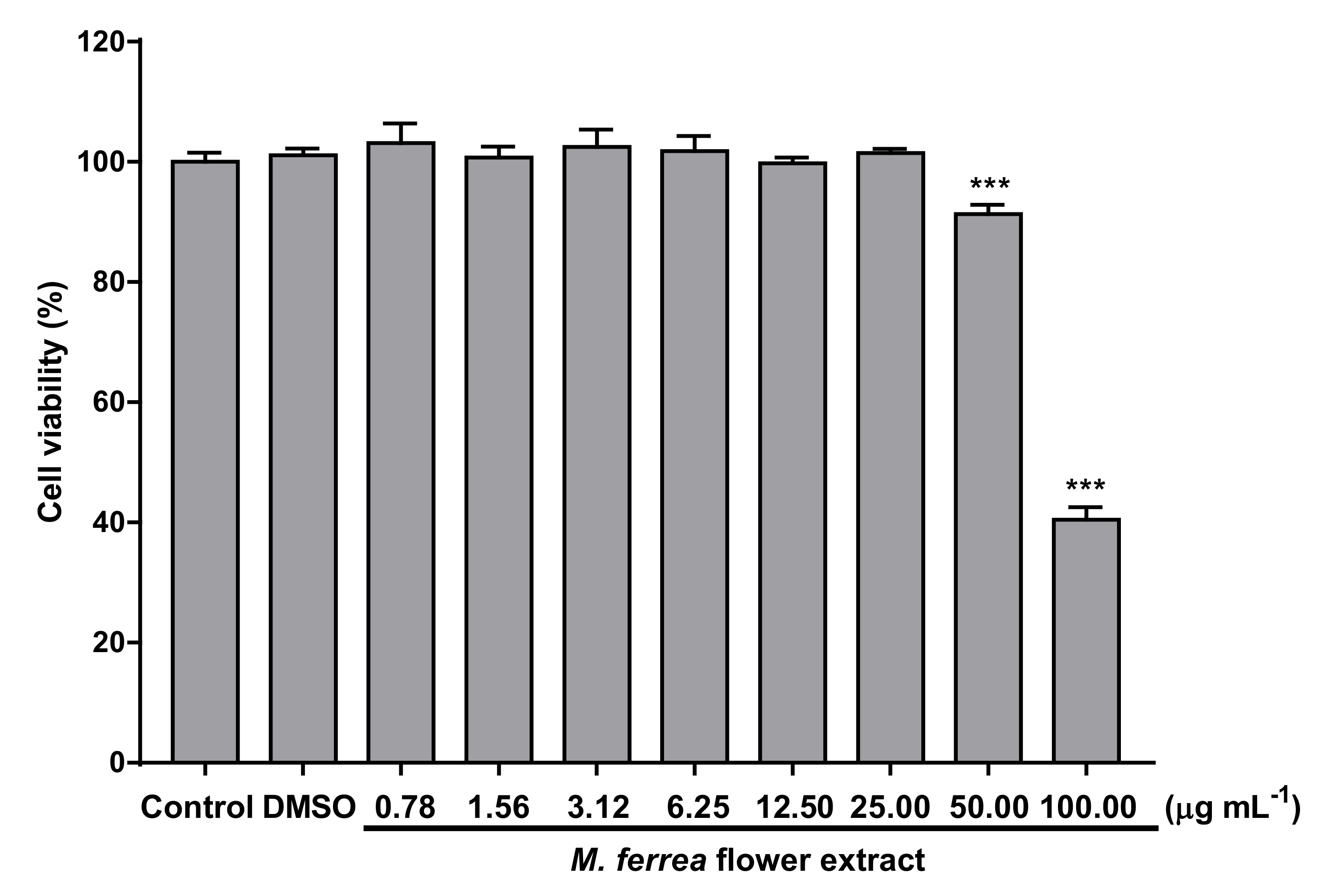
2.7.2. Cytotoxicity
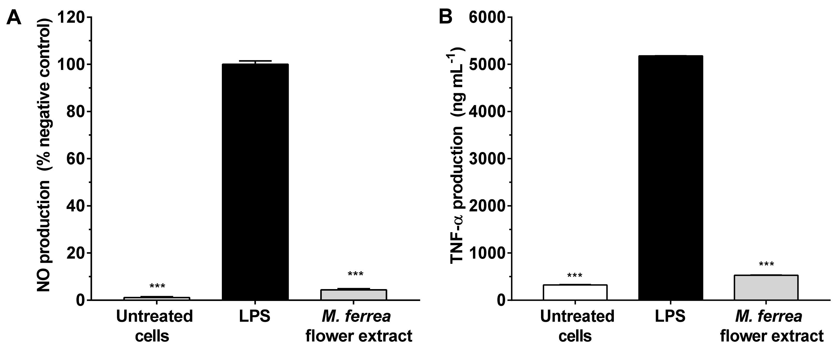
2.7.3. Inhibition of NO Production and Pro-Inflammatory Cytokines
2.8. Anti-Tyrosinase Assay
2.9. Statistical Analysis
3. Results and Discussion
3.1. Extraction
3.2. Total Phenolic Contents and Anti-Oxidant Activities
3.3. Anti-Bacterial Activity
3.4. Anti-Inflammatory Activities
3.5. Anti-Tyrosinase Activity
4. Conclusions
Author Contributions
Funding
Institutional Review Board Statement
Informed Consent Statement
Data Availability Statement
Acknowledgments
Conflicts of Interest
References
- De Canha, M.N.; Komarnytsky, S.; Langhansova, L.; Lall, N. Exploring the anti-acne potential of Impepho [Helichrysum odoratissimum (L.) sweet] to combat Cutibacterium acnes virulence. Front. Pharmacol. 2020, 10, 1559. [Google Scholar] [CrossRef] [Green Version]
- Chen, L.-W.; Chung, H.-L.; Wang, C.-C.; Su, J.-H.; Chen, Y.-J.; Lee, C.-J. Anti-acne effects of cembrene diterpenoids from the cultured soft coral Sinularia flexibilis. Mar. Drugs 2020, 18, 487. [Google Scholar] [CrossRef] [PubMed]
- Fournière, M.; Latire, T.; Souak, D.; Feuilloley, M.G.J.; Bedoux, G. Staphylococcus epidermidis and Cutibacterium acnes: Two major sentinels of skin microbiota and the influence of cosmetics. Microorganisms 2020, 8, 1752. [Google Scholar] [CrossRef] [PubMed]
- Farrar, M.D.; Ingham, E. Acne: Inflammation. Clin. Dermatol. 2004, 22, 380–384. [Google Scholar] [CrossRef] [PubMed]
- Jin, S.; Lee, M.-Y. Kaempferia parviflora extract as a potential anti-acne agent with anti-inflammatory, sebostatic and anti-propionibacterium acnes activity. Int. J. Mol. Sci. 2018, 19, 3457. [Google Scholar] [CrossRef] [Green Version]
- Srihaphon, K.; Wongwat, T.; Lamlertthon, S.; Pitaksuteepong, T. Investigation on the potential application of Morus alba stem extract for inflammatory acne vulgaris. Songklanakarin J. Sci. Technol. 2020, 42, 1319–1325. [Google Scholar]
- Lee, J.W.; Kang, Y.J.; Choi, H.K.; Yoon, Y.G. Fractionated coptis chinensis extract and its bioactive component suppress Propionibacterium acnes-stimulated inflammation in human keratinocytes. J. Microbiol. Biotechnol. 2018, 28, 839–848. [Google Scholar] [CrossRef] [Green Version]
- Kanlayavattanakul, M.; Lourith, N. Therapeutic agents and herbs in topical application for acne treatment. Int. J. Cosmet. Sci. 2011, 33, 289–297. [Google Scholar] [CrossRef] [PubMed]
- Lalla, J.K.; Nandedkar, S.Y.; Paranjape, M.H.; Talreja, N.B. Clinical trials of ayurvedic formulations in the treatment of acne vulgaris. J. Ethnopharmacol. 2001, 78, 99–102. [Google Scholar] [CrossRef]
- Griffith, G.; Trueman, L.; Crowther, T.; Thomas, B.; Smith, B. Onions—A global benefit to health. Phytother. Res. 2002, 16, 603–615. [Google Scholar] [CrossRef] [PubMed]
- Van Wyk, B.E. A broad review of commercially important southern African medicinal plants. J. Ethnopharmacol. 2008, 119, 342–355. [Google Scholar] [CrossRef] [PubMed]
- Raman, A.; Weir, U.; Bloomfield, S.F. Antimicrobial effects of tea tree oil and its major components on Staphylococcus aureus, S. epidermidis and Propionibacterium acnes. Lett. Appl. Microbiol. 1995, 21, 242–245. [Google Scholar] [CrossRef]
- Smitinand, T. Thai Plant Names; Revised Edition; Office of the Forest Herbarium, Department of Natural Park, Wildlife and Plant Conservation: Bangkok, Thailand, 2014; p. 376. [Google Scholar]
- Bunyapraphatsara, N.; Chokchaicharoenporn, O. Indigenous Medicinal Herbs; Prachachon: Bangkok, Thailand, 1999; pp. 534–537. [Google Scholar]
- Kshirsagar, P.R.; Patil, S.M. Phytochemistry and pharmacology of Mesua ferrea L. In Bioactive Compounds in Underutilized Fruits and Nuts; Murthy, H., Bapat, V., Eds.; Reference Series in Phytochemistry; Springer: Cham, Switzerland, 2020; pp. 223–256. [Google Scholar]
- Govindachari, T.R.; Pai, B.R.; Subramaniam, P.S.; Rao, U.R.; Muthukumaraswa, N. Constituents of Mesua ferrea L.–mesuaxanthone A and mesuaxanthone B. Tetrahedron 1967, 23, 243–248. [Google Scholar] [CrossRef]
- Teh, S.S.; Ee, G.C.L.; Rahmani, M.; Taufiq-Yap, Y.H.; Go, R.; Mah, S.H. Pyranoxanthones from Mesua ferrea. Molecules 2011, 16, 5647–5654. [Google Scholar] [CrossRef]
- Ee, G.C.L.; Teh, S.S.; Rahmani, M.; Taufiq-Yap, Y.H.; Go, R.; Mah, S.H. A new furanoxanthone from the root bark of Mesua ferrea. Lett. Org. Chem. 2012, 9, 457–459. [Google Scholar]
- Rasol, N.E.; Naz, H.; Awang, K.; Ridhwan, M.J.M.; Choy, Y.K.; Ismail, N.H. Isomeric polycyclic polyprenylated acylphloroglucinols from the bark of Mesua ferrea (Clusiaceae). Nat. Prod. Commun. 2017, 12, 1283–1286. [Google Scholar] [CrossRef] [Green Version]
- Chukaew, A.; Saithong, S.; Chusri, S.; Limsuwan, S.; Watanapokasin, R.; Voravuthikunchai, S.P.; Chakthong, S. Cytotoxic xanthones from the roots of Mesua ferrea L. Phytochemistry 2019, 157, 64–70. [Google Scholar] [CrossRef]
- Chahar, M.K.; Sanjaya Kumar, D.S.; Geetha, L.; Lokesh, T.; Manohara, K.P. Mesua ferrea L.: A review of the medical evidence for its phytochemistry and pharmacological actions. Afr. J. Pharm. Pharmacol. 2013, 7, 211–219. [Google Scholar] [CrossRef] [Green Version]
- Asif, M.; Jafari, S.F.; Iqbal, Z.; Revadigar, V.; Oon, C.E.; Majid, A.S.A.; Majid, A.M.S.A. Ethnobotanical and phytopharmacological attributes of Mesua ferrea: A mini review. J. Appl. Pharm. Sci. 2017, 7, 242–251. [Google Scholar]
- Rajalakshmi, P.; Vadivel, V.; Ravichandran, N.; Brindha, P. Investigation on pharmacognostic parameters of Sirunagapoo (Mesua ferrea L): A traditional Indian herbal drug. Pharmacogn. J. 2019, 11, 225–230. [Google Scholar] [CrossRef] [Green Version]
- Nićiforović, N.; Mihailović, V.; Mašković, P.; Solujić, S.; Stojković, A.; Muratspahić, D.P. Antioxidant activity of selected plant species; potential new sources of natural antioxidants. Food Chem. Toxicol. 2010, 48, 3125–3130. [Google Scholar] [CrossRef] [PubMed]
- Gordon, M.H.; Paiva-Martins, F.; Almeida, M. Antioxidant activity of hydroxytyrosol acetate compared with that of other olive oil polyphenols. J. Agric. Food Chem. 2001, 49, 2480–2485. [Google Scholar] [CrossRef] [PubMed]
- Gião, M.S.; González-Sanjosé, M.L.; Rivero-Pérez, M.D.; Pereira, C.I.; Pintado, M.E.; Malcata, F.X. Infusions of Portuguese medicinal plants: Dependence of final antioxidant capacity and phenol content on extraction features. J. Sci. Food Agric. 2007, 87, 2638–2647. [Google Scholar] [CrossRef] [PubMed]
- Berker, K.I.; Güçlü, K.; Tor, I.; Apak, R. Comparative evaluation of Fe(III) reducing power-based antioxidant capacity assays in the presence of phenanthroline, batho-phenanthroline, tripyridyltriazine (FRAP), and ferricyanide reagents. Talanta 2007, 72, 1157–1165. [Google Scholar] [CrossRef]
- Bahiense, J.B.; Marques, F.M.; Figueira, M.M.; Vargas, T.S.; Kondratyuk, T.P.; Endringer, D.C.; Scherer, R.; Fronza, M. Potential anti-inflammatory, antioxidant and antimicrobial activities of Sambucus australis. Pharm. Biol. 2017, 55, 991–997. [Google Scholar] [CrossRef] [Green Version]
- Suebsakwong, P.; Chulrik, W.; Chunglok, W.; Li, J.-X.; Yao, Z.-J.; Suksamrarn, A. New triterpenoid saponin glycosides from the fruit fibers of Trichosanthes cucumerina L. RSC Adv. 2020, 10, 10461–10470. [Google Scholar] [CrossRef] [Green Version]
- Athipornchai, A.; Niyomtham, N.; Pabuprapap, W.; Ajavakom, V.; Duca, M.; Azoulay, S.; Suksamrarn, A. Potent tyrosinase inhibitory activity of curcuminoid analogues and inhibition kinetics studies. Cosmetics 2021, 8, 35. [Google Scholar] [CrossRef]
- Leyden, J.J.; McGinley, K.J.; Vowels, B. Propionibacterium acnes colonization in acne and non-acne. Dermatology 1998, 196, 55–58. [Google Scholar] [CrossRef]
- Ozgen, M.; Reese, R.N.; Tulio Jr, A.Z.; Scheerens, J.C.; Miller, A.R. Modified 2,2-azino-bis-3-ethylbenzothiazoline-6-sulfonic acid (ABTS) method to measure antioxidant capacity of selected small fruits and comparison to ferric reducing antioxidant power (FRAP) and 2,2′-diphenyl-1-picrylhydrazyl (DPPH) methods. J. Agric. Food Chem. 2006, 54, 1151–1157. [Google Scholar] [CrossRef]
- Gliszczyńska-Świgło, A. Antioxidant activity of water soluble vitamins in the TEAC (Trolox equivalent antioxidant capacity) and the FRAP (ferric reducing antioxidant power) assays. Food Chem. 2006, 96, 131–136. [Google Scholar] [CrossRef]
- Kwon, S.H.; Wang, Z.; Hwang, S.H.; Kang, Y.-H.; Lee, J.-Y.; Lim, S.S. Comprehensive evaluation of the antioxidant capacity of Perilla frutescens leaves extract and isolation of free radical scavengers using step-wise HSCCC guided by DPPH-HPLC. Int. J. Food Prop. 2017, 20, 921–934. [Google Scholar] [CrossRef] [Green Version]
- He, J.; Xu, L.; Yang, L.; Wang, X. Epigallocatechin gallate is the most effective catechin against antioxidant stress via hydrogen peroxide and radical scavenging activity. Med. Sci. Monit. 2018, 24, 8198–8206. [Google Scholar] [CrossRef] [PubMed]
- Yoon, J.Y.; Kwon, H.H.; Min, S.U.; Thiboutot, D.M.; Suh, D.H. Epigallocatechin-3-gallate improves acne in humans by modulating intracellular molecular targets and inhibiting P. acnes. J. Investig. Dermatol. 2013, 133, 429–440. [Google Scholar] [CrossRef] [PubMed] [Green Version]
- Phimnuan, P.; Yakaew, S.; Yosboonruang, A.; Luangbudnak, W.; Grandmottet, F.; Viyoch, J. Development of anti-acne film from bio-cellulose incorporating Punica granatum peel extract. Walailak J. Sci. Technol. 2018, 16, 765–778. [Google Scholar] [CrossRef]
- Lee, C.J.; Chen, L.G.; Liang, W.L.; Wang, C.C. Multiple activities of Punica granatum Linne against acne vulgaris. Int. J. Mol. Sci. 2017, 18, 141. [Google Scholar] [CrossRef] [PubMed] [Green Version]
- Mogana, R.; Adhikari, A.; Tzar, M.N.; Ramliza, R.; Wiart, C. Antibacterial activities of the extracts, fractions and isolated compounds from Canarium patentinervium Miq. against bacterial clinical isolates. BMC Complement. Med. Ther. 2020, 20, 55. [Google Scholar] [CrossRef] [Green Version]
- Jiamboonsri, P.; Pithayanukul, P.; Bavovada, R.; Chomnawang, M.T. The inhibitory potential of Thai mango seed kernel extract against methicillin-resistant Staphylococcus aureus. Molecules 2011, 16, 6255–6270. [Google Scholar] [CrossRef] [Green Version]
- Belay, D.; Kenubih, A.; Yesuf, M.; Kebede, E.; Yayeh, M.; Birhan, M. Antioxidant and antimicrobial activity of solvent fractions of Calpurnia aurea (Ait.) Benth. (Fabaceae). J. Exp. Pharmacol. 2021, 13, 499–509. [Google Scholar] [CrossRef] [PubMed]
- Shafiei, Z.; Shuhairi, N.N.; Md Fazly Shah Yap, N.; Harry Sibungkil, C.-A.; Latip, J. Antibacterial activity of Myristica fragrans against oral pathogens. Evid. Based Complement. Alternat. Med. 2012, 2012, 1–7. [Google Scholar] [CrossRef] [Green Version]
- Desbois, A.; Lawlor, K. Antibacterial activity of long-chain polyunsaturated fatty acids against Propionibacterium acnes and Staphylococcus aureus. Mar. Drugs 2013, 11, 4544–4557. [Google Scholar] [CrossRef] [Green Version]
- Kim, S.; Oh, S.; Noh, H.; Ji, S.; Lee, S.; Koo, J.; Choi, C.W.; Jhun, H.P. In vitro antioxidant and anti-Propionibacterium acnes activities of cold water, hot water, and methanol extracts, and their respective ethyl acetate fractions, from Sanguisorba officinalis L. roots. Molecules 2018, 23, 3001. [Google Scholar] [CrossRef] [PubMed] [Green Version]
- Moon, S.H.; Roh, H.S.; Kim, Y.H.; Kim, J.E.; Ko, J.Y.; Ro, Y.S. Antibiotic resistance of microbial strains isolated from Korean acne patients. J. Dermatol. 2012, 39, 833–837. [Google Scholar] [CrossRef] [PubMed]
- Lo, C.W.; Lai, Y.K.; Liu, Y.T.; Gallo, R.L.; Huang, C.M. Staphylococcus aureus hijacks a skin commensal to intensify its virulence: Immunization targeting β-hemolysin and CAMP factor. J. Investig. Dermatol. 2011, 131, 401–409. [Google Scholar] [CrossRef] [Green Version]
- Kumar, B.; Pathak, R.; Mary, P.B.; Jha, D.; Sardana, K.; Gautam, H.K. New insights into acne pathogenesis: Exploring the role of acne-associated microbial populations. Dermatol. Sin. 2016, 34, 63–74. [Google Scholar] [CrossRef] [Green Version]
- Hajlaoui, H.; Arraouadi, S.; Noumi, E.; Aouadi, K.; Adnan, M.; Khan, M.A.; Kadri, A.; Snoussi, M. Antimicrobial, antioxidant, anti-acetylcholinesterase, antidiabetic, and pharmacokinetic properties of Carum carvi L. and Coriandrum sativum L. essential oils alone and in combination. Molecules 2021, 26, 3625. [Google Scholar] [CrossRef]
- Peker, E.G.G.; Kaltalioglu, K. Cinnamaldehyde and eugenol protect against LPS-stimulated oxidative stress and inflammation in Raw 264.7 cells. J. Food Biochem. 2021, e13980. [Google Scholar] [CrossRef]
- Tsai, T.H.; Huang, W.C.; Lien, T.J.; Huang, Y.H.; Chang, H.; Yu, C.H.; Tsai, P.J. Clove extract and eugenol suppress inflammatory responses elicited by Propionibacterium acnes in vitro and in vivo. Food Agric. Immunol. 2017, 28, 916–931. [Google Scholar] [CrossRef] [Green Version]
- Aruldass, C.; Marimuthu, M.; Ramanathan, S.; Mansor, S.; Murugaiyah, V. Effects of Mesua ferrea leaf and fruit extracts on growth and morphology of Staphylococcus aureus. Microsc. Microanal. 2013, 19, 254–260. [Google Scholar] [CrossRef]
- Lademann, J.; Richter, H.; Jacobi, U.; Patzelt, A.; Hueber-Becker, F.; Ribaud, C.; Benech-Kieffer, F.; Dufour, E.K.; Sterry, W.; Schaefer, H.; et al. Human percutaneous absorption of a direct hair dye comparing in vitro and in vivo results: Implications for safety assessment and animal testing. Food Chem. Toxicol. 2008, 46, 2214–2223. [Google Scholar] [CrossRef] [PubMed]
- Novilla, A.; Djamhuri, D.S.; Nurhayati, B.; Rihibiha, D.D.; Afifah, E.; Widowati, W. Anti-inflammatory properties of Oolong tea (Camellia sinensis) ethanol extract and epigallocatechin gallate in LPS-induced RAW 264.7 cells. Asian Pac. Trop. Biomed. 2017, 7, 1005–1009. [Google Scholar] [CrossRef]
- Shin, S.; Lee, J.-A.; Son, D.; Park, D.; Jung, E. Anti-skin-aging activity of a standardized extract from Panax ginseng leaves in vitro and in human volunteer. Cosmetics 2017, 4, 18. [Google Scholar] [CrossRef] [Green Version]
- Escobar, S.; Valois, A.; Nielsen, M.; Closs, B.; Kerob, D. Effectiveness of a formulation containing peptides and vitamin C in treating signs of facial ageing: Three clinical studies. Int. J. Cosmet. Sci. 2021, 43, 131–135. [Google Scholar] [CrossRef] [PubMed]
- Rattanawiwatpong, P.; Wanitphakdeedecha, R.; Bumrungpert, A.; Maiprasert, M. Anti-aging and brightening effects of a topical treatment containing vitamin C, vitamin E, and raspberry leaf cell culture extract: A split-face, randomized controlled trial. J. Cosmet. Dermatol. 2020, 19, 671–676. [Google Scholar] [CrossRef] [PubMed] [Green Version]
- Kang, M.C.; Lee, J.-W.; Lee, T.H.; Subedi, L.; Wahedi, H.M.; Do, S.-G.; Shin, E.; Moon, E.-Y.; Kim, S.Y. UP256 inhibits hyperpigmentation by tyrosinase expression/dendrite formation via Rho-dependent signaling and by primary cilium formation in melanocytes. Int. J. Mol. Sci. 2020, 21, 5341. [Google Scholar] [CrossRef] [PubMed]

| Sample | Total Phenolic Contents (µg GAE mg−1) | Anti-Oxidant Activities (IC50 ± SD; µg mL−1) | |||
|---|---|---|---|---|---|
| DPPH | ABTS | FRAP | NO | ||
| M. ferrea extract | 227.23 ± 0.01 | 12.87 ± 1.04 | 16.95 ± 1.04 | 96.33 ± 1.05 | 79.83 ± 1.08 |
| L-Ascorbic acid 1 | - | 3.46 ± 1.09 | 0.86 ± 1.03 | 2.06 ± 1.05 | 0.35 ± 1.25 |
| Microorganism | Diameter of Inhibition Zone (Mean ± SD) (mm) | ||
|---|---|---|---|
| M. ferrea Flower Extract | Clindamycin 1 | Tetracycline 2 | |
| C. acnes | 14.33 ± 0.29 | 60.50 ± 0.62 | - |
| S. aureus | 9.08 ± 0.38 | - | 33.50 ± 0.50 |
| S. epidermidis | 11.50 ± 0.25 | - | 10.00 ± 0.25 |
| Microorganism | M. ferrea Flower Extract (Mean ± SD) | ||
|---|---|---|---|
| MIC (mg mL−1) | MBC (mg mL−1) | MBC/MIC Ratio | |
| C. acnes | 3.12 ± 0.00 | 25.00 ± 0.00 | 8.01 |
| S. aureus | 6.25 ± 0.00 | 12.50 ± 0.00 | 2.00 |
| S. epidermidis | 0.78 ± 0.00 | 1.56 ± 0.00 | 2.00 |
| Sample | Anti-Tyrosinase Activity (IC50 ± SD; µg mL−1) |
|---|---|
| M. ferrea extract | 219.58 ± 3.41 |
| Kojic acid 1 | 46.05 ± 1.16 |
Publisher’s Note: MDPI stays neutral with regard to jurisdictional claims in published maps and institutional affiliations. |
© 2021 by the authors. Licensee MDPI, Basel, Switzerland. This article is an open access article distributed under the terms and conditions of the Creative Commons Attribution (CC BY) license (https://creativecommons.org/licenses/by/4.0/).
Share and Cite
Nakyai, W.; Pabuprapap, W.; Sroimee, W.; Ajavakom, V.; Yingyongnarongkul, B.-e.; Suksamrarn, A. Anti-Acne Vulgaris Potential of the Ethanolic Extract of Mesua ferrea L. Flowers. Cosmetics 2021, 8, 107. https://doi.org/10.3390/cosmetics8040107
Nakyai W, Pabuprapap W, Sroimee W, Ajavakom V, Yingyongnarongkul B-e, Suksamrarn A. Anti-Acne Vulgaris Potential of the Ethanolic Extract of Mesua ferrea L. Flowers. Cosmetics. 2021; 8(4):107. https://doi.org/10.3390/cosmetics8040107
Chicago/Turabian StyleNakyai, Wongnapa, Wachirachai Pabuprapap, Wichuda Sroimee, Vachiraporn Ajavakom, Boon-ek Yingyongnarongkul, and Apichart Suksamrarn. 2021. "Anti-Acne Vulgaris Potential of the Ethanolic Extract of Mesua ferrea L. Flowers" Cosmetics 8, no. 4: 107. https://doi.org/10.3390/cosmetics8040107
APA StyleNakyai, W., Pabuprapap, W., Sroimee, W., Ajavakom, V., Yingyongnarongkul, B.-e., & Suksamrarn, A. (2021). Anti-Acne Vulgaris Potential of the Ethanolic Extract of Mesua ferrea L. Flowers. Cosmetics, 8(4), 107. https://doi.org/10.3390/cosmetics8040107