Article

Comparison of Neck Pain and Posture with Spine Angle Tracking System between Static and Dynamic Computer Monitor Use

1
Department of Biomedical Engineering, Hanyang University, Seoul 04763, Republic of Korea
2
Department of Neurosurgery, Chungnam National University Sejong Hospital, Sejong 30099, Republic of Korea
3
Division of Medical Statistics, Medical Research Collaborating Center, Seoul National University Hospital, Seoul 03080, Republic of Korea
4
Materials & Devices Advanced Research Center, LG Electronics, Seoul 07795, Republic of Korea
5
Department of Neurosurgery, Seoul National University Hospital, Seoul 03080, Republic of Korea
6
Department of Neurosurgery, College of Medicine, Seoul National University, Seoul 03080, Republic of Korea
7
Department of Brain and Cognitive Sciences, Seoul National University, Seoul 08826, Republic of Korea
*
Author to whom correspondence should be addressed.
These authors contributed equally to this work.
Electronics 2024, 13(7), 1363; https://doi.org/10.3390/electronics13071363
Submission received: 5 February 2024 / Revised: 1 April 2024 / Accepted: 2 April 2024 / Published: 4 April 2024
(This article belongs to the Special Issue New Insights Into Smart and Intelligent Sensors)

Abstract

:
This study investigates the effect of dynamic changes in monitor height and tilt on neck pain and posture of computer users. Using a wearable device, we aim to compare neck pain and spine angle between static and dynamic monitors. A spine angle tracking system using the Inertial Measurement Unit (IMU) was proposed, and the accuracy was validated. Eight office workers participated for five hours over two days, and each day used either a static monitor or a dynamic monitor that changed height and tilt every 30 min. The angles of C0, C7, L1, and S1 endplates were estimated using the proposed system. Changes in neck pain and spine angle with time were compared in static and dynamic monitors. The intraclass correlation coefficient confirmed a high concordance between the estimated and actual angles (p < 0.001). Rehabilitation Bioengineering Group (RBG) score increased less in the dynamic monitor compared to the static monitor (p = 0.003). Spinal curvatures are bent in the static monitor compared to the dynamic monitor. The estimated angles aligned well with X-ray measurements. A dynamic monitor that changes height and tilt at regular intervals may reduce neck pain increase and reduce bend forward posture compared to a static monitor.

1. Introduction

Neck pain is common worldwide and causes significant disability and economic costs [1]. One of the main causes of neck pain is forward head posture (FHP) [2]. FHP is defined as a flexed posture of the neck and head adopted for reading and typing while using computers or smartphones [3,4,5,6]. FHP is common among office workers, and this posture overstrains the muscles and ligaments of the neck and back. Thus, it causes pain in the neck, shoulders, and back [7,8,9]. Static posture maintained for a longer time, such as prolonged use of a computer or laptop, and repetitive work are known to be major factors of this posture and neck pain [10,11,12].
Neck pain in office workers has an incidence of approximately 20%, and some studies have reported it as high as 48% [13]. Many studies have shown that computer use increases neck pain [14,15,16], and computer workstation design is also one of the factors contributing to neck pain [13]. Considering that the most crucial interface when using a computer is the monitor, it can be inferred that monitor setting correlates with neck pain. Many studies have been conducted on this issue, and appropriate monitor height and tilt are suggested through these studies [17,18,19,20]. However, previous studies were conducted in a static environment. It is unknown how the dynamic environment of changing the height and tilt of the monitor affects the neck pain and spine curvature of computer users. To observe changes in posture as a function of the monitor’s environment, a continuous posture measurement is required.
X-ray examination provides an accurate spine angle, but it may not be sufficient to explain neck pain as a one-time capture cannot represent neck angles in daily life [21]. Therefore, we propose a system that continuously measures the spine angle using Inertial Measurement Unit (IMU). Wearable devices using IMU have become increasingly popular in recent years for monitoring human movement. IMU is a small sensor that can be attached to the body or integrated into wearable devices to measure acceleration, angular velocity, and magnetic field strength. The sensor can provide valuable information about the orientation of the body, which can be used to monitor and analyze physical activity, gait, and fall risk assessment [22,23,24]. Han et al. used IMU to study the effect of smartphone use on FHP, demonstrating the potential for accurate measurement and providing users with feedback to improve posture [25]. These studies suggest that wearable devices using IMU could be a useful tool for monitoring posture and neck pain in office workers. Therefore, this study aims to develop a wearable IMU system capable of measuring spine angles and using it to compare neck pain and posture in static and dynamic monitor environments.

2. Materials and Methods

2.1. Spine Angle Tracking System

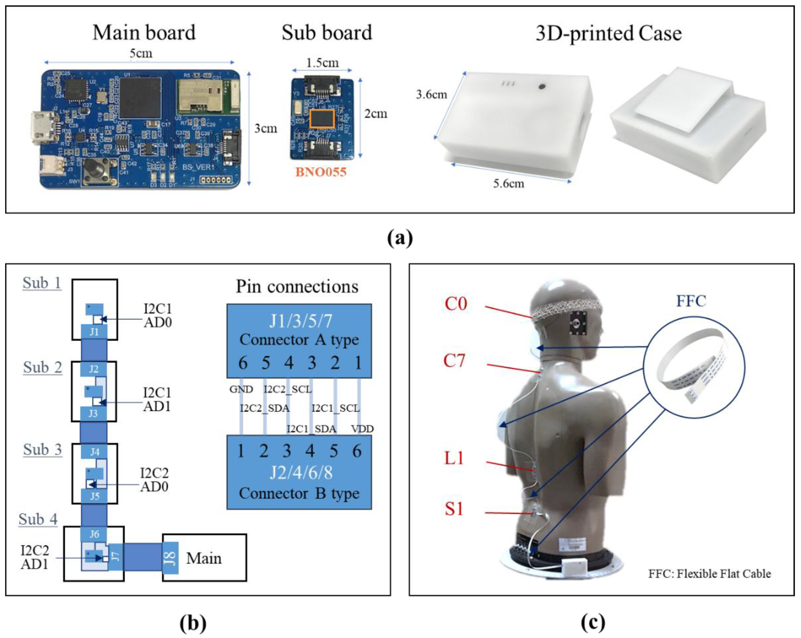
2.1.1. IMU-Embedded Wearable Device

To measure the changes in spine angle during work, we developed a wireless angle measurement system using an IMU sensor chip. IMU sensor is a system composed of gyroscopes, accelerometers, and magnetic sensors. A gyroscope measures an object’s angular velocity, while an accelerometer measures linear acceleration (direction of gravity). A magnetometer detects magnetic fields (vectors towards Earth’s magnetic North). By using these three data, the object’s movement is continuously measured in real-time. In this study, angles were measured using BNO055 (Bosch Sensortec, Reutlingen, Germany), which is known to have the highest accuracy [26]. Figure 1 shows the hardware, schematic diagrams, and placement of the system. Unlike existing wired IMU systems, our system is designed to be wireless, with the main board housed in a small 3D-printed case that can be mounted on a pair of trousers to minimize disturbance during work. Each of the four sub-boards contains one IMU sensor, connected to the main board via an inter-integrated circuit (I2C) interface. The angular data of three planes (sagittal plane, coronal plane, and transverse plane) for each sensor can be obtained via Bluetooth communication with a sampling rate of 10 Hz. The battery life is 6 h, which is sufficient for the experiment. The sub-boards were attached to the occipital protuberance (reflecting McGregor’s line [27], henceforth referred to as C0) C7, L1, and S1 spinous process tips. The length of the flexible flat cable connecting boards was determined by considering the demographic average and range of motion.

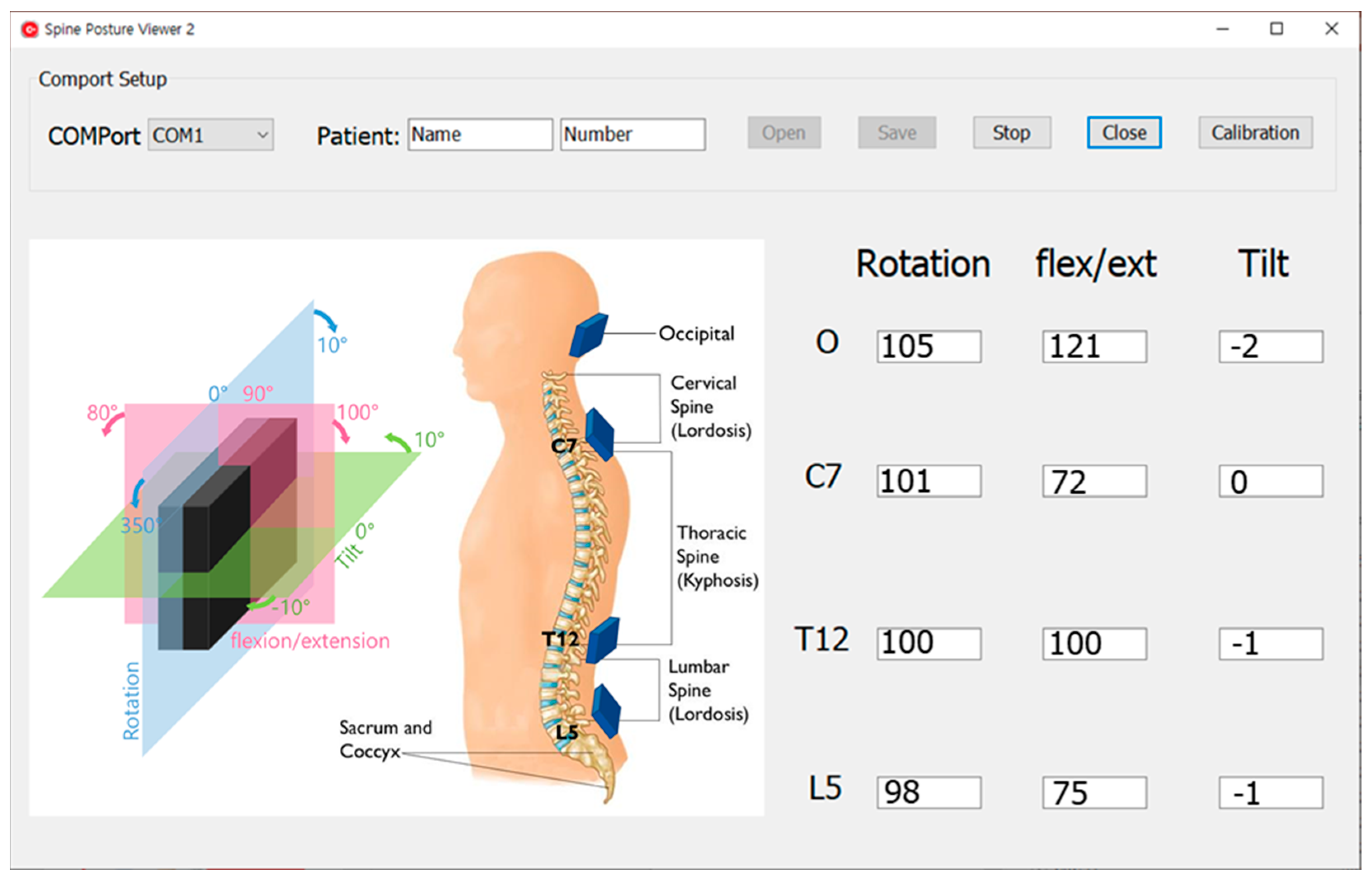
2.1.2. Data Acquisition and User Interface Viewer

Figure 2 shows the user interface viewer for outputting data to a PC. The viewer was written in the C++ programming language in the Embarcadero RAD Studio. With this viewer, angular data from three planes for four sensors are stored with time stamps as a text file on a PC.

2.1.3. System Algorithms

The data processing involves two steps. The initial step is a calibration process that addresses the disparity between the sensor angle and the angle of the vertebral endplate. Prior to conducting the experiment, an X-ray was taken while the system was attached, allowing for the calculation of the following equations for each of the four sensors. These equations serve to correct the difference between the sensor angles and the angles of endplates in the sagittal plane, as measured from the X-ray, facilitating consecutive angle measurements.
C a l i b r a t e d = R a w I M U D i f f D i f f = I n i t I M U X r a y
The second step is the Cobb angle calculation, which describes the degree of curvature of the neck, back, and lumbar. The Cobb angle is the most widely used measurement for quantifying spinal curvature [28]. Since curvature in the Cobb method is defined as the angle between the endplate of the upper vertebra and the endplate of the lower vertebra, each of the four sensors will have three Cobb angles. Therefore, the joint angle between two adjacent sensors is calculated using a rotation matrix [29]. A conceptual diagram of the algorithm, including the individual frames and transformation sequences for computing the joint angle between sensors, is illustrated in Figure 3.
LA and CA define the anatomical frames for L1 and C7, respectively. The sensor measurement frames for L1 and C7 are denoted LM and CM. The z axes of the earth frames LE and CE, are aligned but have different x-y coordinates. As an example, the rotation matrix representing the joint angle from the coordinate system of the fixed C7 endplate to the coordinate system of the moving L1 endplate can be represented as follows:
T C A L A = T C A C M M C S M L 1 T L A L M 1
where S is the alignment matrix between the two earth frames M L . and M C are the orientation matrices of the angles measured by the sensor with respect to their corresponding earth frame, LE, and CE, respectively. T L A L M and T C A C M are the transformation matrices between the sensor frame and the anatomical frame.
The relationship between the anatomical angles and the rotation matrix is given by:
T C A L A = cos β cos γ sin γ cos β sin β cos γ sin β sin α + sin γ cos α cos α cos γ sin α sin β sin γ cos β sin α sin γ sin α cos γ sin β cos α sin γ sin β cos α + cos γ sin α cos β cos α
Solving the corresponding rotation matrix to calculate the anatomical angle as:
Flexion / Extension α = tan 2 1 T C A L A 2,3 T C A L A 3,3 Abduction / Adduction β = sin 1 T C A L A 1,3 Internal / External γ = tan 2 1 T C A L A 1,2 T C A L A 1,1
where t a n 2 1 is the four quadrant arctangent [29]. Of the three anatomical angles, the sagittal plane angle corresponding to flexion/extension was used for further analysis.

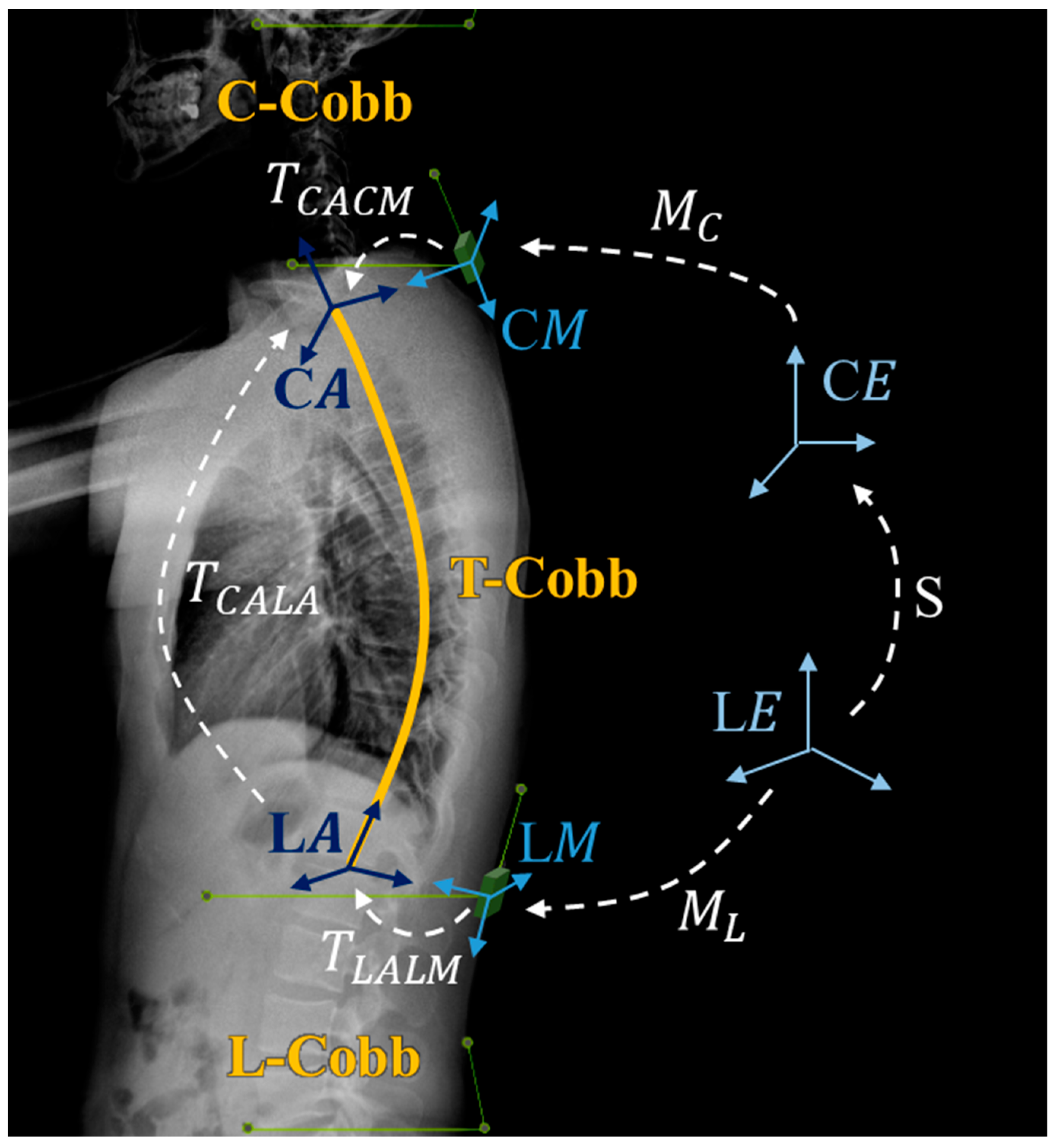
2.2. Evaluation of Sensor Accuracy and Concordance between Sensor Estimates and X-ray Measurements

To evaluate the accuracy of the developed system’s sensors, we conducted 10 measurements each at angles of 30, 60, and 90 degrees.
Additionally, we examined the agreement between the estimated spine angle provided by the system and the actual angle. Considering the potential interference from hair or flesh at the C0 and C7 positions, we captured X-rays in both extension and flexion positions to assess the reliability of the measurements within these sensor clusters.

2.3. Experimental Design

This study is a non-randomized experimental design study for the monitor environment using healthy persons. The primary outcome is to determine if there is a difference in neck pain between static monitor and dynamic monitor. The secondary outcome is whether there is a difference in the spine angle change according to the monitor environment. We recruited office workers who have used computers for more than 5 h a day [14], 3 males and 5 females participated. Their years of service were at least 4 years. The mean age was 29.75 ± 1.39 years, the height was 168.5 ± 7.97 cm, and the weight was 62.25 ± 9.85 kg. Those who had been diagnosed with cervical or lumbar disease, deformities such as scoliosis, or other musculoskeletal disorders, and those who had previously undergone surgery in the spine were not enrolled in the study. Participants were asked to report any significant differences in neck pain compared to their usual daily life before the start of the experiments. We maintained a consistent experimental environment with the same desks, chairs, and monitors at the same place and time. The present study was conducted in accordance with the Declaration of Helsinki and the Guideline for Good Clinical Practice. The study protocol was approved by the Seoul National University Hospital ethics committee/institutional review board (IRB No. 2106-053-1227). A written informed consent was collected from each subject prior to participation.

2.3.1. Static Monitor and Dynamic Monitor

The experiment was conducted over two days with more than one-week intervals. For the first measurement, a conventional static monitor was used. A chair with a back was used. The height of the chair was adjusted so that the elbow was at the height of the desk. The height and tilt of the monitor were adjusted so that the participants felt comfortable before working. The height was defined as the distance between the center of the monitor and the desk. The tilt was defined as the monitor’s slope to a vertical line. A 32-inch monitor was used. As usual, they were asked to work 3 h in the morning, 1 h lunch, and 5 h in the afternoon, and data during the afternoon work were measured. Although it is a rule to take a 10-min break after 50 min of work, participants were allowed to take a break in the middle or continue to work as they were in their actual work. With an interval of 1 week or more after the first measurement, the second measurements were made using a dynamic monitor. The dynamic monitor environment consisted of three monitor settings that the experimenter changed sequentially at 30-min intervals. Each monitor setting was created with a monitor height and tilt that the subject felt comfortable with. The distance between the monitor and the subject was about 55 cm for each monitor setting. The view angle of the gaze looking at the monitor’s center was around 15°, which was in line with the optimal monitor setting [13]. Considering studies that neck pain and electromyogram (EMG) changes occur within 1 h [30], a dynamic monitor environment was configured to change monitor settings at 30-min intervals. Conditions on the other workstations were the same, except that the height and tilt of the monitors were changed at certain times.

2.3.2. Measurements

Each spine angle was measured at 6-min intervals through the IMU sensor while the participant was working. Neck pain was measured every hour using the RBG score which refers to the Rehabilitation Bioengineering Group pain scale, which is pain scale criteria adapted from Balasubramanian et al. that was used for evaluating perceived discomfort and pain from ‘no pain’ (0 points) to ‘unbearable pain’ (5 points) [31]. Details of RBG can be found in Table 1. In addition, monitor settings such as eye-level height, distance from the monitor, and height and tilt of the monitor were also measured for subgroup analysis.

2.4. Statistical Analysis

The intraclass correlation coefficient (ICC) for absolute, single measurement using the two-way mixed effects model was used as an index of concordance between the IMU sensor and X-ray, where 1 represents a total agreement and 0 represents no agreement at all. The primary endpoint was the neck pain measured at every hour of work. Using a linear mixed effects model with the time, the monitor environments, the interaction between time and the monitor environment, and the baseline neck pain as the fixed effects and subjects as the random effects, the difference in neck pain between a static and dynamic monitor environment was estimated and tested. The secondary endpoint was changes in spine angle measured with the proposed system.Angles of C0, C7, L1, and S1 endplate, C0-C7 (C-Cobb), C7-L1 (T-Cobb), and L1-S1 (L-Cobb) were obtained and the amounts of change from the baseline were compared using the mixed effects models: the subject was considered as a random effect, and the time, the monitor environment, and the interaction between time and the monitor environment as the fixed effects. We also examined the association between spine angle and the RBG score adjusting time and the monitor environment using the mixed effect model. The spine angles, the times, monitor environment, the interaction between the spine angles and the times, and the interaction between the spine angles and the monitor environment were considered as the fixed effect, and the subjects as random effects. Since the spine angles were measured at 6-min intervals and the RBG score was obtained at every hour. The average values of the angles for an hour were applied to that analysis. The data were analyzed via the SAS software package version 9.4 (SAS Institute Inc., Cary, NC, USA).

3. Results

3.1. Evaluation of Sensor Accuracy and Concordance between Sensor Estimates and X-ray Measurements

3.1.1. Sensor Accuracy

To verify the sensor accuracy of the developed system, we measured 30, 60, and 90 degrees 10 times each. As a result, we obtained the mean error shown in Table 2, which is similar to the errors in other studies using the BNO055 IMU sensor [26].

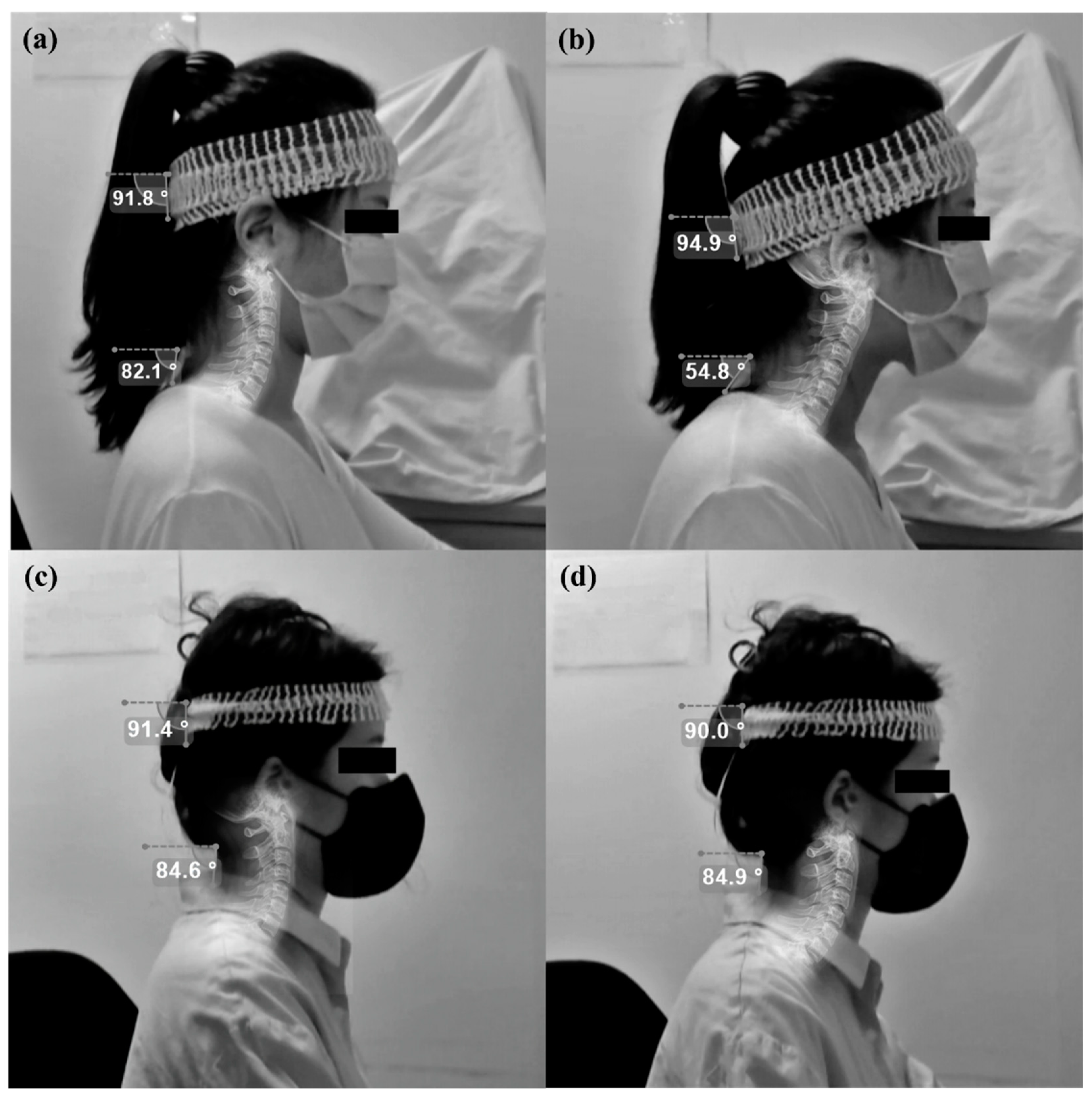
3.1.2. Concordance between Sensor Estimates and X-ray Measurements

The spine angles of flexion and extension by IMU sensor were estimated by correcting the difference between the angles measured by X-ray and IMU sensor in the neutral posture. Table 3 shows the degree of agreement between the C0 and C7 endplate angles measured by X-rays in the flexion and extension postures and those estimated by the IMU sensor. The ICC between the two measurements showed a high concordance rate (p < 0.001).

3.2. Baseline Characteristics

Table 4 shows a result of the Wilcoxon signed-rank test, there was no statistically significant difference between the baseline of neck pain and spine angle on the first day (static monitor) and second day (dynamic monitor). Monitor settings such as eye-level height, monitor distance, and height and tilt of the monitor did not show any difference in the two monitor environments.

3.3. Neck Pain Change in Static and Dynamic Monitor Environment

Figure 4 shows the amount of pain increases over time for each monitor environment. The x-axis is the time (minutes), and the y-axis is the RBG score. The interaction between the monitor environment and time was not significant in neck pain (p = 0.500), which means that the pattern of pain changed over time did not differ depending on the monitor environment. When the time was controlled, the neck pain increase was as small as 0.365 in the dynamic monitor compared to the static monitor (p = 0.003).

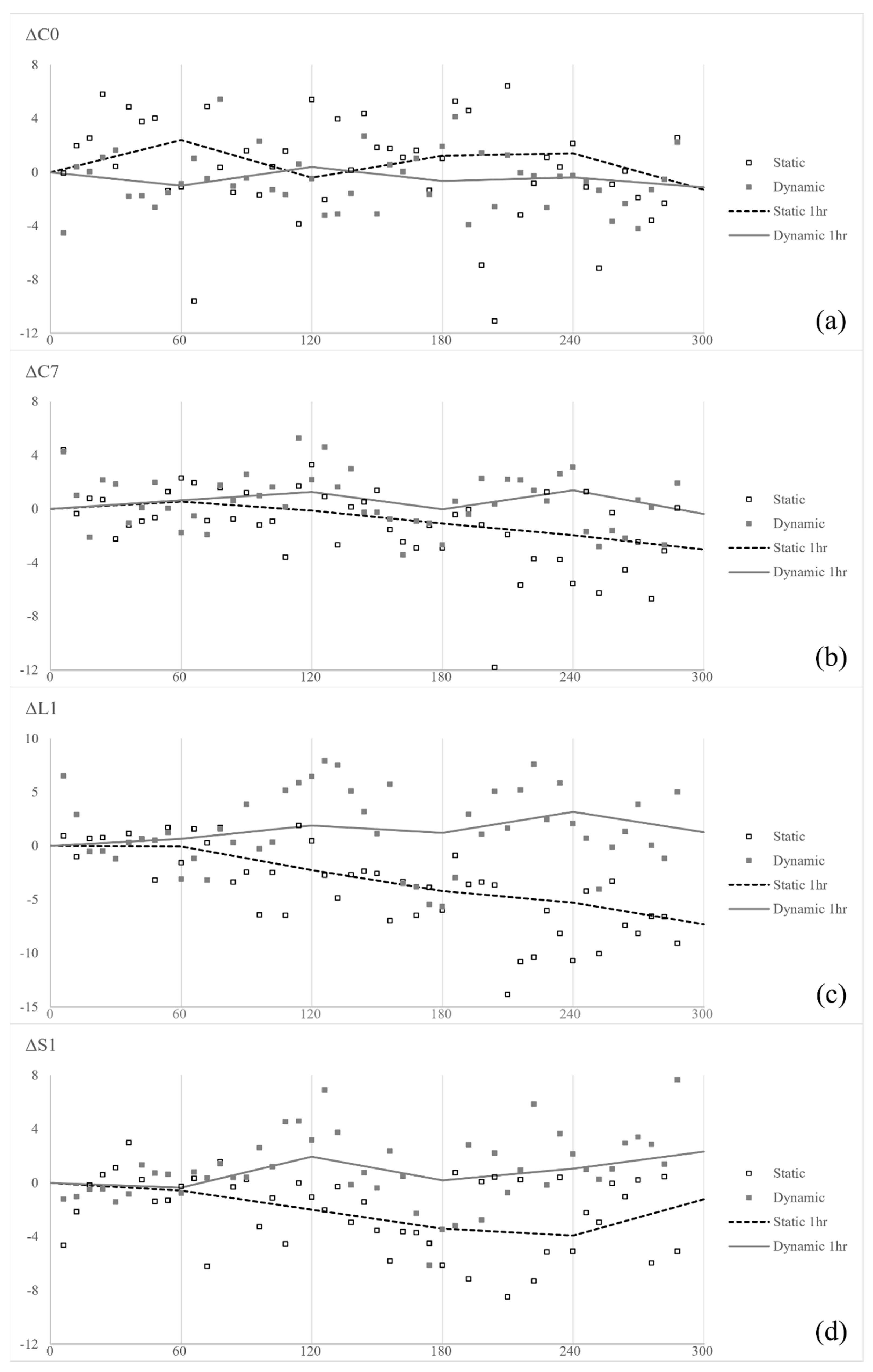
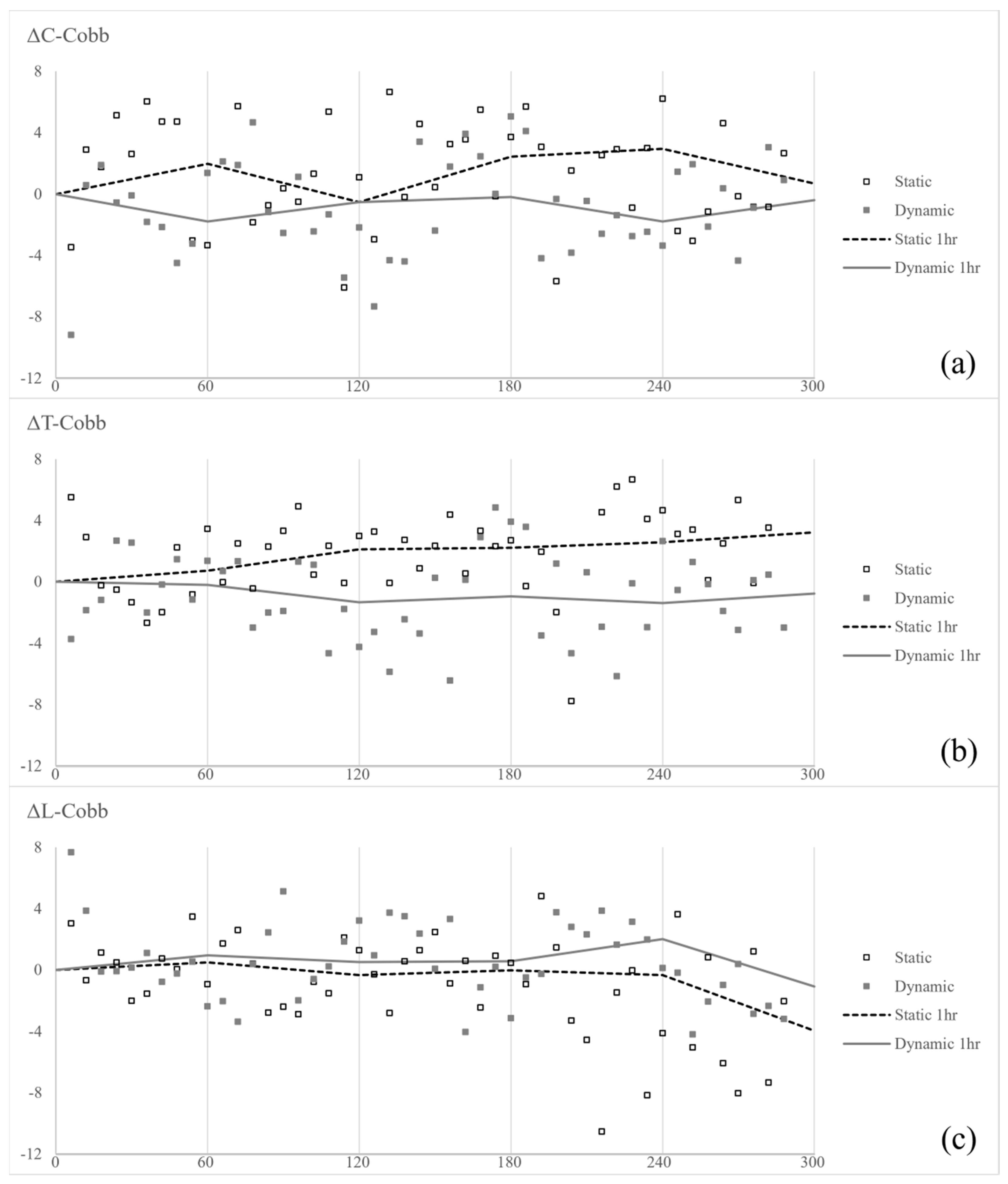
3.4. Spine Angle Change in Static and Dynamic Monitor Environment

Figure 5 depicts the trend of spine angle change from the baseline in the two monitor environments. The y-axis is the degree (°). In the change of C0 angle (ΔC0), the interaction between time and the monitor environment was not significant (p = 0.225), and there was no statistically significant change according to time (p = 0.081) or the monitor environment (p = 0.123). In the change of C7, L1, and S1 angle (ΔC7, ΔL1, and ΔS1), interactions between time and the monitor were statistically significant (p = 0.044; <0.001; 0.021), meaning that the changing pattern of spine angle over time differed depending on the monitor environment. C7 and L1 were flexed forward by −0.01439° and −0.03985° every 1 min in the static monitor, respectively (p = 0.037; <0.001), whereas in the dynamic monitor, there was no change in angle with time (p = 0.675; 0.665). S1 showed a tendency to flex forward by −0.0102° every 1 min on the static monitor (p = 0.089) and stretch back by 0.009069° for the dynamic monitor (p = 0.115), but it was not statistically significant.
Figure 6 shows the change of C-Cobb, T-Cobb, and L-Cobb from baseline (ΔC-Cobb, ΔT-Cobb, and ΔL-Cobb). They had no interaction between time and the monitor environment (p = 0.784; 0.094; 0.240). C-Cobb and T-Cobb did not change significantly over time (p = 0.624; 0.241), whereas L-Cobb became kyphotic by −0.0125° per minute (p = 0.007). In the static monitor, C-Cobb and T-Cobb were more lordotic (2.151°, p = 0.005; 3.0375°, <0.001), and L-Cobb was less lordotic (−1.5056°, p = 0.018) compared to the dynamic monitor.
Figure 7 shows the change in posture of a representative participant. In the static monitor, at the end of the work, C7 was leaned forward with increasing C-Cobb, resulting in a bent forward posture. In contrast, the dynamic monitor had no difference in posture before and after work.

3.5. Association between Neck Pain and Spine Angle or Monitor Setting

Table 5 shows the association between neck pain and spine angle or monitor setting. The interaction between the spine angle and the monitor environment was statistically significant for C7 and ΔS1 (p = 0.022, 0.003). The average neck pain was found to increase by 0.0329 as C0 was extended and 0.0345 as C-Cobb became lordotic (p = 0.024; 0.003). In the C7 angle, there was no significant relation with neck pain in the static monitor (p = 0.674), but in the dynamic monitor, neck pain increased as C7 became flexed (p = 0.002). In the change from baseline, neck pain increased as ΔS1 flexion in a dynamic monitor environment (p = 0.001), but other factors showed no significant correlation. There was no correlation with monitor height or tilt.

4. Discussion

In this study, the effect of a dynamic monitor that changes the height and tilt of the monitor at regular time intervals compared to the conventional static monitor on neck pain was evaluated. In addition, by measuring the spine angle in real-time using the IMU sensor, the effect of the monitor environment on the spine angle change was also compared.
The IMU sensor itself is known to measure orientation with very high accuracy [26]. This study also found that the IMU sensor could reflect the endplate angle in X-rays. The endplate angle and Cobb angle of the spine, which are crucial to clinical diagnosis, could only be measured instantaneously using X-rays. Lau et al. showed that there was no difference (p > 0.05) in the neck angles measured on X-rays between a group of subjects with and without neck pain [21]. However, with the implementation of a spinal angle tracking system, these angles can be continuously tracked throughout an individual’s daily life, providing valuable insights beyond the limitations of sporadic X-ray measurements. Also, we could expand on the comparisons and contributions of our system to traditional 3D video and other types of monitoring systems. Specifically, we emphasize the advantages of our wearable system over existing approaches. Our system offers several key benefits compared to traditional 3D video or other types of systems. A 2D video system primarily captures frontal or side views with limited degrees of freedom. Marker-based 3D video systems rely on vision tracking and can be limited by spatial constraints and computational demands. In contrast, our system allows for more comprehensive monitoring regardless of spatial constraints. By attaching four sensors to designated positions on the spine, we achieve simultaneous and continuous monitoring of spinal angles with high precision and accuracy. Furthermore, our system demonstrates strong agreement with spine angles measured via X-ray, further validating its reliability. Overall, our contribution lies in providing a practical and effective solution for continuous and accurate monitoring of spinal posture, addressing the limitations of existing systems. We recognize the importance of highlighting the broader applications of our developed system beyond ergonomic research. Our system exhibits significant potential for utilization across various fields, extending its relevance and applicability. For instance, beyond its primary application in ergonomic studies, our system can be leveraged for physical activity analysis, fall risk assessment, and gait analysis within clinical settings [22,23,24]. By continuously monitoring joint angles and spinal posture dynamics, our system offers valuable insights into movement patterns and musculoskeletal health, facilitating early intervention and personalized treatment strategies.
The increase in neck pain was less in the dynamic monitor than in the conventional static monitor. Although workstations such as monitor height, tilt, keyboard and mouse positions, and chairs are known as risk factors for neck pain, some authors reported that making these workstation designs optimal did not reduce neck pain [13]. Considering that repetitive motions, static postures, and prolonged computer use are the causes of neck pain [11,13,14], simply changing the arrangement of computer workstations may not be effective in reducing neck pain. In this regard, a dynamic monitor environment that periodically changes monitor settings can reduce neck pain by inducing beneficial posture changes. Since the monitor setting affects the user’s neck posture, it is presumed that the dynamic monitor caused a change in posture in a beneficial direction away from a static posture and repeated movements. Choi et al. conducted a study using a moving monitor that rotates at a speed that the user could not perceive, similarly suggesting that neck fatigue can be less in the moving monitor than in the static monitor [32]. There is also the possibility that changes in the monitor settings may act as a kind of alarm to change posture for subjects when using the dynamic monitor. However, it is difficult to assume that the role of these alarms only appeared in the dynamic monitor since both monitor environments required the RBG questionnaire at regular time intervals in the present study. In addition, similar results were also observed in the study that used a moving monitor that rotates at a speed that the user does not recognize [32]. Therefore, changing the monitor setting regularly may be interpreted to affect neck pain.
FHP is a commonly recognized type of poor head posture, defined as the position of the external auditory meatus anterior to the shoulder [4]. A cadaveric study reveals that increasing C2-C7 sagittal vertical axis (SVA) caused flexion of lower cervical (C2-C7) segments and hyperextension of suboccipital (C0-C1-C2) segments to maintain horizontal gaze [33]. These changes are consistent with the trend of neck angle when using a static monitor in this study. As the FHP position progressed over time, the angle of the C7 endplate tended to lean forward, and the C0-C7 Cobb increased in the conventional static monitor. However, this change did not appear in the dynamic monitor. Choi et al. reported similar results, showing a higher percentage of “good” neck posture on moving monitors compared to static monitors [32]. In Choi’s study, posture was indirectly evaluated by using the angle between the horizontal line and the line extending from the tragus of the ear to C7. Therefore, the spine angle is unknown. Also, if the Cobb angle of the upper cervical segment increased, there was a possibility that a bad posture could be interpreted as good or fair. Although it was a different experiment from our study, which observed changes in spine angle over time by estimating the endplate angle, it can be inferred that the change in monitor setting affects posture from the result of the trend of posture shown in Choi’s experiment. Considering that FHP frequently appears in computer work and is one of the crucial causes of neck fatigue [9,12], it can be inferred that neck pain during computer use can be reduced by preventing the posture change to FHP in the dynamic monitor. Of course, there is a limitation that the upper cervical angle and lower cervical angle could not be measured separately, and the SVA could not be measured through the IMU sensor in this study. However, since extreme postures do not occur in general working postures with elbows placed on a desk or armrest, the values measured in this experiment may be sufficient.
As a result of analyzing the factors related to pain, neck pain showed a tendency to increase with the extension of C0 and flexion of C7. Considering the fact that if the head is placed an inch or two further forward, the load on the cervical spine can be doubled or tripled [34], changes in C0 and C7 appearing in the FHP posture are thought to have affected the neck pain. However, this relationship was not clearly elucidated due to the small number of subjects. Since this experiment was not a suitable structure to find factors affecting neck pain, additional research is needed on which parts of the spine angle are related to neck pain. For the same reason, the monitor setting did not show any correlation with pain. As it has been found that the height and tilt of the monitor are related to neck pain [13], further study is needed to determine whether similar factors affect pain even in a dynamic monitor environment. Thus, to summarize the advantages and disadvantages associated with static and dynamic monitors, dynamic monitors offer the benefit of potentially reducing neck pain and encouraging better posture through periodic adjustments. However, they may introduce distractions due to the frequent need for monitor adjustments. Conversely, static monitors provide stability but can contribute to musculoskeletal deformation over prolonged use. Understanding these factors is crucial for optimizing monitor ergonomics and minimizing the risk of neck pain among computer users.
Notwithstanding the potential exhibited by this study, it is imperative to acknowledge the presence of certain constraints that may impact the comprehensive interpretation and applicability of its findings. First, this study included a small number of participants who could not be blinded. Considering that the incidence of neck pain reached up to 63.0%, although the number of participants was small [13], it was sufficient to observe changes in neck pain and spine angle over time. It was impossible to blind the participants due to the nature of the dynamic monitor in which the monitor setting changes continuously. However, since the purpose was not to compare the excellence of the two monitor environments but to see changes in neck pain and spine angle over time, blinding might not significantly affect the results. In addition, since the changes in spine angle over a long period were difficult to control consciously, the results might not be affected even if it was not blinded. Second, the monitor height and tilt were not controlled, but the participants adjusted the monitor to whatever they desired. However, there was no significant difference in setting between the two monitor environments, so there would be no difficulty accepting the results. In addition, it has the advantage of revealing the effect of dynamic monitoring in real work. The comfortable height and angle were different for each person. Since the applicability of the dynamic monitor environment has been investigated, further studies on appropriate heights and tilt angles are needed. Third, it is questionable whether a value of 0.333 on the RBG scale is a clinically meaningful difference. While our current study provided insights into the effects of dynamic monitor use on neck pain and posture, we recognize the importance of investigating the long-term implications. To address this, future research endeavors will involve extending the duration of experiments and increasing the sample size to further elucidate the impact of dynamic monitor settings on neck pain over time. By conducting longer-term studies, we aim to provide a more nuanced understanding of how dynamic monitor adjustments can influence neck pain and posture among computer users. Additionally, to mitigate potential sources of error and enhance the reliability of our findings, future research endeavors will incorporate a more comprehensive experimental scope. This will involve controlling for variables such as individual variations in ergonomic perception, sleep patterns, participant mood, work patterns, and undisclosed conditions. By expanding the experimental scope and minimizing error margins, we aim to obtain more accurate and practical results that are applicable to real-world scenarios. Fourth, we utilized the Rehabilitation Bioengineering Group (RBG) pain scale to assess neck pain, acknowledging its reliance on subjective pain reports. While this approach provides valuable insights, it may introduce ambiguity in pain measurement due to its subjective nature. To address this limitation and enhance the reliability of our pain assessment, future research will incorporate objective measures alongside subjective reporting. Specifically, we plan to integrate additional bio-signal indicators such as heart rate variability (HRV), which reflects autonomic nervous system activity, and electromyography (EMG), which provides insights into muscle activity. By combining subjective pain reports with objective physiological parameters, we aim to provide a clearer link between dynamic monitor usage and neck pain outcomes. Finally, except for head rotation, only flexion-extension is considered. Therefore, the result of this study may be difficult to apply to a situation where the work typically involves a lot of head rotation. In the same vein, since it was a study on a single monitor setting, additional study is needed to determine whether it will be applied to an environment using dual or more monitors. In addition to long-term experiments with large sample sizes, it is expected that the optimal monitor setting can be recommended in the workplace by observing the individual’s preference for monitor setting. Furthermore, it is possible to grasp the circadian rhythm based on the setting deviation based on the individual’s preference setting. Extending our findings to workdays from a regulatory standpoint involves considering various factors, including workplace ergonomics guidelines and occupational health regulations. One approach could be to conduct further research aimed at developing standardized recommendations for monitor settings based on individual preferences and ergonomic principles. These efforts can contribute to promoting a healthier and more productive work environment.
Despite these limitations, this study observed changes in neck pain and spine angle over a long time in a dynamic monitor environment. In addition, it is meaningful in that this study suggests the possibility of a new workstation environment by revealing that a dynamic monitor may have advantages over a conventional static monitor.

5. Conclusions

In this study, we developed a spine angle tracking system and confirmed that the estimated angles reflected the spine angles on the X-ray well. Dynamic monitor setting that changes the monitor heights and tilts at regular intervals may reduce neck pain increase over time than the conventional static monitor. Also, the kyphotic flexion of C7 in the static monitor did not appear in the dynamic monitor, and the C-Cobb angle was lower. A dynamic monitor environment could be considered a new alternative workstation environment that reduces neck pain and FHP.

Author Contributions

Conceptualization, C.K.C. and J.H.P.; methodology, H.K. and Y.I.W.; software, H.K.; validation, H.K. and J.L.; formal analysis, H.K., Y.I.W., S.K. and Y.C.; investigation, H.K. and Y.I.W.; data curation, H.K. and Y.I.W.; writing—original draft preparation, H.K. and Y.I.W.; writing—review and editing, J.L., I.Y.K. and C.K.C.; visualization, H.K. and Y.I.W.; supervision, J.L., I.Y.K. and C.K.C.; project administration, J.L., I.Y.K. and C.K.C.; funding acquisition, J.H.P. All authors have read and agreed to the published version of the manuscript.

Funding

This study was supported by LG Electronics (Project No. C2021024637). The funder contributed to the study concept and provided the prototype dynamic monitors for the experiment. The funder had no role in the design and conduct of the study; collection, analysis, and interpretation of the data; preparation, review, or approval of the manuscript; and decision to submit the manuscript for publication. The corresponding author had full access to all the data in the study and had final responsibility for the decision to submit the manuscript for publication.

Institutional Review Board Statement

The study was conducted in accordance with the Declaration of Helsinki, and approved by the Ethics Committee/Institutional Review Board of Seoul National University Hospital (No. 2106-053-1227).

Data Availability Statement

The data presented in this study are available on request from the corresponding author due to privacy.

Conflicts of Interest

Jin Ho Park was employed by the company LG Electronics.The authors declare that this study received funding from LG Electronics. The funder was not involved in the study design, collection, analysis, interpretation of data, the writing of this article or the decision to submit it for publication. The authors declare that the research was conducted in theabsence of any commercial or financial relationships that could beconstrued as a potential conflict of interest.

References

  1. Hoy, D.; March, L.; Woolf, A.; Blyth, F.; Brooks, P.; Smith, E.; Vos, T.; Barendregt, J.; Blore, J.; Murray, C.; et al. The global burden of neck pain: Estimates from the global burden of disease 2010 study. Ann. Rheum. Dis. 2014, 73, 1309–1315. [Google Scholar] [CrossRef] [PubMed]
  2. Hoy, D.G.; Protani, M.; De, R.; Buchbinder, R. The epidemiology of neck pain. Best Pract. Res. Clin. Rheumatol. 2010, 24, 783–792. [Google Scholar] [CrossRef] [PubMed]
  3. Yip, C.H.; Chiu, T.T.; Poon, A.T. The relationship between head posture and severity and disability of patients with neck pain. Man. Ther. 2008, 13, 148–154. [Google Scholar] [CrossRef] [PubMed]
  4. Shaghayegh Fard, B.; Ahmadi, A.; Maroufi, N.; Sarrafzadeh, J. Evaluation of forward head posture in sitting and standing positions. Eur. Spine J. 2016, 25, 3577–3582. [Google Scholar] [CrossRef] [PubMed]
  5. Braun, B.L.; Amundson, L.R. Quantitative assessment of head and shoulder posture. Arch. Phys. Med. Rehabil. 1989, 70, 322–329. [Google Scholar] [PubMed]
  6. Nejati, P.; Lotfian, S.; Moezy, A.; Nejati, M. The study of correlation between forward head posture and neck pain in Iranian office workers. Int. J. Occup. Med. Environ. Health 2015, 28, 295–303. [Google Scholar] [CrossRef]
  7. Mahmoud, N.F.; Hassan, K.A.; Abdelmajeed, S.F.; Moustafa, I.M.; Silva, A.G. The Relationship Between Forward Head Posture and Neck Pain: A Systematic Review and Meta-Analysis. Curr. Rev. Musculoskelet. Med. 2019, 12, 562–577. [Google Scholar] [CrossRef] [PubMed]
  8. Falla, D.; Jull, G.; Hodges, P.W. Feedforward activity of the cervical flexor muscles during voluntary arm movements is delayed in chronic neck pain. Exp. Brain Res. 2004, 157, 43–48. [Google Scholar] [CrossRef]
  9. Schomacher, J.; Falla, D. Function and structure of the deep cervical extensor muscles in patients with neck pain. Man. Ther. 2013, 18, 360–366. [Google Scholar] [CrossRef]
  10. Kumar, S. Theories of musculoskeletal injury causation. Ergonomics 2001, 44, 17–47. [Google Scholar] [CrossRef]
  11. Ariens, G.A.; Bongers, P.M.; Douwes, M.; Miedema, M.C.; Hoogendoorn, W.E.; van der Wal, G.; Bouter, L.M.; van Mechelen, W. Are neck flexion, neck rotation, and sitting at work risk factors for neck pain? Results of a prospective cohort study. Occup. Environ. Med. 2001, 58, 200–207. [Google Scholar] [CrossRef] [PubMed]
  12. Sikka, I.; Chawla, C.; Seth, S.; Alghadir, A.H.; Khan, M. Effects of Deep Cervical Flexor Training on Forward Head Posture, Neck Pain, and Functional Status in Adolescents Using Computer Regularly. Biomed. Res. Int. 2020, 2020, 8327565. [Google Scholar] [CrossRef]
  13. Cote, P.; van der Velde, G.; Cassidy, J.D.; Carroll, L.J.; Hogg-Johnson, S.; Holm, L.W.; Carragee, E.J.; Haldeman, S.; Nordin, M.; Hurwitz, E.L.; et al. The burden and determinants of neck pain in workers: Results of the Bone and Joint Decade 2000-2010 Task Force on Neck Pain and Its Associated Disorders. Spine 2008, 33 (Suppl. S4), S60–S74. [Google Scholar] [CrossRef] [PubMed]
  14. Hakala, P.T.; Rimpela, A.H.; Saarni, L.A.; Salminen, J.J. Frequent computer-related activities increase the risk of neck-shoulder and low back pain in adolescents. Eur. J. Public Health 2006, 16, 536–541. [Google Scholar] [CrossRef] [PubMed]
  15. Auvinen, J.; Tammelin, T.; Taimela, S.; Zitting, P.; Karppinen, J. Neck and shoulder pains in relation to physical activity and sedentary activities in adolescence. Spine 2007, 32, 1038–1044. [Google Scholar] [CrossRef] [PubMed]
  16. Briggs, A.M.; Straker, L.M.; Bear, N.L.; Smith, A.J. Neck/shoulder pain in adolescents is not related to the level or nature of self-reported physical activity or type of sedentary activity in an Australian pregnancy cohort. BMC Musculoskelet. Disord. 2009, 10, 87. [Google Scholar] [CrossRef] [PubMed]
  17. Kothiyal, K.; Bjornerem, A.M. Effects of computer monitor setting on muscular activity, user comfort and acceptability in office work. Work 2009, 32, 155–163. [Google Scholar] [CrossRef] [PubMed]
  18. Burgess-Limerick, R.; Plooy, A.; Ankrum, D.R. The effect of imposed and self-selected computer monitor height on posture and gaze angle. Clin Biomech. 1998, 13, 584–592. [Google Scholar] [CrossRef] [PubMed]
  19. Chiou, W.K.; Chou, W.Y.; Chen, B.H. Notebook computer use with different monitor tilt angle: Effects on posture, muscle activity and discomfort of neck pain users. Work 2012, 41 (Suppl. S1), 2591–2595. [Google Scholar] [CrossRef]
  20. Jensen, C. Development of neck and hand-wrist symptoms in relation to duration of computer use at work. Scand. J. Work Environ. Health 2003, 29, 197–205. [Google Scholar] [CrossRef]
  21. Lau, K.T.; Cheung, K.Y.; Chan, M.H.; Lo, K.Y.; Chiu, T.T.W. Relationships between sagittal postures of thoracic and cervical spine, presence of neck pain, neck pain severity and disability. Man. Ther. 2010, 15, 457–462. [Google Scholar] [CrossRef] [PubMed]
  22. Mascret, Q.; Bielmann, M.; Fall, C.-L.; Bouyer, L.J.; Gosselin, B. (Eds.) Real-time human physical activity recognition with low latency prediction feedback using raw IMU data. In Proceedings of the 2018 40th Annual International Conference of the IEEE Engineering in Medicine and Biology Society (EMBC), Honolulu, HI, USA, 18–21 July 2018. [Google Scholar]
  23. Kobsar, D.; Charlton, J.M.; Tse, C.T.; Esculier, J.-F.; Graffos, A.; Krowchuk, N.M.; Thatcher, D.; Hunt, M.A. Validity and reliability of wearable inertial sensors in healthy adult walking: A systematic review and meta-analysis. J. Neuroeng. Rehabil. 2020, 17, 1–21. [Google Scholar] [CrossRef] [PubMed]
  24. Jebelli, H.; Ahn, C.R.; Stentz, T.L. Comprehensive fall-risk assessment of construction workers using inertial measurement units: Validation of the gait-stability metric to assess the fall risk of iron workers. J. Comput. Civ. Eng. 2016, 30, 04015034. [Google Scholar] [CrossRef]
  25. Han, H.; Jang, H.; Yoon, S.W. Novel wearable monitoring system of forward head posture assisted by magnet-magnetometer pair and machine learning. IEEE Sens. J. 2019, 20, 3838–3848. [Google Scholar] [CrossRef]
  26. Lin, Z.R.; Xiong, Y.S.; Dai, H.D.; Xia, X.K. An Experimental Performance Evaluation of the Orientation Accuracy of Four Nine-Axis MEMS Motion Sensors. In Proceedings of the 2017 5th International Conference on Enterprise Systems (ES), Beijing, China, 22–24 September 2017; pp. 185–189. [Google Scholar]
  27. Yoon, S.D.; Lee, C.H.; Lee, J.; Choi, J.Y.; Min, W.K. Occipitocervical inclination: New radiographic parameter of neutral occipitocervical position. Eur. Spine J. 2017, 26, 2297–2302. [Google Scholar] [CrossRef] [PubMed]
  28. Horng, M.-H.; Kuok, C.-P.; Fu, M.-J.; Lin, C.-J.; Sun, Y.-N. Cobb angle measurement of spine from X-ray images using convolutional neural network. Comput. Math. Methods Med. 2019, 2019, 6357171. [Google Scholar] [CrossRef] [PubMed]
  29. Brennan, A.; Zhang, J.; Deluzio, K.; Li, Q. Quantification of inertial sensor-based 3D joint angle measurement accuracy using an instrumented gimbal. Gait Posture 2011, 34, 320–323. [Google Scholar] [CrossRef] [PubMed]
  30. Keown, G.A.; Tuchin, P.A. Workplace factors associated with neck pain experienced by computer users: A systematic review. J. Manip. Physiol. Ther. 2018, 41, 508–529. [Google Scholar] [CrossRef]
  31. Balasubramanian, V.; Jagannath, M.; Adalarasu, K. Muscle fatigue based evaluation of bicycle design. Appl. Ergon. 2014, 45, 339–345. [Google Scholar] [CrossRef]
  32. Choi, K.H.; Cho, M.U.; Park, C.W.; Kim, S.Y.; Kim, M.J.; Hong, B.; Kong, Y.K. A Comparison Study of Posture and Fatigue of Neck According to Monitor Types (Moving and Fixed Monitor) by Using Flexion Relaxation Phenomenon (FRP) and Craniovertebral Angle (CVA). Int. J. Environ. Res. Public Health 2020, 17, 6345. [Google Scholar] [CrossRef]
  33. Patwardhan, A.G.; Havey, R.M.; Khayatzadeh, S.; Muriuki, M.G.; Voronov, L.I.; Carandang, G.; Nguyen, N.L.; Ghanayem, A.J.; Schuit, D.; Patel, A.A.; et al. Postural Consequences of Cervical Sagittal Imbalance: A Novel Laboratory Model. Spine 2015, 40, 783–792. [Google Scholar] [CrossRef] [PubMed]
  34. Hansraj, K.K. Assessment of stresses in the cervical spine caused by posture and position of the head. Surg. Technol. Int. 2014, 25, 277–279. [Google Scholar] [PubMed]
Figure 1. (a) Hardware; (b) schematic diagrams; and (c) placement of the spine angle tracking system.
Electronics 13 01363 g001
Figure 2. User interface viewer.
Electronics 13 01363 g002
Figure 3. Cobb angle calculation algorithm.
Electronics 13 01363 g003
Figure 4. Graph of neck pain according to the monitor.
Electronics 13 01363 g004
Figure 5. Graph of (a) C0 angle (ΔC0); (b) C7 angle (ΔC7); (c) L1 angle (ΔL1); and (d) S1 angle (ΔS1) from the baseline according to the monitor. Square: Spine angle measured for 5 min, Line: 1-h average value of spine angle.
Electronics 13 01363 g005
Figure 6. Graph of (a) C-Cobb angle (ΔC-Cobb); (b) T-Cobb angle (ΔT-Cobb); and (c) L-Cobb angle (ΔL-Cobb) from the baseline according to the monitor. Square: Cobb angle measured for 5 min, Line: 1-h average value of Cobb angle.
Electronics 13 01363 g006
Figure 7. Lateral photographs along with an X-ray schematic. (a) Angles of IMU sensor at the start of the static monitor; (b) angles at the end of the static monitor; (c) angles at the start of the dynamic monitor; and (d) angles at the end of the dynamic monitor.
Electronics 13 01363 g007
Table 1. RBG pain scale.
GradePain Scale Criteria
0No pain
1Very minor, barely felt
2Minor pain not interfering with the work routine
3Moderate pain
4Continuous pain affecting routine cycling
5Unbearable/severe pain
Table 2. Sensor accuracy evaluation results.
Sagittal (°)Transverse (°)Frontal (°)
306090306090306090
Average29.9359.9289.8830.0459.9689.9329.2959.2588.54
Mean error0.090.050.81
Table 3. ICC between X-ray and IMU sensor.
ICC *95% CI *p-Value
C00.9930.987, 0.996<0.001
C70.8760.804, 0.923<0.001
* ICC: intraclass correlation, CI: confidence interval. Absolute, single measurement using the two-way mixed effects model. Boldface indicates statistical significance.
Table 4. Baseline characteristics.
FactorStaticDynamicp-Value
Mean ± SDMedian [Min, Max]Mean ± SDMedian [Min, Max]
RBG0.63 ± 0.920 [0, 2]0.5 ± 0.530.5 [0, 1]0.705
C0, °−9.14 ± 7.11−9.17 [−19.77, 0.26]−8.23 ± 4.12−9.16 [−12.94, −2.09]0.889
C7, °−26.94 ± 6.62−26.85 [−36.73, −16.53]−28.01 ± 6.83−26.62 [−39.82, −17.51]0.575
L1, °3.52 ± 11.91−0.78 [−8.24, 28.35]1.54 ± 7.97−1.18 [−4.32, 19.4]0.674
S1, °−24.59 ± 18.35−28.13 [−56.53, 5.7]−22.72 ± 10.02−23.7 [−39.57, −5.72]0.484
C-Cobb, °17.81 ± 6.7617.5 [9.6, 29.92]19.31 ± 7.6121.25 [6.83, 31.37]0.575
T-Cobb, °−30.11 ± 9.36−30.85 [−41.22, −18.01]−29.59 ± 11.21−28.77 [−50.68, −13.42]0.779
L-Cobb, °27.81 ± 11.224.15 [18.27, 51.53]24.33 ± 12.0323.85 [9.21, 43.86]0.889
Monitor setting
Eye-level, cm48.19 ± 3.8148.55 [41.6, 53.6]47.38 ± 3.7846.70 [40.3, 54.0]0.294
Distance, cm56.04 ± 5.4156.65 [48.4, 64.0]54.13 ± 6.0855.55 [42.9, 63.2]0.290
Height, cm37.65 ± 1.6837.55 [35.4, 40.8]37.86 ± 3.8038.45 [29.0, 44.6]0.186
Tilt, °0.30 ± 1.430.24 [−2.12, 2.50]0.32 ± 3.580.65 [−7.80, 6.42]0.943
Table 5. Association between RBG and the spine angle.
FactorEstimate95% CI *p-Value
C0, °0.03290.00440.06130.024
C7, °
  static−0.0076−0.04350.02830.674
  dynamic−0.0498−0.0802−0.01940.002
L1, °−0.013−0.03280.00690.197
S1, °0.0059−0.00690.01880.359
C-Cobb, °0.03450.01240.05660.003
T-Cobb, °−0.0024−0.02440.01960.829
L-Cobb, °−0.0103−0.0230.00250.114
ΔC0, °−0.02914−0.06690.008630.128
ΔC7, °−0.01038−0.04160.020780.508
ΔL1, °−0.01035−0.02630.005550.198
ΔS1, °
  static0.002103−0.02580.029990.881
  dynamic−0.08655−0.1361−0.0370.001
ΔC-Cobb, °−0.011−0.04490.022910.520
ΔT-Cobb, °0.01523−0.01260.043040.278
ΔL-Cobb, °−0.00103−0.02070.01860.917
Monitor height, cm−0.0063−0.09380.08110.885
Monitor tilt, °0.0018−0.07110.07460.961
* CI: confidence interval. A positive number means extension and lordosis, and a negative number means flexion and kyphosis. Monitor tilt: A positive value means that the top is lying on its back. Boldface indicates statistical significance.
Disclaimer/Publisher’s Note: The statements, opinions and data contained in all publications are solely those of the individual author(s) and contributor(s) and not of MDPI and/or the editor(s). MDPI and/or the editor(s) disclaim responsibility for any injury to people or property resulting from any ideas, methods, instructions or products referred to in the content.

