Systematic Review

Human Gut Microbiota in Coronary Artery Disease: A Systematic Review and Meta-Analysis †

1
Department of Microbiology, Wroclaw Medical University, 50-367 Wroclaw, Poland
2
Department of Biochemistry and Immunochemistry, Wroclaw Medical University, 50-367 Wroclaw, Poland
3
Ninewells Hospital and Medical School, James Arrott Drive, Dundee DD1 9SY, UK
4
Research and Development Centre, Regional Specialist Hospital, 51-124 Wroclaw, Poland
5
Department of Cardiovascular Medicine, Mayo Clinic, Rochester, MN 55905, USA
6
Division of Cardiology, National Defense Medical College, Tokorozawa 359-8513, Japan
7
Department of Population Medicine and Lifestyle Diseases Prevention, Medical University of Bialystok, 15-269 Bialystok, Poland
8
Department of Cardiology, Medical University of Bialystok, 15-089 Bialystok, Poland
9
Clinical Research Centre, Medical University of Bialystok, 15-089 Bialystok, Poland
*
Author to whom correspondence should be addressed.
The work was performed at Wroclaw Medical University.
Metabolites 2022, 12(12), 1165; https://doi.org/10.3390/metabo12121165
Submission received: 19 October 2022 / Revised: 16 November 2022 / Accepted: 18 November 2022 / Published: 23 November 2022
(This article belongs to the Special Issue Metabolic Profiling of Cardiovascular Disease, 2nd Edition)

Abstract

:
In recent years, the importance of the gut microbiome in human health and disease has increased. Growing evidence suggests that gut dysbiosis might be a crucial risk factor for coronary artery disease (CAD). Therefore, we conducted a systematic review and meta-analysis to determine whether or not CAD is associated with specific changes in the gut microbiome. The V3–V4 regions of the 16S rDNA from fecal samples were analyzed to compare the gut microbiome composition between CAD patients and controls. Our search yielded 1181 articles, of which 21 met inclusion criteria for systematic review and 7 for meta-analysis. The alpha-diversity, including observed OTUs, Shannon and Simpson indices, was significantly decreased in CAD, indicating the reduced richness of the gut microbiome. The most consistent results in a systematic review and meta-analysis pointed out the reduced abundance of Bacteroidetes and Lachnospiraceae in CAD patients. Moreover, Enterobacteriaceae, Lactobacillus, and Streptococcus taxa demonstrated an increased trend in CAD patients. The alterations in the gut microbiota composition are associated with qualitative and quantitative changes in bacterial metabolites, many of which have pro-atherogenic effects on endothelial cells, increasing the risk of developing and progressing CAD.

1. Introduction

Various studies on coronary atherosclerosis have revealed that chronic vascular inflammation, which begins with a coronary endothelial injury, is an essential process in the development of coronary artery disease (CAD) [1]. Despite advances in various drug therapies and interventions, CAD remains one of the most common causes of death worldwide [2]. In recent years, data from rigorous preclinical studies and epidemiological studies linking changes in the gut microbiome to vascular endothelial dysfunction and vascular inflammation have accumulated [3].
The gut microbiome comprises over 2000 species, most of which fall into the four main phyla: Bacteroidetes, Firmicutes, Proteobacteria, and Actinobacteria [4]. A diverse, well-balanced gut microbiota is crucial to human health. Gut microbiota consisting of a 100,000,000,000,000 microbes living in a symbiotic host relationship regulate host metabolism, insulin sensitivity, intestinal endocrine, and neurological function. Gut bacteria produce neurotransmitters, influence the maturation and training of the host immune system, neutralize exogenous toxins, and protect against overgrowth pathogens [5,6,7]. Active microbial metabolites, such as short-chain fatty acids, vita-mins, autoinducers, indole derivates, bile acid metabolites, microbial amino acids, and polyamines, directly regulate human physiology [8].
Disruption of the diversity and abundance of the gut microbiota, termed gut dysbiosis, underlies many diseases, including metabolic syndrome associated with obesity and diabetes. Gut dysbiosis underlies inflammatory bowel disease (IBD), irritable bowel disease (IBS), colon cancer, asthma, autism, Parkinson’s disease, Alzheimer’s disease, schizophrenia, depression and cardiovascular diseases [5,9,10,11,12]. An imbalance in the abundance of specific bacterial taxa in the gut microbiota correlates with a deficiency or excess of bacterial metabolites that fundamentally affect the physiological status of the host cells, including endothelial cells. To exemplify, specific tryptophan-derived gut microbiota metabolites, such as indole metabolites and butyrate, serve as regulators of the intestinal barrier, increasing the expression of tight junction proteins that ensure the tightness of the epithelial barrier [13,14,15]. Hence, the depletion of butyrate-producing bacterial taxa, e.g., Lachnospiraceae, may compromise intestinal barrier integrity and leakage of bacterial metabolites into the bloodstream [16]. One of the most critical consequences of intestinal barrier dysfunction in gut dysbiosis is endotoxemia and the activation of immune processes leading to chronic subacute inflammation [17,18]. Apart from immune cells, low-grade endotoxemia affects endothelial cells, contributing to the initiation and progression of atherosclerosis [19,20]. Many studies have shown alterations in the composition and diversity of the gut microbiota in CAD [21].
Nevertheless, it is still uncertain which bacterial taxa are altered in CAD and whether these alterations are consistent across different study populations. Studies of gut microbiota composition are always affected by factors related to the study population, such as age, diet, chronic diseases, medications, and physical activity [22]. In addition, gut microbiota studies are complicated by the choice of sequencing methods and bioinformatic pipelines, which significantly influence the results [23].
A recent study on simulated microbiome datasets highlights the importance of using consistent analytical pipelines. Nearing et al. [24] have shown that the appropriate tools for the differential abundance of microbial taxa testing impact the result. Therefore, this meta-analysis and systematic review aimed to provide an overview of the literature linking changes in gut microbiota with CAD using consistent analytical tools across all the datasets.

2. Materials and Methods

2.1. Systematic Review

This systematic review is registered in the International Prospective Register of Systematic Reviews “PROSPERO” ID CRD42020187549. The study was conducted following the Preferred Reporting Items of Systematic reviews and Meta-Analyses (PRISMA) guidelines.

2.1.1. Search Strategy

A comprehensive literature search was performed. The search terms were “(Human Microbiomes OR Human Microbiome OR Human Microflora OR Microbial Community Structure OR Human Flora OR Composition, Microbial Community) AND (atherosclerosis or atherogenesis or Coronary atherosclerosis or Coronary Artery Disease or Coronary Atherosclerosis) AND ischemic heart disease NOT Review” for Medline on PubMed. The search terms were appropriately modified for other databases and are included in the Supplementary Materials (Supplementary S1). The search strategy was applied to the following databases: PubMed, Scopus, Web of Science, Embase, CINAHL, and Cochrane. We used Google Scholar to search for grey literature; the last search was conducted on 14 December 2021. Before proceeding with the selection of eligible studies, all duplicates were removed.

2.1.2. Inclusion and Exclusion Criteria

We have included studies that analyzed the differences in the profile of the intestinal microbiome between patients with CAD (both stable and unstable) and healthy controls. Only human studies were included, and neither publication status nor language restrictions were imposed. The primary outcome measure was a comparison of relative abundances of bacterial phyla, families, and genera of human gut microflora. We excluded papers in which CAD was diagnosed by clinical symptoms or ECG only, without coronary imaging methods. Articles in which the control group included subjects with coronary artery disease, or the study group consisted of cadavers, were also excluded.

2.1.3. Assessment of Eligibility and Data Extraction

After removing the duplicates, two authors (MC and RB) independently screened obtained studies by titles and abstracts for relevance to the topic of our systematic review. The studies obtained by screening were read in full text, and eligibility was determined based on inclusion and exclusion criteria. The records that did not fulfill the criteria were removed without using automatic tools. Discrepancies were discussed, and a third author (KL) arbitrated if disagreement was not resolved. We have included peer-reviewed papers and those still awaiting review (grey literature). The eligibility assessment of studies is summarized in Figure 1. The study quality was assessed using a modified Newcastle Ottawa Scale (Supplementary).

2.2. Meta-Analysis

The meta-analytical pipeline has been run using Snakemake version 6.6.1. [25]. The characteristics of the studies included in the meta-analysis and systematic review are provided in Table 1. Only studies that provided raw sequences from 16S rRNA sequencing were included in the meta-analysis. The results were obtained by analyzing the samples using two approaches. First, all datasets were analyzed separately, and their results were combined in the random-effects meta-analysis (single study approach). In the second approach, all the samples were analyzed as one combined dataset (combined studies approach).

2.2.1. Readings Preparation and zOTU Picking

We have removed primers using cutadapt [26]. Primers used for the single study approach are provided in the table available in the supplementary. For the combined study approach, we have used 338F (5′-CTACGGGNGGCWGCAG-3′) and 805R (5′-GACTACHVGGGTATCTAATCC-3′) primers, except the Mayo dataset where 926R (5′-CCGTCAATTCMTTTRAGT-3′) was used. Sawicka-Smiarowska dataset [27] was excluded from the combined study approach due to the choice of primers in the study, which did not allow for obtaining sequences that were globally aligned with other studies. Next, all of the readings were truncated to 250 bps leaving us with globally trimmed data (e.g., all reads starting and ending at the same position). Furthermore, reading preparation and zOTU picking have been performed using usearch v11.0.667 and UNOISE. zOTU refers to correct biological sequences with a 100% identity threshold [28,29]. The taxonomy was resolved using DECIPHER IDTAXA [30] with Genome Taxonomy Database (version 06-RS202) [31] as a reference. For downstream analyses, the data have been imported into the phyloseq object [32] and filtered to leave only zOTUs present in at least 20% of the samples and with at least five counts.

2.2.2. Differential Abundance Testing

Differential abundance (DA) testing was performed using three approaches: (1) DESeq2, which models the data using the negative binomial distribution, (2) MetagenomeSeq, which uses a zero-inflated Gaussian model, and (3) ANCOM-BC, which uses an offset-based log-linear model and currently appears to be the best method for controlling the false-discovery ratio without losing statistical power. Only bacteria detected as statistically significant metagenomeSeq, Ancom-BC, or DESeq2 in at least three of 7 studies were included. A comprehensive discussion of the DA testing approaches is out of the scope of our manuscript, and we refer the reader to the large body of research already published on the topic [33]. For all the approaches, we have assumed a significance level of p < 0.05. We have used the Benjamini and Hochberg method to adjust for multiple testing.

2.2.3. Statistical Analysis

Alpha diversity indices (observed zOTUs, Fischer, Chao1, Shannon, ACE, and Simpson indices) were calculated on data rarefied to the depth of 5000. Beta diversity was assessed using Unweighted UniFrac. The statistical significance was determined using the Wilcoxon test for the alpha diversity and PERMANOVA with 999 permutations for the beta diversity. PERMANOVA was run with the vegan R package [34]. The single study approach calculated relative abundances using the mean difference in centered log ratios (CLR). The obtained relative abundances were used to estimate odds ratios using Agresti’s generalized odd ratios [35] as implemented in the genodds package [36]. Next, odd ratios were summarized in a random-effects meta-analysis performed using the meta R package [37].

3. Results

Our search has yielded 1181 articles. After removing the duplicates, 897 unique studies were identified, out of which 21 met inclusion criteria for systematic review. Among these 21 articles, 7 had raw data available and could be used to conduct a meta-analysis. (Figure 1). In the systematic review, the relative abundance of gut microbiota from all 21 eligible papers were compared. Bacterial taxa detected as significant in at least four studies are included in Figure 2. In the meta-analysis, only bacteria detected as significant in at least three of the seven eligible articles were reported. The complete results of the systematic review are provided in Table 1, and the characteristics of studies included in the meta-analysis are available in the Supplement.

3.1. Characteristics of Included Studies

Many factors may affect the gut microbiome composition. We compared all eligible studies by the following: age, gender, BMI, and diabetes mellitus. The complete results are provided in the Supplement.
The average age of participants was between the sixth and seventh decade of life. In five articles, differences in age between groups were significant; in 13 other articles were insignificant, and there were no available data for comparison in three articles.
In the majority of studies, participants were male. In five articles, differences in gender between groups were significant, ten others were insignificant, and six articles had no available data for comparison. The BMI of participants generally had values typical for overweight (BMI = 25–30). In four articles, differences in BMI were significant, in ten the differences were insignificant, and in seven, there were no available data for comparison. There were relatively limited data on the prevalence of diabetes in participants. In four articles, differences in diabetes were significant, in five the differences were insignificant, and in 12, there were no available data for comparison.

3.2. Systematic Review Results

A comprehensive comparison between CAD patients and controls regarding the relative abundance at the phylum level demonstrated changes related to four phyla, i.e., Proteobacteria, Bacteroidetes, Actinomycetota, and Firmicutes. The increase of Gammaproteobacteria has been reported in six (28.6%) studies and the rise of the Enterobacteriaceae family within the Proteobacteria phylum in four (19%) studies in CAD patients. Moreover, four (19%) studies recorded the rise of the Escherichia genus of the Enterobacteriaceae family. Overall, seven (33.3%) articles reported an increased abundance of Gammaproteobacteria taxa (Figure 2, Table 1).
Bacteroidetes phylum decline in CAD was reported in five (23.8%) studies (Table 1; Figure 2). On the contrary, one cross-sectional study reported an increased abundance of Bacteroidetes in CAD compared to the control group. Remarkably, at the genus level, a decrease in the relative abundance of Bacteroides in CAD has been recorded in nine (42.8%) studies. Overall, a decline in the relative abundance of the Bacteroidetes taxa was recorded in 16 (76.2%) of all 21 articles analyzed, making the decrease in these taxa strikingly CAD-related. Furthermore, conflicting results regarding the Prevotellaceae family and Prevotella genus within the Bacteroidales order were associated with CAD, where the relative abundance of these taxa were decreased in two (9.5%) studies and increased in two other studies.
The results regarding the Firmicutes phylum were conflicting. In two (9.5%) studies, the relative abundance of Firmicutes was increased, whereas in three other (14.3%) decreased in CAD compared to the control group. Within the Firmicutes phylum, there were conflicting results concerning the Bacilli class. In two (9.5%) studies, the taxon was increased, whereas in two others decreased in CAD compared to the control group. However, within the Bacilli taxon, the increased relative abundance of the Lactobacillales order was associated with CAD patients in four (19%) studies and the Lactobacillus genus in three (14.3%) studies in contrast to the control group. Moreover, within the Firmicutes phylum, the decrease in the relative abundance of the Lachnospiraceae family of class Clostridia in CAD patients versus the control group was demonstrated in five (23.8%) studies. On the contrary, within the Firmicutes phylum, the Christensenellaceae family abundance was increased in CAD in three (14.3%) studies. The systematic review analysis also demonstrated the increased relative abundance within the Streptococcaceae family and the Streptococcus genus among CAD patients in four (19%) studies compared to the control group. These results indicate a quantitative disturbance within the Firmicutes, mainly concerning an increase in the abundance of Lactobacillus and Streptococcus, as well as bacteria from the Christensenellaceae family, with a concomitant decrease in the abundance of bacteria from the Lachnospiraceae family.
The less prominent changes in the gut microbiota of CAD patients concerned the Coriobacteriales order of the phylum Actinobacteria, which was decreased in four (19%) studies in CAD patients. Changes in the abundance of other bacterial groups, i.e., Desulfovibrio, Parabacteroides, and Fusobacterium in CAD, were recorded in only a few articles (Table 1).
Table 1. Key characteristics of included studies. Legend: RoB—the risk of bias, NOS—Newcastle-Ottawa Scale, “+” significant difference in diversity, “−” no significant difference in diversity, PCI—percutaneous coronary intervention, CABG—coronary artery bypass graft, IBD—inflammatory bowel disease, GIT—gastrointestinal tract, ACS—acute coronary syndrome, UA—unstable angina, SA—stable angina, CAD—coronary artery disease, Ctrl—control, “”increased relative abundance in CAD patients, ““ decreased relative abundance in CAD patients.
RoB
NOS
7107108
Diversity
α β
No
data
++No data
No data+No data
Major outcome-
CAD relative abundance
Bacteroidales
Gammaproteobacteria
Enterobacteriaceae
Prevotella 
Lactobacillales
Bacteroides
Gammaproteobacteria
Firmicutes
Lactobacillales
Enterobacteriaceae 
Lachnospiraceae
Prevotella
Bacilli
Christensellaceae
Prevotellaceae
Bacteroidetes
Firmicutes 
Bacteroidales
Coriobacteriales
Christensellaceae
Prevotellaceae
Enterobacteriaceae 
Escherichia/Shigella 
Major exclusion criteriapatients less than 19 years, pregnancy
abnormal kidney function
Patients with systemic diseases: hepatic, renal, collagen, malignancyother identifiable etiologies of coronary thrombi,
active infection during admission.
heart failure, structural heart disease, history of antibiotic use within 1 month, serious dysfunction of liver or kidneyantibiotics, probiotics, decompensated chronic diseases, oncological diseases
Major inclusion criteria
CAD Control
No prior heart attacks or strokes, and no antibiotic use
within three months before enrollment
-coronary risk factors: hypertension, diabetes,
and/or dyslipidemia,
no previous history of
cardiovascular disease, no evidence of active infection
coronary arteries without stenosisover 50 years, without cardiovascular disease and acute or acute exacerbations of chronic diseases
acute coronary syndrome (STEMI or NSTEMI)-stable angina pectoris, old myocardial infarction, PCI or CABG for m in
6 months interval
ECG criteria of STEMI, angiographically proven coronary thrombi≥50% stenosis in at least one main coronary arteryCAD confirmed by anamnestic data, results of daily ECG Holter monitoring, coronary angiography.
Sample sizeCAD 19
Ctrl 19
CAD 39
Ctrl 30
CAD 22
Ctrl 20
CAD 186
Ctrl 123
CAD 29
Ctrl 30
Technique16S rRNA
Illumina MiSeq
T-RFLP16S rRNA
V3-V4
Illumina
Miseq
16S rRNA
V3-V4
Illumina MiSeq
16S rRNA
V3-V4
Illumina MiSeq
Study designCase-controlCross-sectionalCase-controlCross-sectionalCross-sectional
First author/yearAlhmoud T., et al. [38]
2019
Emoto T.,
et al. [39]
2016
Kwun J., et al. [40]
2020
Zheng Y, et al. [41]
2020
Ivashkin, V., et al. [42]
2019
RoB NOS79897
Diversity
α β
No dataNo data+No dataNo data
No dataNo data
Major outcome-
CAD relative abundance
Bacteroidetes
Lactobacillales
Escherichia coli
R. gnavus
Bacteroides
Streptococcus
Enterococcus
Lactobacillus
Bacteroides
Fusobacterium
Dorea
Streptococcus
Bacteroides vulgatus
Bacteroides dorei
Faecalibacterium prausnitzii
Prevotella copri
Bacteroides
Major exclusion criteriaacute coronary syndrome
systemic disease, including hepatic, renal, collagen disease and malignancy, antibiotic treatment
ongoing infectious diseases, cancer, renal, or hepatic failure, stroke, use of antibiotics within
1 month of sample collection.
kidney dialysis acute infection, gastrointestinal diseases cancer, treatments with antibiotics or probiotics within one monthPatients with: acute coronary
syndrome, with systemic disease: including hepatic, renal, collagen disease and malignancy, antibiotics
heart failure, renal and hepatic disease, malignancies, inflammatory disease
Major inclusion criteria
CAD Control
no history of coronary or another vascular disease, no symptoms indicating angina, no ischemic abnormality in ECGasymptomatic, no history of CAD, renal failure, systemic disease, and strokeNo dataPatients with coronary risk factors: hypertension, diabetes, dyslipidemia
Without a present or past history of coronary or other vascular diseases
Patients with coronary risk factors
stable angina, old myocardial infarction, PCI or CABG at least 6 months interval, 75% stenosis of coronary arteryconfirmed by coronary angiography, and ≥50% stenosis in single or multiple vesselscoronary angiography or coronary computed tomography angioplastystable angina, old myocardial infarction, PCI or CABG ≥ 6
months before the present study.
>75% stenosis.
CAD confirmed by
a coronary angiography
Sample sizeCAD 39
Ctrl 50
CAD 218
Ctrl 187
CAD 67
Ctrl 17
CAD 30
Ctrl 30
CAD 11
Ctrl 10
TechniqueT-RFLPShotgun sequen-
cing
16S rRNA
V4-V5 Illumina Miseq
16S rRNA
V3-V4 Illumina MiSeq
16S rRNA
V3-V4 Illumina MiSeq
Study designCase-controlCross-sectionalCross-sectionalCross-sectionalCross-sectional
First author/yearEmoto T.
et al. [43]
2016
Jie Z.,et al. [44]
2017
Liu Z., et al. [45]
2019
Yoshida N. et al. [46]
2018
Yoshida N., et al. [47]
2019
RoB
NOS
101079
Diversity
α β
++No data+
++++
Major outcome-
CAD relative abundance
Bacteroides
Escherichia
Desulfovibrio
Parabacteroides
Streptococcus
Lacobacillus
Gamaaproteobacteria
Enterobacteriaceae
Prevotellaceae
Escherichia-Shigella
Fusobacterium
Streptococcus
Bacilli
Lactobacillus
Lactobacillales
Coriobacteriales
Ruminococcus Gnavus
Lachnospiraceae
Ruminococcus
Gauvreauii group
Bacteroidetes
Bacteroidia
Bacilli
Gammaproteobacteria
Bacteroidales
Lactobacillales
Major exclusion criteriaNo history of unstable angina, myocardial infarction, stroke, cancers,
coronary revascularization.
history of GIT surgery or organic disease, history of stroke, hypertension, diabetes, kidney disease
infection within one
the month of the study or the use of a probiotic, antacid, antibiotic
prior gastrointestinal surgery, the current administration of antibiotics or probiotics, history of IBD or auto-immune diseases, history of acute or chronic intestinal disease, active cancer
aged below 30 and over 80, history of acute coronary syndrome or typical angina (HC)
Major inclusion criteria
CAD Control
Without significant stenosis in coronary arterieshealthy volunteers from the hospital
health examination center
abnormal peripheral endothelial dysfunction without CAD based on clinical history, non-invasive stress testing, and coronary imaging studiesrandomly selected. sex and age-matched to CAD group, without CAD
SA and AMI criteria. The coronary angiography was performed on all patients.coronary angiography, ECG changeshistory of PCI or CABG, coronary arteries diagnosed by coronary angiography or computed tomography coronary angiographyaged 30–79,
hospitalization 12–18 months before the evaluation for elective PCI ACS: STEMI, NSTEMI, UA
Sample sizeCAD 141
Ctrl 49
CAD 60
Ctrl 30
CAD 88
Ctrl 114
CAD 169
Ctrl 166
Technique16S rRNA
Illumina HiSeq
Phusion High-Fidelity PCR Master Mix16S rDNA
IM-TORNADO
16S rRNA
Study designCross-sectionalCross-sectionalCross-sectionalCross-sectional
First author/yearDong Ch,, et al., 2021 preprint
[48]
Gao J., et al. [49]
2020
Toya T, et al. [50]
2021
Sawicka-
Śmiarowska et al. [27]
2021
RoB
NOS
10101010
Diversity
α β
+No data++
+++
Major outcome-
CAD relative abundance
Prevotellaceae
Fusobacterium
Bacteroides
Parabacteroides
Lachnospiraceae
R. gnavus
Firmicutes
Bacteroides
Bacteroidetes
Bacteroidia
Bacteroides
Bacilli
Firmicutes
Gammaprotebacteria
Lachnospiracea
Escherichia-Shigella
Lactobacillus
Bacteroidia
Lactobacillales
Bacteroidales
Coriobacteriales
Christensellaceae
Streptococcus
Major exclusion criteriaIBD, hepatitis B or cirrhosis, cancer, organ failure, exposure to probiotics or prebiotics
within one month; receiving treatment with antibiotics,
gastrointestinal surgery, current administration of antibiotics and a probiotic, history of IBD and auto-immune diseasesprobiotics, antibiotics within a month before sample
gastrointestinal surgery;history of alcohol abuse, diabetes, gastrointestinal disease
cancer, infectious diseases: IBD, antibiotic or probiotic consumption within 1 month before sample collection
Major inclusion criteria
CAD Control
participants
with no evidence of stenosis in the coronary artery
healthy volunteers,
normal or <50% stenosis in coronary arteries
healthy volunteersno history of CAD and other diseases from the exclusion criteria
coronary angiography, >50% stenosis≥50% stenosis in at least one main coronary artery, patients with ACSCAD confirmed by coronary angiography and CABG or PCI,
residents of southern China, 50–85 years.
CAD confirmed by
a coronary angiography
Sample sizeCAD 45
Ctrl 19
CAD 53
Ctrl 53
CAD 29
Ctrl 34
CAD 70
Ctrl 98
Technique16S rRNA
V4
Ion Torrent
16S rDNA
V3-V5
IM-Tornado
16S rRNA
V3-V5 Illumina Miseq
16S rRNA
V4 Illumina MiSeq
Study designCross-sectionalCross-sectionalCross-sectionalCross-sectional
First author/yearHu Jl, et al. [51]
2021
Toya T, et al. [52]
2020
Cui L., et al. [53]
2017
Zhu Q.et al. [54]
2018
RoB
NOS
977
Diversity
α β
+No dataNo data
++No data
Major outcome-
CAD relative abundance
Firmicutes
Bacteroides
Bacteroidetes
Gammaproteobacteria
Bacteroidia
Desulfovibrio
Prevotella
Bacteroidales
Christensellaceae
LachnospiraceaeBacteroidetes
Bacteroidales
Coriobacteriales
Major exclusion criteriaantibiotics, probiotics,
or prebiotics for at least 3 months before sampling, acute and chronic inflammatory diseases, tumors
Prior gastrointestinal surgery, the current administration of
antibiotics, IBD, malignancy, auto-immune disease
renal disease, malignancy,
ongoing infectious disease, hepatic disease, use of antibiotics within four weeks before sample collection.
Major inclusion criteria
CAD Control
Tibetan native residents
family members of the patients
Without CAD based on clinical history, non-invasive stress testing, and coronary imaging studieshealthy volunteers
Tibetan native residents
coronary artery stenosis >50%
ages 40–70 years
typical symptoms, the ECG pattern, cardiac enzyme raise, coronary angiographyCAD confirmed by coronary angiography, and patients with ≥50% stenosis in single or multiple vessels
Sample sizeCAD 18
Ctrl 23
CAD 19
Ctrl 25
CAD 15
Ctrl 15
Technique16S rRNA
regions
Illumina Hiseq
16S rRNA
V3-V4
Illumina Miseq
16S rRNA
V3-V4
Illumina
Miseq
Study designCross-sectionalCase-control studyCross-sectional
First author/yearLiu F. et al.,
2020 [55]
Chiu et al.,
2022 [56]
Choroszy et. al. [57]
2022

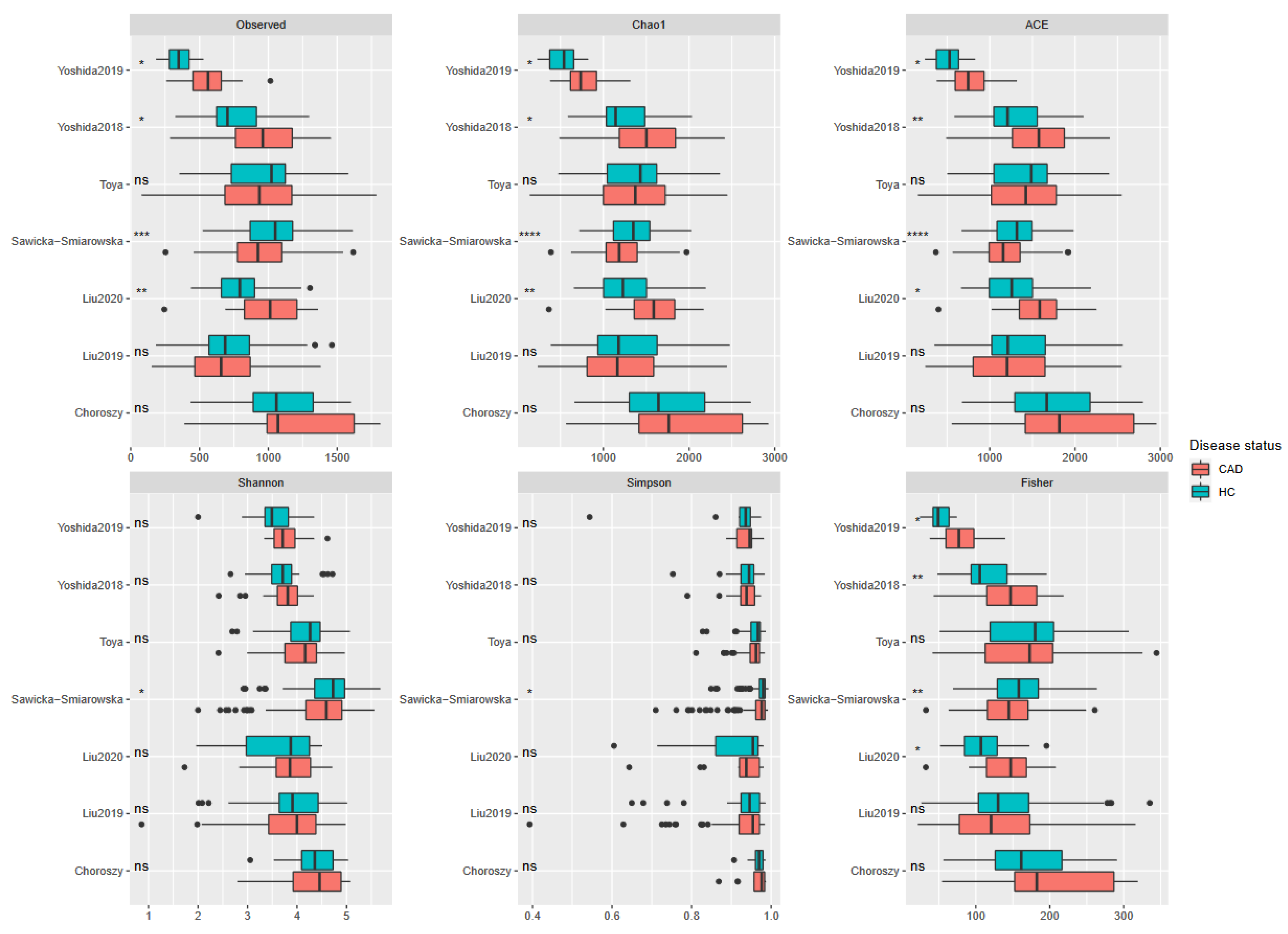
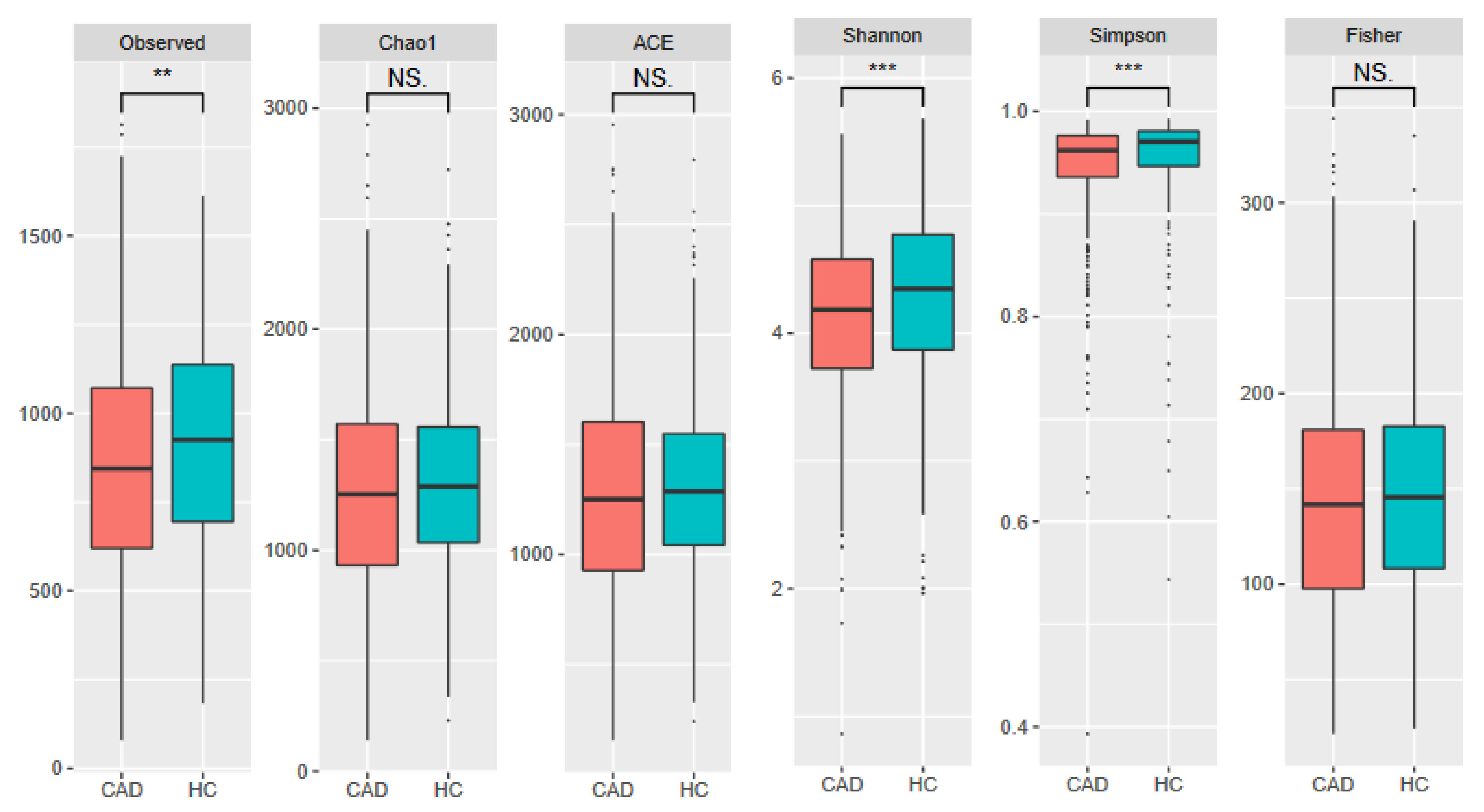
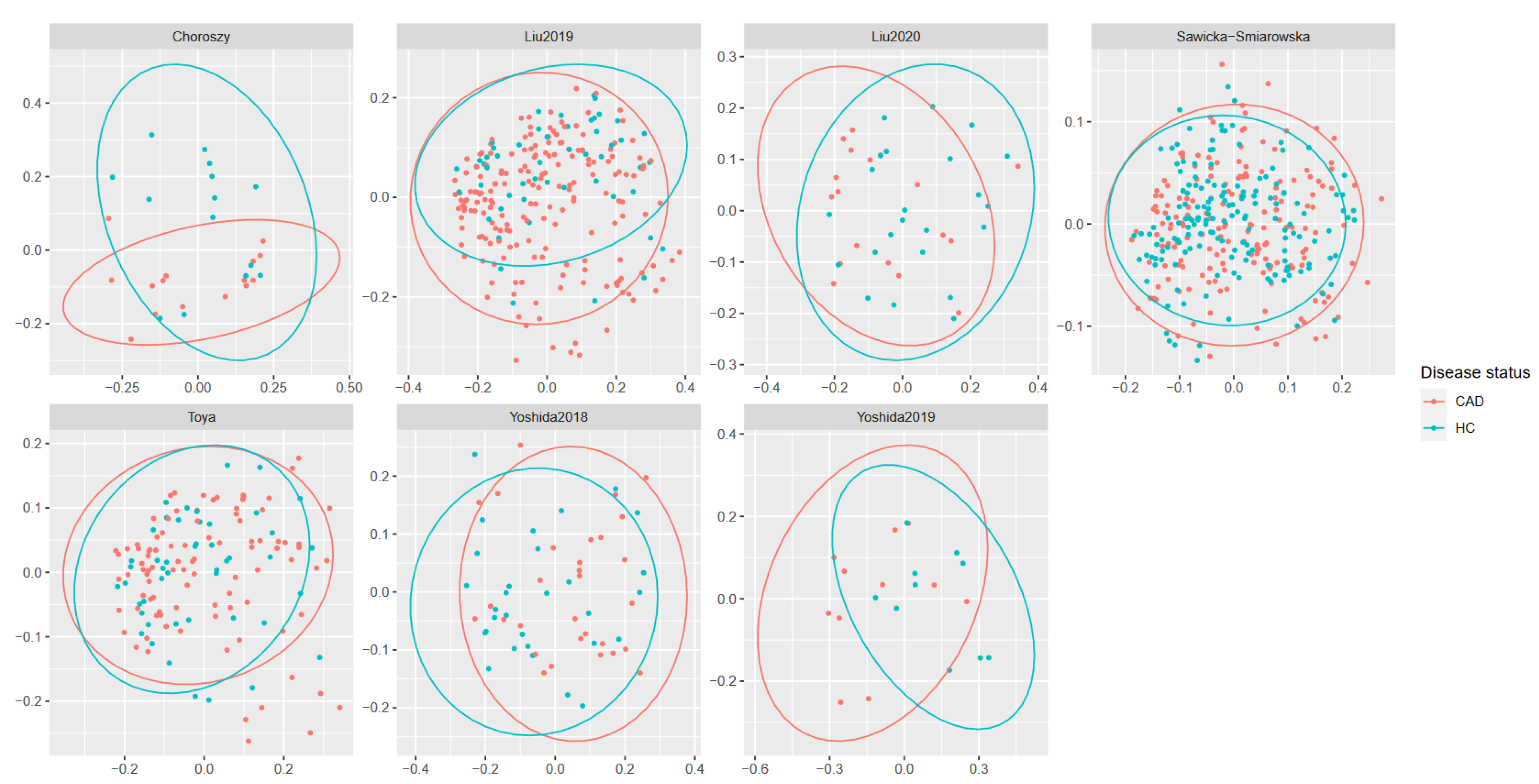
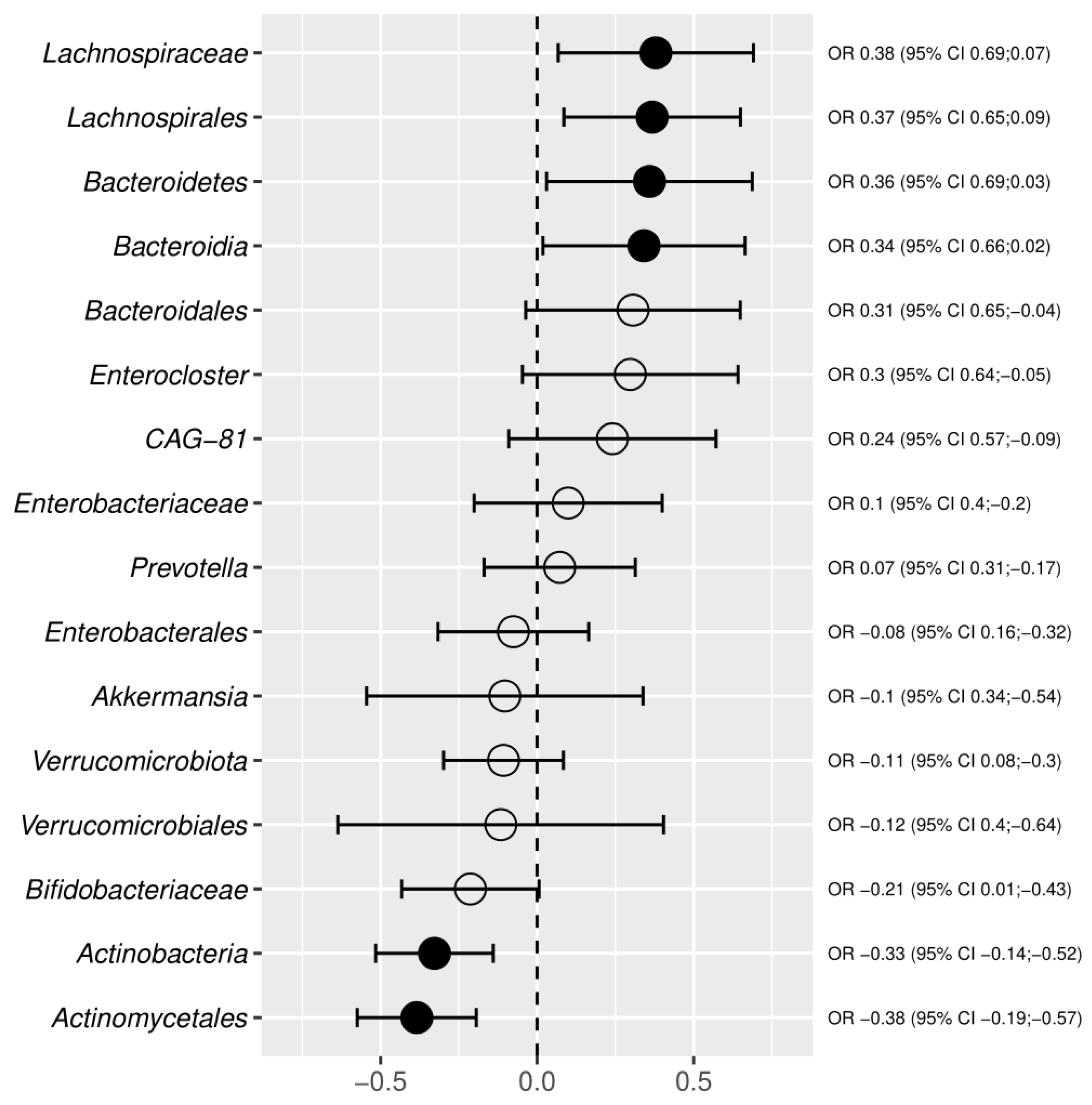
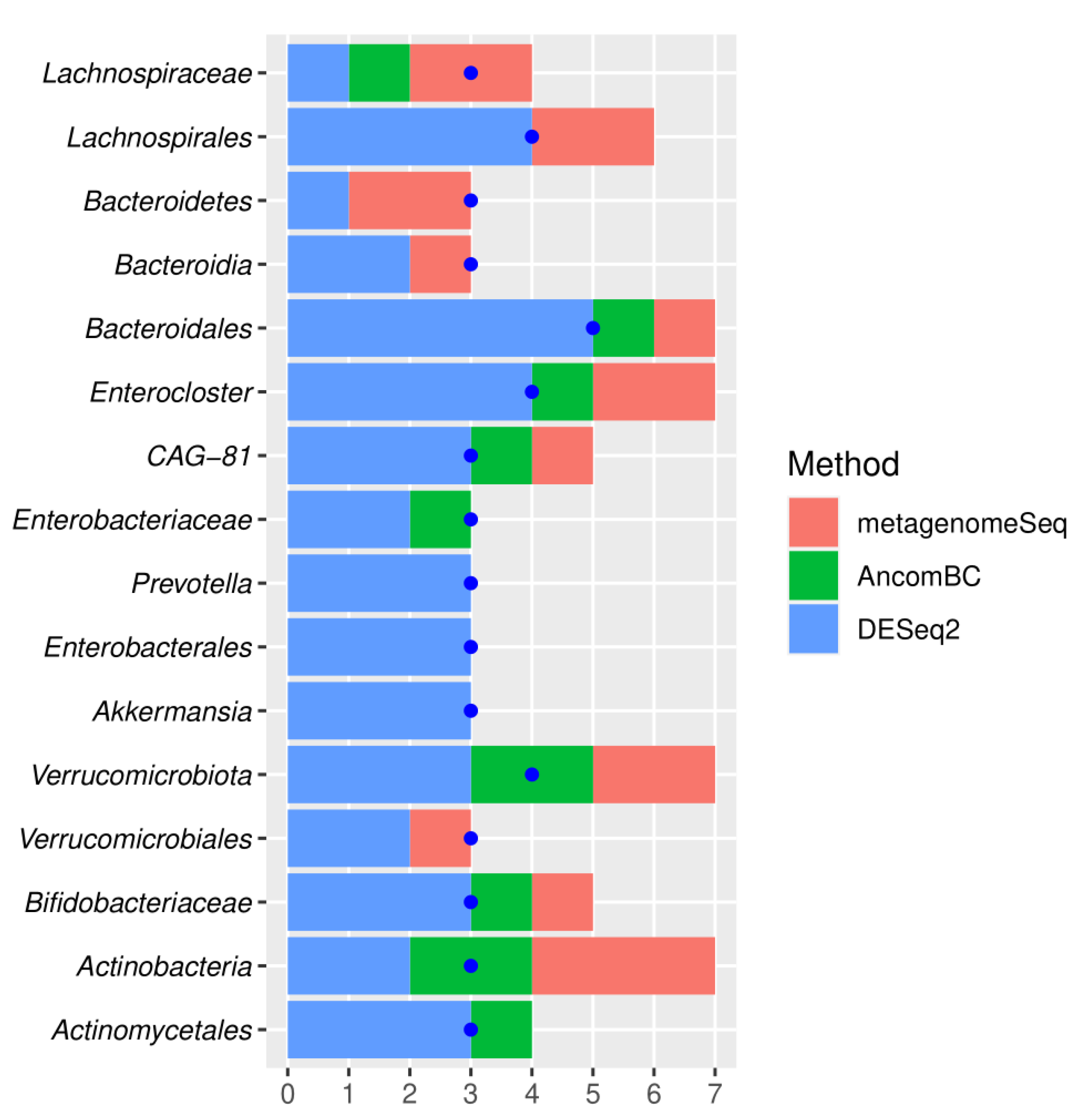
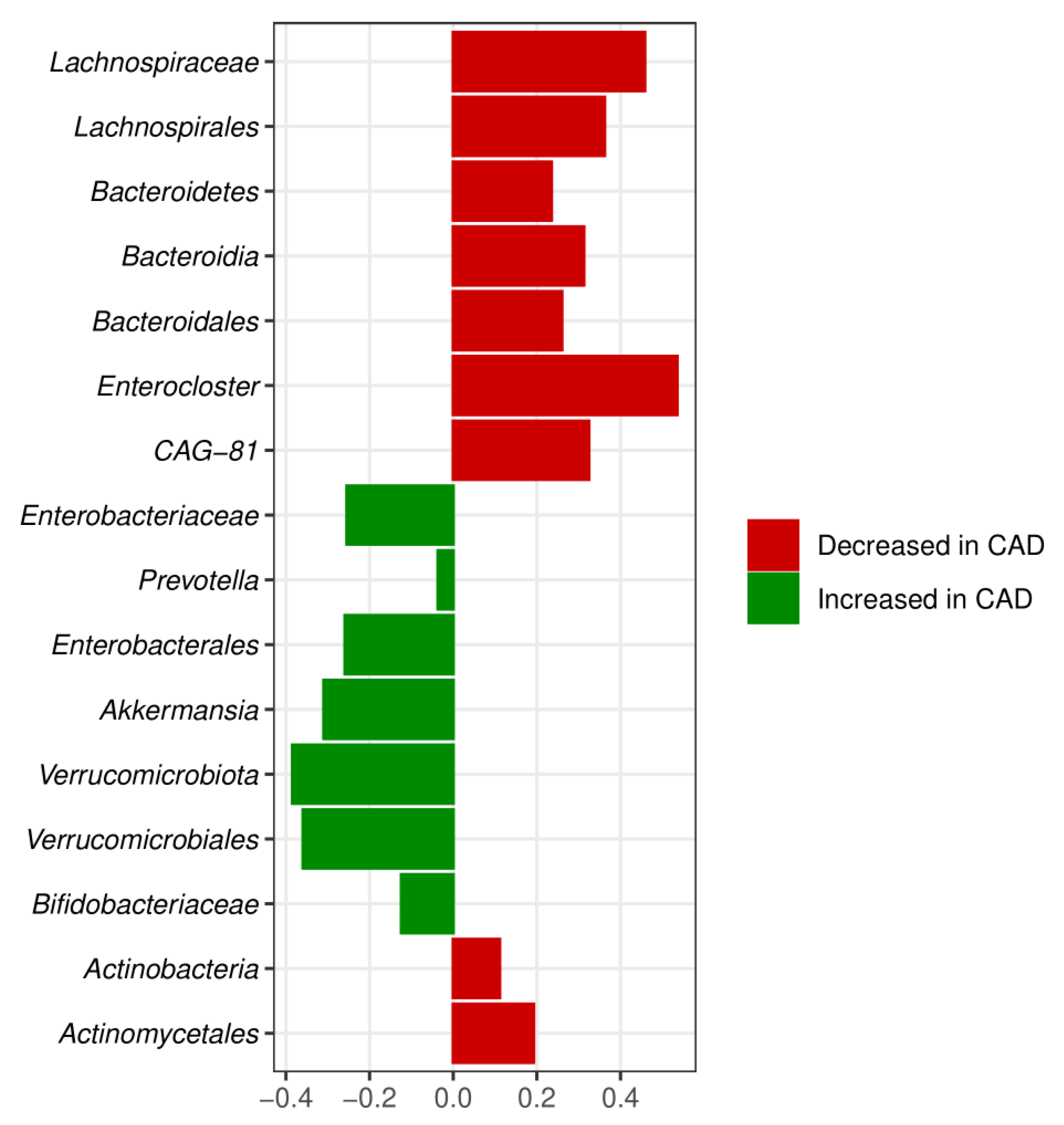
3.3. Meta-Analysis Results

The gut microbiome profile in CAD patients and controls was compared using 16S rDNA gene sequencing in fecal samples. Although 16S rDNA can deliver the resolution required for taxonomic classification at species and strain levels, it generally involves sequencing the entire 16S rRNA gene. Since the study focused only on the V3-V4 region, the meta-analysis of species or lower taxonomic groups was not performed.
Three of the evaluated alpha-diversity measures were decreased in CAD patients. These included Shannon (p = 0.00025) and Simpson indices (p < 0.0001), both of which measure evenness and observed OTUs (p = 0.0015), indicating richness. Other measures (Chao1, ACE, and Fisher indices) evaluated in this study did not significantly differ between groups (Figure 3 and Figure 4). All (except Toya et al. [50]) datasets showed significant differences in beta-diversity (measured with UniFrac) between controls and patients with CAD. However, PCoA plots (Figure 5) showed notable overlap between patients with CAD and healthy controls, without a clear boundary between samples.
The meta-analysis combined CAD with three phyla, i.e., Bacteroidetes, Actinobacteria, and Verrucomicrobiota (Figure 6, Figure 7 and Figure 8). The relative abundance of Bacteroidetes was decreased, whereas Actinobacteria and Verrucomicrobiota were increased in CAD compared to the control group. The Bacteroidetes depletion in CAD was also confirmed at the class, order, and genus levels (Figure 2) supporting the systematic review results. The Actinobacteria overrepresentation in CAD was further confirmed by the increased relative abundance of the order Actinomycetales and the family Bifidobacteriaceae.
Increased Verrucomicrobiota phylum in CAD versus the control group was also associated with increased Verrucomicrobiales order and Akkermansia genus. However, the confidence interval for Akkermansia significantly overlapped between the increase and decrease of abundance, indicating the low reliability of this result (Figure 6).
Additionally, the meta-analysis showed an increase in Proteobacteria at the level of Enterobacteriales and Enterobacteriaceae family. The alterations within the Firmicutes phylum concerned the Lachnospiraceae family and Enterocloster genus, which were decreased in CAD versus the control group. Additionally, the reduced abundance of the CAG-81 genus from a class of Clostridia was observed in CAD patients. Overall, the meta-analysis combined gut microbiota in CAD patients with main bacterial phyla, i.e., Proteobacteria, Bacteroidetes, Actinobacteria, Firmicutes, and Verrucomicrobiota, at different taxonomic ranks. The convergent results of systematic review and meta-analysis concerned Enterobacteriaceae, Bacteroidetes, and Lachnospiraceae. In contrast with the systematic review, the meta-analysis showed differences within the Verrucomicrobiota phylum in CAD patients versus the control group.

4. Discussion

The growing evidence indicates that altered gut microbiota plays a crucial role in coronary CAD as a risk factor for the disease’s outcome [58]. The gut microbiota in healthy individuals claims gut homeostasis, imparts resistance to the colonization of new species, and maintains a symbiotic relationship with the host. On the contrary, the shift in the diversity and abundance of gut microbiota facilitates the overgrowth of potentially pathogenic bacteria causing inflammatory processes and the evolution of various diseases [59]. An imbalance in the quantity of specific bacterial taxa in the gut microbiota correlates with a deficiency or excess of bacterial metabolites that fundamentally affect the physiological status of the host cells, including endothelial cells. Depending on the concentration, bacterial metabolites, e.g., trimethylamine-N-oxide (TMAO), lipopolysaccharide (LPS), or indoxyl sulfate, may yield direct toxic effects on the endothelium or indirect toxicity through their modulatory effects on hormones and biologically active compounds of the host organism [60].
The gut microbiota is dominated by five bacterial phyla, namely Bacteroidetes, Firmicutes, Actinobacteria, Proteobacteria, and Verrucomicrobiota, with Firmicutes and Bacteroidetes accounting for more than 90% of the overall gut microbiome [61]. Interestingly, the most common alterations reported in CAD are related to decreased Bacteroidetes and increased Firmicutes abundance [52].
The systematic review and meta-analysis performed in the study confirmed a significant decrease in the Bacteroidetes taxa in CAD. The decrease of Bacteroidetes in the gut microbiota carries profound health implications. These Gram-negative obligate anaerobic bacteria have several beneficial effects on the human body and play an essential role in maintaining a healthy gut ecosystem [52]. First, Bacteroidetes is involved in the degradation of non-digestible dietary carbohydrates and host-derived carbohydrates from gastrointestinal tract secretions yielding butyrate and acetate, which can lower serum lipid levels by blocking cholesterol synthesis [62,63]. Moreover, the Bacteroidetes taxa esterify absorbable cholesterol to coprostanol, a nonabsorbable sterol excreted in feces, thus lowering the blood level of cholesterol [62]. Notably, the high efficiency of cholesterol to coprostanol metabolism is suggested to reduce the risk of CAD [64]. Second, the Bacteroidetes capsular polysaccharide antigen (PSA) is vital in activating the T-cell-dependent immune response that can affect the development and homeostasis of the host immune system [65,66]. PSA of Bacteroides promotes CD4+ T cell differentiation, the balance of Th1 and Th2 populations, and the differentiation of regulatory T cells (Treg) [67,68]. On an atherosclerotic-prone mice model, Yoshida et al. [46] demonstrated that mice supplementation with Bacteroides ameliorated endotoxemia, reduced TLR4 expression and activation, and lowered plasma levels of pro-atherogenic cytokines such as IL-2, IL-4, IL-6, IL-17A, INF-γ, and TNF-α. Third, some Bacteroides species directly impact microbial LPS synthesis in the human gut, lowering systemic endotoxemia involved in the onset and progression of atherosclerosis. Penta- and tetra-acylated lipids A in LPS of Bacteroides are structurally distinct from the hexa-acylated LPS of E. coli and, in contrast with E. coli LPS, elicit reduced TLR4 response. TLR4 expression increases after LPS exposure in a dose-dependent manner, and TLR-mediated dendritic cell activation and maturation upregulates the histocompatibility complex, costimulatory molecules, cytokine production, and T cell activation. The quantity of Bacteroides tends to be negatively correlated with fecal LPS levels [46,68]. Arumugam et al. [69] demonstrated a higher incidence of symptomatic atherosclerosis in individuals with decreased Bacteroides abundance. Hence, reduced Bacteroides in the gut microbiome seem to be an important factor predisposing to CAD.
The current meta-analysis demonstrated decreased relative abundance of the Lachnospiraceae family of Firmicutes phylum in CAD versus the control group, which is in concordance with other studies [50,54]. Lachnospiraceae, similarly to Bacteroidetes taxa, produce butyrate and reduce cholesterol to coprostanol, lowering blood cholesterol levels [70]. Therefore, the Lachnospiraceae decrease in CAD may amplify the adverse effects of Bacteroides depletion of the gut microbiota. Toya et al. demonstrated that advanced CAD patients had a reduced relative abundance of Lachnospiraceae NK4B4 and other Lachnospiraceae family members, which may suggest a possibility that butyrate depletion could lead to an increase in inflammation. Moreover, the meta-analysis demonstrated in CAD patients decreased Enterocloster genus, comprising re-classified Clostridium species.
According to the systematic review, CAD was associated with an increased abundance of Enterobacteria, Lactobacillus, and Coriobacterium taxa. Lactobacillus is a well-known lactic acid producer with beneficial effects on human health, as demonstrated in several studies [71,72]. However, none of these studies evaluated current Lactobacillus levels before supplementation with these probiotics. Hence, there is a possibility that the overgrowth of specific Lactobacillus strains in gut microbiota may have adverse effects on human health. According to Ferrarese et al. [73], some probiotic strains such as L.acidophilus, L.ingluviei, L. fermentum, and L. delbrueckii are linked to a paradoxical significant weight-gain effect both in animal and human studies. According to Quiepo-Ortuno’s [74] study on an animal model, leptin and ghrelin, energy metabolism hormones, seem to be regulated by lactic acid-producing bacteria. Their study demonstrated a positive correlation between the abundance of Bifidobacterium and Lactobacillus and serum leptin levels and a significant negative correlation between the number of Clostridium, Bacteroides, and Prevotella and serum leptin levels.
Leptin is a hormone produced primarily by adipose cells and enterocytes that helps regulate energy balance by inhibiting hunger, which diminishes fat storage in adipocytes. However, obesity promotes insulin resistance and increases serum insulin levels, and high insulin levels increase leptin levels, eventually leading to leptin resistance in the nervous system and adipose tissue. This causes leptin to not reduce food intake and body weight in obese individuals [75]. Therefore, an increased abundance of Lactobacillus and Bifidobacterium and a decreased abundance of Bacteroides taxa in obese individuals may result in leptin resistance that fuels obesity, a well-known risk factor of CAD. Whether the increase in Lactobacillus is related to leptin levels in humans needs to be determined.
Moreover, it has been shown that the therapeutic effects of statins are attenuated by the abundance of Lactobacillus and Bifidobacterium, which renders these drugs relatively ineffective in decreasing LDL, confirming the putative role of lactic acid bacteria in leptin resistance [76]. Puurunen et al. [77] demonstrated that high plasma leptin levels predict the short-term occurrence of congestive heart failure, cardiac death, and acute coronary syndrome in patients with CAD independently of established risk factors.
On the contrary, ghrelin is a hormone produced by enteroendocrine cells in the gastrointestinal tract that stimulates food intake, fat deposition, and growth hormone release. Moreover, ghrelin and its receptor GHS-R1a have a cardioprotective effect on the cardiovascular system via the modulation of sympathetic activity and hypertension, enhancement of vascular activity and angiogenesis, inhibition of arrhythmias, reduction in heart failure, and inhibition of cardiac remodeling after myocardial infarction [78]. Interestingly, a meta-analysis aimed to summarize the available data regarding the circulating levels of ghrelin in patients with CAD published by Niknam et al. [79] also combined significantly lower circulating ghrelin levels with CAD. According to Torres-Fuentes et al. [80], lactate produced by Lactobacillus and Bifidobacterium attenuates ghrelin-mediated signaling through the GHSR-1a. On an animal model, Queipo-Ortuño et al. [74] demonstrated that serum ghrelin levels were negatively correlated with Bifidobacterium and Lactobacillus and positively correlated with Bacteroides and Prevotella. These data strongly suggest that an overabundance of lactic acid bacteria in the gut microbiome of patients with CAD may negatively impact patients’ energy metabolism leading to weight gain and obesity. Notably, the abundance of Lactobacillus in the gut microbiome is regulated by proatherogenic TMAO, a well-known predictor of cardiovascular disease. Hoyles et al. [81] demonstrated that TMAO stimulates the growth of lactic acid bacteria and lactate production. The level of TMAO, in turn, depends on the abundance of bacterial taxa producing TMAO precursor, i.e., trimethylamine (TMA) from dietary choline, mainly Gamma- and Betaproteobacteria, and some Firmicutes [82]. Interestingly, TMAO also stimulates the growth of Enterobacteriaceae and may account for the increased abundance of Enterobacteriaceae and Lactobacillus taxa in CAD [81]. Additionally, increased Enterobacteria correlates with increased fecal indole and serum indoxyl sulfate, a cardiotoxic uremic toxin [83]. The increase in the plasma levels of these uremic toxins is reported to accelerate the development of atherosclerotic plaque [21]. Moreover, an increased abundance of Gram-negative Enterobacteriaceae taxa may be an essential source of endotoxin. In gut dysbiosis, the endotoxin leaks into the circulation, inducing inflammation and accelerating CAD progression [57].
Jie et al. [44] performed a metagenomic shotgun-sequencing on stool samples from 218 patients with atherosclerotic CVDs and 187 healthy controls and identified, apart from Enterobacteriaceae, an increased abundance of Streptococcus spp. in atherosclerotic cardiovascular disease which is in concordance with the current study. The link between the gut Streptococcus species with atherosclerosis has been demonstrated in the current study and is well-established in other studies [44,84,85]. Hashizume-Takizawa et al. [84] on the hyperlipidemic mouse model demonstrated that atherosclerotic plaque formation increased significantly in the S. sanguis-challenged mice compared to the control group. Moreover, challenged mice showed increased expression levels of mRNAs of proinflammatory cytokines in the aorta and atherosclerosis-related mediators in the blood. The role of 73 Streptococcus species in aortic inflammation and atherosclerosis progression has been confirmed recently by computed tomography-derived coronary artery calcium score in a large cohort of middle-aged Swedes and validated in a geographically separate case-control study of symptomatic atherosclerotic disease. The study supported that gut Streptococcus spp. were independently associated with endogenous and exogenous atherogenic plasma metabolites, inflammatory and infection markers, and bacterial homologs in the oral cavity, which were associated with worse oral health [86].
The Coriobacteriales order, which according to the systematic review, was more abundant in CAD patients than controls, includes pathobionts of human gut microbiota. With Collinsella as its dominant taxon, these bacteria can affect host metabolism by altering intestinal cholesterol absorption, decreasing glycogenesis in the liver, and increasing triglyceride synthesis. Karlsson et al. [86], using shotgun sequencing of the gut metagenome, demonstrated increased Collinsella genus in patients with stenotic atherosclerotic plaques in the carotid artery. According to a study on an animal model, the presence of Corriobacteriaceae in the mouse gut correlated with decreased hepatic glycogen and glucose levels, enhanced triglyceride synthesis, and the activity of Cyp3a11, a hepatic detoxification enzyme [87]. Lahti et al. [88] identified a positive correlation between the abundance of human serum cholesterol and the genus Collinsella. According to biochemical lipid analysis, the Collinsella genus explicitly correlates with total cholesterol and LDL but not HDL, supporting data generated in studies on animal models [86,89].
There are several limitations to our study. First, most studies included in our systematic review were based on 16s rRNA sequencing focused on the V3-V4 regions, which can barely classify microbiota to the strain level. Specific metabolites of bacterial strains may exert pronounced effects on gut homeostasis. Hence, accurate characterization of the gut microbiota at the species level may be of great importance concerning preventing CAD by regulating the abundance of bacterial taxa.
Another limitation is the difference in characteristics of the studied populations in the included studies. Several studies have shown significant differences in age, gender distribution, BMI, and diabetes mellitus frequency, which are also risk factors for CAD. All these factors are also associated with changes in the gut microbiome [90,91,92,93]. Hence, it can be hypothesized that these factors, but not CAD itself, cause the microbiome alterations detected in our study. However, since most of the included studies did not show significant differences in the frequency of these risk factors, we consider this hypothesis unlikely.
The meta-analysis and systematic review pointed out differences in the gut microbiota composition in CAD patients compared to healthy controls. One of the most striking differences was the decrease in the beneficial Bacteroides and Lachnospira combined with the increase in enterobacteria, Actinobacteria, and Verrucomicrobiota in CAD patients. These alterations in the gut microbiota composition are associated with quantitative changes in atherogenic bacterial metabolites, e.g., LPS, TMAO, and uremic toxins, that increase the risk of developing or progressing CAD.
The gut microbiota composition, however, can be modulated by an appropriate diet. Therefore, knowledge and understanding of the role of the gut microbiota in CAD pathomechanism are crucial in preventing the coronary artery disease and slowing its progression. Targeting personalized medicine with dietary selection or supplementation with beneficial bacterial species could help reduce cardiovascular morbidity and mortality. However, before this can happen, an in-depth understanding of how the gut microflora changes under the diet and impacts mutual interactions with the host organism is imperative.

Supplementary Materials

The following supporting information can be downloaded at: https://www.mdpi.com/article/10.3390/metabo12121165/s1. S1: Search strategy; Table S2: PRISMA 2020 for Abstracts Checklist; S3: PRISMA 2020 Checklist; S4: The Newcastle-Ottawa Assessment Quality Scale; Table S5: Characteristics of studies included in the meta-analysis; Table S6: Characteristics of participants included studies.

Author Contributions

Conceptualization, M.C. and B.S.; methodology, M.C. and K.L.; software, K.L.; validation, R.B. and B.S.; formal analysis, K.L.; investigation, M.C., K.L., R.B. and B.S.; resources, T.R.; data curation, A.L., T.T., K.K., E.S.-Ś. and M.N.; writing—original draft preparation, M.C., K.L. and B.S.; writing—review and editing, R.B., A.L. and T.T.; visualization, M.C. and K.L.; supervision, B.S. and T.R.; project administration, B.S.; funding acquisition, M.C. and K.K. All authors have read and agreed to the published version of the manuscript.

Funding

The publication was prepared under the project financed from the funds granted by the Ministry of Education and Science in the “Regional Initiative of Excellence” program for the years 2019–2022, project number 016/RID/2018/19 (individual project number RID Z501.20.009). The part of the data was funded by National Science Centre, Poland project no.: 2017/25/N/NZ5/02765.

Data Availability Statement

The part of data that support the findings of this study are openly available under the accession numbers: PRJDB7456, PRJDB6472, PRJNA550301, PRJNA503710.

Conflicts of Interest

The authors have no conflict of interest to declare. All co-authors have seen and agree with the manuscript’s contents, and there is no financial interest to report. We certify that the submission is original work and is not under review at any other publication. The authors have no competing interests to declare.

References

  1. Medina-Leyte, D.J.; Zepeda-García, O.; Domínguez-Pérez, M.; González-Garrido, A.; Villarreal-Molina, T.; Jacobo-Albavera, L. Endothelial Dysfunction, Inflammation and Coronary Artery Disease: Potential Biomarkers and Promising Therapeutical Approaches. Int. J. Mol. Sci. 2021, 22, 3850. [Google Scholar] [CrossRef] [PubMed]
  2. Khan, M.A.; Hashim, M.J.; Mustafa, H.; Baniyas, M.Y.; Al Suwaidi, S.K.B.M.; AlKatheeri, R.; Alblooshi, F.M.K.; Almatrooshi, M.E.A.H.; Alzaabi, M.E.H.; Al Darmaki, R.S.; et al. Global Epidemiology of Ischemic Heart Disease: Results from the Global Burden of Disease Study. Cureus 2020, 12, e9349. [Google Scholar] [CrossRef] [PubMed]
  3. Maiuolo, J.; Carresi, C.; Gliozzi, M.; Mollace, R.; Scarano, F.; Scicchitano, M.; Macrì, R.; Nucera, S.; Bosco, F.; Oppedisano, F.; et al. The Contribution of Gut Microbiota and Endothelial Dysfunction in the Development of Arterial Hypertension in Animal Models and in Humans. Int. J. Mol. Sci. 2022, 23, 3698. [Google Scholar] [CrossRef] [PubMed]
  4. Thursby, E.; Juge, N. Introduction to the Human Gut Microbiota. Biochem. J. 2017, 474, 1823–1836. [Google Scholar] [CrossRef] [PubMed]
  5. Lynch, S.V.; Pedersen, O. The Human Intestinal Microbiome in Health and Disease. N. Engl. J. Med. 2016, 375, 2369–2379. [Google Scholar] [CrossRef] [PubMed] [Green Version]
  6. Afzaal, M.; Saeed, F.; Shah, Y.A.; Hussain, M.; Rabail, R.; Socol, C.T.; Hassoun, A.; Pateiro, M.; Lorenzo, J.M.; Rusu, A.V.; et al. Human Gut Microbiota in Health and Disease: Unveiling the Relationship. Front. Microbiol. 2022, 13, 999001. [Google Scholar] [CrossRef] [PubMed]
  7. Fan, Y.; Pedersen, O. Gut Microbiota in Human Metabolic Health and Disease. Nat. Rev. Microbiol. 2021, 19, 55–71. [Google Scholar] [CrossRef]
  8. Ghosh, S.; Whitley, C.S.; Haribabu, B.; Jala, V.R. Regulation of Intestinal Barrier Function by Microbial Metabolites. Cmgh 2021, 11, 1463–1482. [Google Scholar] [CrossRef]
  9. Yoo, J.Y.; Sniffen, S.; McGill Percy, K.C.; Pallaval, V.B.; Chidipi, B. Gut Dysbiosis and Immune System in Atherosclerotic Cardiovascular Disease (ACVD). Microorganisms 2022, 10, 108. [Google Scholar] [CrossRef]
  10. Baizabal-Carvallo, J.F.; Alonso-Juarez, M. The Link between Gut Dysbiosis and Neuroinflammation in Parkinson’s Disease. Neuroscience 2020, 432, 160–173. [Google Scholar] [CrossRef]
  11. Janeiro, M.H.; Ramírez, M.J.; Solas, M. Dysbiosis and Alzheimer’s Disease: Cause or Treatment Opportunity? Cell Mol. Neurobiol. 2022, 42, 377–387. [Google Scholar] [CrossRef] [PubMed]
  12. Halverson, T.; Alagiakrishnan, K. Gut Microbes in Neurocognitive and Mental Health Disorders. Ann. Med. 2020, 52, 423–443. [Google Scholar] [CrossRef] [PubMed]
  13. Zhou, A.; Yuan, Y.; Yang, M.; Huang, Y.; Li, X.; Li, S.; Yang, S.; Tang, B. Crosstalk Between the Gut Microbiota and Epithelial Cells Under Physiological and Infectious Conditions. Front. Cell Infect. Microbiol. 2022, 12, 832672. [Google Scholar] [CrossRef]
  14. Fachi, J.L.; Felipe, J.d.S.; Pral, L.P.; da Silva, B.K.; Corrêa, R.O.; de Andrade, M.C.P.; da Fonseca, D.M.; Basso, P.J.; Câmara, N.O.S.; de Sales e Souza, É.L.; et al. Butyrate Protects Mice from Clostridium Difficile-Induced Colitis through an HIF-1-Dependent Mechanism. Cell Rep. 2019, 27, 750–761.e7. [Google Scholar] [CrossRef] [Green Version]
  15. Venkatesh, M.; Mukherjee, S.; Wang, H.; Li, H.; Sun, K.; Benechet, A.P.; Qiu, Z.; Maher, L.; Redinbo, M.R.; Phillips, R.S.; et al. Symbiotic Bacterial Metabolites Regulate Gastrointestinal Barrier Function via the Xenobiotic Sensor PXR and Toll-like Receptor 4. Immunity 2014, 41, 296–310. [Google Scholar] [CrossRef] [Green Version]
  16. Louis, P.; Flint, H.J. Formation of Propionate and Butyrate by the Human Colonic Microbiota. Environ. Microbiol. 2017, 19, 29–41. [Google Scholar] [CrossRef] [Green Version]
  17. Ghosh, T.S.; Das, M.; Jeffery, I.B.; O’Toole, P.W. Adjusting for Age Improves Identification of Gut Microbiome Alterations in Multiple Diseases. Elife 2020, 9, e50240. [Google Scholar] [CrossRef]
  18. Chelakkot, C.; Ghim, J.; Ryu, S.H. Mechanisms Regulating Intestinal Barrier Integrity and Its Pathological Implications. Exp. Mol. Med. 2018, 50, 1–9. [Google Scholar] [CrossRef] [Green Version]
  19. Violi, F.; Cammisotto, V.; Bartimoccia, S.; Pignatelli, P.; Carnevale, R.; Nocella, C. Gut-Derived Low-Grade Endotoxaemia, Atherothrombosis and Cardiovascular Disease. Nat. Rev. Cardiol. 2022, 0123456789. [Google Scholar] [CrossRef]
  20. Akash, M.S.H.; Fiayyaz, F.; Rehman, K.; Sabir, S.; Rasool, M.H. Gut Microbiota and Metabolic Disorders: Advances in Therapeutic Interventions. Crit. Rev. Immunol. 2019, 39, 223–237. [Google Scholar] [CrossRef]
  21. Zhang, B.; Wang, X.; Xia, R.; Li, C. Gut Microbiota in Coronary Artery Disease: A Friend or Foe? Biosci. Rep 2020, 40, BSR20200454. [Google Scholar] [CrossRef] [PubMed]
  22. Hasan, N.; Yang, H. Factors Affecting the Composition of the Gut Microbiota, and Its Modulation. PeerJ 2019, 7, e7502. [Google Scholar] [CrossRef] [PubMed] [Green Version]
  23. Boers, S.A.; Jansen, R.; Hays, J.P. Understanding and Overcoming the Pitfalls and Biases of Next-Generation Sequencing (NGS) Methods for Use in the Routine Clinical Microbiological Diagnostic Laboratory. Eur. J. Clin. Microbiol. Infect. Dis. Off. Publ. Eur. Soc. Clin. Microbiol. 2019, 38, 1059–1070. [Google Scholar] [CrossRef] [PubMed] [Green Version]
  24. Nearing, J.T.; Douglas, G.M.; Hayes, M.G.; MacDonald, J.; Desai, D.K.; Allward, N.; Jones, C.M.A.; Wright, R.J.; Dhanani, A.S.; Comeau, A.M.; et al. Microbiome Differential Abundance Methods Produce Different Results across 38 Datasets. Nat. Commun. 2022, 13, 342. [Google Scholar] [CrossRef]
  25. Köster, J.; Rahmann, S. Snakemake-a Scalable Bioinformatics Workflow Engine. Bioinformatics 2012, 28, 2520–2522. [Google Scholar] [CrossRef] [Green Version]
  26. Martin, M. Cutadapt Removes Adapter Sequences from High-Throughput Sequencing Reads. EMBnet. J. 2011, 17, 10–12. [Google Scholar] [CrossRef]
  27. Sawicka-Śmiarowska, E.; Bondarczuk, K.; Bauer, W.; Niemira, M.; Szalkowska, A.; Raczkowska, J.; Kwasniewski, M.; Tarasiuk, E.; Dubatowka, M.; Lapinska, M.; et al. Gut Microbiome in Chronic Coronary Syndrome Patients. J. Clin. Med. 2021, 10, 5074. [Google Scholar] [CrossRef]
  28. RC, E. Search and Clustering Orders of Magnitude Faster than BLAST. Bioinformatics 2010, 26, 2460–2461. [Google Scholar] [CrossRef] [Green Version]
  29. Edgar, R. UNOISE2: Improved Error-Correction for Illumina 16S and ITS Amplicon Sequencing. bioRxiv 2016, 081257. [Google Scholar] [CrossRef] [Green Version]
  30. Murali, A.; Bhargava, A.; Wright, E.S. IDTAXA: A Novel Approach for Accurate Taxonomic Classification of Microbiome Sequences. Microbiome 2018, 6, 140. [Google Scholar] [CrossRef]
  31. Chaumeil, P.A.; Mussig, A.J.; Hugenholtz, P.; Parks, D.H. GTDB-Tk: A Toolkit to Classify Genomes with the Genome Taxonomy Database. Bioinformatics 2020, 36, 1925–1927. [Google Scholar] [CrossRef] [PubMed]
  32. McMurdie, P.J.; Holmes, S. Phyloseq: An R Package for Reproducible Interactive Analysis and Graphics of Microbiome Census Data. PLoS ONE 2013, 8, e61217. [Google Scholar] [CrossRef] [PubMed] [Green Version]
  33. Lin, H.; Peddada, S. Das Analysis of Compositions of Microbiomes with Bias Correction. Nat. Commun. 2020, 11, 3514. [Google Scholar] [CrossRef] [PubMed]
  34. Oksanen, A.J.; Blanchet, F.G.; Friendly, M.; Kindt, R.; Legendre, P.; Mcglinn, D.; Minchin, P.R.; Hara, R.B.O.; Simpson, G.L.; Solymos, P.; et al. Vegan. Encycl. Food Agric. Ethics 2019, 2, 2395–2396. [Google Scholar] [CrossRef]
  35. Agresti, A. Generalized Odds Ratios for Ordinal Data. Biometrics 1980, 36, 59. [Google Scholar] [CrossRef]
  36. Package, T.; Generalized, T.; Ratios, O.; Rcpp, L. Package ‘Genodds’. 2021. [software].
  37. Balduzzi, S.; Rücker, G.; Schwarzer, G. How to Perform a Meta-Analysis with R: A Practical Tutorial. Evid. Based. Ment. Health 2019, 22, 153–160. [Google Scholar] [CrossRef] [Green Version]
  38. Alhmoud, T.; Kumar, A.; Lo, C.-C.; Al-Sadi, R.; Clegg, S.; Alomari, I.; Zmeili, T.; Gleasne, C.D.; Mcmurry, K.; Dichosa, A.E.K.; et al. Investigating Intestinal Permeability and Gut Microbiota Roles in Acute Coronary Syndrome Patients. Hum. Microbiome J. 2019, 13, 100059. [Google Scholar] [CrossRef]
  39. Emoto, T.; Yamashita, T.; Kobayashi, T.; Sasaki, N.; Hirota, Y.; Hayashi, T.; So, A.; Kasahara, K.; Yodoi, K.; Matsumoto, T.; et al. Characterization of Gut Microbiota Profiles in Coronary Artery Disease Patients Using Data Mining Analysis of Terminal Restriction Fragment Length Polymorphism: Gut Microbiota Could Be a Diagnostic Marker of Coronary Artery Disease. Hear. Vessel. 2016, 32, 39–46. [Google Scholar] [CrossRef] [PubMed]
  40. Kwun, J.S.; Kang, S.H.; Lee, H.J.; Park, H.K.; Lee, W.J.; Yoon, C.H.; Suh, J.W.; Cho, Y.S.; Youn, T.J.; Chae, I.H. Comparison of Thrombus, Gut, and Oral Microbiomes in Korean Patients with ST-Elevation Myocardial Infarction: A Case–Control Study. Exp. Mol. Med. 2020, 52, 2069–2079. [Google Scholar] [CrossRef]
  41. Zheng, Y.Y.; Wu, T.T.; Liu, Z.Q.; Li, A.; Guo, Q.Q.; Ma, Y.Y.; Zhang, Z.L.; Xun, Y.L.; Zhang, J.C.; Wang, W.R.; et al. Gut Microbiome-Based Diagnostic Model to Predict Coronary Artery Disease. J. Agric. Food Chem. 2020, 68, 3548–3557. [Google Scholar] [CrossRef] [PubMed]
  42. Ivashkin, V.T.; Kashukh, Y.A. Impact of L-Carnitine and Phosphatidylcholine Containing Products on the Proatherogenic Metabolite TMAO Production and Gut Microbiome Changes in Patients with Coronary Artery Disease. Vopr. Pitan. 2019, 88, 25–33. [Google Scholar] [CrossRef] [PubMed]
  43. Emoto, T.; Yamashita, T.; Sasaki, N.; Hirota, Y.; Hayashi, T.; So, A.; Kasahara, K.; Yodoi, K.; Matsumoto, T.; Mizoguchi, T.; et al. Analysis of Gut Microbiota in Coronary Artery Disease Patients: A Possible Link between Gut Microbiota and Coronary Artery Disease. J. Atheroscler. Thromb. 2016, 23, 908–921. [Google Scholar] [CrossRef] [PubMed] [Green Version]
  44. Jie, Z.; Xia, H.; Zhong, S.-L.; Feng, Q.; Li, S.; Liang, S.; Zhong, H.; Liu, Z.; Gao, Y.; Zhao, H.; et al. The Gut Microbiome in Atherosclerotic Cardiovascular Disease. Nat. Commun. 2017, 8, 845. [Google Scholar] [CrossRef] [PubMed] [Green Version]
  45. Liu, Z.; Li, J.; Liu, H.; Tang, Y.; Zhan, Q.; Lai, W.; Ao, L.; Meng, X.; Ren, H.; Xu, D.; et al. The Intestinal Microbiota Associated with Cardiac Valve Calcification Differs from That of Coronary Artery Disease. Atherosclerosis 2019, 284, 121–128. [Google Scholar] [CrossRef] [PubMed] [Green Version]
  46. Yoshida, N.; Emoto, T.; Yamashita, T.; Watanabe, H.; Hayashi, T.; Tabata, T.; Hoshi, N.; Hatano, N.; Ozawa, G.; Sasaki, N.; et al. Bacteroides Vulgatus and Bacteroides Dorei Reduce Gut Microbial Lipopolysaccharide Production and Inhibit Atherosclerosis. Circulation 2018, 138, 2486–2498. [Google Scholar] [CrossRef] [PubMed]
  47. Yoshida, N.; Sasaki, K.; Sasaki, D.; Yamashita, T.; Fukuda, H.; Hayashi, T.; Tabata, T.; Osawa, R.; Hirata, K.I.; Kondo, A. Effect of Resistant Starch on the Gut Microbiota and Its Metabolites in Patients with Coronary Artery Disease. J. Atheroscler. Thromb. 2019, 26, 705–719. [Google Scholar] [CrossRef] [Green Version]
  48. Dong, C.; He, Z.; Zhu, Q.; Liu, J.; Gao, F.; Li, K.; Sun, S.; Liu, Q.; Wang, Y.; Tang, Y.; et al. Correlation Network Analyses Based on Metagenomics and Multi-Type Metabolomic Data Identified Biomarkers of Coronary Artery Disease. Res. Sq. 2020, preprint. [Google Scholar] [CrossRef]
  49. Gao, J.; Yan, K.T.; Wang, J.X.; Dou, J.; Wang, J.; Ren, M.; Ma, J.; Zhang, X.; Liu, Y. Gut Microbial Taxa as Potential Predictive Biomarkers for Acute Coronary Syndrome and Post-STEMI Cardiovascular Events. Sci. Rep. 2020, 10, 2639. [Google Scholar] [CrossRef] [Green Version]
  50. Toya, T.; Ozcan, I.; Corban, M.T.; Sara, J.D.; Marietta, E.V.; Ahmad, A.; Horwath, I.E.; Loeffler, D.L.; Murray, J.A.; Lerman, L.O.; et al. Compositional Change of Gut Microbiome and Osteocalcin Expressing Endothelial Progenitor Cells in Patients with Coronary Artery Disease. PLoS ONE 2021, 16, e0249187. [Google Scholar] [CrossRef] [PubMed]
  51. Hu, J.L.; Yao, Z.F.; Tang, M.N.; Tang, C.; Zhao, X.F.; Su, X.; Lu, D.B.; Li, Q.R.; Wang, Z.S.; Yan, Y.; et al. Gut Microbiota Community Shift with Severity of Coronary Artery Disease. Engineering 2021, 7, 1715–1724. [Google Scholar] [CrossRef]
  52. Toya, T.; Corban, M.T.; Marrietta, E.; Horwath, I.E.; Lerman, L.O.; Murray, J.A.; Lerman, A. Coronary Artery Disease Is Associated with an Altered Gut Microbiome Composition. PLoS ONE 2020, 15, e0227147. [Google Scholar] [CrossRef]
  53. Cui, L.; Zhao, T.; Hu, H.; Zhang, W.; Hua, X. Association Study of Gut Flora in Coronary Heart Disease through High-Throughput Sequencing. Biomed Res. Int. 2017, 2017, 3796359. [Google Scholar] [CrossRef] [Green Version]
  54. Zhu, Q.; Gao, R.; Zhang, Y.; Pan, D.; Zhu, Y.; Zhang, X.; Yang, R.; Jiang, R.; Xu, Y.; Qin, H. Dysbiosis Signatures of Gut Microbiota in Coronary Artery Disease. Physiol. Genomics. 2018, 50, 893–903. [Google Scholar] [CrossRef]
  55. Liu, F.; Fan, C.; Zhang, L.; Li, Y.; Hou, H.; Ma, Y.; Fan, J.; Tan, Y.; Wu, T.; Jia, S.; et al. Alterations of Gut Microbiome in Tibetan Patients With Coronary Heart Disease. Front. Cell Infect. Microbiol. 2020, 10, 373. [Google Scholar] [CrossRef]
  56. Chiu, F.-C.; Tsai, C.-F.; Huang, P.-S.; Shih, C.-Y.; Tsai, M.-H.; Hwang, J.-J.; Wang, Y.-C.; Chuang, E.Y.; Tsai, C.-T.; Chang, S.-N. The Gut Microbiome, Seleno-Compounds, and Acute Myocardial Infarction. J. Clin. Med. 2022, 11, 1462. [Google Scholar] [CrossRef]
  57. Choroszy, M.; Litwinowicz, K.; Łaczmanski, Ł.; Roleder, T.; Sobieszczanska, B. Co-Toxicity of Bacterial Metabolites, to Vascular Endothelial Cells in Coronary Arterial Disease Accompanied by Gut Dysbiosis. Nutrients 2022, 14, 424. [Google Scholar] [CrossRef]
  58. Xu, J.; Yang, Y.J. Implications of Gut Microbiome on Coronary Artery Disease. Cardiovasc. Diagn. Ther. 2020, 10, 869–880. [Google Scholar] [CrossRef]
  59. Olvera-Rosales, L.-B.; Cruz-Guerrero, A.-E.; Ramírez-Moreno, E.; Quintero-Lira, A.; Contreras-López, E.; Jaimez-Ordaz, J.; Castañeda-Ovando, A.; Añorve-Morga, J.; Calderón-Ramos, Z.-G.; Arias-Rico, J.; et al. Impact of the Gut Microbiota Balance on the Health-Disease Relationship: The Importance of Consuming Probiotics and Prebiotics. Foods 2021, 10, 1261. [Google Scholar] [CrossRef]
  60. Liu, J.; Tan, Y.; Cheng, H.; Zhang, D.; Feng, W.; Peng, C. Functions of Gut Microbiota Metabolites, Current Status and Future Perspectives. Aging Dis. 2022, 13, 1106–1126. [Google Scholar] [CrossRef]
  61. Rinninella, E.; Raoul, P.; Cintoni, M.; Franceschi, F.; Miggiano, G.A.D.; Gasbarrini, A.; Mele, M.C. What Is the Healthy Gut Microbiota Composition? A Changing Ecosystem across Age, Environment, Diet, and Diseases. Microorganisms 2019, 7, 14. [Google Scholar] [CrossRef] [PubMed]
  62. Kazemian, N.; Mahmoudi, M.; Halperin, F.; Wu, J.C.; Pakpour, S. Gut Microbiota and Cardiovascular Disease: Opportunities and Challenges. Microbiome 2020, 8, 36. [Google Scholar] [CrossRef] [Green Version]
  63. Thomas, F.; Hehemann, J.H.; Rebuffet, E.; Czjzek, M.; Michel, G. Environmental and Gut Bacteroidetes: The Food Connection. Front. Microbiol. 2011, 2, 93. [Google Scholar] [CrossRef] [Green Version]
  64. Kriaa, A.; Bourgin, M.; Potiron, A.; Mkaouar, H.; Jablaoui, A.; Gerard, P.; Maguin, E.; Rhimi, M. Microbial Impact on Cholesterol and Bile Acid Metabolism: Current Status and Future Prospects. J. Lipid Res. 2019, 60, 323–332. [Google Scholar] [CrossRef] [Green Version]
  65. Troy, E.B.; Kasper, D.L. Beneficial Effects of Bacteroides Fragilis Polysaccharides on the Immune System. Front. Biosci. 2010, 15, 25–34. [Google Scholar] [CrossRef] [Green Version]
  66. Wexler, A.G.; Goodman, A.L. An Insider’s Perspective: Bacteroides as a Window into the Microbiome. Nat. Microbiol. 2017 25 2017, 2, 17026. [Google Scholar] [CrossRef] [Green Version]
  67. Zheng, D.; Liwinski, T.; Elinav, E. Interaction between Microbiota and Immunity in Health and Disease. Cell Res. 2020, 30, 492–506. [Google Scholar] [CrossRef]
  68. Yoshida, N.; Yamashita, T.; Kishino, S.; Watanabe, H.; Sasaki, K.; Sasaki, D.; Tabata, T.; Sugiyama, Y.; Kitamura, N.; Saito, Y.; et al. A Possible Beneficial Effect of Bacteroides on Faecal Lipopolysaccharide Activity and Cardiovascular Diseases. Sci. Rep. 2020, 10, 13009. [Google Scholar] [CrossRef]
  69. Arumugam, M.; Raes, J.; Pelletier, E.; Le Paslier, D.; Yamada, T.; Mende, D.R.; Fernandes, G.R.; Tap, J.; Bruls, T.; Batto, J.-M.; et al. Enterotypes of the Human Gut Microbiome. Nature 2011, 473, 174–180. [Google Scholar] [CrossRef] [Green Version]
  70. Vacca, M.; Celano, G.; Calabrese, F.M.; Portincasa, P.; Gobbetti, M.; Angelis, M. De The Controversial Role of Human Gut Lachnospiraceae. Microorganisms 2020, 8, 573. [Google Scholar] [CrossRef]
  71. Heeney, D.D.; Gareau, M.G.; Marco, M.L. Intestinal Lactobacillus in Health and Disease, a Driver or Just along for the Ride? Curr. Opin. Biotechnol. 2018, 49, 140–147. [Google Scholar] [CrossRef]
  72. Kechagia, M.; Basoulis, D.; Konstantopoulou, S.; Dimitriadi, D.; Gyftopoulou, K.; Skarmoutsou, N.; Fakiri, E.M. Health Benefits of Probiotics: A Review. ISRN Nutr. 2013, 2013, 481651. [Google Scholar] [CrossRef] [PubMed] [Green Version]
  73. Ferrarese, R.; Ceresola, E.R.; Preti, A.; Canducci, F. Probiotics, Prebiotics and Synbiotics for Weight Loss and Metabolic Syndrome in the Microbiome Era. Eur. Rev. Med. Pharmacol. Sci. 2018, 22, 7588–7605. [Google Scholar] [CrossRef] [PubMed]
  74. Queipo-Ortuño, M.I.; Seoane, L.M.; Murri, M.; Pardo, M.; Gomez-Zumaquero, J.M.; Cardona, F.; Casanueva, F.; Tinahones, F.J. Gut Microbiota Composition in Male Rat Models under Different Nutritional Status and Physical Activity and Its Association with Serum Leptin and Ghrelin Levels. PLoS ONE 2013, 8, e65465. [Google Scholar] [CrossRef] [Green Version]
  75. Grigor’eva, I.N. Gallstone Disease, Obesity and the Firmicutes/Bacteroidetes Ratio as a Possible Biomarker of Gut Dysbiosis. J. Pers. Med. 2020, 11, 13. [Google Scholar] [CrossRef] [PubMed]
  76. Sun, B.; Li, L.; Zhou, X. Comparative Analysis of the Gut Microbiota in Distinct Statin Response Patients in East China. J. Microbiol. 2018, 56, 886–892. [Google Scholar] [CrossRef] [PubMed]
  77. Puurunen, V.-P.; Kiviniemi, A.; Lepojärvi, S.; Piira, O.-P.; Hedberg, P.; Junttila, J.; Ukkola, O.; Huikuri, H. Leptin Predicts Short-Term Major Adverse Cardiac Events in Patients with Coronary Artery Disease. Ann. Med. 2017, 49, 448–454. [Google Scholar] [CrossRef]
  78. Yuan, M.-J.; Li, W.; Zhong, P. Research Progress of Ghrelin on Cardiovascular Disease. Biosci. Rep. 2021, 41, BSR20203387. [Google Scholar] [CrossRef]
  79. Niknam, M.; Liaghat, T.; Zarghami, M.; Akrami, M.; Shahnematollahi, S.M.; Ahmadipour, A.; Moazzen, F.; Soltanabadi, S. Ghrelin and Ghrelin/Total Cholesterol Ratio as Independent Predictors for Coronary Artery Disease: A Systematic Review and Meta-Analysis. J. Investig. Med. 2022, 70, 759–765. [Google Scholar] [CrossRef]
  80. Torres-Fuentes, C.; Golubeva, A.V.; Zhdanov, A.V.; Wallace, S.; Arboleya, S.; Papkovsky, D.B.; Aidy, S.E.; Ross, P.; Roy, B.L.; Stanton, C.; et al. Short-Chain Fatty Acids and Microbiota Metabolites Attenuate Ghrelin Receptor Signaling. FASEB J. Off. Publ. Fed. Am. Soc. Exp. Biol. 2019, 33, 13546–13559. [Google Scholar] [CrossRef] [Green Version]
  81. Hoyles, L.; Jiménez-Pranteda, M.L.; Chilloux, J.; Brial, F.; Myridakis, A.; Aranias, T.; Magnan, C.; Gibson, G.R.; Sanderson, J.D.; Nicholson, J.K.; et al. Metabolic Retroconversion of Trimethylamine N-Oxide and the Gut Microbiota. Microbiome 2018, 6, 73. [Google Scholar] [CrossRef]
  82. Dalla Via, A.; Gargari, G.; Taverniti, V.; Rondini, G.; Velardi, I.; Gambaro, V.; Visconti, G.L.; De Vitis, V.; Gardana, C.; Ragg, E.; et al. Urinary TMAO Levels Are Associated with the Taxonomic Composition of the Gut Microbiota and with the Choline TMA-Lyase Gene (CutC) Harbored by Enterobacteriaceae. Nutrients 2019, 12, 62. [Google Scholar] [CrossRef] [PubMed] [Green Version]
  83. Cosola, C.; Rocchetti, M.T.; Cupisti, A.; Gesualdo, L. Microbiota Metabolites: Pivotal Players of Cardiovascular Damage in Chronic Kidney Disease. Pharmacol. Res. 2018, 130, 132–142. [Google Scholar] [CrossRef] [PubMed]
  84. Hashizume-Takizawa, T.; Yamaguchi, Y.; Kobayashi, R.; Shinozaki-Kuwahara, N.; Saito, M.; Kurita-Ochiai, T. Oral Challenge with Streptococcus Sanguinis Induces Aortic Inflammation and Accelerates Atherosclerosis in Spontaneously Hyperlipidemic Mice. Biochem. Biophys. Res. Commun. 2019, 520, 507–513. [Google Scholar] [CrossRef] [PubMed]
  85. Sayols-Baixeras, S.; Dekkers, K.F.; Hammar, U.; Baldanzi, G.; Lin, Y.-T.; Ahmad, S.; Nguyen, D.; Varotsis, G.; Pita, S.; Nielsen, N.; et al. Streptococcus Species Abundance in the Gut Is Linked to Subclinical Coronary Atherosclerosis in 8,973 Participants from the SCAPIS Cohort. medRxiv 2022. [Google Scholar] [CrossRef]
  86. Karlsson, F.H.; Fåk, F.; Nookaew, I.; Tremaroli, V.; Fagerberg, B.; Petranovic, D.; Bäckhed, F.; Nielsen, J. Symptomatic Atherosclerosis Is Associated with an Altered Gut Metagenome. Nat. Commun. 2012, 3, 1245. [Google Scholar] [CrossRef] [PubMed] [Green Version]
  87. Claus, S.P.; Ellero, S.L.; Berger, B.; Krause, L.; Bruttin, A.; Molina, J.; Paris, A.; Want, E.J.; de Waziers, I.; Cloarec, O.; et al. Colonization-Induced Host-Gut Microbial Metabolic Interaction. MBio 2011, 2, e00271-10. [Google Scholar] [CrossRef] [PubMed] [Green Version]
  88. Lahti, L.; Salonen, A.; Kekkonen, R.A.; Salojärvi, J.; Jalanka-Tuovinen, J.; Palva, A.; Orešič, M.; de Vos, W.M. Associations between the Human Intestinal Microbiota, Lactobacillus Rhamnosus GG and Serum Lipids Indicated by Integrated Analysis of High-Throughput Profiling Data. PeerJ 2013, 1, e32. [Google Scholar] [CrossRef] [PubMed] [Green Version]
  89. Martínez, I.; Wallace, G.; Zhang, C.; Legge, R.; Benson, A.K.; Carr, T.P.; Moriyama, E.N.; Walter, J. Diet-Induced Metabolic Improvements in a Hamster Model of Hypercholesterolemia Are Strongly Linked to Alterations of the Gut Microbiota. Appl. Environ. Microbiol. 2009, 75, 4175–4184. [Google Scholar] [CrossRef] [Green Version]
  90. Bosco, N.; Noti, M. The Aging Gut Microbiome and Its Impact on Host Immunity. Genes Immun. 2021, 22, 289–303. [Google Scholar] [CrossRef]
  91. Kim, Y.S.; Unno, T.; Kim, B.Y.; Park, M.S. Sex Differences in Gut Microbiota. World J. Mens. Health 2020, 38, 48–60. [Google Scholar] [CrossRef]
  92. Gao, X.; Zhang, M.; Xue, J.; Huang, J.; Zhuang, R.; Zhou, X.; Zhang, H.; Fu, Q.; Hao, Y. Body Mass Index Differences in the Gut Microbiota Are Gender Specific. Front. Microbiol. 2018, 9, 1250. [Google Scholar] [CrossRef] [PubMed]
  93. Han, J.-L.; Lin, H.-L. Intestinal Microbiota and Type 2 Diabetes: From Mechanism Insights to Therapeutic Perspective. World J. Gastroenterol. 2014, 20, 17737–17745. [Google Scholar] [CrossRef]
Figure 1. Flowchart of literature search according to Preferred Reporting Items for Systematic Reviews and Meta-Analyses guidelines.
Metabolites 12 01165 g001
Figure 2. The results of a systematic review. The red circles describe the percentage of articles with increased bacterial relative abundance in CAD patients. Oppositely, green circles describe the percentage of articles with decreased bacterial relative abundance in CAD patients. One circle represents one study analyzed.
Metabolites 12 01165 g002
Figure 3. Alpha diversity results for individual studies. Asterixis describe statistically significant results: *-p ≤ 0.05, **-p ≤ 0.01, ***-p ≤ 0.001, ****-p ≤ 0.0001, ns-non-significant; dots denote outlier values.
Metabolites 12 01165 g003
Figure 4. Alpha-diversity results for combined dataset. Asterixis describe statistically significant results: **-p ≤ 0.01, ***-p ≤ 0.001, ns-non-significant; dots denote outlier values.
Metabolites 12 01165 g004
Figure 5. Beta-diversity measured by UniFrac.
Metabolites 12 01165 g005
Figure 6. The results of random-effects meta-analysis for effect sizes based on the mean differences in centered log ratios. The left side of the graph describes bacteria with increased relative abundance in CAD, and the right side of the chart represents bacteria with decreased relative abundance in CAD. The filled dot denotes a statistically significant result of the random-effects meta-analysis.
Metabolites 12 01165 g006
Figure 7. The results of a meta-analysis. Statistical significance testing for single datasets using metagenomeSeq, AncomBC, and DESeq2. The number of studies in which a given taxon was detected as significant by at least one method is denoted by a blue dot. Stacked bar charts show how many studies given differential abundance testing methods reported substantial results.
Metabolites 12 01165 g007
Figure 8. The results of a meta-analysis. Only bacteria detected as statistically significant metagenomeSeq, Ancom-BC, or DESeq2 in at least three of seven studies were included. Relative abundances (reported as log-fold change) for the pooled dataset.
Metabolites 12 01165 g008
Publisher’s Note: MDPI stays neutral with regard to jurisdictional claims in published maps and institutional affiliations.

