Abstract
Tryptophan metabolism and gut microbiota form an integrated regulatory axis that impacts immunity, metabolism, and cancer. This review consolidated current knowledge on the bidirectional interactions between microbial tryptophan processing and the host. We focused on how the gut microbiome controls tryptophan breakdown via the indole, kynurenine, and serotonin pathways. Dysbiosis of the gut microbiota induces disruptions in tryptophan catabolism which contribute to disorders like inflammatory conditions, neuropsychiatric diseases, metabolic syndromes, and cancer. These disruptions affect immune homeostasis, neurotransmission, and gut-brain communication. Elucidating the mechanisms of microbial tryptophan modulation could enable novel therapeutic approaches like psychobiotics and microbiome-targeted dietary interventions. Overall, further research on the microbiota-tryptophan axis has the potential to revolutionize personalized diagnostics and treatments for improving human health.
1. Introduction
Tryptophan is an indispensable and essential amino acid that plays critical physiological roles as a substrate for protein synthesis and its catabolism is an important microenvironmental factor that is involvedin cancer immune cell responses [1,2,3]. Tryptophan (Trp) is metabolized via three major pathways: more than 90% of dietary tryptophan is metabolized through the kynurenine pathway, which generates several active metabolites such as kynurenine (Kyn), kynurenic acid (Kna), 3-hydroxykynurenine (3-OHKyn), 3-hydroxyanthranilic acid (3HAA), and quinolinic acid [4]. This pathway is induced by proinflammatory stimuli and regulated by indoleamine 2,3-dioxygenase (IDO) and tryptophan 2,3-dioxygenase (TDO) enzymes [5,6]. Dysregulation of the kynurenine pathway has been implicated in cancer, neurodegenerative disorders, and psychiatric diseases [7]. Approximately 5% of dietary tryptophan is metabolized through the indole pathwayby the gut microbiota into various indole derivatives, including indole, indole-3-acetic acid (IAA), indole-3-propionic acid (IPA), and others; it primarily occurs in the gut microbiota [8]. The remaining tryptophan is used to synthesize serotonin and melatonin via the serotonin pathway in the gut and brain [9] (Figure 1).
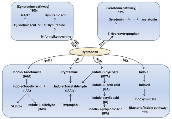
Figure 1.
The three major pathways of tryptophan metabolism. IDO: indoleamine 2,3-dioxygenase, TDO: tryptophan 2,3-dioxygenase, TPH: tryptophan hydroxylase, NAD: nicotinamide adenine dinucleotide, TMO: tryptophan 2-Monooxygenase, TrD: tryptophan Decarboxylase, ArAT: aromatic amino acid aminotransferase, TNA: tryptophanase.
In the past decade, the gut microbiota has emerged as a key regulator of tryptophan metabolism. The colon is home to the densest and most metabolically active community, which comprises more than 1013 individual microbial cells [10] and expresses diverse enzymatic activities capable of utilizing tryptophan [11]. Germ-free mice display increased tryptophan levels along with reduced serotonin, indicating the microbial regulation over host tryptophan metabolism [12]. Certain bacterial species like Clostridium sporogenes and Ruminococcusgnavus have been associated with increased tryptophan catabolism and production of indole metabolites [13]. Conversely, restoringtryptophan levels has been shown to result in the expansion of Lactobacillus, which further led to the conversion oftryptophan into intermediate indole-3-lactic acid (ILA) [14,15].
There is accumulating evidence indicating a bidirectional regulatory axis between microbial tryptophan metabolism and the host immune–intestinal system [16]. Alterations along this axis have been associated with cancer, inflammatory bowel disease (IBD), obesity and type 2 diabetes (T2D), chronic kidney disease (CKD), and autism spectrum disorder (ASD) [12,17,18,19]. Elucidating the mechanisms of microbial tryptophan modulation could lead to novel therapeutic approaches such as psychobiotics and microbiome-targeted dietary interventions. This review summarized current literature on the integrated microbiota-tryptophan metabolic axis in health and disease, mechanisms of cross-talk, and implications for human health and medicine.
2. Tryptophan-Microbiota Interactions in the Healthy State
2.1. Bacterial Species Associated with Tryptophan Metabolism and Metabolite Production
The gut microbiota regulates tryptophan metabolism through microbial enzyme production and conversion into bioactive metabolites [11]. The comparison of conventional mice and germ-free mice showed increased tryptophan availability and reduced kynurenine and serotonin pathway metabolites, indicating microbial catabolism of tryptophan [20,21,22,23]. While the majority of ingested proteins are typically digested and absorbed in the small intestine, it is worth notingthat depending on dietary protein intake, a substantial quantity of proteins and amino acids may transit to the colon [24] where they undergo degradation by a diverse community of commensal bacteria. Specifically, bacterial catabolism of proteins is more pronounced in response to higher dietary protein intake, a reduction in carbohydrate availability within the colon, elevated colonic pH, and an extended transit time through the colon [25,26,27]. This shift towards bacterial proteolytic fermentation is a consequence of the gradual depletion of carbohydrate substrates from the proximal to the distal colon [28]. As a result, the concentration of phenolic compounds, generated through the degradation of aromatic amino acids, is notably more than fourfold higher in the distal colon when compared to the proximal colon [26]. It is important to note that bacterial specialists in proteolysis exhibit lower growth potential compared to generalists and specialists in saccharolysis/lipolysis. This suggests that proteolytic specialists are favored when ecological pressures for rapid bacterial growth are reduced [29]. Nonetheless, it is worth mentioning that the conversion of tryptophan into indole and its derivatives is not exclusive to proteolytic specialists or limited to the distal colon. For example, certain bacterial species such as Lactobacilli have been shown to catabolize tryptophan in the stomach and ileum of mice [30]. Numerous bacterial species have been documented to possess the capability to metabolize tryptophan into indole and related derivatives (Table 1).

Table 1.
Microbiota-derived tryptophan metabolites and gut microbiota (↑ represents an increase, ↓ represents a decrease).
Indoleamine 2,3 dioxygenase 1 (IDO1) is one of the immune checkpoint blockade genes and is highly expressed in many types of tumor cells; it is the most well studied of the enzymes that initiate tryptophan’s catabolism into kynurenine (Kyn) [59,60]. Tryptophan (Trp) metabolism commits to assisting cancer cells to evade immune surveillance [61,62]. Trp depletion inhibits T cells through the activation of general control non-derepressible protein 2 (GCN2) and down-regulation of the mTORC1 complex [63,64]. In addition, the overexpression of IDO and the accumulation of Kyn in tumor tissue can activate regulatory T (Treg) cells, therefore suppressing the functions of effector T (Teff) cells and natural killer (NK) cells and promoting angiogenesis of the tumor [62,65,66,67,68]. There are many studies that have shown reduced Trp levels and increased Kyn pathway metabolites in CRC patients, indicating increased IDO1 activity [69,70,71]. In addition, Trp metabolism by intestinal microbes may have a role in maintaining immune system stability within the body [72]. Recent research confirmed that intestinal microbes can convert tryptophan into indole and related derivatives. For instance, microbes altered the relative abundance of tryptophan metabolites including indole-3-acetic acid (IAA) and indole-3-acetaldehyde (IAAId) in cecal contents [73,74], which serve as endogenous ligands of the aryl hydrocarbon receptor (AhR). Furthermore, AhR activation promotes the maintenance of ILC3 cells that strengthen the integrity of intestinal mucosa by secreting IL-22 which enhances epithelial barrier function and reduces the number of Treg cells [75]. Thus, the regulation of tryptophan metabolism by intestinal microbes is crucial for host immunity.
Specific bacterial strains have been associated with increased tryptophan catabolism and production of bioactive metabolites. For instance, Clostridium sporogenes and Ruminococcusgnavus possess tryptophanase and generate indole derivatives such as indole-3-acetic acid (IAA) [13,76]. Bacteroides thetaiotaomicron generates indole-3-propionic acid (IPA), which can modulate gut barrier function [77]. Lactobacillus spp. can indirectly promote colonic serotonin synthesis by increasing TPH1 expression [78,79,80]. These microbial tryptophan metabolites serve important functions in maintaining intestinal homeostasis. Indole increases the expression of tight junction proteins and mucins to enhance epithelial integrity [81]. IPA promotes intestinal homeostasis by regulating transcript and protein levels of AhR target genes and suppressing cytokine production [82].
2.2. Influence of Diet on the Microbiota-Ryptophan Axis
Numerous studies have established associations between specific dietary patterns and the risk of cancer in humans [83,84,85,86]. Consumption of diets characterized by high fiber content, abundant fruits, yogurt, whole grains, extra virgin olive oil, vegetables, and limited intake of animal products has been consistently linked to a reduced risk of cancer [87,88,89,90]. Conversely, a higher risk of cancer has been associated with the consumption of highly processed foods, for instance, diets rich in animal fats and red meat and lower in dietary fiber intake [91,92]. Additionally, dysbiosis in the gut microbiome, as a consequence of a Western dietary pattern, has been correlated with colorectal cancer [93]. Diet plays a central role in shaping the composition of the microbiome, impacting various microbial communities responsible for maintaining physiological homeostasis, modulating immune responses, and facilitating the breakdown of complex polysaccharides [94,95,96]. Therefore, it is important to explore the intricate connections between diet, the microbiome, and cancer. Recent evidence indicates that diet has an apparent effect on both the composition of the intestinal microbes and tryptophan metabolism. High-fat diets have been associated with decreased production of indole and increased levels of kynurenine metabolites by certain bacterial species [97]. Compared to the high-protein-low-fiber diet, the high-fiber-low-protein diet favored the microbial production of indole-3-acetic acid, indole-3-lactic acid, indole-3-aldehyde, and indole-3-propionic acid in both proximal colon and distal colon compartments of the Simulator of the Human Intestinal Microbial Ecosystem (SHIME) [54]. The SHIME is a unique gut model that simulates the entire gastrointestinal tract, including the stomach, small intestine, and various regions of the colon. It is the only in vitro model that combines the entire gastrointestinal transit into one system. Unlike the Reading model [27], the SHIME utilizes peristaltic pumps to connect the different compartments [98]. In addition, skatole (3-methylindole) is a product of bacterial fermentation of tryptophan in the intestine, and its metabolite indole-3-carbinole (I3C) protects wild type mice against intestinal cancer development and reduces hepatic steatosis in mice fed a high-fat diet [99]. In summary, diet powerfully influences the microbiota-tryptophan axis by modulating the composition and metabolic output of the gut microbiome. Further research is elucidating how specific dietary components shape microbial tryptophan processing and production of bioactive catabolites relevant to health and disease.
2.3. Effects of Probiotics and Prebiotics on Tryptophan Metabolism
Supplementation with Lactobacillus and Bifidobacterium probiotics has been demonstrated to boost plasma tryptophan levels, increase serotonin production, and modify tryptophan catabolism in animal models. Specific probiotic strains can reduce inflammation-induced IDO expression, thus preserving tryptophan levels and enhancing its availability [100]. Moreover, specific probiotics and prebiotics have demonstrated beneficial effects on tryptophan metabolism through the kynurenine pathway. For instance, administering Bifidobacterium infantisto germ-free mice was able to elevate the Kna levels with no effect on the Kyn concentration and therefore normalized the kynurenine-to-tryptophan ratio in these mice [101]. Prebiotic fructo-oligosaccharides increased the relative abundance of Lactobacillus and Bifidobacterium and tryptophan levels in the human intestinal tract [102]. Clinical studies have shown that combining probiotics like Lactobacillus rhamnosusGG with prebiotics further elevates plasma tryptophan levels compared to each intervention alone [103]. This synergistic effect may be attributed to enhanced growth of tryptophan-producing bacteria [104].
These findings highlight how gut microbes and substrates modulate host tryptophan metabolism. Probiotic bacteria and prebiotic fibers can collectively influence tryptophan availability, catabolism, and the production of bioactive metabolites by promoting the growth and metabolic activities of beneficial gut microbiota. Therefore, understanding the specific mechanisms behind microbial tryptophan utilization and metabolite production is essential for gaining deeper insights into this bidirectional interaction crucial for intestinal health.
3. Dysregulation of Gut Microbiota in Disease States
3.1. Evidence for Dysbiosis Disrupting the Axis in Cancer, IBD, Mood Disorders, and ASD
It is widely accepted that the dysbiosis of gut microbiota has been associated with disruptions in tryptophan metabolism along the kynurenine and serotonin pathways, contributing to the pathogenesis of several diseases.
Tryptophan can be degraded to generate kynurenine metabolites. The increased expression of indoleamine enzymes IDO/TDO associated with cancer drives tryptophan degradation, resulting in the formation of N-formylkynurenine. This compound is then hydrolyzed by kynurenine formamidase to produce kynurenine. Kynurenine can subsequently follow two pathways: either forming kynurenic acid or undergoing a cascade of enzymatic reactions to yield NAD+ [105,106,107]. Previous studies have found that one or more of these enzymes were increased in tumors of the pancreas, breast, and brain [108,109,110]. Previousstudies have demonstrated thattumor-produced kynurenine suppresses cancer immune surveillance. Kyn produced by tumor cells can be exported into the tumor microenvironment, causing T cell inactivation and preventing tumor cell clearance. Kyn can also act as an endogenous ligand for theAhR transcription factor, indicating a cell-autonomous role [111,112,113]. It is believed that AhR activation by tumor-derived Kyn triggers a gene expression program that leads to paracrine immune cell suppression [111]. Additionally, this AhR activation promotes cancer cell proliferation and migration in a cell-autonomous way [114]. Previous studies have shown that the expression of AhR is increased in many cancers, for instance, stomach, liver, prostate, head and neck, breast, brain, and skin cancers [111,115,116,117,118,119,120]. This would suggest thatregulating the expression of AhR plays an important role in tumor aggression.
Colorectal cancer (CRC) is one of the main factors contributing to morbidity and mortality, comprising nearly 12% of all annually diagnosed cancers and cancer-related deaths worldwide [121]. In CRC patients, a depletion of tryptophan metabolism by gut microbes such as Fusobacteria, Enterobacteriaceae, and Clostridia coincides with shunting of tryptophan catabolism toward pro-tumorigenic kynurenine metabolites rather than serotonin synthesis [122]. Recent evidence has demonstrated that Lactobacillus gallinarum and its derived ICA could improve anti-PD1 efficacy in CRC by associating with the inhibition of the IDO1/Kyn metabolic circuit as well as the antagonism of Kyn binding to AhR receptors on T cells to inhibit Treg differentiation [123,124,125].
Inflammatory bowel diseases (IBDs) including Crohn’s disease and ulcerative colitis, reveal the significantly differentmetabolic level of Trpbetween healthy individuals and patients. These patients have lower levels of Trp in the serum and feces than healthy subjects [126,127]. Inflammatory bowel disease (IBD) patients exhibit lower concentrations of the AhRagonist IAA in their feces [127]. Interestingly, previous studies have reported an elevated presence of Kyn or higher Kyn/Trp ratios in IBD patients, indicating enhanced tryptophan metabolism through the Kyn pathway during active IBD [128].Additionally, there is an enrichment of Th17 cells and a reduction in Treg cells which can produce anti-inflammatory IL-4 and IL-10, attributed to the interaction of indole metabolites and kynurenine with the AhR on immune cells [77,129]. In general, kynurenine, as an endogenous ligand of AhR, can induce AhR activation when generated in the tumor microenvironment. This function is associated with cancer immunosuppression and sustained activation of AhR, encouraging tumor growth, and affects immune defense [130,131]. However, under inflammatory conditions, AhR activation decreases cytokine production (including TNF, IFNγ, IL-7, IL-12, IL-17, and IL-6) in the intestine, defective AhR activationdetrimentally affects intestinal homeostasis [132]. Appropriate levels of AhR activation are required to maintain intestinal homeostasis. Decreased levels of indole derivatives like IAld can impair intestinal epithelial integrity by inhibiting AhR [30].
In obesity, the relationship between obesity and gut microbiota is a two-way street. Recent studies indicate that the ratio of Firmicutes-to-Bacteroidetes is not a significant factor in human obesity [133]. It is rather important to focus on distinct bacteria which are associated with obesity such as the family Christensenellaceae and the genera Akkermansia, Bifidobacteria, Methanobacteriales, and Lactobacillus [134]. The levels of bacterially generated tryptophan metabolites, including indoles, IPA, and indole sulfuric acid (ISA), are diminished in the blood samples of individuals with type 2 diabetes when compared to lean controls. Elevated serum concentrations of IPA have also been linked to a decreased prevalence of T2D [134]. Several indole derivatives resulting from the gut microbiota’s conversion of tryptophan play a role in the development of metabolic syndrome. For instance, IAA-induced IL-35+ Breg cells have the potential to influence obesity induced by a high-fat diet [135]. Targeting microbial tryptophan catabolism may support weight management efforts.
Autism spectrum disorder (ASD) has been associated with both microbial depletion of tryptophan via the kynurenine pathway and decreased production of serotonin by select bacteria in the gut [136]. These alterations may relate to pathological changes in behavior and social function. Modulating the gut microbiota through dietary interventions has yielded some improvements in behavioral symptoms in autistic children [137]. Optimizing microbial tryptophan metabolism may support microbiota-gut-brain axis communication and ameliorate autism severity.
In depression and anxiety disorders, previous studies have reported a shift in the composition of gut microbes with the capacity to synthesize serotonin and engage with the gut–brain axis. Reduced levels of Lactobacillus and Bifidobacterium spp., coupled with an elevated relative abundance of Alistipes and Ruminococcusgenera, exhibit associations with alterations in tryptophan metabolites along the kynurenine pathway among affected patients [138]. Interventions restoring beneficial serotonin-producing bacteria may hold promise for mood disorders.
3.2. Potential Mechanisms of Tryptophan Modulation by Specific Microbes
Gut microbes may modulate tryptophan and its downstream metabolites through several mechanisms. Tryptophancan be transaminated into indole-3-pyruvic acid via an unstable intermediate by the action of tryptophan aminotransferase [139]. In addition to formation from the kynurenine pathway, kynurenic acid can also be formed from indole-3-pyruvic acid via the unstable kynurenic acid intermediate generated with participation of reactive oxygen species (ROS) [140]. In addition, microbiota can stimulate TPH1 activity by its metabolites (e.g., butyrate), directly influencing serotonin and probably melatonin synthesis [141]. The gut microbes play a significant role in tryptophan metabolism, specializing in production of indoles [11]. They metabolize Trp into indole by tryptophanase, into tryptamine by decarboxylase (ALAAD), and into indole-3-pyruvic acid by Trp aminotransferase. The gut microbes amplify the variety of tryptophan catabolites through oxidative and reductive pathways generating various indole derivatives. IPYAcan be converted into other indole derivatives such as ILA, IA, IPA, and also into IAAId, which can then be further processed into indole-3-acetic acid (IAA) and IAld [17]. These compounds are produced by gut microbiota and can be detected in the circulation, feces, and urine [142,143,144]. When absorbed into the circulation, indole can also be transformed into indoxyl sulfate, which has been linked to chronic kidney diseases and cardiovascular issues [145]. However, other indole derivatives such as ILA, IAA, IPA, and IAld play important roles in maintaining intestinal homeostasis, promoting barrier integrity, stimulating epithelial renewal, and regulating mucosal immune responses [30,81,146,147]. Indole is a potent ligand for AhR [148] and is the main bacterial metabolite of tryptophan. It has been shown to have various protective effects in the gastrointestinal tract. These include regulating bacterial motility, promoting antibiotic resistance, inhibiting invasion of host cells by virulent bacteria, ameliorating intestinal inflammation, suppressing the production of proinflammatory chemokines, and increasing the production of anti-inflammatory cytokines [149]. To sum up, indole derivatives are also endogenous ligands of AhR, including tryptamine, skatole, IA, IAA, ILA, IAld, IAAld, and IPA [30,150,151,152,153,154].
Bacterial genes not only metabolize tryptophan-derived metabolites but also provide substrates that can fuel critical host metabolic pathways, such as short-chain fatty acids (SCFAs) [155]. Propionate, butyrate, and acetate make up the specific SCFAs. In addition, the role of SCFAs in protecting against gut inflammation and regulating colonic Treg homeostasis has been well demonstrated [156,157]. For instance, SCFAs decrease STAT1 expression leading to the inhibition of the IFNγ dependent and STAT1-driven transcription of IDO1. In addition, butyrate impairs IDO1 transcription through a second mechanism in a STAT1-independent manner, which may be attributed to its histone deacetylase (HDAC) inhibitory properties [155]. Moreover, butyrate is able to down-regulate IDO1 expression in human intestinal epithelial cells [158]. As we discussed before, the kyn pathway is closely related to the tumor immune escape mechanisms, with the prerequisite for this process being that kynurenic acid is synthesized by IDO1.Thus, microbes can directly and indirectly control tryptophan metabolism through multiple integrated pathways.
3.3. Contribution to Pathogenesis of Immune, Metabolic, and Disease Progression
Dysregulation of tryptophan metabolism contributes to disease progression. Activation of the kynurenine pathway leads to immunosuppression, facilitating tumor escape [159]. The induction of TDO2 transcription is driven by proinflammatory cytokines such as IFNγand downstream factors including NF-κB and C/EBPβ [160]. Additionally, activation of the AhR by kynurenine establishes a positive feedback loop, further stimulating IDO1 expression [161]. IDO1 exhibits significant overexpression and is indicative of a poor prognosis in numerous malignancies, with a high IDO1 transcript level serving as a universal adverse prognostic factor in solid tumors. Moreover, heightened IDO1 expression correlates with tumor differentiation, distant metastasis, and an advanced clinical stage [162,163]. Proinflammatory cytokines, such as IFNγ, interleukin-1β (IL1β), and tumor necrosis factor alpha (TNFα), activate IDO1 expression via the JAK/STAT pathway [164,165]. Furthermore, similar to TDO2, a high level of IDO1 transcription in cancer cells is sustained through an AhR-IL6-STAT3-driven positive feedback mechanism [166]. Therefore, dysregulation of tryptophan metabolism can lead to immunosuppression and activate the kynurenine pathway, further resulting in significant overexpression of IDO1, which is associated with a poor prognosis in various malignancies. However, the indole derivativesmetabolized by gut microbiota can regulate the expression of IDO1, which eventually affects the immune response. Overall, disrupted microbial tryptophan metabolism contributes substantially to immune, metabolic, and disease progression.
4. Therapeutic Opportunities
4.1. Targeting the Microbiota-Tryptophan Axis for Disease Treatment and Prevention
The gut microbiota plays a crucial role in regulating tryptophan metabolism and diverting tryptophan catabolism towards the production of bioactive compounds that influence immunity, metabolism, neurotransmission, and more. This discovery has indicated novel therapeutic opportunities to target the dysregulated microbiota-tryptophan axis for the prevention or treatment of diverse conditions like cancer, inflammatory bowel disease, mood and cognitive disorders, and neurodevelopmental illnesses [22,58,150,167]. Administering certain indole derivatives in animal models has been shown to have positive effects. For instance, IAA administration resulted in ameliorative colitis symptoms [32]. Administration of IAld has anti-inflammatory effects in treating DSS-induced colitis and improving intestinal inflammation caused by bacterial infections [30]. Additionally, it has been found to increase the production of IL-22, further suppressing inflammatory responses [168]. Moreover, the significant effectiveness of indole-3-acetate (I3A) in reducing steatosis and inflammation in mice models highlights its potential as a safe treatment option for non-alcoholic fatty liver disease (NAFLD) [169]. Administration of IPA has been shown to significantly induce expressionof IL-10 receptor protein 1 in cultured intestinal epithelial T84 cells, which further supports the role of IPA in the maintenance of intestinal immunity [82].
A reduction in IFN-γ production during immune activation and a significant increase in plasma tryptophan levels following chronic administration of Bifidobacteria were observed in the rat experiment [101]. In addition, previous research showed that serotonin can be produced by Lactobacillus plantarum in arginine decarboxylase broth (ADB) [170]. Lactobacillus rhamnosus GG has been reported to promote butyrate production, ultimately stimulating TPH1 activity by butyrate and regulating serotonin synthesis [141,171]. In addition, Lactobacillus amylovorus and Lactobacillus plantarum PS128 have been reported to regulate serotonin synthesis as well [79,80]. These probiotics counteract inflammation-induced tryptophan depletion, suggesting therapeutic utility in depression, anxiety, and other disorders associated with dysregulated tryptophan metabolism [100]. Prebiotic fibers like galacto-oligosaccharides that stimulate indigenous Lactobacillus and Bifidobacterium growth increased SCFAs levels in clinical studies [172], which are the substrates that can mediate tryptophan metabolism in the host. Fecal microbiota transfer from healthy donors modulatedtryptophan and serotonin levels, which indicates microbiota transplantation as another modulatory strategy [78,173]. Potential strategies aim to favorably restructure the gut microbial community and functional capacity to rectify imbalances in tryptophan metabolism through probiotics, prebiotics, fecal microbiota transplantation, and combinatorial therapies. Ongoing investigations aim to identify the most promising probiotic strains, efficacious prebiotics, and optimal microbiota compositional and functional profiles to beneficially impact the microbiota-tryptophan axis across diverse disease contexts.
4.2. Combination with Immunotherapies and IDO Inhibitors
Since tryptophan metabolism regulates immune cell responses, modulating the microbiota-tryptophan axis may provide synergistic benefits when combined with cancer immunotherapies. Previous research has revealed that an abundance of gut-resident Lactobacillus correlates with IDO1 activity and Th17 cells. Supplementation of SIV-infected macaques with Lactobacillus spp. can reduce IDO1 activity and may have the capacity to mitigate the loss of gut barrier-promoting human Th17 cells [174]. Bacterial populations, specifically Bifidobacterium longum, Collinsellaaerofaciens, and Enterococcus faecium, exhibit increased prevalence among individuals who respond favorably to treatment. Conversely, the effectiveness of immune checkpoint blockade therapies is attenuated in the presence of antibiotic administration [175,176,177], which indicates that the combination of certain gut microbes and IDO inhibitors could be a promising treatment. While most current research is focused on the synergistic effects of microbiota and PD-L1 inhibitors, there remains limited literature regarding the concurrent use of IDO1 inhibitors. In fact, IDO inhibitors have led to disappointing results in clinical trials, including epacadostat (INCB024360), BMS-986205, indoximod, navoximod, KHK2455, LY3381916, MK-7162, and NLG802. Notably, clinical trials combining IDO1 inhibitors with other immunotherapies such as PD1 and PD-L1 immune checkpoint inhibitors may be a promising method [178]. However, the combination of microbiota modulation and IDO1 inhibition holds great promise for enhancing the efficacy of immune checkpoint inhibitors. Such combinatorial therapies highlight the potential of targeting dysregulated tryptophan catabolism using the gut microbiota to overcome tumoral immune suppression and improve outcomes across diverse malignancies.
5. Challenges and Limitations of Microbiota-Based Therapies
However, several challenges remain in translating insights from the microbiota-tryptophan axis into viable therapeutic interventions. There aremany critical questionsthat need to be addressed, for instance, identifying optimal single or combined bacterial strains and efficacious doses and timing, understanding inter-individual variability in clinical responses and the impact of diet, and elucidating causal mechanisms linking gut microbiome structure to tryptophan processing. Safety, tolerability, long-term effects, production challenges, and regulatory approval also require further study, particularly for combining microbial therapeutics with small molecules. In addition, the temporal sequence and causal relationships between microbiota alterations and tryptophan pathway disruptions need to be precisely delineated and mapped across different disease settings and populations. Despite current limitations, therapeutically manipulating the gut microbiota and tryptophan metabolism holds remarkable promise for modulating immunity, metabolism, neurotransmission, and more.
6. Summary
In conclusion, the gut microbiota plays a pivotal role in regulating tryptophan metabolism along the kynurenine and serotonin pathways through microbial enzyme production and metabolite generation. Preclinical models and human studies support a regulatory microbiota-tryptophan axis influencing immune function, metabolism, neurotransmission, and disease states. Diet is another key factor influencing the microbiota-tryptophan axis, with high-fat diets decreasing beneficial indole-producing bacteria and increasing tryptophan degradation, while high-fiber diets have the opposite effect. Dysbiosis and disrupted microbial control over tryptophan catabolism contributes to pathogenesis of cancer, IBD, mood disorders, obesity, ASD, and more. The gut microbiota modulates tryptophan availability through direct enzymatic activity as well as production of metabolites like short chain fatty acids that influence host cell tryptophan metabolism (Figure 2). Therapeutic targeting of the microbiota-tryptophan axis through probiotics, prebiotics, and microbiota transplantation shows promise but requires further optimization. Overall, a complex bidirectional relationship exists between gut microbes and host tryptophan metabolism, with intricate effects on physiology and disease. Further research on these interactions may enable novel diagnostics and therapies that harness the microbiota-tryptophan axis to improve human health.
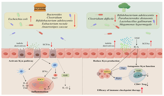
Figure 2.
The impact of different diets on microbiota structure leads to tryptophan metabolism abnormalities. A high-fat diet reduces the levels of indole derivatives and inactivates AhR attenuated expression of IL-22, ultimately leading to proinflammatory outcomes (left); conversely, a high-fiber diet can improve the efficacy of immune checkpoint therapy through specific bacterial strains. Such actions are associated with the IDO1/Kyn metabolic circuit as well as the antagonism of Kyn binding to AhR receptors on T cells to inhibit Treg differentiation (right).
Author Contributions
Y.H., J.L. and S.Y. equally contributed to the writing and reviewing of this manuscript. All authors have read and agreed to the published version of the manuscript.
Funding
This work was strategically funded by the National Natural Science Foundation of China (Grant No. 32170062) and the Hunan Key Laboratory for Bioanalysis of Complex Matrix Samples (2017TP1037). The funders had no role in the study design, data collection and analysis, decision to publish, or preparation of the manuscript.
Acknowledgments
We express our thanks for the pioneering research work carried out by researchers in relevant fields. We are also grateful to FigDraw (https://www.figdraw.com/) for providing rich vector assets for our illustrations, accessed on 10 October 2023.
Conflicts of Interest
S.Y. is an employee of Shanghai Bocimed Pharmaceutical Research Co. Ltd. This paper reflects the views of the scientists and not the company.
Abbreviations
| Trp | tryptophan |
| Kyn | kynurenine |
| Kna | kynurenic acid |
| IDO | indoleamine 2,3-dioxygenase |
| TDO | tryptophan 2,3-dioxygenase |
| TPH | tryptophan hydroxylase |
| NAD | nicotinamide adenine dinucleotide |
| TMO | tryptophan 2-monooxygenase |
| TrD | tryptophan decarboxylase |
| ArAT | aromatic amino acid aminotransferase |
| TNA | tryptophanase |
| IAA | indole-3-acetic acid |
| IPA | indole-3-propionic acid |
| ILA | indole-3-lactic acid |
| IAAId | indole-3-acetaldehyde |
| IPA | indole-3-propionic acid |
| AhR | aryl hydrocarbon receptor |
| I3C | indole-3-carbinole |
| ISA | indole sulfuric acid |
| ROS | reactive oxygen species |
| IPYA | indole-3-pyruvate |
| IA | indole-acrylic acid |
| IAld | indole-3-aldehyde |
| I3A | indole-3-acetate |
References
- Le, N.; Otten, W. Tryptophan Metabolism, from Nutrition to Potential Therapeutic Applications. Amino Acids 2011, 41, 1195–1205. [Google Scholar] [CrossRef]
- Keszthelyi, D.; Troost, F.J.; Masclee, A.A.M. Understanding the Role of Tryptophan and Serotonin Metabolism in Gastrointestinal Function. Neurogastroenterol. Motil. 2009, 21, 1239–1249. [Google Scholar] [CrossRef]
- Badawy, A.A. Neuropharmacology Tryptophan Availability for Kynurenine Pathway Metabolism across the Life Span: Control Mechanisms and Focus on Aging, Exercise, Diet and Nutritional Supplements. Neuropharmacology 2017, 112, 248–263. [Google Scholar] [CrossRef] [PubMed]
- Stone, T.W.; Darlington, L.G. The Kynurenine Pathway as a Therapeutic Target in Cognitive and Neurodegenerative Disorders. Br. J. Pharmacol. 2013, 169, 1211–1227. [Google Scholar] [CrossRef] [PubMed]
- Platten, M.; Wick, W.; Van Den Eynde, B.J. Tryptophan Catabolism in Cancer: Beyond IDO and Tryptophan Depletion. Cancer Res. 2012, 72, 5435–5440. [Google Scholar] [CrossRef] [PubMed]
- Mellor, A.L.; Munn, D.H. IDO Expression by Dendritic Cells: Tolerance and Tryptophan Catabolism. Nat. Rev. Immunol. 2004, 4, 762–774. [Google Scholar] [CrossRef]
- Badawy, A.A.B. Kynurenine Pathway of Tryptophan Metabolism: Regulatory and Functional Aspects. Int. J. Tryptophan Res. 2017, 10, 1178646917691938. [Google Scholar]
- Taleb, S. Tryptophan Dietary Impacts Gut Barrier and Metabolic Diseases. Front. Immunol. 2019, 10, 2113. [Google Scholar] [CrossRef]
- O’Mahony, S.M.; Clarke, G.; Borre, Y.E.; Dinan, T.G.; Cryan, J.F. Serotonin, Tryptophan Metabolism and the Brain-Gut-Microbiome Axis. Behav. Brain Res. 2015, 277, 32–48. [Google Scholar] [CrossRef]
- Sender, R.; Fuchs, S.; Milo, R. Are We Really Vastly Outnumbered? Revisiting the Ratio of Bacterial to Host Cells in Humans. Cell 2016, 164, 337–340. [Google Scholar] [CrossRef]
- Roager, H.M.; Licht, T.R. Microbial Tryptophan Catabolites in Health and Disease. Nat. Commun. 2018, 9, 3294. [Google Scholar] [CrossRef]
- Yano, J.M.; Yu, K.; Donaldson, G.P.; Shastri, G.G.; Ann, P.; Ma, L.; Nagler, C.R.; Ismagilov, R.F.; Mazmanian, S.K.; Elaine, Y. Indigenous Bacteria from the Gut Microbiota Regulate Host Serotonin Biosynthesis. Cell 2015, 161, 264–276. [Google Scholar] [CrossRef] [PubMed]
- Williams, B.B.; Van Benschoten, A.H.; Cimermancic, P.; Donia, M.S.; Zimmermann, M.; Taketani, M.; Ishihara, A.; Kashyap, P.C.; Fraser, J.S.; Fischbach, M.A. Discovery and Characterization of Gut Microbiota Decarboxylases That Can Produce the Neurotransmitter Tryptamine. Cell Host Microbe 2014, 16, 495–503. [Google Scholar] [CrossRef]
- Cervantes-Barragan, L.; Chai, J.N.; Tianero, M.D.; Di Luccia, B.; Ahern, P.P.; Merriman, J.; Cortez, V.S.; Caparon, M.G.; Donia, M.S.; Gilfillan, S.; et al. Lactobacillus Reuteri Induces Gut Intraepithelial CD4(+)CD8alphaalpha(+) T Cells. Science 2017, 357, 806–810. [Google Scholar] [CrossRef] [PubMed]
- Li, C.Y.; Liu, J.X.; Wang, Y.Z.; Wu, Y.M.; Wang, J.K.; Zhou, Y.Y. Influence of Differing Carbohydrate Sources on L-Tryptophan Metabolism by Porcine Fecal Microbiota Studied in Vitro. Livest. Sci. 2009, 120, 43–50. [Google Scholar] [CrossRef]
- Liu, M.; Nieuwdorp, M.; de Vos, W.M.; Rampanelli, E. Microbial Tryptophan Metabolism Tunes Host Immunity, Metabolism, and Extraintestinal Disorders. Metabolites 2022, 12, 834. [Google Scholar] [CrossRef] [PubMed]
- Agus, A.; Planchais, J.; Sokol, H. Gut Microbiota Regulation of Tryptophan Metabolism in Health and Disease. Cell Host Microbe 2018, 23, 716–724. [Google Scholar]
- Liu, J.R.; Miao, H.; Deng, D.Q.; Vaziri, N.D.; Li, P.; Zhao, Y.Y. Gut Microbiota-Derived Tryptophan Metabolism Mediates Renal Fibrosis by Aryl Hydrocarbon Receptor Signaling Activation. Cell. Mol. Life Sci. 2021, 78, 909–922. [Google Scholar] [CrossRef]
- Scheithauer, T.P.M.; Rampanelli, E.; Nieuwdorp, M.; Vallance, B.A.; Verchere, C.B.; van Raalte, D.H.; Herrema, H. Gut Microbiota as a Trigger for Metabolic Inflammation in Obesity and Type 2 Diabetes. Front. Immunol. 2020, 11, 571731. [Google Scholar] [CrossRef]
- Sjögren, K.; Engdahl, C.; Henning, P.; Lerner, U.H.; Tremaroli, V.; Lagerquist, M.K.; Bäckhed, F.; Ohlsson, C. The Gut Microbiota Regulates Bone Mass in Mice. J. Bone Miner. Res. 2012, 27, 1357–1367. [Google Scholar] [CrossRef]
- Marcobal, A.; Kashyap, P.C.; Nelson, T.A.; Aronov, P.A.; Donia, M.S.; Spormann, A.; Fischbach, M.A.; Sonnenburg, J.L. A Metabolomic View of How the Human Gut Microbiota Impacts the Host Metabolome Using Humanized and Gnotobiotic Mice. ISME J. 2013, 7, 1933–1943. [Google Scholar] [CrossRef] [PubMed]
- Gao, K.; Mu, C.L.; Farzi, A.; Zhu, W.Y. Tryptophan Metabolism: A Link between the Gut Microbiota and Brain. Adv. Nutr. 2020, 11, 709–723. [Google Scholar] [CrossRef] [PubMed]
- Gao, K.; Pi, Y.; Mu, C.L.; Farzi, A.; Liu, Z.; Zhu, W.Y. Increasing Carbohydrate Availability in the Hindgut Promotes Hypothalamic Neurotransmitter Synthesis: Aromatic Amino Acids Linking the Microbiota–Brain Axis. J. Neurochem. 2019, 149, 641–659. [Google Scholar] [CrossRef] [PubMed]
- Evenepoel, P.; Claus, D.; Geypens, B.; Hiele, M.; Geboes, K.; Rutgeerts, P.; Ghoos, Y. Amount and Fate of Egg Protein Escaping Assimilation in the Small Intestine of Humans. Am. J. Physiol. Gastrointest. Liver Physiol. 1999, 277, 935–943. [Google Scholar] [CrossRef]
- Cranwell, P.D.; Noakes, D.E.; Hill, K.J. Gastric Secretion and Fermentation in the Suckling Pig. Br. J. Nutr. 1976, 36, 71–86. [Google Scholar] [CrossRef]
- Smith, E.A.; Macfarlane, G.T. Enumeration of Human Colonie Bacteria Producing Phenolic and Indolic Compounds: Effects of PH, Carbohydrate Availability and Retention Time on Dissimilatory Aromatic Amino Acid Metabolism. J. Appl. Bacteriol. 1996, 81, 288–302. [Google Scholar] [CrossRef]
- Macfarlane, G.T.; Cummings, J.H.; Macfarlane, S.; Gibson, G.R. Influence of Retention Time on Degradation of Pancreatic Enzymes by Human Colonic Bacteria Grown in a 3-stage Continuous Culture System. J. Appl. Bacteriol. 1989, 67, 521–527. [Google Scholar] [CrossRef]
- Roager, H.M.; Hansen, L.B.S.; Bahl, M.I.; Frandsen, H.L.; Carvalho, V.; Gøbel, R.J.; Dalgaard, M.D.; Plichta, D.R.; Sparholt, M.H.; Vestergaard, H.; et al. Colonic Transit Time Is Related to Bacterial Metabolism and Mucosal Turnover in the Gut. Nat. Microbiol. 2016, 1, 16093. [Google Scholar] [CrossRef]
- Vieira-Silva, S.; Falony, G.; Darzi, Y.; Lima-Mendez, G.; Garcia Yunta, R.; Okuda, S.; Vandeputte, D.; Valles-Colomer, M.; Hildebrand, F.; Chaffron, S.; et al. Species-Function Relationships Shape Ecological Properties of the Human Gut Microbiome. Nat. Microbiol. 2016, 1, 16088. [Google Scholar] [CrossRef]
- Zelante, T.; Iannitti, R.G.; Cunha, C.; DeLuca, A.; Giovannini, G.; Pieraccini, G.; Zecchi, R.; D’Angelo, C.; Massi-Benedetti, C.; Fallarino, F.; et al. Tryptophan Catabolites from Microbiota Engage Aryl Hydrocarbon Receptor and Balance Mucosal Reactivity via Interleukin-22. Immunity 2013, 39, 372–385. [Google Scholar] [CrossRef]
- Cani, P.D.; Neyrinck, A.M.; Fava, F.; Knauf, C.; Burcelin, R.G.; Tuohy, K.M.; Gibson, G.R.; Delzenne, N.M. Selective Increases of Bifidobacteria in Gut Microflora Improve High-Fat-Diet-Induced Diabetes in Mice through a Mechanism Associated with Endotoxaemia. Diabetologia 2007, 50, 2374–2383. [Google Scholar] [CrossRef] [PubMed]
- Li, M.; Dong, Y.; Zhang, K.; Ma, J.; He, L.; Wei, J.; Jiang, J.; Zhao, Q.; Zhao, Q.; Cao, H. DOP35 High-Fat Diet Reduces Gut Microbiota-Derived Metabolite Indole-Acetic Acid and Aggravates Colitis. J. Crohn’s Colitis 2023, 17, i97–i99. [Google Scholar] [CrossRef]
- Lee, J.H.; Lee, J. Indole as an Intercellular Signal in Microbial Communities. FEMS Microbiol. Rev. 2010, 34, 426–444. [Google Scholar] [CrossRef]
- Martínez, I.; Kim, J.; Duffy, P.R.; Schlegel, V.L.; Walter, J. Resistant Starches Types 2 and 4 Have Differential Effects on the Composition of the Fecal Microbiota in Human Subjects. PLoS ONE 2010, 5, e15046. [Google Scholar] [CrossRef] [PubMed]
- Walker, A.W.; Ince, J.; Duncan, S.H.; Webster, L.M.; Holtrop, G.; Ze, X.; Brown, D.; Stares, M.D.; Scott, P.; Bergerat, A.; et al. Dominant and Diet-Responsive Groups of Bacteria within the Human Colonic Microbiota. ISME J. 2011, 5, 220–230. [Google Scholar] [CrossRef] [PubMed]
- Wu, Z.; Xu, Q.; Wang, Q.; Chen, Y.; Lv, L.; Zheng, B.; Yan, R.; Jiang, H.; Shen, J.; Wang, S.; et al. The Impact of Dietary Fibers on Clostridioides Difficile Infection in a Mouse Model. Front. Cell. Infect. Microbiol. 2022, 12, 1–13. [Google Scholar] [CrossRef] [PubMed]
- Chen, L.; Liu, B.; Ren, L.; Du, H.; Fei, C.; Qian, C.; Li, B.; Zhang, R.; Liu, H.; Li, Z.; et al. High-Fiber Diet Ameliorates Gut Microbiota, Serum Metabolism and Emotional Mood in Type 2 Diabetes Patients. Front. Cell. Infect. Microbiol. 2023, 13, 66. [Google Scholar] [CrossRef]
- Hills, R.D.; Pontefract, B.A.; Mishcon, H.R.; Black, C.A.; Sutton, S.C.; Theberge, C.R. Gut Microbiome: Profound Implications for Diet and Disease. Nutrients 2019, 11, 1613. [Google Scholar] [CrossRef]
- Laird, T.S.; Flores, N.; Leveau, J.H.J. Bacterial Catabolism of Indole-3-Acetic Acid. Appl. Microbiol. Biotechnol. 2020, 104, 9535–9550. [Google Scholar] [CrossRef]
- Hendrikx, T.; Schnabl, B. Indoles: Metabolites Produced by Intestinal Bacteria Capable of Controlling Liver Disease Manifestation. J. Intern. Med. 2019, 286, 32–40. [Google Scholar] [CrossRef]
- Liu, D.; Zhang, S.; Li, S.; Zhang, Q.; Cai, Y.; Li, P.; Li, H.; Shen, B.; Liao, Q.; Hong, Y.; et al. Indoleacrylic Acid Produced by Parabacteroides Distasonis Alleviates Type 2 Diabetes via Activation of AhR to Repair Intestinal Barrier. BMC Biol. 2023, 21, 90. [Google Scholar] [CrossRef] [PubMed]
- Wlodarska, M.; Luo, C.; Kolde, R.; d’Hennezel, E.; Annand, J.W.; Heim, C.E.; Krastel, P.; Schmitt, E.K.; Omar, A.S.; Creasey, E.A.; et al. Indoleacrylic Acid Produced by Commensal Peptostreptococcus Species Suppresses Inflammation. Cell Host Microbe 2017, 22, 25–37.e6. [Google Scholar] [CrossRef] [PubMed]
- Guo, Y.; Bian, X.; Liu, J.; Zhu, M.; Li, L.; Yao, T.; Tang, C.; Ravichandran, V.; Liao, P.; Papadimitriou, K. Dietary Components, Microbial Metabolites and Human Health: Reading between the Lines. Foods 2020, 9, 1045. [Google Scholar] [CrossRef] [PubMed]
- Zhao, Z.H.; Xin, F.Z.; Xue, Y.; Hu, Z.; Han, Y.; Ma, F.; Zhou, D.; Liu, X.L.; Cui, A.; Liu, Z.; et al. Indole-3-Propionic Acid Inhibits Gut Dysbiosis and Endotoxin Leakage to Attenuate Steatohepatitis in Rats. Exp. Mol. Med. 2019, 50, 1–14. [Google Scholar] [CrossRef]
- Jiang, H.; Chen, C.; Gao, J. Extensive Summary of the Important Roles of Indole Propionic Acid, a Gut Microbial Metabolite in Host Health and Disease. Nutrients 2022, 15, 151. [Google Scholar] [CrossRef]
- Kapsetaki, S.E.; Alcaraz, G.M.; Maley, C.C.; Whisner, C.M.; Aktipis, A. Diet, Microbes, and Cancer Across the Tree of Life: A Systematic Review. Curr. Nutr. Rep. 2022, 11, 508–525. [Google Scholar] [CrossRef]
- Maukonen, J.; Saarela, M. Human Gut Microbiota: Does Diet Matter? Proc. Nutr. Soc. 2015, 74, 23–36. [Google Scholar] [CrossRef]
- Zhang, X.; Coker, O.O.; Chu, E.S.H.; Fu, K.; Lau, H.C.H.; Wang, Y.-X.; Chan, A.W.H.; Wei, H.; Yang, X.; Sung, J.J.Y.; et al. Dietary Cholesterol Drives Fatty Liver-Associated Liver Cancer by Modulating Gut Microbiota and Metabolites. Gut 2021, 70, 761–774. [Google Scholar] [CrossRef]
- Li, Y.; Yang, X.; Zhang, J.; Jiang, T.; Zhang, Z.; Wang, Z.; Gong, M.; Zhao, L.; Zhang, C. Ketogenic Diets Induced Glucose Intolerance and Lipid Accumulation in Mice with Alterations in Gut Microbiota And. MBio 2021, 12, e03601-20. [Google Scholar] [CrossRef]
- Tsoi, H.; Chu, E.S.H.; Zhang, X.; Sheng, J.; Nakatsu, G.; Ng, S.C.; Chan, A.W.H.; Chan, F.K.L.; Sung, J.J.Y.; Yu, J. Biosynthesis in Colon Cells to Induce Proliferation and Causes. Gastroenterology 2017, 152, 1419–1433.e5. [Google Scholar] [CrossRef]
- Serger, E.; Luengo-gutierrez, L.; Chadwick, J.S.; Kong, G.; Zhou, L.; Crawford, G.; Danzi, M.C.; Myridakis, A.; Brandis, A.; Bello, A.T.; et al. The Gut Metabolite Indole-3 Propionate Promotes Nerve Regeneration and Repair. Nature 2022, 607, 585–592. [Google Scholar] [CrossRef] [PubMed]
- Chen, J.; Xiao, Y.; Li, D.; Zhang, S.; Wu, Y.; Zhang, Q.; Bai, W. New Insights into the Mechanisms of High-Fat Diet Mediated Gut Microbiota in Chronic Diseases. iMeta 2023, 2, e69. [Google Scholar] [CrossRef]
- Su, X.; Gao, Y.; Yang, R. Gut Microbiota-Derived Tryptophan Metabolites Maintain Gut and Systemic Homeostasis. Cells 2022, 11, 2296. [Google Scholar] [CrossRef] [PubMed]
- Huang, Z.; Boekhorst, J.; Fogliano, V.; Capuano, E.; Wells, J.M. Impact of High-Fiber or High-Protein Diet on the Capacity of Human Gut Microbiota To Produce Tryptophan Catabolites. J. Agric. Food Chem. 2023, 71, 6956–6966. [Google Scholar] [CrossRef]
- Koga, J.; Adachi, T.; Hidaka, H. Molecular Cloning of the Gene for Indolepyruvate Decarboxylase from Enterobacter Cloacae. MGG Mol. Gen. Genet. 1991, 226, 10–16. [Google Scholar] [CrossRef] [PubMed]
- Krishnan, S.; Ding, Y.; Saedi, N.; Choi, M.; Sridharan, G.V.; Sherr, D.H.; Yarmush, M.L.; Alaniz, R.C.; Jayaraman, A.; Lee, K. Gut Microbiota-Derived Tryptophan Metabolites Modulate Inflammatory Response in Hepatocytes and Macrophages. Cell Rep. 2018, 23, 1099–1111. [Google Scholar] [CrossRef] [PubMed]
- Jensen, M.T.; Cox, R.P.; Jensen, B.B. 3-Methylindole (Skatole) and Indole Production by Mixed Populations of Pig Fecal Bacteria. Appl. Environ. Microbiol. 1995, 61, 3180–3184. [Google Scholar] [CrossRef]
- Li, S. Modulation of Immunity by Tryptophan Microbial Metabolites. Front. Nutr. 2023, 10, 1–20. [Google Scholar] [CrossRef]
- Dai, X.; Zhu, B.T. Indoleamine 2,3-Dioxygenase Tissue Distribution and Cellular Localization in Mice: Implications for Its Biological Functions. J. Histochem. Cytochem. 2010, 58, 17–28. [Google Scholar] [CrossRef]
- Theate, I.; Van Baren, N.; Pilotte, L.; Moulin, P.; Larrieu, P.; Renauld, J.C.; Herve, C.; Gutierrez-Roelens, I.; Marbaix, E.; Sempoux, C.; et al. Extensive Profiling of the Expression of the Indoleamine 2,3-Dioxygenase 1 Protein in Normal and Tumoral Human Tissues. Cancer Immunol. Res. 2015, 3, 161–172. [Google Scholar] [CrossRef]
- Platten, M.; von Knebel Doeberitz, N.; Oezen, I.; Wick, W.; Ochs, K. Cancer Immunotherapy by Targeting IDO1/TDO and Their Downstream Effectors. Front. Immunol. 2015, 6, 673. [Google Scholar] [CrossRef] [PubMed]
- Prendergast, G.C.; Smith, C.; Thomas, S.; Mandik-Nayak, L.; Laury-Kleintop, L.; Metz, R.; Muller, A.J. Indoleamine 2,3-Dioxygenase Pathways of Pathogenic Inflammation and Immune Escape in Cancer. Cancer Immunol. Immunother. 2014, 63, 721–735. [Google Scholar] [CrossRef] [PubMed]
- Prendergast, G.C.; Malachowski, W.P.; DuHadaway, J.B.; Muller, A.J. Discovery of IDO1 Inhibitors: From Bench to Bedside. Cancer Res. 2017, 77, 6795–6811. [Google Scholar] [CrossRef]
- Munn, D.H.; Mellor, A.L. Indoleamine 2,3-Dioxygenase and Tumor-Induced Tolerance. J. Clin. Investig. 2007, 117, 1147–1154. [Google Scholar] [CrossRef]
- Ricciuti, B.; Leonardi, G.C.; Puccetti, P.; Fallarino, F.; Bianconi, V.; Sahebkar, A.; Baglivo, S.; Chiari, R.; Pirro, M. Targeting Indoleamine-2,3-Dioxygenase in Cancer: Scientific Rationale and Clinical Evidence. Pharmacol. Ther. 2019, 196, 105–116. [Google Scholar] [CrossRef] [PubMed]
- Moon, Y.W.; Hajjar, J.; Hwu, P.; Naing, A. Targeting the Indoleamine 2,3-Dioxygenase Pathway in Cancer. J. Immunother. Cancer 2015, 3, 51. [Google Scholar] [CrossRef]
- Harden, J.L.; Egilmez, N.K. Indoleamine 2,3-Dioxygenase and Dendritic Cell Tolerogenicity. Immunol. Investig. 2012, 41, 738–764. [Google Scholar] [CrossRef]
- Liu, M.; Wang, X.; Wang, L.; Ma, X.; Gong, Z.; Zhang, S.; Li, Y. Targeting the IDO1 Pathway in Cancer: From Bench to Bedside. J. Hematol. Oncol. 2018, 11, 100. [Google Scholar] [CrossRef]
- Liu, X.; Shin, N.; Koblish, H.K.; Yang, G.; Wang, Q.; Wang, K.; Leffet, L.; Hansbury, M.J.; Thomas, B.; Rupar, M.; et al. Selective Inhibition of IDO1 Effectively Regulates Mediators of Antitumor Immunity. Blood 2010, 115, 3520–3530. [Google Scholar] [CrossRef]
- Walczak, K.; Da̧browski, W.; Langner, E.; Zgrajka, W.; Piłat, J.; Kocki, T.; Rzeski, W.; Turski, W.A. Kynurenic Acid Synthesis and Kynurenine Aminotransferases Expression in Colon Derived Normal and Cancer Cells. Scand. J. Gastroenterol. 2011, 46, 903–912. [Google Scholar] [CrossRef]
- Engin, A.B.; Karahalil, B.; Karakaya, A.E.; Engin, A. Helicobacter Pylori and Serum Kynurenine-Tryptophan Ratio in Patients with Colorectal Cancer. World J. Gastroenterol. 2015, 21, 3636–3643. [Google Scholar] [CrossRef]
- Teunis, C.; Nieuwdorp, M.; Hanssen, N. Interactions between Tryptophan Metabolism, the Gut Microbiome and the Immune System as Potential Drivers of Non-Alcohol Liver Disease (NAFLD) and Metabolic Diseases. Metabolites 2022, 12, 514. [Google Scholar] [CrossRef]
- Ju, T.; Kong, J.Y.; Stothard, P.; Willing, B.P. Defining the Role of Parasutterella, a Previously Uncharacterized Member of the Core Gut Microbiota. ISME J. 2019, 13, 1520–1534. [Google Scholar] [CrossRef]
- Yang, C.; Du, Y.; Ren, D.; Yang, X.; Zhao, Y. Gut Microbiota-Dependent Catabolites of Tryptophan Play a Predominant Role in the Protective Effects of Turmeric Polysaccharides against DSS-Induced Ulcerative Colitis. Food Funct. 2021, 12, 9793–9807. [Google Scholar] [CrossRef] [PubMed]
- Parker, B.J.; Wearsch, P.A.; Veloo, A.C.M.; Rodriguez-Palacios, A. The Genus Alistipes: Gut Bacteria with Emerging Implications to Inflammation, Cancer, and Mental Health. Front. Immunol. 2020, 11, 906. [Google Scholar] [CrossRef] [PubMed]
- Dodd, D.; Spitzer, M.H.; Van Treuren, W.; Merrill, B.D.; Hryckowian, A.J.; Higginbottom, S.K.; Le, A.; Cowan, T.M.; Nolan, G.P.; Fischbach, M.A.; et al. A Gut Bacterial Pathway Metabolizes Aromatic Amino Acids into Nine Circulating Metabolites. Nature 2017, 551, 648–652. [Google Scholar] [CrossRef] [PubMed]
- Li, K.; Hao, Z.; Du, J.; Gao, Y.; Yang, S.; Zhou, Y. Bacteroides Thetaiotaomicron Relieves Colon Inflammation by Activating Aryl Hydrocarbon Receptor and Modulating CD4+T Cell Homeostasis. Int. Immunopharmacol. 2021, 90, 107183. [Google Scholar] [CrossRef]
- Xiao, L.; Yan, J.; Yang, T.; Zhu, J.; Li, T.; Wei, H.; Chen, J. Fecal Microbiome Transplantation from Children with Autism Spectrum Disorder Modulates Tryptophan and Serotonergic Synapse Metabolism and Induces Altered Behaviors in Germ-Free Mice. mSystems 2021, 6, e01343-20. [Google Scholar] [CrossRef]
- Liu, Z.; Ling, Y.; Peng, Y.; Han, S.; Ren, Y.; Jing, Y.; Fan, W.; Su, Y.; Mu, C.; Zhu, W. Regulation of Serotonin Production by Specific Microbes from Piglet Gut. J. Anim. Sci. Biotechnol. 2023, 14, 1–15. [Google Scholar] [CrossRef]
- Chen, C.M.; Wu, C.C.; Huang, C.L.; Chang, M.Y.; Cheng, S.H.; Lin, C.T.; Tsai, Y.C. Lactobacillus Plantarum PS128 Promotes Intestinal Motility, Mucin Production, and Serotonin Signaling in Mice. Probiotics Antimicrob. Proteins 2022, 14, 535–545. [Google Scholar] [CrossRef]
- Bansal, T.; Alaniz, R.C.; Wood, T.K.; Jayaraman, A. The Bacterial Signal Indole Increases Epithelial-Cell Tight-Junction Resistance and Attenuates Indicators of Inflammation. Proc. Natl. Acad. Sci. USA 2010, 107, 228–233. [Google Scholar] [CrossRef]
- Alexeev, E.E.; Lanis, J.M.; Kao, D.J.; Campbell, E.L.; Kelly, C.J.; Battista, K.D.; Gerich, M.E.; Jenkins, B.R.; Walk, S.T.; Kominsky, D.J.; et al. Microbiota-Derived Indole Metabolites Promote Human and Murine Intestinal Homeostasis through Regulation of Interleukin-10 Receptor. Am. J. Pathol. 2018, 188, 1183–1194. [Google Scholar] [CrossRef]
- Whisner, C.M.; Athena Aktipis, C. The Role of the Microbiome in Cancer Initiation and Progression: How Microbes and Cancer Cells Utilize Excess Energy and Promote One Another’s Growth. Curr. Nutr. Rep. 2019, 8, 42–51. [Google Scholar] [CrossRef] [PubMed]
- Sobhani, I.; Amiot, A.; Baleur, Y.; Levy, M.; Auriault, M.L.; Van Nhieu, J.T.; Delchier, J.C. Microbial Dysbiosis and Colon Carcinogenesis: Could Colon Cancer Be Considered a Bacteria-Related Disease? Therap. Adv. Gastroenterol. 2013, 6, 215–229. [Google Scholar] [CrossRef] [PubMed]
- Sepich-Poore, G.D.; Zitvogel, L.; Straussman, R.; Hasty, J.; Wargo, J.A.; Knight, R. The Microbiome and Human Cancer. Science 2021, 371. [Google Scholar] [CrossRef] [PubMed]
- Dzutsev, A.; Goldszmid, R.S.; Viaud, S.; Zitvogel, L.; Trinchieri, G. The Role of the Microbiota in Inflammation, Carcinogenesis, and Cancer Therapy. Eur. J. Immunol. 2015, 45, 17–31. [Google Scholar] [CrossRef]
- Makarem, N.; Lin, Y.; Bandera, E.V.; Jacques, P.F.; Parekh, N. Concordance with World Cancer Research Fund/American Institute for Cancer Research (WCRF/AICR) Guidelines for Cancer Prevention and Obesity-Related Cancer Risk in the Framingham Offspring Cohort (1991–2008). Cancer Causes Control 2015, 26, 277–286. [Google Scholar] [CrossRef]
- Romagnolo, D.F.; Selmin, O.I. Flavonoids and Cancer Prevention: A Review of the Evidence. J. Nutr. Gerontol. Geriatr. 2012, 31, 206–238. [Google Scholar] [CrossRef]
- Abdull Razis, A.F.; Mohd Noor, N. Cruciferous Vegetables: Dietary Phytochemicals for Cancer Prevention. Asian Pacific J. Cancer Prev. 2013, 14, 1565–1570. [Google Scholar] [CrossRef]
- Rodríguez-García, C.; Sánchez-Quesada, C.; Algarra, I.; Gaforio, J.J. The High-Fat Diet Based on Extra-Virgin Olive Oil Causes Dysbiosis Linked to Colorectal Cancer Prevention. Nutrients 2020, 12, 1705. [Google Scholar] [CrossRef]
- Oostindjer, M.; Alexander, J.; Amdam, G.V.; Andersen, G.; Bryan, N.S.; Chen, D.; Corpet, D.E.; De Smet, S.; Dragsted, L.O.; Haug, A.; et al. The Role of Red and Processed Meat in Colorectal Cancer Development: A Perspective. Meat Sci. 2014, 97, 583–596. [Google Scholar] [CrossRef] [PubMed]
- Cross, A.J.; Ferrucci, L.M.; Risch, A.; Graubard, B.I.; Ward, M.H.; Park, Y.; Hollenbeck, A.R.; Schatzkin, A.; Sinha, R. A Large Prospective Study of Meat Consumption and Colorectal Cancer Risk: An Investigation of Potential Mechanisms Underlying This Association. Cancer Res. 2010, 70, 2406–2414. [Google Scholar] [CrossRef] [PubMed]
- Foegeding, N.J.; Jones, Z.S.; Byndloss, M.X. Western Lifestyle as a Driver of Dysbiosis in Colorectal Cancer. DMM Dis. Model. Mech. 2021, 14, 1–6. [Google Scholar] [CrossRef] [PubMed]
- McFall-Ngai, M.; Hadfield, M.G.; Bosch, T.C.G.; Carey, H.V.; Domazet-Lošo, T.; Douglas, A.E.; Dubilier, N.; Eberl, G.; Fukami, T.; Gilbert, S.F.; et al. Animals in a Bacterial World, a New Imperative for the Life Sciences. Proc. Natl. Acad. Sci. USA 2013, 110, 3229–3236. [Google Scholar] [CrossRef] [PubMed]
- Kho, Z.Y.; Lal, S.K. The Human Gut Microbiome—A Potential Controller of Wellness and Disease. Front. Microbiol. 2018, 9, 1–23. [Google Scholar] [CrossRef]
- Thaiss, C.A.; Zmora, N.; Levy, M.; Elinav, E. The Microbiome and Innate Immunity. Nature 2016, 535, 65–74. [Google Scholar] [CrossRef]
- Laurans, L.; Venteclef, N.; Haddad, Y.; Chajadine, M.; Alzaid, F.; Metghalchi, S.; Sovran, B.; Denis, R.G.P.; Dairou, J.; Cardellini, M.; et al. Genetic Deficiency of Indoleamine 2,3-Dioxygenase Promotes Gut Microbiota-Mediated Metabolic Health. Nat. Med. 2018, 24, 1113–1120. [Google Scholar] [CrossRef]
- Van de Wiele, T.; Van den Abbeele, P.; Ossieur, W.; Possemiers, S.; Marzorati, M. The Simulator of the Human Intestinal Microbial Ecosystem (SHIME®). In The Impact of Food Bioactives on Health. In Vitro and Ex Vivo Models; Verhoeckx, K., Cotter, P., López-Expósito, I., Mackie, T.L.A., Eds.; Springer: Cham, Switzerland, 2015; Chapter 27. [Google Scholar] [CrossRef]
- Choi, Y.; Yanagawa, Y.; Kim, S.; Park, T. Involvement of SIRT1-AMPK Signaling in the Protective Action of Indole-3-Carbinol against Hepatic Steatosis in Mice Fed a High-Fat Diet. J. Nutr. Biochem. 2013, 24, 1393–1400. [Google Scholar] [CrossRef]
- Desbonnet, L.; Garrett, L.; Clarke, G.; Kiely, B.; Cryan, J.F.; Dinan, T.G. Effects of the Probiotic Bifidobacterium Infantis in the Maternal Separation Model of Depression. Neuroscience 2010, 170, 1179–1188. [Google Scholar] [CrossRef]
- Desbonnet, L.; Garrett, L.; Clarke, G.; Bienenstock, J.; Dinan, T.G. The Probiotic Bifidobacteria Infantis: An Assessment of Potential Antidepressant Properties in the Rat. J. Psychiatr. Res. 2008, 43, 164–174. [Google Scholar] [CrossRef]
- Tandon, D.; Haque, M.M.; Gote, M.; Jain, M.; Bhaduri, A.; Dubey, A.K.; Mande, S.S. A Prospective Randomized, Double-Blind, Placebo-Controlled, Dose-Response Relationship Study to Investigate Efficacy of Fructo-Oligosaccharides (FOS) on Human Gut Microflora. Sci. Rep. 2019, 9, 5473. [Google Scholar] [CrossRef] [PubMed]
- Radford-Smith, D.E.; Anthony, D.C. Prebiotic and Probiotic Modulation of the Microbiota–Gut–Brain Axis in Depression. Nutrients 2023, 15, 1880. [Google Scholar] [CrossRef]
- Kouchaki, E.; Tamtaji, O.R.; Salami, M.; Bahmani, F.; Daneshvar Kakhaki, R.; Akbari, E.; Tajabadi-Ebrahimi, M.; Jafari, P.; Asemi, Z. Clinical and Metabolic Response to Probiotic Supplementation in Patients with Multiple Sclerosis: A Randomized, Double-Blind, Placebo-Controlled Trial. Clin. Nutr. 2017, 36, 1245–1249. [Google Scholar] [CrossRef]
- Zhai, L.; Spranger, S.; Binder, D.C.; Gritsina, G.; Lauing, K.L.; Giles, F.J.; Wainwright, D.A. Molecular Pathways: Targeting IDO1 and Other Tryptophan Dioxygenases for Cancer Immunotherapy. Clin. Cancer Res. 2015, 21, 5427–5433. [Google Scholar] [CrossRef] [PubMed]
- Essa, M.M.; Subash, S.; Braidy, N.; Al-Adawi, S.; Lim, C.K.; Manivasagam, T.; Guillemin, G.J. Role of NAD+, Oxidative Stress, and Tryptophan Metabolism in Autism Spectrum Disorders. Int. J. Tryptophan Res. 2013, 6, 15–28. [Google Scholar] [CrossRef] [PubMed]
- Palego, L.; Betti, L.; Rossi, A.; Giannaccini, G. Tryptophan Biochemistry: Structural, Nutritional, Metabolic, and Medical Aspects in Humans. J. Amino Acids 2016, 2016, 8952520. [Google Scholar] [CrossRef] [PubMed]
- Liu, J.J.; Raynal, S.; Bailbé, D.; Gausseres, B.; Carbonne, C.; Autier, V.; Movassat, J.; Kergoat, M.; Portha, B. Expression of the Kynurenine Pathway Enzymes in the Pancreatic Islet Cells. Activation by Cytokines and Glucolipotoxicity. Biochim. Biophys. Acta-Mol. Basis Dis. 2015, 1852, 980–991. [Google Scholar] [CrossRef]
- Heng, B.; Lim, C.K.; Lovejoy, D.B.; Bessede, A.; Gluch, L.; Guillemin, G.J. Understanding the Role of the Kynurenine Pathway in Human Breast Cancer Immunobiology. Oncotarget 2016, 7, 6506–6520. [Google Scholar] [CrossRef]
- Adams, S.; Braidy, N.; Bessesde, A.; Brew, B.J.; Grant, R.; Teo, C.; Guillemin, G.J. The Kynurenine Pathway in Brain Tumor Pathogenesis. Cancer Res. 2012, 72, 5649–5657. [Google Scholar] [CrossRef]
- Opitz, C.A.; Litzenburger, U.M.; Sahm, F.; Ott, M.; Tritschler, I.; Trump, S.; Schumacher, T.; Jestaedt, L.; Schrenk, D.; Weller, M.; et al. An Endogenous Tumour-Promoting Ligand of the Human Aryl Hydrocarbon Receptor. Nature 2011, 478, 197–203. [Google Scholar] [CrossRef]
- Liu, Y.; Liang, X.; Dong, W.; Fang, Y.; Lv, J.; Zhang, T.; Fiskesund, R.; Xie, J.; Liu, J.; Yin, X.; et al. Tumor-Repopulating Cells Induce PD-1 Expression in CD8+ T Cells by Transferring Kynurenine and AhR Activation. Cancer Cell 2018, 33, 480–494.e7. [Google Scholar] [CrossRef] [PubMed]
- Hughes, T.; Briercheck, E.L.; Freud, A.G.; Trotta, R.; McClory, S.; Scoville, S.D.; Keller, K.; Deng, Y.; Cole, J.; Harrison, N.; et al. AHR Prevents Human IL-1R1hi ILC3 Differentiation to Natural Killer Cells. Cell Rep. 2014, 8, 150–162. [Google Scholar] [CrossRef] [PubMed]
- Murray, I.A.; Patterson, A.D.; Perdew, G.H. Aryl Hydrocarbon Receptor Ligands in Cancer: Friend and Foe. Nat. Rev. Cancer 2014, 14, 801–814. [Google Scholar] [CrossRef] [PubMed]
- Stanford, E.A.; Ramirez-Cardenas, A.; Wang, Z.; Novikov, O.; Alamoud, K.; Koutrakis, P.; Mizgerd, J.P.; Genco, C.A.; Kukuruzinska, M.; Monti, S.; et al. Role for the Aryl Hydrocarbon Receptor and Diverse Ligands in Oral Squamous Cell Carcinoma Migration and Tumorigenesis. Mol. Cancer Res. 2016, 14, 696–706. [Google Scholar] [CrossRef] [PubMed]
- Andersson, P.; McGuire, J.; Rubio, C.; Gradin, K.; Whitelaw, M.L.; Pettersson, S.; Hanberg, A.; Poellinger, L. A Constitutively Active Dioxin/Aryl Hydrocarbon Receptor Induces Stomach Tumors. Proc. Natl. Acad. Sci. USA 2002, 99, 9990–9995. [Google Scholar] [CrossRef] [PubMed]
- Moennikes, O.; Loeppen, S.; Buchmann, A.; Andersson, P.; Ittrich, C.; Poellinger, L.; Schwarz, M. A Constitutively Active Dioxin/Aryl Hydrocarbon Receptor Promotes Hepatocarcinogenesis in Mice. Cancer Res. 2004, 64, 4707–4710. [Google Scholar] [CrossRef]
- Richmond, O.; Ghotbaddini, M.; Allen, C.; Walker, A.; Zahir, S.; Powell, J.B. The Aryl Hydrocarbon Receptor Is Constitutively Active in Advanced Prostate Cancer Cells. PLoS ONE 2014, 9, e95058. [Google Scholar] [CrossRef]
- Vacher, S.; Castagnet, P.; Chemlali, W.; Lallemand, F.; Meseure, D.; Pocard, M.; Bieche, I.; Perrot-Applanat, M. High AHR Expression in Breast Tumors Correlates with Expression of Genes from Several Signaling Pathways Namely Inflammation and Endogenous Tryptophan Metabolism. PLoS ONE 2018, 13, e0190619. [Google Scholar] [CrossRef]
- Corre, S.; Tardif, N.; Mouchet, N.; Leclair, H.M.; Boussemart, L.; Gautron, A.; Bachelot, L.; Perrot, A.; Soshilov, A.; Rogiers, A.; et al. Sustained Activation of the Aryl Hydrocarbon Receptor Transcription Factor Promotes Resistance to BRAF-Inhibitors in Melanoma. Nat. Commun. 2018, 9, 1–13. [Google Scholar] [CrossRef]
- Sung, H.; Ferlay, J.; Siegel, R.L.; Laversanne, M.; Soerjomataram, I.; Jemal, A.; Bray, F. Global Cancer Statistics 2020: GLOBOCAN Estimates of Incidence and Mortality Worldwide for 36 Cancers in 185 Countries. CA. Cancer J. Clin. 2021, 71, 209–249. [Google Scholar] [CrossRef]
- Wang, T.; Cai, G.; Qiu, Y.; Fei, N.; Zhang, M.; Pang, X.; Jia, W.; Cai, S.; Zhao, L. Structural Segregation of Gut Microbiota between Colorectal Cancer Patients and Healthy Volunteers. ISME J. 2012, 6, 320–329. [Google Scholar] [CrossRef] [PubMed]
- Dai, Z.; Coker, O.O.; Nakatsu, G.; Wu, W.K.K.; Zhao, L.; Chen, Z.; Chan, F.K.L.; Kristiansen, K.; Sung, J.J.Y.; Wong, S.H.; et al. Multi-Cohort Analysis of Colorectal Cancer Metagenome Identified Altered Bacteria across Populations and Universal Bacterial Markers. Microbiome 2018, 6, 70. [Google Scholar] [CrossRef] [PubMed]
- Sugimura, N.; Li, Q.; Chu, E.S.H.; Lau, H.C.H.; Fong, W.; Liu, W.; Liang, C.; Nakatsu, G.; Su, A.C.Y.; Coker, O.O.; et al. Lactobacillus Gallinarum Modulates the Gut Microbiota and Produces Anti-Cancer Metabolites to Protect against Colorectal Tumourigenesis. Gut 2022, 71, 2011–2021. [Google Scholar] [CrossRef] [PubMed]
- Fong, W.; Li, Q.; Ji, F.; Liang, W.; Lau, H.C.H.; Kang, X.; Liu, W.; To, K.K.-W.; Zuo, Z.; Li, X.; et al. Lactobacillus Gallinarum-Derived Metabolites Boost Anti-PD1 Efficacy in Colorectal Cancer by Inhibiting Regulatory T Cells through Modulating IDO1/Kyn/AHR Axis. Gut 2023. [Google Scholar] [CrossRef]
- Shiomi, Y.; Nishiumi, S.; Ooi, M.; Hatano, N.; Shinohara, M.; Yoshie, T.; Kondo, Y.; Furumatsu, K.; Shiomi, H.; Kutsumi, H.; et al. GCMS-Based Metabolomic Study in Mice with Colitis Induced by Dextran Sulfate Sodium. Inflamm. Bowel Dis. 2011, 17, 2261–2274. [Google Scholar] [CrossRef]
- Lamas, B.; Richard, M.L.; Leducq, V.; Pham, H.P.; Michel, M.L.; Da Costa, G.; Bridonneau, C.; Jegou, S.; Hoffmann, T.W.; Natividad, J.M.; et al. CARD9 Impacts Colitis by Altering Gut Microbiota Metabolism of Tryptophan into Aryl Hydrocarbon Receptor Ligands. Nat. Med. 2016, 22, 598–605. [Google Scholar] [CrossRef]
- Sofia, M.A.; Ciorba, M.A.; Meckel, K.; Lim, C.K.; Guillemin, G.J.; Weber, C.R.; Bissonnette, M.; Pekow, J.R. Tryptophan Metabolism through the Kynurenine Pathway Is Associated with Endoscopic Inflammation in Ulcerative Colitis. Inflamm. Bowel Dis. 2018, 24, 1471–1480. [Google Scholar] [CrossRef]
- Salimi Elizei, S.; Poormasjedi-Meibod, M.S.; Wang, X.; Kheirandish, M.; Ghahary, A. Kynurenic Acid Downregulates IL-17/1L-23 Axis in Vitro. Mol. Cell. Biochem. 2017, 431, 55–65. [Google Scholar] [CrossRef]
- Leclerc, D.; Staats Pires, A.C.; Guillemin, G.J.; Gilot, D. Detrimental Activation of AhR Pathway in Cancer: An Overview of Therapeutic Strategies. Curr. Opin. Immunol. 2021, 70, 15–26. [Google Scholar] [CrossRef]
- Gabriely, G.; Wheeler, M.A.; Takenaka, M.C.; Quintana, F.J. Role of AHR and HIF-1α in Glioblastoma Metabolism. Trends Endocrinol. Metab. 2017, 28, 428–436. [Google Scholar] [CrossRef]
- Pernomian, L.; Duarte-Silva, M.; de Barros Cardoso, C.R. The Aryl Hydrocarbon Receptor (AHR) as a Potential Target for the Control of Intestinal Inflammation: Insights from an Immune and Bacteria Sensor Receptor. Clin. Rev. Allergy Immunol. 2020, 59, 382–390. [Google Scholar] [CrossRef] [PubMed]
- Magne, F.; Gotteland, M.; Gauthier, L.; Zazueta, A.; Pesoa, S.; Navarrete, P.; Balamurugan, R. The Firmicutes/Bacteroidetes Ratio: A Relevant Marker of Gut Dysbiosis in Obese Patients? Nutrients 2020, 12, 1474. [Google Scholar] [CrossRef] [PubMed]
- Waters, J.L.; Ley, R.E. The Human Gut Bacteria Christensenellaceae Are Widespread, Heritable, and Associated with Health. BMC Biol. 2019, 17, 83. [Google Scholar] [CrossRef] [PubMed]
- Su, X.; Zhang, M.; Qi, H.; Gao, Y.; Yang, Y.; Yun, H.; Zhang, Q.; Yang, X.; Zhang, Y.; He, J.; et al. Gut Microbiota–Derived Metabolite 3-Idoleacetic Acid Together with LPS Induces IL-35+ B Cell Generation. Microbiome 2022, 10, 1–20. [Google Scholar] [CrossRef] [PubMed]
- Hsiao, E.Y.; McBride, S.W.; Hsien, S.; Sharon, G.; Hyde, E.R.; McCue, T.; Codelli, J.A.; Chow, J.; Reisman, S.E.; Petrosino, J.F.; et al. Microbiota Modulate Behavioral and Physiological Abnormalities Associated with Neurodevelopmental Disorders. Cell 2013, 155, 1451–1463. [Google Scholar] [CrossRef]
- Strati, F.; Cavalieri, D.; Albanese, D.; De Felice, C.; Donati, C.; Hayek, J.; Jousson, O.; Leoncini, S.; Renzi, D.; Calabrò, A.; et al. New Evidences on the Altered Gut Microbiota in Autism Spectrum Disorders. Microbiome 2017, 5, 24. [Google Scholar] [CrossRef] [PubMed]
- Evans, S.J.; Bassis, C.M.; Hein, R.; Assari, S.; Flowers, S.A.; Kelly, M.B.; Young, V.B.; Ellingrod, V.E.; McInnis, M.G. The Gut Microbiome Composition Associates with Bipolar Disorder and Illness Severity. J. Psychiatr. Res. 2017, 87, 23–29. [Google Scholar] [CrossRef] [PubMed]
- Badawy, A.A.B.; Guillemin, G.J. Species Differences in Tryptophan Metabolism and Disposition. Int. J. Tryptophan Res. 2022, 15. [Google Scholar] [CrossRef]
- Ramos-Chávez, L.A.; Lugo Huitrón, R.; González Esquivel, D.; Pineda, B.; Ríos, C.; Silva-Adaya, D.; Sánchez-Chapul, L.; Roldán-Roldán, G.; Pérez de la Cruz, V. Relevance of Alternative Routes of Kynurenic Acid Production in the Brain. Oxid. Med. Cell. Longev. 2018, 2018, 5272741. [Google Scholar] [CrossRef]
- Lukić, I.; Ivković, S.; Mitić, M.; Adžić, M. Tryptophan Metabolites in Depression: Modulation by Gut Microbiota. Front. Behav. Neurosci. 2022, 16, 1–17. [Google Scholar] [CrossRef]
- Anesi, A.; Rubert, J.; Oluwagbemigun, K.; Orozco-Ruiz, X.; Nöthlings, U.; Breteler, M.M.B.; Mattivi, F. Metabolic Profiling of Human Plasma and Urine, Targeting Tryptophan, Tyrosine and Branched Chain Amino Acid Pathways. Metabolites 2019, 9, 261. [Google Scholar] [CrossRef] [PubMed]
- Darkoh, C.; Chappell, C.; Gonzales, C.; Okhuysen, P. A Rapid and Specific Method for the Detection of Indole in Complex Biological Samples. Appl. Environ. Microbiol. 2015, 81, 8093–8097. [Google Scholar] [CrossRef] [PubMed]
- Pavlova, T.; Vidova, V.; Bienertova-Vasku, J.; Janku, P.; Almasi, M.; Klanova, J.; Spacil, Z. Urinary Intermediates of Tryptophan as Indicators of the Gut Microbial Metabolism. Anal. Chim. Acta 2017, 987, 72–80. [Google Scholar] [CrossRef] [PubMed]
- Mosterd, C.M.; Kanbay, M.; van den Born, B.J.H.; van Raalte, D.H.; Rampanelli, E. Intestinal Microbiota and Diabetic Kidney Diseases: The Role of Microbiota and Derived Metabolites Inmodulation of Renal Inflammation and Disease Progression. Best Pract. Res. Clin. Endocrinol. Metab. 2021, 35, 101484. [Google Scholar] [CrossRef] [PubMed]
- Jennis, M.; Cavanaugh, C.R.; Leo, G.C.; Mabus, J.R.; Lenhard, J.; Hornby, P.J. Microbiota-Derived Tryptophan Indoles Increase after Gastric Bypass Surgery and Reduce Intestinal Permeability in Vitro and in Vivo. Neurogastroenterol. Motil. 2018, 30, e13178. [Google Scholar] [CrossRef] [PubMed]
- Shimada, Y.; Kinoshita, M.; Harada, K.; Mizutani, M.; Masahata, K.; Kayama, H.; Takeda, K. Commensal Bacteria-Dependent Indole Production Enhances Epithelial Barrier Function in the Colon. PLoS ONE 2013, 8, e80604. [Google Scholar] [CrossRef]
- Hubbard, T.D.; Murray, I.A.; Bisson, W.H.; Lahoti, T.S.; Gowda, K.; Amin, S.G.; Patterson, A.D.; Perdew, G.H. Adaptation of the Human Aryl Hydrocarbon Receptor to Sense Microbiota-Derived Indoles. Sci. Rep. 2015, 5, 12689. [Google Scholar] [CrossRef]
- Gao, J.; Xu, K.; Liu, H.; Liu, G.; Bai, M.; Peng, C.; Li, T.; Yin, Y. Impact of the Gut Microbiota on Intestinal Immunity Mediated by Tryptophan Metabolism. Front. Cell. Infect. Microbiol. 2018, 8, 13. [Google Scholar] [CrossRef]
- Lavelle, A.; Sokol, H. Gut Microbiota-Derived Metabolites as Key Actors in Inflammatory Bowel Disease. Nat. Rev. Gastroenterol. Hepatol. 2020, 17, 223–237. [Google Scholar] [CrossRef]
- Kurata, K.; Kawahara, H.; Nishimura, K.; Jisaka, M.; Yokota, K.; Shimizu, H. Skatole Regulates Intestinal Epithelial Cellular Functions through Activating Aryl Hydrocarbon Receptors and P38. Biochem. Biophys. Res. Commun. 2019, 510, 649–655. [Google Scholar] [CrossRef]
- Zhao, H.; Chen, L.; Yang, T.; Feng, Y.L.; Vaziri, N.D.; Liu, B.L.; Liu, Q.Q.; Guo, Y.; Zhao, Y.Y. Aryl Hydrocarbon Receptor Activation Mediates Kidney Disease and Renal Cell Carcinoma. J. Transl. Med. 2019, 17, 1–14. [Google Scholar] [CrossRef] [PubMed]
- Wong, C.B.; Tanaka, A.; Kuhara, T.; Xiao, J.Z. Potential Effects of Indole-3-Lactic Acid, a Metabolite of Human Bifidobacteria, on NGF-Induced Neurite Outgrowth in PC12 Cells. Microorganisms 2020, 8, 398. [Google Scholar] [CrossRef] [PubMed]
- Aoki, R.; Aoki-Yoshida, A.; Suzuki, C.; Takayama, Y. Indole-3-Pyruvic Acid, an Aryl Hydrocarbon Receptor Activator, Suppresses Experimental Colitis in Mice. J. Immunol. 2018, 201, 3683–3693. [Google Scholar] [CrossRef] [PubMed]
- Sun, M.; Wu, W.; Liu, Z.; Cong, Y. Microbiota Metabolite Short Chain Fatty Acids, GPCR, and Inflammatory Bowel Diseases. J. Gastroenterol. 2017, 52, 1–8. [Google Scholar] [CrossRef] [PubMed]
- Coutzac, C.; Jouniaux, J.M.; Paci, A.; Schmidt, J.; Mallardo, D.; Seck, A.; Asvatourian, V.; Cassard, L.; Saulnier, P.; Lacroix, L.; et al. Systemic Short Chain Fatty Acids Limit Antitumor Effect of CTLA-4 Blockade in Hosts with Cancer. Nat. Commun. 2020, 11, 1–13. [Google Scholar] [CrossRef] [PubMed]
- Kles, K.A.; Chang, E.B. Short-Chain Fatty Acids Impact on Intestinal Adaptation, Inflammation, Carcinoma, and Failure. Gastroenterology 2006, 130, 100–105. [Google Scholar] [CrossRef]
- Martin-Gallausiaux, C.; Larraufie, P.; Jarry, A.; Béguet-Crespel, F.; Marinelli, L.; Ledue, F.; Reimann, F.; Blottière, H.M.; Lapaque, N. Butyrate Produced by Commensal Bacteria Down-Regulates Indolamine 2,3-Dioxygenase 1 (IDO-1) Expression via a Dual Mechanism in Human Intestinal Epithelial Cells. Front. Immunol. 2018, 9, 2838. [Google Scholar] [CrossRef]
- Gouasmi, R.; Ferraro-Peyret, C.; Nancey, S.; Coste, I.; Renno, T.; Chaveroux, C.; Aznar, N.; Ansieau, S. The Kynurenine Pathway and Cancer: Why Keep It Simple When You Can Make It Complicated. Cancers 2022, 14, 2793. [Google Scholar] [CrossRef]
- Kudo, T.; Prentzell, M.T.; Mohapatra, S.R.; Sahm, F.; Zhao, Z.; Grummt, I.; Wick, W.; Opitz, C.A.; Platten, M.; Green, E.W. Constitutive Expression of the Immunosuppressive Tryptophan Dioxygenase TDO2 in Glioblastoma Is Driven by the Transcription Factor C/EBPβ. Front. Immunol. 2020, 11, 1–9. [Google Scholar] [CrossRef]
- Novikov, O.; Wang, Z.; Stanford, E.A.; Parks, A.J.; Ramirez-Cardenas, A.; Landesman, E.; Laklouk, I.; Sarita-Reyes, C.; Gusenleitner, D.; Li, A.; et al. An Aryl Hydrocarbon Receptor-Mediated Amplification Loop That Enforces Cell Migration in ER-/PR-/Her2- Human Breast Cancer Cells. Mol. Pharmacol. 2016, 90, 674–688. [Google Scholar] [CrossRef]
- Yu, C.P.; Fu, S.F.; Chen, X.; Ye, J.; Ye, Y.; Kong, L.D.; Zhu, Z. The Clinicopathological and Prognostic Significance of IDO1 Expression in Human Solid Tumors: Evidence from a Systematic Review and Meta-Analysis. Cell. Physiol. Biochem. 2018, 49, 134–143. [Google Scholar] [CrossRef] [PubMed]
- Wang, S.; Wu, J.; Shen, H.; Wang, J. The Prognostic Value of IDO Expression in Solid Tumors: A Systematic Review and Meta-Analysis. BMC Cancer 2020, 20, 471. [Google Scholar] [CrossRef] [PubMed]
- Currier, A.R.; Ziegler, M.H.; Riley, M.M.; Babcock, T.A.; Telbis, V.P.; Carlin, J.M. Tumor necrosis factor-and lipopolysaccharide Enhance Interferon-Induced antichlamydial indoleamine Dioxygenase Activity Independently. J. Interf. Cytokine Res. 2000, 20, 369–376. [Google Scholar] [CrossRef] [PubMed]
- Robinson, C.M.; Hale, P.T.; Carlin, J.M. The Role of IFN-γ and TNF-α-Responsive Regulatory Elements in the Synergistic Induction of Indoleamine Dioxygenase. J. Interf. Cytokine Res. 2005, 25, 20–30. [Google Scholar] [CrossRef] [PubMed]
- Litzenburger, U.M.; Opitz, C.A.; Sahm, F.; Rauschenbach, K.J.; Trump, S.; Winter, M.; Ott, M.; Ochs, K.; Lutz, C.; Liu, X.; et al. Constitutive IDO Expression in Human Cancer Is Sustained by an Autocrine Signaling Loop Involving IL-6, STAT3 and the AHR. Oncotarget 2014, 5, 1038–1051. [Google Scholar] [CrossRef] [PubMed]
- Li, Y.; Liu, N.; Ge, Y.; Yang, Y.; Ren, F.; Wu, Z. Tryptophan and the Innate Intestinal Immunity: Crosstalk between Metabolites, Host Innate Immune Cells, and Microbiota. Eur. J. Immunol. 2022, 52, 856–868. [Google Scholar] [CrossRef] [PubMed]
- Monteleone, I.; Rizzo, A.; Sarra, M.; Sica, G.; Sileri, P.; Biancone, L.; MacDonald, T.T.; Pallone, F.; Monteleone, G. Aryl Hydrocarbon Receptor-Induced Signals up-Regulate IL-22 Production and Inhibit Inflammation in the Gastrointestinal Tract. Gastroenterology 2011, 141, 237–248.e1. [Google Scholar] [CrossRef]
- Ding, Y.; Yanagi, K.; Yang, F.; Callaway, E.; Cheng, C.; Hensel, M.E.; Menon, R.; Alaniz, R.C.; Lee, K.; Jayaraman, A.; et al. Oral Supplementation of Gut Microbial Metabolite Indole-3-Acetate Alleviates Diet-Induced Steatosis and Inflammation in Mice. Elife 2023, 12. [Google Scholar] [CrossRef]
- Özoğul, F.; Kuley, E.; Özoğul, Y.; Özoğul, İ. The Function of Lactic Acid Bacteria on Biogenic Amines Production by Food-Borne Pathogens in Arginine Decarboxylase Broth. Food Sci. Technol. Res 2012, 18, 795–804. [Google Scholar] [CrossRef]
- Lin, R.; Sun, Y.; Mu, P.; Zheng, T.; Mu, H.; Deng, F.; Deng, Y.; Wen, J. Lactobacillus Rhamnosus GG Supplementation Modulates the Gut Microbiota to Promote Butyrate Production, Protecting against Deoxynivalenol Exposure in Nude Mice. Biochem. Pharmacol. 2020, 175, 113868. [Google Scholar] [CrossRef]
- Scholtens, P.A.; Goossens, D.A.M.; Staiano, A. Stool Characteristics of Infants Receiving Short-Chain Galacto-Oligosaccharides and Long-Chain Fructo-Oligosaccharides: A Review. World J. Gastroenterol. 2014, 20, 13446–13452. [Google Scholar] [CrossRef] [PubMed]
- Sun, W.; Guo, Y.; Zhang, S.; Chen, Z.; Wu, K.; Liu, Q.; Liu, K.; Wen, L.; Wei, Y.; Wang, B.; et al. Fecal Microbiota Transplantation Can Alleviate Gastrointestinal Transit in Rats with High-Fat Diet-Induced Obesity via Regulation of Serotonin Biosynthesis. Biomed Res. Int. 2018, 2018, 8308671. [Google Scholar] [CrossRef] [PubMed]
- Vujkovic-Cvijin, I.; Swainson, L.A.; Chu, S.N.; Ortiz, A.M.; Santee, C.A.; Petriello, A.; Dunham, R.M.; Fadrosh, D.W.; Lin, D.L.; Faruqi, A.A.; et al. Gut-Resident Lactobacillus Abundance Associates with IDO1 Inhibition and Th17 Dynamics in SIV-Infected Macaques. Cell Rep. 2015, 13, 1589–1597. [Google Scholar] [CrossRef]
- Gopalakrishnan, V.; Spencer, C.N.; Nezi, L.; Reuben, A.; Andrews, M.C.; Karpinets, T.V.; Prieto, P.A.; Vicente, D.; Hoffman, K.; Wei, S.C.; et al. Gut Microbiome Modulates Response to Anti-PD-1 Immunotherapy in Melanoma Patients. Science 2018, 359, 97–103. [Google Scholar] [CrossRef] [PubMed]
- Pushalkar, S.; Hundeyin, M.; Daley, D.; Zambirinis, C.P.; Kurz, E.; Mishra, A.; Mohan, N.; Aykut, B.; Usyk, M.; Torres, L.E.; et al. The Pancreatic Cancer Microbiome Promotes Oncogenesis by Induction of Innate and Adaptive Immune Suppression. Cancer Discov. 2018, 8, 403–416. [Google Scholar] [CrossRef] [PubMed]
- Santoni, M.; Piva, F.; Conti, A.; Santoni, A.; Cimadamore, A.; Scarpelli, M.; Battelli, N.; Montironi, R. Gut Microbiome Influences Efficacy of PD-1-Based Immunotherapy Against Epithelial Tumors. Eur. Urol. 2018, 74, 521–522. [Google Scholar] [CrossRef]
- Opitz, C.A.; Somarribas Patterson, L.F.; Mohapatra, S.R.; Dewi, D.L.; Sadik, A.; Platten, M.; Trump, S. The Therapeutic Potential of Targeting Tryptophan Catabolism in Cancer. Br. J. Cancer 2020, 122, 30–44. [Google Scholar] [CrossRef]
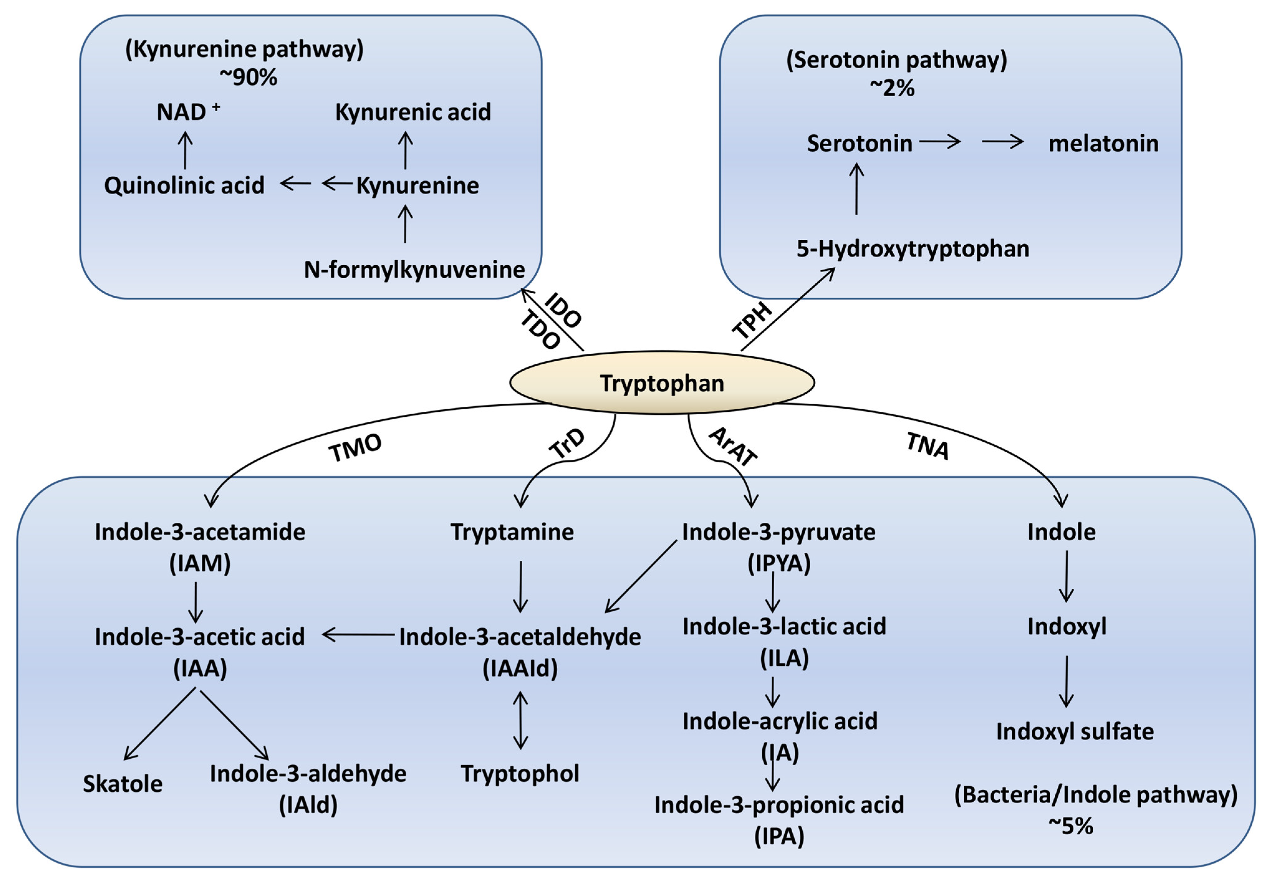
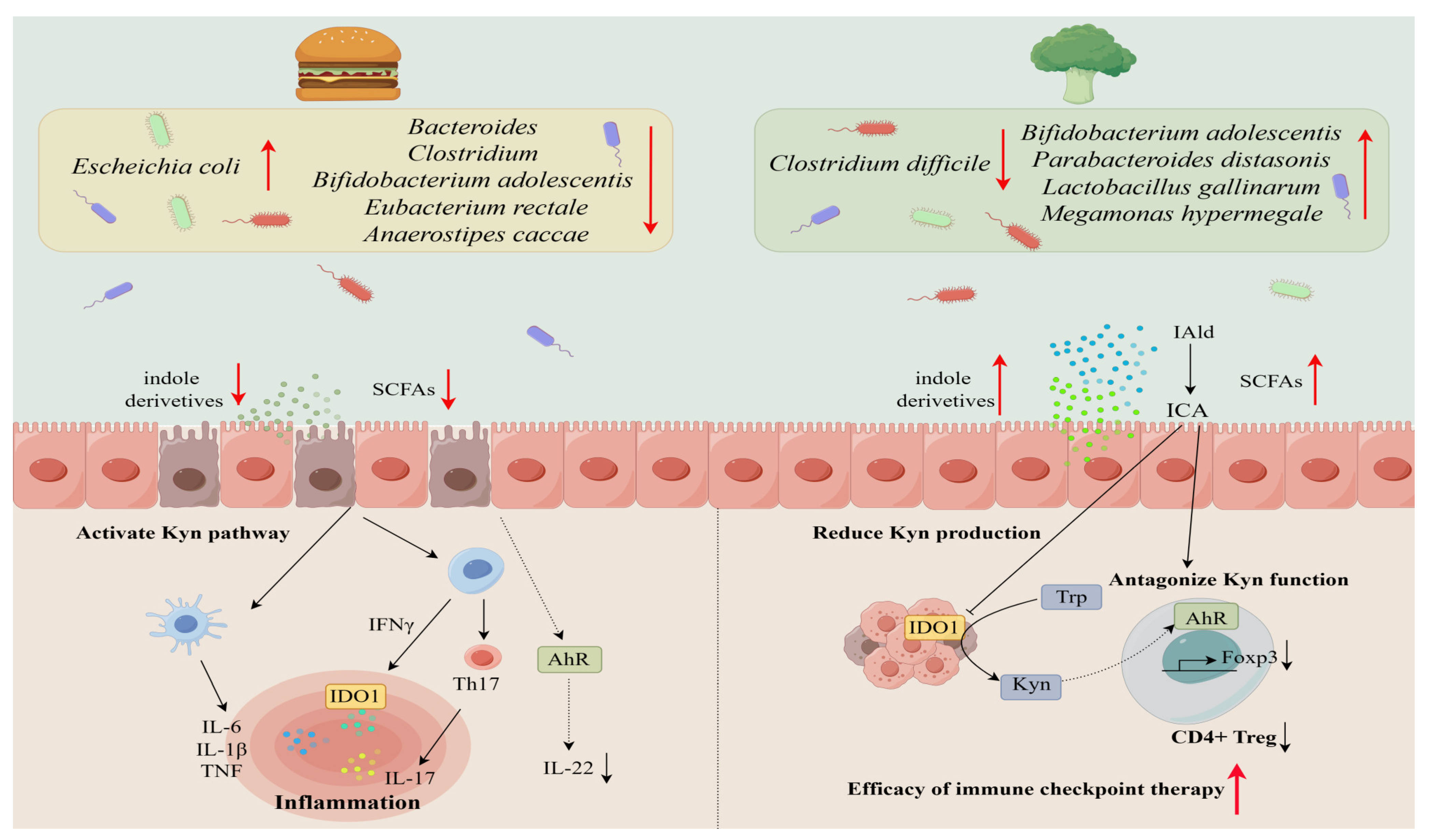
Disclaimer/Publisher’s Note: The statements, opinions and data contained in all publications are solely those of the individual author(s) and contributor(s) and not of MDPI and/or the editor(s). MDPI and/or the editor(s) disclaim responsibility for any injury to people or property resulting from any ideas, methods, instructions or products referred to in the content. |
© 2023 by the authors. Licensee MDPI, Basel, Switzerland. This article is an open access article distributed under the terms and conditions of the Creative Commons Attribution (CC BY) license (https://creativecommons.org/licenses/by/4.0/).