Article

Effect of a Multistrain Probiotic on Feline Gut Health through the Fecal Microbiota and Its Metabolite SCFAs

1
College of Animal Science and Technology, Nanjing Agricultural University, Nanjing 210095, China
2
Department of Medical Cell Biology and Genetics, Health Science Center, Shenzhen University, Shenzhen 518060, China
*
Author to whom correspondence should be addressed.
Metabolites 2023, 13(2), 228; https://doi.org/10.3390/metabo13020228
Submission received: 12 January 2023 / Revised: 30 January 2023 / Accepted: 2 February 2023 / Published: 3 February 2023
(This article belongs to the Special Issue Nutrient Metabolism Studies in Companion Animals)

Abstract

:
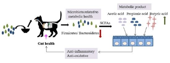
With the increasing awareness of raising pets following scientific methods, people are becoming increasingly more interested in the nutrition and health of pets, especially their intestinal health, which has become a research hotspot. Both Saccharomyces boulardii and Pediococcus acidilactici are probiotics with strong probiotic properties that can maintain the balance of intestinal flora. However, the role of Saccharomyces boulardii and Pediococcus acidilactici in felines has not been comprehensively studied to date. The aim of this study is to investigate the effect of multistrain probiotics consisting of Saccharomyces boulardii and Pediococcus acidilactici on the gut health of felines by modulating gut microbes and the production of metabolite SCFAs. The results show that the multistrain probiotic did not alter the intestinal microbial diversity and structure of short-haired domestic cats, promoted the colonization of beneficial bacteria, increased the levels of microbiota-derived SCFAs and fecal antioxidants, and reduced the levels of fecal inflammatory markers. In conclusion, the use of a multistrain probiotic in healthy, short-haired domestic cats can promote gut health by modulating gut microbes, improving microbiota-derived SCFA production, reducing inflammatory conditions, and improving antioxidant status. These results provide new insights for further exploration of the role of probiotics in the gut microbiome of cats.

Graphical Abstract

1. Introduction

Pet nutrition and health are attracting increased attention in the literature, as pets play a greater role in the daily lives of human beings. The gastrointestinal microbiome of pets is closely related to pet health. Gut microbes, which are typically categorized as commensal, pathogenic, or opportunistic bacteria [1], are observed in the animal’s digestive tract, and each of them has its own distinct flora balance. Intestinal microorganisms participate in the digestion, absorption, and metabolism of nutrients in the host body and play an important regulatory role in the metabolism of nutrients, such as lipid metabolism, carbohydrate metabolism, protein metabolism, and vitamin synthesis. The composition and status of the gut microbiome are closely associated with the intestinal health of the host, and the imbalance of the host intestinal microbiome may lead to a series of disorders in the body, such as gastrointestinal diseases, metabolic diseases, obesity, and immune system diseases.
Probiotics are defined as “live microorganisms that are beneficial to the host when ingested in sufficient amounts” [2], and the most commonly studied in the literature are Lactobacillus, Bifidobacterium, and yeasts [3]. Probiotics are capable of lowering the pH value, creating an acidic environment, reducing the reproduction of harmful microorganisms, increasing the number of beneficial bacteria, interacting directly with immune cells [4], and participating in regulating the immune balance of digestive tract flora, thereby contributing to the prevention and treatment of gastrointestinal-related diseases. Studies have proven that probiotics are effective in the treatment of acute infectious diarrhea, antibiotic-associated diarrhea, Clostridium difficile-associated diarrhea, hepatic encephalopathy, ulcerative colitis, irritable bowel syndrome, functional gastrointestinal disorders, and necrotizing enterocolitis [5,6,7,8,9].
The function of probiotics is closely related to the species of microorganisms colonized in the intestines. The interaction occurring among probiotics, host cells, and intestinal flora is a key factor affecting host health. Probiotics can regulate intestinal mucosal immunity, interact with symbiotic bacteria or potentially harmful pathogens, and produce metabolites, such as short-chain fatty acids (SCFAs), which help to inhibit and eliminate potential pathogens, thereby improving the intestinal microbial environment, strengthening the intestinal barrier, and reducing inflammation, with the purpose of enhancing antigen-specific immune responses [10]. An aqueous probiotic suspension was studied in in vitro human intestinal models, and it was observed that probiotics could increase the production of SCFAs and anti-inflammatory cytokines [11]. Studies using Lactobacillus paracasei SSP in d-galactose-induced aging mice determined that probiotics improved memory and learning ability in mice by increasing SCFA production and inhibiting cell apoptosis and brain injury [12]. The growth of bacteria in microorganisms affects the type and amount of fermentation products. After sugar is phosphorylated, bacteria can be converted to pyruvate or pyruvate and additional acetyl phosphates by glycolysis (the Embden–Meyerhof–Parnassian pathway), the Enter–Doudoroff pathway, or the Bifidobacterium pathway [13].
Saccharomyces boulardii is the only yeast among intestinal probiotics. Unlike other Saccharomyces cerevisiae strains, Saccharomyces boulardii is physiologically and metabolically tolerant to low pH levels and resistant to the gastric environment [14]. Saccharomyces boulardii exhibits distinct probiotic properties, including improved intestinal barrier function, competitive exclusion of pathogens, production of antimicrobial peptides, and immunomodulatory and nutritional effects [15]. Saccharomyces boulardii significantly ameliorates colon damage and regulates inflammatory responses in mice with colitis induced by dextran sulfate sodium (DSS) through changes in the microbiome composition and SCFA metabolism [16]. Pediococcus acidilactici is a facultative anaerobic bacterium of Firmicutes with a high salt tolerance, and it plays an important role in enhancing immunity, producing bacteriocins, maintaining the balance of intestinal flora, and improving the intestinal barrier [17]. The intervention of Pediococcus acidilactici FZU106 was observed to obviously increase SCFA levels in high-fat diet (HFD)-induced hyperlipidemic rats, which was strongly associated with changes in gut microbial composition and metabolism [18]. Saccharomyces boulardii has been observed to be safe for use in dogs with chronic enteritis and to control clinical symptoms better than standard treatment alone [19]. The use of Enterococcus faecium SF68 in cats infected with latent feline herpesvirus 1 (FHV-1) reduced morbidity and maintained fecal microbiota diversity [20]. Studies on Saccharomyces boulardii and Pediococcus acidilactici in pet cats have rarely been reported, and this trial investigated the effect of a multistrain probiotic preparation added to the diet on intestinal fecal microbial diversity, microbiota-derived SCFA production, antioxidative status, and fecal inflammatory markers in healthy short-haired domestic cats. This study provides an innovative platform to further investigate the significance of Saccharomyces boulardii and Pediococcus acidilactici with respect to the intestinal health of companion animals.

2. Materials and Methods

2.1. Study Probiotic

The multistrain probiotic preparation contained approximately 2.0 × 10¹⁰ CFU/g of Saccharomyces boulardii and approximately 2.5 × 10¹⁰ CFU/g of Pediococcus acidilactici mixed in a 1:1 ratio. Saccharomyces boulardii was supplied by the Lallemand Animal Nutrition Group and is the strain used in Levucell SB®. Pediococcus acidilactici (Mito 5051) was provided by Imagilin Technology LLC (Frederick, MD, USA). In the test, both the multistrain probiotic preparation and a basic diet were provided by Nanjing Xintengrui Biotechnology Co., Ltd. (Nanjing, China).

2.2. Animals

A total of 12 short-haired domestic cats between the ages of 2 and 4 years with an average body condition score of 3.5 points (on a 5-point scale) were used in this study. On day 10 of the trial, 1 of the 12 cats in the control group (male) suffered trauma and withdrew from the study; additionally, a cat (male) in the probiotic group dropped out of the study due to a skin disease that developed during the trial. Therefore, 10 cats (4 males and 6 females) were fully involved in the entire trial. They were randomly divided into control and probiotic groups, each containing two male and three female cats (n = 5). Animal experiments were approved by the Laboratory Animal Welfare Ethics Review Committee of Nanjing Agricultural University.

2.3. Experimental Design

A total of 12 short-haired domestic cats were fed with a cat basal diet and acclimatized for 1 week with free access to water. After 1 week of acclimatized feeding, the cats were randomly divided into 2 groups of 6 replicates each, with 1 cat per replicate. The control group was fed a basic diet, and the probiotic group was fed a multistrain probiotic preparation on the basis of the control group. The dosage of probiotics was added according to the cat’s weight (0.5 g/kg).

2.4. Sample Collection and Fecal Microbiota Analysis

Fresh fecal specimens obtained from all subjects were collected on day 28. According to previously described methods [21], DNA extraction and sequencing were completed by Shanghai Biozeron Biotechnology Co., Ltd. (Shanghai, China). The V3–V4 region of the bacterial 16S rRNA gene was amplified by PCR using primers 341F and 806R. The purification of amplification products was performed using an AxyPrepDNA gel extraction kit (Axygen Biosciences, Union City, CA, USA). Based on the preliminary results of electrophoresis, PCR products were detected and quantified with a QuantiFluor-ST™ Blue Fluor Quantitative System (Promega Corporation, Madison, WI, USA). Isomolar pooled and paired-end sequences of purified amplification products on the Illumina platform (2 × 250) were obtained according to the procedure described in [22].

2.5. Fermentation Metabolite Analysis

Concentrations of short-chain fatty acids (SCFAs) were determined by gas chromatography according to the method described in a previous study [23].

2.6. Enzyme-Linked Immunosorbent Assay (ELISA) for Fecal Inflammatory Markers

After mixing feces with normal saline in equal amounts, fecal supernatant samples were collected for further analysis. The concentration of inflammatory markers, such as fecal calprotectin (FC), lactoferrin (LF), matrix metalloproteinase-9 (MMP-9), and myeloperoxidase (MPO), in the supernatant were determined by an enzyme-linked immunosorbent assay (ELISA); then, the assay procedure was performed using a cat CALP ELISA kit (ANG-E71001C), cat LTF ELSA kit (ANG-E71005C), cat MMP-9 ELISA kit (ANG-E71013C), and cat MPO ELISA kit (ANG-E71014C) according to manufacturer’s instructions (Nanjing Jiancheng Bioengineering Institute, Nanjing, China).

2.7. Fecal Supernatant Antioxidant Capacity Analysis

The contents of glutathione (GSH), superoxide dismutase (SOD), and malondialdehyde (MDA) in fecal supernatants on day 28 were determined using GSH (A006), SOD (A001), and MDA (A003) commercial assay kits, respectively (Nanjing Jiancheng Bioengineering Institute, Nanjing, China).

2.8. Statistical Analysis

The data were analyzed using SPSS 23.0 statistical software with an independent t-test and expressed as mean ± standard deviation (SD). The difference between the two means was considered statistically significant when p < 0.05. Data visualization was performed using GraphPad Prism version 8.0.2.

3. Results

3.1. Analysis of Intestinal Flora Structure of Healthy Cats

As shown in Figure 1, the number of OTUs in the control group was 435, and the number of OTUs in the probiotic group was 450, with a total of 369 OTUs in common between the two groups. The number of OTUs in the probiotic group was 81, compared to 66 in the control group alone.

3.2. Phylum-Level Structural Analysis

A few species (genus level) with the highest sampling frequency (the 30 most dominant species) were selected, and the phylum taxonomic information corresponding to the species was displayed; species with the same color indicating their origin from the same phylum were also displayed. The results show that there were five dominant phyla (Figure 2A), namely Actinobacteriota, Bacteroidota, Desulfobacterota, Firmicutes, and Fusobacteriota.
The results show (Figure 2B) that the control and probiotic groups were mainly composed of Actinobacteriota, Bacteroidota, and Firmicutes, whereas the control group was dominated by Firmicutes. The phylum Firmicutes had the highest relative phylum-level frequency in the probiotic group, followed by the phylum Bacteroidota. Table 1 shows that there was no significant difference in the relative abundance of microorganisms at the phylum level between the control and probiotic groups (p > 0.05).

3.3. Genus-Level Structural Analysis

At the genus level (Figure 3), the microbes with a high abundance in the control group were Peptoclostridium and Blautia, whereas the content of Bacteroides was higher in the probiotic group. There was no significant difference in the relative abundance of the top-10 microbial species between the control and probiotic groups (p > 0.05).

3.4. Alpha Diversity Analysis

The Chao index indicates species abundance. The ACE, Shannon, and Simpson indices are used to calculate species richness, and the evenness index indicates the homogeneity of the microbial community. As shown in Table 2, there were no significant differences (p > 0.05).

3.5. Dimension Reduction Analysis

Principal coordinate analysis (PCoA) is based on sample information reflected in the form of points on a two-dimensional plane. Small sample intervals indicate species with similar compositional structures; therefore, samples with a high community structure similarity tend to cluster together, whereas samples with considerable community differences are placed far apart. In Figure 4A,B, the PCoA analysis shows that there was no significant separation between fecal samples of the control and probiotic groups (ANOSIM similarity analysis, weighted UniFrac distance p = 0.052, and unweighted UniFrac distance p = 0.265).
The weighted UniFrac distance reflects the similarity of gut microbial communities in terms of phylogenetic tree overlap. The greater the distance, the greater the species and abundance of microbial communities between the two groups. As shown in Figure 4C, the weighted UniFrac distances between cats in the probiotic group were greater than those between the control groups, and the difference was highly significant (p < 0.01).

3.6. LEFSe Difference Analysis

Figure 5A presents species with significant differences in abundance between groups. The differences are represented by LDA (linear discriminant analysis) scores, and the histogram length represents the contribution of different species (LDA score). Figure 5A shows that 16 species of Firmicutes and Proteobacteria play an important role in the control group. These 16 groups of bacteria are mainly Bacillus and Clostridium of the phylum Firmicutes and Proteobacteria. The three groups of bacteria that play an important role in the probiotic group are Pediococcus, Bacillaceae, and Bacillus of Firmicutes.
Furthermore, Figure 5B shows the clade of different species, with circles radiating from the inside to the outside representing the taxonomic levels ranging from phylum to genus (or species). The red nodes indicate microbial taxa that play an important role in the control group, and green nodes indicate microbial taxa that play an important role in the probiotic group. Microbiota that played an important role in the control group were Erysipelatoclostridiaceae, Erysipelotrichales, Lachnospiraceae, Lachnospirales, Peptostreptococcaceae, Peptostreptococcales_Tissierellales, and Clostridia. An important player in the probiotic group is Bacillaceae.

3.7. Microbiota-Derived SCFA Production

The effect of a multistrain probiotic on fecal fermentation metabolites is presented in Figure 6. Both fecal butyric acid and total SCFA concentrations were elevated in the probiotic group compared to the control group (p < 0.05). There were no differences in acetic and propionic acid quantities as compared to the control group.

3.8. Fecal-Related Inflammatory Indicators

Concentrations of inflammatory indicators in cat feces at each stage were determined using ELISA assay kits. As shown in Figure 7, the probiotic group presented less myeloperoxidase (MPO) and fecal calprotectin (FC) activities on day 28 than the control group (p < 0.05).

3.9. Fecal Antioxidant Capacity

As can be observed in Figure 8, compared with the control group, the probiotic group presented higher (p < 0.05) superoxide dismutase (SOD) and (p < 0.01) glutathione (GSH) activity. There was no significant difference in malondialdehyde (MDA) activity between the control and probiotic groups.

4. Discussion

Despite the fact that probiotics can improve host health by modulating the gut microbiota, typically through metabolic interactions mediated by stimulation of resident bacteria to alter the abundance of pathogenic bacteria, they indirectly alter the resident microbiota through metabolic interactions or by interacting with host epithelial cells and the epithelial immune system [24]. However, probiotics have not been well studied in the literature in relation to cats. In this study, we investigated the effect of a multistrain probiotic containing Saccharomyces boulardii and Pediococcus acidilactici in the diet on the intestinal flora of healthy short-haired domestic cats. The presented results indicate that the colonization of beneficial bacteria promotes gut health by modulating the gut microbiota, reducing inflammatory conditions, improving microbiota-derived SCFA production, and improving antioxidant status.
Saccharomyces boulardii belongs to Saccharomyces cerevisiae, with strong probiotic properties and antipathogen abilities. Studies have determined that Saccharomyces boulardii plays a significant role in the prevention of antibiotic-associated diarrhea [25]. Mixtures of probiotics such as Saccharomyces boulardii and Bifidobacterium reduce the severity of E. coli diarrhea in rats [26]. Saccharomyces boulardii mediates proinflammatory responses through yeast-secreted factors and signaling molecules that control inflammation, thereby regulating gastrointestinal inflammation [27]. Pediococcus acidilactici has the remarkable characteristics of lactic acid bacteria and can colonize the intestinal tract. Its metabolite organic acids can participate in physiological responses, such as by improving the balance of intestinal flora, promoting digestion and absorption, and improving immune performance [17]. Pediococcus acidilactici isolated from the feces of unweaned puppies was fed to weaned puppies, and it was observed that Pediococcus acidilactici can promote digestion and improve serum antioxidant capacity, thereby restoring the puppies’ appetite and reducing transportation stress [28]. Adding Pediococcus acidilactici to the diet of weaned piglets can increase the number of intestinal lactic acid bacteria, reduce the severity of diarrhea [29], and improve intestinal health. Studies have determined that a single- or multistrain probiotics can regulate intestinal function and treat gastrointestinal diseases [30]. How to correctly select the type and combination of probiotic strains is the key to achieving such outcomes.
OTUs are designed to facilitate analytical phylogenetic or population genetics studies, classifying and distinguishing all sequences according to similarity levels. In this study, compared with the control group, the probiotic group had 81 unique OTUs. Alpha refers to the diversity of fecal microbial communities and represents the richness and uniformity of species within the sample. In this study, there was no significant difference in the alpha diversity between the control and probiotic groups, which is consistent with Lin’s study of dogs using a fermentation product of Saccharomyces cerevisiae, which showed no change in the fecal alpha diversity [31]. Minamoto observed that the bacterial diversity and microbial community of dogs with idiopathic inflammatory bowel disease (IBD) were significantly reduced compared to those of the healthy control group [32]. In this study, a multistrain probiotic did not change the diversity of the gut microbiome of short-haired domestic cats, suggesting that a multistrain probiotic can be used safely in short-haired domestic cats.
The results of principal coordinate analysis (PCoA) in this study indicated no change in the microbial community structure between the control and probiotic groups. The weighted UniFrac distance map shows that the weighted UniFrac distance between the cats in the probiotic group is significantly greater than that in the control group, indicating that the differences in species and abundance of microbial communities between the two groups are great, which may be the result of individual differences between short-haired domestic cats. In a previous study, when multiple synbiotic preparations were administered to healthy cats and dogs, there was no significant difference in fecal microflora; however, it led to an increase in the abundance of probiotics in the feces of healthy cats and dogs [33]. Rossi studied the effect of multistrain probiotic SLAB51TM on cats suffering from chronic constipation and Hirschsprung’s disease and observed that it could significantly improve their clinical condition, with a significant increase in Lactobacillus spp. and Bacteroidetes; however, there was no significant difference in the microbiota between healthy controls and constipated cats [34]. LEFSe differential analysis showed that the 16 types of bacteria of the phylum Firmicutes and Proteobacteria played an important role in the control group. The study determined that the abundance of Proteobacteria is related to the increase in Escherichia coli, which belongs to Proteobacteria, has adhesion ability, invades the body, produces toxins, stimulates inflammatory cytokines, and is closely related to gastrointestinal diseases [35,36]. Pediococcus and Bacillaceae played an important role in the probiotic group. Bacillaceae have strong probiotic properties, such as the activity of secreted enzymes, which can help the host digest nutritional compounds and facilitate intestinal colonization [37]. The significant increase in the probiotic group may be associated with the use of a multistrain probiotic containing Pediococcus acidilactici, which can produce bacteriocin from ribosomes, colonize the intestine, regulate the composition of the intestinal microbiota, improve the host immune response, and enhance intestinal barrier function [38,39]. Studies have determined that bacteriocin-producing Pediococcus acidilactici can relieve constipation in mice and regulate intestinal flora [40]. The microencapsulation of probiotic strains by lyophilization can maintain microbial viability in cats and improve Lactobacillus count, exerting a positive effect on cat gut microbiota [41]. The supplementation of Lactobacillus acidophilus in healthy cat diets resulted in an increase the number of Lactobacillus in their feces and a decrease in the number of E. coli, which can effectively improve the fecal quality parameters and intestinal health of healthy adult cats [42]. In this study, the addition of Saccharomyces boulardii and Pediococcus acidilactici to the diet of healthy cats did not change the fecal microbial community structure, which varied due to individual differences; however, it promoted the colonization and abundance of beneficial bacterial strains and may have a positive effect on the gut microbiota of healthy cats.
The presented results show that there were no significant differences at the phylum and genus levels of microorganisms between the control and probiotics groups, and the dominant intestinal microbiota of the two groups are Firmicutes, Bacteroidetes, Actinobacteria, etc. Previous studies have also shown that the dominant microbiota of dogs’ and cats’ intestinal microorganisms are Firmicutes and Bacteroidetes [43], and their abundance values differ according to individual differences. The treatment of dextran sulfate sodium (DSS)-induced rats with Saccharomyces boulardii resulted in a decrease in Firmicutes/Bacteroidetes [44], whereas colitis was associated with increased numbers of Firmicutes and decreased numbers of Bacteroidetes. Bacteroidetes are abundant in healthy microbiomes. A decreased microbial abundance of Bacteroidetes was observed in cats with chronic enteropathy (CE) [45]. Firmicutes are associated with the production of short-chain fatty acids and other metabolites [46,47]. The host gut microbiota produces metabolites such as short-chain fatty acids (SCFAs) through fermentation, which play an important role in host/pathogen interactions [48]. The most common SCFAs are acetic, propionic, and butyric acids, which are involved in immunomodulatory and anti-inflammatory responses [49]. In this study, the concentrations of butyric acid and total SCFAs in the probiotic group were significantly higher than those in the control group. Coincidentally, previous studies have shown that DSS-induced rats have significantly increased acetic, propionic, and butyric acid levels, as well as total SCFA production, following the addition of multistrain probiotics, with a tendency to restore normal rats’ short-chain fatty acid levels (unpublished). Butyric acid has the strongest anti-inflammatory effect among all SCFAs [50]. Studies have shown that SCFAs play a crucial role in alleviating intestinal dysbiosis and maintaining intestinal homeostasis [51]. The gut microbiota is closely linked to host gut health, and an imbalance in the microbiota may cause host dybiosis, as well as intestinal and systemic diseases [52]. A characteristic feature of gut microbial dysbiosis is a decrease in obligate anaerobic flora, such as Firmicutes and Bacteroidetes, whereas there is an increase in facultative anaerobic bacteria, such as Enterobacteriaceae and Escherichia coli [53]. This study shows that a multistrain probiotic can improve the intestinal immune regulation and anti-inflammatory ability of healthy cats by adjusting the proportion of microbial compositions and increasing the production of SCFAs. Paradoxically, the multistrain probiotic did not alter the feline gut microbial diversity and structure presented in this study, although SCFA production was increased. It has been suggested that this may be due to probiotics playing a more significant role in the gut functional level compared to gut microbial composition [33]. Not coincidentally, a previous study reported that Lactobacillus helveticus Bar13 and B. longum Bar33 intake in humans promoted a significant increase in acetate and valeric acid levels but did not alter the structure of the fecal microbiota [54]. Meanwhile, inulin or short-chain fructooligosaccharides were reported to alter the short-chain fatty acid content in dog feces and modestly alter fecal flora abundance [55]. Therefore, further studies on the effect of probiotics on gut microbial function are necessary.
The degree of intestinal inflammation can be manifested by neutrophil derivatives, such as fecal calprotectin (FC), myeloperoxidase (MPO), and matrix metalloproteinases (MMPs) in feces [56]. Fecal calprotectin has an antibacterial effect and is proportional to the severity of inflammation [57]. Myeloperoxidase is known to catalyze the production of hypochlorous acid and promote the production of reactive oxygen species (ROS) and reactive nitrogen species (RNS), mediating oxidative stress and inflammation [58]. Matrix metalloproteinase-9 also activates underlying inflammatory molecules to modulate the inflammatory response [59]. Compared with the control group, the concentration of calprotectin and myeloperoxidase was lower in the probiotic group. Interestingly, Peptostreptococcaceae and Peptostreptococcales_Tissierellales, which are microbiota associated with inflammation, were relatively more abundant in the control group [60]. The content of MPO and FC of inflammatory markers in the probiotic group was significantly reduced, indicating that the addition of probiotics can regulate the intestinal inflammation-related flora and induce a decrease in the level of inflammatory markers, thereby reducing the inflammatory response. In our previous study, we observed that in DSS-induced rats, Peptostreptococcales and Peptostreptococcales_Tissierellales were relatively abundant, with the highest levels of inflammatory markers MPO, MMP-9, and FC, suggesting a relationship between inflammatory markers and inflammation-related gut flora (unpublished). Moreover, the antioxidant activity of superoxide dismutase (SOD) and glutathione (GSH) in the probiotic group was significantly increased. Studies have shown that the probiotic mixture affects the production of SCFAs in aging mice, and SCFAs may regulate antioxidant enzymes by inducing the expression of Nrf2 or HO-1, thereby directing antioxidant effects [12]. Studies have determined that probiotic-treated fermented sausages can not only reduce human inflammatory markers but also improve antioxidant plasma markers and increase butyric acid production [61]. The administration of Saccharomyces boulardii, which are associated with inflammation, has been observed to reduce FC levels in healthy adult dogs and maintain the stability of the intestinal environment [62]. Lactobacillus fermentum reduced the production of MDA and MPO and inhibited the release of proinflammatory cytokine tumor necrosis factor-α (TNF-α), IFN-γ, IL-1β, IL-6, and IL-12, effectively alleviating the symptoms of dextran sulfate sodium (DSS)-induced rats [63]. In a previous study, we observed that treatment administered with a multistrain probiotic in DSS-induced rats significantly reduced the levels of fecal inflammatory markers MPO, MMP-9, and FC; inhibited the activation of proinflammatory cytokines IL-6, IL-1β, interferon IFN-γ, and tumor necrosis factor TNF-α; increased the levels of antioxidant stress parameters SOD and GSH; and reduced the inflammatory state of rats (unpublished), consistent with the results obtained in this study. Following multistrain probiotic treatment, in this study, we observed that the metabolites of SCFA production may play a crucial role in the anti-inflammatory and antioxidant effects of cats.

5. Conclusions

In the present study, feeding a multistrain probiotic to healthy short-haired domestic cats did not alter their gut microbial structure and diversity. In addition, it promoted the colonization of beneficial bacteria (Pediococcus and Bacillaceae) and decreased the proportion of Firmicutes/Bacteroidetes. It was observed that the production of butyric acid and total SCFAs in feces increased, which was associated with Firmicutes and Bacteroidetes. Furthermore, SCFAs may have induced anti-inflammatory and antioxidant effects, not only reducing the concentration of fecal inflammatory markers MPO and FC but also increasing the activity of antioxidant enzymes SOD and GSH. Therefore, the presented results indicate that the colonization of beneficial bacteria promotes gut health by modulating gut microbes, improving microbiota-derived SCFA production, reducing the inflammatory state, and improving antioxidant status. Further studies on the effect of probiotics on the intestinal functional level (i.e., production of SCFAs) are needed. However, due to the influence of individual differences among cats and owner compliance, few studies have been conducted on cat gastrointestinal health in the literature. Importantly, it is necessary to explore the role of ideal probiotic strains in cat intestinal diseases to provide a theoretical basis to improve cat intestinal health.

Author Contributions

Conceptualization, L.L.; methodology, L.L.; software, Y.L. (Yifei Li); validation, Z.L., Y.L. (Yanan Li), C.Y. and M.Y.; formal analysis, Z.L.; investigation, Z.L. and Y.L. (Yifei Li); resources, L.L.; data curation, Y.L. (Yifei Li); writing—original draft preparation, Y.L. (Yifei Li); writing—review and editing, L.L., I.A. and Y.L. (Yifei Li); supervision, L.L.; project administration, L.L., Z.L. and Y.L. (Yifei Li); funding acquisition, L.L. All authors have read and agreed to the published version of the manuscript.

Funding

This research received no external funding.

Institutional Review Board Statement

The animal study protocol was approved by the Laboratory Animal Welfare Ethics Review Committee of Nanjing Agricultural University (SYXK (SU) 2017-0027).

Informed Consent Statement

Not applicable.

Data Availability Statement

The data underlying this article will be shared upon reasonable request to the corresponding author. The data are not publicly available due to the probiotics product is under development and patent application.

Acknowledgments

The authors would like to express their gratitude to Nanjing Xintengrui Biotechnology Co., Ltd., for this research.

Conflicts of Interest

The authors declare no conflict of interest.

References

  1. Zhang, R.J. Absorption and metabolism of microorganisms and host nutrients in the digestive tract. China Feed 2003, 2, 11–14. [Google Scholar] [CrossRef]
  2. Food and Agriculture Organization of the United Nations; World Health Organization. Probiotics in Food: Health and Nutritional Properties and Guidelines for Evaluation; Food and Agriculture Organization of the United Nations; World Health Organization: Rome, Italy, 2006; p. viii. 50p. [Google Scholar]
  3. Kligler, B.; Cohrssen, A. Probiotics. Am. Fam. Physician 2008, 78, 1073–1078. [Google Scholar]
  4. Ricci, A.; Tagliacarne, S.C.; Valsecchi, C.; Boggini, T.; Cattaneo, F.; Licari, A.; Caimmi, S.; Castellazzi, A.M. Probiotics and Inflammatory Bowel Diseases. J. Biol. Regul. Homeost. Agents 2015, 29, 96–113. [Google Scholar]
  5. Collinson, S.; Deans, A.; Padua-Zamora, A.; Gregorio, G.V.; Li, C.; Dans, L.F.; Allen, S.J. Probiotics for treating acute infectious diarrhoea. Cochrane Database Syst. Rev. 2020, 12, CD003048. [Google Scholar] [CrossRef] [PubMed]
  6. Shan, L.S.; Hou, P.; Wang, Z.J.; Liu, F.R.; Chen, N.; Shu, L.H.; Zhang, H.; Han, X.H.; Han, X.X.; Cai, X.X.; et al. Prevention and treatment of diarrhoea with Saccharomyces boulardii in children with acute lower respiratory tract infections. Benef. Microbes 2013, 4, 329–334. [Google Scholar] [CrossRef] [PubMed]
  7. Shen, J.; Zuo, Z.X.; Mao, A.P. Effect of probiotics on inducing remission and maintaining therapy in ulcerative colitis, Crohn’s disease, and pouchitis: Meta-analysis of randomized controlled trials. Inflamm. Bowel. Dis. 2014, 20, 21–35. [Google Scholar] [CrossRef]
  8. Korterink, J.J.; Ockeloen, L.; Benninga, M.A.; Tabbers, M.M.; Hilbink, M.; Deckers-Kocken, J.M. Probiotics for childhood functional gastrointestinal disorders: A systematic review and meta-analysis. Acta Paediatr. 2014, 103, 365–372. [Google Scholar] [CrossRef] [PubMed]
  9. AlFaleh, K.; Anabrees, J. Probiotics for prevention of necrotizing enterocolitis in preterm infants. Cochrane Database Syst. Rev. 2014, 9, CD005496. [Google Scholar] [CrossRef]
  10. Markowiak, P.; Slizewska, K. Effects of Probiotics, Prebiotics, and Synbiotics on Human Health. Nutrients 2017, 9, 1021. [Google Scholar] [CrossRef]
  11. Moens, F.; Van den Abbeele, P.; Basit, A.W.; Dodoo, C.; Chatterjee, R.; Smith, B.; Gaisford, S. A four-strain probiotic exerts positive immunomodulatory effects by enhancing colonic butyrate production in vitro. Int. J. Pharm. 2019, 555, 1–10. [Google Scholar] [CrossRef]
  12. Ho, S.T.; Hsieh, Y.T.; Wang, S.Y.; Chen, M.J. Improving effect of a probiotic mixture on memory and learning abilities in d-galactose-treated aging mice. J. Dairy Sci. 2019, 102, 1901–1909. [Google Scholar] [CrossRef] [PubMed]
  13. Markowiak-Kopec, P.; Slizewska, K. The Effect of Probiotics on the Production of Short-Chain Fatty Acids by Human Intestinal Microbiome. Nutrients 2020, 12, 1107. [Google Scholar] [CrossRef] [PubMed] [Green Version]
  14. McFarland, L.V. Chapter 18—Common Organisms and Probiotics: Saccharomyces boulardii. In The Microbiota in Gastrointestinal Pathophysiology; Floch, M.H., Ringel, Y., Allan Walker, W., Eds.; Academic Press: Boston, MA, USA, 2017; pp. 145–164. [Google Scholar]
  15. Pais, P.; Almeida, V.; Yilmaz, M.; Teixeira, M.C. Saccharomyces boulardii: What Makes It Tick as Successful Probiotic? J. Fungi 2020, 6, 78. [Google Scholar] [CrossRef]
  16. Li, B.; Zhang, H.; Shi, L.; Li, R.; Luo, Y.; Deng, Y.; Li, S.; Li, R.; Liu, Z. Saccharomyces boulardii alleviates DSS-induced intestinal barrier dysfunction and inflammation in humanized mice. Food Funct. 2022, 13, 102–112. [Google Scholar] [CrossRef] [PubMed]
  17. Wang, Y.N.; Hu, S.Y.; Yu, L.Z. Research progress of Pediococcus acidilactici in animal production. Chin. J. Vet. Med. 2021, 57, 54–57. [Google Scholar]
  18. Zhang, Q.; Guo, W.L.; Chen, G.M.; Qian, M.; Han, J.Z.; Lv, X.C.; Chen, L.J.; Rao, P.F.; Ai, L.Z.; Ni, L. Pediococcus acidilactici FZU106 alleviates high-fat diet-induced lipid metabolism disorder in association with the modulation of intestinal microbiota in hyperlipidemic rats. Curr. Res. Food Sci. 2022, 5, 775–788. [Google Scholar] [CrossRef]
  19. D’Angelo, S.; Fracassi, F.; Bresciani, F.; Galuppi, R.; Diana, A.; Linta, N.; Bettini, G.; Morini, M.; Pietra, M. Effect of Saccharomyces boulardii in dogs with chronic enteropathies: Double-blinded, placebo-controlled study. Vet. Rec. 2018, 182, 258. [Google Scholar] [CrossRef]
  20. Lappin, M.R.; Veir, J.K.; Satyaraj, E.; Czarnecki-Maulden, G. Pilot study to evaluate the effect of oral supplementation of Enterococcus faecium SF68 on cats with latent feline herpesvirus. J. Feline Med. Surg. 2009, 11, 650–654. [Google Scholar] [CrossRef]
  21. Yan, X.; Zhang, H.; Lin, A.; Su, Y. Antagonization of Ghrelin Suppresses Muscle Protein Deposition by Altering Gut Microbiota and Serum Amino Acid Composition in a Pig Model. Biology 2022, 11, 840. [Google Scholar] [CrossRef] [PubMed]
  22. Lin, C.; Wan, J.; Su, Y.; Zhu, W. Effects of Early Intervention with Maternal Fecal Microbiota and Antibiotics on the Gut Microbiota and Metabolite Profiles of Piglets. Metabolites 2018, 8, 89. [Google Scholar] [CrossRef]
  23. Yu, M.; Zhang, C.; Yang, Y.; Mu, C.; Su, Y.; Yu, K.; Zhu, W. Long-term effects of early antibiotic intervention on blood parameters, apparent nutrient digestibility, and fecal microbial fermentation profile in pigs with different dietary protein levels. J. Anim. Sci. Biotechnol. 2017, 8, 60. [Google Scholar] [CrossRef]
  24. Derrien, M.; van Hylckama Vlieg, J.E. Fate, activity, and impact of ingested bacteria within the human gut microbiota. Trends Microbiol. 2015, 23, 354–366. [Google Scholar] [CrossRef] [PubMed]
  25. McFarland, L.V. Systematic review and meta-analysis of Saccharomyces boulardii in adult patients. World J. Gastroenterol. 2010, 16, 2202–2222. [Google Scholar] [CrossRef]
  26. Bisson, J.F.; Hidalgo, S.; Rozan, P.; Messaoudi, M. Preventive effects of different probiotic formulations on travelers’ diarrhea model in wistar rats: Preventive effects of probiotics on TD. Dig. Dis. Sci. 2010, 55, 911–919. [Google Scholar] [CrossRef]
  27. Pothoulakis, C. Review article: Anti-inflammatory mechanisms of action of Saccharomyces boulardii. Aliment. Pharmacol. Ther. 2009, 30, 826–833. [Google Scholar] [CrossRef]
  28. Wang, C.F.; Wang, J.Z.; Yang, G.L.; Shi, C.W.; Li, N.; Shan, B.L.; Gu, W.; Wang, H. A Strain of Canine Pediococcus Acidilactici and Its Application. China Patent CN109182184B, 20 July 2021. [Google Scholar]
  29. Liu, G.Y.; Sun, H.T.; Liu, C.; Wang, Y.P.; Zhao, X.Y.; Wang, C. Effects of Pediococcus acidilactici on Growth Performance and Intestinal Microflora of Weaned Piglets. Shandong Agric. Sci. 2020, 52, 131–135. [Google Scholar] [CrossRef]
  30. Thomas, C.M.; Versalovic, J. Probiotics-host communication: Modulation of signaling pathways in the intestine. Gut Microbes 2010, 1, 148–163. [Google Scholar] [CrossRef]
  31. Lin, C.Y.; Alexander, C.; Steelman, A.J.; Warzecha, C.M.; de Godoy, M.R.C.; Swanson, K.S. Effects of a Saccharomyces cerevisiae fermentation product on fecal characteristics, nutrient digestibility, fecal fermentative end-products, fecal microbial populations, immune function, and diet palatability in adult dogs1. J. Anim. Sci. 2019, 97, 1586–1599. [Google Scholar] [CrossRef]
  32. Minamoto, Y.; Otoni, C.C.; Steelman, S.M.; Buyukleblebici, O.; Steiner, J.M.; Jergens, A.E.; Suchodolski, J.S. Alteration of the fecal microbiota and serum metabolite profiles in dogs with idiopathic inflammatory bowel disease. Gut Microbes 2015, 6, 33–47. [Google Scholar] [CrossRef] [PubMed]
  33. Garcia-Mazcorro, J.F.; Lanerie, D.J.; Dowd, S.E.; Paddock, C.G.; Grützner, N.; Steiner, J.M.; Ivanek, R.; Suchodolski, J.S. Effect of a multi-species synbiotic formulation on fecal bacterial microbiota of healthy cats and dogs as evaluated by pyrosequencing. FEMS Microbiol. Ecol. 2011, 78, 542–554. [Google Scholar] [CrossRef]
  34. Rossi, G.; Jergens, A.; Cerquetella, M.; Berardi, S.; Di Cicco, E.; Bassotti, G.; Pengo, G.; Suchodolski, J.S. Effects of a probiotic (SLAB51™) on clinical and histologic variables and microbiota of cats with chronic constipation/megacolon: A pilot study. Benef. Microbes 2018, 9, 101–110. [Google Scholar] [CrossRef]
  35. De la Fuente, M.; Franchi, L.; Araya, D.; Díaz-Jiménez, D.; Olivares, M.; Álvarez-Lobos, M.; Golenbock, D.; González, M.J.; López-Kostner, F.; Quera, R.; et al. Escherichia coli isolates from inflammatory bowel diseases patients survive in macrophages and activate NLRP3 inflammasome. Int. J. Med. Microbiol. IJMM 2014, 304, 384–392. [Google Scholar] [CrossRef]
  36. Kotlowski, R.; Bernstein, C.N.; Sepehri, S.; Krause, D.O. High prevalence of Escherichia coli belonging to the B2+D phylogenetic group in inflammatory bowel disease. Gut 2007, 56, 669–675. [Google Scholar] [CrossRef] [PubMed] [Green Version]
  37. Jeżewska-Frąckowiak, J.; Seroczyńska, K.; Banaszczyk, J.; Jedrzejczak, G.; Żylicz-Stachula, A.; Skowron, P.M. The promises and risks of probiotic Bacillus species. Acta Biochim. Pol. 2018, 65, 509–519. [Google Scholar] [CrossRef] [PubMed]
  38. Heeney, D.D.; Zhai, Z.; Bendiks, Z.; Barouei, J.; Martinic, A.; Slupsky, C.; Marco, M.L. Lactobacillus plantarum bacteriocin is associated with intestinal and systemic improvements in diet-induced obese mice and maintains epithelial barrier integrity in vitro. Gut Microbes 2019, 10, 382–397. [Google Scholar] [CrossRef] [PubMed]
  39. Kommineni, S.; Bretl, D.J.; Lam, V.; Chakraborty, R.; Hayward, M.; Simpson, P.; Cao, Y.; Bousounis, P.; Kristich, C.J.; Salzman, N.H. Bacteriocin production augments niche competition by enterococci in the mammalian gastrointestinal tract. Nature 2015, 526, 719–722. [Google Scholar] [CrossRef]
  40. Qiao, Y.; Qiu, Z.; Tian, F.; Yu, L.; Zhao, J.; Zhang, H.; Zhai, Q.; Chen, W. Pediococcus acidilactici Strains Improve Constipation Symptoms and Regulate Intestinal Flora in Mice. Front. Cell. Infect. Microbiol. 2021, 11, 655258. [Google Scholar] [CrossRef]
  41. Rodrigues, B.M.; Olivo, P.M.; Osmari, M.P.; Vasconcellos, R.S.; Ribeiro, L.B.; Bankuti, F.I.; Pozza, M.S.S. Microencapsulation of Probiotic Strains by Lyophilization Is Efficient in Maintaining the Viability of Microorganisms and Modulation of Fecal Microbiota in Cats. Int. J. Microbiol. 2020, 2020, 1293481. [Google Scholar] [CrossRef]
  42. Fusi, E.; Rizzi, R.; Polli, M.; Cannas, S.; Giardini, A.; Bruni, N.; Marelli, S.P. Effects of Lactobacillus acidophilus D2/CSL (CECT 4529) supplementation on healthy cat performance. Vet. Rec. Open 2019, 6, e000368. [Google Scholar] [CrossRef] [PubMed]
  43. He, S.S.; Jian, S.J.; Wen, C.Y.; Wen, J.W.; Kuang, T.; Tong, A.; Deng, B.C. Research advances on gut microbiota of cats. China Feed 2022, 13, 92–98. [Google Scholar] [CrossRef]
  44. Rodriguez-Nogales, A.; Algieri, F.; Garrido-Mesa, J.; Vezza, T.; Utrilla, M.P.; Chueca, N.; Garcia, F.; Rodriguez-Cabezas, M.E.; Galvez, J. Intestinal anti-inflammatory effect of the probiotic Saccharomyces boulardii in DSS-induced colitis in mice: Impact on microRNAs expression and gut microbiota composition. J. Nutr. Biochem. 2018, 61, 129–139. [Google Scholar] [CrossRef] [PubMed]
  45. Marsilio, S.; Pilla, R.; Sarawichitr, B.; Chow, B.; Hill, S.L.; Ackermann, M.R.; Estep, J.S.; Lidbury, J.A.; Steiner, J.M.; Suchodolski, J.S. Characterization of the fecal microbiome in cats with inflammatory bowel disease or alimentary small cell lymphoma. Sci. Rep. 2019, 9, 19208. [Google Scholar] [CrossRef]
  46. Rossi, G.; Pengo, G.; Caldin, M.; Palumbo Piccionello, A.; Steiner, J.M.; Cohen, N.D.; Jergens, A.E.; Suchodolski, J.S. Comparison of microbiological, histological, and immunomodulatory parameters in response to treatment with either combination therapy with prednisone and metronidazole or probiotic VSL#3 strains in dogs with idiopathic inflammatory bowel disease. PLoS ONE 2014, 9, e94699. [Google Scholar] [CrossRef]
  47. Suchodolski, J.S.; Camacho, J.; Steiner, J.M. Analysis of bacterial diversity in the canine duodenum, jejunum, ileum, and colon by comparative 16S rRNA gene analysis. FEMS Microbiol. Ecol. 2008, 66, 567–578. [Google Scholar] [CrossRef]
  48. Wei, Z.; Xiao, C.; Guo, C.; Zhang, X.; Wang, Y.; Wang, J.; Yang, Z.; Fu, Y. Sodium acetate inhibits Staphylococcus aureus internalization into bovine mammary epithelial cells by inhibiting NF-κB activation. Microb. Pathog. 2017, 107, 116–121. [Google Scholar] [CrossRef]
  49. Ratajczak, W.; Rył, A.; Mizerski, A.; Walczakiewicz, K.; Sipak, O.; Laszczyńska, M. Immunomodulatory potential of gut microbiome-derived short-chain fatty acids (SCFAs). Acta Biochim. Pol. 2019, 66, 1–12. [Google Scholar] [CrossRef] [PubMed]
  50. Kuczyńska, B.; Wasilewska, A.; Biczysko, M.; Banasiewicz, T.; Drews, M. Short chain fatty acids—Mechanisms of action, potential clinical indications and nutritional indications. Now. Lek. 2011, 80, 299–304. [Google Scholar]
  51. Parada Venegas, D.; De la Fuente, M.K.; Landskron, G.; González, M.J.; Quera, R.; Dijkstra, G.; Harmsen, H.J.M.; Faber, K.N.; Hermoso, M.A. Short Chain Fatty Acids (SCFAs)-Mediated Gut Epithelial and Immune Regulation and Its Relevance for Inflammatory Bowel Diseases. Front. Immunol. 2019, 10, 277. [Google Scholar] [CrossRef]
  52. DuPont, A.W.; DuPont, H.L. The intestinal microbiota and chronic disorders of the gut. Nat. Rev. Gastroenterol. Hepatol. 2011, 8, 523–531. [Google Scholar] [CrossRef] [PubMed]
  53. Wilson, M. Microbial Inhabitants of Humans: Their Ecology and Role in Health and Disease; Cambridge University Press: Cambridge, UK, 2004. [Google Scholar] [CrossRef]
  54. Vitali, B.; Ndagijimana, M.; Cruciani, F.; Carnevali, P.; Candela, M.; Guerzoni, M.E.; Brigidi, P. Impact of a synbiotic food on the gut microbial ecology and metabolic profiles. BMC Microbiol. 2010, 10, 4. [Google Scholar] [CrossRef]
  55. Barry, K.A.; Hernot, D.C.; Middelbos, I.S.; Francis, C.; Dunsford, B.; Swanson, K.S.; Fahey, G.C., Jr. Low-level fructan supplementation of dogs enhances nutrient digestion and modifies stool metabolite concentrations, but does not alter fecal microbiota populations. J. Anim. Sci. 2009, 87, 3244–3252. [Google Scholar] [CrossRef] [PubMed]
  56. Jakubczyk, D.; Leszczyńska, K.; Górska, S. The Effectiveness of Probiotics in the Treatment of Inflammatory Bowel Disease (IBD)-A Critical Review. Nutrients 2020, 12, 1973. [Google Scholar] [CrossRef] [PubMed]
  57. Manceau, H.; Chicha-Cattoir, V.; Puy, H.; Peoc’h, K. Fecal calprotectin in inflammatory bowel diseases: Update and perspectives. Clin. Chem. Lab. Med. 2017, 55, 474–483. [Google Scholar] [CrossRef] [PubMed]
  58. Chen, S.; Chen, H.; Du, Q.; Shen, J. Targeting Myeloperoxidase (MPO) Mediated Oxidative Stress and Inflammation for Reducing Brain Ischemia Injury: Potential Application of Natural Compounds. Front. Physiol. 2020, 11, 433. [Google Scholar] [CrossRef]
  59. Deleon-Pennell, K.Y.; Altara, R.; Yabluchanskiy, A.; Modesti, A.; Lindsey, M.L. The circular relationship between matrix metalloproteinase-9 and inflammation following myocardial infarction. IUBMB Life 2015, 67, 611–618. [Google Scholar] [CrossRef]
  60. Jin, S.; Zhao, D.; Cai, C.; Song, D.; Shen, J.; Xu, A.; Qiao, Y.; Ran, Z.; Zheng, Q. Low-dose penicillin exposure in early life decreases Th17 and the susceptibility to DSS colitis in mice through gut microbiota modification. Sci. Rep. 2017, 7, 43662. [Google Scholar] [CrossRef]
  61. Pérez-Burillo, S.; Pastoriza, S.; Gironés, A.; Avellaneda, A.; Pilar Francino, M.; Rufián-Henares, J.A. Potential probiotic salami with dietary fiber modulates metabolism and gut microbiota in a human intervention study. J. Funct. Foods 2020, 66, 103790. [Google Scholar] [CrossRef]
  62. Meineri, G.; Martello, E.; Atuahene, D.; Miretti, S.; Stefanon, B.; Sandri, M.; Biasato, I.; Corvaglia, M.R.; Ferrocino, I.; Cocolin, L.S. Effects of Saccharomyces boulardii Supplementation on Nutritional Status, Fecal Parameters, Microbiota, and Mycobiota in Breeding Adult Dogs. Vet. Sci. 2022, 9, 389. [Google Scholar] [CrossRef]
  63. Zhou, X.; Liu, H.; Zhang, J.; Mu, J.; Zalan, Z.; Hegyi, F.; Takács, K.; Zhao, X.; Du, M. Protective effect of Lactobacillus fermentum CQPC04 on dextran sulfate sodium-induced colitis in mice is associated with modulation of the nuclear factor-κB signaling pathway. J. Dairy Sci. 2019, 102, 9570–9585. [Google Scholar] [CrossRef]
Figure 1. OTU Venn diagram.
Metabolites 13 00228 g001
Figure 2. Phylum-level structural analysis. (A) The 30 most dominant species in all samples. (B) Relative abundance of the gut microbiota at the phylum level.
Metabolites 13 00228 g002
Figure 3. Relative abundance of the gut microbiota at the genus level.
Metabolites 13 00228 g003
Figure 4. Dimension reduction analysis. (A) Principal coordinate analysis (PCoA) of weighted UniFrac distances of 16S rRNA genes. (B) Principal coordinate analysis (PCoA) of unweighted UniFrac distances of 16S rRNA genes. (C) UniFrac distance matrix plot. The pink circles represent the control group, while the blue circles represent the probiotic group. **: the difference between the control and probiotic groups was extremely significant (p < 0.01).
Metabolites 13 00228 g004
Figure 5. LEFSe difference analysis. (A) LDA scores for microbial taxa with significant roles in both groups. (B) 16S rRNA sequencing analysis of differences in functional gene abundance in the guts of cats in the two groups.
Metabolites 13 00228 g005
Figure 6. Effect of a multistrain probiotic preparation on fecal fermentation metabolites in healthy short-haired domestic cats. (AD) Concentrations of acetic acid (A), propionic acid (B), butyric acid (C), and total SCFAs (D) detected by gas chromatography. Data are presented as mean ± SD. * p < 0.05; ns, p > 0.05 compared with the control group.
Metabolites 13 00228 g006
Figure 7. Effect of a multistrain probiotic preparation on inflammatory markers in feces in healthy short-haired domestic cats. (AD) Concentrations of matrix metalloproteinase-9 (A), myeloperoxidase (B), calprotectin (C), and lactoferrin (D) in feces. Data are presented as mean ± SD. * p < 0.05; ns, p > 0.05 compared with the control group.
Metabolites 13 00228 g007
Figure 8. Effect of a multistrain probiotic preparation on fecal antioxidant capacity in healthy short-haired domestic cats. (AC) Concentrations of malondialdehyde (A), superoxide dismutase (B), and glutathione (C) detected by commercial assay kits. Data are presented as mean ± SD. * p < 0.05; ** p < 0.01; ns, p > 0.05 compared with the control group.
Metabolites 13 00228 g008
Table 1. Relative abundance of major flora at the phylum level (%).
PhylumControlProbiotics
Bacteroidota12.30 ± 8.4325.08 ± 19.68
Firmicutes78.07 ± 13.6653.93 ± 22.14
Data are presented as mean ± SD. There was no significant difference between the control and probiotic groups (p > 0.05).
Table 2. Statistical analysis of microbial alpha diversity.
GroupChaoACEShannonSimpsonEvenness
Control316.68 ± 27.52315.58 ± 29.333.95 ± 0.250.05 ± 0.020.70 ± 0.03
Probiotics284.35 ± 71.53283.44 ± 70.473.55 ± 0.410.07 ± 0.030.64 ± 0.04
Data are presented as mean ± SD. There was no significant difference between control and probiotic groups (p > 0.05).
Disclaimer/Publisher’s Note: The statements, opinions and data contained in all publications are solely those of the individual author(s) and contributor(s) and not of MDPI and/or the editor(s). MDPI and/or the editor(s) disclaim responsibility for any injury to people or property resulting from any ideas, methods, instructions or products referred to in the content.