Review

Nicotinamide N-Methyltransferase: An Emerging Protagonist in Cancer Macro(r)evolution

by
Richard B. Parsons
1,* and
Paul D. Facey
2
1
Institute of Pharmaceutical Science, King’s College London, 150 Stamford Street, London SE1 9NH, UK
2
Singleton Park Campus, Swansea University Medical School, Swansea University, Swansea SA2 8PP, UK
*
Author to whom correspondence should be addressed.
Biomolecules 2021, 11(10), 1418; https://doi.org/10.3390/biom11101418
Submission received: 28 July 2021 / Revised: 17 September 2021 / Accepted: 24 September 2021 / Published: 28 September 2021
(This article belongs to the Special Issue Functions of Nicotinamide N-Methyltransferase in Cancer)

Abstract

:
Nicotinamide N-methyltransferase (NNMT) has progressed from being considered merely a Phase II metabolic enzyme to one with a central role in cell function and energy metabolism. Over the last three decades, a significant body of evidence has accumulated which clearly demonstrates a central role for NNMT in cancer survival, metastasis, and drug resistance. In this review, we discuss the evidence supporting a role for NNMT in the progression of the cancer phenotype and how it achieves this by driving the activity of pro-oncogenic NAD+-consuming enzymes. We also describe how increased NNMT activity supports the Warburg effect and how it promotes oncogenic changes in gene expression. We discuss the regulation of NNMT activity in cancer cells by both post-translational modification of the enzyme and transcription factor binding to the NNMT gene, and describe for the first time three long non-coding RNAs which may play a role in the regulation of NNMT transcription. We complete the review by discussing the development of novel anti-cancer therapeutics which target NNMT and provide insight into how NNMT-based therapies may be best employed clinically.

1. Introduction

The ability of any mammalian cell to survive relies upon its need to convert fuel, most commonly glucose, into ATP energy. Non-neoplastic tissues do this via the process of oxidative phosphorylation, comprising glycolysis, the Krebs cycle, and the mitochondrial respiratory chain. In contrast, in neoplastic cells, energy metabolism undergoes a metabolic shift, called the Warburg effect, whereby cells rely predominantly upon glycolysis for their energy needs (for a full in-depth review of the Warburg effect in cancer, see [1]). This change in ATP supply source reduces the number of ATP molecules produced per glucose molecule from a (theoretical) maximum of 38 to 2 [2]; such a marked decrease seems counterproductive to cell survival at first glance. However, this shift in metabolism is essential for tumour growth and survival as it provides the necessary building blocks for tumour mass production [3,4] as well as generating a cellular microenvironment which promotes tumour growth over that of surrounding non-neoplastic tissues [5,6,7]. Pyruvate is diverted from entry into the mitochondria and towards the formation of lactate due to increased lactate dehydrogenase A and pyruvate kinase M2 expressions [8,9]. Additionally, pyruvate dehydrogenase kinase 1 activity is increased, which in turn phosphorylates, and thus inhibits the activity of, pyruvate dehydrogenase, the rate-limiting step in the conversion of pyruvate into acetyl-CoA [1]. The subsequent increase in cellular lactate is excreted out of the cell. Accumulating glycolysis intermediates are diverted into the pentose phosphate pathway (PPP) to synthesise the biochemical components required for tumour growth, such as nucleotides and lipids [10,11]. A small proportion of pyruvate enters oxidative phosphorylation, which is essential for regenerating the NAD(P)H used by these pathways, hence mitochondrial activity slows but does not stop (Figure 1) [12,13]. The decrease in ATP production which arises because of the Warburg effect is thought to be compensated for in part by an increase in the rate of glycolysis [14,15], fuelled by increased expression of the glucose transporter and subsequent increase in glucose uptake [16]. Additionally, glycolytic ATP synthesis is approximately 100-fold more rapid than mitochondrial ATP synthesis [17], thus cellular ATP can be maintained at levels similar to those observed in healthy cells [18].
The Warburg effect is not the only change within the neoplastic cell to occur. Tumour cells are characterised by changes in epigenetic regulation, decreased apoptosis, alterations in cellular signalling, promotion of metastasis and development of drug resistance [19]. How to tie all these seemingly disparate processes together is still unclear. In this review, we introduce a new player in the evolution of the cancer phenotype—nicotinamide N-methyltransferase (NNMT)—and discuss the evidence which places NNMT in the centre of a web of pathways which promotes the metabolic and cellular changes observed in many cancer cells. We will also discuss the challenges faced in designing cancer therapeutics based upon NNMT and show that NNMT-based therapies are potentially a macrorevolution in cancer treatment.

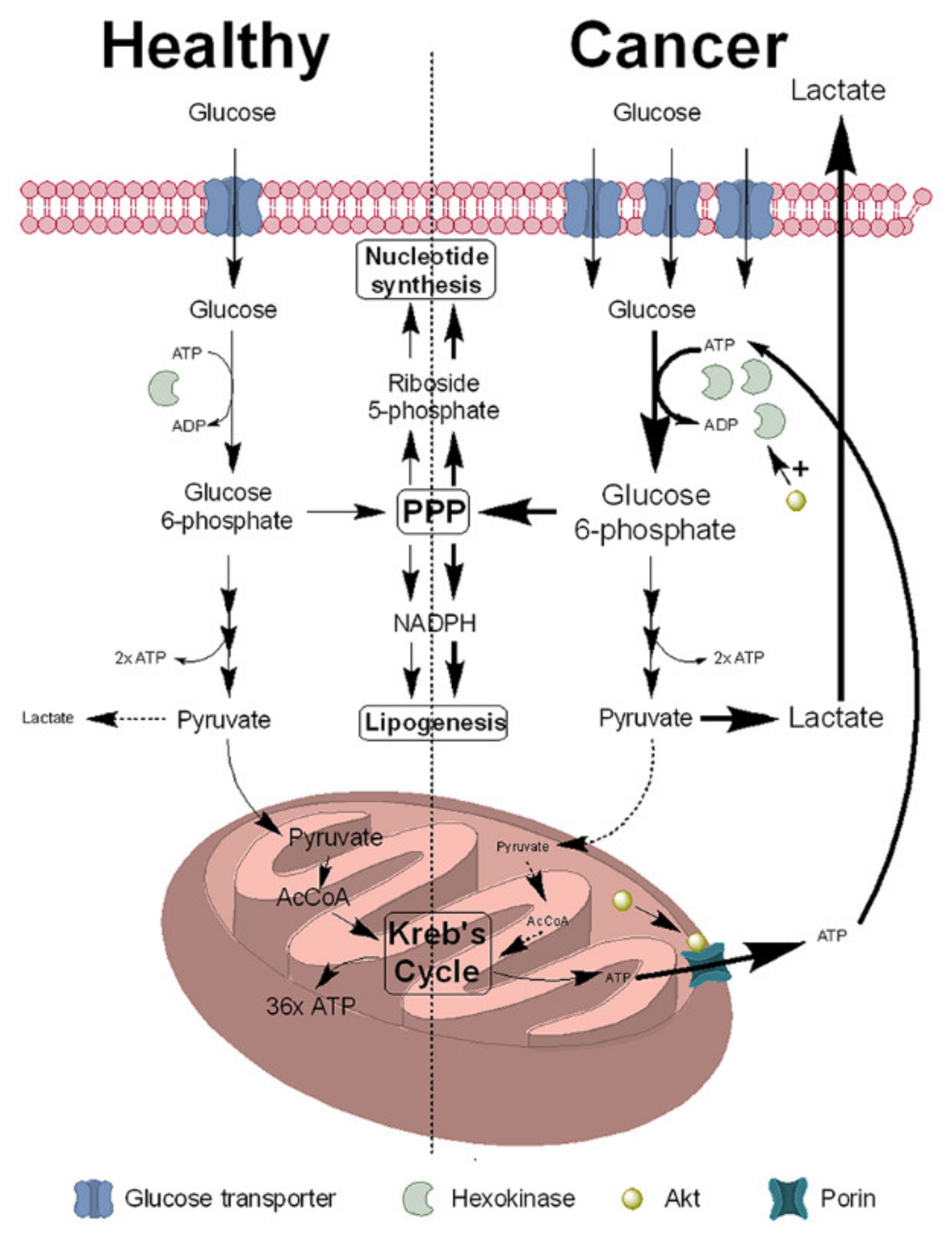
2. NNMT Expression Is Increased in Cancer Cells to Support the Warburg Effect

2.1. NAD+ Synthesis

NAD+ is essential for the survival of the cell. In the form of NADH, it is required for the donation of protons and electrons to the mitochondrial respiratory chain for the generation of ATP [20]. NAD+ is also a substrate for several proteins, for example PARP1, which repairs single-strand DNA breaks [21], and sirtuins, which play a role in epigenetic regulation and energy metabolism [22,23]. Cleavage of NAD+ by NAD+-dependent enzymes releases nicotinamide, which is the physiological inhibitor of these enzymes [24,25]. NAD+ is also required for cell signalling pathways, serving as precursor for the calcium mobilisers 2″-O-acetyl-ADP-D-ribose (OAADPr), ADP ribose and cyclic ADP ribose. The synthesis of these also liberate nicotinamide [26,27].
In mammals, NAD+ can be synthesised from tryptophan via the kynurenine pathway; however, this is unable to sustain normal cellular levels of NAD+ [28]. Instead, NAD+ levels in mammals are maintained by synthesis from nicotinamide via the sequential actions of nicotinamide phosphoribosyltransferase (NAMPT) and nicotinamide adenine mononucleotide adenylyltransferase (NMNAT) [29]. Although nicotinamide is available in the diet, the majority (approximately 99%) is derived from the salvage pathway, involving the release of nicotinamide by NAD+-dependent signalling pathways and its reincorporation into NAD+ via NAMPT (Figure 2) [30].

2.2. NNMT Function and Regulation of NAD+ Synthesis

NNMT is a 29 kDa cytosolic enzyme which is responsible for the N-methylation of nicotinamide into 1-methylnicotinamide (MNA), using S-adenosylmethionine (SAM) as methyl donor (Figure 2). The majority of NNMT expression is in the liver, with significant levels found in other tissues such as the brain, kidney, adipose tissue, endothelium, thyroid and pancreas [31,32,33,34,35,36]. Its activity regulates NAD+ synthesis via the reduction in available nicotinamide [37]. NNMT expression is increased in several disease pathologies, including neurodegenerative diseases such as Alzheimer’s [38] and Parkinson’s disease [39,40], hepatic cirrhosis [41], atherosclerosis [42], pulmonary hypertension [43], acute hepatitis [44], fatty liver disease [45], obesity and metabolic syndrome [46,47], peripheral occlusive arterial disease [48] and chronic obstructive pulmonary disease [49]. Several thousand single nucleotide polymorphisms (SNPs) have been reported in the NNMT gene, many of which have been linked to disease and other biochemical outcomes, a full review of which has been published elsewhere [50].

2.3. NNMT Promotes the Cancer Phenotype

NNMT expression is significantly increased in many cancers (for a full review see [51]). It is thought that cancer cells arise from transformed stem cells, requiring dedifferentiation to become transformed [52,53]. NNMT has been shown to modulate the epigenetic environment in the differentiation process of stem cells [54,55] and is elevated in cancer stem cells compared to non-tumour cells [55,56,57]. Increased amounts of NNMT alter the methylation status of the genome by reducing available SAM, favouring a pattern of gene expression which promotes the dedifferentiated state and thus drives the evolution of the cell towards a cancer phenotype [56,58,59,60]. Hence, it is likely that NNMT holds a central role in the promotion of the cancer phenotype.
We have investigated the cellular effects of NNMT upon tumour cells using the human neuroblastoma cell line SH-SY5Y. The attraction of using this cell line is that it has no endogenous expression of NNMT, making it an ideal knock-in/knock-out type of in vitro model [37]. We generated a knock-in NNMT-expressing cell line (S.NNMT.LP), which revealed that NNMT is not required for tumour survival; cells continue to grow in the absence of NNMT. Instead, the expression of NNMT provided a survival advantage to the tumour cell by reducing cellular apoptosis and necrosis, decreasing susceptibility to mitotoxins and increasing ATP synthesis [37].
Considering NNMT’s role in regulating NAD+ levels, these cytotrophic effects seem counterintuitive. In our S.NNMT.LP cell line, NNMT expression reduced both cellular NAD+ and NADH content, resulting in an approximately 50% reduction in the NAD+: NADH ratio [37]. It has been suggested that limiting NAD+ availability will counter the beneficial effects of the Warburg effect, likely by restricting the activity of NAD+-dependent enzymes such as NAD kinase which shuttles NAD+ into the PPP [4,29]. Additionally, the activity of DNA repair enzymes such as PARP1 are inhibited by restricted NAD+ availability, which halts tumour progression [29]. However, these reductions in the NAD+:NADH ratio replicate what is observed within cancer cells [61]. Additionally, this reduction was not toxic to the tumour cell, as S.NNMT.LP demonstrated a significant reduction in both apoptosis and necrosis compared to wild-type SH-SY5Y [37].
A recent study by Bockwoldt and colleagues has provided an answer for this paradox. Using mathematical modelling of kinetic constants obtained from published literature for NNMT and NAMPT, they suggested that NNMT and NAMPT co-evolved in vertebrates to maintain NAD+ synthesis whilst maximising the activities of NAD+-dependent pathways essential for the survival of the cell. These pathways produce significant amounts of nicotinamide, which is the physiological inhibitor of NAD+-consuming enzymes [62]. NNMT, with its relative high km for nicotinamide (200 µm) [63], removes the excess nicotinamide generated, thus ensuring that cellular levels of nicotinamide are maintained at low levels, a process which is supported by the induction of NNMT expression by nicotinamide (Figure 3). NAMPT, with its high affinity for nicotinamide as evidenced by its nmolar km, can effectively initiate NAD+ synthesis at the low levels of nicotinamide necessary to prevent its inhibitory block of NAD+-consuming enzymes [62]. Thus, by sacrificing a proportion of NADH synthesis, it can be proposed that the tumour cell ensures the activities of NAD+-dependent enzymes can continue at the levels necessary for tumour cell survival and progression by inducing NNMT expression. This cooperation between NNMT and NAMPT is significantly more efficient for the synthesis of NAD+ compared to its synthesis using nicotinic acid via nicotinamidase (NADA), the pathway present in invertebrates such as Drosophila as well as yeast and bacteria [62,64].

2.4. NNMT Induces Sirtuin Expression and Activity

One of the key outcomes we identified arising from expression of NNMT in S.NNMT.LP cells is an increase in both complex I (CxI) activity and ATP synthesis. This arose due to an increase in the stability of CxI subunits such as NDUFS3 [37]. What is missing is how NNMT mediates these effects. Recent evidence suggests that one such possibility is via sirtuins, DNA histone deacetylases involved in epigenetic regulation which have been implicated in longevity in several in vitro and in vivo models [23,65,66]. They use NAD+ as a substrate, and in common with other NADases their activity is inhibited by nicotinamide. The activation of sirtuins increases mitochondrial function, possibly via the direct deacetylation of subunits of CxI such as NDUFA9 which results in their activation [67]. The co-regulation of NNMT and sirtuin expressions and activities were first linked by Estep and colleagues who demonstrated that calorie restriction in mice increases the expression of both NNMT and sirtuin-1 [68]. We demonstrated that the expression of NNMT in S.NNMT.LP cells increased the amount of sirtuin 1 and 3 proteins which we showed, using siRNA silencing of sirtuin-3 expression, mediated the increased CxI activity and cellular ATP synthesis we observed. Furthermore, the increased amount of sirtuin protein was accompanied by the activation of sirtuin-3 activity as evidenced by the reduction in the acetylation of the SH-SY5Y proteome, an effect most likely mediated via the removal of nicotinamide inhibition by NNMT [69]. Subsequent studies have shown that MNA binds directly to sirtuins, via an as-yet unidentified binding site, preventing its degradation and thus maintaining cellular sirtuin protein levels [70,71,72,73]. In support of this, we demonstrated that incubation of SH-SY5Y cells with MNA increased both CxI activity and ATP synthesis, and increased NDUFS3 stability, an effect likely due to the interaction of MNA with sirtuins [37].

2.5. NNMT Increases Mitochondrial Function

Mitochondrial activity is still essential for the generation of NAD(P)H and FAD to support the Warburg effect [12,13]. Sirtuins regulate the expression of several downstream pro-mitochondrial genes such as uncoupling protein-2 (UCP2), peroxisome proliferator-activated receptor-γ (PPAR-γ) and PPAR-γ coactivator 1-α (PGC1α) [74,75]. UCP2 is a mitochondrial uncoupling protein, resident in the inner mitochondrial membrane, which dissipates the proton motive force by allowing protons to flow down their concentration gradient back into the mitochondrial matrix. The consequence of this uncoupling is to reduce oxidative stress-mediated generation of reactive oxygen species (ROS) [76,77]. PPAR-γ is a nuclear receptor which activates the transcription growth factor-β signalling [78] and the mammalian target of rapamycin (mTOR) [79] pathways and interacts with PGC1α [80]. PGC1α is a transcriptional coactivator which regulates the expression of genes involved in energy metabolism and is the master regulator of mitochondrial biogenesis [81,82]. Hence, NNMT may induce pro-mitochondrial changes in gene expression and subsequent mitochondrial activity which support the Warburg effect. This is evidenced by the 2-fold increase in ATP synthesis we observed in S.NNMT.LP cells, which arose from an increase in mitochondrial activity as evidenced by a 300% increase in CxI activity and a 60% increase in oxygen consumption [37]. Whilst these increases seem somewhat large and out of step with the concept of reduced reliance upon mitochondrial generation of ATP, they are relative to activities observed in wild-type SH-SY5Y cells, which are cancer cells and thus rely predominantly upon cytosolic ATP production. Hence, the absolute increases in these mitochondrial measures are relatively small and indicate a maintenance of mitochondrial activity rather than a wholesale activation of mitochondrial function. This fits well with the hypothesis that mitochondrial function is primarily supporting NAD(P)H and FAD recycling to support the increase in the activities of the PPP and lipid biosynthetic pathways, rather than to increase ATP synthesis. The enhanced mitochondrial function we have reported correlates with other studies which have shown activation of mitochondrial activity in cancer [13].
A further potential effect of increased NNMT expression is a reduction in oxidative stress arising from the rapid proliferation of the tumour biomass. Although ROS generation is essential for the progression of the tumour phenotype [83,84,85,86] and the promotion of tumour growth [87,88], the tumour cell also needs to protect itself against the increased production of ROS to prevent the activation of apoptotic pathways and thus tumour cell death [89,90,91]. Hence, the tumour must navigate a fine balance between the beneficial and deleterious effects of enhanced ROS production and thus must keep ROS levels within a narrow window to ensure survival and proliferation [92,93]. Tumour mitochondria are a major source of such ROS [88,92,94,95], and ROS protective mechanisms such as enhanced glutathione levels have been shown to be increased in many tumours [92,93,96]. The much higher increase in CxI activity compared to ATP generation and oxygen consumption in our S.NNMT.LP cell line suggests a degree of uncoupling of the mitochondrial respiratory chain, as evidenced by increased expression of UCP2 protein in S.NNMT.LP cells. The increase in the expression of UCP2 correlated with decreased markers of oxidative stress such as superoxide and isoprostane F2α, plus an increase in the GSH:GSSG ratio [97]. The expression of UCP2 is increased in cancer and in cancer cachexia in response to increasing oxidative stress, the consequence of which is an increase in their apoptotic threshold [77,98,99,100,101]. Hence, the expression of NNMT has a 2-fold beneficial effect on the mitochondria, the first to maintain their function to support the Warburg effect, and the second to assist in maintaining ROS generation within a pro-oncogenic range.

2.6. NNMT Activates the Akt Signalling Pathway

A key feature of cancer metastasis is the induction of the Akt pathway [102]. Akt, also known as protein kinase B, is a component of the mTOR pathway and is central to the regulation of a number of fundamental cell signalling pathways involved in survival, proliferation, drug resistance and differentiation [103]. Induction and activation of the Akt signalling pathway, along with the presence of gain of function mutations, are observed in a wide number of cancers including thyroid, breast, colon and pancreatic cancers [104] and as such aberrant Akt signalling is highly oncogenic [105,106]. Akt signalling is induced by NNMT [107,108], which is mediated by the induction and cleavage of ephrin B2 [108] and subsequent binding to the ephrin B receptor [109].
The consequences for NNMT-mediated induction of Akt activity are significant for the survival and proliferation of the tumour cell. In clear renal cell carcinoma (cRCC) cells, increased Akt signalling induces the expression of matrix metalloproteinase-2, the consequence of which is the remodelling of the extracellular matrix and the subsequent promotion of tumour invasion during metastasis [107]. Activation of the Akt signalling pathways reduces cellular apoptosis by inhibiting p53, reducing the expression of the pro-apoptotic protein Bim and increasing the translation of anti-apoptotic genes such as glycogen synthase kinase-3 [110]. In line with these activities, reduced apoptosis is observed in both S.NNMT.LP cells [37] and in human breast cancer cells [111].
Akt also has several other mitochondrially-targeted effects in cancer. Akt induces hexokinase expression, necessary to phosphorylate the increased levels of glucose entering the cell via the glucose transporter due to the Warburg effect. Additionally, mitochondrial Akt binds to mitochondrial porin, which redirects mitochondrial ATP to support the increase in hexokinase-mediated phosphorylation of glucose [112]. The mitochondrial pool of Akt also promotes tumour development by phosphorylating the mitochondrial calcium uniporter, resulting in increased levels of mitochondrial calcium [113].
The activation of the Akt pathway has been shown to be a central regulator of drug resistance in cancer, arising from interactions with pro-survival pathways such as the MAPK, Notch and Wnt/β-catenin signalling pathways [114]. Inhibition of the Akt pathway has been shown to overcome resistance to cisplatin in a variety of cancer types [115,116,117,118], hence targeting the Akt signalling pathway is an attractive target for current cancer therapy studies [114]. To date, a small number of studies have investigated the effect of NNMT upon drug resistance in cancer. We have shown that NNMT protects against mitochondrial toxins in S.NNMT.LP cells [37,119,120,121]. NNMT induces resistance of colorectal cancer cells to 5-fluorouracil (5-FU) [122] and has been implicated in the resistance of cervical squamous cell carcinoma to treatment [123]. Increased expression of NNMT in non-small-cell lung cancer cells results in resistance to EGFR-tyrosine kinase inhibitors [124]. To date, no study has linked these effects to increased phosphorylation and thus activation of Akt. Although pathways such as the ASK1-p38-MAPK pathway have been implicated in NNMT-mediated resistance of colorectal cancer cells to 5-FU [125], given the strong cytoprotective effects of Akt signalling, along with the well-documented regulation of Akt phosphorylation demonstrated by us and others in a wide variety of cancers, it is likely that the NNMT–Akt axis is heavily involved in acquired drug resistance.

3. NNMT Promotes the Epithelial-to-Mesenchymal Transition

A key role for NNMT in the cancer cell is the promotion of the epithelial-to-mesenchymal transition (EMT), which involves the conversion of epithelial cells into mesenchymal cells characterised by an increase in stem cell phenotype and behaviour [126]. This process is essential for the development of increased invasiveness and thus promotion of metastasis [127]. This in turn mediated the activation of matrix-degrading enzymes such as matrix metalloproteases [128]. An additional effect of the EMT is to reduce apoptosis [129]. As discussed earlier in this review, increased NNMT expression in cancer cells replicates all these effects, demonstrating the central importance of NNMT expression to the EMT. This was elegantly demonstrated recently by Eckert and colleagues who showed that NNMT was central in driving the transition of non-neoplastic fibroblasts into cancer-associated fibroblasts, a process which they successfully prevented therapeutically in an in vivo model of ovarian cancer [59]. For a more complete review of this topic, please see [130].

4. Linking NNMT with Its Cellular Functions

4.1. Epigenetic Regulation

One key question is how to link increased NNMT expression with its functions within the cell. One such mechanism is via epigenetic regulation arising from reduction in intracellular SAM concentration. SAM is the methyl donor for all methylation reactions and is produced by the adenylation of methionine by methionine adenylyltransferase [29]. Methyltransferases are found in diverse pathways such as Phase II metabolism [131,132], the regulation of protein activity [133,134] and the regulation of gene expression [135,136]. DNA methylation is a key process in regulating gene expression, in which methylation of CpG islands prevents expression of genes [135]. Due to the sole requirement for SAM as a cofactor in methylation reactions, the regulation of DNA expression is therefore heavily influenced by SAM availability.
There is a growing body of evidence which suggests that NNMT regulates the expression of pro-oncogenic genes in cancer via the regulation of intracellular SAM concentrations. By reducing available SAM levels, NNMT reduces DNA CpG island methylation, the consequence of which is enhanced gene expression. For example, increased NNMT expression in pancreatic cancer creates a “metabolic sink” whereby intracellular SAM levels are reduced due to NNMT overactivity, the consequence of which is the upregulation of the expression of several cancer-related genes such as SNAI2, ADAMTS6, TGFB2, LAMB3 and CNTN1 [137]. NNMT expression replicates many of the effects of these genes, such as promoting the EMT, extracellular matrix remodelling and activation of cell signalling pathways [59], hence NNMT-mediated changes in epigenetic regulation are likely responsible for some of its pro-tumour effects.

4.2. Regulation of NAD+-Dependent Pathways

NNMT also influences gene expression indirectly via the regulation of intracellular NAD+ and nicotinamide levels. Pathways which use NAD+ as a substrate are also inhibited by nicotinamide, the by-product of NAD+ cleavage. As described earlier in this review, NNMT co-evolved with NAMPT to maximise the activity of NAD+-dependent enzymes despite increasing levels of nicotinamide arising from their activity [62]. Many of these pathways are pro-survival and their activities are dysregulated in many cancers. One such example is sirtuins. Another example is PARP-1. PARP-1 is an NAD+-dependent DNA repair enzyme whose primary activity is the repair of both single-strand and double-strand breaks [138]. When DNA damage is too high, PARP-1 initiates cell death rather than DNA repair [139], the fate of which is determined by the availability of the intracellular NAD+ pool [140]. Patients with bladder cancer undergo nicotinamide infusion prior to radiotherapy to inhibit DNA repair by PARP-1. Increased NNMT expression reduces the efficacy of radiotherapy by removing the inhibitory block of nicotinamide upon PARP-1 [141,142,143]. PARP-1 also positively regulates the expression of many pro-oncogenic genes via the PARylation of DNA histones [144]. Inhibition of PARP-1 is an emerging therapeutic target in cancer therapy [138], thus its activation by the reduction in cellular nicotinamide levels by NNMT may contribute to the pro-oncogenic effect of PARP-1.

4.3. Synthesis of 1-Methylnicotinamide

One possibly mediator of the effects of NNMT is its product of nicotinamide N-methylation, MNA. Long considered merely an excretion product, a considerable body of evidence suggests that it has pharmacological activity in conditions ranging from inflammation and hepatotoxicity through to thrombosis and chronic obstructive pulmonary disease [41,145]. The first evidence for an important pharmacological activity of MNA was reported by Chlopicki and colleagues [145], in which they demonstrated a dose-dependent thrombolytic effect in Wistar rats at doses ranging from 30–100 mg/kg. This thrombolytic response was independent of changes in arterial blood pressure; instead, the effects of MNA were mediated via an increase in cyclooxygenase-2 (COX-2)-mediated release of prostacyclin (PGI2). MNA reduced apoptosis of SH-SY5Y human neuroblastoma cells in a dose-dependent manner [37], in addition to replicating many of the effects of NNMT expression upon tumour-promoting biochemical changes such as increased ATP synthesis and CxI activity, plus changes in cell morphology [37,108], all cognisant with changes observed during metastasis in cancers such as colorectal carcinoma [146,147,148] and breast cancer [149]. Elevated MNA levels correlate with lower survival rate and shorter life expectancy of patients with cervical cancer and thus most likely contributes to the disease progression [150]. Increased MNA synthesis in colorectal cancer xenographs in vivo increases their resistance to 5-FU via the inhibition of apoptosis, which is mediated by the attenuation of the ASK1-p38 MAPK pathway [125]. MNA also binds directly to sirtuin-1 and stabilises it, thus increasing its activity. One consequence of this stabilisation is the enhancement of chemoresistance of breast cancer cells to the anti-tumour agents adriamycin and paclitaxel [70]. Currently, the location of this allosteric site for MNA is unknown.
In contrast, a recent study showed that the role of MNA in promoting tumour development may not be as clear cut as first thought. Despite enhancing tumour vasculature formation in 4T1 murine mammary gland tumours, MNA inhibited metastatic development in the lungs of mice ectopically inoculated with 4T1 cells. Furthermore, when co-administered with the cytostatic anti-tumour agent cyclophosphamide, lung metastases were reduced by 80% [151]. It is also possible that MNA may exert its anti-metastatic effect via increased release of PGI2 [145], a molecular which has been shown to prevent metastasis in a number of in vitro and in vivo models [152,153,154]. The reasons for such discrepancies between studies is unclear but may reflect differences in tumour types investigated and analysis methodologies employed. It does raise the possibility, however, that MNA may have a mixed mode effect upon tumour progression and survival. For example, it is possible that the pro-tumour effects of MNA arises from its binding and consequent stabilisation and activation of sirtuins [70,71,72,73], the consequences of which for the tumour cell include pro-oncogenic changes in epigenetic regulation and protein acetylation [22,23] along with enhancement of chemoresistance [70]. Anti-tumour effects of MNA may at the same time arise from its inhibition of NNMT activity [1,31,63,155] and its subsequent effects upon nicotinamide levels, NAD+ synthesis and cellular SAM availability, the consequences of which include decreased activity of NAD+-dependent pro-oncogenic enzymes such as PARP-1 [141,142,143,144] and reversal of pro-oncogenic changes in epigenetic regulation [56,58,59,60], as well as increased production of PGI2. What is clear is that further research is required to fully elucidate the role of MNA in tumour survival and progression.

5. Mechanisms Underlying Increased NNMT Activity in Cancer Cells

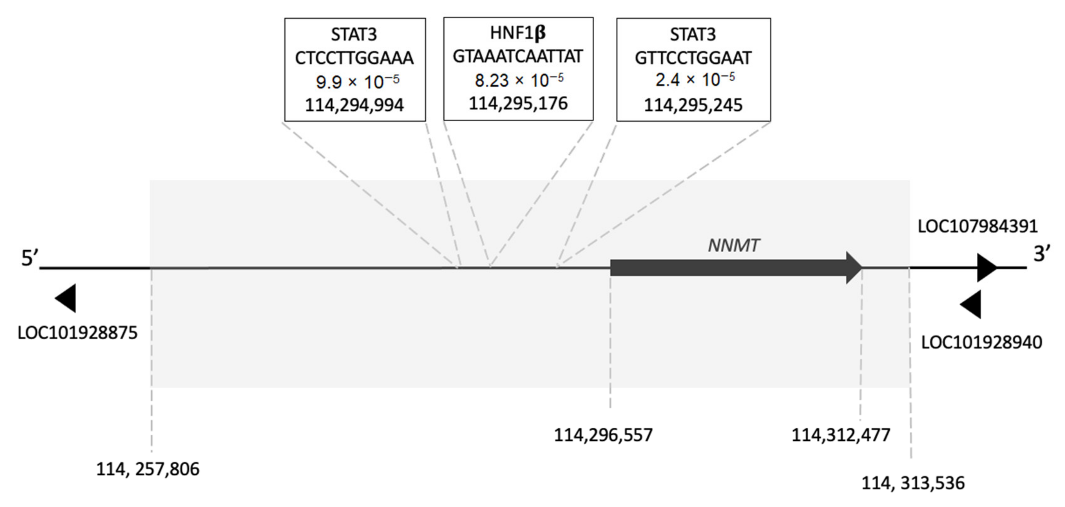
5.1. Transcription Factor Binding Sites in the NNMT Gene

Despite the widely recognised importance of NNMT overexpression in cancer, few studies have investigated how NNMT expression is regulated. It is known that the coding domain sequence produces two transcripts, NNMT-201 and NNMT-203, and these encode the same translated protein of 264 amino acids. However, NNMT-201 and NNMT-203 differ in their transcription initiation sites (TIS). In addition to these two coding transcripts, the latest version of the human genome (GRCh38.p13. refseq accession NC_000011.10) indicates that there are an additional three transcript variants of NNMT which appear to be shorter in length than both NNMT-201 and NNMT-203. Their role is unclear, but they may play a role in the regulation of mRNA processing [50]. Scrutiny of the genomic neighbourhood surrounding NNMT also reveals three, long non-coding RNAs (lncRNA) (LOC101928875, LOC101928940 and LOC107984391) that are highlighted here for the first time. All three of these lncRNAs are within the NNMT genomic neighbourhood, although LOC101928875 is approximately 97 kb upstream of NNMT and is located within another gene region. LOC101928940 and LOC107984391 are approximately 47 kb and 59 kb downstream of NNMT, respectively (Figure 4). Whether these three lncRNAs play any role in the regulation of NNMT expression is so far unknown and is yet to be investigated.
Within the NNMT promoter region, which has been proposed to extend 2 kb upstream of the NNMT coding domain sequence [50], several putative transcription factor binding sites (TFBS) have been identified [36,156,157,158]; however, only the presence of STAT3 and HNF-1β regulatory sites have been experimentally demonstrated [36,156,159]. We have subsequently performed a more in-depth bioinformatic analysis of the NNMT gene to identify the location of the two known TFBSs, STAT3 and HNF-1β, both within its 5′-flanking region and introns. Using position weighted matrices of STAT3 (MA0114.2) and HNF-1β (MA0153.2) from JASPAR [160], the NNMT promoter region and introns were scrutinised for putative TFBS using FIMO [161] implemented in the MEME Suite [162]. In agreement with our previous work [50] we identified, with high probability, two putative STAT3 recognition sites (GTTCCTGGAAT and CTCCTTGGAAA) at positions 114,295,245 and 114,294,994 plus a putative HNF-1β recognition site (GTAAATCAATTAT) at position 114,295,176. We also identified further putative STAT3 and HNF-1β TFBS in this region but disregarded them due to either low probability scores or because they were found some distance away from the gene itself.
The presence of several STAT3, and a single HNF-1β TFBS in the NNMT promoter region suggest a mechanism for the observed induction of NNMT expression in cancer. STAT3 is constitutively activated in many cancers, promoting cancer progression, proliferation, metastasis [163,164] and drug resistance via the regulation of signalling pathways including Akt [165,166,167]. Overexpression of HNF-1β has been implicated in tumour progression in cancers such as clear cell carcinoma of the ovary, liver, pancreas, kidney, endometrium, and prostate [168,169,170,171,172]. HNF-1β leads to the development of chemoresistance to carboplatin in clear cell ovarian cancer via the upregulation of glutathione synthesis [173] and has also been suggested to promote the Warburg effect and ROS reduction [174]. Interestingly, our analysis also identified several putative STAT3 and HNF-1β TFBSs throughout introns 2 and 4 (Table 1); however, whether these sites are functional is yet to be determined.

5.2. NNMT Single-Nucleotide Polymorphisms and Cancer

Over 12,000 SNPs are currently known for the NNMT gene, many of which are associated with either disease or other physiological conditions such as increased athletic performance (for a full, in-depth review of this topic, please see [50]). In total, 21 different cancers have a somatic SNP in the NNMT gene; however, only 102 SNPs are represented in a combined 119 instances of cancer, thus it is impossible to determine whether SNPs are causative or coincidental. Additionally, functional confirmation of the effects of these SNPs upon protein activity, protein expression or mRNA stability is lacking [50].

5.3. Post-Translational Modifications

Post-translational modification of proteins regulates diverse functions such as enzyme activity, protein folding, cellular trafficking, and protein-protein interactions [175,176]. However, very little research into the post-translational modification of NNMT in both healthy and cancerous tissues, and in non-cancer diseases, has been undertaken. The first post-translational modification to be functionally confirmed for NNMT was phosphorylation in gastric tumours by casein kinase 2 (CK2). The consequence of this phosphorylation for NNMT was not determined. Subsequent eukaryotic linear motif searching of the NNMT sequence revealed several further potential modification sites, including phosphorylation consensus sequences for glycogen synthase kinase 3, protein kinase A and protein-directed kinase (a component of the MAPK pathway) along with predicted sites for glycosaminoglycation and sumoylation [177]. Further analysis, using the PhosphoSitePlus v.6.5.9.3 webtool [178], predicts several further potential post-translational modification sites, including acetylation, ubiquitinylation, succinylation and methylation (Table 2). However, the experimental confirmation of these post-translational modification sites has yet to be undertaken.
CK2 is implicated in the proliferation a wide variety of tumours, including gastric tumours [180], and to promote chemoresistance to anti-tumour agents such as paclitaxel [181]. CK2 inhibition by CX-4945 mitigates the Warburg effect [180], an effect possibly mediated at least in part via reduced phosphorylation of NNMT. MAPK [182], PKA [183] and GSK3 [184] signal transduction pathways are all implicated in tumour progression and resistance. Thus, functional confirmation of the presence of these regulatory sites in the NNMT protein sequence will provide further insight into the promotion of the cancer phenotype by NNMT.
Citrulline is a non-coding amino acid which is produced via the deamidation of protein arginine residues by protein arginine deiminases (PAD) [185], resulting in the production of a positively-charged residue [186,187]. Nemmara and colleagues revealed the presence of three citrullination sites, R18, R132 and R181, in the NNMT protein sequence. Of the three sites, citrullination of R132 resulted in reduced SAM binding via the disruption of the conformation of loop 7 of the NNMT structure, the consequence of which was the inactivation of NNMT activity. The citrullination of the remaining two sites had no effect upon NNMT activity [179]. Abnormal PAD activity is observed in many cancers such as triple-negative breast cancer [188], hence this may mediate the effects of NNMT observed in many cancers.

6. NNMT and Cancer—Cause or Consequence?

Accumulating evidence is pointing to a central role for NNMT in the correct functioning of the cell, many of which are hijacked by the cancer cell to promote growth and survival. The predicted co-evolution of a high km NNMT alongside a low km NAMPT in vertebrates [62,64] raises the question—has the evolution of NNMT driven the evolution of the cancer phenotype? Cancer cells appear to have taken advantage of the NNMT/NAMPT pathway to promote the neoplastic phenotype by increasing the activity of NAD+-dependent processes such as sirtuins and PARP-1. Mitochondrial function is also stimulated, most likely to provide the NAD+ and FAD required to maintain glycolysis and the Krebs cycle as part of the Warburg effect. Finally, Akt signalling is also activated, the consequence of which is the stimulation of anti-apoptotic processes and the induction drug resistance, an effect also induced by NNMT-mediated stabilisation of sirtuin-1 via MNA.
A chain of events can therefore be envisioned which can describe the metabolic changes occurring because of increased NNMT expression in the tumour cell. Upon neoplastic transformation, there is as shift in ATP generation towards glycolysis [1]. Despite a reduction in the reliance upon the mitochondria for ATP production, there is still a need to replenish the large amounts of NAD+ and FAD being consumed by glycolysis and the PPP, which are obtained from the mitochondria [10,11]. Increased NNMT expression contributes towards meeting this need by increasing mitochondrial function [37]. As part of the neoplastic transformation, the increased activities of NAD+-dependent pro-oncogenic enzymes such as PARP1 and sirtuins generate significant amounts of nicotinamide, the physiological inhibitor of NAD+-consuming enzymes [62], thus the consequence of this is the inhibition of their respective activities. NNMT prevents this by N-methylating nicotinamide, thus removing this inhibitory block, whilst ensuring NAD+ synthesis occurs at optimal levels via the NMNT/NAMPT axis [62,64]. Finally, NNMT expression also increases Akt phosphorylation [107,108], thus enhancing pro-oncogenic signalling within the cell, resulting in effects such as the promotion of metastasis [107], inhibition of apoptosis [37,111], promotion of drug resistance [114,119,120,121] and supporting glycolysis by increasing hexokinase expression [112]. Hence, NNMT sits at the centre of a web of cellular processes, each of which contribute to the promotion of the neoplastic phenotype by supporting the metabolic shift of the cell towards glycolysis whilst maintaining NAD+ synthesis and signalling as well as inducing pro-oncogenic processes (Figure 5).
One way to address this question is to observe whether cancer affects those species which do not express NNMT but use NADA instead, for example Drosophila. NADA hydrolyses nicotinamide, producing nicotinic acid which then enters the NAD+ synthetic pathway via nicotinic acid adenylyltransferase (NaMNAT) [189,190] (Figure 2). What is key about this pathway is that it does not use SAM, thus one of the major routes by which NNMT influences the cancer phenotype—epigenetic regulation via reduced DNA methylation—is absent. Drosophila cancer models are routinely used to reproduce aspects of human cancers such as metastasis, cachexia and drug resistance [191,192]. The presence of the Hippo signalling pathway in Drosophila, and its dysregulation in many human carcinomas, makes them a simple yet effective model of human cancers [193]. Most importantly, Drosophila develop naturally occurring tumours [194]. Taken with the fact that NNMT expression is not required for a basic cancer phenotype [37], it is likely that NNMT plays no role in the initiation of tumorigenesis but instead is recruited as part of an evolution of the tumour phenotype to provide a survival advantage over surrounding non-cancerous cells. This is evidenced by (1) pro-oncogenic changes in epigenetic regulation [59], promotion of the activities of pro-oncogenic NAD+-dependent pathways such as sirtuins and PARP-1 [69,141,142,143], maintenance of mitochondrial function and support of the Warburg effect [37,97,195], activation of pro-oncogenic signalling pathways such as Akt [107,108] and protection against anti-cancer drugs [37,119,120,121,122,123,124,125]. We have shown that SH-SY5Y cells expressing NNMT demonstrate reduced cell death in response to toxins compared to wild-type SH-SY5Y which have no endogenous NNMT expression [37,119], and as discussed elsewhere in this review, numerous other studies have demonstrated increased drug resistance in response to increased NNMT expression.

7. Therapeutic Targeting of NNMT

With such a central role in the regulation of cellular processes underlying cancer progression, it is no surprise that NNMT is now a focus of intense cancer drug studies [130]. The ability to target a specific enzyme to reverse many pro-oncogenic pathways in a single hit is very attractive. Such approaches which target for example transcription factors have the potential for significant off-target effects due to their ubiquitous nature in many tissues and their involvement in essential cellular functions for non-tumour cells [196,197]. The drawback to NNMT-targeted therapy is that it is unlikely to act as a stand-alone drug. Evidence for this lies with those tumours in which NNMT is not a feature, for example human neuroblastomas [37]. This seeming weakness is probably its greatest strength, as it suggests that off-target effects for NNMT-based therapies will be minimal. It is therefore likely that the best use for NNMT-based cancer therapies is as an adjunct to anti-tumour drugs, acting to prevent tumour metastasis and burden and to reduce the survival advantage afforded to tumour cells by increased NNMT expression. Currently, two general approaches are being investigated, small-molecule inhibitors of NNMT activity and small-molecule inhibitors of NNMT expression (Figure 6).

7.1. Small-Molecule Inhibitors of NNMT Activity

Many research groups, including our own, are now actively developing inhibitors of NNMT as anti-cancer therapies (for a comprehensive review of the current state of NNMT inhibitor development, see [198]). The first description of an NNMT inhibitor which was not S-adenosylhomocysteine (SAH) or MNA identified S-adenosylethionine as a potent inhibitor, although this was significantly less effective than SAH [199]. In recent years, rapid progress in inhibitor design has been made in a relatively short amount of time due to our increased understanding of the interactions between nicotinamide, SAM and the NNMT active site [30,198]. The key to unlocking this understanding was the first publication of the crystal structure of NNMT co-crystalised with nicotinamide and SAH by Peng and colleagues [200]. The majority of the first small-molecule inhibitors developed were focussed upon targeting NNMT in obesity and alcohol-related fatty liver disease [201,202,203,204,205]. Many of these inhibitors have yet to be tested in in vitro cancer models systems, and questions remain about their selectivity towards NNMT in relation to other methyltransferases as well as their cell permeability. This is particularly important for NNMT’s family members phenylethanolamine N-methyltransferase (PNMT), responsible for the conversion of noradrenaline to adrenaline [206], and indolethylamine N-methyltransferase (INMT), which is involved in tryptamine and serotonin metabolism [207].

7.2. Nicotinamide Analogues

The active site of NNMT is relatively large, containing three binding pockets, one for nicotinamide, one for the adenosine of SAM and one for the amino acid portion of SAM [198,208]. This at first glance would appear to be beneficial, as it provides a large canvas for designing inhibitors which bind to these pockets. However, the drawback is the ubiquitous use of SAM as the methyl donor in methylation reactions and their use in almost all areas of cell function and survival. Inhibitors such as SAH and sinefungin, a close structural analogue of SAM, are non-selective and inhibit all methyltransferases, including NNMT [63].
Initial studies of NNMT substrate specificity revealed a wide substrate profile [209], which for a long period of time was the basis for labelling NNMT merely as an enzyme of Phase II metabolism. The poor rates of N-methylation of many of these substrates would indicate that they are in fact competitive inhibitors of NNMT. In a recent study, we performed a wide-ranging screen of potential NNMT substrates revealed that the majority were poor substrates. The catalytic efficiencies ranged from 59% to 4% for the close structural analogues thionicotinamide and nicotinimidamide, respectively [63]. The kcat for the most poorly performing, nicotinimidamide, was 0.0135 s−1, which equates to a turnover time of approximately 71 s to N-methylate one molecule, suggesting activity more closely related to that of an inhibitor. A further poor substrate, 4-phenylpyridine (4-PP) [63], demonstrated substrate inhibition kinetics arising from its 180° rotational symmetry of binding in the NNMT active site. Furthermore, the kcat was 0.00057 s−1, a turnover time of 1754 s or approximately 30 min per molecule [121]. What these studies show is that molecules which bind to the active site can provide significant inhibition of activity. However, the high concentrations of inhibitor required to elicit inhibition, due to the high ki of substrate-inhibitors such as 4-PP (4 mM) [121] and high km for compounds such as nicotinimidamide compared to nicotinamide (1.3 mM vs. 0.2 mM) [63], make these small-molecule analogues unsuitable.
Others have had more success. The first successful inhibitor to be reported was NNMTi, which was shown to reverse high-fat diet-induced obesity in mice [204,205]. NNMTi has an inhibitory concentration (IC50) of 1.2 μM. In human cancer-associated fibroblasts, NNMTi increased histone methylation and tubulin acetylation, and in an in vivo model of ovarian cancer metastasis it reduced tumour burden. It is important to note that NNMTi did not affect the viability of CAFs or ovarian cancer cells [59], thus supporting the hypothesis stated earlier in this review that NNMT-targeted drugs may prevent tumour metastasis and thus burden but not induce tumour cell death. Crucially, all these effects were specific to NNMT-expressing cells, suggesting that off-target effects with such an approach would be minimal [59].
5-Methylquinoline is a small-molecule inhibitor of NNMT which selectively inhibits HeLa cell proliferation without having any effect upon HEK-293 proliferation [210]. This selectivity towards the inhibition of tumour rather than stem cell proliferation suggests that NNMT inhibitors are likely to be selective for tumour cells, thus reducing the possibility of deleterious off-target effects for non-tumour tissues. Yuanhuadine (YD) is a naturally derived anti-tumour agent used for the treatment of cancers of the lung [211]. YD binds to the nicotinamide binding site of NNMT, resulting in inhibition with an IC50 of 400 nM. YD also reversed NNMT-mediated resistance of non-small cell lung cancer cells to 5-FU [124]. The anti-bacterial compound thiotetramycin and its derivatives demonstrated mild inhibitory effects against NNMT [212]. JBSNF-000088 (a substrate inhibitor) [201,202], 6-methylaminonicotinamide [213] and methylated quinolinium analogues such as 5-amino-1-methylquinoline [34,214], are nicotinamide analogues which all have IC50 values in the single μM range. Recent studies have described inhibitors with IC50 values in the low nM range [202], such as pyrimidine 5-carboxamide which has an IC50 of 74 nM [215]. However, to date, none of these have been tested in any in vitro or in vivo cancer models.

7.3. Covalent Inhibitors

A small number of studies have developed covalent inhibitors of NNMT, which bind to the active site and prevent access for nicotinamide. Such inhibitors have been developed primarily as pharmacological tools for investigating methyltransferase enzymes, rather than as therapeutic treatments for disease. The first to be developed, RS004, is based upon the structure of SAH, and covalently links to Cys165 in the NNMT active site. This residue is unique to NNMT, thus providing selectivity towards NNMT. RS004 demonstrated NNMT inhibitory efficacy with an IC50 of 1 μM [216]. Lee and colleagues produced α-chloroacetamide as inhibitors with low μM activities; however, cellular efficacy and specificity towards NNMT were low [217]. 4-chloro-3-ethynylpyridine is a suicide inhibitor of NNMT, requiring prior N-methylation by NNMT before covalently binding to Cys159. The IC50 of this molecule was 36 μM [218]. Covalent inhibitors represent approximately 30% of drugs currently in clinical use and have many benefits, such as high potency, less frequent dosage, wide therapeutic windows, and reduced development of drug resistance. Such inhibitors are rapidly becoming more abundant in cancer drug design [218]. However, they do come with increased risk of toxicity and consequences from off-target effects [219,220]. As yet, no covalent NNMT inhibitor has been screened for specificity for NNMT over its family members PNMT and INMT, instead relying on the unique cysteine target residue in the NNMT active site for their selectivity. Furthermore, none have been tested in an in vivo model, therefore the potential for their toxicity is unknown.

7.4. Bisubstrate Inhibitors

Recent efforts have focussed upon the design and synthesis of bisubstrate inhibitors. Such compounds comprise, within a single molecule, moieties designed to bind to both the nicotinamide and SAM binding sites. In this manner, it is proposed that substrate inhibitors increase the inhibitory efficacy and the enzyme selectivity over that possible with small molecule, nicotinamide analogue-type inhibitors. Using this approach, inhibitors with low EC50 values have been successfully developed, some of which demonstrate efficacy in in vitro cancer models and increased specificity towards NNMT.
Taking advantage of our novel UHP-HILIC-MS-based NNMT activity assay [63], we designed and synthesised the first bisubstrate inhibitors which mimicked the NNMT methylation reaction transition state with µM efficacy. The lead compound, the trivalent molecule (S)-2-amino-4-((((2R,3S,4R,5R)-5-(6-amino-9H-purin-9-yl)-3,4-dihydroxytetrahydrofuran-2-yl)methyl)(3-carbamoyl-benzyl)-amino)butanoic acid (compound 45), binds to the nicotinamide, adenosine and amino acid binding pockets of the active site, and is a potent inhibitor of NNMT with an IC50 (29 µM) on par with that of MNA (25 µM) and sinefungin (17 µM) [205]. Further optimisation of the nicotinamide moiety and the linker connecting the amino acid moiety of compound 45 resulted in a compound, (S)-2-Amino-4-((((2R,3S,4R,5R)-5-(6-amino-9H-purin-9-yl)-3,4-dihydroxytetrahydrofuran-2-yl)methyl)(napthalen-2-ylmethyl)amino)butanoic acid (compound 78) with an IC50 of 1.7 µM. Furthermore, compound 78 inhibited the proliferation of HSC-2 human oral cancer cells [221]. At the time of writing, the specificity of these inhibitors for NNMT over other methyltransferases has yet to be published. Our most recent inhibitor, (S)-2-amino-4((((2R,3S,4R,5R)-5-(6-amino-9H-purin-9-yl)-3,4-dihydroxytetrahydrofuran-2-yl)methyl)((E)-3-(4-cyanophenyl)allyl)-amino)butanoic acid (compound 17u), replaces the nicotinamide mimic common in current bisubstrate inhibitors with an electron-deficient para-cyano aromatic group coupled with a trans-alkene linker, resulting in an IC50 of 1.7 nM [222]. Inhibitor selectivity studies revealed that compound 17u has 3000-fold higher activity towards NNMT than PNMT. In vitro toxicity assays revealed that compound 17u significantly reduced the viability of HSC-2human oral cancer, A549 human lung carcinoma and T24 bladder cancer cell lines.
Other groups have also designed bisubstrate inhibitors of NNMT. Babault and colleagues developed the bisubstrate mimic MS2734 which has an IC50 of 14 µM. However, a methyltransferase screen revealed significant inhibitory activity against DOT1L, PRMT7, BCDIN3D and SMYD2, all proteins involved in epigenetic regulation via histone methylation and miRNA methylation and reported to be dysregulated in cancer [223]. Although not an unwelcome outcome towards tumour cells, the roles of these genes in non-tumour cells may give rise to the possibility of off-target effects. In addition, no screening of its effects upon PNMT and INMT or their efficacy in vitro were carried out [123]. Policarpo and colleagues reported bisubstrate inhibitors with similar efficacies, with methyltransferase screening revealing significant inhibition of INMT, but not PNMT. Toxicity screening of their most potent compound (NS1) revealed no toxicity towards the U2OS human bone sarcoma cell line, which the authors reported was due to poor cell permeability [224]. Chen and colleagues reported the synthesis of bisubstrate inhibitors which utilise a propargyl linkage between the nicotinamide analogue and the adenosine moieties, with one (compound 2a, LL320) having a ki of 7 nM. Crucially, specificity screening demonstrated that this molecule exhibited no interaction with either INMT or PNMT, although significant efficacy was observed towards SAH hydrolase (SAHH) [225], essential in the cleavage of SAH and subsequent release of its inhibitory effect upon SAM-utilising enzymes, along with the synthesis of SAM via homocysteine and methionine [226]. Cell permeability screening demonstrated that these molecules did not cross the outer plasma membrane [225].
It must be noted that, in contrast to our studies which used a nicotinamide-based enzyme assay coupled with the direct detection of MNA production using LC–MS, the studies of Babault, Policarpo and Chen all used a SAHH-based fluorescence assay using quinoline as substrate. The use of the SAHH-based fluorescence assay is therefore complicated by the marked inhibition of SAHH by compound 2a, for which it has an IC50 of approximately 3 µM [223]. We also reported that NNMT demonstrated only 15% of the catalytic activity towards quinoline compared to nicotinamide, along with possessing substrate inhibition kinetics [63]. What is clear from these studies is that bisubstrate-based inhibitors offer increased efficacy along with increased specificity over and above those of nicotinamide analogues which, due to the nature of the compounds, provide significant scope for further optimisation. One hurdle which clearly needs to be overcome is to enhance the cell permeability of these molecules, using derivatisation with moieties which promote cell permeability. One possibility is to attach moieties which will promote cellular uptake via transporters; however, this raises the very significant possibility of drug–drug interactions [227,228].

7.5. Small-Molecule Inhibitors of NNMT Expression

One avenue for targeting NNMT is via its expression. Although, as described previously in this review, several TFBSs have been predicted, only two—STAT3 and HNF-1β—have been functionally confirmed. STAT3 is of particular interest because of its involvement in cancer progression and development of drug resistance. We reported that crispene E, a cis-clerodane diterpene isolated from Tinospora crispa, inhibited the dimerisation of STAT3 both via an interaction with its SH2 domain and the downregulation of its gene transcription. This correlated with a decrease in the expression of STAT3 target genes, including NNMT. Crispene E had no effect upon the expression of STAT1, thus demonstrating its selectivity. In vitro efficacy and selectivity screening revealed that crispene E was significantly toxic towards the STAT3-positive human breast cancer cell line MDA-MB-231, with an EC50 of 5.35 µM, but had no effect against the STAT3-null human leukoblastic leukaemia cell line A4 [159]. A further approach for targeting STAT3 is via its phosophorylation, essential for its transcription factor activity [229,230]. Inhibition of STAT3 phosphorylation using vanillin was shown to inhibit NNMT expression; however, further in vitro and in vivo screening is required to confirm this [122].
YD was shown to supress NNMT mRNA production, which although the exact mechanism was not elucidated, possibly involved in the induction of miR-449a expression [124]. miR-449a is a microRNA whose expression inhibits tumour growth, invasion, and metastasis as well as inducting apoptosis and cellular differentiation [231].
The pleiotropic effects of transcription factors, and the importance of these pathways for non-neoplastic cells, means that they can have wide-ranging effects upon cellular physiology and reduce their cell selectivity. The ability to target NNMT activity using specific inhibitors means that such interventions can be fine-tuned to affect only those pathways affected by enhanced NNMT expression. Additionally, by their very selectivity, off-target effects can be minimised using inhibitors of NNMT activity rather than NNMT expression, making cancer therapeutics much more effective and tolerable for the patient. Therefore, although effective at reducing NNMT expression, the lack of specificity of small-molecule inhibitors of NNMT expression means that it is likely that NNMT inhibitors will be favoured as the most promising therapeutic approach going forward.
Figure 6. Structures of inhibitors of NNMT activity and expression. Numbers in italics are citations reporting NNMT inhibitory activity or downregulation of expression.
Biomolecules 11 01418 g006

8. Conclusions

Since the first description of the methylation of nicotinamide in the 1940s [232,233] and its link to SAM usage [234], it was long thought that the role of NNMT was merely as a component of Phase II metabolism, N-methylating pyridine-containing compounds and regulating cellular nicotinamide levels [209]. This perception continued until the mid-1980s, after which papers reporting the increased N-methylation of nicotinamide in cancer began to be periodically published, albeit at a relatively low rate [130]. The intervening 20 years have seen a rapid increase in interest in the role of NNMT in cancer, fuelled by the increasing knowledge of its involvement in the many cellular pathways essential to both tumour and non-tumour survival. The accumulated evidence of thirty years of research which we have presented in this review clearly shows that increased NNMT expression induces a wide variety of pro-oncogenic effects which serves to support the Warburg effect, to promote tumour metastasis, and to provide the tumour cell with a survival advantage over non-neoplastic cells, a hypothesis which has also been proposed by others [195]. We now have multiple research groups regularly publishing studies of inhibitors with ever-improving binding affinities, inhibitory efficacies, and enzyme selectivity profile. Although unlikely to lead to a cure for cancer when used alone, it is likely that when used as an adjunct therapy NNMT inhibitors will significantly improve the efficacy of other treatments as well as reduce the possibility for the development of drug resistance. It also opens the avenue for the management of cancers for which currently there are limited therapeutic options, for example bladder cancer [235,236]. It is highly likely that, within 5–10 years, we will see NNMT-based therapeutics entering human clinical trials, with the first entering clinical use within 10–15 years.

Author Contributions

Both R.B.P. and P.D.F. conceived and wrote the manuscript. R.B.P. performed the biochemical analysis of NNMT activity in HepG2 cells shown in Figure 3. P.D.F. performed the bioinformatic analysis of the NNMT gene shown in Figure 4. All authors have read and agreed to the published version of the manuscript.

Funding

This research received no external funding.

Institutional Review Board Statement

Not applicable.

Informed Consent Statement

Not applicable.

Data Availability Statement

All novel data reported in this manuscript can be obtained from the authors upon reasonable request.

Acknowledgments

The authors would like to acknowledge the contributions of numerous colleagues and students who have contributed to the work reviewed in this manuscript. The authors would particularly like to thank David Ramsden, The University of Birmingham for his valued guidance, mentorship and friendship over the last two decades and without whom this journey would not have begun.

Conflicts of Interest

The authors declare no conflict of interest.

References

  1. Vaupel, P.; Multhoff, G. Revisiting the Warburg effect: Historical dogma versus current understanding. J. Physiol. 2021, 599, 1745–1757. [Google Scholar] [CrossRef]
  2. Mookerjee, S.A.; Gerencser, A.A.; Nicholls, D.A.; Brand, M.D. Quantifying intracellular rated of glycolytic and oxidative ATP production and consumption using extracellular flux measurements. J. Biol. Chem. 2017, 292, 7189–7207. [Google Scholar] [CrossRef] [Green Version]
  3. Cairns, R.A.; Harris, I.S.; Mak, T.W. Regulation of cancer metabolism. Nat. Rev. 2011, 11, 85–95. [Google Scholar] [CrossRef] [PubMed] [Green Version]
  4. Hamanaka, R.B.; Chandel, N.S. Cell Biology. Warburg effect and redox balance. Science 2011, 334, 1219–1220. [Google Scholar] [CrossRef] [PubMed]
  5. Le, A.; Cooper, C.R.; Gouw, A.M.; Dinavahi, R.; Maitra, A.; Deck, L.M.; Royer, R.E.; Vander Jagt, D.L.; Semenza, G.L.; Dang, C.V. Inhibition of lactate dehydrogenase A induces oxidative stress and inhibits tumor progression. Proc. Natl. Acad. Sci. USA 2010, 107, 2037–2042. [Google Scholar] [CrossRef] [PubMed] [Green Version]
  6. Hjelemand, A.B.; Heddleston, J.M.; Choudhary, G.S.; MacSwords, J.; Lathia, J.D.; McLendon, R.; Lindner, D.; Sloan, A.; Rich, J.N. Acidic stress promotes a glioma stem cell phenotype. Cell Death Differ. 2011, 18, 829–840. [Google Scholar] [CrossRef] [PubMed] [Green Version]
  7. Peppicelli, S.; Bianchini, F.; Toti, A.; Laurenzana, A.; Fibbi, G.; Calorini, L. Extracellular acidity strengthens mesenchymal stem cells to promote melanoma progression. Cell Cycle 2015, 14, 3088–3100. [Google Scholar] [CrossRef] [PubMed]
  8. Draoui, N.; Feron, O. Lactate shuttle at a glance: From physiological paradigms to anti-cancer treatments. Dis. Model. Mech. 2011, 4, 727–732. [Google Scholar] [CrossRef] [Green Version]
  9. Alquraishi, M.; Puckett, D.L.; Alani, D.S.; Humidat, A.S.; Frankel, V.D.; Donohoe, D.R.; Whelan, J.; Bettaieb, A. Pyruvate kinase M2: A simple molecule with complex functions. Free Radic. Biol. Med. 2019, 143, 176–192. [Google Scholar] [CrossRef]
  10. Alfarouk, K.O.; Ahmed, S.B.M.; Elliott, R.L.; Benoit, A.; Alqahtani, S.S.; Ibrahim, M.E.; Bashir, A.H.H.; Alhoufie, S.T.S.; Elhassan, G.O.; Wales, C.C.; et al. The pentose phosphate pathway dynamics in cancer and its dependency on intracellular pH. Metabolites 2020, 10, 285. [Google Scholar] [CrossRef]
  11. Jin, L.; Zhou, Y. Crucial role of the pentose phosphate pathway in malignant tumours. Oncol. Lett. 2019, 17, 4213–4221. [Google Scholar] [PubMed] [Green Version]
  12. Vyas, S.; Zaganjor, W.; Haigis, M.C. Mitochondria and cancer. Cell 2016, 166, 555–566. [Google Scholar] [CrossRef] [PubMed]
  13. Grasso, D.; Zampieri, L.X.; Capeloa, T.; Van de Velde, J.A.; Sonveaux, P. Mitochondria in cancer. Cell Stress 2020, 4, 114–146. [Google Scholar] [CrossRef]
  14. Gatenby, R.A.; Gillies, R.J. Glycolysis in cancer: A potential target for therapy. Int. J. Biochem. Cell Biol. 2007, 39, 1358–1366. [Google Scholar] [CrossRef]
  15. Kim, J.-W.; Dang, C.V. Cancer’s molecular sweet tooth and the Warburg effect. Cancer Res. 2006, 66, 8927–8930. [Google Scholar] [CrossRef] [Green Version]
  16. Ancey, P.-B.; Contat, C.; Meylan, E. Glucose transporters in cancer—From tumor cells to the tumor microenvironment. FEBS J. 2018, 286, 2926–2943. [Google Scholar] [CrossRef]
  17. Vaupel, P.; Schmidberger, H.; Mayer, A. The Warburg effect: Essential part of metabolic reprogramming and central contributor to cancer progression. Int. J. Radiat. Biol. 2019, 95, 912–919. [Google Scholar] [CrossRef] [PubMed]
  18. Yang, Y.; Karakhanova, S.; Hartwig, W.; D’Haese, J.G.D.; Philippov, P.P.; Werner, J.; Bazhin, A.V. Mitochondria and mitochondrial ROS in cancer: Novel targets for anticancer therapy. J. Cell Physiol. 2016, 231, 2570–2581. [Google Scholar] [CrossRef]
  19. Park, J.H.; Pyun, W.Y.; Park, H.W. Cancer metabolism: Phenotype, signaling and therapeutic targets. Cells 2020, 9, 2308. [Google Scholar] [CrossRef] [PubMed]
  20. Rich, P.R.; Marechal, A. The mitochondrial respiratory chain. Essays Biochem. 2010, 47, 1–23. [Google Scholar] [PubMed] [Green Version]
  21. Pascal, J.M. The comings and goings of PARP-1 in response to DNA damage. DNA Rep. 2018, 71, 177–182. [Google Scholar] [CrossRef] [PubMed]
  22. Liu Haigis, M.C.; Guarente, L. Mammalian sirtuins—Emerging roles in physiology, aging and calorie restriction. Genes Dev. 2006, 20, 2913–2921. [Google Scholar] [CrossRef] [PubMed] [Green Version]
  23. Watroba, M.; Dudek, I.; Skoda, M.; Stangret, A.; Rzodkiewicz, P.; Skukiewicz, D. Sirtuins, epigenetics and longevity. Aging Res. Rev. 2017, 40, 11–19. [Google Scholar] [CrossRef] [PubMed]
  24. Hwang, E.S.; Song, S.B. Nicotinamide is an inhibitor of SIRT in vitro, but can be a stimulator in cells. Cell Mol. Life Sci. 2017, 74, 3347–3362. [Google Scholar] [CrossRef] [PubMed]
  25. Song, S.B.; Park, J.S.; Chung, G.J.; Lee, I.H.; Hwang, E.S. Diverse therapeutic efficacies and more diverse mechanisms of nicotinamide. Metabolomics 2019, 15, 137. [Google Scholar] [CrossRef] [PubMed]
  26. Jackson, M.D.; Denu, J.M. Structural identification of 2′- and 3′-O-acetyl-ADP-ribose as novel metabolites derived from the Sir2 family of β-NAD+-dependent histone/protein deacetylases. J. Biol. Chem. 2002, 277, 18535–18544. [Google Scholar] [CrossRef] [PubMed] [Green Version]
  27. Meszaros, L.G.; Bak, J.; Chu, A. Cyclic ADP-ribose as an endogenous regulator of the non-skeletal type ryanodine receptor Ca2+ channel. Nature 1993, 364, 76–79. [Google Scholar] [CrossRef] [PubMed]
  28. Magni, G.; Orsomando, G.; Raffelli, N.; Ruggieri, S. Enzymology of mammalian NAD metabolism in health and disease. Front. Biosci. 2008, 13, 6135–6154. [Google Scholar] [CrossRef] [Green Version]
  29. Chiarugi, A.; Dolle, C.; Felici, R.; Ziegler, M. The NAD metabolome—A key determinant of cancer cell biology. Nat. Rev. Cancer 2012, 12, 741–752. [Google Scholar] [CrossRef]
  30. Canto, C.; Menzies, K.; Auwerx, J. NAD+ metabolism and the control of energy homeostasis—A balancing act between mitochondria and the nucleus. Cell Metab. 2015, 22, 31–53. [Google Scholar] [CrossRef] [Green Version]
  31. Aksoy, S.; Szumlanski, C.L.; Weinshilboum, R.M. Human liver nicotinamide N-methyltransferase: cDNA cloning, expression and biochemical characterisation. J. Biol. Chem. 1994, 269, 14835–14840. [Google Scholar] [CrossRef]
  32. Smith, M.-L.; Burnett, D.; Bennett, P.; Waring, R.H.; Brown, H.M.; Williams, A.C.; Ramsden, D.B. A direct correlation between nicotinamide N-methyltransferase activity and protein levels in human liver cytosol. Biochem. Biophys. Acta 1998, 1422, 238–244. [Google Scholar] [CrossRef]
  33. Seifert, R.; Hoshino, J.; Kroger, H. Nicotinamide methylation. Tissue distribution, developmental and neoplastic changes. Biochem. Biophys. Acta 1984, 801, 259–264. [Google Scholar] [CrossRef]
  34. Campagna, R.; Mateuszuk, L.; Wojnar-Lason, K.; Kaczara, P.; Tworzydlo, A.; Kij, A.; Bujok, R.; Mlynarski, J.; Wang, Y.; Sartini, D.; et al. Nicotinamide N-methyltransferase in endothelium protects against oxidant stress-induced endothelial injury. Biochim. Biophys. Acta Mol. Cell Res. 2021, 1868, 119082. [Google Scholar] [CrossRef] [PubMed]
  35. Riederer, M.; Erwa, W.; Zimmerman, R.; Frank, S.; Zechner, R. Adipose tissue as a source of nicotinaminde N-methyltransferase and homocysteine. Atherosclerosis 2009, 204, 412–417. [Google Scholar] [CrossRef]
  36. Xu, J.; Capezzone, M.; Xu, X.; Hershman, J.M. Activation of nicotinamide N-methyltransferase gene promoter by hepatocyte nuclear factor-1β in human papillary thyroid cancer cells. Mol. Endocrinol. 2005, 19, 527–539. [Google Scholar] [CrossRef] [PubMed] [Green Version]
  37. Parsons, R.B.; Aravindan, S.; Kadampeswaran, A.; Evans, E.A.; Sandhu, K.K.; Levy, E.R.; Thomas, M.G.; Austen, B.M.; Ramsden, D.B. The expression of nicotinamide N-methyltransferase increases ATP synthesis and protects SH-SY5Y neuroblastoma cells against the toxicity of Complex I inhibitors. Biochem. J. 2011, 436, 145–155. [Google Scholar] [CrossRef] [Green Version]
  38. Kocinaj, A.; Chaudhury, T.; Uddin, M.S.; Junaid, R.R.; Ramsden, D.B.; Hondhamuni, G.; Klamt, F.; Parsons, L.; Parsons, R.B. High expression of nicotinamide N-methyltransferase in patients with sporadic Alzheimer’s disease. Mol. Neurobiol. 2021, 58, 1769–1781. [Google Scholar] [CrossRef] [PubMed]
  39. Parsons, R.B.; Smith, M.-L.; Williams, A.C.; Waring, R.H.; Ramsden, D.B. Expression of nicotinamide N-methyltransferase (E.C. 2.1.1.1) in the Parkinsonian brain. J. Neuropathol. Exp. Neurol. 2002, 61, 111–124. [Google Scholar] [CrossRef] [Green Version]
  40. Parsons, R.B.; Smith, S.W.; Waring, R.H.; Williams, A.C.; Ramsden, D.B. High expression of nicotinamide N-methyltransferase in patients with idiopathic Parkinson’s disease. Neurosci. Lett. 2003, 342, 13–16. [Google Scholar] [CrossRef]
  41. Pumpo, M.D.; Sarnelli, G.; Spinella, S.; Budillon, G.; Cuomo, R. The metabolism of nicotinamide in human liver cirrhosis: A study on N-methylnicotinamide and 2-pyridone-5-carboxamide production. Am. J. Gastroenterol. 2001, 96, 1183–1187. [Google Scholar] [CrossRef]
  42. Mateuszuk, L.; Komich, T.I.; Slominska, E.; Gajda, M.; Wojcik, L.; Lomnicka, M.; Gwozdz, P.; Chlopicki, S. Activation of nicotinamide N-methyltransferase and increased formation of 1-methylnicotinamide (MNA) in atherosclerosis. Pharmacol. Rep. 2009, 61, 76–85. [Google Scholar] [CrossRef] [Green Version]
  43. Fedorowicz, A.; Mateuszuk, L.; Kopec, G.; Skorka, T.; Kutryb-Zajac, B.; Zakrzewska, A.; Walczak, M.; Jakubowski, A.; Lomnicka, M.; Slominska, E.; et al. Activation of nicotinamide N-methyltransferase (NNMT)-1-methylnicotinamide (MNA) pathway in pulmonary hypertension. Respir. Res. 2016, 17, 108. [Google Scholar] [CrossRef] [Green Version]
  44. Sternak, M.; Khomich, T.I.; Jakubowski, A.; Szafarz, M.; Sczcepanski, W.; Bialas, M.; Stojak, M.; Szymura-Oleksiak, J.; Chlopicki, S. Nicotinamide N-methyltransferase (NNMT) and 1-methylnicotinamide (MNA) in experimental hepatitis induced by concanavalin A in the mouse. Pharmacol. Rep. 2010, 62, 483–493. [Google Scholar] [CrossRef]
  45. Komatsu, M.; Kanda, T.; Urai, H.; Kurokochi, A.; Kitahama, R.; Shigaki, S.; Ono, T.; Yukioka, H.; Hasegawa, K.; Tokuyama, H.; et al. NNMT activation can contribute to the development of fatty liver disease by modulating the NAD+ metabolism. Sci. Rep. 2018, 8, 8637. [Google Scholar] [CrossRef] [Green Version]
  46. Giuliante, R.; Sartini, D.; Bacchetti, T.; Rocchetti, R.; Kloting, I.; Polidori, C.; Ferretti, G.; Emanuelli, M. Potential involvement of nicotinamide N-methyltransferase in the pathogenesis of metabolic syndrome. Metab. Syndr. Relat. Disord. 2015, 13, 165–170. [Google Scholar] [CrossRef] [PubMed]
  47. Kraus, D.; Yang, Q.; Kong, D.; Banks, A.S.; Zhang, L.; Rodgers, J.T.; Pirinen, E.; Pulinilkunnil, T.C.; Gong, F.; Wang, Y.-C.; et al. Nicotinamide N-methyltransferase knockdown protects against diet-induced obesity. Nature 2014, 508, 258–262. [Google Scholar] [CrossRef] [PubMed] [Green Version]
  48. Bubenek, S.; Natase, A.; Niculescu, A.M.; Tomescu, S.; Popescu, I.; Dima, S. Asssessment of gene expression profiles in peripheral occlusive arterial disease. Can. J. Cardiol. 2012, 28, 712–720. [Google Scholar] [CrossRef]
  49. Kim, H.C.; Mofarrahi, M.; Vassilakopoulos, T.; Maltais, F.; Sigala, I.; Debigare, R.; Bellenis, I.; Hussain, S.N.A. Expression and functional significance of nicotinamide N-methyltransferase in skeletal muscles of patients with chronic obstructive pulmonary disease. Am. J. Respir. Crit. Care Med. 2010, 181, 797–805. [Google Scholar] [CrossRef]
  50. Ramsden, D.B.; Waring, R.H.; Parsons, R.B.; Barlow, D.J.; Williams, A.C. Nicotinamide N-methyltransferase: Genomic connection to disease. Int. J. Tryptophan Res. 2020, 13, 1178646920919770. [Google Scholar] [CrossRef] [PubMed]
  51. Ramsden, D.B.; Waring, R.H.; Barlow, D.J.; Parsons, R.B. Nicotinamide N-methyltransferase in health and cancer. Int. J. Tryptophan Res. 2017, 10, 1178646917691739. [Google Scholar] [CrossRef] [Green Version]
  52. Mosteiro, L.; Pantoja, C.; Alcazar, N.; Marion, R.M.; Chondronasiou, D.; Rovira, M.; Fernandez-Marcos, P.J.; Munoz-Martin, M.; Blanco-Aparicio, C.; Pastor, J.; et al. Tissue damage and senescence provide critical signals for cellular reprogramming in vivo. Science 2016, 354, aaf4445. [Google Scholar] [CrossRef]
  53. Najafi, M.; Farhood, B.; Mortezaee, K. Cancer stem cells (CSCs) in cancer progression and therapy. J. Cell Physiol. 2019, 234, 8381–8395. [Google Scholar] [CrossRef] [PubMed]
  54. Pozzi, V.; Salvolini, E.; Lucarini, G.; Salvucci, A.; Campagna, R.; Rubini, C.; Sartini, D.; Emanuelli, M. Cancer stem cell enrichment is associated with enhancement of nicotinamide N-methyltransferase expression. IUBMB Life 2020, 72, 1415–1425. [Google Scholar] [CrossRef] [PubMed]
  55. Sperber, H.; Mathieu, J.; Wang, Y.; Ferreccio, A.; Hesson, J.; Xu, Z.; Fischer, K.A.; Devi, A.; Detraux, D.; Gu, H.; et al. The metabolome regulates the epigenetic landscape during naïve and primed human embryonic stem cell transition. Nat. Cell Biol. 2015, 17, 1523–1535. [Google Scholar] [CrossRef] [PubMed]
  56. Jung, J.; Kim, L.J.Y.; Wang, X.; Wu, Q.; Sanvoranart, T.; Hubert, C.G.; Prager, B.C.; Wallace, L.C.; Jin, X.; Mack, S.C.; et al. Nicotinamide metabolism regulates glioblastoma stem cell maintenance. JCI Insight 2017, 2, e90019. [Google Scholar] [CrossRef] [PubMed] [Green Version]
  57. Pozzi, V.; Sartini, D.; Rochetti, R.; Santarelli, A.; Rubini, C.; Morganti, S.; Giuliante, R.; Calabrese, S.; Di Ruscio, G.; Orlando, F.; et al. Identification and characterization of cancer stem cells from head and neck squamous cell carcinoma cell lines. Cell Physiol. Biochem. 2015, 36, 784–798. [Google Scholar] [CrossRef] [PubMed]
  58. Kujundzic, R.N.; Prpic, M.; Dakovic, N.; Dabelic, N.; Tomljanovic, M.; Mojzes, A.; Frobe, A.; Troelj, K.G. Nicotinamide N-methyltransferase in acquisition of stem cell properties and therapy resistance to cancer. Int. J. Mol. Sci. 2021, 22, 5681. [Google Scholar] [CrossRef] [PubMed]
  59. Eckert, M.A.; Coscia, F.; Chryplewicz, A.; Chang, J.W.; Hernandez, K.M.; Pan, S.; Tienda, S.M.; Nahotko, D.A.; Li, G.; Blazenovic, I.; et al. Proteomics reveals NNMT as a master metabolic regulator of cancer-associated fibroblasts. Nature 2019, 569, 723–728. [Google Scholar] [CrossRef]
  60. Matei, D.; Nephew, K.P. Epigenetic attire in ovarian cancer: The Emperor’s new clothes. Cancer Res. 2020, 80, 3775–3785. [Google Scholar] [CrossRef]
  61. DeBerardinis, R.J.; Lum, J.J.; Hatzivassiliou, G.; Thompson, C.B. The biology of cancer: Metabolic reprogramming fuels cell growth and proliferation. Cell Metab. 2008, 7, 11–20. [Google Scholar] [CrossRef] [Green Version]
  62. Bockwoldt, M.; Houry, D.; Niere, M.; Gossmann, T.I.; Reinartz, I.; Schug, A.; Ziegler, M.; Heiland, I. Identification of evolutionary and kinetic drivers of NAD-dependent signalling. Proc. Natl. Acad. Sci. USA 2019, 116, 15957–15966. [Google Scholar] [CrossRef] [PubMed] [Green Version]
  63. van Haren, M.J.; Torano, J.S.; Sartini, D.; Emanuelli, M.; Parsons, R.B.; Martin, N.I. A rapid and efficient assay for the characterisation of substrates and inhibitors for nicotinamide N-methyltransferase. Biochemistry 2016, 55, 5307–5315. [Google Scholar] [CrossRef] [PubMed] [Green Version]
  64. Gossmann, T.I.; Ziegler, M.; Puntervoll, P.; de Figueiredo, L.P.; Schuster, S.; Heiland, I. NAD+ biosynthesis and salvage—A phylogenetic perspective. FEBS J. 2012, 279, 3355–3363. [Google Scholar] [CrossRef] [PubMed]
  65. Bellizzi, D.; Rose, G.; Cavalcante, P.; Covello, G.; Dato, S.; De Rango, F.; Greco, V.; Maggiolini, M.; Feraco, E.; Mari, V.; et al. A novel VNTR enhancer within the SIRT gene, a human homologue of SIR2, is associated with survival at oldest ages. Genomics 2005, 85, 258–263. [Google Scholar] [CrossRef] [PubMed]
  66. Lee, S.-H.; Lee, J.-H.; Lee, H.-Y.; Min, K.-J. Sirtuin signalling in cellular senescence and aging. BMB Rep. 2019, 52, 24–34. [Google Scholar] [CrossRef] [Green Version]
  67. Ahn, B.-H.; Kim, H.-S.; Song, S.; Lee, I.H.; Vassilopoulos, A.; Deng, C.-X.; Finkel, T. A role for the mitochondrial deacetylase Sirt3 in regulating energy homeostasis. Proc. Natl. Acad. Sci. USA 2008, 105, 14447–14452. [Google Scholar] [CrossRef] [Green Version]
  68. Estep, P.W., II; Warner, J.B.; Bulyk, M.L. Short-term calorie restriction in male mice feminizes gene expression and alters key regulators of conserved aging regulatory pathways. PLoS ONE 2009, 4, e5242. [Google Scholar] [CrossRef] [Green Version]
  69. Liu, K.Y.; Mistry, R.J.; Aguirre, C.A.; Fasouli, E.S.; Thomas, M.G.; Klamt, F.; Ramsden, D.B.; Parsons, R.B. Nicotinamide N-methyltransferase increases complex I activity in SH-SY5Y cells via sirtuin-3. Biochem. Biophys. Res. Commun. 2015, 467, 491–496. [Google Scholar] [CrossRef]
  70. Wang, Y.; Zeng, J.; Wu, W.; Xie, S.; Yu, H.; Li, G.; Zhu, T.; Li, F.; Lu, J.; Wang, G.Y.; et al. Nicotinamide N-methyltransferase enhances chemoresistance in breast cancer through SIRT1 protein stabilization. Breast Cancer Res. 2019, 21, 64. [Google Scholar] [CrossRef] [Green Version]
  71. Hong, S.; Moreno-Navarette, J.M.; Wei, X.; Kikukawa, Y.; Tzameli, I.; Prasad, D.; Lee, Y.; Asara, J.M.; Fernandez-Real, J.M.; Maratos-Flier, E.; et al. Nicotinamide N-methyltransferase regulates hepatic nutrient metabolism through Sirt1 protein stabilization. Nat. Med. 2015, 21, 887–894. [Google Scholar] [CrossRef] [Green Version]
  72. You, Z.; Liu, Y.; Liu, X. Nicotinamide N-methyltransferase enhances the progression of prostate cancer by stabilizing sirtuin 1. Oncol. Lett. 2018, 15, 9195–9201. [Google Scholar] [CrossRef]
  73. Schmeisser, K.; Mansfeld, J.; Kuhlow, D.; Weimer, S.; Priebe, S.; Heiland, I.; Birringer, M.; Groth, M.; Segref, A.; Kanfi, Y.; et al. Role of sirtuins in lifespan regulation is linked to methylation of nicotinamide. Nat. Chem. Biol. 2013, 9, 693–700. [Google Scholar] [CrossRef] [Green Version]
  74. Haigis, M.C.; Sinclair, D.A. Mammalian sirtuins: Biological insights and disease relevance. Ann. Rev. Pathol. 2010, 5, 253–295. [Google Scholar] [CrossRef] [Green Version]
  75. Feng, H.; Wang, J.-Y.; Yu, B.; Cong, X.; Zhang, W.-G.; Li, L.; Liu, L.-M.; Zhou, Y.; Zhang, C.-L.; Gu, P.-L.; et al. Peroxisome proliferator-activated receptor-γ coactivator 1-α inhibits vascular calcification through sirtuin 3-mediated reduction of mitochondrial oxidative stress. Antiox. Redox. Signal. 2019, 31, 75–91. [Google Scholar] [CrossRef]
  76. Samudio, I.; Fiegl, M.; Andreeff, M. Mitochondrial uncoupling and the Warburg effect: Molecular basis for the reprogramming of cancer cell metabolism. Cancer Res. 2009, 69, 2163–2166. [Google Scholar] [CrossRef] [PubMed] [Green Version]
  77. Valle, A.; Oliver, J.; Roca, P. Role of uncoupling proteins in cancer. Cancers 2010, 2, 567–591. [Google Scholar] [CrossRef] [PubMed]
  78. Rekar, A.K.; Goswami, M.T.; Krishnapuram, R.; Standiford, T.J.; Keshamouni, V.G. Molecular cross-regulation between PPAR-γ and other signaling pathways: Implications for lung cancer therapy. Lung Cancer 2011, 72, 154–159. [Google Scholar] [CrossRef] [Green Version]
  79. Vella, V.; Nicoloso, M.L.; Giuliano, S.; Bellomo, M.; Belfiore, A.; Malaguarnera, R. PPAR-γ agonists as antineoplastic agents in cancers with dysregulated IGF axis. Front. Endocrinol. 2017, 8, 31. [Google Scholar] [CrossRef] [PubMed] [Green Version]
  80. Puigserver, P. Tissue-specific regulation of metabolic pathways through the transcriptional coactivator PGC1-α. Int. J. Obes. 2005, 29, S5–S9. [Google Scholar] [CrossRef] [Green Version]
  81. Thirupathi, A.; de Souza, C.T. Multi-regulatory network of ROS: The interconnection of ROS, PGC-alpha, and AMPK-SIRT1 during exercise. J. Physiol. Biochem. 2017, 73, 487–494. [Google Scholar] [CrossRef]
  82. Scarpulla, R.C. Metabolic control of mitochondrial biogenesis through the PGC-family regulatory network. Biochem. Biophys. Acta 2011, 1813, 1269–1278. [Google Scholar] [CrossRef] [Green Version]
  83. Halliwell, B. Oxidative stress and cancer: Have we moved forward? Biochem. J. 2007, 401, 1–11. [Google Scholar] [CrossRef]
  84. Martindale, J.L.; Holbrook, N.J. Cellular response to oxidative stress: Signalling for suicide and survival. J. Cell Physiol. 2002, 192, 1–15. [Google Scholar] [CrossRef]
  85. Chaterjee, R.; Chatterjee, J. ROS and oncogenesis with special reference to EMT and stemness. Eur. J. Cell Biol. 2020, 99, 151073. [Google Scholar] [CrossRef] [PubMed]
  86. Ghoneum, A.; Abdulfattah, A.Y.; Warren, B.O.; Shu, J.; Said, N. Redox homeostasis and metabolism in cancer: A complex mechanism and potential targeted therapeutics. Int. J. Mol. Sci. 2020, 21, 3100. [Google Scholar] [CrossRef] [PubMed]
  87. Salganiak, R.I. The benefits and hazards of antioxidants: Controlling apoptosis and other protective mechanisms in cancer patients and the human population. J. Am. Coll. Nutr. 2001, 20, 464S–472S. [Google Scholar] [CrossRef] [PubMed]
  88. Sabharwal, S.S.; Schumaker, P.T. Mitochondrial ROS in cancer: Initiators, amplifiers or an Achilles’ heel? Nat. Rev. Cancer 2014, 14, 709–721. [Google Scholar] [CrossRef] [Green Version]
  89. Kerr, J.F.; Winterford, C.M.; Harmon, B.V. Apoptosis. Its significance in cancer and cancer therapy. Cancer 1994, 73, 2013–2026. [Google Scholar] [CrossRef]
  90. Rodic, S.; Vincent, M.D. Reactive oxygen species (ROS) are a key determinant of cancer’s metabolic phenotype. Int. J. Cancer 2017, 142, 440–448. [Google Scholar] [CrossRef]
  91. Aggarwal, V.; Tuli, H.S.; Varol, A.; Thakral, F.; Yerer, M.B.; Sak, K.; Varol, M.; Jain, A.; Khan, M.A.; Sethi, D. Role of reactive oxygen species in cancer progression: Molecular mechanisms and recent advancements. Biomolecules 2019, 9, 735. [Google Scholar] [CrossRef] [Green Version]
  92. Del Pilae Sosa Idelchik, M.; Begley, U.; Begley, T.J.; Melendez, J.A. Mitochondrial ROS control of cancer. Semin. Cancer Biol. 2017, 47, 57–66. [Google Scholar] [CrossRef] [PubMed]
  93. Bansal, A.; Simon, M.C. Glutathione metabolism in cancer progression and treatment resistance. J. Cell Biol. 2018, 217, 2291–2298. [Google Scholar] [CrossRef] [PubMed] [Green Version]
  94. Echtay, K.S. Mitochondrial uncoupling proteins—What is their physiological role? Free Radic. Biol. Med. 2007, 43, 1351–1371. [Google Scholar] [CrossRef] [PubMed]
  95. Korshunov, S.S.; Skulachex, V.P.; Starkov, A.A. High protonic potential actuates a mechanism of production of reactive oxygen species in mitochondria. FEBS Lett. 1997, 416, 15–18. [Google Scholar] [CrossRef] [Green Version]
  96. Li, B.; Lee, D.S.M.; Walton, Z.E.; Ochocki, J.D.; Mathew, L.K.; Mancuso, A.; Gade, T.P.F.; Keith, B.; Nissim, I.; Simon, M.C. Fructose-1, 6-bisphosphatase opposes renal carcinoma progression. Nature 2014, 513, 251–255. [Google Scholar] [CrossRef] [PubMed] [Green Version]
  97. Mistry, R.J.; Klamt, F.; Ramsden, D.B.; Parsons, R.B. Nicotinamide N-methyltransferase expression in SH-SY5Y human neuroblastoma cells decreases oxidative stress. J. Biochem. Mol. Toxicol. 2020, 34, e22439. [Google Scholar] [CrossRef]
  98. Yu, J.; Shi, L.; Lin, W.; Lu, B.; Zhao, Y. UCP2 promotes proliferation and chemoresistance through regulating the NF-κB/β-catenin axis and mitochondrial ROS in gallbladder cancer. Biochem. Pharmacol. 2020, 172, 113745. [Google Scholar] [CrossRef]
  99. Robinson, A.J.; Hopkins, G.L.; Rastogi, N.; Hodges, M.; Doyle, M.; Davies, S.; Hole, P.S.; Omidvar, N.; Darley, R.L.; Tonks, A. Reactive oxygen species drives proliferation in acute myeloid leukemia via the glycolytic regulator PFKFB3. Cancer Res. 2020, 80, 937–949. [Google Scholar] [CrossRef] [Green Version]
  100. Sanchis, D.; Busquets, S.; Alvarez, B.; Ricquier, D.; Lopez-Soriano, F.J.; Argiles, J.M. Skeletal muscle UCP2 and UCP3 gene expression in a rat cancer cachexia model. FEBS Lett. 1998, 436, 415–418. [Google Scholar] [CrossRef] [Green Version]
  101. Bing, C.; Brown, M.; King, P.; Collins, P.; Tisdale, M.J.; Williams, G. Increased gene expression of brown fat uncoupling protein (UCP)1 and skeletal muscle UCP2 and UCP3 in MAC16-induced cancer cachexia. Cancer Res. 2000, 60, 2405–2410. [Google Scholar]
  102. Osaki, M.; Oshimura, M.; Ito, H. PI3K-Akt pathway: Its functions and alterations in human cancer. Apoptosis 2004, 9, 667–676. [Google Scholar] [CrossRef]
  103. Narayanankutty, A. PI3K/Akt/mTOR pathway as a therapeutic target for colorectal cancer: A review of preclinical and clinical evidence. Curr. Drug Targets 2019, 20, 1217–1226. [Google Scholar] [CrossRef]
  104. Revathidevi, S.; Munirajan, A.K. Akt in cancer: Mediator and more. Semin. Cancer Biol. 2019, 59, 80–91. [Google Scholar] [CrossRef] [PubMed]
  105. Hoxhaj, G.; Manning, B.D. The PI3K-AKT network at the interface of oncogenic signalling and cancer metabolism. Nat. Rev. Cancer 2020, 20, 74–88. [Google Scholar] [CrossRef]
  106. Shorning, B.Y.; Dass, M.S.; Smalley, M.J.; Pearson, H.B. The PI3K-AKT-mTOR pathway and prostate cancer: At the crossroads of AR, MAPK, and WNT signaling. Int. J. Mol. Sci. 2020, 21, 4507. [Google Scholar] [CrossRef]
  107. Tang, S.-W.; Yang, T.-C.; Lin, W.-C.; Chang, W.-H.; Wang, C.-C.; Lai, M.-K.; Lin, J.-Y. Nicotinamide N-methyltransferase induces cellular invasion through activating matrix metalloproteinase-expression in clear cell renal cell carcinoma cells. Carcinogenesis 2011, 32, 138–145. [Google Scholar] [CrossRef] [Green Version]
  108. Thomas, M.G.; Saldanha, M.; Mistry, R.J.; Dexter, D.T.; Ramsden, D.B.; Parsons, R.B. Nicotinamide N-methyltransferase expression in SH-SY5Y neuroblastoma and N27 mesencephalic neurones induces changes in cell morphology via ephrin-B2 and Akt signalling. Cell Death Dis. 2013, 4, e669. [Google Scholar] [CrossRef] [PubMed] [Green Version]
  109. Darling, T.K.; Lamb, T.J. Emerging roles for Eph receptors and ephrin ligands in immunity. Front. Immunol. 2019, 10, 1473. [Google Scholar] [CrossRef] [PubMed] [Green Version]
  110. Franke, T.F.; Hornik, C.P.; Segev, L.; Shostak, G.A.; Sugimoto, C. PI3K/Akt and apoptosis: Size matters. Oncogene 2003, 22, 8983–8998. [Google Scholar] [CrossRef] [Green Version]
  111. Zhang, J.; Wang, Y.; Li, G.; Yu, H.; Xie, X. Down-regulation of nicotinamide N-methyltransferase induces apoptosis in human breast cancer cells via the mitochondria-mediated pathway. PLoS ONE 2014, 9, e89202. [Google Scholar] [CrossRef] [PubMed]
  112. Wallace, D.C. Mitochondria and cancer: Warburg addressed. Cold Spring Harb. Symp. Quant. Biol. 2005, 70, 363–374. [Google Scholar] [CrossRef] [Green Version]
  113. Marchi, S.; Corricelli, M.; Branchini, A.; Vitto, V.A.M.; Missiroli, S.; Morciano, G.; Perrone, M.; Ferrarese, M.; Giorgi, C.; Pinotti, M.; et al. Akt-mediated phosphorylation of MICU1 regulates mitochondrial Ca2+ levels and tumor growth. EMBO J. 2019, 38, e99435. [Google Scholar] [CrossRef]
  114. Kaboli, P.J.; Salimian, F.; Aghapour, S.; Xiang, S.; Zhao, Q.; Li, M.; Wu, X.; Du, F.; Zhao, Y.; Shen, J.; et al. Akt-targeted therapy as a promising strategy to overcome drug resistance in breast cancer—A comprehensive review from chemotherapy to immunotherapy. Pharmacol. Res. 2020, 156, 104806. [Google Scholar] [CrossRef]
  115. Yu, M.; Qi, B.; Wu, X.; Xu, J.; Liu, X. Baicalein increases cisplatin sensitivity of A549 lung adenocarcinoma cells via PI3K/Akt/NFκB pathway. Biomed. Pharmacother. 2017, 90, 677–685. [Google Scholar] [CrossRef]
  116. Deng, J.; Bai, X.; Feng, X.; Beretov, J.; Graham, P.; Li, Y. Inhibition of PI3K/Akt/mTor signaling pathway alleviates ovarian cancer chemoresistance through reversing epithelial-mesenchymal transition and decreasing cancer stem cell marker expression. BMC Cancer 2019, 19, 618. [Google Scholar] [CrossRef] [PubMed]
  117. Cao, W.-Q.; Zhai, X.-Q.; Ma, J.-W.; Fu, X.-Q.; Zhao, B.-S.; Zhang, P.; Fu, X.-Y. Natural borneol sensitizes human glioma cells to cisplatin-induced apoptosis by triggering ROS-mediated oxidative damage and regulation of MAPKs and PI3K/AKT pathway. Pharm. Biol. 2020, 58, 72–79. [Google Scholar] [CrossRef] [Green Version]
  118. Yu, L.; Xu, H.; Zhang, S.; Chen, J.; Yu, Z. SDC1 promotes cisplatin resistance in hepatic carcinoma cells via PI3K-AKT pathway. Hum. Cell 2020, 33, 721–729. [Google Scholar] [CrossRef]
  119. Milani, Z.H.; Ramsden, D.B.; Parsons, R.B. Neuroprotective effects of nicotinamide N-methyltransferase and its metabolite 1-methylnicotinamide. J. Biochem. Mol. Toxicol. 2013, 27, 451–456. [Google Scholar] [CrossRef]
  120. Thomas, M.G.; Sartini, D.; Emanuelli, M.; van Haren, M.J.; Martin, N.I.; Mountford, D.M.; Barlow, D.J.; Klamt, F.; Ramsden, D.B.; Reza, M.; et al. Nicotinamide N-methyltransferase catalyses the N-methylation of the endogenous β-carboline norharman: Evidence for a novel detoxification pathway. Biochem. J. 2016, 473, 3253–3267. [Google Scholar] [CrossRef] [PubMed] [Green Version]
  121. Van Haren, M.J.; Thomas, M.G.; Sartini, D.; Barlow, D.J.; Ramsden, D.B.; Emanuelli, M.; Klamt, F.; Martin, N.I.; Parsons, R.B. The kinetic analysis of the N-methylation of 4-phenylpyridine by nicotinamide N-methyltransferase: Evidence for a novel mechanism of substrate inhibition. Int. J. Biochem. Cell Biol. 2018, 98, 127–136. [Google Scholar] [CrossRef] [PubMed] [Green Version]
  122. Li, G.; Kong, B.; Tong, Q.; Li, Y.; Chen, L.; Zeng, J.; Yu, H.; Xie, X.; Zhang, J. Vanillin downregulates NNMT and attenuates NNMT-related resistance to 5-fluorouracil via ROS-induced cell apoptosis in colorectal cancer cells. Oncol. Rep. 2021, 45, 110. [Google Scholar] [CrossRef] [PubMed]
  123. Akar, S.; Harmankaya, I.; Ugras, S.; Celik, C. Expression and clinical significance of nicotinamide N-methyltransferase in cervical squamous cell carcinoma. Int. J. Gynecol. Pathol. 2019, 39, 289–295. [Google Scholar] [CrossRef]
  124. Bach, D.-H.; Kim, D.; Bae, S.Y.; Kim, W.K.; Hong, J.-Y.; Lee, H.-J.; Rajasekaran, N.; Kwon, S.; Fan, Y.; Luu, T.-T.-T.; et al. Targeting nicotinamide N-methyltransferase and miR-449a in EGFR-TKI-resistant non-small-cell lung cancer cells. Mol. Ther. Nucleic Acids 2018, 11, 455–467. [Google Scholar] [CrossRef] [PubMed] [Green Version]
  125. Xie, X.; Liu, H.; Wang, Y.; Zhou, Y.; Yu, H.; Li, G.; Ruan, Z.; Li, F.; Wang, X.; Zhang, J. Nicotinamide N-methyltransferase enhances resistance to 5-fluorouracil in colorectal cancer cells through inhibition of the ASK1-pMAPK pathway. Oncotarget 2016, 7, 45837–45848. [Google Scholar] [CrossRef] [Green Version]
  126. Zhang, Y.; Weinberg, R.A. Epithelial-to-mesenchymal transition in cancer: Complexity and opportunities. Front. Med. 2018, 12, 361–373. [Google Scholar] [CrossRef] [Green Version]
  127. Poylak, K.; Weinberg, R.A. Transitions between epithelial and mesenchymal states: Acquisition of malignant and stem cell traits. Nat. Rev. 2009, 9, 265–273. [Google Scholar] [CrossRef] [PubMed]
  128. Scheau, C.; Badarau, I.A.; Costache, R.; Caruntu, C.; Mihai, G.L.; Didilescu, A.C.; Constantin, C.; Neagu, M. The role of matrix metalloproteinases in the epithelial-mesenchymal transition of hepatocellular carcinoma. Anal. Cell Pathol. 2019, 2019, 9423907. [Google Scholar] [CrossRef] [Green Version]
  129. Su, Z.; Yang, Z.; Xu, Y.; Chen, Y.; Yu, Q. Apoptosis, necroptosis, and cancer metastasis. Mol. Cancer 2015, 14, 48. [Google Scholar] [CrossRef] [PubMed] [Green Version]
  130. Parsons, R.B. Nicotinamide N-methyltransferase and metastasis: A new player in cancer therapeutics. Biotarget 2019, 3, 20. [Google Scholar] [CrossRef]
  131. Muller, T. Catechol-O-methyltransferase enzyme: Cofactor S-adenosyl-L-methionine and related mechanisms. Int. Rev. NeuroBiol. 2010, 95, 49–71. [Google Scholar]
  132. Yoshikawa, T.; Nakamura, T.; Yanai, K. Histamine N-methyltransferase in the brain. Int. J. Mol. Sci. 2019, 20, 737. [Google Scholar] [CrossRef] [PubMed] [Green Version]
  133. Wesche, J.; Kuhn, S.; Kessler, B.M.; Salton, M.; Wolf, A. Protein arginine methylation: A prominent modification and its demethylation. Cell Mol. Life Sci. 2017, 74, 3305–3315. [Google Scholar] [CrossRef]
  134. Rui, L.; Wei, X.; Jiang, D.-S. Protein methylation functions as the posttranslational modification switch to regulate autophagy. Cell Mol. Life Sci. 2019, 76, 3711–3722. [Google Scholar]
  135. Kulis, M.; Esteller, M. DNA methylation and cancer. Adv. Genet. 2010, 70, 27–56. [Google Scholar]
  136. Husmann, D.; Gozani, O. Histone lysine methyltransferases in biology and disease. Nat. Struct. Mol. Biol. 2019, 26, 880–889. [Google Scholar] [CrossRef] [PubMed]
  137. Ulanovskaya, O.A.; Zuhl, A.M.; Cravatt, B.F. NNMT promotes epigenetic remodelling in cancer by creating a metabolic methylation sink. Nat. Chem. Biol. 2013, 9, 300–306. [Google Scholar] [CrossRef] [PubMed] [Green Version]
  138. Deshmukh, D.; Qiu, Y. Role of PARP-1 in prostate cancer. J. Clin. Exp. Urol. 2015, 3, 1–12. [Google Scholar]
  139. Burkle, A. PARP-1: A regulator of genomic stability linked with mammalian longevity. ChemBiochem 2001, 2, 725–728. [Google Scholar] [CrossRef]
  140. Alano, C.C.; Garnier, P.; Ying, W.; Higashi, Y.; Kauppinen, T.M.; Swanson, R.A. NAD+ depletion is necessary and sufficient for poly(ADP-ribose)polymerase-1-mediated neuronal death. J. Neurosci. 2010, 30, 2967–2978. [Google Scholar] [CrossRef]
  141. D’Andrea, F.P.; Horsman, M.R.; Kassem, M.; Overgaard, J.; Safwat, A. Tumourigenicity and radiation resistance of mesenchymal stem cells. Acta Oncol. 2012, 51, 669–679. [Google Scholar] [CrossRef] [PubMed] [Green Version]
  142. D’Andrea, F.P.; Safwat, A.; Kassem, M.; Gautier, L.; Overgaard, J.; Horsman, M.R. Cancer stem cell overexpression of nicotinamide N-methyltransferase enhances cellular radiation resistance. Radiother. Oncol. 2011, 99, 373–378. [Google Scholar] [CrossRef] [PubMed]
  143. Kassem, H.S.; Sanger, V.; Cowan, R.; Clarke, N.; Margison, G.P. A potential role of heat shock proteins and nicotinamide N-methyltransferase in predicting response to radiation in bladder cancer. Int. J. Cancer 2002, 101, 454–460. [Google Scholar] [CrossRef]
  144. Krishnakumar, R.; Gamble, M.J.; Frizzell, K.M.; Berrocal, J.G.; Kininis, M.; Kraus, W.L. Reciprocal binding of PARP-1 and histone H1 promoters specifies transcriptional outcomes. Science 2008, 319, 819–821. [Google Scholar] [CrossRef]
  145. Gebicki, J.; Sysa-Jedrzejowska, A.; Adamus, J.; Wozniacka, A.; Rybak, M.; Zielonka, J. 1-Methylnicotinamide (MNA), a primary metabolite of nicotinamide, exerts anti-thrombotic activity mediated via a cyclooxygenase-2/prostacyclin pathway. Br. J. Pharmacol. 2007, 152, 230–239. [Google Scholar]
  146. Huang, Q.; Chen, Z.; Cheng, P.; Jiang, Z.; Wang, Z.; Huang, Y.; Yang, C.; Pan, J.; Qiu, F.; Huang, J. LYRM2 directly regulates complex I activity to support tumor growth in colorectal cancer by oxidative phosphorylation. Cancer Lett. 2019, 455, 36–47. [Google Scholar] [CrossRef]
  147. Marquez, J.; Kratchmarova, I.; Akimov, V.; Unda, F.; Ibarretxe, G.; Clerigue, A.S.; Osinalde, N.; Badiola, I. NADH dehydrogenase complex I is overexpressed in incipient metastatic murine colon cancer cells. Oncol. Rep. 2019, 41, 742–752. [Google Scholar] [CrossRef] [Green Version]
  148. Xinyou, X.; Yu, H.; Wang, Y.; Zhou, Y.; Li, G.; Ruan, Z.; Li, F.; Wang, X.; Liu, H.; Zhang, J. Nicotinamide N-methyltransferase enhances the capacity of tumorigenesis associated with the promotion of cell cycle progression in human colorectal cancer cells. Ann. Biochem. Biophys. 2014, 564, 52–66. [Google Scholar]
  149. Yu, H.; Zhou, X.; Wang, Y.; Huang, X.; Yang, J.; Zeng, J.; Li, G.; Xie, X.; Zhang, J. Nicotinamide N-methyltransferase inhibits autophagy induced by oxidative stress through suppressing the AMPK pathway in breast cancer cells. Cancer Cell Int. 2020, 20, 191. [Google Scholar] [CrossRef] [PubMed]
  150. Ji, M.; Na, N.; Lin, S. Diagnostic value of serum N1-methylnicotinamide in cervical cancer patients. Clin. Lab. 2021, 67. [Google Scholar] [CrossRef] [PubMed]
  151. Blazejczyk, A.; Switalska, M.; Chlopicki, S.; Marcinek, A.; Gebicki, J.; Nowak, M.; Nasulewicz-Goldeman, A.; Wietrzyk, J. 1-Methylnicotinamide and its structural analogue 1,4-dimethylpyridine for the prevention of cancer metastasis. J. Exp. Clin. Cancer Res. 2016, 35, 110. [Google Scholar] [CrossRef] [PubMed] [Green Version]
  152. Minami, Y.; Sasaki, T.; Bochimoto, H.; Kawabe, J.-I.; Endo, S.; Hira, Y.; Watanabe, T.; Okumura, S.; Hasebe, N.; Ohsaki, Y. Prostaglandin I2 analog suppresses lung metastasis by recruiting pericytes in tumor angiogenesis. Int. J. Oncol. 2015, 46, 548–554. [Google Scholar] [CrossRef] [PubMed] [Green Version]
  153. Ahn, J.-H.; Lee, K.-T.; Choi, Y.S.; Choi, J.-H. Iloprost, a prostacyclin analog, inhibits the invasion of ovarian cancer cells by downregulating matrix metallopeptidase (MMP-2) through the IP-dependent pathway. Prostaglandins Other Lipid Mediat. 2018, 134, 47–56. [Google Scholar] [CrossRef] [PubMed]
  154. Schneider, M.R.; Tang, D.G.; Schirner, M.; Honn, K.V. Prostacyclin and its analogues: Antimetastatic effects and mechanisms of action. Cancer Metastasis Rev. 1994, 13, 349–364. [Google Scholar] [CrossRef] [PubMed]
  155. Luo, L.; Shang, F.-F.; Long, H.; Jiang, L.; Zhu, R.; Zhao, Q.; Gu, H.; Kong, J.; Xu, W.; Zhao, Y.; et al. Role of nicotinamide N-methyltransferase in dorsal striatum in cocaine place preference. Neuropsychopharmacology 2017, 42, 2333–2343. [Google Scholar] [CrossRef] [Green Version]
  156. Tomida, M.; Ohtake, H.; Yokota, T.; Kobayashi, Y.; Kurosomi, M. Statup-regulates expression of nicotinamide N-methyltransferease in human cancer cells. J. Cancer Res. Clin. Oncol. 2008, 134, 551–559. [Google Scholar] [CrossRef]
  157. Katiyar, S.; Jiao, X.; Addya, S.; Ertel, A.; Covarrubias, Y.; Rose, V.; Casimiro, M.C.; Zhou, J.; Lisanti, M.P.; Nasim, T.; et al. Mammary gland selective excision of c-jun identifies its role in mRNA splicing. Cancer Res. 2012, 72, 1023–1034. [Google Scholar] [CrossRef] [Green Version]
  158. Nabokikh, A.; Ilhan, A.; Bilban, M.; Gartner, W.; Vila, G.; Niederle, B.; Nielsen, J.H.; Wagner, O.; Base, W.; Luger, A.; et al. Reduced TGF-beta expression and its target genes in human insulinomas. Exp. Clin. Endocrinol. Diabetes 2007, 115, 674–682. [Google Scholar] [CrossRef]
  159. Mantaj, J.; Rahman, S.M.A.; Bokshi, B.; Hasan, C.M.; Jackson, P.J.M.; Parsons, R.B.; Rahman, K.M. Crispene E, a cis-clerodane diterpene inhibits STAT3 dimerization in breast cancer cells. Org. Biomol. Chem. 2015, 13, 3882–3886. [Google Scholar] [CrossRef]
  160. Available online: http://jaspar.genereg.net/ (accessed on 27 July 2021).
  161. Grant, C.E.; Bailey, T.L.; Noble, W.S. FIMO: Scanning for occurrences of a given motif. Bioinformatics 2011, 27, 1017–1018. [Google Scholar] [CrossRef] [Green Version]
  162. Bailey, T.L.; Boden, M.; Buske, F.A.; Frith, M.; Grant, C.E.; Clementi, L.; Ren, J.; Li, W.W.; Noble, W.S. MEME SUITE: Tools for motif discovery and searching. Nucleic Acids Res. 2009, 37, W202–W208. [Google Scholar] [CrossRef]
  163. Ma, J.-H.; Qin, L.; Li, X. Role of STAT3 signaling pathway in breast cancer. Cell Commun. Signal. 2020, 18, 33. [Google Scholar] [CrossRef] [Green Version]
  164. Rostami, N.; Nikkhoo, A.; Ajjoolabady, A.; Azizi, G.; Hojjat-Farsangi, M.; Ghalamfarsa, G.; Yousefi, B.; Yousefi, M.; Jadidi-Niaragh, F. S1PR1 as a novel promising therapeutic target in cancer therapy. Mol. Diagn. Ther. 2019, 23, 467–487. [Google Scholar] [CrossRef]
  165. Wang, T.; Fahrmann, J.F.; Lee, H.; Li, Y.-J.; Tripathi, S.C.; Yue, C.; Zhang, C.; Lifshitz, V.; Song, J.; Yuan, Y.; et al. JAK/STAT3-regulated fatty acid β-oxidation is critical for breast cancer stem cell self-renewal and chemoresistance. Cell Metab. 2018, 27, 136–150. [Google Scholar] [CrossRef] [Green Version]
  166. Wang, S.; Yao, Y.; Yao, M.; Fu, P.; Wang, W. Interleukin-2 promotes triple negative breast cancer cells migration and paclitaxel resistance through JAK-STAT3/MAPKs/AKT signaling pathways. Biochem. Biophys. Res. Commun. 2018, 503, 1605–1609. [Google Scholar] [CrossRef]
  167. Liu, C.; Xing, H.; Guo, C.; Yang, Z.; Wang, Y.; Wang, Y. MiR-124 reversed the doxorubicin resistance of breast cancer stem cells through STAT3/HIF-signaling pathways. Cell Cycle 2019, 18, 2215–2217. [Google Scholar] [CrossRef]
  168. Lida, Y.; Okamoto, A.; Hollis, R.L.; Gourley, C.; Herrington, C.S. Clear cell carcinoma of the ovary: A clinical and molecular perspective. Int. J. Gynecol. Cancer 2021, 31, 605–616. [Google Scholar]
  169. Yu, D.-D.; Guo, S.-W.; Jing, Y.-Y.; Dong, Y.-L.; Wei, L.-X. A review on hepatocyte nuclear factor-1beta and tumor. Cell Biosci. 2015, 5, 58. [Google Scholar] [CrossRef] [Green Version]
  170. Kim, L.; Liao, J.; Zhang, M.; Talamonti, M.L.; Bentrem, D.; Rao, S.; Yang, G.Y. Clear cell carcinoma of the pancreas: Histopathologic features and a unique biomarker: Hepatocyte nuclear factor-1beta. Mod. Pathol. 2008, 21, 1075–1083. [Google Scholar] [CrossRef] [Green Version]
  171. Kao, Y.C.; Lin, M.C.; Lin, W.C.; Jeng, Y.M.; Mao, T.L. Utility of hepatocyte nuclear factor-1beta as a diagnostic marker in ovarian carcinomas with clear cells. Histopathology 2012, 61, 760–768. [Google Scholar] [CrossRef]
  172. Setiawan, V.W.; Haessler, J.; Schumacher, F.; Cote, M.L.; Deelman, E.; Fesinmeyer, M.D.; Hendersen, B.E.; Jackson, R.D.; Vockler, J.-S.; Wilkens, L.R.; et al. HNF1B and endometrial cancer risk: Results from the PAGE study. PLoS ONE 2012, 7, e303090. [Google Scholar] [CrossRef] [PubMed]
  173. Lopes-Coelho, F.; Gouveia-Fernandes, S.; Goncalves, L.G.; Nunes, C.; Faustino, I.; Silva, F.; Felix, A.; Pereira, S.A.; Serpa, J. HNF1β drives glutathione (GSH) synthesis underlying intrinsic carboplatin resistance of ovarian clear cell carcinoma (OCCC). Tumor Biol. 2016, 37, 4813–4829. [Google Scholar] [CrossRef] [PubMed]
  174. Mandai, M.; Amano, Y.; Amano, Y.; Yamaguchi, K.; Matsumura, N.; Baba, T.; Konishi, I. Ovarian clear cell carcinoma meets metabolism; HNF-1β confers survival benefits through the Warburg effect and ROS reduction. Oncotarget 2015, 6, 30704–30714. [Google Scholar] [CrossRef]
  175. Narita, T.; Weinert, B.T.; Choudhary, C. Functions and mechanisms of non-histone protein acetylation. Nat. Rev. Mol. Cell Biol. 2019, 20, 156–174. [Google Scholar] [CrossRef] [PubMed]
  176. Madzharova, E.; Kastl, P.; Sabino, F.; auf dem Keller, U. Post-translational modification-dependent activity of matric metalloproteinases. Int. J. Mol. Sci. 2019, 20, 3077. [Google Scholar] [CrossRef] [PubMed] [Green Version]
  177. Lim, B.-H.; Cho, B.-I.; Kim, Y.N.; Kim, J.W.; Park, S.-T.; Lee, C.-W. Overexpression of nicotinamide N-methyltransferase in gastric cancer tissues and its potential post-translational modification. Exp. Mol. Med. 2006, 38, 455–465. [Google Scholar] [CrossRef] [PubMed] [Green Version]
  178. Available online: httpps://www.phosphosite.org/proteinAction?id=14006&showAllSites=true#appletMsg (accessed on 26 July 2021).
  179. Nemmara, V.V.; Tilvawala, R.; Salinger, A.J.; Miller, L.; Bguyen, S.H.; Weerapana, E.; Thompson, P.R. Citrullination inactivates nicotinamide N-methyltransferase. ACS Chem. Biol. 2018, 13, 2663–2672. [Google Scholar] [CrossRef] [Green Version]
  180. Tang, S.; Yuan, Y.; Liu, Z.; He, Y.; Pan, D. Casein kinase 2 inhibitor CX-4945 elicits an anti-Warburg effects through the downregulation of Tap73 and inhibits gastric tumorigenesis. Biochem. Biophys. Res. Commun. 2020, 530, 686–691. [Google Scholar] [CrossRef]
  181. Jung, M.; Park, K.H.; Kim, H.M.; Kim, T.S.; Zhang, X.; Park, S.-M.; Beom, S.-H.; Kim, H.S.; Cheong, J.-H.; Chung, H.C.; et al. Inhibiting casein kinase overcomes paclitaxel resistance in gastric cancer. Gastric Cancer 2019, 22, 1153–1163. [Google Scholar] [CrossRef]
  182. Lee, S.; Rauch, J.; Kolch, W. Targeting MAPK signaling in cancer: Mechanisms of drug resistance and sensitivity. Int. J. Mol. Sci. 2020, 21, 1102. [Google Scholar] [CrossRef] [Green Version]
  183. Miller, W.R. Regulatory subunits of PKA and breast cancer. Ann. N. Y. Acad. Sci. 2002, 968, 37–48. [Google Scholar] [CrossRef] [PubMed]
  184. Duda, P.; Akula, S.M.; Abrams, S.L.; Steelman, L.S.; Martelli, A.M.; Cocco, L.; Ratti, S.; Candido, S.; Libra, M.; Montalto, G.; et al. Targeting GSK3 and associated signalling pathways involved in cancer. Cells 2020, 9, 1110. [Google Scholar] [CrossRef] [PubMed]
  185. Tilvawala, R.; Nguyen, S.H.; Maurais, A.J.; Nemmara, V.V.; Nagar, M.; Salinger, A.J.; Nagpal, S.; Weerapana, E.; Thompson, P.R. The rheumatoid arthritis-associated citrullinome. Cell Chem. Biol. 2018, 25, 691–704. [Google Scholar] [CrossRef] [PubMed] [Green Version]
  186. Witalison, W.E.; Thompson, P.R.; Hofseth, L.J. Protein arginine deiminases and associated citrullination: Physiological functions and diseases associated with dysregulation. Curr. Drug Targets 2015, 16, 700–710. [Google Scholar] [CrossRef] [PubMed]
  187. Mondal, S.; Thompson, P.R. Protein arginine deiminases (PADs): Biochemistry and chemical biology of protein citrullination. Acc. Chem. Res. 2019, 52, 818–832. [Google Scholar] [CrossRef] [PubMed]
  188. Qin, H.; Liu, X.; Li, F.; Miao, L.; Li, T.; Xu, B.; An, X.; Muth, A.; Thompson, P.R.; Coonrod, S.A.; et al. PAD1 promotes epithelial-mesenchymal transition and metastasis in triple-negative breast cancer cells by regulating MEK1-ERK1/2-MMPsignaling. Cancer Lett. 2017, 409, 30–41. [Google Scholar] [CrossRef] [Green Version]
  189. Balan, V.; Miller, G.S.; Kaplan, L.; Balan, K.; Chong, Z.-Z.; Li, F.; Kaplun, A.; Van Berkum, M.F.A.; Arking, R.; Freeman, D.C.; et al. Life span extension and neuronal cell protection by Drosophila nicotinamidase. J. Biol. Chem. 2008, 283, 27810–27819. [Google Scholar] [CrossRef] [Green Version]
  190. Zhai, R.G.; Rizzi, M.; Garavaglia, S. Nicotinamide/nicotinic acid mononucleotide adenylyltransferase, new insights into an ancient enzyme. Cell. Mol. Life Sci. 2009, 66, 2805–2818. [Google Scholar]
  191. Enomoto, M.; Siow, C.; Igaki, T. Drosophila as a cancer model. Adv. Exp. Med. Biol. 2018, 1076, 173–194. [Google Scholar]
  192. Herranz, H.; Eichenlaub, T.; Cohen, S.M. Cancer in Drosophila: Imaginal discs as a model for epithelial tumor formation. Curr. Top. Dev. Biol. 2016, 116, 181–199. [Google Scholar]
  193. Harvey, K.F.; Zhang, X.; Thomas, D.M. The Hippo pathway and human cancer. Nat. Rev. Cancer 2013, 13, 246–257. [Google Scholar] [CrossRef] [PubMed]
  194. Salomon, R.N.; Jackson, F.R. Tumors of the testis and midgut in aging flies. Fly 2008, 2, 265–268. [Google Scholar] [CrossRef] [PubMed] [Green Version]
  195. Cui, Y.; Yang, D.; Wang, W.; Zhang, L.; Liu, H.; Ma, S.; Guo, W.; Yao, M.; Zhang, K.; Li, W.; et al. Nicotinamide N-methyltransferase decreases 5-fluorouracil sensitivity in human esophageal squamous cell carcinoma through metabolic reprogramming and promoting the Warburg effect. Mol. Carcinog. 2020, 59, 940–954. [Google Scholar] [CrossRef] [PubMed]
  196. Brown, A.K.; Webb, A.E. Regulation of FOXO factors in mammalian cells. Curr. Top. Dev. Biol. 2018, 127, 165–192. [Google Scholar]
  197. Frum, T.; Ralston, A. Cell signaling and transcription factors regulating cell fate during formation of the mouse blastocyst. Trends Genet. 2015, 31, 402–410. [Google Scholar] [CrossRef] [Green Version]
  198. Gao, Y.; Martin, N.I.; van Haren, M.J. Nicotinamide N-methyl transferase (NNMT): An emerging therapeutic target. Drug Discov. Today 2021. [Google Scholar] [CrossRef]
  199. Cox, R. Nicotinamide methyltransferase inhibition by S-adenosylethionine. Cancer Lett. 1983, 17, 295–300. [Google Scholar] [CrossRef]
  200. Peng, Y.; Sartini, D.; Pozzi, V.; Wilk, D.; Emanuelli, M.; Yee, V.C. Structural basis of substrate recognition in human nicotinamide N-methyltransferase. Biochemistry 2011, 50, 7800–7808. [Google Scholar] [CrossRef] [Green Version]
  201. Song, Q.; Chen, Y.; Wang, J.; Hao, L.; Huang, C.; Griffiths, A.; Sun, Z.; Zhou, Z.; Song, Z. ER stress-induced upregulation of NNMT contributes to alcohol-related fatty liver development. J. Hepatol. 2020, 73, 783–793. [Google Scholar] [CrossRef]
  202. Kannt, A.; Rajagopal, S.; Kadnur, S.V.; Suresh, J.; Bhamidipati, R.K.; Swaminathan, S.; Hallur, M.S.; Kristam, R.; Elvert, R.; Czech, J.; et al. Nicotinamide N-methyltransferase for the treatment of metabolic disorders. Sci. Rep. 2018, 8, 3660. [Google Scholar] [CrossRef]
  203. Kannt, A.; Rajagopal, S.; Hallur, M.S.; Swamy, I.; Kristam, R.; Dhakshinamoorthy, S.; Czech, J.; Zech, G.; Schreuder, H.; Ruf, S. Novel inhibitors of nicotinamide N-methyltransferase for the treatment of metabolic disorders. Molecules 2021, 26, 991. [Google Scholar] [CrossRef]
  204. Sampson, C.M.; Dimet, A.L.; Neelakantan, H.; Ogunseye, K.O.; Stevenson, H.L.; Hommel, J.D.; Watowich, S.J. Combined nicotinamide N-methyltransferase inhibition and reduced-calorie diet normalises body composition and enhanced metabolic benefits in obese mice. Sci. Rep. 2021, 11, 5637. [Google Scholar] [CrossRef]
  205. Neelakantan, H.; Vance, V.; Wetzel, M.D.; Wang, H.-Y.L.; McHardy, S.F.; Finnerty, C.C.; Hommel, J.D.; Watowich, S.J. Selective and membrane-permeable small molecule inhibitors of nicotinamide N-methyltransferase reverse high-fat diet-induced obesity in mice. Biochem. Pharmacol. 2018, 147, 141–152. [Google Scholar] [CrossRef]
  206. Ziegler, M.G.; Bao, X.; Kennedy, B.P.; Joyner, A.; Enns, R. Location, development, control, and function of extraadrenal phenylethanolamine N-methyltransferase. Ann. N. Y. Acad. Sci. 2002, 971, 76–82. [Google Scholar] [CrossRef] [PubMed]
  207. Thompson, M.A.; Moon, E.; Kim, U.-J.; Xu, J.; Siciliano, M.J.; Weinshilboum, R.M. Human indolethylamine N-methyltransferase: cDNA cloning and expression, gene cloning, and chromosomal localisation. Genomics 1999, 61, 285–297. [Google Scholar] [CrossRef] [PubMed]
  208. Gao, Y.; van Haren, M.J.; Moret, E.E.; Rood, J.J.M.; Sartini, D.; Salvucci, A.; Emanuelli, M.; Craveur, P.; Babault, N.; Jin, J.; et al. Bisubstrate inhibitors of nicotinamide N-methyltransferase (NNMT) with enhanced activity. J. Med. Chem. 2019, 62, 6597–6614. [Google Scholar] [CrossRef] [Green Version]
  209. Alson, T.A.; Abeles, R.H. Substrate specificity of nicotinamide methyltransferase isolated from porcine liver. Arch. Biochem. Biophys. 1988, 260, 601–608. [Google Scholar] [CrossRef]
  210. Akar, S.; Duran, T.; Azzawri, A.A.; Kocak, N.; Celik, C.; Yildirim, H.I. Small molecule inhibitor of nicotinamide N-methyltransferase shows anti-proliferative activity in HeLa cells. J. Obstet. Gynaecol. 2021. [Google Scholar] [CrossRef] [PubMed]
  211. Hong, J.Y.; Chung, H.J.; Lee, H.J.; Park, H.J.; Lee, S.K. Growth inhibition of human lung cancer cells via down-regulation of epidermal growth factor receptor signaling by yuanhuadine, a daphnane diterpene from Dapne genkwa. J. Nat. Prod. 2011, 74, 2102–2108. [Google Scholar] [CrossRef]
  212. Rothe, M.L.; Li, J.; Garibay, E.; Moore, B.S.; McKinnie, S.M.K. Synthesis, bioactivity, and enzymatic modification of antibacterial thiotetromycin derivatives. Org. Biomol. Chem. 2019, 17, 3416–3423. [Google Scholar] [CrossRef]
  213. Ruf, S.; Hallur, M.S.; Anchan, N.K.; Swamy, I.N.; Murugesan, K.R.; Sarkar, S.; Narasimhulu, L.K.; Putta, V.P.P.R.; Shaik, S.; Chandrasekar, D.V.; et al. Novel nicotinamide analog as an inhibitor of nicotinamide N-methyltransferase. Bioorg. Med. Chem. Lett. 2018, 28, 922–925. [Google Scholar] [CrossRef] [PubMed]
  214. Neelakantan, H.; Wang, H.-Y.; Vance, V.; Hommel, J.D.; McHardy, S.F.; Watowich, S.J. Structure-activity relationship for small molecule inhibitors of nicotinamide N-methyltransferase. J. Med. Chem. 2017, 60, 5015–5028. [Google Scholar] [CrossRef] [PubMed]
  215. Sabnis, R.W. Novel pyrimidine-5-carboxamide compounds as NNMT inhibitors for treating diabetes. ACS Med. Chem. Lett. 2021, 12, 538–539. [Google Scholar] [CrossRef]
  216. Horning, B.D.; Suciu, R.M.; Ghadiri, D.; Ulanovskaya, O.; Matthews, M.L.; Lum, K.M.; Backus, K.; Brown, S.J.; Rosen, H.; Cravatt, B.F. Chemical proteomic profiling of human methyltransferases. J. Am. Chem. Soc. 2016, 138, 13335–13343. [Google Scholar] [CrossRef] [PubMed] [Green Version]
  217. Lee, S.-Y.; Suciu, R.M.; Horning, B.D.; Vinogradova, E.V.; Ulanovskaya, O.A.; Cravatt, B.F. Covalent inhibitors of nicotinamide N-methyltransferase (NNMT) provide evidence for target engagement challenges in situ. Bioorg. Med. Chem. Lett. 2018, 28, 2682–2687. [Google Scholar] [CrossRef]
  218. Sen, S.; Mondal, S.; Zheng, L.; Salinger, A.J.; Fast, W.; Weerapana, E.; Thompson, P.R. Development of a suicide inhibition based protein labelling (SIBLing) strategy for nicotinamide N-methyltransferase. ACS Chem. Biol. 2019, 14, 613–618. [Google Scholar] [CrossRef]
  219. Bauer, R.A. Covalent inhibitors in drug discovery: From accidental discoveries to avoided liabilities and designed therapies. Drug Discov. Today 2015, 20, 1061–1073. [Google Scholar] [CrossRef]
  220. De Cesco, S.; Kurian, J.; Dufresne, C.; Mittermaier, A.K.; Moitessier, N. Covalent inhibitors design and discovery. Eur. J. Med. Chem. 2017, 138, 96–114. [Google Scholar] [CrossRef]
  221. van Haren, M.J.; Taig, R.; Kuppens, J.; Torano, J.S.; Moret, E.E.; Parsons, R.B.; Sartini, D.; Emanuelli, M.; Martin, N.I. Inhibitors of nicotinamide N-methyltransferase designed to mimic the methylation reaction transition state. Org. Biomol. Chem. 2017, 15, 6656–6667. [Google Scholar] [CrossRef] [Green Version]
  222. Gao, Y.; van Haren, M.J.; Buis, N.; Innocenti, P.; Zhang, Y.; Sartini, D.; Campagna, R.; Emanuelli, M.; Parsons, R.B.; Jespers, W.; et al. Potent inhibition of nicotinamide N-methyltransferase by alkene-linked bisubstrate mimics bearing electron deficient aromatics. J. Med. Chem. 2021, 64, 12938–12963. [Google Scholar] [CrossRef]
  223. Babault, N.; Allali-Hassani, A.; Li, F.; Fan, J.; Yue, A.; Ju, K.; Liu, F.; Vedadi, M.; Liu, J.; Jin, J. Discovery of bisubstrate inhibitors of nicotinamide N-methyltransferase (NNMT). J. Med. Chem. 2018, 61, 1541–1551. [Google Scholar] [CrossRef] [PubMed]
  224. Policarop, R.L.; Decultot, L.; May, E.; Kuzmic, P.; Carlson, S.; Huang, D.; Chu, V.; Wright, B.A.; Dhakshinamoorthy, S.; Kannt, A.; et al. High-affinity alkynyl bisubstrate inhibitors of nicotinamide N-methyltransferase (NNMT). J. Med. Chem. 2019, 62, 9837–9873. [Google Scholar] [CrossRef] [PubMed] [Green Version]
  225. Chen, D.; Li, L.; Diaz, K.; Iyamu, I.; Yadav, R.; Noinaj, N.; Huang, R. Novel propargyl-linked bisubstrate analogs as tight-binding inhibitors for nicotinamide N-methyltransferase. J. Med. Chem. 2019, 62, 10783–10797. [Google Scholar] [CrossRef] [PubMed]
  226. Tehlivets, O.; Malanovic, N.; Visram, M.; Pavkov-Keller, T.; Keller, W. S-Adenosyl-L-homocysteine hydrolase and methylation disorders: Yeast as a model system. Biochim. Biophys. Acta 2013, 1832, 204–215. [Google Scholar] [CrossRef] [PubMed] [Green Version]
  227. Schlessinger, A.; Welch, M.A.; van Vlijmen, H.; Korzekwa, K.; Swaan, P.W.; Matsson, P. Molecular modelling of drug-transporter interactions—An international transporter consortium perspective. Clin. Pharmacol. Ther. 2018, 104, 818–835. [Google Scholar] [CrossRef]
  228. Varma, M.V.; Lai, Y.; El-Kattan, A.F. Molecular properties associated with transporter-mediated drug disposition. Adv. Drug Deliv. Rev. 2017, 116, 92–99. [Google Scholar] [CrossRef]
  229. Fathi, N.; Rashidi, G.; Khodadadi, A.; Shahi, S.; Sharifi, S. STAT3 and apoptosis challenges in cancer. Int. J. Biol. Macromol. 2018, 117, 993–1001. [Google Scholar] [CrossRef]
  230. Kim, M.; Morales, L.D.; Jang, I.-S.; Cho, Y.-Y.; Kim, D.J. Protein tyrosine phosphatases as potential regulators of STAT3 signaling. Int. J. Mol. Sci. 2018, 19, 2708. [Google Scholar] [CrossRef] [Green Version]
  231. Yong-Ming, H.; Ai-Jun, J.; Xiao-Yue, X.; Jian-Wei, J.; Chen, Y.; Ye, C. miR-449a: A potential therapeutic agent for cancer. Anticancer Drugs 2017, 28, 1067–1078. [Google Scholar] [CrossRef]
  232. Ellinger, P.; Coulson, R.A. The urinary elimination of nicotinamide methochloride by man. Biochem. J. 1944, 38, 265–270. [Google Scholar] [CrossRef] [Green Version]
  233. Ellinger, P.; Abdel Kader, M.M. Nicotinamide metabolism in mammals. Biochem. J. 1949, 44, 77–87. [Google Scholar] [CrossRef] [PubMed] [Green Version]
  234. Mann, P.J.G.; Quastel, J.H. Nicotinamide, cozymase and tissue metabolism. Biochem. J. 1941, 35, 502–517. [Google Scholar] [CrossRef] [PubMed] [Green Version]
  235. Lenis, A.T.; Lec, P.M.; Chamie, K.; Mshs, M.D. Bladder cancer: A review. JAMA 2020, 324, 1980–1991. [Google Scholar] [CrossRef]
  236. DeGeorge, K.C.; Holt, H.R.; Hodges, S.C. Bladder cancer: Diagnosis and treatment. Am. Fam. Physician 2017, 96, 507–514. [Google Scholar]
Figure 1. Summary of the metabolic changes within tumour cells arising from the Warburg effect. In healthy cells, glucose is taken up and converted into ATP via oxidative phosphorylation. In the tumour cell, mitochondrial oxidation of pyruvate via the Krebs cycle and the mitochondrial respiratory chain is reduced, mediated by increased expression of lactate dehydrogenase A and pyruvate kinase M2 which shuttles pyruvate into lactate production. The loss in ATP synthesis is compensated by increased expression of the glucose transporter and hexokinase expression, which is induced by Akt signalling. The increase in hexokinase activity is supported by the diversion of mitochondrial ATP to hexokinase, mediated by the binding of Akt to mitochondrial porin. Intermediate products of glycolysis are diverted into the pentose phosphate pathway (PPP), which generates the riboside 5-phosphate and NADPH arising from the increased demand for nucleotide synthesis and lipogenesis arising from the increasing tumour mass. Excess lactate is excreted from the cell, acidifying the extracellular environment, and thus promoting the tumour phenotype over non-tumour cells.
Biomolecules 11 01418 g001
Figure 2. Summary of NAD+ synthesis using nicotinamide as substrate. In vertebrates, NAD+ is synthesised primarily from nicotinamide, obtained either from the diet or the actions of NAD+-consuming enzymes, via the salvage pathway. NAD+ is also cleaved by the NAD+-ase SARM1, releasing nicotinamide plus the signalling molecule (cyclic)ADP-ribose. Levels of nicotinamide are regulated by the N-methylation of nicotinamide, producing 1-methylnicotinamide, by nicotinamide N-methyltransferase. In invertebrates, NAD+ is synthesised from nicotinic acid, produced via the deamination of nicotinamide by nicotinamidase. The key intermediates in both pathways are boxed. MNA = 1-methylnicotinamide; NAM = nicotinamide; N(a)MN = nicotinamide(ate) mononucleotide; N(a)AD+ = nicotinamide(ate) adenine mononucleotide; NA = nicotinic acid; NNMT = nicotinamide N-methyltransferase; N(a)A(M)PT = nicotinamide(ate) phosphoribosyltransferase; N(a)MNAT = nicotinamide(ate) mononucleotide adenylyltransferase; NADA = nicotinamidase; NADS = NAD+ synthase; SAM = S-adenosylmethionine; SAH = S-adenosylhomocysteine; PPi = pyrophosphate; NH3 = glutamine; PPRP = phosphoribosyl diphosphate.
Biomolecules 11 01418 g002
Figure 3. Effect of increased nicotinamide concentration upon NNMT activity in HepG2 hepatocarcinoma cells. Cells were incubated in cell culture media (control) or media supplemented with 100 µM nicotinamide (nicotinamide) for 24 h. Cells were harvested, supernatant prepared and NNMT activity assayed. Results were calculated and expressed as nmol 1-methylnicotinamide produced/hr/mg protein ± S.D. Statistical analysis comprised Student’s t-test with Welch correction (n = 3). *** = p < 0.001.
Biomolecules 11 01418 g003
Figure 4. Schematic representation of the NNMT genomic neighbourhood showing the position of putative STAT3 and HNF-1 transcription binding sites (TFBS) and three lncRNAs. Sites were predicted in FIMO with p-values for each predicted TFBS given below the retrieved motif. Coordinates of all features taken from GRCh38.p13 (accession NC_000011.10). lncRNAs are indicated with “LOC” numbers. Shaded area = NNMT gene regions. Arrows are not drawn to scale.
Biomolecules 11 01418 g004
Figure 5. Summary of the pro-oncogenic cellular effects of NNMT expression. Numbers represent citations in the text.
Biomolecules 11 01418 g005
Table 1. Intronic STAT3 and HNF-1β transcription factor binding sites in the NNMT gene.
Intron aIntron Coordinates bIdentified Transcription Factor Binding Motifs
STAT3HNF-1β
2114,262,935–114,296,427CTTCCTGGAAT
GTTCCTGGAAT
CTGCTGGGAAC
GTTCTGGAAAA
GTGCTAGGAAG
CTTTTGGGAAA
TTCCTGGGAAA
TTCCTGGGAAA
GTTCCTGAAAA
CTCCTTGGAAA
CTGCTAGAAAA
GTCAATTATTTAC
TTTAAAAATTAAT
GTTATTAATTACC
GTAAATCAATTAT
TTGAATTATTAAT
4114,298,160–114,312,044TTTCTAGGAAT
ATTCTGGAAAA
GTTCCTGGAAC
TTAAATGATTGAT
TTCAATGATTTAT
a Intron number taken from the NNMT1 transcript. b Intron coordinates taken from chromosome 11 GRCh38.p13.
Table 2. Predicted and experimentally demonstrated post-translational modification sites in the NNMT protein sequence.
ResidueModification aEnzyme(s) bPrediction/Confirmation
Serine-3PhosphorylationCK1, GSK3PhosphoSite [178] (site), Lim et al. [177] (site & enzymes)
Serine-7PhosphorylationCK1Nemmara et al. [179]
Lysine-8Acetylation-PhosphoSite
Ubiquitinylation-
Methylation-
Succinylation-
Tyrosine-11Phosphorylation-PhosphoSite
Arginine-18CitrullinationPADNemmara et al.
Lysine-23Ubiquitinylation-PhosphoSite
Succinylation-
Tyrosine-24Phosphorylation-PhosphoSite
Tyrosine-25Phosphorylation-PhosphoSite
Lysine-26Ubiquitinylation-PhosphoSite
Serine-29PhosphorylationCK1Lim et al.
Serine-32PhosphorylationCK1, PKA2Lim et al.
Serine-35Phosphorylation-PhosphoSite
Lysine-39Acetylation-PhosphoSite
Ubiquitinylation-PhosphoSite
Lysine-43Acetylation-PhosphoSite
Ubiquitinylation-PhosphoSite
Lysine-47Ubiquitinylation-PhosphoSite
Serine-64PhosphorylationCK1Lim et al.
Serine-73PhosphoroylationCK2 c, GSK3Lim et al.
Serine-77PhosphorylationCK2 cLim et al.
Lysine-96Ubiquitinylation-PhosphoSite
Lysine-99Sumoylation-Lim et al.
Lysine-100Ubiquitinylation-PhosphoSite
Serine-108PhosphorylationGSK3, ProDKinPhosphoSite (site), Lim et al. (site & enzymes)
Tyrosine-113Phosphorylation-PhosphoSite
Lysine-123Ubiquitinylation-PhosphoSite
Arginine-132CitrullinationPADNemmara et al.
Lysine-136Ubiquitinylation-PhosphoSite
Arginine-181CitrullinationPADNemmara et al.
Tyrosine-203Phosphorylation-PhosphoSite
Tyrosine-204Phosphorylation-PhosphoSite
Lysine-210Ubiquitinylation-PhosphoSite
Serine-239PhosphorylationGSK3Lim et al.
Serine-241PhosphorylationCK1, GSK3Lim et al.
Serine-261PhosphorylationPKA1Lim et al.
Residues experimentally demonstrated to undergo post-translational modification are shown in bold. The remaining post-translational modifications are yet to be experimentally confirmed. a Modifications are colour-coded according to the type of modification. b CK1 = creatine kinase-1; CK2 = creatine kinase-2; PAD = protein arginine deiminases; GSK3 = glycogen synthase kinase-3; ProDKin = protein-directed kinase-1; PKA1 = protein kinase A1; PKA2 = protein kinase A2; - = not determined. c The phosphorylation of NNMT by CK2 was demonstrated experimentally using a cell-free assay [177], hence it was not possible to identify which site(s) underwent phosphorylation and whether this was limited to monophosphorylation.
Publisher’s Note: MDPI stays neutral with regard to jurisdictional claims in published maps and institutional affiliations.

