Article

Hydrophobic Rose Bengal Derivatives Exhibit Submicromolar-to-Subnanomolar Activity against Enveloped Viruses

by
Anna A. Rubekina
1,†,
Polina N. Kamzeeva
2,†,
Vera A. Alferova
2,3,
Elena Yu. Shustova
4,
Ekaterina S. Kolpakova
4,
Elizaveta V. Yakovchuk
4,
Evgenia V. Karpova
4,
Maria O. Borodulina
4,
Evgeny S. Belyaev
5,
Alexei A. Khrulev
2,
Vladimir A. Korshun
2,
Evgeny A. Shirshin
1,6,
Liubov I. Kozlovskaya
4,7 and
Andrey V. Aralov
2,*
1
Faculty of Physics, M.V. Lomonosov Moscow State University, 1-2 Leninskie Gory, 119234 Moscow, Russia
2
Shemyakin-Ovchinnikov Institute of Bioorganic Chemistry, Russian Academy of Sciences, Miklukho-Maklaya 16/10, 117997 Moscow, Russia
3
Gause Institute of New Antibiotics, Russian Academy of Sciences, Bolshaya Pirogovskaya 11, 119021 Moscow, Russia
4
Chumakov Scientific Center for Research and Development of Immune-and-Biological Products, Russian Academy of Sciences (Institute of Poliomyelitis), 108819 Moscow, Russia
5
Frumkin Institute of Physical Chemistry and Electrochemistry, Russian Academy of Science, 119071 Moscow, Russia
6
Laboratory of Clinical Biophotonics, Biomedical Science and Technology Park, I.M. Sechenov First Moscow State Medical University, Trubetskaya Str. 8-2, 119991 Moscow, Russia
7
Institute of Translational Medicine and Biotechnology, Sechenov First Moscow State Medical University, Trubetskaya Str. 8-2, 119991 Moscow, Russia
*
Author to whom correspondence should be addressed.
These authors contributed equally to this work.
Biomolecules 2022, 12(11), 1609; https://doi.org/10.3390/biom12111609
Submission received: 19 October 2022 / Revised: 28 October 2022 / Accepted: 30 October 2022 / Published: 1 November 2022
(This article belongs to the Special Issue Interplay between Viral Infections and Metabolic and Redox Pathway)

Abstract

:
Rose Bengal (RB) is an anionic xanthene dye with multiple useful biological features, including photosensitization properties. RB was studied extensively as a photosensitizer, mostly for antibacterial and antitumor photodynamic therapy (PDT). The application of RB to virus inactivation is rather understudied, and no RB derivatives have been developed as antivirals. In this work, we used a synthetic approach based on a successful design of photosensitizing antivirals to produce RB derivatives for virus photoinactivation. A series of n-alkyl-substituted RB derivatives was synthesized and evaluated as antiviral photosensitizers. The compounds exhibited similar 1O2 generation rate and efficiency, but drastically different activities against SARS-CoV-2, CHIKV, and HIV; with comparable cytotoxicity for different cell lines. Submicromolar-to-subnanomolar activities and high selectivity indices were detected for compounds with C4-6 alkyl (SARS-CoV-2) and C6-8 alkyl (CHIKV) chains. Spectrophotometric assessment demonstrates low aqueous solubility for C8-10 congeners and a significant aggregation tendency for the C12 derivative, possibly influencing its antiviral efficacy. Initial evaluation of the synthesized compounds makes them promising for further study as viral inactivators for vaccine preparations.

Graphical Abstract

1. Introduction

Emerging and re-emerging viral pathogens are responsible for significant morbidity and pose a serious threat to public health around the world. Thus, the development of broad-spectrum antivirals that act simultaneously on a variety of structurally and phylogenetically diverse viruses is needed. However, currently approved broad-spectrum antivirals suffer from such disadvantages as poor bioavailability, low selectivity, and rapid body clearance [1,2,3,4]; thus, the research and development of new candidates is ongoing. In general, this type of anti-pathogenic agent targets a conserved pathway or proteins which different viruses use to infect host cells, or common structural features of the viruses. Among host targets, proteases [5], inosine monophosphate dehydrogenase (IMPDH) [6], and polymerases [7,8] are of noteThe outer lipid membrane, in turn, represents an attractive target shared by all enveloped viruses causing socially significant infections. Importantly, these membranes are not encoded by the viral genome; therefore, viral membrane-targeting antivirals would probably raise the barrier for variation-induced resistance.
Photosensitizing antivirals are a promising class of drug candidates against enveloped viruses [9]. Among a variety of structurally and spectrally distinct compounds [10], rigid amphipathic fusion inhibitors (RAFIs) [11,12,13,14,15] and thiazolidine derivatives [16,17] look especially attractive. These compounds selectively influence viral envelopes and inhibit viral fusion at nanomolar concentrations with no significant cytotoxicity, as the activity-determining mechanism of action for both classes is currently assumed to be the modification of phospholipids in the viral lipid bilayer via singlet oxygen (1O2) production under light exposure [17,18]. Recently, an effective 1O2-based virus inactivation strategy using thiazolidine derivatives for the preparation of inactivated vaccines has been proposed [19]. This approach avoids the disadvantages of conventional virus deactivators, such as modification of viral surface proteins and carcinogenesis for formalin [20,21,22] and toxicity for aziridine derivatives [23].
Rose Bengal (RB) is an anionic xanthene dye, a derivative of fluorescein, that demonstrated activity upon illumination against herpes simplex virus 1 [24,25], Friend leukemia virus [26], and Sendai, influenza, human immunodeficiency, and vesicular stomatitis viruses [27]. Medical applications of RB are mostly limited by its short half-life; therefore, various delivery systems have been developed [28]. RB found wide application in sono-photodynamic therapy acting predominantly via a type 2 photoreaction—generation of 1O2 [28]. Derivatization enables a wide array of possibilities for biological property tuning; nonetheless, for RB, derivatization efforts have mostly been limited to inactivation of bacteria [29,30] and cancer cells [31]. Here we report the antiviral activity, cell cytotoxicity, efficiency of 1O2 generation, and aqueous solubility of a series of RB derivatives with various hydrophobic n-alkyl chains.

2. Materials and Methods

2.1. General

All reagents and solvents were commercially available, unless otherwise mentioned, and used without further purification. 1H and 13C NMR spectra were registered on a Bruker Avance III 600 spectrometer at 600 and 150 MHz, respectively. Chemical shifts are given in δ (ppm) units relative to residual 1H or 13C signal from DMSO-d6 as reference. Multiplicity is shown using the following abbreviations: s for singlet, t for triplet, and m for multiplet. Coupling constants (J) are given in Hz. A Thermo Scientific LTQ Orbitrap hybrid instrument (Thermo Electron Corp., Bremen, Germany) was used to register ESI HR mass spectra in continuous flow direct sample infusion (positive ion mode).

2.2. Synthesis

To a solution of Rose Bengal, disodium salt 1 (1.0 g; 1.0 mmol) in DMF, the corresponding 1-bromoalkane (2.7 mmol) was added at room temperature in one portion. The resulting solution was stirred at 80 °C for 6 h. The reaction mixture was concentrated in vacuo, and the residue was stirred with diethyl ether (20 mL) overnight. The precipitate was filtered and the filter cake was washed with diethyl ether (2 × 5 mL), followed by the stirring with water (5 mL) overnight. The precipitate was filtered and dried in vacuo, affording ester 2 as a dark brown powder.

2.2.1. Rose Bengal N-Butyl Ester, Monosodium Salt 2a

Yield 91%. 1H NMR (600 MHz, DMSO-d6): δ 7.46 (s, 2H), 3.92 (t, J = 6.3 Hz, 2H), 1.12–1.06 (m, 2H), 0.95–0.88 (m, 2H), 0.64 (t, J = 7.4 Hz, 3H). 13C NMR (150 Hz, DMSO-d6): δ 171.7 (2C), 163.0, 156.9 (2C), 139.1, 135.9 (2C), 134.7, 134.2, 133.6, 131.7, 129.8, 128.6, 110.0 (2C), 97.1 (2C), 75.7 (2C), 65.8, 29.6, 18.3, 13.4. HRMS (ESI) m/z: calcd for C24H13Cl4I4O5+ [M-Na+2H]+: 1028.5690; found 1028.5675.

2.2.2. Rose Bengal N-Hexyl Ester, Monosodium Salt 2b

Yield 89%. 1H NMR (600 MHz, DMSO-d6): δ 7.47 (s, 2H), 3.92 (t, J = 6.4 Hz, 2H), 1.13–1.04 (m, 4H), 1.03–0.97 (m, 2H), 0.93–0.86 (s, 2H), 0.79 (t, J = 7.3 Hz, 3H). 13C NMR (150 Hz, DMSO-d6): δ 171.5 (2C), 163.0, 156.8 (2C), 139.1, 136.0 (2C), 134.7, 134.2, 133.6, 131.7, 129.7, 128.6, 110.4 (2C), 97.0 (2C), 75.8 (2C), 66.0, 30.6, 27.5, 24.5, 21.7, 13.9. HRMS (ESI) m/z: calcd for C26H17Cl4I4O5+ [M-Na+2H]+: 1056.6003; found 1056.5986.

2.2.3. Rose Bengal N-Octyl Ester, Monosodium Salt 2c

Yield 93%. 1H NMR (600 MHz, DMSO-d6): δ 7.50 (s, 2H), 3.92 (t, J = 6.3 Hz, 2H), 1.27–1.21 (m, 2H), 1.19–1.12 (m, 2H), 1.10–1.04 (m, 4H), 1.04–0.97 (m, 2H), 0.90–0.83 (m, 2H), 0.84 (t, J = 7.3 Hz, 3H). 13C NMR (150 Hz, DMSO-d6): δ 171.2 (2C), 163.0, 156.7 (2C), 139.2, 136.2 (2C), 134.8, 134.2, 133.6, 131.7, 129.6, 128.6, 110.8 (2C), 96.9 (2C), 75.9 (2C), 66.0, 31.1, 28.3, 28.2, 27.5, 24.8, 21.9, 13.9. HRMS (ESI) m/z: calcd for C28H21Cl4I4O5+ [M-Na+2H]+: 1084.6316; found 1084.6304.

2.2.4. Rose Bengal N-Decyl Ester, Monosodium Salt 2d

Yield 83%. 1H NMR (600 MHz, DMSO-d6): δ 7.45 (s, 2H), 3.92 (t, J = 6.4 Hz, 2H), 1.28–1.14 (m, 8H), 1.11–1.05 (m, 4H), 1.04–0.98 (m, 2H), 0.92–0.86 (m, 2H), 0.85 (t, – = 7.1 Hz, 3H). 13C NMR (150 Hz, DMSO-d6): δ 171.6 (2C), 163.0, 156.9 (2C), 139.1, 135.9 (2C), 134.7, 134.2, 133.6, 131.7, 129.8, 128.6, 110.1 (2C), 97.1 (2C), 75.8 (2C), 66.0, 31.2, 28.8, 28.6, 28.5, 28.4, 27.5, 24.8, 22.0, 13.8. HRMS (ESI) m/z: calcd for C30H25Cl4I4O5+ [M-Na+2H]+: 1112.6629; found 1112.6621.

2.2.5. Rose Bengal N-Dodecyl Ester, Monosodium Salt 2e

Yield 72%. 1H NMR (600 MHz, DMSO-d6): δ 7.45 (s, 2H), 3.91 (t, J = 6.4 Hz, 2H), 1.28–1.14 (m, 12H), 1.11–1.04 (m, 4H), 1.04–0.98 (m, 2H), 0.91–0.85 (m, 2H), 0.85 (t, J = 7.1 Hz, 3H). 13C NMR (150 Hz, DMSO-d6): δ 171.6 (2C), 163.0, 156.9 (2C), 139.1, 136.0 (2C), 134.7, 134.2, 133.6, 131.7, 129.8, 128.6, 110.1 (2C), 97.1 (2C), 75.8 (2C), 66.0, 31.2, 28.9, 28.9, 28.8, 28.6, 28.5, 28.4, 27.5, 24.8, 22.0, 13.8. HRMS (ESI) m/z: calcd for C32H29Cl4I4O5+ [M-Na+2H]+: 1140.6942; found 1140.6929.

2.3. Assessment of 1O2 Generation

Absorption spectra were measured in the 300–750 nm spectral range using a UV–Vis Lambda 25 spectrophotometer (Perkin Elmer, Waltham, MA, USA). Steady-state fluorescence measurements were performed on a FluoroMax-4 spectrofluorometer (HORIBA Jobin Yvon SAS, Longjumeau, France). Fluorescence spectra in the 515–650 nm range were measured at the 504 nm excitation wavelength. The spectral width of excitation and emission slits was set to 1 nm.
1 μM solutions of RB 1 or its derivatives 2ae in methanol were used to test the photosensitizing ability. To determine the efficiency of 1O2 generation, commercially available fluorescent dye SOSG (Singlet Oxygen Sensor Green, Thermo-Fisher Scientific, Madison, WI, USA) at the final concentration of 10 μM was used (https://tools.thermofisher.com/content/sfs/manuals/mp36002.pdf, accessed on 17 October 2022). All samples were photo-oxidized by irradiation with LED (λ = 520 nm, intensity 10 mW/cm2) for 10 min at room temperature. Fluorescence spectra were measured every 30 s during irradiation. Solutions of (±)-α-tocopherol (Sigma-Aldrich, St. Louis, MO, USA) in methanol at 0.1 or 1 mg/mL concentrations were used to study its ability to quench 1O2 generation caused by RB 1.

2.4. Solubility

Weighted samples were dissolved in DMSO to a 5 mM concentration and then diluted with phosphate-buffered saline (PBS) to a 50 μM concentration (PBS containing 1% DMSO). For RB 1, an additional 10-fold dilution in PBS containing 1% DMSO was performed to a 5 μM concentration. The suspensions were sonicated for 5 min, shaken for 30 min, and centrifuged at 12,000 rpm for 10 min. The supernatants were photometrically analyzed and concentrations of 1, 2a, and 2b were roughly evaluated, taking the molar absorption coefficient to be equal to 95,000 [32]. Since derivatives 2ce seem to form aggregates, causing two absorption maxima to appear, their concentration has not been evaluated.

2.5. Biological Activity

2.5.1. Cells and Viruses

Vero cells originate from WHO Biologicals (Geneva, Switzerland). RD cells originate from the NIBSC (UK) cell collection (Accession No. 081003). MT-4 cells were obtained via the NIH AIDS Research and Reference Reagent Program (ARP-120).
SARS-CoV-2 strain PIK35 (GISAID EPI_ISL_428852), CHIKV strain Nic (GenBank IDs MN271691-2), Enterovirus A Coxsackievirus A16 (CVA16) isolate 49360 (GenBank ID MK704491), Enterovirus B Echovirus 13 (E13) isolate 57088 (GenBank ID MK704490), and Enterovirus C vaccine strain Sabin 1 of poliovirus type 1 (PV1, GenBank ID AY184219) were stored as infected cell suspension at −70 °C and used for cytopathic effect inhibition tests. HIV-1 strain NL4-3 was obtained via cell transfection with pNL4-3 (ARP2006, NIBSC, Potters Bar, UK).

2.5.2. Cell Viability Assay (for Vero Cells)

Eight two-fold dilutions of RB 1 or its derivatives 2ae (5 mM or 0.2 mM stock solutions), and DMSO as a negative control, were prepared in DMEM (Chumakov FSC R&D IBP RAS, Russia). Compound dilutions were added to confluent Vero cell monolayers in 2 replicates. The final concentration series of eight dilutions started from 100 µM. After incubation at 37 °C in a CO2-incubator for 5 days, the culture medium was substituted with a resazurin solution (0.15 mg/mL). Cells were incubated at 37 °C in a CO2-incubator for 4 h. Then, 20 µL of 10% SDS were added to stop the reaction. Fluorescence was measured with Fluoroskan (ThermoFisher Scientific) at λex = 544 nm and λem = 590 nm. Statistical analysis and fluorescence curves were prepared with Microsoft Excel 2013. The 50% cytotoxic concentration (CC50) was calculated (compound concentration required to induce reduction of fluorescence by 50%).

2.5.3. Cell Viability Assay (for RD and MT-4 Cells)

Two-fold dilutions of RB 1 or its derivatives 2ae (5 mM stock solutions), and DMSO as a negative control, were prepared in an appropriate cell culture medium (Chumakov FSC R&D IBP RAS, Russia). MT-4 or RD cell suspensions were added to the wells with compound dilutions and the DMSO control (approximately 2 × 10⁴–1 × 10⁵ cells per well). After incubation at 37 °C in a CO2-incubator for 7 or 5 days, respectively, cells were analyzed by microscopy. Cytotoxic concentration (CC50) values were calculated using the Kärber method [21].

2.5.4. Virus-Induced Cytopathic Effect Inhibition Test (SARS-CoV-2, CHIKV)

The procedure is as previously described [33]. In brief, eight 2-fold dilutions of 5 mM (or 100 µM) DMSO stock solutions of RB 1 or its derivatives 2ae were prepared in DMEM (Chumakov FSC R&D IBP RAS, Russia). Compound dilutions were mixed with equal volumes of a virus suspension containing 100 50% cell culture infectious doses (CCID50) per well. After 1 h incubation at 37 °C, virus-compound mixtures were added to confluent Vero cell monolayers in 2 replicates. Control cells were treated with the same sequential concentrations of DMSO as in the compound dilutions (negative control), or with N4-hydroxycytidine (NHC) [34] (positive control). After a 5-day incubation at 37 °C, the cytopathic effect (CPE) was visually assessed via microscope. EC50 values were calculated using the Kärber method [35]. The experiment was repeated at least 2 times for each compound. Each experiment included virus dose titration to assure an acceptable dose range.

2.5.5. Cytopathic Effect Inhibition Test (HIV)

The procedure is as previously described [36], with some changes. In brief, eight two-fold dilutions of 5 mM stock solutions of the compounds in RPMI-1640 (Chumakov FSC R&D IBP RAS, Russia) were prepared in two replicates. Compound dilutions were mixed with equal volumes of a virus suspension containing 100 CCID50. Control cells were treated with the same sequential concentrations of DMSO as in the compound dilutions (negative control), or with AZT (positive control). Then, an MT-4 cell suspension (approx. 2 × 10⁴ cells per well) in RPMI-1640 containing 10% FBS (Invitrogen, South America) was added to the experimental mixtures. The final concentration series started from 50 µM. Each experiment included titration of the virus dose in the inoculate to assure an acceptable dose range. After a 7-day incubation (5% CO2, 37 °C), CPE was visually assessed via microscope. EC50 values were calculated using the Kärber method [21]. All experimental procedures were performed in two replicates and were repeated twice.

2.5.6. Cytopathic Effect Inhibition Test (CVA16, E13 and PV1)

The procedure is as previously described [37]. In brief, eight 2-fold dilutions of 5 mM stock solutions of the compounds in 2 × EMEM (Chumakov FSC R&D IBP RAS, Russia) were prepared in two replicates to obtain the final concentration series starting from 100 μM. Compound dilutions were mixed with equal volumes of an enterovirus suspension containing 100 CCID50. Control cells were treated with the same sequential concentrations of DMSO as in the compound dilutions (negative control), or with N4-hydroxycytidine (NHC) [22] (positive control). After 1 h incubation at 36.5 °C, an RD cell suspension (approx. 1 × 10⁵ cells per well) in 2 × EMEM containing 5% FBS was added to the compound-virus mixtures. Each experiment included titration of the virus dose in the inoculate to assure an acceptable dose range. After a 5-day incubation at 37 °C, CPE signs were visually assessed via microscope. EC50 values were calculated using the Kärber method [21].

3. Results and Discussion

Here, we synthesized five derivatives 2ae of a known photosensitizer, Rose Bengal (RB) 1 [38], containing an alkyl substituent of various lengths (from C4 to C12) at the RB carboxyl moiety according to a reported method [39,40,41]. Briefly, a mixture of RB 1 and the corresponding 1-bromoalkane was heated at 80 °C for 6 h and concentrated, and the starting materials were removed by sequential stirring in diethyl ether and water, followed by filtration and drying in vacuo, affording target derivatives 2ae (Scheme 1). The structure and the elemental composition of 2ae were confirmed by NMR spectroscopy and HRMS spectrometry, respectively (see Section 2.2 Synthesis and Figure S3).
Antiviral activity of the synthesized derivatives 2ae against SARS-CoV-2/CHIKV was evaluated on Vero cells, while MT-4 and RD cells were used for HIV and enteroviruses (CVA16/E13/PV1) (Table S1), respectively. RB 1 and N4-hydroxycytidine (NHC) [34] or azidothymidine (AZT) [42,43] were used as positive controls with the same (RB) or different (lethal mutagenesis for NHC [44] and reverse transcriptase inhibition for AZT [42]) mechanisms of action. Among the compounds studied, 2a exhibited activity against SARS-CoV-2 at nanomolar concentrations and was superior to RB 1 in terms of activity and SI, while 2b has activity comparable to RB, but with a higher SI (Table 1), suggesting the optimal alkyl substituent length of C4 (n-butyl) for this virus. As the length of the alkyl substituent in the series increases further, a reduction in activity and SI is observed. For CHIKV, the most potent derivatives 2b and 2c, with n-hexyl (C6) and n-octyl (C8) tethers, respectively, demonstrated activity in subnanomolar concentrations and a two orders of magnitude higher efficiency compared to RB 1. Despite the presence of some cytotoxicity in the micromolar range for Vero cells, low effective concentrations provide significant SI (for example, about 230,000 for 2c), making these RB derivatives promising as deactivators in the development of vaccines against the Chikungunya virus [45]. All tested compounds also show activity against HIV; however, it is comparable to the observed cytotoxicity for MT-4 cells. RB has much lower activity against HIV than reported previously [27], which seems to be caused by the lack of preliminary illumination before the treatment of the cells. Finally, the compounds failed to inhibit nonenveloped enteroviruses like CVA16 [46], E13 and PV1 [47] (Table S1), thus supporting the viral membrane-dependent mechanism of action common to this class of antivirals [17].
Several factors may contribute to observed activity against the studied viruses and cell cytotoxicity, including the ability of the derivatives to generate 1O2, their solubility in physiological media and formation of dimers or aggregates.
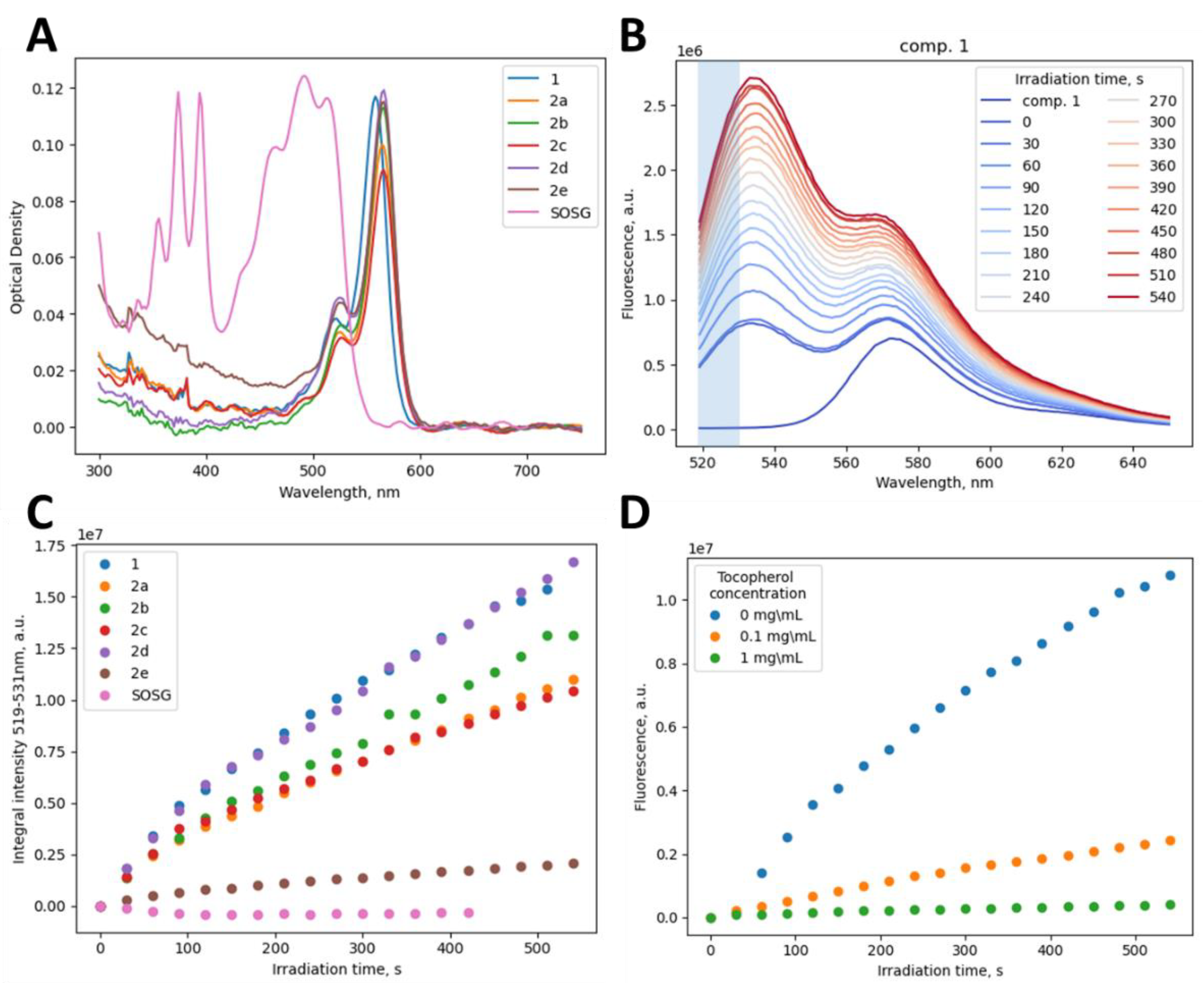
First, the ability of RB 1 and its derivatives 2ae to generate 1O2 was evaluated (Figure 1). The absorption spectra of the compounds and SOSG in methanol in the visible spectral range are shown in Figure 1A. The optical density of all samples was close to 0.12, they had the same spectral shape with the maximum at approximately 565 nm. They had non-zero optical density at 520 nm; therefore, their irradiation at 520 nm should lead to 1O2 generation. SOSG can also be a photosensitizer, so we first checked whether the dye could generate a significant amount of 1O2 under experimental conditions. The fluorescence intensity of SOSG did not change during irradiation, suggesting the increase in the dye’s fluorescence was associated solely with 1O2 generation by RB 1 and its derivatives (Figure S1).
Samples were irradiated at 520 nm for the specified lengths of time, and then fluorescence spectra were recorded at the 504 nm excitation wavelength (Figure 1B). The amount of generated 1O2 can be estimated from integral intensity in the 519–531 nm range, where only the signal from SOSG is detected, and the intrinsic fluorescence of RB and its derivatives is close to 0.
Integral fluorescence as a function of 1O2 generation time is shown in Figure 1C. By approximating the data with an exponential function (Equation (1)), it is possible to compare RB and its derivatives in terms of 1O2 generation rate (k) and the amount of 1O2 generated (amplitude, A) (Table 2). The 1O2 generation rate is approximately the same for all compounds, but the amount of generated 1O2 (directly proportional to the amplitude A) is slightly different for all compounds except 2e. For derivative 2e, it was minimal, while the concentration (Cgen) was the highest, suggesting that 2e is the least effective photosensitizer (Table 2). These results are consistent with 2e having the lowest antiviral activity in the series (Table 1). Taking into account the equal ability to generate 1O2, the hydrophobicity of the tether might be the main parameter influencing the antiviral activity against SARS-CoV-2 and CHIKV. For HIV and cells, alkyl length has practically no effect on activity and cytotoxicity, respectively (Table 1).
I 519 530   nm = C A × e k t
The effect of (±)-α-tocopherol, a well-known antioxidant [48], on the photosensitization ability of 2a, the derivative with the optimum combined activity against SARS-CoV-2 and CHIKV, was also studied. 1O2 generation was determined the same way as in previous experiments. The results are shown in Figure 1D. The presence of (±)-α-tocopherol reduced the efficiency of 1O2 generation, reaching a 2 orders of magnitude reduction for the tocopherol concentration of 1 mg/mL. Thus, the adverse effects of RB derivatives on host cells can likely be mitigated by the presence of antioxidants within organisms [49].
The next factor that could affect the observed activity of the compounds is their solubility in aqueous media used to assess antiviral activity and cytotoxicity. Solubility in PBS containing 1% DMSO was evaluated spectrophotometrically (Figure S2). Similarly to RB 1, derivatives 2a and 2b exist in a mixture of monomeric (λmax 549 nm) and dimeric (λmax 515 nm) forms [50], with the proportion of dimers increasing with higher alkyl chain lengths. For this reason, we made a rough estimate the solubility based on absorption intensity for the monomeric form at 549 nm, judging that the dimeric form is not a photosensitizer [50] and likely does not contribute to 1O2-mediated antiviral activity. The saturation concentration (Csat) for RB 1 and 2ab was significantly higher than EC50 for SARS-CoV-2 and CHIKV and comparable to EC50 for HIV (Table 1). Derivatives 2ce seem to form higher aggregates, which lead to the lowest excited singlet state energy level being split and two absorption maxima appearing at higher (λmax 568 nm) and lower (λmax 532 nm) wavelengths (549 nm) [51]. The formation of dimers/aggregates does not allow estimating the solubility of 2ce, but the area under curve (AUC) for 2e is higher than for 2cd and even 2b, which did not form any supramolecular assemblies. Thus, although 2e appears to have higher solubility than derivatives 2bd with shorter alkyl substituents, its amount of generated 1O2 being the lowest in the series is responsible for its lower antiviral activity and cell cytotoxicity (Table 1 and Table 2). To conclude this part, while RB 1 and its congeners 2ab with shorter (C4 and C6) alkyl substituents have Csat higher than or comparable with EC50, low solubility and aggregate formation have likely affected the observed antiviral activity of 2de, with longer (C10 and C12) alkyl groups.
To conclude, the effect of the introduction of fatty alkyl (C4–C12) substituents at the carboxy functionality of Rose Bengal on cell cytotoxicity and activity against a number of enveloped viruses was studied. Photophysical studies and the lack of activity against non-enveloped viruses indirectly support the mechanism of action associated with the impact of generated 1O2 on viral membranes. A reduction in aqueous solubility and aggregate formation with increasing alkyl group length may affect antiviral activity. The observed submicromolar-to-subnanomolar activity against SARS-CoV-2 and CHIKV and the extremely high selectivity index for alkyl-modified RB derivatives opens up an opportunity to develop broad-spectrum viral inactivants for vaccine preparations.

Supplementary Materials

The following supporting information can be downloaded at: https://www.mdpi.com/article/10.3390/biom12111609/s1, Table S1: Antiviral activity of RB and 2a–e against non-enveloped viruses and cytotoxicity for RD cells; Figure S1: Fluorescence spectra of SOSG during LED irradiation (520 nm); Figure S2: Absorption spectra of RB 1 (an additional 10-fold dilution) and its derivatives 2a–e in PBS containing 1% DMSO, used for solubility evaluation; Figure S3: NMR spectra.

Author Contributions

Conceptualization, A.V.A.; methodology, A.A.R., E.A.S., L.I.K. and A.V.A.; investigation, A.A.R., P.N.K., E.Y.S., E.S.K., E.V.Y., E.V.K., M.O.B., E.S.B. and A.A.K.; data curation, A.A.R., L.I.K. and A.V.A.; visualization, V.A.A.; writing—original draft preparation, A.A.R., V.A.A., V.A.K., L.I.K. and A.V.A.; writing—review and editing, V.A.A., E.A.S., L.I.K. and A.V.A.; supervision, A.V.A.; project administration, A.V.A.; funding acquisition, A.V.A. All authors have read and agreed to the published version of the manuscript.

Funding

This work was funded by a grant from the Ministry of Science and Higher Education of Russian Federation (agreement No. 075-15-2021-1049 to AVA).

Institutional Review Board Statement

Not applicable.

Informed Consent Statement

Not applicable.

Data Availability Statement

Not applicable.

Conflicts of Interest

The authors declare no conflict of interest.

References

  1. Nguyen, T.H.T.; Guedj, J.; Anglaret, X.; Laouénan, C.; Madelain, V.; Taburet, A.-M.; Baize, S.; Sissoko, D.; Pastorino, B.; Rodallec, A.; et al. Favipiravir pharmacokinetics in Ebola-Infected patients of the JIKI trial reveals concentrations lower than targeted. PLoS Negl. Trop. Dis. 2017, 11, e0005389. [Google Scholar] [CrossRef] [PubMed] [Green Version]
  2. Agrawal, U.; Raju, R.; Udwadia, Z.F. Favipiravir: A new and emerging antiviral option in COVID-19. Med. J. Armed Forces India 2020, 76, 370–376. [Google Scholar] [CrossRef] [PubMed]
  3. Müller, W.E.; Maidhof, A.; Taschner, H.; Zahn, R.K. Virazole (1-β-d-ribofuranosyl-1,2,4-triazole-3-carboxamide; A cytostatic agent. Biochem. Pharmacol. 1977, 26, 1071–1075. [Google Scholar] [CrossRef]
  4. Jorgensen, S.C.; Kebriaei, R.; Dresser, L.D. Remdesivir: Review of Pharmacology, Pre-clinical Data, and Emerging Clinical Experience for COVID-19. Pharmacother. J. Hum. Pharmacol. Drug Ther. 2020, 40, 659–671. [Google Scholar] [CrossRef]
  5. Johnson, E.M.; Doyle, J.D.; Wetzel, J.D.; McClung, R.P.; Katunuma, N.; Chappell, J.D.; Washington, M.K.; Dermody, T.S. Genetic and Pharmacologic Alteration of Cathepsin Expression Influences Reovirus Pathogenesis. J. Virol. 2009, 83, 9630–9640. [Google Scholar] [CrossRef] [Green Version]
  6. Satoh, S.; Mori, K.; Onomura, D.; Ueda, Y.; Dansako, H.; Honda, M.; Kaneko, S.; Ikeda, M.; Kato, N. Ribavirin suppresses hepatic lipogenesis through inosine monophosphate dehydrogenase inhibition: Involvement of adenosine monophosphate-activated protein kinase-related kinases and retinoid X receptor α. Hepatol. Commun. 2017, 1, 550–563. [Google Scholar] [CrossRef]
  7. Furuta, Y.; Takahashi, K.; Shiraki, K.; Sakamoto, K.; Smee, D.F.; Barnard, D.L.; Gowen, B.B.; Julander, J.G.; Morrey, J.D. T-705 (favipiravir) and related compounds: Novel broad-spectrum inhibitors of RNA viral infections. Antivir. Res. 2009, 82, 95–102. [Google Scholar] [CrossRef]
  8. Te, H.S.; Randall, G.; Jensen, D.M. Mechanism of action of ribavirin in the treatment of chronic hepatitis C. Gastroenterol. Hepatol. 2007, 3, 218–225. [Google Scholar]
  9. Mariewskaya, K.; Tyurin, A.; Chistov, A.; Korshun, V.; Alferova, V.; Ustinov, A. Photosensitizing Antivirals. Molecules 2021, 26, 3971. [Google Scholar] [CrossRef]
  10. Kunstek, H.; Vreken, F.; Keita, A.; Hamblin, M.R.; Dumarçay, F.; Varbanov, M. Aspects of Antiviral Strategies Based on Different Phototherapy Approaches: Hit by the Light. Pharmaceuticals 2022, 15, 858. [Google Scholar] [CrossRef]
  11. Proskurin, G.V.; Orlov, A.A.; Brylev, V.; Kozlovskaya, L.I.; Chistov, A.A.; Karganova, G.G.; Palyulin, V.A.; Osolodkin, D.I.; Korshun, V.A.; Aralov, A.V. 3′-O-Substituted 5-(perylen-3-ylethynyl)-2′-deoxyuridines as tick-borne encephalitis virus reproduction inhibitors. Eur. J. Med. Chem. 2018, 155, 77–83. [Google Scholar] [CrossRef] [PubMed]
  12. Colpitts, C.C.; Ustinov, A.V.; Epand, R.F.; Epand, R.M.; Korshun, V.A.; Schang, L.M. 5-(Perylen-3-yl)Ethynyl-arabino-Uridine (aUY11), an Arabino-Based Rigid Amphipathic Fusion Inhibitor, Targets Virion Envelope Lipids To Inhibit Fusion of Influenza Virus, Hepatitis C Virus, and Other Enveloped Viruses. J. Virol. 2013, 87, 3640–3654. [Google Scholar] [CrossRef] [PubMed] [Green Version]
  13. Aralov, A.V.; Proskurin, G.V.; Orlov, A.A.; Kozlovskaya, L.I.; Chistov, A.A.; Kutyakov, S.V.; Karganova, G.G.; Palyulin, V.A.; Osolodkin, D.I.; Korshun, V.A. Perylenyltriazoles inhibit reproduction of enveloped viruses. Eur. J. Med. Chem. 2017, 138, 293–299. [Google Scholar] [CrossRef] [PubMed]
  14. St.Vincent, M.R.; Colpitts, C.C.; Ustinov, A.V.; Muqadas, M.; Joyce, M.A.; Barsby, N.L.; Epand, R.F.; Epand, R.M.; Khramyshev, S.A.; Valueva, O.A.; et al. Rigid amphipathic fusion inhibitors, small molecule antiviral compounds against enveloped viruses. Proc. Natl. Acad. Sci. USA 2010, 107, 17339–17344. [Google Scholar] [CrossRef] [PubMed] [Green Version]
  15. Slesarchuk, N.A.; Khvatov, E.V.; Chistov, A.A.; Proskurin, G.V.; Nikitin, T.D.; Lazarevich, A.I.; Ulanovskaya, A.A.; Ulashchik, E.A.; Orlov, A.; Jegorov, A.V.; et al. Simplistic perylene-related compounds as inhibitors of tick-borne encephalitis virus reproduction. Bioorganic Med. Chem. Lett. 2020, 30, 127100. [Google Scholar] [CrossRef] [PubMed]
  16. Wolf, M.C.; Freiberg, A.N.; Zhang, T.; Akyol-Ataman, Z.; Grock, A.; Hong, P.W.; Li, J.; Watson, N.F.; Fang, A.Q.; Aguilar, H.C.; et al. A broad-spectrum antiviral targeting entry of enveloped viruses. Proc. Natl. Acad. Sci. USA 2010, 107, 3157–3162. [Google Scholar] [CrossRef] [PubMed] [Green Version]
  17. Vigant, F.; Lee, J.; Hollmann, A.; Tanner, L.B.; Ataman, Z.A.; Yun, T.; Shui, G.; Aguilar, H.C.; Zhang, D.; Meriwether, D.; et al. A Mechanistic Paradigm for Broad-Spectrum Antivirals that Target Virus-Cell Fusion. PLoS Pathog. 2013, 9, e1003297. [Google Scholar] [CrossRef]
  18. Vigant, F.; Hollmann, A.; Lee, J.; Santos, N.C.; Jung, M.E.; Lee, B. The Rigid Amphipathic Fusion Inhibitor dUY11 Acts through Photosensitization of Viruses. J. Virol. 2014, 88, 1849–1853. [Google Scholar] [CrossRef] [PubMed] [Green Version]
  19. Zeng, L.; Wang, M.-D.; Ming, S.-L.; Li, G.-L.; Yu, P.-W.; Qi, Y.-L.; Jiang, D.-W.; Yang, G.-Y.; Wang, J.; Chu, B.-B. An effective inactivant based on singlet oxygen-mediated lipid oxidation implicates a new paradigm for broad-spectrum antivirals. Redox Biol. 2020, 36, 101601. [Google Scholar] [CrossRef]
  20. Moghaddam, A.; Olszewska, W.; Wang, B.; Tregoning, J.S.; Helson, R.; Sattentau, Q.J.; Openshaw, P.J.M. A potential molecular mechanism for hypersensitivity caused by formalin-inactivated vaccines. Nat. Med. 2006, 12, 905–907. [Google Scholar] [CrossRef]
  21. Blackburn, N.; Besselaar, T. A study of the effect of chemical inactivants on the epitopes of Rift Valley fever virus glycoproteins using monoclonal antibodies. J. Virol. Methods 1991, 33, 367–374. [Google Scholar] [CrossRef]
  22. Swenberg, J.A.; Moeller, B.C.; Lu, K.; Rager, J.E.; Fry, R.C.; Starr, T.B. Formaldehyde Carcinogenicity Research: 30 Years and Counting for Mode of Action, Epidemiology, and Cancer Risk Assessment. Toxicol. Pathol. 2012, 41, 181–189. [Google Scholar] [CrossRef] [PubMed] [Green Version]
  23. Bahnemann, H.G. Inactivation of viral antigens for vaccine preparation with particular reference to the application of binary ethylenimine. Vaccine 1990, 8, 299–303. [Google Scholar] [CrossRef]
  24. Roat, M.I.; Romanowski, E.; Araullo-Cruz, T.; Gordon, Y.J. The Antiviral Effects of Rose Bengal and Fluorescein. Arch. Ophthalmol. 1987, 105, 1415–1417. [Google Scholar] [CrossRef] [PubMed]
  25. Brooks, S.E.; Kaza, V.; Nakamura, T.; Trousdale, M.D. Photoinactivation of Herpes Simplex Virus by Rose Bengal and Fluorescein: In Vitro and in Vivo Studies. Cornea 1994, 13, 43–50. [Google Scholar] [CrossRef] [PubMed]
  26. Stevenson, N.R.; Lenard, J. Antiretroviral activities of hypericin and rose bengal: Photodynamic effects on Friend leukemia virus infection of mice. Antivir. Res. 1993, 21, 119–127. [Google Scholar] [CrossRef]
  27. Lenard, J.; Rabson, A.; Vanderoef, R. Photodynamic inactivation of infectivity of human immunodeficiency virus and other enveloped viruses using hypericin and rose bengal: Inhibition of fusion and syncytia formation. Proc. Natl. Acad. Sci. USA 1993, 90, 158–162. [Google Scholar] [CrossRef] [PubMed] [Green Version]
  28. Demartis, S.; Obinu, A.; Gavini, E.; Giunchedi, P.; Rassu, G. Nanotechnology-based rose Bengal: A broad-spectrum biomedical tool. Dye. Pigment. 2021, 188, 109236. [Google Scholar] [CrossRef]
  29. Ferreira, A.M.; Carmagnola, I.; Chiono, V.; Gentile, P.; Fracchia, L.; Ceresa, C.; Georgiev, G.; Ciardelli, G. Surface modification of poly(dimethylsiloxane) by two-step plasma treatment for further grafting with chitosan–Rose Bengal photosensitizer. Surf. Coat. Technol. 2013, 223, 92–97. [Google Scholar] [CrossRef]
  30. Tegos, G.P.; Hamblin, M.R. Phenothiazinium Antimicrobial Photosensitizers Are Substrates of Bacterial Multidrug Resistance Pumps. Antimicrob. Agents Chemother. 2006, 50, 196–203. [Google Scholar] [CrossRef] [Green Version]
  31. Chen, H.-J.; Zhou, X.-B.; Wang, A.-L.; Zheng, B.-Y.; Yeh, C.-K.; Huang, J.-D. Synthesis and biological characterization of novel rose bengal derivatives with improved amphiphilicity for sono-photodynamic therapy. Eur. J. Med. Chem. 2018, 145, 86–95. [Google Scholar] [CrossRef] [PubMed]
  32. Ludvíková, L.; Friš, P.; Heger, D.; Šebej, P.; Wirz, J.; Klán, P. Photochemistry of rose bengal in water and acetonitrile: A comprehensive kinetic analysis. Phys. Chem. Chem. Phys. 2016, 18, 16266–16273. [Google Scholar] [CrossRef] [PubMed]
  33. Kozlovskaya, L.I.; Volok, V.P.; Shtro, A.A.; Nikolaeva, Y.V.; Chistov, A.A.; Matyugina, E.S.; Belyaev, E.S.; Jegorov, A.V.; Snoeck, R.; Korshun, V.A.; et al. Phenoxazine nucleoside derivatives with a multiple activity against RNA and DNA viruses. Eur. J. Med. Chem. 2021, 220, 113467. [Google Scholar] [CrossRef] [PubMed]
  34. Sheahan, T.P.; Sims, A.C.; Zhou, S.; Graham, R.L.; Pruijssers, A.J.; Agostini, M.L.; Leist, S.R.; Schäfer, A.; Dinnon, K.H., 3rd; Stevens, L.J.; et al. An orally bioavailable broad-spectrum antiviral inhibits SARS-CoV-2 in human airway epithelial cell cultures and multiple coronaviruses in mice. Sci. Transl. Med. 2020, 12, eabb5883. [Google Scholar] [CrossRef] [Green Version]
  35. Kärber, G. Beitrag zur kollektiven Behandlung pharmakologischer Reihenversuche. Arch. Exp. Pathol. Pharmakol. 1931, 162, 480–483. [Google Scholar] [CrossRef]
  36. Ryazantsev, D.Y.; Myshkin, M.Y.; Alferova, V.A.; Tsvetkov, V.B.; Shustova, E.Y.; Kamzeeva, P.N.; Kovalets, P.V.; Zaitseva, E.R.; Baleeva, N.S.; Zatsepin, T.S.; et al. Probing GFP Chromophore Analogs as Anti-HIV Agents Targeting LTR-III G-Quadruplex. Biomolecules 2021, 11, 1409. [Google Scholar] [CrossRef]
  37. Zenchenko, A.A.; Oslovsky, V.E.; Varizhuk, I.V.; Karpova, E.V.; Osolodkin, D.I.; Kozlovskaya, L.I.; Ishmukhametov, A.A.; Drenichev, M.S. Cytotoxicity reduction by O-nicotinoylation of antiviral 6-benzylaminopurine ribonucleosides. Toxicol. Vitr. 2022, 82, 105355. [Google Scholar] [CrossRef]
  38. Jodlbauer, A.; Von Tappeiner, H. Die Beteiligung Des Sauerstoffs Bei Der Wirkung Fluorescierender Stoffe. Dtsch. Arch. Klin. Med. 1905, 82, 520–546. [Google Scholar]
  39. Lamberts, J.J.M.; Schumacher, D.R.; Neckers, D.C. Novel rose bengal derivatives: Synthesis and quantum yield studies. J. Am. Chem. Soc. 1984, 106, 5879–5883. [Google Scholar] [CrossRef]
  40. Sugita, N.; Kawabata, K.-I.; Sasaki, K.; Sakata, A.I.; Umemura, S.-I. Synthesis of Amphiphilic Derivatives of Rose Bengal and Their Tumor Accumulation. Bioconjugate Chem. 2007, 18, 866–873. [Google Scholar] [CrossRef]
  41. Pereira, P.C.D.S.; Costa, P.F.D.A.; Pellosi, D.S.; Calori, I.R.; Vilsinski, B.H.; Estevão, B.M.; Hioka, N.; Caetano, W. Photophysical properties and interaction studies of Rose Bengal derivatives with biomimetic systems based in micellar aqueous solutions. J. Mol. Liq. 2017, 230, 674–685. [Google Scholar] [CrossRef]
  42. Mitsuya, H.; Weinhold, K.J.; Furman, P.A.; Clair, M.H.S.; Lehrman, S.N.; Gallo, R.C.; Bolognesi, D.; Barry, D.W.; Broder, S. 3’-Azido-3’-deoxythymidine (BW A509U): An antiviral agent that inhibits the infectivity and cytopathic effect of human T-lymphotropic virus type III/lymphadenopathy-associated virus in vitro. Proc. Natl. Acad. Sci. USA 1985, 82, 7096–7100. [Google Scholar] [CrossRef] [PubMed]
  43. Fischl, M.A.; Richman, D.D.; Grieco, M.H.; Gottlieb, M.S.; Volberding, P.A.; Laskin, O.L.; Leedom, J.M.; Groopman, J.E.; Mildvan, D.; Schooley, R.T.; et al. The efficacy of azidothymidine (AZT) in the treatment of patients with AIDS and AIDS-related complex. A double-blind, placebo-controlled trial. N. Engl. J. Med. 1987, 317, 185–191. [Google Scholar] [CrossRef]
  44. Zhou, S.; Hill, C.S.; Sarkar, S.; Tse, L.V.; Woodburn, B.M.D.; Schinazi, R.F.; Sheahan, T.P.; Baric, R.S.; Heise, M.T.; Swanstrom, R. β-d-N4-hydroxycytidine Inhibits SARS-CoV-2 Through Lethal Mutagenesis But Is Also Mutagenic To Mammalian Cells. J. Infect. Dis. 2021, 224, 415–419. [Google Scholar] [CrossRef] [PubMed]
  45. Valneva Successfully Completes Pivotal Phase 3 Trial of Single-Shot Chikungunya Vaccine Candidate. Available online: https://valneva.com/press-release/valneva-successfully-completes-pivotal-phase-3-trial-of-single-shot-chikungunya-vaccine-candidate/ (accessed on 18 October 2022).
  46. Mao, Q.; Wang, Y.; Yao, X.; Bian, L.; Wu, X.; Xu, M.; Liang, Z. Coxsackievirus A16: Epidemiology, Diagnosis, and Vaccine. Hum. Vaccines Immunother. 2013, 10, 360–367. [Google Scholar] [CrossRef] [PubMed] [Green Version]
  47. Wells, A.I.; Coyne, C.B. Enteroviruses: A Gut-Wrenching Game of Entry, Detection, and Evasion. Viruses 2019, 11, 460. [Google Scholar] [CrossRef] [Green Version]
  48. Fatima, K.; Masood, N.; Luqman, S. Quenching of singlet oxygen by natural and synthetic antioxidants and assessment of electronic UV/Visible absorption spectra for alleviating or enhancing the efficacy of photodynamic therapy. Biomed. Res. Ther. 2016, 3, 8. [Google Scholar] [CrossRef]
  49. Stockert, J.C.; Herkovits, J. Photodynamic toxicity and its prevention by antioxidative agents in Bufo arenarum embryos. Toxicology 2003, 192, 211–218. [Google Scholar] [CrossRef]
  50. Mendes, B.; Kassumeh, S.; Aguirre-Soto, A.; Pei, Q.; Heyne, B.; Kochevar, I.E. Influence of Rose Bengal Dimerization on Photosensitization. Photochem. Photobiol. 2021, 97, 718–726. [Google Scholar] [CrossRef]
  51. Kasha, M. Energy Transfer Mechanisms and the Molecular Exciton Model for Molecular Aggregates. Radiat. Res. 1963, 20, 55. [Google Scholar] [CrossRef]
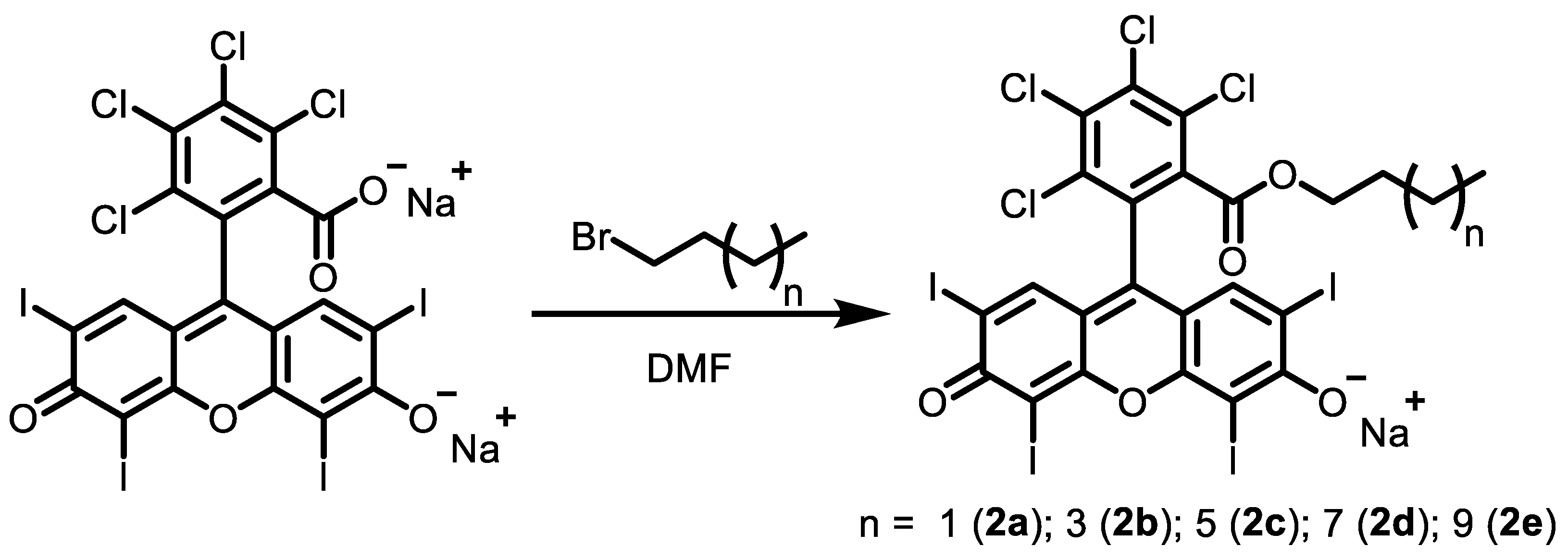
Scheme 1. Preparation of n-alkyl-substituted RB derivatives.
Biomolecules 12 01609 sch001
Figure 1. Steady-state optical properties of the investigated compounds: (A) Absorption spectra of non-irradiated samples; (B) typical fluorescence spectra of an irradiated sample containing RB 1 and SOSG (the integration area (519–530 nm) is highlighted with blue); (C) integral fluorescence intensity in the 519–530 nm spectral range as a function of photo-oxidation time; (D) integral fluorescence intensity in the 519–530 nm spectral range as a function of photo-oxidation time of SOSG and 2a in methanol with or without (±)-α-tocopherol.
Biomolecules 12 01609 g001
Table 1. Antiviral and cytotoxic properties of the studied compounds.
CmpdEC50, µM (M ± SD) aCC50, µM
(M ± SD) b
SI c
SARS-CoV-2CHIKVHIVVero
Cells
MT-4
Cells
SARS-CoV-2CHIKVHIV
10.5 ± 0.20.11 ± 0.0518 ± 743 ± 10>5091391>3
2a0.05 ± 0.020.004 ± 0.0026.3 ± 2.68 ± 115 ± 416918602.4
2b0.4 ± 0.30.0007 ± 0.00034.4 ± 1.860 ± 1515 ± 414690,9093.4
2c5.3 ± 1.30.0002 ± 0.00018.8 ± 3.753 ± 2542 ± 1010230,4354.8
2d6.6 ± 3.10.002 ± 0.0018.8 ± 2.660 ± 1542 ± 10928,5714.8
2e43 ± 100.038 ± 0.030>50>100>50>2>2632n/d
Positive
control
4.7 ± 2.9 d23.6 ± 10.2 d0.01 e>100>5>21>4>455
a Effective concentration required to inhibit virus-induced cytopathic effect by 50%. b Compound concentration required to induce a reduction of fluorescence by 50%. c Selectivity index (SI) is the ratio of CC50 for the appropriate cell line to EC50 for the studied virus. d NHC was used as a positive control. e AZT was used as a positive control.
Table 2. Photophysical properties and solubility of the studied compounds.
Cmpd1O2 GenerationSolubility
Cgen, μM ak, ×10−3 1/s bA, ×10−7 a.u.f cCsat, μM d
11.31.863.23>50.0
2a1.21.432.283.1
2b1.30.974.041.0
2c1.52.231.98ND
2d1.11.293.54ND
2e2.32.210.635ND
a Compound concentration used in 1O2 generation experiments. b 1O2 generation rate. c Amplitude is a measure of 1O2 generation efficiency proportional to the amount of 1O2 bound by SOSG. d Saturation concentration of RB and its derivatives in PBS containing 1% DMSO.
Publisher’s Note: MDPI stays neutral with regard to jurisdictional claims in published maps and institutional affiliations.