Article

The Juvenile-Hormone-Responsive Factor AmKr-h1 Regulates Caste Differentiation in Honey Bees

1
Honeybee Research Institute, Jiangxi Agricultural University, Nanchang 330045, China
2
Jiangxi Province Key Laboratory of Honeybee Biology and Beekeeping, Nanchang 330045, China
*
Author to whom correspondence should be addressed.
These authors contributed equally to this work.
Biomolecules 2023, 13(11), 1657; https://doi.org/10.3390/biom13111657
Submission received: 3 September 2023 / Revised: 23 October 2023 / Accepted: 28 October 2023 / Published: 17 November 2023
(This article belongs to the Section Molecular Biology)

Abstract

:
Honey bees are typical model organisms for the study of caste differentiation, and the juvenile hormone (JH) is a crucial link in the regulatory network of caste differentiation in honey bees. To investigate the mechanism of JH-mediated caste differentiation, we analyzed the effect of the JH response gene AmKr-h1 on this process. We observed that AmKr-h1 expression levels were significantly higher in queen larvae than in worker larvae at the 48 h, 84 h, and 120 h larval stages, and were regulated by JH. Inhibiting AmKr-h1 expression in honey bee larvae using RNAi could lead to the development of larvae toward workers. We also analyzed the transcriptome changes in honey bee larvae after AmKr-h1 RNAi and identified 191 differentially expressed genes (DEGs) and 682 differentially expressed alternative splicing events (DEASEs); of these, many were related to honey bee caste differentiation. Our results indicate that AmKr-h1 regulates caste differentiation in honey bees by acting as a JH-responsive gene.

1. Introduction

In honey bee colonies, the queen and worker are diploids, developing from fertilized eggs and, although they share the same genetic material, they differ significantly in morphological characteristics, behavior, and longevity [1]. Currently, nutrition [2], epigenetic modifications [3], and hormones [4] are considered to be important factors in regulating the differentiation of queen–worker castes. Of them, juvenile hormone (JH) is known to play a key role in this process [5].
JH is a pleiotropic hormone secreted by the insect corpora allata (CA) and released into the hemolymph [6]. It interacts antagonistically with 20-hydroxyecdysone (20E) to maintain larval traits in insects, where 20E induces molting and metamorphosis, and JH prevents the metamorphosis caused by 20E. During the last larva instar, the JH titer drops sharply, resulting in the pupation of completely metamorphosed insects and the emergence of incompletely metamorphosed insect adults [7,8,9,10]. In the honey bee, JH is a central regulator of the non-genetic diversity of social castes. Its physiological role in the honey bee is not only limited to maintaining the juvenile state but also has a significant ability to regulate growth and development, caste differentiation, and affects longevity [11,12]. Queen larvae have larger corpora allata and significantly higher JH titres in their hemolymph compared to worker larvae, especially at the critical point of caste differentiation, where the JH titre threshold determines the developmental trajectory of the larvae (development into a queen or worker). This suggests that caste differentiation is accompanied by strict regulation of JH synthesis and metabolism [5]. In addition, JH also regulates the synthesis of vitellogenin (Vg), which promotes ovarian development in honey bees, resulting in higher levels of Vg and more ovarian tubes in queens [13,14].
Krüppel homolog 1 (Kr-h1), a C2H2 zinc finger (Znf) transcription factor, is an early responder gene for JH that can be activated by the JH receptors Met and Gce in Drosophila [9]. It plays a crucial role in the JH signaling pathway by regulating the expression of its downstream target genes. In many insects, the JH response elements (JHREs) in the Kr-h1 promoter have been identified as a canonical E-box (CACGTG) or a C-box (CACGCG) [15,16,17], which are the typical binding elements for bHLH-PAS proteins and are also essential for JH-induced gene expression via Met [18,19]. The Kr-h1 is highly expressed in the larval stage in insects and decreases in the pupal stage, which is consistent with changes in JH titres in haemolymph [20,21,22]. The main function of Kr-h1 is to repress the metamorphosis of insects by transmitting JH signaling. It also regulates insects’ yolk production and egg maturation [23,24,25], but the regulatory effect of Kr-h1 on yolk production varies across species. For example, down-regulation of Kr-h1 in Locusta migratoria reduced the mRNA expression level of Vg by an average of 95%, which significantly inhibited oocyte maturation and ovarian development [26]. However, in Tribolium castaneum the low expression of Kr-h1 could only down-regulate Vg by about 30% [27]. In addition, Kr-h1 is also involved in the development of the nervous system of Drosophila larvae [28] and foraging behavior of honey bees [29].
In the JH signaling pathway, Kr-h1 is downstream of the JH receptors Met and Gce and can be up-regulated by JH [30,31]. Due to the antagonistic effect of JH and 20E, AmKr-h1 can inhibit 20E-induced metamorphosis by transducing JH signaling to suppress the expression of the major 20E response genes during the larval stage when JH titres are high [7,32]. At the terminal instar of insect larvae, the JH titre decreases dramatically and low-expressing AmKr-h1 is unable to override high 20E signaling, thus, 20E-induced metamorphosis occurs [9].
Since AmKr-h1 is regulated by JH, we speculate that AmKr-h1 may be the key regulator in the queen–worker caste differentiation. Therefore, in the present study, we analyzed the function of AmKr-h1 in the caste differentiation in honey bees using RNAi, and analyzed transcriptome changes in the bee larvae after AmKr-h1 RNAi.

2. Materials and Methods

2.1. Insects

The honey bee colonies (Apis mellifera ligustica) used in this study were bred at the Honeybee Research Institute of Jiangxi Agricultural University (28.46 N, 115.49 E), Nanchang, Jiangxi, China.

2.2. Sequence Analysis of AmKr-h1 Gene

The exon–intron structure was analyzed by mapping the mRNA sequence (GenBank accession no.: NM_001242470.1, NM_001011566.1) of the AmKr-h1 to the A. mellifera genome at the NCBI website from the A. mellifera AmKr-h1 gene deposited in the GenBank database (https://www.ncbi.nlm.nih.gov/genbank/, accessed on 20 March 2022). The structural domains of the AmKr-h1 protein were analyzed at the SMART online website (http://smart.embl-heidelberg.de/, accessed on 20 March 2022). Multiple sequence alignments of amino acid sequences downloaded from NCBI were performed using the CLUSTALX v1.81 software [33].

2.3. Expression Analysis Using qRT-PCR

A queen was restricted on an empty comb to lay eggs for six hours. After the eggs hatched into larvae, some of the larvae were transferred to queen cells, while the remaining larvae continued to be bred in worker cells. Six biological replicates of both queen larvae and worker larvae were sampled at 48 h, 84 h, and 120 h after hatching of the eggs for qRT-PCR analysis. During sampling, the larvae were washed three times with double-distilled water to remove any royal jelly, and then were stored in liquid nitrogen for further use.
Total RNA was extracted from the collected samples using the TrizolUp Kit (Transgen, ER501-01, Beijing, China), and the RNA concentration of each sample was measured using a spectrophotometer. A total of 1 µg of RNA was taken from each sample and reverse transcribed into cDNA using M-MLV reverse transcriptase (TakaRa, RR047A, Dalian, China). The Gapdh (GenBank accession no.: NM_001014994.1) gene was used as the internal reference.
The qRT-PCR primer sequences of each gene were designed using the Primer Premier 5.0 software based on their mRNA sequences (Table S1). The qRT-PCR reaction system was as follows: 5.0 µL of SYBR GREEN, 0.2 µL of ROX correction fluid, 0.4 µL of forward primer (0.0125 nmol/μL), 0.4 µL of reverse primer (0.0125 nmol/μL), 1.0 µL of cDNA (150 μg/μL), and 3.0 µL of ddH2O. The qRT-PCR amplification conditions were as follows: 95 °C for 10 min; followed by 40 cycles of 95 °C for 15 s, Tm for 1 min. Four technical replicates were set up for each biological replicate. The data were analyzed using the 2−ΔΔCT method [34] and t-tests were performed using the SPSS 26.0 software to analyze the differences in gene expression.

2.4. JH Treatment

The queen of an A. mellifera colony was controlled on an empty comb to lay eggs for 6 h. When the eggs hatched, the larvae were transferred to a 24-well culture plate containing 300 µL of artificially prepared food (6% fructose, 6% glucose, 1% yeast, 37% distilled water, and 50% royal jelly) per well. The larvae were randomly divided into three groups: to the first group (JH), 1 µL of juvenile hormone III (JH III) (APExBIO, purity ≥ 65.00%) solution diluted with ethanol (10 µg/µL) was dripped on the back; the second group (ET) received 1 µL of ethanol on the back as a control; the third group (NO) was normal larvae that received no treatment. Then, the larvae were moved to an incubator at 34 °C and 85% humidity. For the next 2 days, the JH group and ethanol group continued to be treated with JH III or ethanol three times per day. Meanwhile, each group received 150 µL of artificial foods twice daily.
Twenty-four hours after treatment completion, the larvae in each group were collected for mRNA expression analysis using qRT-PCR. For each treatment group, seven biological replicates with two larvae per replicate were sampled. The samples were washed three times with double-distilled water and immediately snap-frozen in liquid nitrogen, and then stored in liquid nitrogen for RNA extraction. The qRT-PCR protocol detailed in Section 2.3 was followed. The differences in the expression of AmKr-h1 between the JH and two control groups were analyzed by ANOVA using SPSS 26.0.

2.5. RNAi Treatment

The siRNA sequences of AmKr-h1 and the control (Con) were as follows: AmKr-h1, sense: GGUACAUACGCGUACGCAUTT, antisense: AUGCGUACGCGUAUGUACCTT; Con, sense: UUCUCCGAACGUGUCACGUTT, antisense: ACGUGACACGUUCGGAGAATT. The siRNA was synthesized by Genepharma (Shanghai, China).
Once again, a queen of an A. mellifera colony was limited to an empty comb to lay eggs for 6 h. After hatching of the eggs, the one-day-old larvae were transferred to a 24-well culture plate containing 300 µL of artificially prepared food in each well. The plates were kept in an incubator at 34 °C and 85% humidity. Then, the larvae were fed twice per day with 150 μL of artificial food. When they reached 60 h, the larvae were randomly divided into two groups and injected with the AmKr-h1 siRNA solution (solubilizing siRNA with RNase-free water; the concentration was 2 μg/μL) and the NT siRNA solution (solubilizing Con with RNase-free water; the concentration was 2 μg/μL), respectively. The NT group was set as the control group. Each larva was injected with 400 ng of siRNA and then provided with 300 µL of artificial food and kept in an incubator (34 °C, 85% humidity). When the larvae were 3 days old, they were transferred to a new plate and fed twice per day with 200 µL of artificial food until pupation.
At 48 h after manual injection of siRNA, ten surviving larvae were selected and set as five biological replicates (each containing two larvae) for detecting the efficiency of RNAi by qRT-PCR. Total RNA extraction, cDNA synthesis, and qRT-PCR were performed as described above. The remaining larvae were kept in the incubator until emergence. The morphological traits of 35 newly emerged bees from the Con group and 35 bees from the AmKr-h1 RNAi group were measured as follows. Firstly, bees were frozen to death on ice. Then, the body weight of each bee in each group was weighed with an electronic balance, and the length of the body, proboscis, and forewing of each bee was measured by a stereomicroscope (Guiguang, GL-99TI, Guilin, China) with the tiny creature morphological measurement and data analysis system of the Optec OPTPro software (cnoptec, version: x86, Chongqing, China). All data were analyzed by t-test in SPSS 26.0.

2.6. cDNA Library Construction and Sequencing

Total RNA was extracted from both the RNAi group and Con group samples (each sample containing two larvae). After measuring the quality and concentration of the RNA samples, the mRNA molecules were enriched using Oligo (dT) magnetic beads, and then fragmentation buffer was added to break them randomly into small fragments. Using these small fragments as templates, double-stranded cDNA was synthesized by reverse transcriptase. Then, cDNA fragments were purified, end-repaired, and poly(A) tails and Illumina sequencing adaptors were added. The cDNA fragments were isolated using AMPure XPbeads and were PCR-amplified, the PCR products were purified again to build a cDNA sequencing library. Finally, the cDNA libraries were sequenced using the Illumina high-throughput sequencing platform.
The raw sequences (raw reads) obtained from sequencing were filtered with the fastp software by removing adaptors, N (no base information could be determined) ratio > 10%, low-quality (the number of bases with quality value Q ≤ 10 accounted for more than 50% of the whole read), and trimmed sequences < 500 bp in length, to obtain clean sequences (clean reads). Clean reads were compared with the A. mellifera reference genome sequences (https://ftp.ncbi.nlm.nih.gov/genomes/all/GCF/003/254/395/GCF_003254395.2_Amel_HAv3.1/, accessed on 19 January 2022).

2.7. Screening of DEGs and DEASEs

The expression level of each gene was calculated by counting the number of reads matched to each gene with the Cufflinks v2.2.1 software. Differentially expressed genes were screened by DESeq using ∣log2(fold change)∣ > 2 and p-value < 0.05 as the criteria. Then, a GO and KEGG enrichment analysis was performed with the DEGs. Differentially expressed alternative splicing events were analyzed using the rMATS v4.1.2 software with FDR < 0.05 as a screening criterion. The reliability of the RNA-seq results of nine DEGs was verified using qRT-PCR, as mentioned above.

3. Results

3.1. AmKr-h1 Encodes a Zinc Finger Protein

The mRNA sequence of A. mellifera AmKr-h1 is 1756 bp long, containing three exons and encoding a protein of 500 aa, containing eight C2H2 zinc finger (ZnF_C2H2) domains (Figure 1A). The AmKr-h1 protein is highly conserved in the ZnF_C2H2 domain region compared with those of Aedes aegypti, T. castaneum, Drosophila melanogaster, and Bombyx mori (Figure 1B).

3.2. AmKr-h1 Has Higher Expression in Queen Larvae and Is Regulated by JH

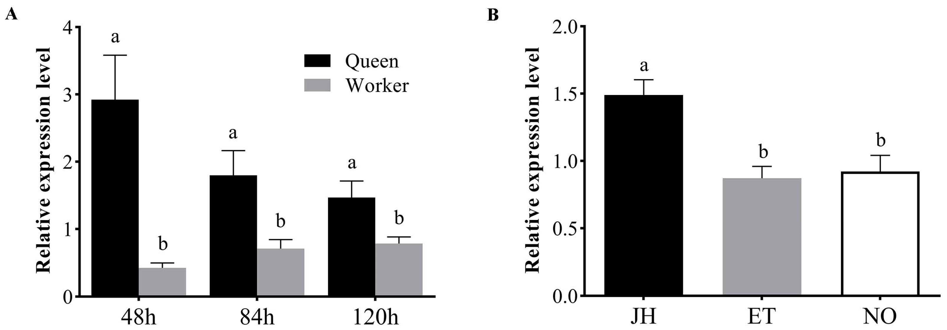
The relative mRNA expression levels of AmKr-h1 between queen larvae and worker larvae at 48 h, 84 h, and 120 h after the hatching of eggs were measured using quantitative real-time polymerase chain reaction (qRT-PCR). The results showed that the expression levels of the AmKr-h1 gene in the queen larvae were significantly higher than in the worker bee larvae at all the three time points (t-test, p < 0.05) (Figure 2A).
We investigated the effect of JH on the expression of AmKr-h1. After treatment of the larvae with JH, the expression level of AmKr-h1 in the JH group was significantly higher than that in the ethanol group and the untreated group (ANOVA, p < 0.05), and there was no significant difference between the ethanol group and the no treatment group (ANOVA, p > 0.05) (Figure 2B).

3.3. AmKr-h1 Affects Castes Differentiation of Queen–Worker

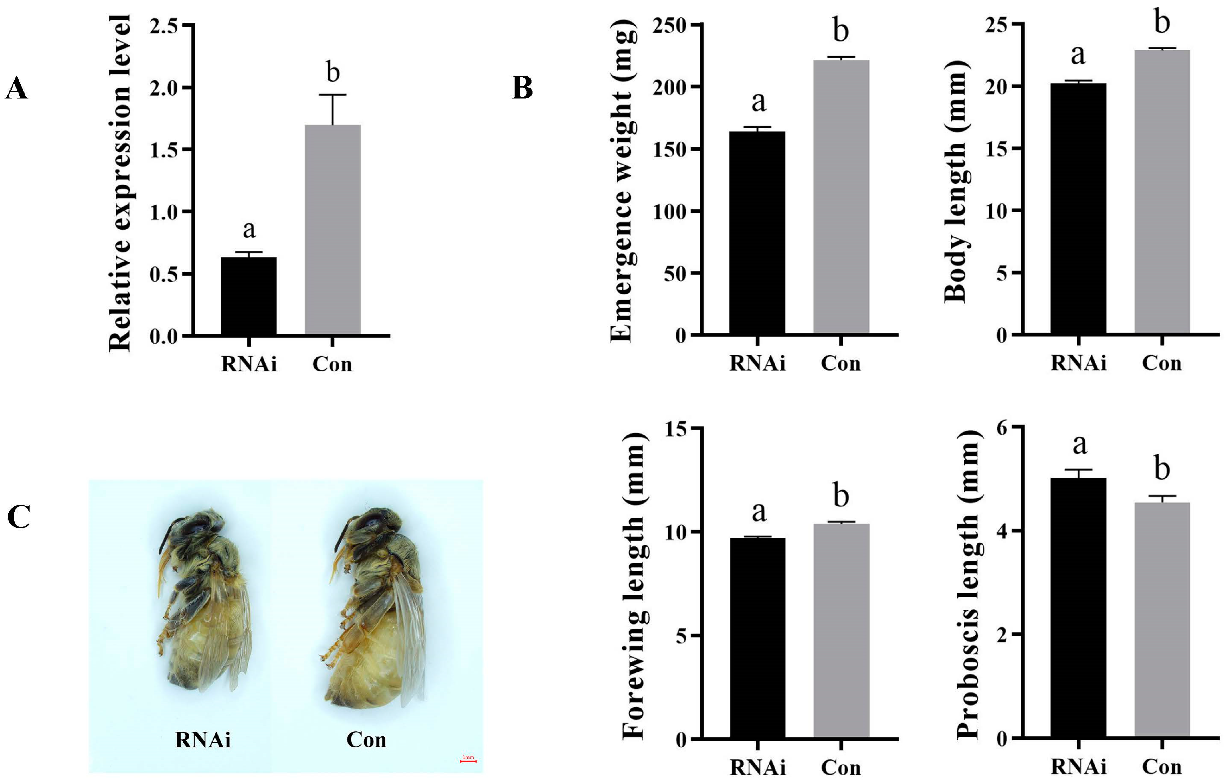
The expression changes in AmKr-h1 after RNAi were analyzed using qRT-PCR, and the results showed that the expression levels of AmKr-h1 in the RNAi group were significantly lower than in the Con group (t-test, p < 0.05) (Figure 3A). This indicated that our RNAi experiment was effective.
The morphological indexes of the newly emerged adult bees in the RNAi and the Con groups were measured. After knocking down the expression of AmKr-h1, the emergence weight, body length, and forewing length of the newly emerged bees in the RNAi group decreased significantly compared with the Con group (t-test, p < 0.05), while the proboscis length was significantly longer than in the Con group (t-test, p < 0.05) (Figure 3B). The bees in the RNAi group were significantly smaller than those in the Con group (Figure 3C). This indicates that reduced AmKr-h1 expression can lead to the differentiation of larvae into workers.

3.4. Summary of Transcriptome Sequencing Data

The number of raw reads obtained after sequencing for each biological replicate in the RNAi and Con groups ranged from 45,302,028 to 62,997,770, and the number of clean reads remaining after filtering low-quality reads and ambiguous reads ranged from 43,925,842 to 60,493,146. The percentages of Q20 and Q30 bases in each sample were higher than 97.16% and 92.36%, respectively. The GC content of clean reads in each sample was between 40.66 and 41.41%. The number of reads matched to the honey bee reference genome ranged from 41,673,370 to 57,479,542, with the proportion ranging from 89.45% to 95.28%, and the uniquely matched reads ranged from 40,781,360 to 56,370,089, with the proportion ranging from 87.76% to 93.83% (Table S2).
The correlation coefficients between biological replicates in the RNAi and Con groups were from 0.9388 to 0.9972, indicating high reliability between biological replicates (Table S3).

3.5. DEGs between the RNAi and Con Groups

A total of 191 DEGs were identified between the RNAi and Con groups. Among them, 85 genes were up-regulated and 106 genes were down-regulated in the RNAi group compared with the Con group (Figure 4A, Table S4).
The KEGG pathway enrichment analysis of the DEGs showed that 16 KEGG pathways were significantly enriched (p-value < 0.05), including “longevity-regulating pathway—multiple species”, “biotin metabolism”, “vitamin digestion and absorption”, “linoleic acid metabolism”, “terpenoid backbone biosynthesis”, “alpha-linolenic acid metabolism”, “protein processing in endoplasmic reticulum”, “steroid hormone biosynthesis”, “arachidonic acid metabolism”, “ether lipid metabolism”, “lysine biosynthesis”, “influenza A”, “glycerophospholipid metabolism”, “AMPK signaling pathway”, “tryptophan metabolism”, and “biosynthesis of unsaturated fatty acids”. Among these, the “longevity-regulating pathway—multiple species”, “steroid hormone biosynthesis”, and “AMPK signaling pathway” were associated with honey bee caste differentiation (Figure 4B, Table S5), including a total of eleven DEGs (Figure 4C), and the other pathways were mainly related to metabolism.
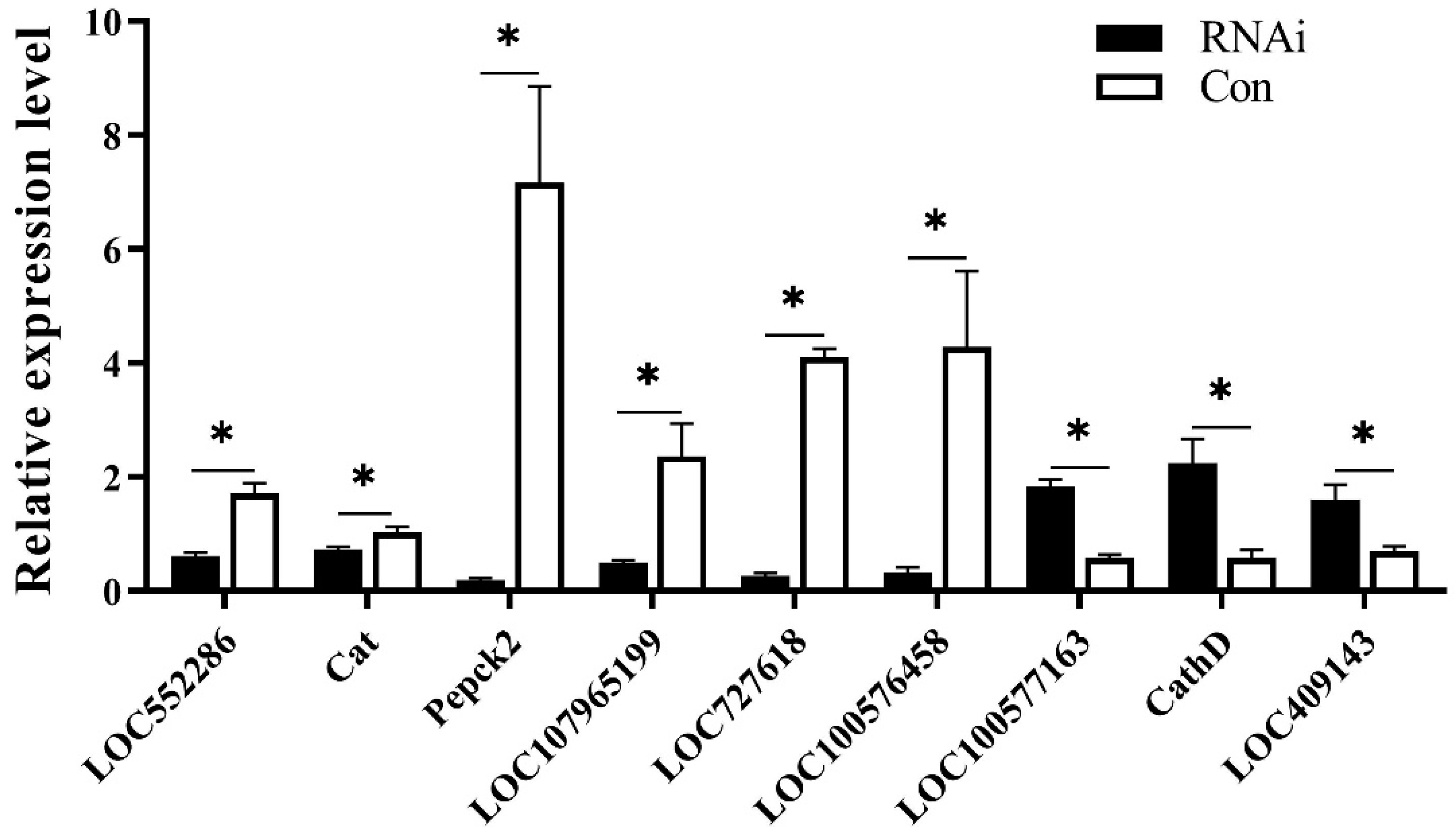
Nine DEGs were selected for verifying the reliability of the RNA-seq results using qRT-PCR, including LOC552286, Catalase (Cat, LOC443552), Phosphoenolpyruvate carboxykinase 2 (Pepck 2, LOC412843), LOC107965199, LOC727618, LOC100576458, LOC100577163, Cathepsin D (CathD, LOC409341), and LOC409143. The results indicated that all of them were significantly differentially expressed between the RNAi group and the control group, while six of these genes were down-regulated and three genes up-regulated in the RNAi group. These results are consistent with the RNA-seq results, indicating that the transcriptome sequencing results are reliable (Figure 5).

3.6. DEASEs between the RNAi and Control Groups

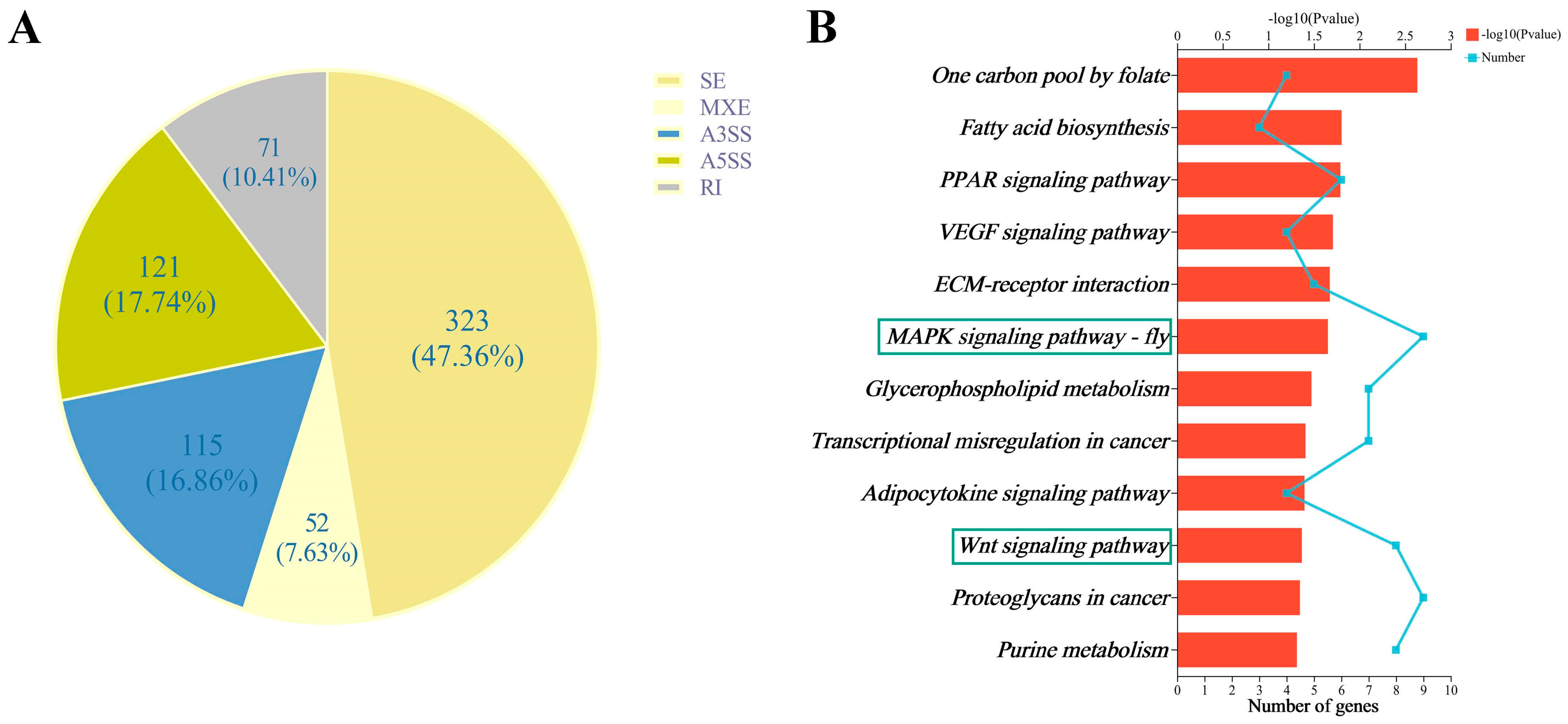
In this study, five alternative splicing forms between the RNAi and Con groups were analyzed, including skipped exon (SE), alternative 5′ splice site (A5SS), alternative 3′ splice site (A3SS), mutually exclusive exons (MXE), and retained intron (RI), and a total of 42,174 and 44,051 splicing forms were detected in the three biological replicates from the RNAi and Con groups, respectively, of which skipped exon (SE) was the most splicing type. In addition, there were 682 DEASEs related to 495 genes between the RNAi and Con groups, including 323 SE, 121 A5SS, 115 A3SS, 52 MXE, and 71 RI (Figure 6A), related to 262, 113, 101, 41, and 49 genes, respectively (Figure 6A, Table S6). Of the genes related to these DEASEs, HR3 (LOC408586), Hex110 (LOC551648), and Br-C (LOC552255) were involved in honey bee caste differentiation.
The KEGG pathway analysis of the DEASE-related genes revealed that a total of 12 KEGG pathways were significantly enriched (p-value < 0.05), including “one carbon pool by folate”, “fatty acid biosynthesis”, “PPAR signaling pathway”, “VEGF signaling pathway”, “ECM-receptor interaction”, “MAPK signaling pathway—fly”, “glycerophospholipid metabolism”, “transcriptional misregulation in cancer”, “adipocytokine signaling pathway”, “Wnt signaling pathway”, “proteoglycans in cancer”, and “purine metabolism” (Figure 6B, Table S7). Among these, the “MAPK signaling pathway—fly” and the “Wnt signaling pathway” were associated with honey bee caste differentiation.

4. Discussion

In the present study, we found that RNAi of the AmKr-h1 gene resulted in the development of honey bee larvae into workers. In honey bees, JH is the principal factor influencing caste differentiation. AmKr-h1 is the response gene downstream of the JH receptors Met and Gce, which are early response genes to JH [30,31]. Data from our results suggest that JH regulates the caste differentiation in honey bees by acting on the AmKr-h1 gene.
We analyzed gene expression changes in honey bee larvae after AmKr-h1 gene knockdown and identified many differentially expressed genes associated with honey bee caste differentiation, including Cat (LOC443552), CathD (LOC409341), and Pepck2 (LOC412843).
The Cat gene encodes a catalase important to the cellular antioxidant system. It can guide the conversion of oxygen radicals in cells into hydrogen peroxide, with lower oxidative activity, and then be broken down, thus reducing the degree of cellular oxidation [35]. Studies have shown that Cat is significantly expressed at a higher level in queen larvae than in worker larvae, suggesting that the queen is more efficient in removing reactive oxygen species (ROS) and has a stronger antioxidant capacity than workers [36]. In our results, the expression of the Cat gene was down-regulated after AmKr-h1 RNAi, suggesting that AmKr-h1 may influence the antioxidant capacity of honey bees by regulating the Cat gene.
The CathD gene encodes a lysosomal aspartate protease. In insects, lysosome-mediated organelle degradation is particularly important in liposomal remodeling during the vitellogenesis cycle of female larvae [37,38]. In the fat bodies of insects with periodic egg maturation, lysosomes play a key role in terminating vitellogenesis by selectively degrading the secretory machinery involved in the mass production of yolk protein precursors [39]. During the termination of vitellogenesis in A. aegypti, the number of lysosomes in the fat body increases significantly [40]. In the present study, the expression of the CathD gene was up-regulated after RNAi of the AmKr-h1 gene, indicating that AmKr-h1 can suppress the expression of CathD, which leads to a decrease in aspartate protease activity and, thus, promotes honey bee ovarian development.
The Pepck2 encodes a phosphoenolpyruvate carboxykinase (PEPCK) that is closely associated with pyruvate homeostasis [41]. In Helicoverpa armigera, high levels of pyruvate promote growth and development, and low levels of pyruvate lead to reduced metabolic activity [42]. In the present study, the expression of Pepck2 was significantly down-regulated after RNAi of the AmKr-h1 gene, indicating that the AmKr-h1 gene may affect the growth and development of honey bees by regulating Pepck2.
Alternative splicing has a significant effect on caste differentiation [43]. Among the genes associated with DEASEs, we found three genes associated with honey bee caste differentiation: HR3, Hex110, and Br-C. The HR3 gene encodes for a 20E-induced transcription factor that acts as a developmental switch in 20E-regulated insect development and metamorphosis [44]. In A. aegypti, inhibition of HR3 gene expression by RNAi resulted in diminutive ovaries and a significantly reduced number of eggs [45]. Hexamerins are mainly involved in the dynamic changes in amino acid storage and utilization that occur during insect development and they can also function as JH-binding proteins [46,47,48]. In Reticulitermes flavipes, inhibition of hexamerins’ expression promotes the differentiation of larvae into workers [49]. The Hex110 gene is highly expressed in the ovaries of the queens and is associated with the reproductive function in honey bees [50]. Br-C, a 20E response gene, is essential for maintaining the ecdysteroid titer [51]. During larval–pupal metamorphosis, Ecdysone signaling induces programmed cell death by inducing Br-C gene expression, eliminating larval tissues, and promoting adult tissue formation [52]. RNAi of the Br-C gene in Drosophila larvae at 120 h leads to a delay in pupal development [53]. In the present study, the expression levels of the HR3 and Hex110 isoforms were down-regulated and the Br-C isoform was up-regulated after inhibiting AmKr-h1 expression, indicating that AmKr-h1 may influence the caste differentiation in honey bees by positively regulating the expression levels of the HR3 and Hex110 isoforms while negatively regulating the Br-C isoform.
Through a KEGG pathway enrichment analysis of DEGs and DEASE-related genes, we found that several important pathways related to caste differentiation in honey bees were significantly enriched, such as the longevity-regulating pathway—multiple species, AMPK signaling pathway, MAPK signaling pathway—fly, and Wnt signaling pathway. This suggests that the JH signaling pathway has a cross-talk with the Wnt signaling pathway. It suggests that AmKr-h1, upon receiving juvenile hormone signals, may induce differentiation of queen and worker bees by acting on these critical pathways, but further experimental verification is needed.

5. Conclusions

In summary, our results indicate that JH regulates caste differentiation in honey bees by acting on AmKr-h1. Moreover, we identified a large number of differentially expressed genes after AmKr-h1 knockdown, and future studies should focus on elucidating the specific pathways downstream of AmKr-h1 that affect phenotypic differentiation between honey bee castes.

Supplementary Materials

The following supporting information can be downloaded at: https://www.mdpi.com/article/10.3390/biom13111657/s1, Table S1: Specific primers used for quantitative real-time PCR; Table S2: Summary of the transcriptome sequencing data; Table S3: The correlation coefficients between biological replicates; Table S4: DEGs between the RNAi and Con groups; Table S5: The significantly enriched KEGG pathways of the DEGs; Table S6: DEASEs between the RNAi and Con groups; Table S7: The significantly enriched KEGG pathways of the DEASESs.

Author Contributions

Conceptualization, Z.-L.W.; methodology, Z.-X.G. and Z.-L.W.; software, Z.-X.G. and F.-P.C.; validation, Z.-X.G., F.-P.C. and J.-N.X.; formal analysis, Z.-X.G. and F.-P.C.; investigation, Z.-X.G., F.-P.C. and J.-N.X.; resources, W.-Y.Y.; data curation, Z.-X.G. and F.-P.C.; writing—original draft preparation, Z.-X.G.; writing—review and editing, Z.-L.W.; visualization, Z.-X.G. and F.-P.C.; supervision, Z.-L.W.; project administration, Z.-L.W.; funding acquisition, Z.-L.W. All authors have read and agreed to the published version of the manuscript.

Funding

This research was funded by the National Natural Science Foundation of China, grant number 32160134 and 31402147.

Institutional Review Board Statement

Not applicable.

Informed Consent Statement

Not applicable.

Data Availability Statement

The raw data of the RNA-seq are available from the NCBI SRA database, under accession number PRJNA996350.

Acknowledgments

The authors would like to thank ZhenXiu Zeng for helping manage the A. mellifera colony, which made the experiment much smoother.

Conflicts of Interest

The authors declare no conflict of interest. The funders had no role in the design of the study; in the collection, analyses, or interpretation of data; in the writing of the manuscript; or in the decision to publish the results.

References

  1. Barchuk, A.R.; Cristino, A.S.; Kucharski, R.; Costa, L.F.; Simões, Z.L.; Maleszka, R. Molecular determinants of caste differentiation in the highly eusocial honeybee Apis mellifera. BMC Dev. Biol. 2007, 7, 70. [Google Scholar] [CrossRef] [PubMed]
  2. Kamakura, M. Royalactin induces queen differentiation in honeybees. Nature 2011, 473, 478–483. [Google Scholar] [CrossRef] [PubMed]
  3. Kucharski, R.; Maleszka, J.; Foret, S.; Maleszka, R. Nutritional Control of Reproductive Status in Honeybees via DNA Methylation. Science 2008, 319, 1827–1830. [Google Scholar] [CrossRef] [PubMed]
  4. Shuel, R.W.; Dixon, S.E. The early establishment of dimorphism in the female honeybee, Apis mellifera L. Insect. Soc. 1960, 7, 265–282. [Google Scholar] [CrossRef]
  5. Hartfelder, K.; Engels, W. 2 Social Insect Polymorphism: Hormonal Regulation of Plasticity in Development and Reproduction in the Honeybee. In Current Topics in Developmental Biology; Pedersen, R.A., Schatten, G.P., Eds.; Academic Press: Cambridge, MA, USA, 1998; Volume 40, pp. 45–77. ISBN 0070-2153. [Google Scholar]
  6. Kotaki, T.; Shinada, T.; Kaihara, K.; Ohfune, Y.; Numata, H. Structure Determination of a New Juvenile Hormone from a Heteropteran Insect. Org. Lett. 2009, 11, 5234–5237. [Google Scholar] [CrossRef]
  7. Belles, X.; Santos, C.G. The MEKRE93 (Methoprene tolerant-Krüppel homolog 1-E93) pathway in the regulation of insect metamorphosis, and the homology of the pupal stage. Insect Biochem. Mol. Biol. 2014, 52, 60–68. [Google Scholar] [CrossRef]
  8. Jindra, M.; Bellés, X.; Shinoda, T. Molecular basis of juvenile hormone signaling. Curr. Opin. Insect Sci. 2015, 11, 39–46. [Google Scholar] [CrossRef]
  9. Li, K.; Jia, Q.Q.; Li, S. Juvenile hormone signaling—A mini review. Insect Sci. 2018, 26, 600–606. [Google Scholar] [CrossRef]
  10. Liu, S.; Li, K.; Gao, Y.; Liu, X.; Chen, W.; Ge, W.; Feng, Q.; Palli, S.R.; Li, S. Antagonistic actions of juvenile hormone and 20-hydroxyecdysone within the ring gland determine developmental transitions in Drosophila. Proc. Natl. Acad. Sci. USA 2018, 115, 139–144. [Google Scholar] [CrossRef]
  11. Jindra, M.; Palli, S.R.; Riddiford, L.M. The Juvenile Hormone Signaling Pathway in Insect Development. Annu. Rev. Entomol. 2013, 58, 181–204. [Google Scholar] [CrossRef]
  12. Roy, S.; Saha, T.T.; Zou, Z.; Raikhel, A.S. Regulatory Pathways Controlling Female Insect Reproduction. Annu. Rev. Entomol. 2018, 63, 489–511. [Google Scholar] [CrossRef] [PubMed]
  13. Wirtz, P.; Beetsma, J. Induction of caste differentiation in the honeybee (Apis mellifera) by juvenile hormone. Entomol. Exp. Appl. 1972, 15, 517–520. [Google Scholar] [CrossRef]
  14. Chen, S.; Lin, C.; Lu, K. cDNA isolation, expression, and hormonal regulation of yolk protein genes in the oriental fruit fly, Bactrocera dorsalis (Hendel) (Diptera: Tephritidae). J. Insect Physiol. 2012, 58, 763–770. [Google Scholar] [CrossRef] [PubMed]
  15. Kayukawa, T.; Tateishi, K.; Shinoda, T. Establishment of a versatile cell line for juvenile hormone signaling analysis in Tribolium castaneum. Sci. Rep. 2013, 3, 1570. [Google Scholar] [CrossRef]
  16. He, Q.; Wen, D.; Jia, Q.; Cui, C.; Wang, J.; Palli, S.R.; Li, S. Heat Shock Protein 83 (Hsp83) Facilitates Methoprene-tolerant (Met) Nuclear Import to Modulate Juvenile Hormone Signaling. J. Biol. Chem. 2014, 289, 27874–27885. [Google Scholar] [CrossRef]
  17. Chen, X.; Hu, Y.; Zheng, H.; Cao, L.; Niu, D.; Yu, D.; Sun, Y.; Hu, S.; Hu, F. Transcriptome comparison between honey bee queen- and worker-destined larvae. Insect Biochem. Mol. Biol. 2012, 42, 665–673. [Google Scholar] [CrossRef]
  18. Li, M.; Liu, P.; Wiley, J.D.; Ojani, R.; Bevan, D.R.; Li, J.; Zhu, J. A steroid receptor coactivator acts as the DNA-binding partner of the methoprene-tolerant protein in regulating juvenile hormone response genes. Mol. Cell. Endocrinol. 2014, 394, 47–58. [Google Scholar] [CrossRef]
  19. Kewley, R.J.; Whitelaw, M.L.; Chapman-Smith, A. The mammalian basic helix–loop–helix/PAS family of transcriptional regulators. Int. J. Biochem. Cell Biol. 2004, 36, 189–204. [Google Scholar] [CrossRef]
  20. Martín, D.; Chafino, S.; Franch-Marro, X. How stage identity is established in insects: The role of the Metamorphic Gene Network. Curr. Opin. Insect Sci. 2021, 43, 29–38. [Google Scholar] [CrossRef]
  21. Truman, J.W. The Evolution of Insect Metamorphosis. Curr. Biol. 2019, 29, R1252–R1268. [Google Scholar] [CrossRef]
  22. Jindra, M. Where did the pupa come from? The timing of juvenile hormone signalling supports homology between stages of hemimetabolous and holometabolous insects. Philos. Trans. R. Soc. B-Biol. Sci. 2019, 374, 20190064. [Google Scholar] [CrossRef] [PubMed]
  23. Pecasse, F.; Beck, Y.; Ruiz, C.; Richards, G. Krüppel-homolog, a Stage-Specific Modulator of the Prepupal Ecdysone Response, Is Essential for Drosophila Metamorphosis. Dev. Biol. 2000, 221, 53–67. [Google Scholar] [CrossRef]
  24. Minakuchi, C.; Namiki, T.; Shinoda, T. Krüppel homolog 1, an early juvenile hormone-response gene downstream of Methoprene-tolerant, mediates its anti-metamorphic action in the red flour beetle Tribolium castaneum. Dev. Biol. 2009, 325, 341–350. [Google Scholar] [CrossRef] [PubMed]
  25. Lozano, J.; Belles, X. Conserved repressive function of Krüppel homolog 1 on insect metamorphosis in hemimetabolous and holometabolous species. Sci. Rep. 2011, 1, 163. [Google Scholar] [CrossRef]
  26. Song, J.; Wu, Z.; Wang, Z.; Deng, S.; Zhou, S. Krüppel-homolog 1 mediates juvenile hormone action to promote vitellogenesis and oocyte maturation in the migratory locust. Insect Biochem. Mol. Biol. 2014, 52, 94–101. [Google Scholar] [CrossRef] [PubMed]
  27. Parthasarathy, R.; Sun, Z.; Bai, H.; Palli, S.R. Juvenile hormone regulation of vitellogenin synthesis in the red flour beetle, Tribolium castaneum. Insect Biochem. Mol. Biol. 2010, 40, 405–414. [Google Scholar] [CrossRef]
  28. Fichelson, P.; Brigui, A.; Pichaud, F. Orthodenticle and Kruppel homolog 1 regulate Drosophila photoreceptor maturation. Proc. Natl. Acad. Sci. USA 2012, 109, 7893–7898. [Google Scholar] [CrossRef]
  29. Fussnecker, B.; Grozinger, C. Dissecting the role of Kr-h1 brain gene expression in foraging behavior in honey bees (Apis mellifera). Insect Mol. Biol. 2008, 17, 515–522. [Google Scholar] [CrossRef]
  30. Abdou, M.; Peng, C.; Huang, J.; Zyaan, O.; Wang, S.; Li, S.; Wang, J. Wnt Signaling Cross-Talks with JH Signaling by Suppressing Met and gce Expression. PLoS ONE 2011, 6, e26772. [Google Scholar] [CrossRef]
  31. Bernardo, T.J.; Dubrovsky, E.B. The Drosophila Juvenile Hormone Receptor Candidates Methoprene-tolerant (MET) and Germ Cell-expressed (GCE) Utilize a Conserved LIXXL Motif to Bind the FTZ-F1 Nuclear Receptor. J. Biol. Chem. 2012, 287, 7821–7833. [Google Scholar] [CrossRef]
  32. Dubrovsky, E. Hormonal cross talk in insect development. Trends Endocrinol. Metab. 2005, 16, 6–11. [Google Scholar] [CrossRef] [PubMed]
  33. Thompson, J.D.; Gibson, T.J.; Higgins, D.G. Multiple sequence alignment using ClustalW and ClustalX. Curr. Protoc. Bioinform. 2003, 2.3.1–2.3.22. [Google Scholar] [CrossRef] [PubMed]
  34. Schmittgen, T.D.; Livak, K.J. Analyzing real-time PCR data by the comparative CT method. Nat. Protoc. 2008, 3, 1101–1108. [Google Scholar] [CrossRef]
  35. Lei, X.G.; Zhu, J.; Cheng, W.; Bao, Y.; Ho, Y.; Reddi, A.R.; Holmgren, A.; Arnér, E.S.J. Paradoxical Roles of Antioxidant Enzymes: Basic Mechanisms and Health Implications. Physiol. Rev. 2016, 96, 307–364. [Google Scholar] [CrossRef] [PubMed]
  36. Santos, D.E.; Souza, A.D.O.; Tibério, G.J.; Alberici, L.C.; Hartfelder, K. Differential expression of antioxidant system genes in honey bee (Apis mellifera L.) caste development mitigates ROS-mediated oxidative damage in queen larvae. Genet. Mol. Biol. 2020, 43, e20200173. [Google Scholar] [CrossRef] [PubMed]
  37. Raikhel, A.S. Lysosomes in the cessation of vitellogenin secretion by the mosquito fat body; selective degradation of Golgi complexes and secretory granules. Tissue Cell 1986, 18, 125–142. [Google Scholar] [CrossRef]
  38. Raikhel, A.S. Role of lysosomes in regulating of vitellogenin secretion in the mosquito fat body. J. Insect Physiol. 1986, 32, 597–604. [Google Scholar] [CrossRef]
  39. Dittmer, N.T.; Raikhel, A.S. Analysis of the mosquito lysosomal aspartic protease gene: An insect housekeeping gene with fat body-enhanced expression. Insect Biochem. Mol. Biol. 1997, 27, 323–335. [Google Scholar] [CrossRef]
  40. Raikhel, A.S.; Lea, A.O. Previtellogenic development and vitellogenin synthesis in the fat body of a mosquito: An ultrastructural and immunocytochemical study. Tissue Cell 1983, 15, 281–299. [Google Scholar] [CrossRef]
  41. Nye, C.K.; Hanson, R.W.; Kalhan, S.C. Glyceroneogenesis Is the Dominant Pathway for Triglyceride Glycerol Synthesis in Vivo in the Rat. J. Biol. Chem. 2008, 283, 27565–27574. [Google Scholar] [CrossRef]
  42. Wang, T.; Geng, S.L.; Guan, Y.M.; Xu, W.H. Deacetylation of metabolic enzymes by Sirt2 modulates pyruvate homeostasis to extend insect lifespan. Aging 2018, 10, 1053–1072. [Google Scholar] [CrossRef] [PubMed]
  43. He, X.J.; Barron, A.B.; Yang, L.; Chen, H.; He, Y.Z.; Zhang, L.Z.; Huang, Q.; Wang, Z.L.; Wu, X.B.; Yan, W.Y.; et al. Extent and complexity of RNA processing in honey bee queen and worker caste development. iScience 2022, 25, 104301. [Google Scholar] [CrossRef] [PubMed]
  44. Xu, J.; Tan, A.; Palli, S.R. The function of nuclear receptors in regulation of female reproduction and embryogenesis in the red flour beetle, Tribolium castaneum. J. Insect Physiol. 2010, 56, 1471–1480. [Google Scholar] [CrossRef] [PubMed]
  45. Mane-Padros, D.; Cruz, J.; Cheng, A.; Raikhel, A.S. A Critical Role of the Nuclear Receptor HR3 in Regulation of Gonadotrophic Cycles of the Mosquito Aedes aegypti. PLoS ONE 2012, 7, e45019. [Google Scholar] [CrossRef]
  46. Burmester, T.; Scheller, K. Ligands and receptors: Common theme in insect storage protein transport. Die Naturwissenschaften 1999, 86, 468–474. [Google Scholar] [CrossRef]
  47. Braun, R.P.; Wyatt, G.R. Sequence of the Hexameric Juvenile Hormone-binding Protein from the Hemolymph of Locusta migratoria. J. Biol. Chem. 1996, 271, 31756–31762. [Google Scholar] [CrossRef]
  48. Ismail, S.M.U.O.; Gillott, C. Identification, characterization, and developmental profile of a high molecular weight, juvenile hormone-binding protein in the hemolymph of the migratory grasshopper, Melanoplus sanguinipes. Arch. Insect Biochem. Physiol. 1995, 29, 415–430. [Google Scholar] [CrossRef]
  49. Zhou, X.; Oi, F.M.; Scharf, M.E. Social Exploitation of Hexamerin: RNAi Reveals a Major Caste-Regulatory Factor in Termites. Proc. Natl. Acad. Sci. USA 2006, 103, 4499–4504. [Google Scholar] [CrossRef]
  50. Martins, J.R.; Nunes, F.M.; Cristino, A.S.; Simoes, Z.L.; Bitondi, M.M. The four hexamerin genes in the honey bee: Structure, molecular evolution and function deduced from expression patterns in queens, workers and drones. BMC Mol. Biol. 2010, 11, 23. [Google Scholar] [CrossRef]
  51. Xiang, Y.; Liu, Z.; Huang, X. br regulates the expression of the ecdysone biosynthesis gene npc1. Dev. Biol. 2010, 344, 800–808. [Google Scholar] [CrossRef]
  52. Yamanaka, N.; Rewitz, K.F.; O’Connor, M.B. Ecdysone control of developmental transitions: Lessons from Drosophila research. Annu. Rev. Entomol. 2013, 58, 497–516. [Google Scholar] [CrossRef] [PubMed]
  53. Moeller, M.E.; Danielsen, E.T.; Herder, R.; O Connor, M.B.; Rewitz, K.F. Dynamic feedback circuits function as a switch for shaping a maturation-inducing steroid pulse in Drosophila. Development 2013, 140, 4730–4739. [Google Scholar] [CrossRef] [PubMed]
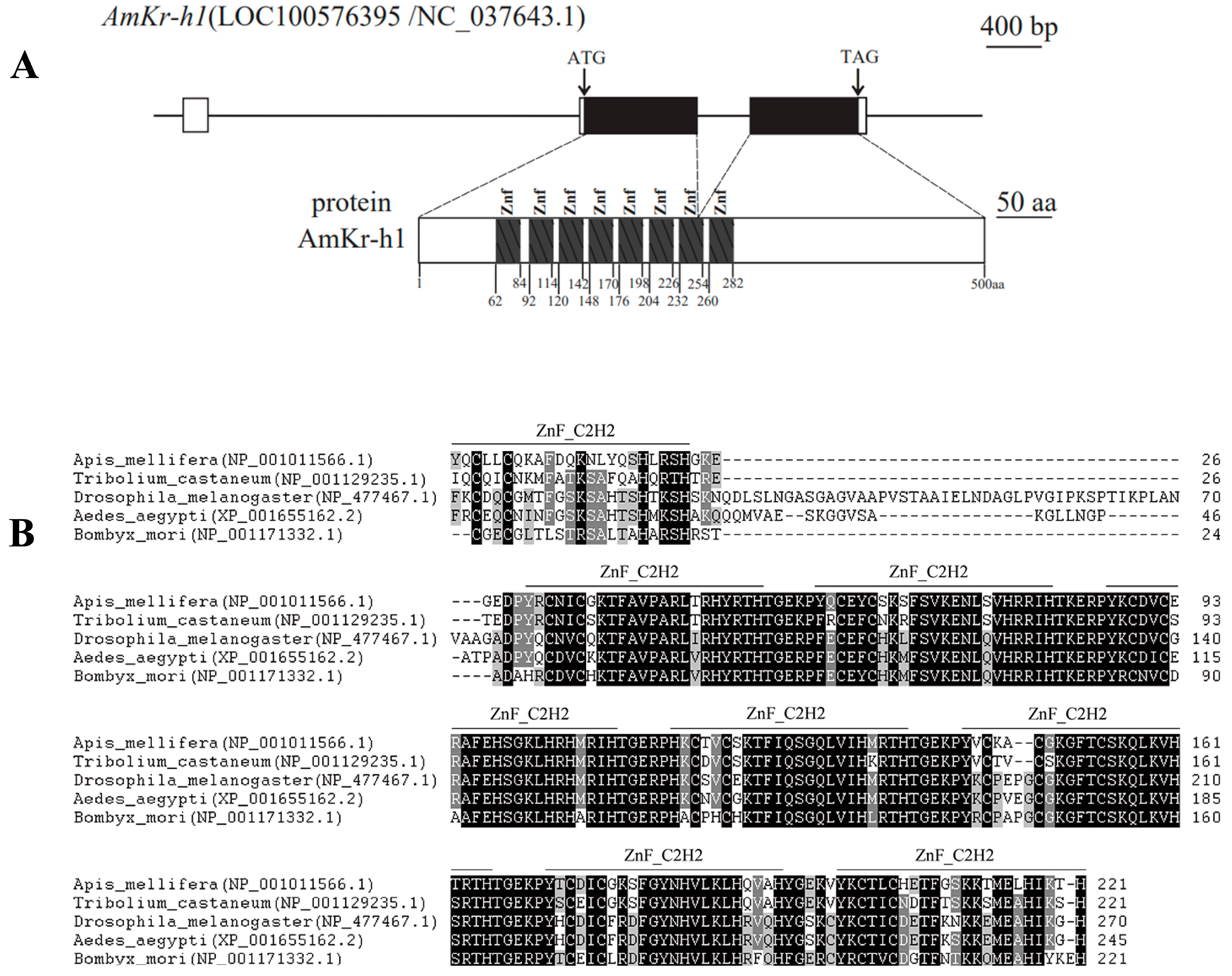
Figure 1. The intron/exon structure and amino acid sequence comparison of the AmKr-h1 gene of insects. (A) The intron/exon structure and predicted protein of AmKr-h1 gene. In the upper part, boxes, lines, and black boxes represent exons, introns, and coding regions, respectively; (B) Amino acid sequence comparison of the C2H2 zinc finger domains between the Kr-h1 protein of A. mellifera and other species.
Biomolecules 13 01657 g001
Figure 2. Differences in expression of AmKr-h1 between A. mellifera queen larvae and worker larvae as well as expression changes after JH treatment. (A) Relative mRNA expression levels of AmKr-h1 in queen larvae and worker larvae at 48 h, 84 h, and 120 h of the larval stage. Different lowercase letters represent significant differences at p < 0.05; the same in (B). (B) Relative mRNA expression levels of AmKr-h1 in larvae of Apis mellifera after JH treatment. JH: the group treated with juvenile hormone III; ET: the group treated with ethanol; NO: the group without treatment.
Biomolecules 13 01657 g002
Figure 3. Effect of AmKr-h1 on honey bee caste differentiation. (A) Expression change of AmKr-h1 in larvae of A. mellifera after RNAi. Different lowercase letters represent a significant difference at p < 0.05; the same in (B). (B) Morphological changes in newly emerged bees after AmKr-h1 RNAi. (C) An example of morphological change in newly emerged bees after AmKr-h1 RNAi. “Con” represents control group.
Biomolecules 13 01657 g003
Figure 4. DEGs between the RNAi and Con groups. (A) Up- and down-regulated DEGs in the RNAi group compared to the control group in the larval stage of A. mellifera. Both the RNAi group and control group contain three biological replicates. (B) The significantly enriched KEGG pathways of the DEGs identified between the RNAi group and the control group. Pathways marked with a box are those associated with caste differentiation. (C) The DEGs involved in honey bee caste differentiation. “Con” represents control group.
Biomolecules 13 01657 g004
Figure 5. Verification of DEGs using qRT-PCR. Quantitative RT-PCR verification of nine DEGs between the RNAi and control (Con) groups. Asterisk (*) represents significant difference at p < 0.05.
Biomolecules 13 01657 g005
Figure 6. DEASEs between the RNAi and Con groups of honey bee. (A) Differential alternative splicing events between the RNAi group and control (Con) group. (B) The significant enriched KEGG pathways between the RNAi group and control (Con) group. The pathways marked with a box are associated with honey bee caste differentiation.
Biomolecules 13 01657 g006
Disclaimer/Publisher’s Note: The statements, opinions and data contained in all publications are solely those of the individual author(s) and contributor(s) and not of MDPI and/or the editor(s). MDPI and/or the editor(s) disclaim responsibility for any injury to people or property resulting from any ideas, methods, instructions or products referred to in the content.