Technical Note

A Bacterial Platform for Studying Ubiquitination Cascades Anchored by SCF-Type E3 Ubiquitin Ligases

1
Ministry of Education Key Laboratory of Cell Activities and Stress Adaptations, School of Life Sciences, Lanzhou University, Lanzhou 730000, China
2
Gansu Province Key Laboratory of Gene Editing for Breeding, School of Life Sciences, Lanzhou University, Lanzhou 730000, China
3
School of Chemical Engineering Ocean and Life Science, Dalian University of Technology, Panjin 124221, China
*
Authors to whom correspondence should be addressed.
Biomolecules 2024, 14(10), 1209; https://doi.org/10.3390/biom14101209
Submission received: 28 May 2024 / Revised: 20 September 2024 / Accepted: 23 September 2024 / Published: 25 September 2024
(This article belongs to the Section Biomacromolecules: Proteins)

Abstract

:
Ubiquitination is one of the most important post-translational modifications in eukaryotes. The ubiquitination cascade includes ubiquitin-activating enzymes (E1), ubiquitin-conjugating enzymes (E2), and ubiquitin ligases (E3). The E3 ligases, responsible for substrate recognition, are the most abundant and varied proteins in the cascade and the most studied. SKP1-CUL1-F-Box (SCF)-type E3 ubiquitin ligases are multi-subunit RING (Really Interesting New Gene) E3 ubiquitin ligases, composed of CUL1 (Cullin 1), RBX1 (RING BOX 1), SKP1 (S-phase Kinase-associated Protein 1), and F-box proteins. In vitro ubiquitination assays, used for studying the specific recognition of substrate proteins by E3 ubiquitin ligases, require the purification of all components involved in the cascade, and for assays with SCF-type E3 ligases, additional proteins (several SCF complex subunits). Here, the Duet expression system was used to co-express E1, E2, ubiquitin, ubiquitylation target (substrate), and the four subunits of a SCF-type E3 ligase in E. coli. When these proteins co-exist in bacterial cells, ubiquitination occurs and can be detected by Western Blot. The effectiveness of this bacterial system for detecting ubiquitination cascade activity was demonstrated by replicating both AtSCFTIR1-mediated and human SCFFBXO28-mediated ubiquitylation in bacteria. This system provides a basic but adaptable platform for the study of SCF-type E3 ubiquitin ligases.

1. Introduction

Ubiquitin (Ub) is a highly conserved protein composed of 76 amino acids. A single Ub (monoubiquitination) or Ub chains (polyubiquitination) can be conjugated to proteins. Ubiquitination is a major regulatory mechanism for protein degradation and impacts functions such as DNA repair, transcription, cell differentiation, cell cycle regulation, and stress responses [1]. The covalent attachment of ubiquitin or ubiquitin chains to the target protein requires a multi-step, ATP-dependent process controlled by ubiquitin-activating enzymes (E1s), ubiquitin-binding enzymes (E2s), and ubiquitin ligases (E3s), which is also known as the ubiquitination cascade. The cascade begins with an E1 forming a thioester bond with the C-terminal carboxyl group of ubiquitin. Next, the thioester-linked ubiquitin is transferred to a cysteinyl residue of an E2 enzyme. Finally, the E3 ubiquitin ligase binds directly to the substrate and catalyzes the transfer of Ub from E2 to a lysine residue of the target protein [2,3].
E1’s initiation of the coupling cascade has little effect on target specificity. There are only two E1 subtypes in Arabidopsis thaliana, one of which may be nuclear-localized [4]. In contrast, the E2 enzymes comprise a large family, including at least 37 E2 genes, which are divided into 12 subfamilies and eight E2-like genes in the A. thaliana genome [5,6,7]. In the ubiquitination cascade, E3 is responsible for recruiting target proteins for ubiquitination and is the major substrate recognition component of this pathway. The number of E3 is particularly large in eukaryotes, with 1400 predicted in A. thaliana [2]. Based on the presence of conserved domains and the mechanism of ubiquitin transfer to the substrate protein, E3s can be divided into four groups: RING (Really Interesting New Gene), U-box, HECT (Homologous to the E6AP C-Terminus), and RBR (RING-between RING) [8].
Among the four groups of E3 ligases, RING E3s are the most abundant in eukaryotes. The characteristic of this group is the presence of a Ring-Finger domain. The Ring-Finger domain contains 8eightconserved amino acid residues (Cys or His) that coordinate two zinc ions, which binds to an E2-ubiquitin conjugate to transfer ubiquitin to a lysine residue of the target protein [9,10]. Based on their subunit composition and mechanism of action, the RING E3 group can be further divided into several subgroups, including monomer RING E3s (that can function as monomers, homodimers, or heterodimers), Cullin-RING Ligases (CRLs), and the anaphase-promoting complex [8,11,12].
CRLs are the most widespread class of E3s in eukaryotes. Studies to date have shown that all eukaryotes contain a family of Cullin proteins and that homologs for most Cullin proteins can be identified across kingdoms. CRLs consist of several subunits. The Cullin subunit serves as a scaffold, recruiting a RING-Finger domain containing the RBX1 (RING BOX 1) subunit to its C-terminal region and binding one or more additional Cullin-specific subunit(s), which serve as substrate receptors and as adaptors, to its N-terminus [12]. There are three types of CRLs in plants, each built on a different Cullin: CUL1, CUL3, and CUL4. The CUL1-scaffolded E3 ligases are the most intensively studied CRLs due to their important function in plants [13].
For CUL1, the adaptor subunit is SKP (S-phase Kinase-associated Protein) [10,14], which together form SCF (SKP1-CUL1-F-Box) complexes. Within the SCF complex, the SKP adaptor protein binds an F-box protein that is responsible for substrate recognition and determines the specificity of the SCF complex, so the name of the F-box subunit is written in superscript to distinguish between different SCF-type E3 ligases. SCF-type E3 ligases are a highly flexible class of ubiquitin ligases. Within the proteomes of different species, a large number of F-box and SKP proteins is present, suggesting a conserved combinatorial organization of the SCF complex. The same CUL1-RBX1 works with different F-boxes and SKPs to mediate the ubiquitination of different target proteins [13].
While studying ubiquitination, researchers face several major challenges: (1) The high redundancy of components in the ubiquitination cascade (i.e., redundancy between E2/E3s and their ubiquitylation targets) makes it difficult to determine the connections between them in vivo; (2) The instability of ubiquitinated proteins in cells due to rapid deubiquitylation or degradation by the 26S proteasome, which makes them difficult to detect; and (3) The required activation of ubiquitination modifications in eukaryotic cells, which usually occur as a component of cellular signaling under specific circumstances [15]. For these reasons, in vitro experiments are often used to reconstruct ubiquitination cascades for research. However, many E3 ligases and their substrates are unstable and difficult to purify in their full-length forms. Moreover, ubiquitination is a highly dynamic and complex biochemical process involving the interaction of multiple enzymes and proteins. Reconstructing ubiquitination in the tube requires not only maintaining the activity of these components but also their interactions, which is very challenging under experimental manipulation [15]. For the multi-subunit E3 ubiquitin ligase SCF complex, it is even more difficult to reproduce the ubiquitination cascade in vitro. To make in vitro experiments easier to perform, some researchers developed a bacterial ubiquitination system for detecting the ubiquitination by singular E3 ligases to their particular substrates [15,16,17,18,19]. Co-expressing components of the entire ubiquitination cascade in Escherichia coli has successfully reproduced the ubiquitination cascade in a bacterial system. In addition to its convenience and ease, the other advantage of this method is that the ubiquitination tagging for degradation does not naturally exist in E. coli cells, and therefore stable ubiquitinated proteins can accumulate, which facilitates subsequent studies [15,16]. However, to date, there are no reports of this system being used for multi-subunit E3 ubiquitin ligases. Here, we extend the use of this bacterial system to ubiquitin cascades containing the multi-subunit E3 ubiquitin ligase SCF complex. When CUL1, RBX1, SKP1, and F-box protein are co-expressed with other components of the ubiquitination cascade, ubiquitination can occur in E. coli cells. In this paper, we selected AtSCFTIR1 and the human HsSCFFBXO28 as representatives of SCF-type E3 ligases to evaluate this system.

2. Materials and Methods

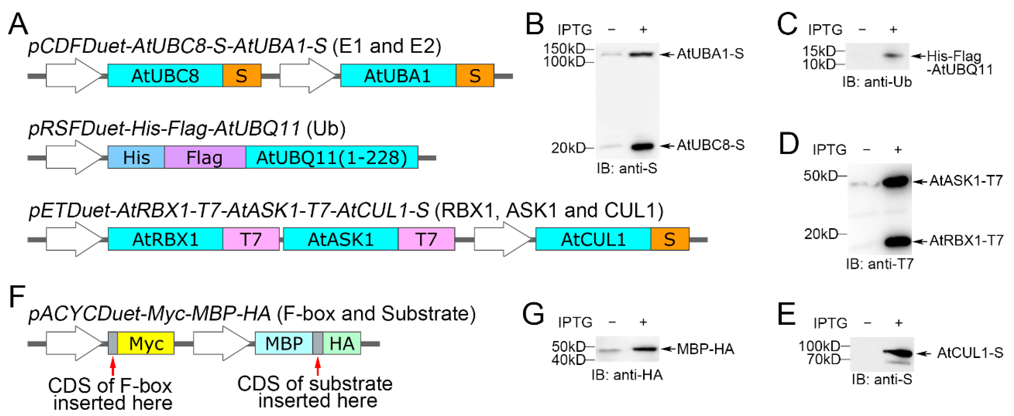
2.1. Plasmid Design for the Expression of Proteins Associated with the Ubiquitination Cascade

The Duet expression system was used to co-express the E1, E2, ubiquitin, ubiquitylation target (substrate), and four subunits of an SCF-type E3 ligase in E. coli. The original plasmids pETDuet-1 (Cat. NO. 71146), pACYCDuet-1 (Cat. NO. 71147), pCDFDuet-1 (Cat. NO. 71340), and pRSFDuet-1 (Cat. NO. 71341) were ordered from MERCK (Darmstadt, Germany). Details of the plasmids can be found on “https://www.merckmillipore.com/ (assessed on 25 September 2024)”.
The genes encoding the ubiquitination proteins of A. thaliana, namely AtUBA1 (Ubiquitin Activating Enzyme 1), AtUBC8 (Ubiquitin Conjugating Enzyme 8), AtUBQ11 (Ubiquitin 11), AtRBX1, AtASK1 (Arabidopsis SKP1 Homologue 1), and AtCUL1, were cloned into a variety of compatible Duet expression vectors, each with a different antibiotic resistance selective marker. In order to better detect these proteins in this experimental system, we changed the coding sequences corresponding to the original short tagging peptides in some of the Duet expression vectors. Three plasmids, pCDFDuet-AtUBC8-S-AtUBA1-S, pRSFDuet-His-Flag-AtUBQ11, and pETDuet-AtRBX1-T7-AtASK1-T7-AtCUL1-S, were constructed as shown in (Figure 1A).
The coding sequences of AtUBC8 (E2) and AtUBA1 (E1) in the first and second Multiple Cloning Sites (MCSs) of the pCDFDuet-1 vector, respectively, had an S-Tag fused at the C-end of each of them. AtUBQ11 encodes a Ub polymer with three repeats of the 228-bp coding sequence connected head to tail. One 228-bp fragment was cloned at the C-terminal of a His-Flag tag in the pRSFDuet-1 vector. In vector pETDuet-1, there are only two MCSs for the expression of exogenous proteins, but we wanted to simultaneously express three subunits of the SCF complex (AtRBX1, AtASK1, and AtCUL1) using a single vector. To accomplish this, an open reading frame containing two cistrons was designed. The coding sequences of AtRBX1-T7 and AtASK1-T7 with stop codons were cloned in tandem with an RBS (ribosome binding site) added in front of the AtASK1-T7 sequence. This cassette was inserted into the first MCS of the pETDuet-1 vector. Next, the coding sequence of AtCUL1 was inserted into the second MCS of the pETDuet-1 vector.
Each of the three constructed vectors was independently transfected into E. coli strain BL21(DE3). After cultivation and induction with IPTG, the crude protein was extracted. Protein-specific antibodies were used for the Western Blot (WB) detection of these tagged proteins (Table 1). As shown in Figure 1B–E, we observed all expected proteins by immunoblot.
The F-box and ubiquitylation target protein (substrate) were cloned into the pACYCDuet-1 vector for expression. The His and S tags in this vector were replaced with Myc and MBP-HA, respectively (Figure 1F), to ensure that the F-box and substrate were tagged differently from other components in the cascade. This modified plasmid was named pACYCDuet-Myc-MBP-HA. As shown in Figure 1G, MBP-HA can be detected using WB. The coding sequence of Myc was too short, so we did not detect it before verifying the validity of this system. However, after performing the ubiquitination cascade, the F-box fused with Myc was detected.

2.2. Plasmid Construction

To construct the pCDFDuet-AtUBA1-S plasmid, the CDS of AtUBA1 was amplified by primers UBA1 F/R (Table S1), and then inserted between the EcoRV and KpnI sites of pCDFDuet-1.
To construct the pCDFDuet-AtUBC8-S or pCDFDuet-AtUBC8-S-AtUBC1-S plasmids, the sequence of AtUBC8-S was amplified by primers UBC8-S F/R (Table S1), and then inserted into the EcoRI site of pCDFDuet-1 and pCDFDuet-AtUBA1-S, respectively.
To construct the pRSFDuet-His-Flag-AtUBQ11 plasmid, the sequence of HIS-Flag-UBQ11(1-228) was amplified by primers HIS-FLAG-UBQ F/R (Table S1), and then inserted between the NcoI and KpnI sites of pRSFDuet-1.
The construction of the pETDuet-AtRBX1-T7-AtASK1-T7-AtCUL1-S plasmid was performed in two steps. First, the sequence of AtRBX1-T7-RBS-AtASK1-T7 was synthesized by Genewiz (Suzhou, China) and inserted between the NcoI and SalI sites of pETDuet-1. In the second step, the CDS of AtCUL1 was amplified by primers CUL1 F/R (Table S1) and then inserted into the KpnI site of the plasmid obtained in the first step.
To construct the pACYCDuet-Myc-MBP-HA plasmid, the sequence of MCS-Myc-T7 promoter-lac operator-RBS-MBP-MCS-HA was synthesized by Genewiz and then inserted between the NcoI and XmaJI sites of pACYCDuet-1.
To construct the pACYCDuet-AtTIR1-Myc-MBP-HA or pACYCDuet-AtTIR1P10A-Myc-MBP-HA plasmid, the CDSs of AtTIR1 or AtTIR1P10A [20] were amplified by primers TIR1 F/R (Table S1) and then inserted into the BamHI site of pACYCDuet-Myc-MBP-HA.
To construct the pACYCDuet-AtTIR1-Myc-MBP-AtIAA6-HA and pACYCDuet-Myc-MBP-AtIAA6-HA plasmids, the CDS of AtIAA6 was amplified by primers IAA6 F/R (Table S1) and then inserted into the Eco147I site of pACYCDuet-AtTIR1-Myc-MBP-HA and pACYCDuet-Myc-MBP-HA, respectively.
To construct the pETDuet-AtRBX1-T7-HsSKP1-T7-AtCUL1-S plasmid, the sequence of T7-RBS-HsSKP1 was synthesized by Genewiz and then inserted between the BamHI and SgsI sites of pETDuet-AtRBX1-T7-AtASK1-T7-AtCUL1-S.
To construct the pACYCDuet-HsFBXO28-Myc-MBP-HA or pACYCDuet-ΔHsFBXO28-Myc-MBP-HA plasmid, the CDS of HsFBXO28 or ΔHsFBXO28 [21] was synthesized by Genewiz and then inserted between the BamHI and EcoRV sites of pACYCDuet-Myc-MBP-HA.
All DNA fragments were amplified using ApexHF HS DNA Polymerase (Cat. NO. AG12207) or TransStart® FastPfu Fly DNA Polymerase (Cat. NO. AP231), which were purchased from Accurate Biotechnology (Changsha, China) and TransGen Biotech (Beijing, China), respectively. The DNA fragments obtained by PCR were ligated into the vector backbone using the pEASY®-Basic Seamless Cloning and Assembly Kit (Cat. NO. CU201), which was purchased from TransGen Biotech (Beijing, China). All gene sequences corresponding to prokaryotic proteins were sequenced by AuGCT DNA-SYN Biotechnology (Yangling, China).

2.3. Co-Expression of Recombinant Proteins in E. coli

The different expression vectors constructed above were individually transformed or transformed in different combinations into Escherichia coli BL21 (DE3) (TransGen Biotech, China, Cat. NO. CD601) and cultured in 2× Yeast extract and Tryptone (2 × YT) liquid medium containing the corresponding antibiotic at 37 °C. When the absorbance at 600 nm reached 0.5–0.6, 0.5 mM of isopropyl β-D-1-thiogalactopyranoside (IPTG) was added to induce the expression of the target protein(s), generally at 25 °C for 8-10 h with shaking at 200 rpm. IPTG (Cat. NO. I1020) was purchased from Solarbio (Beijing, China). The E. coli cells were collected and lysed by Scientz-IID Ultrasonic Homogenizer of Ningbo Scientz Biotechnology (Ningbo, China). Crude proteins were used to detect recombinant proteins or for ubiquitination assays.

2.4. SDS-PAGE and Immunoblot Analysis for Detecting the Ubiquitination Cascade

For immunoblot analysis, the crude proteins were separated on 12% SDS-polyacrylamide gels (PAGEs). The electrophoresed proteins were transferred to a 0.45 μm nitrocellulose membrane using a wet transfer system and incubated with different antibodies. Anti-S (Cat. NO. K200013M) and anti-T7 (Cat. NO. K200008M) were purchased from Solarbio (Beijing, China), anti-HA (Cat. NO. M20021) and anti-Myc (Cat. NO. M20019) were purchased from Abmart (Shanghai, China), and anti-ubiquitin (Cat. NO. AF1705) was purchased from Beyotime (Shanghai, China). The secondary antibody Goat Anti-Rabbit Mouse IgG-HRP (Cat. NO. M21003) was purchased from Abmart (Shanghai, China) and used as per the manufacturer’s instructions. Horseradish peroxidase was detected using the Super ECL Western Blotting Substrate (Cat. NO. SL1350), which was purchased from Coolaber (Beijing, China).

2.5. Accession Numbers

The gene sequence data from this article can be found at “https://www.ncbi.nlm.nih.gov/ (assessed on 25 September 2024)” data libraries under the following accession numbers: AtUBA1 (NCBI Gene ID: 817562, TAIR:AT2G30110), AtUBC8 (NCBI Gene ID: 834173, TAIR:AT5G41700), AtUBQ11 (NCBI Gene ID: 825847, TAIR:AT4G05050), AtRBX1 (NCBI Gene ID: 832179, TAIR:AT5G20570), AtCUL1 (NCBI Gene ID: 825648, TAIR:AT4G02570), AtASK1 (NCBI Gene ID: 843928, TAIR:AT1G75950), AtTIR1 (NCBI Gene ID: 825473, TAIR:AT3G62980), AtIAA6 (NCBI Gene ID: 841717, TAIR:AT1G52830), HsSKP1 (NCBI Gene ID: 6500, HGNC:HGNC:10899), and HsFBXO28 (NCBI Gene ID: 23219, HGNC:HGNC:29046).

3. Results

3.1. Auto-Ubiquitination Activity of AtSCFTIR1 in Recombinant E. coli

To verify that the SCF-type E3 ubiquitin ligase-mediated ubiquitination cascade can be reconstructed in bacteria, we chose the well-studied plant F-box protein AtTIR1 (Transport Inhibitor Response 1) to test our system. As receptors for the plant hormone auxin, AtTIR1 and its homologs, the AtAFBs (Auxin Signaling F-box proteins), play key roles in the auxin signaling pathway. Auxin regulates the transcription of auxin response genes through the action of TIR1/AFBs, Aux/IAA transcriptional suppressors, and ARFs (Auxin Response Factors). In general, Aux/IAAs work by binding directly to ARFs and recruiting the corepressor TPL (TOPLESS) to chromatin [22,23]. In plants, Aux/IAAs can be sensed by TIR1/AFBs, which interact with ASK1, RBX1, and CUL1 to form SCFTIR1/AFBs ubiquitin ligase complexes. In the presence of auxin, the SCFTIR1/AFBs ligases interact with Aux/IAAs to mediate their degradation.
Previous studies have shown that most E3 ubiquitin ligases can also undergo auto-ubiquitination when their specific substrate protein is missing [24]. Although F-box proteins are only one subunit of SCF-type E3 ubiquitin ligases, the auto-ubiquitination of F-box proteins has been demonstrated in yeast and human systems [25,26]. For the auxin receptor AtTIR1, there are no reports that it can undergo auto-ubiquitination, but Yu’s study in 2015 has shown that AtTIR1 is subject to autocatalytic degradation when assembled into an SCF complex [27]. Now, the degradation of poly-ubiquitin-tagged proteins is well known. For an SCF-type E3 ubiquitin ligase, its catalytic domain Ring-Finger is located in the RBX1 subunit. This means that the F-box protein in monomeric form is devoid of E3 ubiquitin ligase activity. If AtTIR1 fails to mediate its own degradation without assembling into the AtSCFTIR1 complex, it is possible that AtTIR1 undergoes auto-ubiquitination before autocatalytic degradation.
Based on the above hypothesis, we wanted to test the auto-ubiquitination activity of the AtSCFTIR1 E3 ubiquitin ligase using this bacterial system. As shown in Figure 2A, the vector pACYCDuet-AtTIR1-Myc-MBP-HA is constructed to express AtTIR1-Myc (73.3 kD). The E. coli BL21(DE3) strain containing multiple vectors, pCDFDuet-AtUBC8-S-AtUBA1-S, pRSFDuet-His-Flag-AtUBQ11, pETDuet-AtRBX1-T7-AtASK1-T7-AtCUL1-S and pACYCDuet-AtTIR1-Myc-MBP-HA, was cultured, and gene expression was induced by 0.5 mM of IPTG. As negative controls lacking E1 or E2 of the ubiquitination cascade, we also constructed the vectors pCDFDuet-AtUBA1-S and pCDFDuet-AtUBC8-S for co-transforming BL21(DE3) with the other plasmids (Figure 2B).
The total crude protein was separated by SDS-PAGE and subjected to Western Blot. The anti-Myc antibody detected AtTIR1-Myc in a ladder pattern, which implies that AtTIR1-Myc undergoes poly-ubiquitination modification (Figure 2C). The laddering pattern was also observed with the anti-ubiquitin antibody, but was not observed when any component of the ubiquitination cascade was absent (Figure 2C). These results indicate that AtSCFTIR1-Myc has auto-ubiquitination activity, whose auto-ubiquitination can only be observed in the presence of other three subunits of the SCF complex (Figure 2C), which is consistent with previous studies showing that only AtTIR1 itself does not mediate its own degradation [27]. In addition, previous research has shown a specific mutation of the F-box domain of TIR1 (AtTIR1P10A) prevents its interaction with AtASK1 [20]. In our experiment, no auto-ubiquitination of AtTIR1P10A-Myc was detected when AtTIR1P10A-Myc replaced AtTIR1-Myc (Figure 2C), further indicating that AtTIR1-Myc cannot undergo auto-ubiquitination when it fails to assemble into the AtSCFTIR1-Myc complex.

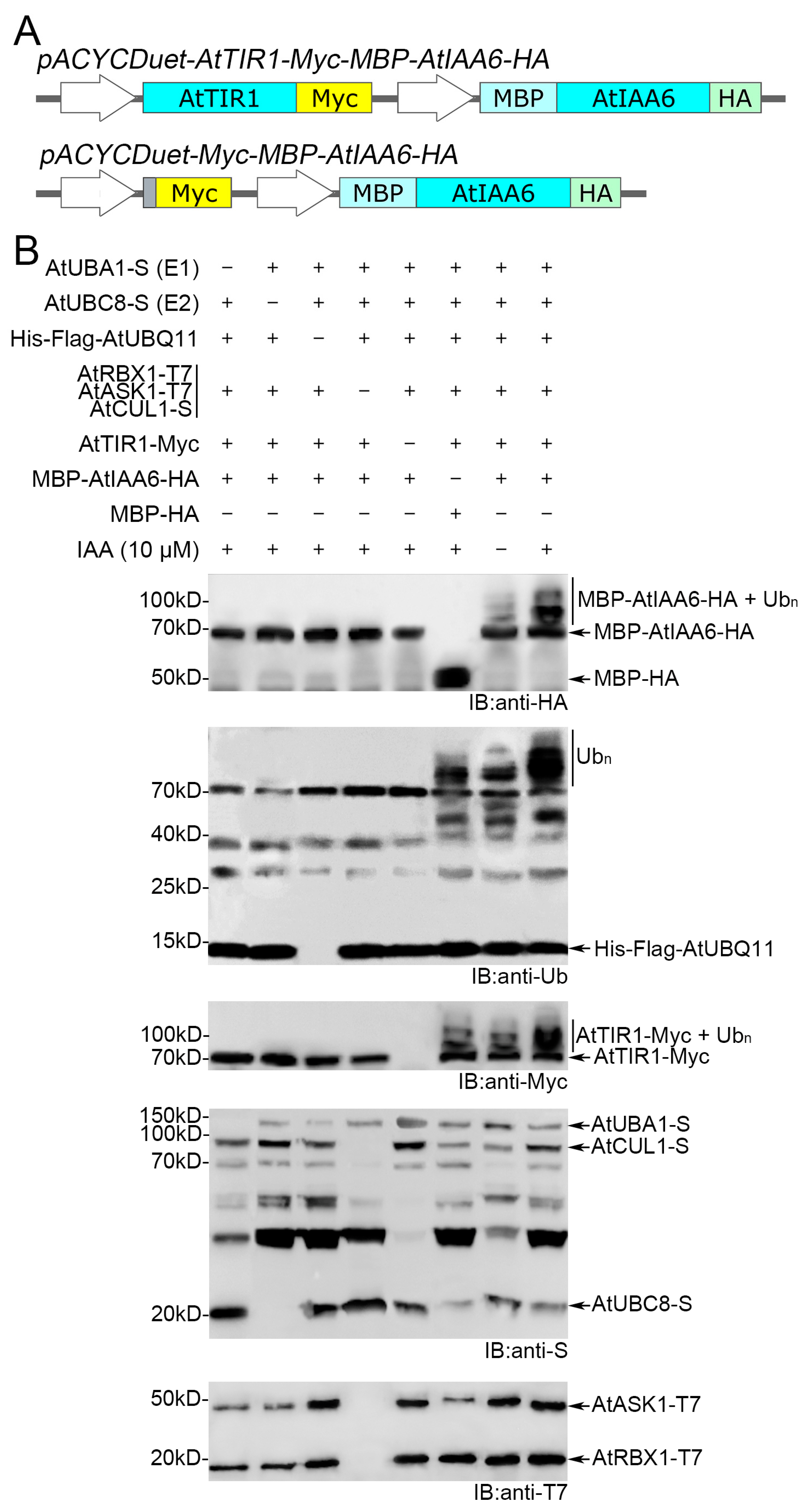
3.2. AtSCFTIR1 Can Catalyze AtIAA6 Ubiquitination in the Bacterial System

A previous study has shown that AtSCFTIR1 mediates AtIAA6 (a member of the Aux/IAA family) ubiquitination both in vitro and in vivo [28]. Therefore, we used the AtIAA6 as a substrate for AtSCFTIR1 to verify whether the bacterial system can analyze substrate ubiquitination. In order to co-express AtIAA6 with other components in the ubiquitination cascade, the vectors pACYCDuet-Myc-MBP-AtIAA6-HA (for the negative control) and pACYCDuet-AtTIR1-Myc-MBP-AtIAA6-HA were constructed (Figure 3A). The E. coli strain harboring AtUBA1-S, AtUBC8-S, His-FLAG-AtUBQ11, AtCUL1-S, AtRBX1-T7, AtASK1-T7, AtTIR1-Myc, and MBP-AtIAA6-HA (67.2 kD) was cultured. Since AtIAA6 interacted in an auxin-dependent manner with AtTIR1 [28], we added the naturally occurring auxin IAA (indole-3-acetic acid) when inducing gene expression with IPTG.
Then, the crude protein extracts were obtained by crushing the bacteria, separated by SDS-PAGE, and analyzed by WB with the corresponding antibodies. A laddering pattern was observed in the presence of all components when using substrate specific anti-HA, but was not observed when replacing MBP-AtIAA6-HA with MBP-HA (Figure 3B). When compared side by side, the immunoreactivity of HA is significantly stronger in the presence of IAA than in the absence of IAA (Figure 3B), coinciding with the idea that IAA can act as a molecular glue to enhance the interaction between TIR1 and AUX/IAA proteins [28,29]. It is noteworthy that, when using anti-ubiquitin for immunoblotting, protein ladders are also observed without MBP-AtIAA6-HA, because AtTIR1-Myc can undergo auto-ubiquitination, which can also be observed in the Myc-blot (Figure 3B). As expected, AtUBA1, AtUBC8, Ub, and AtSCFTIR1 are clearly required for AtIAA6 ubiquitination (Figure 3B). This is consistent with Winkler’s results [28], and also shows that this recombinant system can be used to analyze substrate ubiquitination, further confirming the feasibility of this recombinant system.

3.3. SCF Complex Composed of Heterologous Subunits Showed Ubiquitin Ligase Activity

For SCF-type multi-subunit E3 ubiquitin ligases, the Cullin subunit serves as a scaffold, while the RBX1 subunit endows the SCF complex with the ability to transfer ubiquitin from the E2 to either F-box proteins or specific substrates [10]. Cul and RBX1 are conserved in eukaryotes, and mammalian CUL1 and RBX1 can assemble with Arabidopsis ASK1 and the F-box protein to form active SCF complexes [28]. To verify that our system can also be used to study the ubiquitination cascade of SCF-type E3 ubiquitin ligases in animals, we wanted to replicate the recently reported auto-ubiquitination of the human HsFBXO28 in bacteria [21]. Since the F-BOX protein can only be recognized by the specific SKP protein [10,14], we replaced AtASK1 in the bacterial system with HsSKP1.
As shown in Figure 4A, the vector pETDuet-AtRBX1-T7-HsSKP1-T7-AtCUL1-S was constructed to express AtRBX1-T7, AtCUL1-S, and HsSKP1-T7 (20.5 kD). The vector pACYCDuet-HsFBXO28-Myc-MBP-HA was constructed to express HsFBXO28-Myc (47.3 kD). Following the study of Cai et al. [21], the deletion of the F-box domain (ΔHsFBXO28-Myc, 41.6 kD) was used as a negative control. The E. coli strain harboring HsSKP1-T7, HsFBXO28-Myc, and other components of the ubiquitination cascade derived from A. thaliana was cultured and induced. The crude protein extracts were separated by SDS-PAGE and analyzed by WB with the corresponding antibodies. A laddering pattern was observed in the presence of all components when using anti-Myc or anti-ubiquitin (Figure 4B). There was no auto-ubiquitination of ΔHsFBXO28-Myc detected when ΔHsFBXO28-Myc replaced HsFBXO28-Myc, which is consistent with previous studies by Cai et al. [21]. This result indicate that our system can also be applied to study the ubiquitination cascade of SCF-type E3 ubiquitin ligases in organisms other than plants.

4. Discussion

To date, countless different E3 ubiquitin ligases have been predicted in eukaryotic genomes on the basis of structural motifs. These E3s belong to different groups, among which the SCF-type E3 ubiquitin ligase group is the largest and best characterized [30]. F-box proteins confer the SCF-type ubiquitin ligases the ability to recognize target proteins, which is dependent on their C-terminal variable recruitment module. In eukaryotes, the F-box protein family is the largest known protein superfamily, and many F-box proteins with different C-terminal motifs have been identified as SCF components. The number of F-box proteins varies widely among organisms and shows little correlation with their complexity, genome size, or life cycles [31,32,33]. For example, there are 20 F-box proteins in budding yeast (S. cerevisiae), 27 in fruit flies (D. melanogaster), 69 in humans, 520 in C. elegans, and a staggering 897 in the model plant A. thaliana [31,32,33,34]. Interestingly, there is little overlap between F-box proteins across organisms, e.g., only six F-box proteins have apparent orthologs conserved between humans and flies [31,32]. Moreover, there are significant differences in F-box gene loci among closely related species. For instance, A. thaliana and A. lyrata, which diverged only five Mya, differ by 453 F-box loci, with A. thaliana acquiring 109 loci and losing 468 loci [33]. Current evolutionary studies speculated that huge differences in the F-box proteins between species show the rapid evolution of this superfamily, which potentially reflects a central role for ubiquitination in driving biological adaptations [33,35].
Consistent with evolutionary studies, more than 20 years of research on A. thaliana F-box proteins has revealed their important functions [36]. F-box proteins are critical regulators of many biological pathways, including phytohormone signaling pathways, floral development, seed germination, leaf senescence, self-incompatibility, lateral shoot branching, control of cell cycle, regulation of circadian clock, floral meristem formation, floral organ identity determination, and photomorphogenesis [36,37,38], and very much decide the fate of a plant. Although the number of F-box proteins in mammals such as humans is much smaller than in A. thaliana, they participate in numerous vital physiological and pathological processes, including cell cycle regulation, cell proliferation, gene expression regulation, apoptosis, and signal transduction [39]. In particular, there are concerns about the important roles of F-box proteins in carcinogenesis and tumor progression [40].
Although there are established roles for F-box proteins in many diverse pathways, the majority of the predicted F-box proteins has no known substrates. Further research on deciphering the physiological and biochemical functions of the F-box proteins in cells is absolutely necessary. Here, we present an improved, simple, and quick bacterial system for the study of SCF-type E3 ubiquitin ligases and their substrates. In vitro ubiquitination analysis using this system avoids the purification of every component in the cascade and every subunit of the SCF complex, which reduces the difficulty and increases the efficiency of the experiments. This system can also be used to obtain ubiquitination-modified target proteins in large quantities through bacterial culture, which facilitates later biochemical, biophysical, and crystallographic analyses. Furthermore, this system is expected to be used for the high-throughput screening of E3 ubiquitin ligase targets in future studies [15,17], which may boost research on the SCF complex and may help delineating F-box-substrate pairs.
In this article, we applied this system to reproduce both AtSCFTIR1-mediated and a mixed-species SCFFBXO28-mediated ubiquitylation. Special care should be taken to select the paired SKP and F-box protein when using this system, as some F-box proteins might be recognized only by specific SKPs. Our assay could also be used with E3 and substrates from other plant species. Additionally, our assay can also be applied to single-subunit E3 ligases by replacing the SCF components with a monomeric E3 ligase, while considering the replacement of suitable E2 [41].

5. Conclusions

In vitro ubiquitination detection of multi-subunit E3 ubiquitin ligase SCF complex has always been a challenging experiment. Here, we present an improved, simple, and quick bacterial system for the study of SCF-type E3 ubiquitin ligases and their substrates. When CUL1, RBX1, SKP1, and F-box protein are co-expressed with other components of the ubiquitination cascade, ubiquitination can occur in E. coli cells. By using this system, the ubiquitination cascade of AtSCFTIR1 and mixed-species SCFFBXO28 was reproduced in bacteria, which implies that this platform is generally applicable to the study of SCF complexes in eukaryotes. Using this system avoids the purification of every component in the cascade and every subunit of the SCF complex, which reduces the difficulty and increases the efficiency of the experiments.

Supplementary Materials

The following supporting information can be downloaded at: https://www.mdpi.com/article/10.3390/biom14101209/s1, Table S1: Primers used in this study. Figure S1. Uncropped Western Blot images corresponding to Figure 1B–E,G. Figure S2: Uncropped Western Blot images corresponding to Figure 2C. Figure S3: Uncropped Western Blot images corresponding to Figure 3B. Figure S4: Uncropped Western Blot images corresponding to Figure 4B. Attached file contains all plasmid maps and sequences in this study.

Author Contributions

L.W. (Lei Wu) and L.W. (Li Wang) conceived and designed the experiments; Z.-X.P. performed the major research; J.-L.W., Y.-Y.L., L.-Y.L., Y.-T.T., Z.-H.W. and B.-L.L. participated in the construction of the recombinant plasmids; Professor G.-Q.G. reviewed the manuscript before submitting it and gave many constructive comments; L.W. (Lei Wu) and Z.-X.P. wrote the paper. All authors have read and agreed to the published version of the manuscript.

Funding

This work was supported by the Natural Science Foundation of Gansu Province (Grant 22JR5RA462) and the Fundamental Research Funds for the Central Universities (Grant lzujbky-2023-22) awarded to Lei Wu; the Natural Science Foundation of Gansu Province (Grant 23JRRA1141) and the Fundamental Research Funds for the Central Universities (Grant lzujbky-2023-it20) awarded to Zuo-Xian Pu; and the Natural Science Foundation of Gansu Province (Grant 24JRRA509) awarded to Jun-Li Wang.

Institutional Review Board Statement

Not applicable.

Informed Consent Statement

Not applicable.

Data Availability Statement

All data generated or analyzed during this study are available in this published article and its Supplementary Information. The plasmids will be available at Addgene “https://www.addgene.org/ (assessed on 25 September 2024)” or MiaoLingBio “http://www.miaolingbio.com/ (assessed on 25 September 2024)”.

Acknowledgments

We thank the Core Facility of the School of Life Sciences, Lanzhou University, for helping us with our experiments.

Conflicts of Interest

The authors declare no conflicts of interest.

References

  1. Pickart, C.M. Mechanisms Underlying Ubiquitination. Annu. Rev. Biochem. 2001, 70, 503–533. [Google Scholar] [CrossRef]
  2. Callis, J. The Ubiquitination Machinery of the Ubiquitin System. Arab. Book 2014, 12, e0174. [Google Scholar] [CrossRef]
  3. Kwon, Y.T.; Ciechanover, A. The Ubiquitin Code in the Ubiquitin-Proteasome System and Autophagy. Trends Biochem. Sci. 2017, 42, 873–886. [Google Scholar] [CrossRef]
  4. Hatfield, P.M.; Gosink, M.M.; Carpenter, T.B.; Vierstra, R.D. The ubiquitin-activating enzyme (E1) gene family in Arabidopsis thaliana. Plant J. 1997, 11, 213–226. [Google Scholar] [CrossRef]
  5. Bachmair, A.; Novatchkova, M.; Potuschak, T.; Eisenhaber, F. Ubiquitylation in plants: A post-genomic look at a post-translational modification. Trends Plant Sci. 2001, 6, 463–470. [Google Scholar] [CrossRef]
  6. Vierstra, R.D. The ubiquitin/26S proteasome pathway, the complex last chapter in the life of many plant proteins. Trends Plant Sci. 2003, 8, 135–142. [Google Scholar] [CrossRef]
  7. Xu, L.; Ménard, R.; Berr, A.; Fuchs, J.; Cognat, V.; Meyer, D.; Shen, W.H. The E2 ubiquitin-conjugating enzymes, AtUBC1 and AtUBC2, play redundant roles and are involved in activation of FLC expression and repression of flowering in Arabidopsis thaliana. Plant J. 2009, 57, 279–288. [Google Scholar] [CrossRef]
  8. Morreale, F.E.; Walden, H. SnapShot: Types of Ubiquitin Ligases. Cell 2016, 165, 248–248.e1. [Google Scholar] [CrossRef]
  9. Schulman, B.A.; Carrano, A.C.; Jeffrey, P.D.; Bowen, Z.; Kinnucan, E.R.E.; Finnin, M.S.; Elledge, S.J.; Harper, J.W.; Pagano, M.; Pavletich, N.P. Insights into SCF ubiquitin ligases from the structure of the Skp1–Skp2 complex. Nature 2000, 408, 381–386. [Google Scholar] [CrossRef] [PubMed]
  10. Zheng, N.; Schulman, B.A.; Song, L.; Miller, J.J.; Jeffrey, P.D.; Wang, P.; Chu, C.; Koepp, D.M.; Elledge, S.J.; Pagano, M.; et al. Structure of the Cul1–Rbx1–Skp1–F boxSkp2 SCF ubiquitin ligase complex. Nature 2002, 416, 703–709. [Google Scholar] [CrossRef] [PubMed]
  11. Komander, D.; Rape, M. The Ubiquitin Code. Annu. Rev. Biochem. 2012, 81, 203–229. [Google Scholar] [CrossRef] [PubMed]
  12. Zheng, N.; Shabek, N. Ubiquitin Ligases: Structure, Function, and Regulation. Annu. Rev. Biochem. 2017, 86, 129–157. [Google Scholar] [CrossRef]
  13. Hotton, S.K.; Callis, J. Regulation of Cullin RING Ligases. Annu. Rev. Plant Biol. 2008, 59, 467–489. [Google Scholar] [CrossRef]
  14. Frescas, D.; Pagano, M. Deregulated proteolysis by the F-box proteins SKP2 and β-TrCP: Tipping the scales of cancer. Nat. Rev. Cancer 2008, 8, 438–449. [Google Scholar] [CrossRef] [PubMed]
  15. Levin-Kravets, O.; Keren-Kaplan, T.; Attali, I.; Sharon, I.; Tanner, N.; Shapira, D.; Rathi, R.; Persaud, A.; Shohat, N.; Shusterman, A.; et al. E. coli-Based Selection and Expression Systems for Discovery, Characterization, and Purification of Ubiquitylated Proteins. Methods Mol. Biol. 2018, 1844, 155–166. [Google Scholar] [CrossRef]
  16. Keren-Kaplan, T.; Attali, I.; Motamedchaboki, K.; Davis, B.A.; Tanner, N.; Reshef, Y.; Laudon, E.; Kolot, M.; Levin-Kravets, O.; Kleifeld, O.; et al. Synthetic biology approach to reconstituting the ubiquitylation cascade in bacteria. EMBO J. 2012, 31, 378–390. [Google Scholar] [CrossRef]
  17. Su, L.; Lineberry, N.; Huh, Y.; Soares, L.; Fathman, C.G. A novel E3 ubiquitin ligase substrate screen identifies Rho guanine dissociation inhibitor as a substrate of gene related to anergy in lymphocytes. J. Immunol. 2006, 177, 7559–7566. [Google Scholar] [CrossRef]
  18. Rosenbaum, J.C.; Fredrickson, E.K.; Oeser, M.L.; Garrett-Engele, C.M.; Locke, M.N.; Richardson, L.A.; Nelson, Z.W.; Hetrick, E.D.; Milac, T.I.; Gottschling, D.E.; et al. Disorder targets misorder in nuclear quality control degradation: A disordered ubiquitin ligase directly recognizes its misfolded substrates. Mol. Cell 2011, 41, 93–106. [Google Scholar] [CrossRef]
  19. Han, Y.F.; Sun, J.H.; Yang, J.; Tan, Z.Y.; Luo, J.J.; Lu, D.P. Reconstitution of the plant ubiquitination cascade in bacteria using a synthetic biology approach. Plant J. 2017, 91, 766–776. [Google Scholar] [CrossRef]
  20. Gray, W.M.; del Pozo, J.C.; Walker, L.; Hobbie, L.; Risseeuw, E.; Banks, T.; Crosby, W.L.; Yang, M.; Ma, H.; Estelle, M. Identification of an SCF ubiquitin-ligase complex required for auxin response in Arabidopsis thaliana. Genes Dev. 1999, 13, 1678–1691. [Google Scholar] [CrossRef]
  21. Cai, L.; Liu, L.; Li, L.; Jia, L. SCFFBXO28-mediated self-ubiquitination of FBXO28 promotes its degradation. Cell. Signal. 2020, 65, 109440. [Google Scholar] [CrossRef]
  22. Yu, Z.; Zhang, F.; Friml, J.; Ding, Z. Auxin signaling: Research advances over the past 30 years. J. Integr. Plant Biol. 2022, 64, 371–392. [Google Scholar] [CrossRef]
  23. Friml, J. Fourteen Stations of Auxin. Cold Spring Harb. Perspect. Biol. 2022, 14, a039859. [Google Scholar] [CrossRef]
  24. Stone, S.L.; HauksdótTir, H.; Troy, A.; Herschleb, J.; Kraft, E.; Callis, J. Functional Analysis of the RING-Type Ubiquitin Ligase Family of Arabidopsis. Plant Physiol. 2005, 137, 13–30. [Google Scholar] [CrossRef]
  25. Scaglione, K.M.; Bansal, P.K.; Deffenbaugh, A.E.; Kiss, A.; Moore, J.M.; Korolev, S.; Cocklin, R.; Goebl, M.; Kitagawa, K.; Skowyra, D. SCF E3-Mediated Autoubiquitination Negatively Regulates Activity of Cdc34 E2 but Plays a Nonessential Role in the Catalytic Cycle In Vitro and In Vivo. Mol. Cell. Biol. 2007, 27, 5860–5870. [Google Scholar] [CrossRef]
  26. Yen, H.-C.S.; Elledge, S.J. Identification of SCF ubiquitin ligase substrates by global protein stability profiling. Science 2008, 322, 923–929. [Google Scholar] [CrossRef]
  27. Yu, H.; Zhang, Y.; Moss, B.L.; Bargmann, B.O.R.; Wang, R.H.; Prigge, M.; Nemhauser, J.L.; Estelle, M. Untethering the TIR1 auxin receptor from the SCF complex increases its stability and inhibits auxin response. Nat. Plants 2015, 1, 1–8. [Google Scholar] [CrossRef]
  28. Winkler, M.; Niemeyer, M.; Hellmuth, A.; Janitza, P.; Christ, G.; Samodelov, S.L.; Wilde, V.; Majovsky, P.; Trujillo, M.; Zurbriggen, M.D.; et al. Variation in auxin sensing guides AUX/IAA transcriptional repressor ubiquitylation and destruction. Nat. Commun. 2017, 8, 15706. [Google Scholar] [CrossRef]
  29. Niemeyer, M.; Castillo, E.M.; Ihling, C.H.; Iacobucci, C.; Wilde, V.; Hellmuth, A.; Hoehenwarter, W.; Samodelov, S.L.; Zurbriggen, M.D.; Kastritis, P.L.; et al. Flexibility of intrinsically disordered degrons in AUX/IAA proteins reinforces auxin co-receptor assemblies. Nat. Commun. 2020, 11, 2277. [Google Scholar] [CrossRef]
  30. Lechner, E.; Achard, P.; Vansiri, A.; Potuschak, T.; Genschik, P. F-box proteins everywhere. Curr. Opin. Plant Biol. 2006, 9, 631–638. [Google Scholar] [CrossRef]
  31. Skaar, J.R.; D’ANgiolella, V.; Pagan, J.K.; Pagano, M. SnapShot: F Box Proteins II. Cell 2009, 137, 1358.e1–1358.e2. [Google Scholar] [CrossRef]
  32. Skaar, J.R.; Pagan, J.K.; Pagano, M. SnapShot: F Box Proteins I. Cell 2009, 137, 1160–1160.e1. [Google Scholar] [CrossRef]
  33. Hua, Z.; Zou, C.; Shiu, S.-H.; Vierstra, R.D. Phylogenetic comparison of F-Box (FBX) gene superfamily within the plant kingdom reveals divergent evolutionary histories indicative of genomic drift. PLoS ONE 2011, 6, e16219. [Google Scholar] [CrossRef]
  34. Thomas, J.H. Adaptive evolution in two large families of ubiquitin-ligase adapters in nematodes and plants. Genome Res. 2006, 16, 1017–1030. [Google Scholar] [CrossRef]
  35. Hua, Z.; Pool, J.E.; Schmitz, R.J.; Schultz, M.D.; Shiu, S.-H.; Ecker, J.R.; Vierstra, R.D. Epigenomic programming contributes to the genomic drift evolution of the F-Box protein superfamily in Arabidopsis. Proc. Natl. Acad. Sci. USA 2013, 110, 16927–16932. [Google Scholar] [CrossRef]
  36. Saxena, H.; Negi, H.; Sharma, B. Role of F-box E3-ubiquitin ligases in plant development and stress responses. Plant Cell Rep. 2023, 42, 1133–1146. [Google Scholar] [CrossRef]
  37. Zhang, X.; Gonzalez-Carranza, Z.H.; Zhang, S.; Miao, Y.; Liu, C.-J.; Roberts, J.A. F-Box Proteins in Plants. Annu. Plant Rev. Online 2019, 2, 307–328. [Google Scholar] [CrossRef]
  38. Abd-Hamid, N.-A.; Ahmad-Fauzi, M.-I.; Zainal, Z.; Ismail, I. Diverse and dynamic roles of F-box proteins in plant biology. Planta 2020, 251, 68. [Google Scholar] [CrossRef]
  39. Lee, E.K.; A Diehl, J. SCFs in the new millennium. Oncogene 2014, 33, 2011–2018. [Google Scholar] [CrossRef] [PubMed]
  40. Yang, Y.; Xie, Q.; Hu, C.; Xu, J.; Chen, L.; Li, Y.; Luo, C. F-box proteins and gastric cancer: An update from functional and regulatory mechanism to therapeutic clinical prospects. Int. J. Med. Sci. 2024, 21, 1575–1588. [Google Scholar] [CrossRef] [PubMed]
  41. Turek, I.; Tischer, N.; Lassig, R.; Trujillo, M. Multi-tiered pairing selectivity between E2 ubiquitin-conjugating enzymes and E3 ligases. J. Biol. Chem. 2018, 293, 16324–16336. [Google Scholar] [CrossRef] [PubMed]
Figure 1. Expression of all components of the ubiquitination cascade by the Duet vector system. (A) The structures of pCDFDuet-AtUBC8-S-AtUBA1-S, pRSFDuet-His-Flag-AtUBQ11, and pETDuet-AtRBX1-T7-AtASK1-T7-AtCUL1-S. AtUBC8 and AtUBA1 are S-tagged at their C-termini. AtUBQ11 is His-FLAG-tagged at its N-terminus. AtRBX1 and AtASK1 are T7-tagged at their C-termini. AtCUL1 is S-tagged at its C-terminus. (BE) The E. coli strains BL21(DE3) containing the individual plasmids in A were cultured separately. Crude proteins obtained by lysing these bacteria via ultrasonication were used to detect recombinant proteins using immunoblot. (F) The vector pACYCDuet-Myc-MBP-HA designed for the expression of the Myc-tagged F-box protein and MBP-HA-tagged substrate protein. (G) After obtaining crude proteins from the lysed bacteria containing the plasmid in F, an HA-blot is used to detect MBP-HA. In B-E and G, crude proteins from strains not induced by IPTG were used for negative controls, with some recombinant proteins showing a weak, leaky expression. (A,F) Turquoise represents CDS of different genes, orange represents S-tagged, blue represents His-tagged, purple represents Flag-tagged, pink represents T7-tagged, yellow represents Myc-tagged, cyan represents MBP-tagged, and light green represents HA-tagged. Original images can be found in Figure S1.
Biomolecules 14 01209 g001
Figure 2. Reconstitution of AtSCFTIR1 auto-ubiquitination in Escherichia coli bacteria. (A) The structures of pACYCDuet-AtTIR1-Myc-MBP-HA and pACYCDuet-AtTIR1P10A-Myc-MBP-HA. AtTIR1 or AtTIR1P10A are Myc-tagged at their C-termini. (B) The structures of pCDFDuet-AtUBA1-S and pCDFDuet-AtUBC8-S. AtUBA1 and AtUBC8 are S-tagged at their C-termini. (C) The auto-ubiquitination of AtSCFTIR1 is detected after the co-expression of all components of the ubiquitination cascade, except the substrate in E coli. The strains missing one or more of these components served as the negative controls. The mutation of AtTIR1P10A-Myc co-expressed with other components of the ubiquitination cascade served as another negative control. Auto-ubiquitination activities of AtSCFTIR1 were analyzed by Western Blot with anti-Myc or anti-ubiquitin antibodies. AtUBA1, AtUBC8, and AtCUL1 were analyzed by WB with anti-S antibodies. AtRBX1 and AtASK1 were analyzed by WB with anti-T7 antibodies. (A,B) Turquoise represents CDS of different genes, orange represents S-tagged, yellow represents Myc-tagged, cyan represents MBP-tagged, and light green represents HA-tagged. Original images can be found in Figure S2.
Biomolecules 14 01209 g002
Figure 3. Ubiquitination of AtIAA6 by AtSCFTIR1 in Escherichia coli bacteria. (A) The structures of pACYCDuet-AtTIR1-Myc-MBP-AtIAA6-HA and pACYCDuet-Myc-MBP-AtIAA6-HA. AtTIR1 is Myc-tagged at its C-terminus. AtIAA6 is MBP-tagged at its N-terminus and HA-tagged at its C-terminus. Turquoise represents CDS of different genes, orange represents S-tagged, yellow represents Myc-tagged, cyan represents MBP-tagged, and light green represents HA-tagged. (B) The ubiquitination of AtIAA6 is detected after the co-expression of all components of the ubiquitination cascade in E coli. The strains missing one or more of these components served as negative controls. Auxin IAA was added to all expression systems, except to the control. The ubiquitination of AtIAA6 was analyzed by WB with anti-HA. The auto-ubiquitination of AtTIR1 was analyzed by WB with anti-Myc. The ubiquitination of AtIAA6 and AtTIR1 was detected simultaneously using anti-ubiquitin. AtUBA1, AtUBC8, and AtCUL1 were analyzed by WB with anti-S. AtRBX1 and AtASK1 were analyzed by WB with anti-T7. Original images can be found in Figure S3.
Biomolecules 14 01209 g003
Figure 4. Reconstitution of SCFFBXO28 auto-ubiquitination in Escherichia coli. (A) The structures of pETDuet-AtRBX1-T7-HsSKP1-T7-AtCUL1-S, pACYCDuet-HsFBXO28-Myc-MBP-HA, and pACYCDuet-ΔHsFBXO28-Myc-MBP-HA. The human protein HsHFBXO28 and its inactive form ΔHsFBXO28 are Myc-tagged at their C-termini. Turquoise represents CDS of different genes, orange represents S-tagged, purple represents Flag-tagged, pink represents T7-tagged, yellow represents Myc-tagged, cyan represents MBP-tagged, and light green represents HA-tagged. (B) The auto-ubiquitination of SCFFBXO28 is detected after the co-expression of all components of the ubiquitination cascade, except substrate in E coli. SCFFBXO28 was composed of heterologous subunits: AtCUL1, AtRBX1, HsSKP1, and HsFBXO28. The strains missing one or more of these components served as negative controls. Mutated ΔHsFBXO28-Myc served as another negative control. Auto-ubiquitination activities of SCFFBXO28 were analyzed by WB with anti-Myc or anti-ubiquitin. AtUBA1, AtUBC8, and AtCUL1 were analyzed by WB with anti-S. AtRBX1 and HsSKP1 were analyzed by WB with anti-T7. Original images can be found in Figure S4.
Biomolecules 14 01209 g004
Table 1. Molecular weight (MW) of proteins involved in ubiquitination cascade.
ProteinAtUBC8-SAtUBA1-SHis-Flag-UbAtRBX1-T7AtASK1-T7AtCUL1-SMBP-HA
MW20.9 kD123.9 kD13.6 kD17.9 kD44.5 kD90.8 kD46.0 kD
Disclaimer/Publisher’s Note: The statements, opinions and data contained in all publications are solely those of the individual author(s) and contributor(s) and not of MDPI and/or the editor(s). MDPI and/or the editor(s) disclaim responsibility for any injury to people or property resulting from any ideas, methods, instructions or products referred to in the content.

