Article

Integrated Analysis of lncRNAs and mRNAs Reveals Complex Gene Network Mediated by lncRNAs and Regulatory Function of MuLRR-RLK-AS in Response to Phytoplasma Infection in Mulberry

1
College of Forestry, Shandong Agricultural University, Taian 271018, China
2
College of Life Sciences, Shandong Agricultural University, Taian 271018, China
*
Authors to whom correspondence should be addressed.
These authors contributed equally to this study.
Biomolecules 2024, 14(3), 308; https://doi.org/10.3390/biom14030308
Submission received: 5 February 2024 / Revised: 27 February 2024 / Accepted: 1 March 2024 / Published: 5 March 2024

Abstract

:
Phytoplasma disease is one of the most serious infectious diseases that affects the growth and development of mulberry. Long non-coding RNAs (lncRNAs) play an important role in plants’ defense systems; however, the contribution of lncRNAs in the response to phytoplasma infection in mulberry is still largely unknown. Herein, strand-specific RNA sequencing was performed to profile the mRNAs and lncRNAs involved in the response to phytoplasma infection in mulberry, and a total of 4169 genes were found to be differentially expressed (DE) between healthy and phytoplasma-infected leaves. Moreover, 1794 lncRNAs were identified, of which 742 lncRNAs were DE between healthy and infected leaves. Target prediction showed that there were 68 and 44 DE lncRNAs which may function as cis and trans-regulators, targeting 54 and 44 DE genes, respectively. These DE target genes are associated with biological processes such as metabolism, signaling, development, transcriptional regulation, etc. In addition, it was found that the expression of the antisense lncRNA (MuLRR-RLK-AS) of the leucine-rich repeat receptor-like protein kinase gene (MuLRR-RLK) was decreased in the phytoplasma-infected leaves. Interestingly, it was found that overexpression of MuLRR-RLK-AS can inhibit the expression of MuLRR-RLK. Moreover, it was found that the expression levels of PTI-related and MAPK genes in the transgenic MuLRR-RLK Arabidopsis plants were significantly higher than those in the wild-type plants when inoculated with pathogens, and the transgenic plants were conferred with strong disease resistance. Our results demonstrate that MuLRR-RLK-AS, as a trans-regulatory factor, can inhibit the expression of the MuLRR-RLK gene and is a negative regulatory factor for mulberry resistance. The information provided is particularly useful for understanding the functions and mechanisms of lncRNAs in the response to phytoplasma infection in mulberry.

1. Introduction

As the only feed tree species of silkworm and an important material basis of sericulture, mulberry has been cultivated for thousands of years in China [1]. Moreover, the leaves, fruits, branches, bark, and roots of mulberry are also of high edible, medicinal, and feeding value [2]. In addition, as an ecological tree species, mulberry also plays an important role in sand prevention and control, rocky desertification control, saline alkali land control, soil and water conservation, and returning farmland to forest, and its economic and ecological value has been paid more and more attention [3]. However, this important economic and ecological tree is susceptible to many pathogens and pests, and more than 100 mulberry diseases have been found, among which is mulberry yellowing dwarf disease, caused by phytoplasma; this disease is not only rapidly infectious, but also explosive and destructive, and often leads to the destruction of large mulberry gardens, which seriously limits the realization of the feeding, medicinal, and ecological value of mulberry trees [4,5,6]. In recent years, with the rapid development of molecular plant pathology and molecular biology, the research on the mechanism of phytoplasma disease has gradually deepened, and a large number of studies have been carried out to analyze mulberry defense responses at transcript, protein, metabolite, and epigenetic levels [6,7,8,9,10]. However, the information obtained is still insufficient to reveal the molecular mechanism underlying mulberry phytoplasma disease.
Plants have evolved a set of elaborate strategies for coping with pathogen infection. In addition to protein-coding genes, which have important regulatory role as key regulators in plant defense responses, increasing evidence shows that some non-coding RNAs (ncRNAs) also play an important role in plant immune response and are important elements of gene regulation and plant resistance mechanisms [11]. lncRNAs are a diverse class of RNA molecules more than 200 nucleotides in length and have no protein-coding capacity [12]. In recent years, with the rapid development of high-throughput sequencing, a large number of lncRNAs have been identified from various plants, and many lncRNAs have been functionally characterized in some plants [13,14,15,16,17,18,19,20,21]. It has been shown that lncRNAs could act in a cis or trans fashion to control gene expression at transcriptional and post-transcriptional levels, executing as signals, decoys, guides, or scaffolds [22,23], and many studies have shown that lncRNAs can act as “competing endogenous RNA” (ceRNA) by competing with mRNAs to bind to miRNAs and changing the expression of the miRNA target genes [24]. Some lncRNAs have been suggested to play important regulatory roles in plant defense systems and to be involved in responses to viral, fungal, and bacterial infections in plants [14,25,26,27,28,29,30,31,32,33,34,35,36,37,38,39,40]. Phytoplasma is an unculturable obligate parasitic pathogen, and despite the fact that several potential virulence factors have been discovered recently, its pathogenic mechanism is still unknown [41,42]. Studies examining plant–phytoplasma interactions have reported that a large number of non-coding RNAs were regulated following infection with phytoplasma. For example, many miRNAs associated with phytoplasma infection have been identified in mulberry [8,10], and some lncRNAs involved in phytoplasma infection have been recognized in Paulownia tomentosa [43,44]. It was suggested that lncRNAs may play an important role in plant–phytoplasma interactions. However, few lncRNAs have been experimentally tested for their biological functions in plant–phytoplasma interactions. Furthermore, it is evident that lncRNAs have less evolutionary conservation across species and lower expression levels than protein-coding genes but have a higher degree of time- or tissue-specific expression patterns [21,45]. Therefore, the existing information is still insufficient to understand the molecular mechanisms of lncRNAs involved in plant–phytoplasma interactions. The identification and characterization of species-specific phytoplasma-responsive lncRNAs is of great significance.
In the present study, strand-specific RNA transcriptome sequencing was performed to profile the lncRNAs involved in the response of mulberry to phytoplasma infection, and the differentially expressed lncRNAs were identified and their functions were discussed. Moreover, the functions of the antisense lncRNA (MuLRR-RLK-AS) of the leucine-rich repeat receptor-like protein kinase gene (MuLRR-RLK) were analyzed. The information provided will provide important clues for revealing insights into the roles of lncRNAs in mulberry–phytoplasma interactions and better understanding of the molecular mechanisms of phytoplasma pathogenicity.

2. Materials and Methods

2.1. Plant Materials and Growth Conditions

Mulberry seedlings (Morus multicaulis Perr.) were grafted with phytoplasma-infected scions to inoculate them with phytoplasma, and the grafted seedlings showing phytoplasma disease symptoms, such as stunting, witches’-broom, and yellowing of the leaves, were confirmed by PCR amplification of the 16S rRNA gene of phytoplasma with the primers TAAAAGACCTAGCAATAGG and CAATCCGAACTGAGACTGT to confirm successful inoculation with phytoplasma as described previously [46]. All the healthy and infected mulberry seedlings were cultivated in a greenhouse under a light/dark regime (16 h light, 100 μmol·m−2·s−1 photon flux density/8 h dark) at 25 °C ± 1 °C with a humidity of 50–60%. Arabidopsis thaliana (Col-0) and Nicotiana benthamiana seedlings were cultured in incubators at 22 °C and 24 °C, respectively, under a light/dark regime (16 h light, 100 μmol·m−2·s−1 photon flux density/8 h dark) and humidity of 50–60%.

2.2. Construction of cDNA Libraries and High-Throughput Sequencing

Total RNA was extracted from the leaves of healthy and phytoplasma infected mulberry seedlings using the RNA extraction kit (Invitrogen Corporation, Carlsbad, CA, USA) and then treated with Epicentre’s Ribo-Zero™ kit (Epicentre, Madison, Wisconsin, USA) to deplete the ribosome RNA. The quality of the RNA was confirmed using the Agilent 2100 Bioanalyzer (Agilent Technologies, Santa Clara, CA, USA) (2100 RIN C7.0 and 28S/18S C0.7). Thereafter the strand-specific libraries were constructed using the Illumina TruSeq Stranded RNA Kit (Illumina, San Diego, CA, USA) and sequenced on an Illumina HiSeq 2000 sequencer at a depth of 10 million reads per library. Raw sequencing data were filtered to discard the adapter contaminations, shorter fragments < 200 bp, and low-quality tags (Q20 < 20). Then, the clean reads obtained were merged and aligned to the M. notabilis reference genome (https://www.ncbi.nlm.nih.gov/genome/?term=Morus; accessed on 24 June 2013) sequence using the spliced read aligner TopHat, and the mapped reads were assembled de novo using Cufflinks (2.0.2) to construct transcript sequences.

2.3. Identification of lncRNAs and Target Prediction

Assembled transcripts were then compared with the reference genome using Cuffcompare program from the Cufflinks package (2.0.2). According to the annotation of the M. notabilis genome sequence, unknown transcripts other than those identified as known protein coding transcripts were used to screen for putative lncRNAs. First, the assembly transcripts were compared with the reference transcripts, and only the five kinds of class transcripts, including a transfrag, that completely fall within a reference intron, unknown intergenic transcripts, exonic overlapping on the antisense chain of a reference transcript, and generic exonic overlapping with a reference transcript, were retained and defined as “i”, “u”, “x”, and “o”, respectively. Second, the transcripts with a length > 200 bp and an exon count exceeding one were saved for the next step. Finally, lncRNAs were screened using four analytical tools, namely, the Coding Potential Calculator (CPC) (https://cpc.cbi.pku.edu.cn/) (with e-value “1 × 10⁻¹⁰”), the Coding-Non-Coding Index (CNCI) (https://github.com/www-bioinfo-org/CNCI), PLEK v1.2 (https://sourceforge.net/projects/plek/), and Pfam (https://pfam.xfam.org) (E 0.001–domE 0.001), to screen these ncRNAs for RNA length > 200 nt and ORF < 50aa.
To understand biological function of the DE lncRNAs, their potential target genes were predicted based on cis-acting and trans-acting modes. Based on the genomic location of the lncRNAs and protein coding genes, the protein coding genes within 5 kb upstream or downstream of the lncRNA were selected, and if their Pearson correlation coefficient with lncRNA was ≥0.8, they were selected as potential target genes of the lncRNAs. If a protein coding gene was on the antisense strand of the lncRNA and had a complementary sequence > 50 nt with the lncRNA and the correlation between the protein coding gene and lncRNA was ≥0.8, the protein coding gene was predicted as the potential target gene for the lncRNAs that act in trans-acting mode.

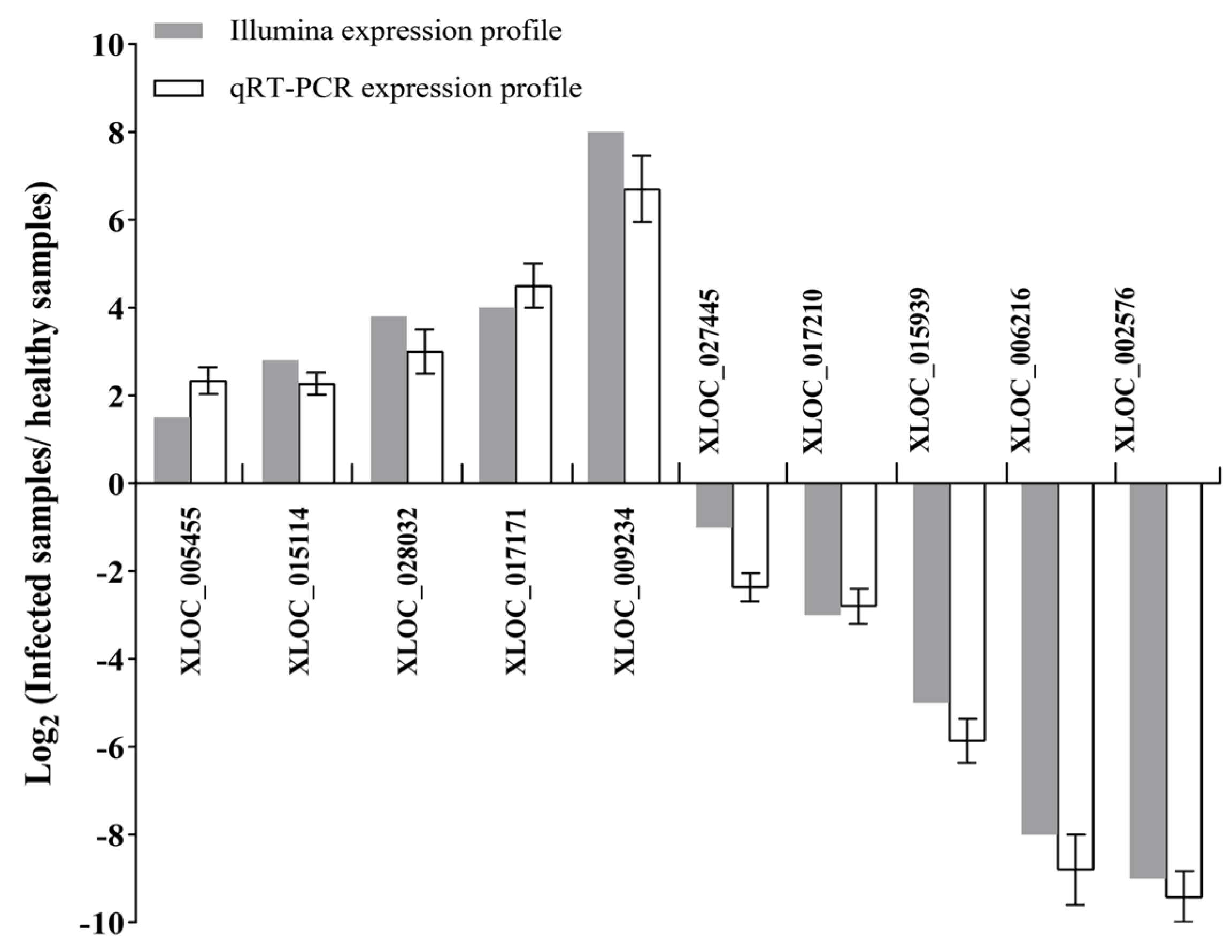
2.4. Gene Expression Analysis and qRT-PCR Validation

The expressions values of genes were calculated using the RPKM (reads per kb per million reads) method, and statistical analysis was carried out according to Poisson distribution [44]. Only the genes with expression values which changed more than twice (p ≤ 0.05) between healthy and phytoplasma-infected leaves were designated as significantly differentially expressed genes. To verify the reliability of the high-throughput analysis results, some mRNAs and lncRNAs were randomly selected following the principle of random sampling for qRT-PCR analysis, and the qRT-PCR was carried out on the CFX96TM real-time machine (Bio-Rad) using SYBR Green Premix Ex Taq TM (Takara Biotechnology, Tokyo, Japan). Then, the expression of the gene was normalized to EF1-a transcript abundance and calculated using the 2−ΔΔCT method [47], and the samples were analyzed in triplicate.

2.5. Gene Ontology Analysis

Assembled transcripts were used as query sequences for BLASTN searches against the reference M. notabilis database with online tools (https://morus.swu.edu.cn/morusdb/blast) to obtain the gene IDs for the transcripts, and gene ontology (GO) analysis was performed based on the gene IDs obtained using the online tool (https://morus.swu.edu.cn/morusdb/searchgo) with the ANNEX annotation augmentation function (version 2.3.1) according to the method described before [48,49].

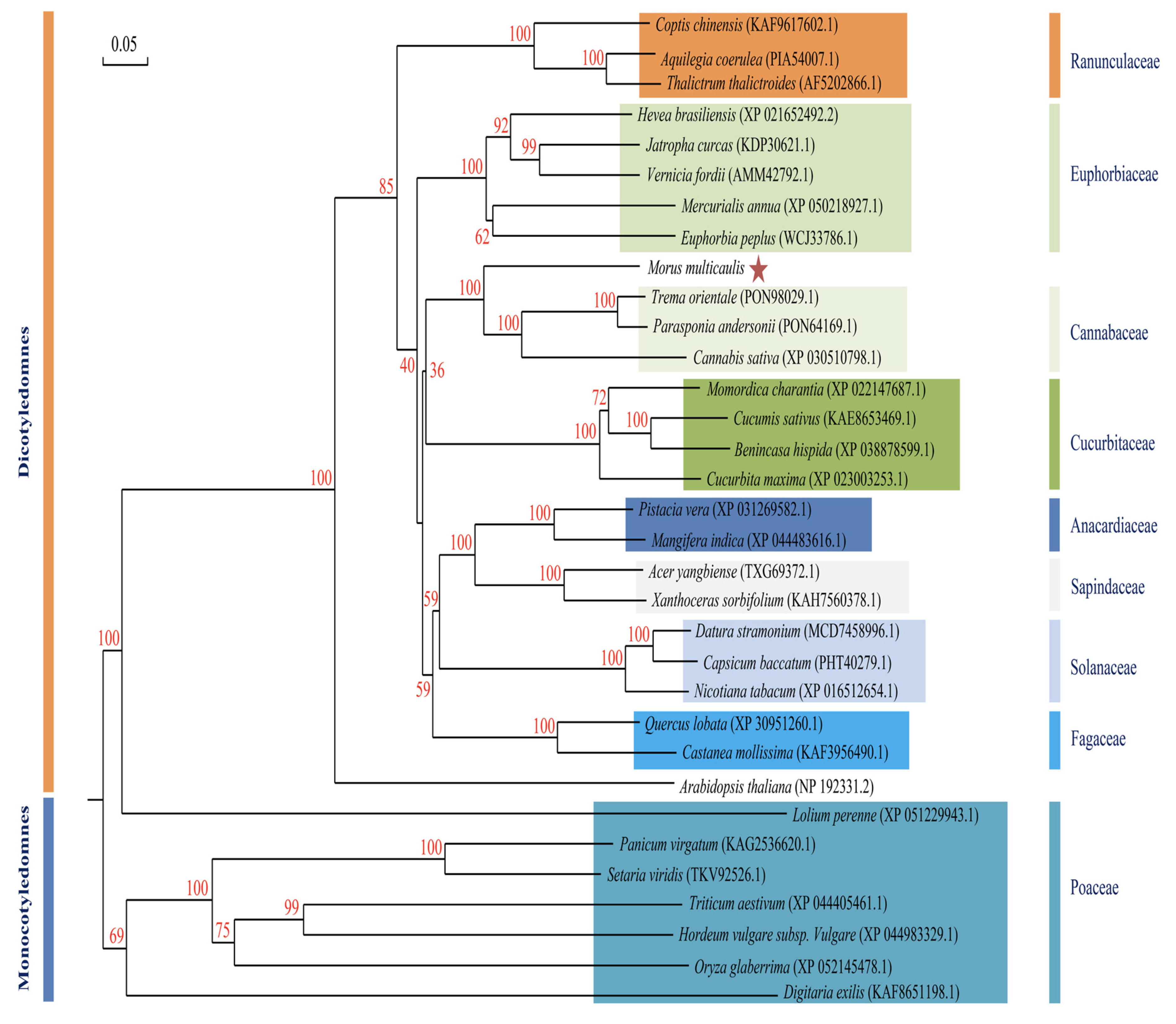
2.6. Gene Cloning and Phylogenetic Analysis

Reverse transcriptase M-MLV (Promega) was used to synthesize cDNA with the RNA isolated, and then the cDNA obtained was used for PCR amplifications with the specific primers (for MuLRR-RLK: ATGGCCAAACTAAGCCTCCTACT; CTAAGCTCTTCTCCTTCTCCGT; for MuLRR-RLK-AS: TGAGTTTTTGCATCGATGATGGGA; GTGCCTTCCCATCGCCATCGTT) designed based on the nucleotide sequence of the gene. Then, the target DNA fragment was recovered from the PCR products by electrophoresis and subcloned into the pMD18-T vector (Takara, Dalian, China) and sequenced. The multiple alignments between the amino acid sequences encoded by genes and the homologous proteins from other plants were performed using the DNAMAN program. The phylogenetic tree of the proteins from diverse species was generated with the neighbor-joining method in the MEGA program (version 6.0) with 1000 bootstraps. Modeling of the three-dimensional structure of the protein was performed using the SWISS-MODEL server.

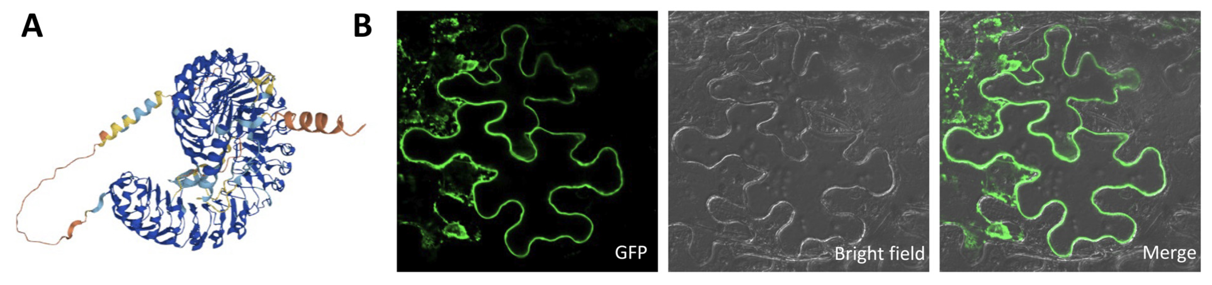
2.7. Subcellular Localization

To observe the subcellular localization of the target protein, the gene coding the protein was fused with the green fluorescent protein gene (GFP) and was cloned into the binary vector pBI121 under the control of 35S. The Agrobacteria containing the vector constructed above infiltrated the N. benthamiana leaf epidermal cells. Then, the small sections of the infiltrated leaves were excised 48 to 72 h after infiltration using a Bio-Rad MRC1024 confocal laser scanning microscope (Bio-Rad, Hemel Hempstead, UK) with an excitation wavelength of 488 nm and an emission wavelength of 505–530 nm at 400× magnification.

2.8. Promoter Activity Analysis

The specific primers were designed based on the M. notabilis genome (https://www.ncbi.nlm.nih.gov/genome/?term=Morus; accessed on 24 June 2013) to amplify a 2000 bp upstream region of the MuLRR-RLK (named pMuLRR-RLK) or MuLRR-RLK-AS gene (named pMuLRR-RLK-AS). Then, the 35S promoter in the binary vector pBI121 was replaced by the promoters, respectively, to create the expression vector pMuLRR-RLK::GUS or pMuLRR-RLK-AS::GUS. The vector created was then transferred into Agrobacterium strain GV3101, which was used to infiltrate tobacco leaves, and the GUS expression in the infiltrated leaves was assessed by histochemical staining [50].

2.9. Production of Transgenic Arabidopsis

For production of transgenic Arabidopsis, MuLRR-RLK or MuLRR-RLK-AS was introduced into the expression vector pBI121 under the control of the 35S promoter, respectively, to construct their expression vectors. Meanwhile, both the MuLRR-RLK and MuLRR-RLK-AS genes were ligated into the same modified expression vector pLGNL under the control of the 35S promoter, respectively, to construct their co-expression vector. Then, the vectors constructed were introduced into A. tumefaciens strain GV3101, which was used to transform Arabidopsis with the floral dip method [51]. The seeds of transgenic plants were selected, and the T3 generation transgenic seeds were used for subsequent experiments.

2.10. Plant Treatment

In the case of exogenous SA treatments, 5 mmol·L−1 SA solution was evenly sprayed onto the adaxial surfaces of mulberry leaves. The CFU of Pseudomonas syringae pv. tomato DC3000 (Pst. DC3000) assay was performed with the serial dilution method, and the bacterial suspensions (105 CFU·mL−1) of Pst. DC3000 were injected into the rosette leaves of Arabidopsis seedlings to perform inoculation. All of the inoculated seedlings above and the control mulberry seedlings sprayed or injected with sterilized water were incubated in a humidified 95% chamber for 48 h to ensure successful inoculation. The rosette leaves of Arabidopsis were detached, the adaxial surface of the leaves was inoculated with the mycelium plugs (Φ 2 mm) from the actively growing Botrytis cinerea colonies, and the leaves inoculated with PDA medium plugs (Φ 2 mm) were used as controls. All of the inoculated leaves and controls were placed in covered Petri dishes, and the disease severity was examined daily. All the treatments were conducted independently at least three times.

2.11. Detection of Colony-Forming Units

The leaves inoculated with Pst. DC3000 were ground in sterile water, and the suspension was serially diluted in a ten-fold series in sterile water. Then, 100 μL of each dilution was spread-plated onto King’s B medium and incubated at 28 °C. After 48 h of incubation, colony forming units were counted. All of the experiments were independently repeated at least three times.

2.12. Statistic Analysis

For the high-throughput sequencing analysis, three biological replicates with healthy and infected leaves were used for strand-specific transcriptome analysis. For gene function, expression, and other physiology and biochemistry assays, all the experiments were bioreplicated at least three times. The results were analyzed by analysis of variance, and when the p-values were less than 0.05, the difference was considered significant.

3. Results

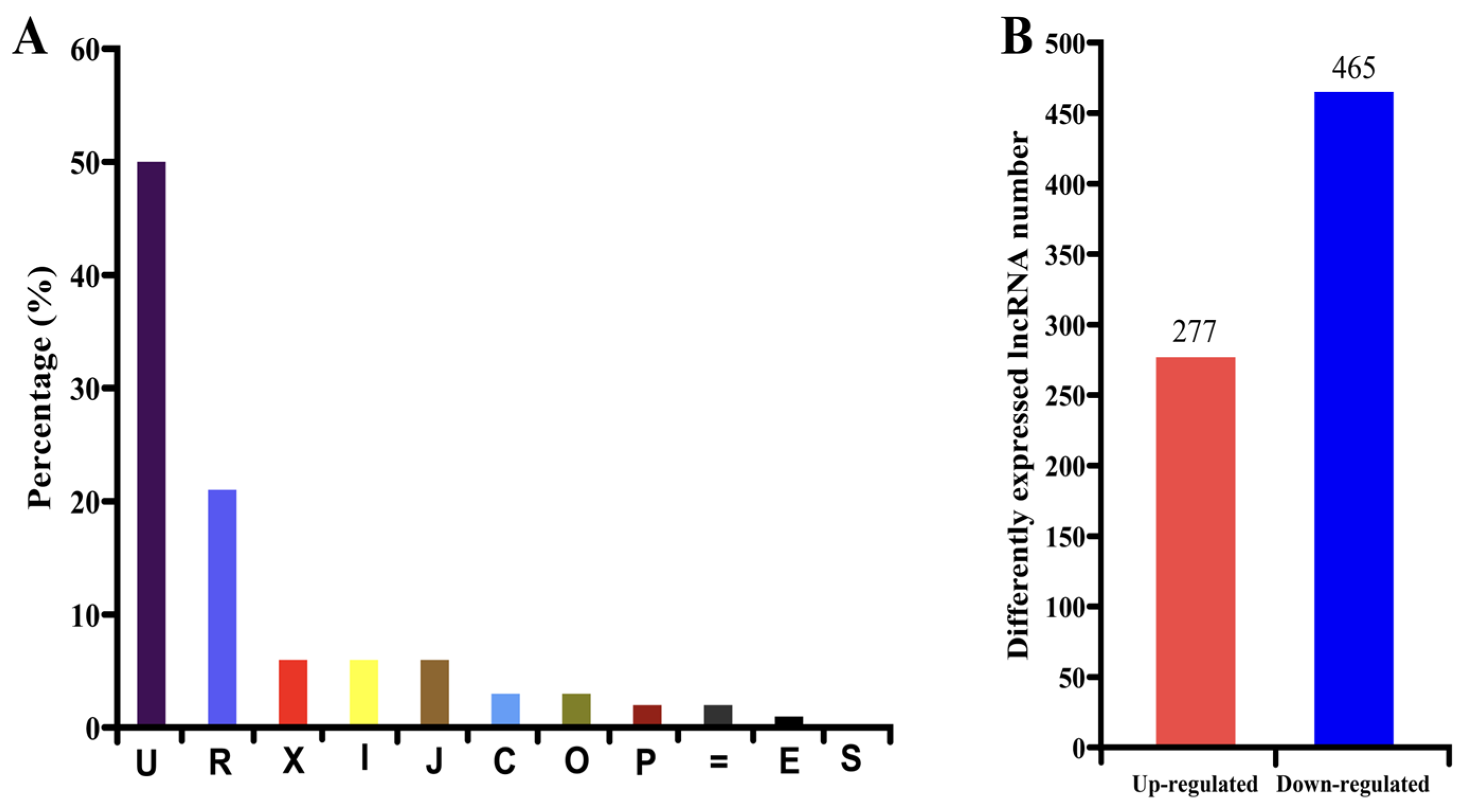
3.1. High-Throughput Sequencing and DEG Analysis

Using the Illumina platform, the strand-specific transcriptome libraries of healthy (HL) and phytoplasma-infected mulberry leaves (IL) were sequenced, and a total of 36.0 and 37.5 million clean reads were obtained in the HL and IL libraries, respectively. After calculating the expression level (FPKM) of each gene, a total of 4169 genes were found to be differentially expressed between HL and IL libraries, among which 1287 genes were up-regulated and 2882 genes were down-regulated in the IL libraries (Figure 1A). All of the differentially expressed genes (DEGs) detected are shown in Supplementary Table S1.
Through gene ontology (GO) analysis of the DEGs, they were classified into 17 functional categories (Figure 1B). The first category of genes was associated with cellular components and processes, the genes involved in translation and post-translation modification belonged to the second category, and the third category included the genes related to metabolic processes. Interestingly, it was found that 3% and 2% of the DEGs detected were associated with stress and environment response and signaling pathways, respectively. The other categories of DEGs included transcription and post-transcription modifications, carbohydrate metabolic processes, growth and development, etc. This indicates that the regulatory network of the genes in response to phytoplasma infection is complex in mulberry. The detailed functional GO terms of the DEGs are provided in Supplementary Table S1.
To verify the reliability of the gene expression profiles obtained by RNA-seq, 10 DEGs including the up-regulated and down-regulated genes responsive to phytoplasma infection were randomly selected for qRT-PCR validation (Figure 2). The qRT-PCR results showed that the expression levels of all the selected genes show similar change profiles between qRT-PCR and RNA-seq results, indicating that the results obtained by RNA-seq are reliable.

3.2. Identification and Characterization of lncRNAs

In addition to the above mRNAs, a large number of lncRNAs were also identified. In total, 1794 unique lncRNAs were obtained from the HL and IL libraries. Based on the locations of the lncRNAs obtained in the genome, 50.5% of the lncRNAs were located in the intergenic regions, 7.0% of the lncRNAs were exonic overlaps with known transcripts on the opposite strand, 7.0% of them were transfrags falling entirely within intron of known transcripts, and 6.9% of them were potentially novel isoforms (Figure 3A). To identify phytoplasma-responsive lncRNAs, the expression levels of the lncRNAs in HL samples and IL samples were evaluated. Of these 1794 lncRNAs, 742 lncRNAs were differentially expressed between HL samples and IL samples, including 277 up-regulated lncRNAs and 465 down-regulated lncRNAs (Figure 3B and Supplementary Table S2).
In order to confirm the reliability of the expression patterns of the lncRNAs obtained, the expression levels of 10 randomly selected lncRNAs were analyzed by qRT−PCR. The results showed consistent results between RNA-seq and qRT−PCR data (Figure 4), suggesting that the expression patterns of lncRNAs obtained are highly reliable, and the DE lncRNAs selected may play major roles in response to phytoplasma infection in mulberry.

3.3. Target Prediction of the Differentially Expressed lncRNAs (DELs)

Previous studies have shown that lncRNAs are preferentially located in the genomic regions adjacent to the genes they regulate. In order to reveal the potential functions and regulatory mechanism of lncRNAs in response to phytoplasma infection, firstly, the protein-coding genes located within <2 kb from the lncRNAs were predicted. A total of 621 genes were predicted as target genes for the 1122 lncRNAs, of which 465 were differentially expressed lncRNAs (DELs). Further analysis showed that 54 DEGs were potential target genes of 68 DELs, of which 13 were up-regulated and 41 were down-regulated in the infected leaves. Interestingly, it was found that the lncRNAs and their target genes not only exhibit the same expression trend, but also exhibit opposite expression trends. In addition, it was found that some target genes could be targeted by multiple lncRNAs, indicating that the regulatory mechanism of lncRNAs is complex (Table 1 and Supplementary Table S3). GO term analyses showed that these differentially expressed targeted genes were enriched in diverse biological processes, such as the metabolic process, development, defense responses, etc. (Table 1 and Supplementary Table S3). Therefore, these DELs may participate in the response to phytoplasma infection through various pathways by regulating the expression of their target genes.
Among the lncRNAs obtained, some are nature antisense transcripts (lncNATs) that were transcribed from the opposite strand of protein-coding regions and may regulate the expression of their sense transcripts. From our data, 378 lncNATs were identified and found to overlap with 365 protein-coding genes at their opposite strands. Among these lncNATs, there were only 44 differentially expressed lncNATs whose target genes were also differentially expressed. Furthermore, it was found that there were only nine lncNATs and their target genes showing the opposite expression tendency during phytoplasma infection (Table 2 and Supplementary Table S4). These lncNATs may also participate in the response to phytoplasma infection in mulberry by regulating the expression of corresponding genes.

3.4. Characterization of MuLRR-RLK-AS Trans-Target Gene MuLRR-RLK

Our data showed that the expression level of lncNAT XLOC_027445 (LRR-RLK-AS) was decreased, while the expression level of its trans-target gene, the putative LRR receptor-like serine/threonine-protein kinase gene (LRR-RLK), was increased in the phytoplasma-infected leaves, and these results were confirmed by qRT-PCR (Figure 2 and Figure 4). As we have seen, the LRR-RLK gene has not been reported to be associated with phytoplasma infection, and the gene in mulberry has not been characterized. To explore the roles of the LRR-RLK and LRR-RLK-AS in the response to phytoplasma infection in mulberry, the LRR-RLK gene was cloned from mulberry (M. multicaulis Perr.) and designated as MuLRR-RLK. The MuLRR-RLK gene encodes a protein of 819 amino acid residues, with a predicted molecular weight of 89.923 kDa and a pI of 5.98. Multiple sequence alignment analysis showed that the amino acids of MuLRR-RLK protein have high sequence identity with other plant LRR-RLK proteins, indicating that MuLRR-RLK is an evolutionarily conserved protein (Figure 5A). Protein structure analysis showed that MuLRR-RLK has structural characteristics of the LRR-RLK protein family, including a signaling peptide (SP) (1–20aa), four leucine-rich repeats (LRR) (112–173aa; 210–269aa; 520–577aa; 658–716aa) at the N-terminus, a transmembrane (TM) domain (784–809aa), and an intracellular Ser/Thr kinase domain (KD) at the C-terminus (Figure 5B).
Phylogenetic analysis of MuLRR-RLK and LRR-RLKs from other plants showed that the closest homology was between MuLRR-RLK and LRR-RLKs from Trema orientale and Cannabis sativa (Figure 6). Subcellular localization analysis showed that MuLRR-RLK localized to the plasma membrane as other LRR-RLK proteins (Figure 7), suggesting it may have similar roles as other LRR-RLK proteins which play important roles in perceiving and transmitting signals arising from different environmental conditions.

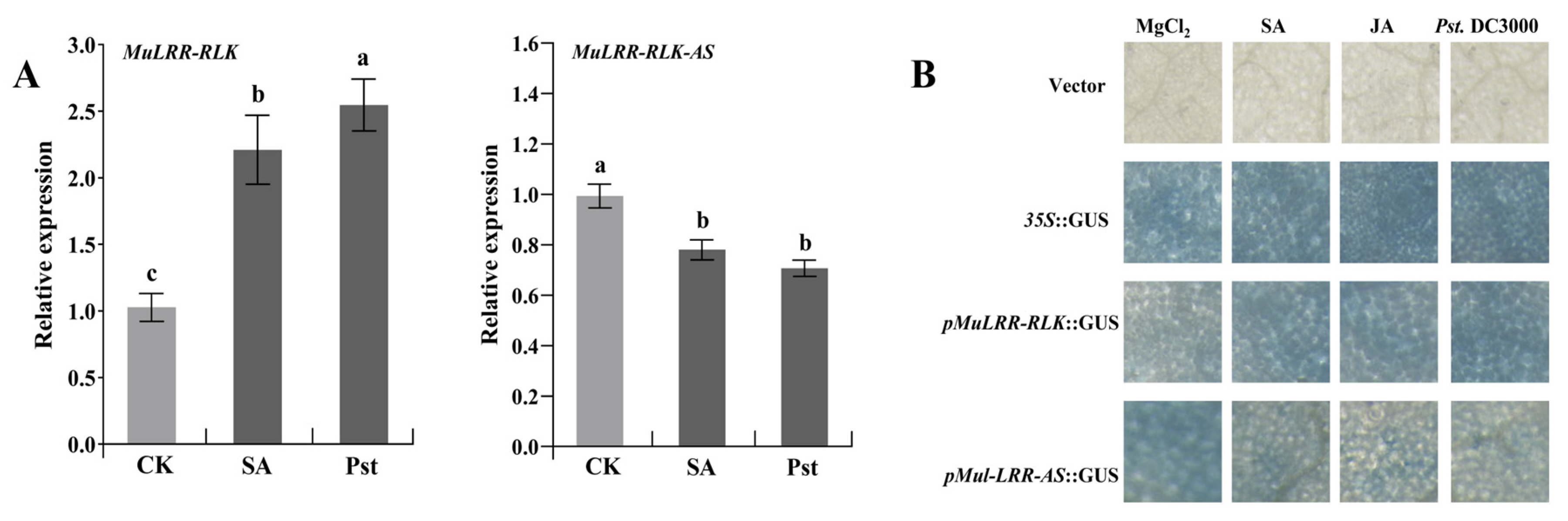
3.5. Expression Patterns of MuLRR-RLK and MuLRR-RLK-AS

To analyze the induced expression patterns of MuLRR-RLK and MuLRR-RLK-AS, the mulberry seedlings were challenged with Pst. DC3000 or SA, respectively. qRT-PCR analysis showed that the expression level of MuLRR-RLK was increased in the leaves challenged by Pst. DC3000 or SA. However, the expression level of MuLRR-RLK-AS was decreased in the leaves challenged (Figure 8A). Meanwhile, the putative promoters of the MuLRR-RLK and MuLRR-RLK-AS were cloned and submitted to the PlantCARE database to detect their cis-elements. The results showed that besides the common TATA-boxes and basic cis-acting elements, both pMuLRR-RLK-AS and pMuLRR-RLK contain various abiotic and biotic stress-responsive elements, including ABRE elements, MBS elements, TCA elements, G-boxes, ARE, Box4, P-boxes, TATC-boxes, etc. (Table 3 and Table 4). In addition, the pMuLRR-RLK or pMuLRR-RLK-AS was fused with GUS and transiently expressed in the tobacco leaves. Staining results showed that the GUS gene driven by pMuLRR-RLK was induced after inoculation of Pst. DC3000 or SA treatment (Figure 8B), while the GUS gene driven by pMuLRR-RLK-AS was repressed after inoculation of Pst. DC3000 or SA treatment. These data indicated that MuLRR-RLK and MuLRR-RLK-AS may be involved in mulberry resistance to pathogens, and SA may modulate their expression in mulberry.

3.6. MuLRR-RLK-AS Represses the Expression of MuLRR-RLK

Because an efficient regeneration system has not yet been established in mulberry, it is difficult to obtain transgenic mulberry trees. The nucleotide sequence identity of MuLRR-RLK and its homologue in Arabidopsis was low (74.6%). To verify the suppression of MuLRR-RLK-AS on the expression of MuLRR-RLK, the expression vector of MuLRR-RLK and the vector co-expressing MuLRR-RLK and MuLRR-RLK-AS were constructed, respectively, and transgenic Arabidopsis plants overexpressing MuLRR-RLK (OE) or co-expressing MuLRR-RLK and MuLRR-RLK-AS (CO-OE) were generated. The results of genome PCR analysis showed that both MuLRR-RLK and MuLRR-RLK-AS genes were integrated into the Arabidopsis genome (Figure 9A,B). Moreover, qRT−PCR analysis results indicated that the MuLRR-RLK gene was successfully expressed in the OE plants, and the expression levels of MuLRR-RLK were significantly higher in the OE Arabidopsis than those in the WT plants (Figure 9C). Although the expression levels of MuLRR-RLK-AS were significantly higher in the CO-OE plants than those in the WT plants, the expression level of MuLRR-RLK was significantly lower than that in the OE plants. This confirmed that the expression of MuLRR-RLK-AS effectively represses the expression of MuLRR-RLK.

3.7. MuLRR-RLK-AS Is a Negative Regulator of Plant Disease Resistance

Firstly, the CK, OE, and CO-OE Arabidopsis plants obtained above were challenged by B. cinerea to explore the possible role of MuLRR-RLK-AS in response to fungal pathogen infection. Four DAI, obviously expanding necrotic lesions were observed around the inoculated sites on the inoculated leaves of CK and CO-OE plants. In contrast, only mild disease symptoms could be observed on the surface of the inoculated leaves of OE plants (Figure 10A). In addition, the CK, OE, and CO-OE Arabidopsis plants were inoculated with Pst. DC3000 to explore the roles of MuLRR-RLK-AS in the response to pathogen infection. Three days after inoculation (DAI), the challenged leaves of CK and CO-OE plants showed obvious grayish brown lesion with chlorosis around the inoculation sites, especially the samples of CK showing severe disease symptoms. In contrast, no obvious disease symptoms were observed in the leaves of OE plants, although occasional mild chlorosis or necrosis was observed (Figure 10B,C). In addition, the bacterial populations of Pst. DC3000 strains in the inoculated leaves were determined, and the results showed that the strain number in the OE plants leaves was the lowest. Although the strain number in the CO-OE leaves was lower than that in the CK leaves, it was still significantly higher than those in the OE leaves (Figure 10D). This is consistent with the symptom phenotype of the leaves, indicating that overexpression of MuLRR-RLK-AS may inhibit the expression of MuLRR-RLK, weakening the resistance of the plant to Pst. DC3000. These results indicate that overexpression of MuLRR-RLK in Arabidopsis enhanced resistance to Pst. DC3000 and B. cinerea, while MuLRR-RLK-AS weakens plant disease resistance by repressing the expression of MuLRR-RLK.

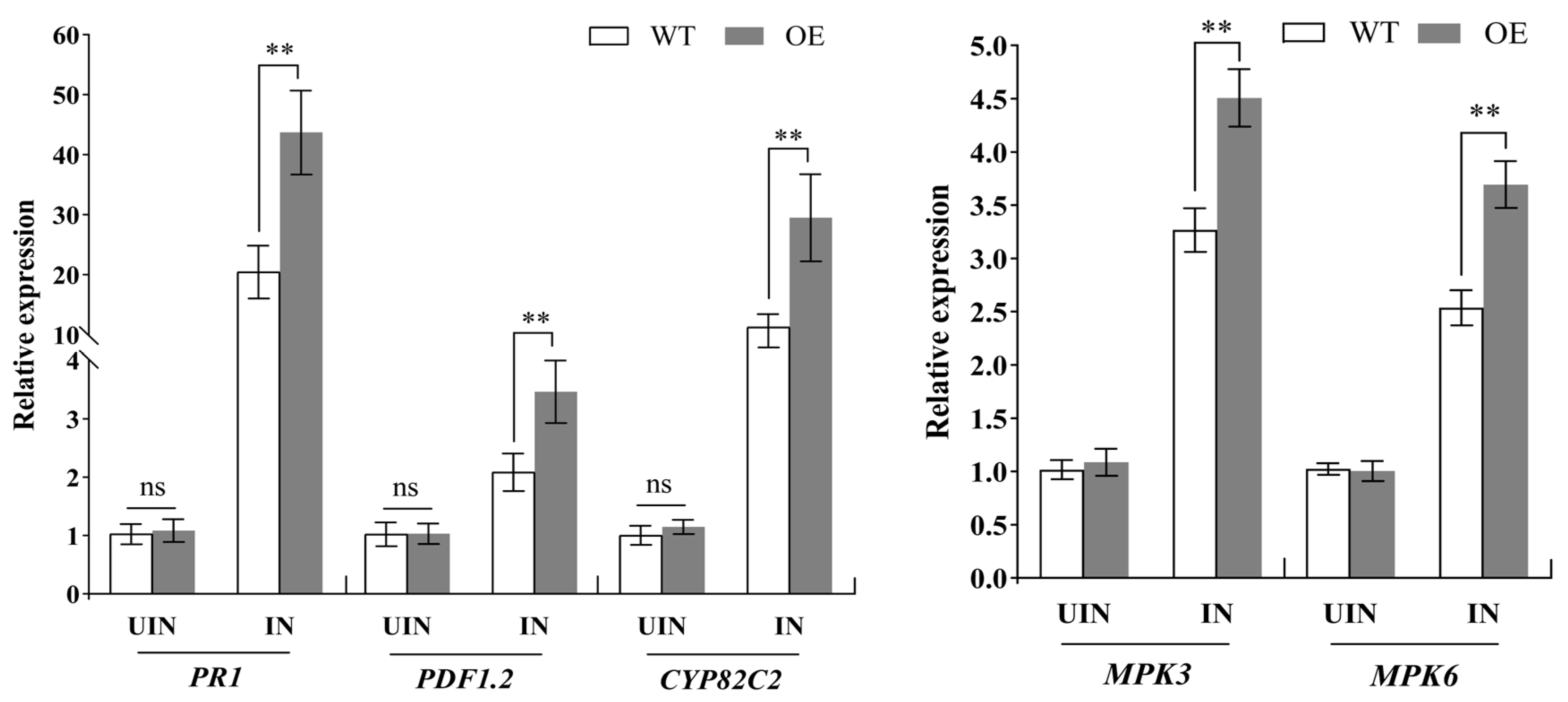
3.8. Ectopic Expression of MuLRR-RLK Affects the Expression of Defense-Related Genes

According to the above results, MuLRR-RLK-AS weakens plant disease resistance by repressing the expression of MuLRR-RLK. To explore why the MuLRR-RLK gene was involved in plant disease resistance, the expression change in some defense-related genes in the transgenic MuLRR-RLK plants was evaluated. The results showed that there was no significant difference in the expression levels of the pathogenesis-related protein 1 gene (PR-1), plant defensin gene (PDF1.2), and cytochrome P450 protein gene (CYP82C2) between wild-type and transgenic MuLRR-RLK plants, and their expression levels were all very low. This indicates that overexpression of the MuLRR-RLK gene in Arabidopsis may not affect the basic expression levels of these defense-related genes without pathogen inoculation. However, it was found that 24 h post-inoculation with Pst. DC3000, the expression levels of PR-1, PDF1.2, and CYP82C2 in the transgenic MuLRR-RLK Arabidopsis plants were higher than those in the wild-type plants (Figure 11). Since the MPK3 and MPK6 genes have been shown to have important roles in plant innate immune responses, to determine whether MuLRR-RLK affects the expression of the two genes, their expression levels in the MuLRR-RLK-overexpressing and wild-type Arabidopsis were measured. Similar to the expression patterns of the disease resistance-related genes mentioned above, there was no significant difference in the expression levels of MPK3 and MPK6 genes between the wild-type and transgenic MuLRR-RLK plants without pathogen inoculation. However, the expression levels of the two genes in transgenic plants were significantly higher than those in the wild-type plants when the plants were inoculated with Pst. DC3000 (Figure 11). These results indicate that when plants are infected with pathogens, MuLRR-RLK may play an important role in the disease resistance response by activating the MAPK cascade reaction, regulating the expression of defense genes.

4. Discussion

4.1. LncRNAs Are Involved in Regulating Gene Expression in Both Cis- and Trans-Manners in Response to Phytoplasma Infection

Recently, lncRNAs have been recognized as important regulators of plant responses to biotic and abiotic stresses [52]. In this study, an RNA-seq approach was used to investigate the transcriptomic changes in response to phytoplasma infection in mulberry, and 1794 novel lncRNAs were identified. To our knowledge, this is the first work to globally identify the lncRNAs responsive to phytoplasma infection in mulberry, and the results provided here can be useful for future research in this direction.
Previous studies have shown that lncRNAs, as cis- and trans-regulators, may regulate gene expression at both the transcriptional and posttranscriptional levels in various ways [53]. Cis-acting lncRNAs usually regulate the transcription of genes in close genomic proximity by recruiting or displacing transcription factors at the promoters of the neighboring genes [54]. To explore the potential roles of the cis-acting lncRNAs in response to phytoplasma infection in mulberry, an integrated DE lncRNA and coding gene analysis was performed to identify the cis-regulatory networks of lncRNAs, and 68 lncRNA-mRNA pairs were found. Furthermore, it was found that there were 37 lncRNAs with a positive correlation with their neighboring mRNAs, while only 31 neighboring lncRNA–mRNA pairs were found to be negatively correlated (Table 1). This suggests that the lncRNAs can cis-regulate the neighboring protein-coding genes in both positive and negative ways in response to phytoplasma infection in mulberry. However, the mechanism of the relationship between lncRNA–mRNA pairs needs to be further studied.
In addition to cis-regulatory roles, lncRNAs transcribed from an opposite strand of sense RNA in the same genomic regions (known as natural antisense transcript, lncNAT) may regulate the expression of sense RNA as trans-regulators by transcriptional or post-transcriptional mechanisms [13,55]. In this study, 44 DE mRNA–lncNAT pairs were identified (Table 2). Although the negative regulatory effects of lncNATs on the coding genes are more expectable, there were only nine DE lncNATs. Their target transcripts showed the opposite expression tendency, and the expression of most of DE lncNATs positively correlates with the expression of their targets (Table 2). It was suggested that some plant lncNATs can facilitate DNA methylation, histone modifications, chromatin conformation changes, and eventually up-regulate or down-regulate the transcription of their sense genes at the translational level. In addition, some plant lncNATs may affect mRNA decay by nucleases, mask miRNA binding sites, modulate protein translation, or produce endogenous siRNAs to execute RNA interference at the post-transcription level [22,56,57,58,59]. Therefore, further studies are needed to understand the action mechanism of these lncNATs.
Furthermore, GO enrichment analysis of the targeted genes of these DE cis- and trans-acting lncRNAs indicated that they were associated with various biological processes, such as the metabolic process, signal transduction, development, transcription regulatory, etc. (Supplementary Tables S3 and S4). Therefore, the DE lncRNAs may play important roles in diverse biologic processes, and their regulatory networks involved in the response to phytoplasma infection in mulberry are intricate. On one hand, the changes in these DE lncRNAs may disturb the expression of the genes involved in the normal growth and developmental and metabolic processes, resulting in the symptoms of shoot proliferation, witches’-broom, yellowing of the leaves, etc. On the other hand, the changes in DE lncRNAs may alter the expression of some resistance genes, thereby improving the resistance of mulberry to phytoplasma. Further experiments are required to uncover the action mechanism of these lncRNA–mRNA pairs that modulate the response of mulberry to phytoplasma.

4.2. MuLRR-RLK-AS Plays an Important Role in Regulating MuLRR-RLK Gene Expression in Response to Phytoplasma Infection

Leucine-rich repeat receptor-like protein kinases (LRR-RLKs) comprise one of the largest groups of receptor-like protein kinases (RLKs), which serve as receptors for signaling transduction pathways in plants [60,61]. It was suggested that LRR-RLKs are highly conserved and usually contain an extracellular, tandemly organized LRR domain, a single-pass transmembrane domain, and a functional protein kinase domain [62]. Our study showed that MuLRR-RLK protein structure has a high homology to other LRR-RLK proteins (Figure 5), indicating that it might have similar biological functions as other LRR-RLK proteins in the same subfamilies. It was reported that LRR-RLKs play vital roles in plant growth, development, and the responses to environmental stress, and some LRR-RLKs are associated with plant resistance to bacterial and fungal pathogen invasion [63,64,65]. However, to our knowledge, this is the first report that LRR-RLK genes are associated with the response to phytoplasma infection. In this study, MuLRR-RLK was identified as a phytoplasma-responsive gene. In addition, our data showed that although there was no significant difference in the expressions of PTI-related and MAPK genes between transgenic MuLRR-RLK and wild-type Arabidopsis plants, the expression levels of these genes in the transgenic plants were significantly higher than those in wild-type plants when inoculated with Pst. DC3000 (Figure 11). Meanwhile, our results showed that the MuLRR-RLK protein is localized on the plasma membrane (Figure 7B), and ectopic overexpression of the MuLRR-RLK gene confers the transgenic Arabidopsis plants with enhanced disease tolerance to Pst. DC3000 and B. cinerea (Figure 10). These results indicated that MuLRR-RLK might be involved in sensing signals arising from biotic stress and regulating defense responses, and the expression of MuLRR-RLK may be a strategy to overcome phytoplasma in diseased mulberry plants. Further research is needed to determine the molecular mechanism by which MuLRR-RLK senses the signals triggered by phytoplasma infection and mediates the resistance response to phytoplasma in mulberry.
Our results indicate that the expression of the MuLRR-RLK gene and the activity of its promoter are both induced by SA, while SA inhibits the expression of the MuLRR-RLK-AS gene and the activity of its promoter (Figure 8). It was reported that the levels of endogenous SA in the phytoplasma-infected plants are significantly increased [66]. Therefore, the increase in endogenous SA in the phytoplasma-infected leaves not only enhances the transcriptional activity of the promoter of MuLRR-RLK and increases the expression of MuLRR-RLK, but also reduces the post-transcriptional inhibitory effects of MuLRR-RLK-AS on MuLRR-RLK by inhibiting the expression of MuLRR-RLK-AS, ultimately leading to an increase in the expression of MuLRR-RLK. In the absence of pathogen infection, the SA level in plants is very low, and the expression level of MuLRR-RLK-AS may be higher, which inhibits the expression of MuLRR-RLK and thus maintains the expression of disease resistance-related genes at a low level. This may be beneficial for maintaining normal plant growth and development. Therefore, the SA level in plants may be an important factor in balancing the expression levels of MuLRR-RLK-AS and MuLRR-RLK, and the regulation of MuLRR-RLK-AS on the expression of MuLRR-RLK mediated by SA may be an important resistance response of mulberry to phytoplasma infection. Further research is needed to elucidate the exact mechanism behind this process.

5. Conclusions

In conclusion, the dynamic expression profiles of lncRNAs during the response to phytoplasma infection in mulberry were explored using high-throughput sequencing, and the DE lncRNAs and their target genes were identified. Moreover, the roles of the DE lncRNAs were discussed. Our results proved that MuLRR-RLK-AS acting as a trans-regulator can regulate the expression of MuLRR-RLK, which is a positive regulator of mulberry resistance. The information provided is particularly useful for understanding the functions and mechanisms of lncRNAs during the response to phytoplasma infection in mulberry.

Supplementary Materials

The following supporting information can be downloaded at: https://www.mdpi.com/article/10.3390/biom14030308/s1, Table S1. Differentially expressed genes between healthy and infected mulberry leaves. Table S2. Differentially expressed lnRNAs between healthy and infected mulberry leaves. Table S3. Association analysis of differentially expressed lncRNAs and their co-localization differentially expressed target genes. Table S4. Association analysis of the differentially expressed lncRNAs and their differentially expressed target genes.

Author Contributions

Visualization and supervision, Y.G. and X.J.; Performed the experiments, Z.L. (Zixuan Liu), C.L. and T.Z.; Analyzed the data, L.Y., Q.S. and G.W.; Writing—review and editing, Z.L. (Zhaoyang Liu). All authors have read and agreed to the published version of the manuscript.

Funding

This work was supported by the National Natural Science Foundation of China (32172799 and 32171748) and the Modern Agricultural Technology System of Shandong Province (No. SDAIT-18-04).

Data Availability Statement

The transcriptome data have been deposited to GenBank with the dataset code SRR28089553 and SRR28089552.

Conflicts of Interest

The authors declare no conflict of interest.

References

  1. Dhanyalakshmi, K.H.; Nataraja, K.N. Mulberry (Morus spp.) has the features to treat as a potential perennial model system. Plant Signal Behav. 2018, 13, e1491267. [Google Scholar] [CrossRef]
  2. Yuan, Q.; Zhao, L. The mulberry (Morus alba L.) Fruit-a review of characteristic components and health benefits. J. Agric. Food Chem. 2017, 65, 10383–10394. [Google Scholar] [CrossRef] [PubMed]
  3. He, N.; Zhang, C.; Qi, X.; Zhao, S.; Tao, Y.; Yang, G.; Lee, T.H.; Wang, X.; Cai, Q.; Li, D.; et al. Draft genome sequence of the mulberry tree. Morus Notabilis. Nat. Commun. 2013, 4, 2445. [Google Scholar] [CrossRef] [PubMed]
  4. Liu, C.; Dong, X.; Xu, Y.; Dong, Q.; Wang, Y.; Gai, Y.; Ji, X. Transcriptome and DNA methylome reveal insights into phytoplasma infection responses in mulberry (Morus multicaulis Perr.). Front. Plant Sci. 2021, 12, 697702. [Google Scholar] [CrossRef] [PubMed]
  5. Jiang, H.; Wei, W.; Saiki, T.; Kawakita, H.; Watanabe, K.; Sato, M. Distribution patterns of mulberry dwarf phytoplasma in reproductive organs, winter buds, and roots of mulberry trees. J. Gen. Plant Pathol. 2004, 70, 168–173. [Google Scholar] [CrossRef]
  6. Luo, L.; Zhang, X.; Meng, F.; Wang, Y.; Zhou, Y.; Liu, J. Draft genome sequences resources of mulberry dwarf phytoplasma strain MDGZ-01 associated with mulberry yellow dwarf (MYD) diseases. Plant Dis. 2022, 106, 2239–2242. [Google Scholar] [CrossRef] [PubMed]
  7. Gai, Y.P.; Yuan, S.S.; Liu, Z.Y.; Zhao, H.N.; Liu, Q.; Qin, R.L.; Fang, L.J.; Ji, X.L. Integrated phloem sap mRNA and protein expression analysis reveals phytoplasma-infection responses in mulberry. Mol. Cell Proteom. 2018, 17, 1702–1719. [Google Scholar] [CrossRef]
  8. Gai, Y.P.; Zhao, H.N.; Zhao, Y.N.; Zhu, B.S.; Yuan, S.S.; Li, S.; Guo, F.Y.; Ji, X.L. MiRNA-seq-based profiles of miRNAs in mulberry phloem sap provide insight into the pathogenic mechanisms of mulberry yellow dwarf disease. Sci. Rep. 2018, 8, 812. [Google Scholar] [CrossRef]
  9. Gai, Y.P.; Han, X.J.; Li, Y.Q.; Yuan, C.Z.; Mo, Y.Y.; Guo, F.Y.; Liu, Q.X.; Ji, X.L. Metabolomic analysis reveals the potential metabolites and pathogenesis involved in mulberry yellow dwarf disease. Plant Cell Environ. 2014, 37, 1474–1490. [Google Scholar] [CrossRef] [PubMed]
  10. Gai, Y.P.; Li, Y.Q.; Guo, F.Y.; Yuan, C.Z.; Mo, Y.Y.; Zhang, H.L.; Wang, H.; Ji, X.L. Analysis of phytoplasma-responsive sRNAs provide insight into the pathogenic mechanisms of mulberry yellow dwarf disease. Sci. Rep. 2014, 4, 5378. [Google Scholar] [CrossRef]
  11. Song, L.; Fang, Y.; Chen, L.; Wang, J.; Chen, X. Role of non-coding RNAs in plant immunity. Plant Commun. 2021, 2, 100180. [Google Scholar] [CrossRef] [PubMed]
  12. Mercer, T.R.; Dinger, M.E.; Mattick, J.S. Long non-coding RNAs: Insights into functions. Nat. Rev. Genet. 2009, 10, 155–159. [Google Scholar] [CrossRef] [PubMed]
  13. Xu, Y.C.; Zhang, J.; Zhang, D.Y.; Nan, Y.H.; Ge, S.; Guo, Y.L. Identification of long noncoding natural antisense transcripts (lncNATs) correlated with drought stress response in wild rice (Oryza nivara). BMC Genom. 2021, 22, 424. [Google Scholar] [CrossRef] [PubMed]
  14. Zhang, L.; Liu, J.; Cheng, J.; Sun, Q.; Zhang, Y.; Liu, J.; Li, H.; Zhang, Z.; Wang, P.; Cai, C.; et al. lncRNA7 and lncRNA2 modulate cell wall defense genes to regulate cotton resistance to Verticillium wilt. Plant Physiol. 2022, 189, 264–284. [Google Scholar] [CrossRef] [PubMed]
  15. Cui, J.; Jiang, N.; Meng, J.; Yang, G.; Liu, W.; Zhou, X.; Ma, N.; Hou, X.; Luan, Y. LncRNA33732-respiratory burst oxidase module associated with WRKY1 in tomato- Phytophthora infestans interactions. Plant J. 2019, 97, 933–946. [Google Scholar] [CrossRef]
  16. Jiang, N.; Cui, J.; Hou, X.; Yang, G.; Xiao, Y.; Han, L.; Meng, J.; Luan, Y. Sl-lncRNA15492 interacts with Sl-miR482a and affects Solanum lycopersicum immunity against Phytophthora infestans. Plant J. 2020, 103, 1561–1574. [Google Scholar] [CrossRef]
  17. Hong, Y.; Zhang, Y.; Cui, J.; Meng, J.; Chen, Y.; Zhang, C.; Yang, J.; Luan, Y. The lncRNA39896-miR166b-HDZs module affects tomato resistance to Phytophthora infestans. J. Integr. Plant Biol. 2022, 64, 1979–1993. [Google Scholar] [CrossRef]
  18. Liu, G.; Liu, F.; Wang, Y.; Liu, X. A novel long noncoding RNA CIL1 enhances cold stress tolerance in Arabidopsis. Plant Sci. 2022, 323, 111370. [Google Scholar] [CrossRef]
  19. Liu, N.; Xu, Y.; Li, Q.; Cao, Y.; Yang, D.; Liu, S.; Wang, X.; Mi, Y.; Liu, Y.; Ding, C.; et al. A lncRNA fine-tunes salicylic acid biosynthesis to balance plant immunity and growth. Cell Host Microbe. 2022, 30, 1124–1138.e8. [Google Scholar] [CrossRef] [PubMed]
  20. Zhou, Y.F.; Zhang, Y.C.; Sun, Y.M.; Yu, Y.; Lei, M.Q.; Yang, Y.W.; Lian, J.P.; Feng, Y.Z.; Zhang, Z.; Yang, L.; et al. The parent-of-origin lncRNA MISSEN regulates rice endosperm development. Nat. Commun. 2021, 12, 6525. [Google Scholar] [CrossRef] [PubMed]
  21. Ransohoff, J.D.; Wei, Y.; Khavari, P.A. The functions and unique features of long intergenic non-coding RNA. Nat. Rev. Mol. Cell Biol. 2018, 19, 143–157. [Google Scholar] [CrossRef]
  22. Wang, Y.; Deng, X.W.; Zhu, D. From molecular basics to agronomic benefits: Insights into noncoding RNA-mediated gene regulation in plants. J. Integr. Plant Biol. 2022, 64, 2290–2308. [Google Scholar] [CrossRef] [PubMed]
  23. Quinn, J.; Chang, H. Unique features of long non-coding RNA biogenesis and function. Nat. Rev. Genet. 2016, 17, 47–62. [Google Scholar] [CrossRef] [PubMed]
  24. Wang, Z.; Liang, S.; Liu, S.; Meng, Z.; Wang, J.; Liang, S. Sequence pre-training-based graph neural network for predicting lncRNA-miRNA associations. Brief. Bioinform. 2023, 24, bbad317. [Google Scholar] [CrossRef] [PubMed]
  25. Ai, G.; Li, T.; Zhu, H.; Dong, X.; Fu, X.; Xia, C.; Pan, W.; Jing, M.; Shen, D.; Xia, A.; et al. BPL3 binds the long non-coding RNA nalncFL7 to suppress FORKED-LIKE7 and modulate HAI1-mediated MPK3/6 dephosphorylation in plant immunity. Plant Cell 2023, 35, 598–616. [Google Scholar] [CrossRef]
  26. Li, C.; Lai, X.; Yu, X.; Xiong, Z.; Chen, J.; Lang, X.; Feng, H.; Wan, X.; Liu, K. Plant long noncoding RNAs: Recent progress in understanding their roles in growth, development, and stress responses. Biochem. Biophys. Res. Commun. 2023, 671, 270–277. [Google Scholar] [CrossRef]
  27. Wang, J.Y.; Yu, W.G.; Yang, Y.W.; Li, X.; Chen, T.Z.; Liu, T.L.; Ma, N.; Yang, X.; Liu, R.Y.; Zhang, B.L. Genome-wide analysis of tomato long non-coding RNAs and identification as endogenous target mimic for microRNA in response to TYLCV infection. Sci. Rep. 2015, 5, 16946. [Google Scholar] [CrossRef]
  28. Zhou, Y.; Cho, W.K.; Byun, H.S.; Chavan, V.; Kil, E.J.; Lee, S.; Hong, S.W. Genome wide identification of long non-coding RNAs in tomato plants irradiated by neutrons followed by infection with tomato yellow leaf curl virus. PeerJ 2019, 7, e6286. [Google Scholar] [CrossRef]
  29. Zhang, T.; Liang, Q.; Li, C.; Fu, S.; Kundu, J.K.; Zhou, X.; Wu, J. Transcriptome analysis of rice reveals the lncRNA-mRNA regulatory network in response to rice black-streaked dwarf virus infection. Viruses 2020, 12, 951. [Google Scholar] [CrossRef]
  30. Gao, R.; Liu, P.; Irwanto, N.; Loh, R.; Wong, S.M. Upregulation of LINC-AP2 is negatively correlated with AP2 gene expression with Turnip crinkle virus infection in Arabidopsis thaliana. Plant Cell Rep. 2016, 35, 2257–2267. [Google Scholar] [CrossRef]
  31. Kang, S.H.; Sun, Y.D.; Atallah, O.O.; Huguet-Tapia, J.C.; Noble, J.D.; Folimonova, S.Y. A long non-coding RNA of Citrus tristeza virus: Role in the virus interplay with the host immunity. Viruses 2019, 11, 436. [Google Scholar] [CrossRef]
  32. Zhu, Q.H.; Stephen, S.; Taylor, J.; Helliwell, C.A.; Wang, M.B. Long noncoding RNAs responsive to Fusarium oxysporum infection in Arabidopsis thaliana. New Phytol. 2014, 201, 574–584. [Google Scholar] [CrossRef]
  33. Wang, N.; Cao, P.; Xia, W.; Fang, L.; Yu, H. Identification and characterization of long non-coding RNAs in response to early infection by Melampsoralarici-populina using genome-wide high-throughput RNA sequencing. Tree Genet. Genomes. 2017, 13, 34. [Google Scholar] [CrossRef]
  34. Yao, Z.; Chen, Q.; Chen, D.; Zhan, L.; Zeng, K.; Gu, A.; Zhou, J.; Zhang, Y.; Zhu, Y.; Gao, W.; et al. The susceptibility of sea-island cotton recombinant inbred lines to Fusarium oxysporum f. sp. vasinfectum infection is characterized by altered expression of long noncoding RNAs. Sci. Rep. 2019, 9, 2894. [Google Scholar] [CrossRef]
  35. Jiang, N.; Cui, J.; Shi, Y.; Yang, G.; Zhou, X.; Hou, X.; Meng, J.; Luan, Y. Tomato lncRNA23468 functions as a competing endogenous RNA to modulate NBS-LRR genes by decoying miR482b in the tomato-Phytophthora infestans interaction. Hortic. Res. 2019, 6, 28. [Google Scholar] [CrossRef]
  36. Zhao, X.; Sun, X.; Chen, Y.; Wu, H.; Liu, Y.; Jiang, Y.; Xie, F.; Chen, Y. Mining of long non-coding RNAs with target genes in response to rust based on full-length transcriptome in Kentucky bluegrass. Front. Plant Sci. 2023, 14, 1158035. [Google Scholar] [CrossRef] [PubMed]
  37. Wang, Z.; Liu, Y.; Li, L.; Li, D.; Zhang, Q.; Guo, Y.; Wang, S.; Zhong, C.; Huang, H. Whole transcriptome sequencing of Pseudomonas syringae pv. actinidiae-infected kiwifruit plants reveals species specific interaction between long non-coding RNA and coding genes. Sci. Rep. 2017, 7, 4910. [Google Scholar] [CrossRef] [PubMed]
  38. Yu, Y.; Zhou, Y.F.; Feng, Y.Z.; He, H.; Lian, J.P.; Yang, Y.W.; Lei, M.Q.; Zhang, Y.C.; Chen, Y.Q. Transcriptional landscape of pathogen-responsive lncRNAs in rice unveils the role of ALEX1 in jasmonate pathway and disease resistance. Plant Biotechnol. J. 2020, 18, 679–690. [Google Scholar] [CrossRef] [PubMed]
  39. Seo, J.S.; Sun, H.X.; Park, B.S.; Huang, C.H.; Yeh, S.D.; Jung, C.; Chua, N.H. ELF18-INDUCED LONG-NONCODING RNA associates with mediator to enhance expression of innate immune response genes in Arabidopsis. Plant Cell 2017, 29, 1024–1038. [Google Scholar] [CrossRef] [PubMed]
  40. Rosli, H.G.; Sirvent, E.; Bekier, F.N.; Ramos, R.N.; Pombo, M.A. Genome-wide analysis uncovers tomato leaf lncRNAs transcriptionally active upon Pseudomonas syringae pv. tomato challenge. Sci. Rep. 2021, 11, 24523. [Google Scholar] [CrossRef]
  41. Wang, J.; Song, L.; Jiao, Q.; Yang, S.; Gao, R.; Lu, X.; Zhou, G. Comparative genome analysis of jujube witches’-broom Phytoplasma, an obligate pathogen that causes jujube witches’-broom disease. BMC Genom. 2018, 19, 689. [Google Scholar] [CrossRef] [PubMed]
  42. Tokuda, R.; Iwabuchi, N.; Kitazawa, Y.; Nijo, T.; Suzuki, M.; Maejima, K.; Oshima, K.; Namba, S.; Yamaji, Y. Potential mobile units drive the horizontal transfer of phytoplasma effector phyllogen genes. Front. Genet. 2023, 14, 1132432. [Google Scholar] [CrossRef] [PubMed]
  43. Cao, Y.B.; Fan, G.Q.; Zhai, X.Q.; Dong, Y.P. Genome-wide analysis of lncRNAs in Paulownia tomentosa infected with phytoplasmas. Acta Physiol. Plant. 2018, 40, 49. [Google Scholar] [CrossRef]
  44. Fan, G.; Cao, Y.; Wang, Z. Regulation of long noncoding RNAs responsive to phytoplasma infection in Paulownia tomentosa. Int. J. Genom. 2018, 2018, 3174352. [Google Scholar] [CrossRef] [PubMed]
  45. Deng, P.; Liu, S.; Nie, X.; Weining, S.; Wu, L. Conservation analysis of long non-coding RNAs in plants. Sci. China Life Sci. 2018, 61, 190–198. [Google Scholar] [CrossRef]
  46. Ji, X.; Gai, Y.; Zheng, C.; Mu, Z. Comparative proteomic analysis provides new insights into mulberry dwarf responses in mulberry (Morus alba L.). Proteomics 2009, 9, 5328–5339. [Google Scholar] [CrossRef] [PubMed]
  47. Livak, K.J.; Schmittgen, T.D. Analysis of relative gene expression data using real-time quantitative PCR and the 2−ΔΔCT method. Methods 2001, 25, 402–408. [Google Scholar] [CrossRef] [PubMed]
  48. Mazziotta, C.; Cervellera, C.F.; Badiale, G.; Vitali, I.; Touzé, A.; Tognon, M.; Martini, F.; Rotondo, J.C. Distinct retinoic gene signatures discriminate Merkel cell polyomavirus-positive from -negative Merkel cell carcinoma cells. J. Med. Virol. 2023, 95, e28949. [Google Scholar] [CrossRef]
  49. Tomczak, A.; Mortensen, J.M.; Winnenburg, R.; Liu, C.; Alessi, D.T.; Swamy, V.; Vallania, F.; Lofgren, S.; Haynes, W.; Shah, N.H.; et al. Interpretation of biological experiments changes with evolution of the Gene Ontology and its annotations. Sci. Rep. 2018, 8, 5115. [Google Scholar] [CrossRef]
  50. Jefferson, R.A.; Kavanagh, T.A.; Bevan, M.W. GUS fusions: Beta-glucuronidase as a sensitive and versatile gene fusion marker in higher plants. EMBO J. 1987, 6, 3901–3907. [Google Scholar] [CrossRef]
  51. Harrison, S.J.; Mott, E.K.; Parsley, K.; Aspinall, S.; Gray, J.C.; Cottage, A. A rapid and robust method of identifying transformed Arabidopsis thaliana seedlings following floral dip transformation. Plant Methods 2006, 2, 19. [Google Scholar] [CrossRef]
  52. Patra, G.K.; Gupta, D.; Rout, G.R.; Panda, S.K. Role of long non coding RNA in plants under abiotic and biotic stresses. Plant Physiol. Biochem. 2023, 194, 96–110. [Google Scholar] [CrossRef]
  53. Sun, Q.; Hao, Q.; Prasanth, K.V. Nuclear long noncoding RNAs: Key regulators of gene expression. Trends Genet. 2018, 34, 142–157. [Google Scholar] [CrossRef] [PubMed]
  54. Wang, Y.; Luo, X.; Sun, F.; Hu, J.; Zha, X.; Su, W.; Yang, J. Overexpressing lncRNA LAIR increases grain yield and regulates neighbouring gene cluster expression in rice. Nat. Commun. 2018, 9, 3516. [Google Scholar] [CrossRef] [PubMed]
  55. Feng, S.; Long, X.; Gao, M.; Zhao, Y.; Guan, X. Global identification of natural antisense transcripts in Gossypium hirsutum and Gossypium barbadense under chilling stress. iScience 2023, 26, 107362. [Google Scholar] [CrossRef] [PubMed]
  56. Zhao, X.; Li, J.; Lian, B.; Gu, H.; Li, Y.; Qi, Y. Global identification of Arabidopsis lncRNAs reveals the regulation of MAF4 by a natural antisense RNA. Nat. Commun. 2018, 9, 5056. [Google Scholar] [CrossRef] [PubMed]
  57. Wang, K.C.; Chang, H.Y. Molecular mechanisms of long noncoding RNAs. Mol. Cell 2011, 43, 904–914. [Google Scholar] [CrossRef] [PubMed]
  58. Marchese, F.P.; Raimondi, I.; Huarte, M. The multidimensional mechanisms of long noncoding RNA function. Genome Biol. 2017, 18, 206. [Google Scholar] [CrossRef] [PubMed]
  59. Mercer, T.; Mattick, J. Structure and function of long noncoding RNAs in epigenetic regulation. Nat. Struct. Mol. Biol. 2013, 20, 300–307. [Google Scholar] [CrossRef]
  60. Yan, J.; Su, P.; Meng, X.; Liu, P. Phylogeny of the plant receptor-like kinase (RLK) gene family and expression analysis of wheat RLK genes in response to biotic and abiotic stresses. BMC Genom. 2023, 24, 224. [Google Scholar] [CrossRef]
  61. Wang, L.; Xu, G.; Li, L.; Ruan, M.; Bennion, A.; Wang, G.L.; Li, R.; Qu, S. The OsBDR1-MPK3 module negatively regulates blast resistance by suppressing the jasmonate signaling and terpenoid biosynthesis pathway. Proc. Natl. Acad. Sci. USA 2023, 120, e2211102120. [Google Scholar] [CrossRef] [PubMed]
  62. Sun, J.; Li, L.; Wang, P.; Zhang, S.; Wu, J. Genome-wide characterization, evolution, and expression analysis of the leucine-rich repeat receptor-like protein kinase (LRR-RLK) gene family in Rosaceae genomes. BMC Genom. 2017, 18, 763. [Google Scholar] [CrossRef]
  63. Wang, Z.; Cheng, J.; Fan, A.; Zhao, J.; Yu, Z.; Li, Y.; Zhang, H.; Xiao, J.; Muhammad, F.; Wang, H.; et al. LecRK-V, an L-type lectin receptor kinase in Haynaldia villosa, plays positive role in resistance to wheat powdery mildew. Plant Biotechnol. J. 2018, 16, 50–62. [Google Scholar] [CrossRef]
  64. Hu, L.; Ye, M.; Kuai, P.; Ye, M.; Erb, M.; Lou, Y. OsLRR-RLK1, an early responsive leucine-rich repeat receptor-like kinase, initiates rice defense responses against a chewing herbivore. New Phytol. 2018, 219, 1097–1111. [Google Scholar] [CrossRef]
  65. Liu, L.; Liu, J.; Xu, N. Ligand recognition and signal transduction by lectin receptor-like kinases in plant immunity. Front. Plant Sci. 2023, 14, 1201805. [Google Scholar] [CrossRef] [PubMed]
  66. Dermastia, M. Plant hormones in phytoplasma infected plants. Front. Plant Sci. 2019, 10, 477. [Google Scholar] [CrossRef] [PubMed]
Figure 1. Statistics of the differentially expressed genes and their distribution percentage in different categories. (A) Statistics of the differentially expressed genes (DEGs); (B) gene ontology (GO) analysis of the DEGs.
Biomolecules 14 00308 g001
Figure 2. Verification of the DEG expression patterns by qRT−PCR. The EF1-α was used as a reference gene to evaluate the relative expression levels of the selected genes with the 2−ΔΔCt method. The efficiency of all the qRT−PCR reactions was 95%~105%. The column indicates the log2 ratio of the relative expression levels of the genes of phytoplasma−infected leaves versus healthy leaves, and values are reported as means ± SD.
Biomolecules 14 00308 g002
Figure 3. Categories of lncRNAs obtained (A) and statistics of differentially expressed lncRNAs (DELs) (B). ‘U’ represents an unknown intergenic transcript; ‘R’ represents a repeat sequence which was determined to refer to the soft-masked reference sequence and applied to transcripts with at least 50% lower case; ‘X’ represents an exonic overlap with a known transcript on the opposite strand; ‘I’ indicates a transfrag that entirely falls within an intron of a reference transcript; ‘J’ represents a potential novel isoform (fragment) which has at least one splice junction shared with a reference transcript; ‘C’ represents contained; ‘O’ represents an lncRNA that has generic exonic overlap with a known transcript; ‘P’ represents a possible polymerase run-on fragment that is within 2K bases of a reference transcript; ‘=’ represents complete match with the intron chain; ‘E’ indicates single-exon transfrag overlapping of an exon and at least 10 bp of an intron of a reference gene, indicating that it may be a pre-mRNA fragment; ‘S’ represents an intron of the transfrag overlapping with an intron of a known transcript on the opposite strand.
Biomolecules 14 00308 g003
Figure 4. Verification of the expression patterns of the differentially expressed lncRNAs by qRT−PCR. Using EF1-α as a reference gene, the expression levels of the selected lncRNAs were analyzed with the 2−ΔΔCt method. The efficiency of all the qRT−PCR reactions was 95%~105%. The column indicates the log2 ratio of the relative expression levels of the lncRNAs of phytoplasma−infected leaves versus healthy leaves, and values are reported as means ± SD, n = 3 in each group.
Biomolecules 14 00308 g004
Figure 5. Alignment of the deduced amino acid sequences of MuLRR-RLK protein with other plant LRR-RLKs (A) and the domain organization of MuLRR-RLK (B). The identical amino acid residues are displayed in black, while the amino acids with >75% similarity are shaded in red and the amino acids with >50% similarity are shaded in blue. The heavy point indicates the conserved domain. SP, signal peptide; LRR, leucine-rich repeats; TM, transmembrane region; KD, Ser/Thr kinase domain.
Biomolecules 14 00308 g005
Figure 6. Phylogenetic analyses of MuLRR-RLK proteins from different plants. The phylogenetic tree was generalized using the neighbor-joining method. The numbers on the nodes are bootstrap values, and the scale indicates genetic distance. GenBank accession numbers of the proteins are shown in brackets. Red star indicates MuLRR-RLK protein.
Biomolecules 14 00308 g006
Figure 7. Predicted three-dimensional structure (A) and subcellular localization of MuLRR-RLK (B). MuLRR-RLK-GFP fusion protein was transiently expressed in Nicotiana benthamiana leaf epidermal cells and visualized with a confocal laser scanning microscope (Zeiss LSM880, Zeiss, Jena, Germany). The left image shows the cell with a GFP signal, and the bright-field view of the same cells is shown in the middle image. The right image indicates the overlays of the fluorescent and bright-field images.
Biomolecules 14 00308 g007
Figure 8. Induced expression profiles of MuLRR-RLK and MuLRR-RLK-AS. (A) Expression profiles of MuLRR-RLK and MuLRR-RLK-AS in mulberry leaves induced by Pst. DC3000 or SA. EF1-α was chosen as a reference gene for the qRT−PCR analysis, data represent the mean values of three biological replicates ± standard deviation, and different letters above the columns indicate significant differences at the 5% level according to the Duncan’s multiple range tests. Different letters indicate significant differences at p < 0.05 (Duncan’s multiple range test) (B) Transient expression of the GUS gene controlled by pMuLRR-RLK and pMuLRR-RLK-AS in N. benthamiana leaves, respectively. Tobacco leaves that were infiltrated were sampled at 6 h after SA treatments and at 36 h after Pst. DC3000 inoculation, respectively. Pst indicates Pst. DC3000.
Biomolecules 14 00308 g008
Figure 9. Expression levels of MuLRR-RLK in the transgenic Arabidopsis plants overexpressing MuLRR-RLK and co-expressing MuLRR-RLK and MuLRR-RLK-AS. (A,B) Genome PCR analysis showing that the MuLRR-RLK and MuLRR-RLK-AS genes were integrated into the genomes of transgenic Arabidopsis plants. (C) Expression levels of MuLRR-RLK in the transgenic Arabidopsis plants overexpressing MuLRR-RLK and co-expressing MuLRR-RLK and MuLRR-RLK-AS. The gene expression levels were evaluated via the ΔCt method with the EF1-α gene as a reference gene. Assays were performed three times with three replicates each time. Values are given as the mean ± SD of three experiments in each group. The double asterisks indicate significant differences at p < 0.05. CK indicates the transgenic empty vector plant. OE indicates the transgenic MuLRR-RLK plant. CO-OE indicates the co-expressing MuLRR-RLK and MuLRR-RLK-AS Arabidopsis plant.
Biomolecules 14 00308 g009
Figure 10. Negative regulation of MuLRR−RLK−AS on plant disease resistance to B. cinerea and Pst. DC 3000. (A) Symptoms on the leaves inoculated with B. cinerea observed 4 DAI. (B,C) Symptoms on the detached Arabidopsis leaves infiltrated with Pst. DC3000 observed 3 DAI. (D) Bacterial populations in the Arabidopsis leaves inoculated Pst. DC3000. Three independent experiments were analyzed 36 h after inoculation, with three replicates each time. Values in bar graphs are given as the mean ± SD of three experiments.The double asterisks indicate significant differences at p < 0.05. CK: Transgenic empty vector Arabidopsis seedlings. OE: Transgenic MuLRR−RLK Arabidopsis seedlings. CO−OE: Arabidopsis seedlings co−expressing MuLRR−RLK and MuLRR−RLK−AS.
Biomolecules 14 00308 g010
Figure 11. Regulation of MuLRR-RLK in the expression of defense-related genes, MPK3, and MPK6. With EF1-α as a reference gene, the gene expression levels were detected by qRT-qPCR using the comparative Ct method. The experiment was conducted three times, repeated three times each time, and the values in the bar graphs are the mean ± SD of three experiments. The double asterisk above the bar indicates significant difference at p < 0.05 according to Student’s t-test, and ns above the bar indicates no significant difference (p > 0.05). WT: Wild-ype Arabidopsis seedlings. OE: Transgenic MuLRR-RLK Arabidopsis seedlings. IN and UIN indicate the plants with or without pathogen inoculation, respectively.
Biomolecules 14 00308 g011
Table 1. Association analysis of the differentially expressed lncRNAs and their co-localization differentially expressed target genes.
LncRNAsUp/Down StreamCo-Localization Differentially Expressed Target Genes
IDlog2FoldChange (IL/HL)p ValueUp/DownIDlog2FoldChange (IL/HL)p ValueUp/DownDescription
XLOC_000009−1.6599046912.01 × 10−5DownUpstream_2kXP_010086539.1−6.2854022199.71 × 10−11DownUncharacterized protein LOC21384651
XLOC_003582−1.3577485644.19 × 10−63DownDownstream_2kXP_010089960.11.6227645332.69 × 10−30UpNADP-dependent glyceraldehyde-3-phosphate dehydrogenase
XLOC_0041042.301761610UpUpstream_2kXP_010090441.1−1.4988058578.26 × 10−9DownNAC domain containing protein 50 isoform X1
XLOC_004314−11.627638754.50 × 10−161DownDownstream_2kXP_010090692.15.0443941195.83 × 10−8UpUncharacterized protein
XLOC_005352−1.3596398187.41 × 10−74DownDownstream_2kXP_010091710.1−1.751612370.00017364DownHypothetical protein
XLOC_005353−2.1423798171.19 × 10−54DownDownstream_2kXP_010091710.1−1.751612370.00017364DownHypothetical protein
XLOC_006216−8.0879825420.000355214DownDownstream_2kXP_010092498.111.061734660UpHypothetical protein
XLOC_006461−2.2207258883.24 × 10−11DownUpstream_2kXP_010092702.1−2.1154772178.81 × 10−8DownPentatricopeptide repeat-containing protein
XLOC_006690−1.4968012792.07 × 10−6DownDownstream_2kXP_010092905.11.3255079852.24 × 10−35UpCommon plant regulatory factor 1
XLOC_006961−1.325775534.79 × 10−5DownUpstream_2kXP_010093164.1−2.1914260711.03 × 10−19DownHypothetical protein
XLOC_007096−1.0485214382.67 × 10−8DownDownstream_2kXP_010093303.1−1.4150374991.18 × 10−5DownATP-dependent zinc metalloprotease FtsH
XLOC_0083732.9024447465.16 × 10−6UpUpstream_2kXP_010094597.11.3783944314.77 × 10−91Up4-coumarate--CoA ligase-like 5
XLOC_008398−2.456560830.474176DownUpstream_2kXP_010094597.11.3783944314.77 × 10−91Up4-coumarate--CoA ligase-like 5
XLOC_016866−1.1087415373.09 × 10−6DownDownstream_2kXP_010094598.1−1.7083449167.10 × 10−19DownProtein ROOT PRIMORDIUM DEFECTIVE 1
XLOC_009663−2.0438186611.86 × 10−23DownUpstream_2kXP_010095745.1−2.1724042869.14 × 10−119DownPyruvate kinase isozyme A
XLOC_011123−1.6995116751.81 × 10−14DownDownstream_2kXP_010097263.1−1.5702409957.69 × 10−21DownHypothetical protein
XLOC_0119291.0091101522.46 × 10−50UpDownstream_2kXP_010097937.1−1.6534422396.36 × 10−61DownRetrovirus-related Pol polyprotein from transposon TNT 1–94
XLOC_011968−3.4501651560.000183881DownDownstream_2kXP_010097994.1−6.0874628410.00017296DownGDT1-like protein 2
XLOC_012270−1.2718366511.07 × 10−23DownUpstream_2kXP_010098256.1−1.1151226161.46 × 10−36DownDEAD-box ATP-dependent RNA helicase ISE2
XLOC_012337−2.4389801840.474176DownUpstream_2kXP_010098340.1−2.8508565610.00017039DownZinc finger CCCH domain-containing protein 58
XLOC_012366−2.4504771860.0266538DownUpstream_2kXP_010098340.1−2.8508565610.00017039DownZinc finger CCCH domain-containing protein 58
XLOC_014320−2.0869892565.76 × 10−12DownDownstream_2kXP_010100223.1−1.0473057151.35 × 10−7DownUbiquitin carboxyl-terminal hydrolase 10
XLOC_014820−1.8713616766.68 × 10−98DownDownstream_2kXP_010100733.1−4.0370893190.00018388DownHypothetical protein
XLOC_015257−2.8532831090.474176DownUpstream_2kXP_010101095.1−1.4610498972.36 × 10−7DownHistone-lysine N-methyltransferase
XLOC_01572412.611847844.13 × 10−69UpDownstream_2kXP_010101540.1−2.7220499070DownGDSL esterase/lipase
XLOC_017908−1.92009521.66 × 10−142DownUpstream_2kXP_010103622.1−1.9722930716.04 × 10−21DownDihydroorotate dehydrogenase (quinone)
XLOC_018202−1.7436222635.76 × 10−133DownUpstream_2kXP_010103913.1−1.5454341373.59 × 10−9DownHypothetical protein
XLOC_0183322.515716060.000105589UpDownstream_2kXP_010104046.1−1.9068905969.50 × 10−8DownHypothetical protein
XLOC_0183331.5086254782.35 × 10−15UpDownstream_2kXP_010104046.1−1.9068905969.50 × 10−8DownHypothetical protein
XLOC_0183341.1365962351.14 × 10−12UpDownstream_2kXP_010104046.1−1.9068905969.50 × 10−8DownHypothetical protein
XLOC_0183521.2748453298.73 × 10−16UpUpstream_2kXP_010104047.1−1.4656635724.78 × 10−11DownGlucuronoxylan 4-O-methyltransferase 1
XLOC_0186001.2322389570.000128139UpDownstream_2kXP_010104288.1−1.0809593212.26 × 10−9DownUncharacterized protein LOC21388859
XLOC_018675−1.4765212014.78 × 10−16DownUpstream_2kXP_010104373.1−1.8624964760.00022881DownTwo-component response regulator-like protein
XLOC_018680−2.1799557712.14 × 10−6DownUpstream_2kXP_010104373.1−1.8624964760.00022881DownTwo-component response regulator-like protein
XLOC_018692−2.1392471362.33 × 10−37DownUpstream_2kXP_010104373.1−1.8624964760.00022881DownTwo-component response regulator-like protein
XLOC_01869612.22760093.01 × 10−28UpUpstream_2kXP_010104373.1−1.8624964760.00022881DownTwo-component response regulator-like protein
XLOC_019166−1.1209890212.10 × 10−94DownDownstream_2kXP_010104915.1−1.1002739081.37 × 10−28DownUncharacterized protein LOC21406865
XLOC_019555−2.582037060.0595904DownDownstream_2kXP_010105201.1−5.0874628412.00 × 10−5DownSn1-specific diacylglycerol lipase beta
XLOC_01981014.107331521.15 × 10−47UpDownstream_2kXP_010105413.1−1.705714662.40 × 10−31DownPutative GDP-L-fucose synthase 2
XLOC_020110−1.0300502972.09 × 10−24DownUpstream_2kXP_010105705.1−1.6503222338.17 × 10−45DownGuanylate kinase 2
XLOC_0201121.2439591677.39 × 10−25UpUpstream_2kXP_010105705.1−1.6503222338.17 × 10−45DownGuanylate kinase 2
XLOC_020267−1.0093446578.55 × 10−6DownDownstream_2kXP_010105833.1−2.3219280951.57 × 10−10DownPre-mRNA-processing protein 40A isoform X2
XLOC_020549−3.4683086582.93 × 10−10DownUpstream_2kXP_010106108.1−7.67948018.38 × 10−20DownHypothetical protein
XLOC_020799−3.1748010498.27 × 10−9DownUpstream_2kXP_010106332.14.5589672921.88 × 10−156UpHypothetical protein
XLOC_020823−1.2905724629.68 × 10−10DownUpstream_2kXP_010106349.1−3.0195907281.52 × 10−41DownHypothetical protein
XLOC_020815−1.5502047514.40 × 10−5DownDownstream_2kXP_010106354.12.6747122131.36 × 10−243UpG-type lectin S-receptor-like serine/threonine-protein kinase
XLOC_021165−1.2209137173.65 × 10−8DownUpstream_2kXP_010106679.11.857087044.53 × 10−17UpInorganic pyrophosphatase 1
XLOC_021166−2.9997156552.63 × 10−30DownUpstream_2kXP_010106679.11.857087044.53 × 10−17UpInorganic pyrophosphatase 1
XLOC_021967−1.4591419273.89 × 10−9DownDownstream_2kXP_010107456.1−3.3785116232.13 × 10−13Downuncharacterized protein LOC21385704 isoform X1
XLOC_022178−1.0747333391.69 × 10−54DownDownstream_2kXP_010107670.11.481126692.62 × 10−11Upprobable indole-3-acetic acid-amido synthetase GH3.6
XLOC_0230912.6456941091.02 × 10−6UpDownstream_2kXP_010108540.1−2.2644998153.96 × 10−15Downhypothetical protein
XLOC_024046−2.4585900430.230884DownDownstream_2kXP_010109450.1−1.6382607271.48 × 10−60Downmitochondrial carrier protein MTM1 isoform X1
XLOC_024072−1.3637203871.03 × 10−15DownDownstream_2kXP_010109466.1−1.4696950848.90 × 10−40Downhypothetical protein
XLOC_0242501.5119439854.91 × 10−15UpDownstream_2kXP_010109645.11.0302146139.23 × 10−51Uptranscription factor DIVARICATA
XLOC_024279−2.9687240680.474176DownUpstream_2kXP_010109661.1−2.1642828551.62 × 10−187Downpentatricopeptide repeat-containing protein
XLOC_024407−1.2184498561.04 × 10−22DownUpstream_2kXP_010109786.1−1.5130695823.18 × 10−11Downuncharacterized protein At4g10930
XLOC_025437−2.5615794710.474176DownDownstream_2kXP_010110803.11.129635282.27 × 10−5UpRING finger protein B
XLOC_025452−2.4747694680.474176DownUpstream_2kXP_010110804.15.7201220848.40 × 10−129Upleucoanthocyanidin reductase
XLOC_0254533.771290586.73 × 10−9UpUpstream_2kXP_010110804.15.7201220848.40 × 10−129Upleucoanthocyanidin reductase
XLOC_0254631.4483117745.88 × 10−9UpUpstream_2kXP_010110804.15.7201220848.40 × 10−129Upleucoanthocyanidin reductase
XLOC_0256051.2681197661.27 × 10−140UpDownstream_2kXP_010110915.1−1.7766217951.26 × 10−42Downhypothetical protein
XLOC_0258451.0860541030.000228706UpUpstream_2kXP_010111144.1−5.5849625012.31 × 10−6Downhypothetical protein
XLOC_0259061.3136311497.94 × 10−25UpUpstream_2kXP_010111198.1−1.2521870243.51 × 10−8Downputative E3 ubiquitin-protein ligase RING1a isoform X2
XLOC_0259072.2901160815.36 × 10−23UpUpstream_2kXP_010111198.1−1.2521870243.51 × 10−8Downputative E3 ubiquitin-protein ligase RING1a isoform X2
XLOC_025909−1.4401845899.82 × 10−6UpUpstream_2kXP_010111198.1−1.2521870243.51 × 10−8Downputative E3 ubiquitin-protein ligase RING1a isoform X2
XLOC_026031−1.4401845899.82 × 10−6DownDownstream_2kXP_010111442.11.0152667570.0001083Uphypothetical protein
XLOC_026652−2.3550177680.474176DownUpstream_2kXP_010111922.1−2.302562771.38 × 10−15Downuncharacterized protein LOC21393124
XLOC_026790−1.1239601819.54 × 10−5DownUpstream_2kXP_010112073.1−2.6924909651.52 × 10−20DownATP-dependent Clp protease ATP-binding subunit ClpX
Table 2. Association analysis of the differentially expressed lncRNAs and their differentially expressed target genes.
LncRNAsTarget Genes
IDlog2FoldChange (B/J)p ValueUp/DownIDlog2FoldChange (BB/JJ)p ValueUp/DownDescrition
XLOC_0015903.8374419020.000471504UpXP_010088049.12.6059018386.15 × 10−43UpChaperone protein DnaJ
XLOC_0030174.0096642690.012449373UpXP_010089382.1−1.154170932.37 × 10−8DownPAP-specific phosphatase HAL2-like protein
XLOC_003046−2.4330709310.014546012DownXP_010089404.1−2.066671043.31 × 10−9DownHypothetical protein L484_013795
XLOC_003503−1.5996537711.22743 × 10−11DownXP_010089852.1−7.0334230022.00 × 10−5DownSubtilisin-like protease
XLOC_004551−1.7798445070.034037713DownXP_010090841.1−1.3856536924.59 × 10−6DownPutative polygalacturonase
XLOC_0049542.3796632847.51328 × 10-−5UpXP_010091231.12.0113839960UpHypothetical protein L484_005255 ]
XLOC_005131−2.311980780.003175704DownXP_010091411.1−2.0214797277.08 × 10−11DownChlorophyll a-b binding protein 16
XLOC_005342−8.7324557662.79104 × 10−56DownXP_010091645.12.4797802645.94 × 10−26UpUDP-glycosyltransferase 89A2
XLOC_006046−1.5957616982.7063 × 10−13DownXP_010092313.1−2.6629650131.18 × 10−40DownPutative galacturonosyltransferase 12
XLOC_006462−2.0223574124.62933 × 10−36DownXP_010092676.1−1.695386492.14 × 10−20DownHypothetical protein L484_019750
XLOC_009619−1.0205063486.48115 × 10−7DownXP_010095699.1−1.1561192022.20 × 10−7DownPutative endo-1,4-beta-xylanase C
XLOC_010285−1.4796090080.00017518DownXP_010096371.15.1699250010.00017525UpMyb-related protein B
XLOC_011021−2.3565974151.18979 × 10−10DownXP_010097040.1−2.9385994551.17 × 10−8DownHypothetical protein L484_003871
XLOC_011087−1.1895370551.92884 × 10−8DownXP_010097097.1−1.3103810650DownHypothetical protein L484_019536
XLOC_011132−1.3257688663.97956 × 10−11DownXP_010097155.1−1.8300749997.78 × 10−6DownATPase 9, plasma membrane-type
XLOC_011724−1.8922724817.40323 × 10−13DownXP_010097661.1−1.5235619564.05 × 10−14DownPhytosulfokine receptor 2
XLOC_012537−1.4372840730.002462778DownXP_010098515.1−1.6599245584.93 × 10−27DownHypothetical protein L484_025954
XLOC_012821−1.5454820341.99649 × 10−14DownXP_010098738.1−1.747233932.02 × 10−8DownPectinesterase 3
XLOC_0129231.1514488575.66295 × 10−6UpXP_010098833.1−1.273648083.01 × 10−6DownUDP-glycosyltransferase 73C3
XLOC_013301−1.1357888660.000554496DownXP_010099215.1−1.9634741243.86 × 10−34DownHypothetical protein L484_010155
XLOC_013697−2.5874720549.67889 × 10−17DownXP_010099597.1−1.172908865.38 × 10−6DownHistone H3-like centromeric protein
XLOC_015378−1.5343665050.000274527DownXP_010101197.1−1.0210616160.000108539DownHypothetical protein L484_015001
XLOC_016174−1.6599682925.73067 × 10−20DownXP_010101961.1−2.4080847392.01 × 10−5DownHypothetical protein L484_011978
XLOC_0164489.0178245010.008623184UpXP_010102287.15.9848931081.89 × 10−19UpHypothetical protein L484_024569
XLOC_017317−2.247887761.18979 × 10−10DownXP_010103054.1−2.8151744277.64 × 10−92DownHypothetical protein L484_001885
XLOC_017564−1.7806522225.73067 × 10−20DownXP_010103289.14.9307373383.35 × 10−92Up3’-hydroxy-N-methyl-(S)-coclaurine 4’-O-methyltransferase
XLOC_018767−2.147339328.58013 × 10−35DownXP_010104430.1−4.0874628410.000183881DownHypothetical protein L484_016029
XLOC_019411−1.5979338780.006303773DownXP_010105051.1−2.2972660413.97 × 10−15DownHypothetical protein L484_001492
XLOC_020739−2.7723989162.36945 × 10−8DownXP_010106301.1−3.7958592833.06 × 10−10DownMagnesium-transporting ATPase, P-type 1
XLOC_0207891.0430803614.3553 × 10−7UpXP_010106332.14.5589672921.88 × 10−156UpHypothetical protein L484_004731
XLOC_021217−1.3072074948.68908 × 10−7DownXP_010106725.1−1.6347155365.88 × 10−5DownMyb-related protein Myb4
XLOC_021596−2.4432383280.003175704DownXP_010107118.1−1.0935741159.06 × 10−5DownNAC domain-containing protein 1
XLOC_022461−1.9114114490.031545424DownXP_010107850.1−2.2757352857.51 × 10−53DownHypothetical protein L484_027437
XLOC_0225912.2845008048.77802 × 10−9UpXP_010108041.11.835189131.25 × 10−6UpExocyst complex component 7
XLOC_0228401.4711195770.008783891UpXP_010108284.1−6.9068905963.53 × 10−19DownHypothetical protein L484_007137
XLOC_022990−9.9881023797.94406 × 10−5DownXP_010108424.12.035623918.43 × 10−11UpNudix hydrolase 15
XLOC_0250859.7470835616.51127 × 10−23UpXP_010110394.11.0947186570.000123589UpHypothetical protein L484_022797
XLOC_025632−2.6756880537.45831 × 10−12DownXP_010110923.1−1.809374092.02 × 10−73DownOmega-hydroxypalmitate O-feruloyl transferase
XLOC_026011−2.8476035727.57567 × 10−8DownXP_010111400.11.9611109871.28 × 10−25UpMultiple C2 and transmembrane domain-containing protein 2
XLOC_026715−2.992573682.63073 × 10−53DownXP_010111977.1−3.890770932.45 × 10−17DownPutative peptide/nitrate transporter
XLOC_026802−2.7268062114.61238 × 10−40DownXP_010112045.1−1.8073549221.62 × 10−8DownEndoglucanase 9
XLOC_027017−1.2712690545.03723 × 10−10DownXP_010112258.1−1.3325753390.000230944DownMADS-box transcription factor 27
XLOC_027018−2.1859486721.18979 × 10−10DownXP_010112258.1−1.3325753390.000230944DownMADS-box transcription factor 27
XLOC_027445−1.0818488580.033465549DownXP_010112683.14.0243841595.29 × 10−30UpPutative LRR receptor-like serine/threonine-protein kinase
Table 3. Cis-acting elements of the pMuLRR-RLK-AS.
Site NameNumberSequenceFunction
ABRE2ACGTGCis-acting element involved in abscisic acid responsiveness
ACE1GACACGTATGCis-acting element involved in light responsiveness
ARE1AAACCACis-acting regulatory element essential for anaerobic induction
ATCT-motif1AATCTAATCCPart of a conserved DNA module involved in light responsiveness
AuxRR-core1GGTCCATCis-acting regulatory element involved in auxin responsiveness
Box 41ATTAATPart of a conserved DNA module involved in light responsiveness
CAAT-box31CCAATCommon cis-acting element in promoter and enhancer regions
CAT-box1GCCACTCis-acting regulatory element related to meristem expression
GARE-motif1TCTGTTGGibberellin-responsive element
GATA-motif1GATAGGAPart of a light responsive element
G-Box3TACGTGCis-acting regulatory element involved in light responsiveness
MBS1CGGTCAMYB binding site involved in drought-inducibility
P-box1CCTTTTGGibberellin-responsive element
TATA-box13TATACore promoter element of approximately −30 transcription starts
TATC-box1TATCCCACis-acting element involved in gibberellin-responsiveness
TCA-element1CCATCTTTTTCis-acting element involved in salicylic acid responsiveness
TCT-motif1TCTTACPart of a light responsive element
TGA-element1AACGACAuxin-responsive element
Table 4. Cis-acting elements of the pMuLRR-RLK.
Site NameNumberSequenceFunction
ABRE2ACGTGCis-acting element involved in abscisic acid responsiveness
ARE2AAACCACis-acting regulatory element essential for anaerobic induction
Box 41ATTAATPart of a conserved DNA module involved in light responsiveness
CAAT-box46CCAATCommon cis-acting element in promoter and enhancer regions
CAT-box1GCCACTCis-acting regulatory element related to meristem expression
CGTCA-motif2CGTCACis-acting regulatory element involved in meja-responsiveness
G-Box3TACGTGCis-acting regulatory element involved in light responsiveness
GCN4_motif1TGAGTCACis-regulatory element involved in endosperm expression
GT1-motif1GGTTAALight responsive element
LTR1CCGAAACis-acting element involved in low-temperature responsiveness
MBS1CGGTCAMYB binding site involved in drought-inducibility
P-box1CCTTTTGGibberellin-responsive element
O2-site2GATGA(C/T)(A/G)TG(A/G)Cis-acting regulatory element involved in zein metabolism regulation
TATA-box46TATACore promoter element of approximately −30 transcription starts
TC-rich repeats1GTTTTCTTACCis-acting element involved in defense and stress responsiveness
TCA-element1CCATCTTTTTCis-acting element involved in salicylic acid responsiveness
TCT-motif1TCTTACPart of a light responsive element
Disclaimer/Publisher’s Note: The statements, opinions and data contained in all publications are solely those of the individual author(s) and contributor(s) and not of MDPI and/or the editor(s). MDPI and/or the editor(s) disclaim responsibility for any injury to people or property resulting from any ideas, methods, instructions or products referred to in the content.

