Article

An Early Gestation Plasma Inflammasome in Rural Bangladeshi Women

1
Department of Epidemiology, Johns Hopkins Bloomberg School of Public Health, Baltimore, MD 21205, USA
2
Department of Epidemiology, School of Public Health, University of Washington, Seattle, WA 98105, USA
3
Cancer Prevention Fellowship Program, Division of Cancer Prevention, National Cancer Institute, Rockville, MD 20850, USA
4
Department of International Health (Human Nutrition), Johns Hopkins Bloomberg School of Public Health, Baltimore, MD 21205, USA
5
Department of Nutritional Sciences, Pennsylvania State University, University Park, PA 16802, USA
6
SomaLogic Inc., Boulder, CO 80301, USA
7
Department of Environmental Health and Engineering, Johns Hopkins Bloomberg School of Public Health, Baltimore, MD 21205, USA
8
The JiVitA Project, Rangpur 8240, Bangladesh
*
Author to whom correspondence should be addressed.
Biomolecules 2024, 14(7), 736; https://doi.org/10.3390/biom14070736
Submission received: 29 May 2024 / Revised: 17 June 2024 / Accepted: 18 June 2024 / Published: 21 June 2024
(This article belongs to the Special Issue Human Reproductive Biology: Uncertainties and Controversies)

Abstract

:
Circulating α1-acid glycoprotein (AGP) and C-reactive protein (CRP) are commonly measured to assess inflammation, but these biomarkers fail to reveal the complex molecular biology of inflammation. We mined the maternal plasma proteome to detect proteins that covary with AGP and CRP. In 435 gravida predominantly in <12-week gestation, we correlated the relative quantification of plasma proteins assessed via a multiplexed aptamer assay (SOMAScan®) with AGP and CRP, quantified by immunoassay. We defined a plasma inflammasome as protein correlates meeting a false discovery rate <0.05. We examined potential pathways using principal component analysis. A total of 147 and 879 of 6431 detected plasma proteins correlated with AGP and CRP, respectively, of which 61 overlapped with both biomarkers. Positive correlates included serum amyloid, complement, interferon-induced, and immunoregulatory proteins. Negative correlates were micronutrient and lipid transporters and pregnancy-related anabolic proteins. The principal components (PCs) of AGP were dominated by negatively correlated anabolic proteins associated with gestational homeostasis, angiogenesis, and neurogenesis. The PCs of CRP were more diverse in function, reflecting cell surface and adhesion, embryogenic, and intracellular and extra-hepatic tissue leakage proteins. The plasma proteome of AGP or CRP reveals wide proteomic variation associated with early gestational inflammation, suggesting mechanisms and pathways that merit future research.

1. Introduction

Pregnancy is a dynamic, inflammation-regulated state that enables and protects the mother, placenta, and fetus to coexist, progressing through complex stages of implantation, growth, and development [1,2,3]. Early pregnancy is characterized by a slightly proinflammatory phase, accompanied by major maternal immune adaptations [4], when immune cells (macrophages, natural killer cells, and dendritic cells) infiltrate the decidua and surround the newly forming trophoblast cells to support placental development, implantation, and decidual formation [1]. The trophoblast sends signals to recruit immune cells toward the site of implantation, which is believed to switch immune cell differentiation toward an anti-inflammatory state in order to support fetal growth into the second trimester [1]. Despite immense advances in the knowledge of inflammation in reproductive biology [5], the exploration of the diversity, function, and potential public health application of the biomarkers of gestational inflammation, especially in low-resource settings, remains modest.
Population-based epidemiologic studies have long restricted the assessment of inflammation to the measurement of two serum biomarkers, α-1 acid glycoprotein (AGP) as an indicator of chronic inflammation, and C-reactive protein (CRP) as a marker of acute inflammation [6,7,8], frequently using them to adjust other health or nutrition indicators (e.g., micronutrient status) for modulating the effects of presumed infection, despite the unclear functional roles of either in immune surveillance or response [9,10,11]. Deploying only two biomarkers also masks the immense complexity of inflammation and contributions of numerous proteins functioning locally and systemically in innate and adaptive immune systems [12,13]. Further, AGP and CRP may act in similar or opposing directions, rising and falling in plasma on different timelines and reflecting different multisystem sequences and levels of response to stress from infection, chronic disease, environmental exposure, or other metabolic or normal physiologic stress, including pregnancy [7,14,15,16].
Plasma proteomics embodies an array of high-throughput methodologies capable of detecting and quantifying an abundance of thousands of circulating proteins in a small sample of plasma [17]. Increasingly deployed to identify potentially predictive biomarkers of disease [18,19], nutritional status [20], and inflammation in children [21], plasma proteomics has been explored as a means to reveal physiological [22,23] and pathological biomarkers in pregnancy [23]. Previously, we used tandem mass spectrometry to detect 100 and 90 plasma proteins associated with AGP and CRP, respectively, at a false discovery rate (q) of < 0.10 in Nepalese school-aged children, proposing the term plasma inflammasome [21]. Included in these clusters were proteins involved in homeostatic and induced host defense, nutrient metabolism, and tissue repair, revealing an array of potential biomarkers with which to assess and differentiate the types of inflammation in populations [21].
In this study, we extend the exploration and quantification of a plasma inflammasome, linked to global inflammatory biomarkers of AGP and CRP, in a population sample of rural Bangladeshi women who were predominantly in the first trimester of pregnancy. Employing a high-throughput modified aptamer capture methodology [17,18], we characterize a vast plasma inflammasome comprising homeostatic, anabolic, catabolic, proinflammatory, and resolving biomarkers that, with further research, may lead to panels of indicators of early gestational inflammation.

2. Materials and Methods

2.1. JiVitA-3 Trial

We used archived biospecimens of participants in the JiVitA-3 study, a cluster-randomized, double-masked trial conducted in the Gaibandha District of northern Bangladesh from 2008 to 2012 that examined the prophylactic efficacy of daily antenatal multiple micronutrient (MM) versus iron–folic acid (IFA) supplementation in reducing adverse pregnancy outcomes. Details on the study design, population, and effects on pregnancy outcomes have been reported previously [24]. Briefly, this location was selected because it typifies rural life in Bangladesh with respect to the extent of malnutrition, agriculture, infrastructure, health care, diet, and demographic factors [25].
Married, nonpregnant women aged 12–45 years living in 596 sectors (clusters of 25–400 households) across 19 rural unions were enlisted into a home-based pregnancy surveillance system. Trained data collectors visited a total of >127,000 listed women every 5 weeks to detect new pregnancies by eliciting a history of amenorrhea in the previous month and pregnancy confirmation by urine testing. A total of 44,567 consenting pregnant women were enrolled into the study at a median of 9 (interquartile range: 7–12, 5 weeks) weeks’ gestation. Participants underwent a baseline interview about the previous week’s diet, frequencies of work-related activities, presence of 23 morbidity symptoms (including nausea, vomiting, severe headache, high and low fever, diarrhea) conditioned on a positive history of ≥1 symptomatic days in the previous month, and social, educational, and ownership variables that were weighted and modeled into living standards and wealth indices [26]. Trained staff measured participants’ height, weight, and arm circumference. Further trial procedures, including 3rd trimester, birth, and post-partum assessments of mothers and infants have been reported elsewhere [25]. The current study was restricted to biospecimens from the trial’s baseline assessment.
This proteomics study focused on 435 women with complete demographic, epidemiological, socioeconomic, biochemical, and other assessment data from the trial. Written informed consent was obtained from all participants. The JiVitA-3 trial was approved by the Bangladesh Medical Research Council, approved annually by the Institutional Review Board at Johns Hopkins Bloomberg School of Public Health (BSPH, IRB number: IRB00000570) on 10 December 2007, with latest annual approval on 27 October 2023. A Data and Safety Monitoring Board regularly followed the trial which was registered with ClinicalTrials.gov (NCT00860470). Data from the present study will not be shared publicly and will follow the parent study’s (JiVitA trial) data management and sharing procedures.

2.2. Biochemical Substudy

A total of 64 contiguous sectors (approximately 10% of all sectors), centrally located in the trial area and balanced on supplement allocation, comprised a substudy area in which pregnant women identified during surveillance were invited to receive additional anthropometric (triceps and subscapular skinfolds), bioelectrical impedance, clinical (including blood pressure), and biochemical assessments [27]. Fasting blood specimens of 5–6 mL were collected from 2070 subjects at home in sodium-heparin-containing evacuated tubes, transported to a field laboratory in opaque coolers, centrifuged to plasma, aliquoted, and stored and shipped under liquid nitrogen to BSPH. Biospecimens were stored at −80 °C until analysis. At BSPH, α1-acid glycoprotein (AGP) and C-reactive protein (CRP) were measured using a radial immunodiffusion kit (Kent Laboratories, Bellingham, WA, USA) and a clinical chemistry analyzer (Immulite 1000, Siemens Diagnostics, Munich, Germany), respectively. The laboratory coefficients of variation (CVs) for AGP were 10.0% and 5.8% for CRP. Laboratory technicians were not aware of the intervention status. A cut-off of >1.0 g/L for AGP and >5.0 mg/L for CRP was used to identify individuals with inflammation [27].

2.3. Proteomics Substudy

A proteomics sampling frame of N = 1423 of a total of 2070 trial substudy plasma specimens in the JiVitA-3 bioarchive was constructed based on several criteria: having no incomplete or missing demographic, epidemiological, socioeconomic, biochemical, or other assessment data from the trial. The characteristics of the 1423 selected women without any missing data were highly comparable to the 747 women excluded for having any missing information. From this sampling frame, n = 450 women who were predominantly in their 1st trimester (baseline) plasma samples, 225 from each of the two trial supplement allocation groups were randomly selected for proteomics analysis.
Aliquots of 150 µL for each of the 450 pregnant women, plus 20 randomly sampled, masked duplicates from the same group of women, were sent to SomaLogic (Boulder, CO, USA) where samples were analyzed by SomaScan® (SomaLogic, Boulder, CO, USA), a highly sensitive aptamer-based proteomics assay which utilizes chemically modified nucleotides, called SOMAmer® (Slow Off-rate Modified Aptamer) reagents that bind to proteins throughout the fM-to-µM range [17,28]. Through a previously described sequence of the capture, recapture, and washing process [17,28], SomaScan detected and quantified 7596 aptamer-bound protein targets, and the median coefficient of variation (CV) was approximately 5%. We excluded 233 protein targets that were bound to FC-mouse and 73 protein targets that the SomaScan assay identified as non-proteins (e.g., hybridization control elution aptamers or classified as non-human proteins). Of the remaining 7290 protein targets, 859 isomers were detected as duplicate proteins (i.e., >1 targets aligning with a given protein). Therefore, 6431 proteins had unique Entrez Gene and UniProt identifiers. Our analyses were based on the 7290 protein targets, and all were log2-transformed to reduce skewness. Masked duplicates generated from our study participants showed excellent SomaLogic assay reproducibility (median Pearson r = 0.92, median CV = 4.8%).
Among the original 450 specimens, 14 specimens flagged by the SomaScan assay as being of poor quality due to insufficient volume were excluded, and one specimen was not analyzed, leaving 435 specimens available for evaluating plasma proteins associated with AGP. An additional 15 specimens were excluded due to the lack of biochemically determined CRP data, leaving 420 specimens for exploring proteins associated with CRP. CRP was log2-transformed to improve the normality of the distribution.

2.4. Statistical Analysis

Following the examination of 1st trimester continuous and categorical characteristics, we employed simple linear regression models to obtain Pearson correlation coefficients (r) quantifying the linear relationship between 7290 protein targets and plasma AGP and CRP concentrations. A chance-corrected p-value [(FDR, q) [29] of <0.05] was adopted to define suites of plasma proteins associated with AGP and CRP, referred to as the AGPome and CRPome, respectively. To explain variances in the distributions of AGP and CRP, we performed principal component analysis (PCA) and explored linear combinations of proteins that were significantly associated with AGP and CRP. The first three principal components (PCs) that explained approximately 40% of the variation in AGP and CRP were selected. We also constructed correlation matrices with the top 40 proteins associated with AGP, based on q-values, to provide a view of the diversity of biological function and covariability. Lastly, we estimated the risks of having a positive 7-day history of maternal morbidity symptoms (nausea, vomiting, severe headache, high fever, and diarrhea) associated with each PC by estimating odds ratios (ORs) with associated 95% confidence intervals (CIs). All analyses were performed using R version 4.1.2.

3. Results

Table 1 displays comparability between the 435 women whose specimens were analyzed and 973 women whose samples were not included. Notably, 23% of women included in the current analysis had no formal schooling, one-third were <20 years, 30% were multiparous, 44% were underweight, and half were short in stature. The median gestational age at blood draw was 10 weeks (interquartile range: 8.0–12.3, 4.3 weeks). Approximately 20% and 11% had AGP and CRP concentrations > 1 g/L and 5 mg/L, respectively.
Of the 7290 protein targets, the AGPome comprised 147 protein targets (2.3%) at q < 0.05 (Supplemental Tables S1 and S2). Fifty-five protein targets were positively correlated with AGP, including serum amyloid proteins (APCS, SAA1, SAA2, and SAA4), complement proteins (C1q, C1QC, CFB, CFI, and C9), interferon-induced proteins (IFIT3, MX1), immune signaling and regulatory proteins (LILRA1/5, IGSF3, and TIMD4), and CRP (q = 5.65 × 10⁻⁶). Of the top 50 proteins associated with AGP, major negative correlates included lipocalins (RBP4) and apolipoproteins (APOA1, APOC1, APOM), sex hormones (SHBG), and pregnancy-related proteins (CSH1, GPC3) (Table 2).
Of 879 protein targets (13.7%) comprising the CRPome, 438 were positive correlates and 441 negative correlates, meeting a q < 0.05 (Supplemental Tables S3 and S4). CRP (SomaLogic assay) was strongly positively correlated with our biochemical indicator of CRP (r = 0.89; FDR value = 8.48 × 10−144) (Table 3). Of the top 50 proteins associated with CRP, most were also correlates of AGP [serum amyloid (APCS, SAA1/2), complement proteins (C1QC, CFB, CFI, C9), lipocalins (RBP4), and apolipoprotein (APOA5)].
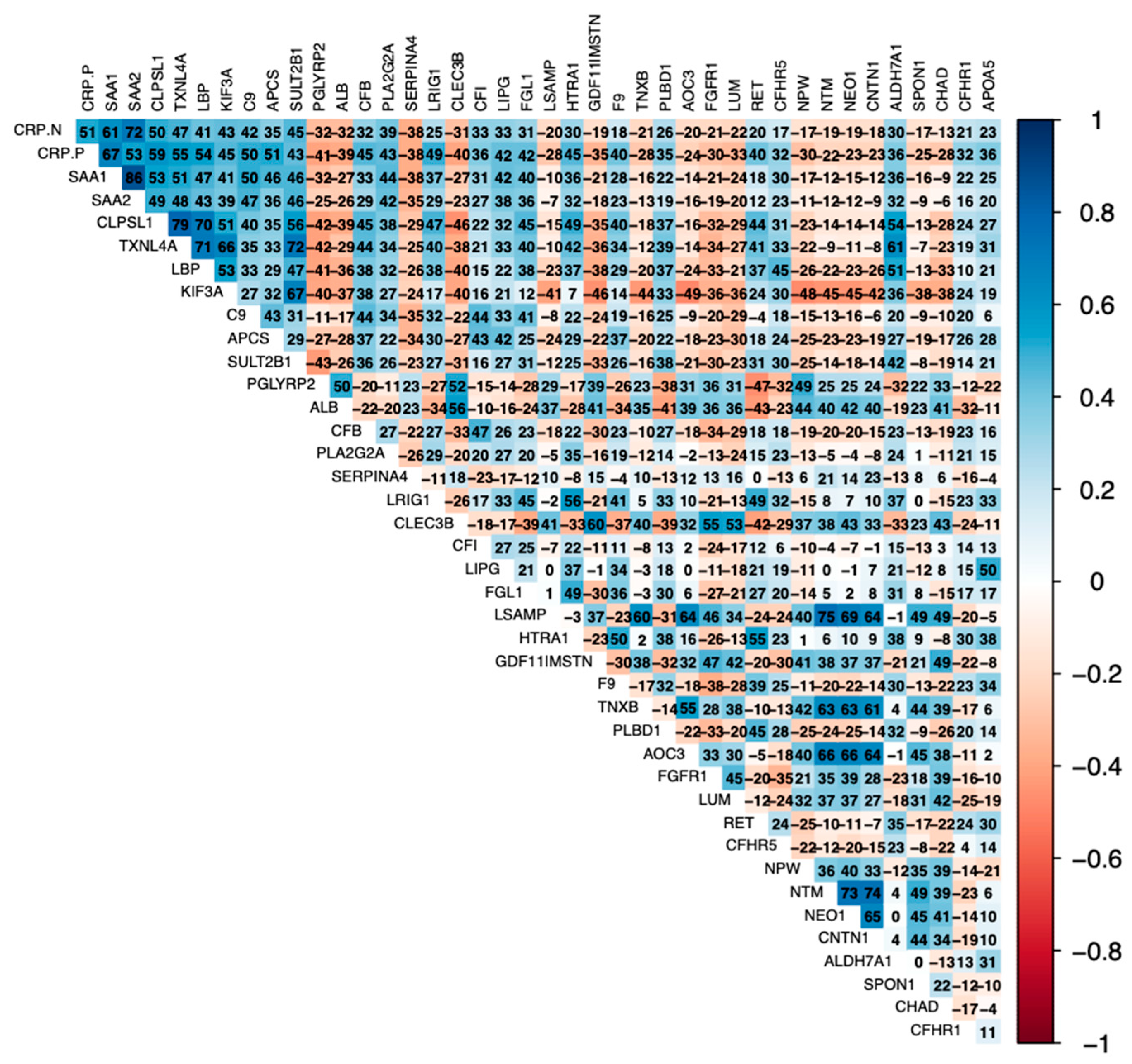
Positive correlates of AGP [complement proteins (C9, CFI, C1QC, CFB), serum amyloids (SAA1/2, APCS), and CRP] were positively correlated with each other (Figure 1). Further, IGSF3 was highly positively correlated with SERPINA4, SHBG, TFF3, and CCN5. Similarly, positive correlates of CRP were positively correlated with each other (serum amyloids, LBP, LIPG, complement proteins) (Figure 2). Negative correlates of CRP (APOC3, FGFR1, LUM, NPW, NTM, NEO1, CNTN1) were inversely related to proinflammatory serum amyloids, LBP, and complement proteins.
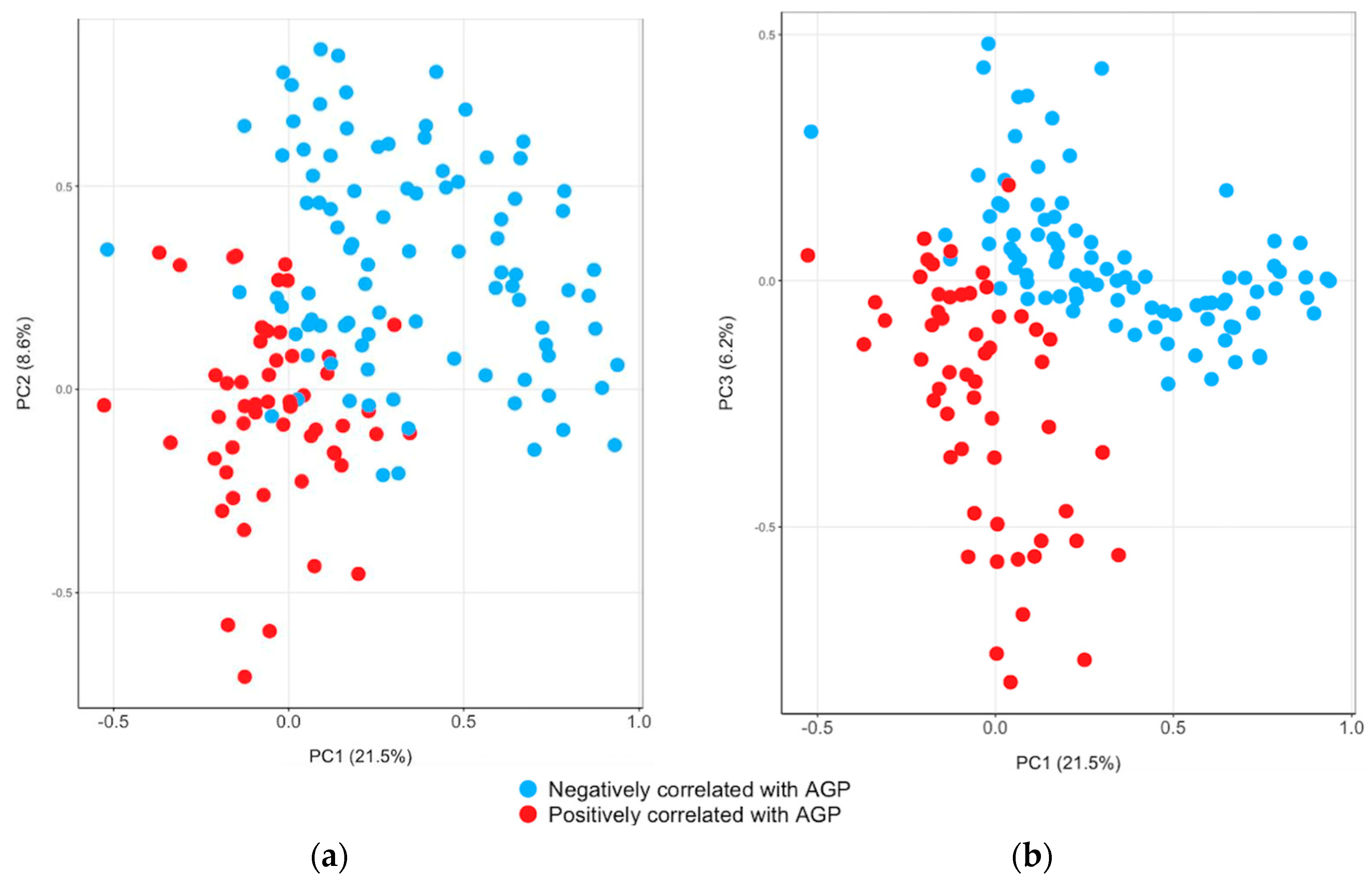
The PCA identified three components that explained 21.5%, 8.6%, and 6.2% of the variance in AGP (Supplemental Table S5; Figure 3). All proteins in the first component of AGP were negatively correlated with AGP, except for coagulation factor XI (F11). The first component of AGP had high positive loadings on CSH1 and PGF and negative loadings on ALB and F11. In the second component, the majority of proteins were negatively correlated with AGP. The second component of AGP had high loadings on B4GALT6, EGFLAM, and protogenin. The third component had high negative loadings on complement, serum amyloids (SAA1/2, APCS), and LBP and positive loadings on SERPINA4 and HSPA1A.
For CRP, the first to third components explained 24.4%, 8.9%, and 5.4% of the variance (Supplemental Table S6; Figure 4). The principal components of CRP were more diverse than the those of AGP. The majority of proteins with high positive loadings in the first component were dominated by cell surface, cell adhesion (neurotrimin, netrin receptors), and embryogenesis-related proteins. The second component of CRP had high loadings on intracellular and extra-hepatic tissue leakage proteins. The third component had high positive loadings in CSH1, glypican-3, pregnancy-specific beta-1-glycoprotein 11, and intestinal barrier proteins that support gastrointestinal immunity (e.g., TFF3) and other normal functions.
The PCs were associated with maternal morbidity symptoms (Table 4). The first (OR: 2.28, 95% CI: 1.59, 3.29), second (OR: 2.05, 95% CI: 1.37 3.11), and third (OR: 1.69, 95% CI: 1.08 to 2.77) scores of AGP per standard deviation were associated with higher odds of women reporting a high fever in the previous week. The PCs of AGP were associated with higher odds of nausea, vomiting, and severe headache. Similarly, the second or third PCs of CRP were associated with higher odds of vomiting, severe headache, or high fever.

4. Discussion

In this study of first-trimester gravida in Bangladesh, we identified a vast plasma inflammasome in early gestation, defined as proteins that covaried with the common inflammatory biomarkers AGP or CRP. The AGPome and CRPome proteins highlight the intricacy of pro- and anti-inflammatory and homeostatic processes to which these two biomarkers are linked. Among the inflammasome correlates are subsets of proteins associated with maternal morbidity, especially recent high fever, headache, and vomiting. It is possible that PCA provided proteins that are down- and up-regulated in maternal inflammation.
In this aptamer-based study, generating highly precise relative quantification and lacking missing data, we set q < 0.05, which accommodated an unknown breadth of early gestational inflammation. Alternatively, in an earlier mass spectrometric study in young Nepalese children, requiring a substantial imputation of missing values, we employed a more stringent threshold of q < 0.01 [21]. In both studies, there were similar but far from identical compositions of the plasma AGPome and CRPome. For example, in both life stages, positive correlates of AGP and CRP included complement proteins, protease inhibitors (SERPING1, SERPINA10), and serum amyloid A-1/A-2 proteins, all of which are expected to increase with inflammation [21]. In our study, negative correlates of AGP and CRP expectedly included transporters of micronutrients and lipids and anabolic hormone regulators. Among the negative correlates, RBP4 transports retinol, APOA-1 transports lipids, thyroxine-binding globulin (SERPINA7) transports thyroid hormones [30], and sex hormone-binding protein (SHBG) binds and mediates the bioavailability of sex steroids [31]. Many such proteins are characterized as anti-inflammatory [32,33,34,35]. In addition to these proteins, many observed correlates of AGP and/or CRP in this study might reflect complement-mediated apoptosis and the resultant release of cellular components into plasma, which naturally occur throughout pregnancy [36,37].
Our analysis identified a strong, negative correlation of AGP with plasma kallistatin, a circulating serine protease inhibitor with pleiotropic antioxidant, anti-inflammatory, and immunoregulatory functions affecting cardiovascular homeostasis [38]. A low plasma kallistatin level has recently been associated with an increased risk of chorioamnionitis in women experiencing preterm labor [39] and the preterm rupture of membranes [40], implicating its potential utility as a biomarker of chorionic inflammation.
Lipopolysaccharide-binding protein (LBP), an acute-phase protein involved in eliciting an innate immune response, was positively correlated with AGP and CRP. LBP is secreted by hepatocytes into the serum and interacts with the Gram-negative bacterial lipopolysaccharide (LPS) with dual functions to neutralize proinflammatory responses against pathogens by promoting phagocytosis and the clearance of the pathogen and helping to bring the LPS antigen to its cognate receptor, CD14-toll-like receptor 4, on mononuclear target cells [41]. This latter interaction induces a proinflammatory innate immune response. Although it is not certain whether infection is present in the study population, given the presence of morbidity symptoms in early pregnancy, there are likely proinflammatory processes occurring [1,4,42].
Several proteins thought to function in intracellular immunity were correlated with AGP and CRP, suggesting processes of cellular death, protein degradation, and release into circulation. However, it may also indicate the extracellular systemic delivery of proteins, which may occur through the loading and transport of small extracellular vesicles (sEVs) [43,44]. Indeed, the interferon-induced protein with tetratricopeptide repeats 3 (IFIT3), a cytosolic protein with antiproliferative and antiviral properties that was positively correlated with AGP, has been previously located in sEVs and shown to exhibit a regulatory activity of protein expression in the vesicle and intercellular communication [44]. It is also possible that cell surface receptors (e.g., IL-22RA, the immunoglobulin receptor IGDCC4, TIMD-4, and CD74) that correlated with AGP and CRP in our study may have similar effector roles in sEVs. Any of these proteins may have physiological function in plasma, warranting further investigation.
The early gestation plasma inflammasome revealed paradoxical associations, perhaps unique to pregnancy. For example, chorionic somatomammotropin hormone 1/2 (CSH 1/2) and glypican-3 (GPC3) were negatively correlated with and received high loading for principal component 1 for AGP but were positively correlated with CRP. CSH1/2 is a part of the somatotropin and prolactin class of hormones, expressed in the placenta, whose expression rises early in gestation and remains high throughout pregnancy [45] as a peptide hormone enabling maternal–fetal nutrient transfer and fetal growth [46]. GPC3 is involved in regulating cellular growth, differentiation, and the regulation of growth factors involved in Wnt, hippo, and Hedgehog signaling pathways [47,48], possibly affecting morphogenesis, limb patterning, and skeletal development [49]. Plasma pregnancy-specific glycoprotein beta-1 (PSG1), a positive correlate of CRP secreted by the placenta, has been expressed in pregnant women from day 3 post-fertilization when the blastocyst implants to the uterine wall [50,51], a proinflammatory process requiring careful homeostatic control. PSGs have been observed to help establish the maternal–fetal vasculature [52]. Discordant associations observed between these proteins and AGP and CRP may reflect that AGP and CRP are differentially sensitivity to the timing of the inflammatory response. CRP responds more immediate than AGP; as CRP begins to resolve, AGP becomes elevated [16].
Complement surveillance is integral to innate immune responses to inflammation [53], which is critical for controlled utero-placental growth [36,37]. Many complement components were variably correlated with AGP and CRP. The complement C1q subcomponent subunit C, a key initiator of complement pathway activation, was positively correlated with both AGP and CRP [53,54]. Several early cascade proteins correlated with CRP but not AGP, including C2, a protease important early in the classical pathway, complement components C3 and C5 that mediate inflammation, and complement factor H-related proteins 1 and 5, which are inhibitory factors of the alternative pathway that rise in the first trimester [53]. Complement factor B (CFB), component 9 (C9), and factor I (CFI) were positively correlated with both AGP and CRP. CFB promotes the alternative complement activation pathway and has been shown to rise during the first trimester [53], while C9 is involved in downstream activation to form the membrane attack complex leading to the death of damaged cells. As a serum protease, activated CFI deattenuates complement activation through the degradation of certain complement proteins [55], thus protecting host tissues from inappropriate or prolonged complement activation. While seemingly paradoxical, a positive correlation of CFI with both CRP and AGP might illustrate an increased circulation of CFI as a zymogen [56], to be activated as needed to control inflammatory processes. These results appear to reflect immense heterogeneity in pro- and anti-inflammatory circuits underlying these two biomarkers.
Seeming paradoxical associations extended to other signaling molecules involved in immunoregulation. For example, the cytokine IL-10, a protein that acts within type 2 immunity to down-regulate inflammation [57], was negatively correlated with AGP. Similarly, the ubiquitin-like protein, ISG15, is an interferon-induced protein of the innate immune system that acts both intracellularly and extracellularly to regulate inflammatory responses. Its negative association with AGP, a more gradual and longer acting biomarker than CRP, may have been reflecting a chronic, low-level proinflammatory state [58].
The three examined PCs offered an ordered view of clusters of proteins explaining more to less variance in plasma concentrations of AGP and CRP, possibly reflecting the biological prominence of proteins involved in the down- and up-regulation of inflammation. The first and second components of AGP comprised predominantly negative correlates of AGP, anabolic agents linked to the maintenance of pregnancy (PGRMC2 [59]), embryogenesis (protogenin [60], DDR1 required for blastocyst implantation [61]), neurogenesis (TMEM132A [62], gliomedin [63]) including axon growth (semaphorin 6/7A [64]) and synapse formation (pikachurin [65]), and organ development (DDR1, fibulin-1 [66]). The third component of AGP included a broader mix of proinflammatory proteins, including those of the complement cascade, CRP, and serum amyloids. The composition of these sequential components may reflect priorities to tolerate the conceptus, promote embryo-feto-placental growth and development, and protect early pregnancy from inflammation while responding to infectious threats evident by the concurrent activation of acute-phase and catabolic proteins. The essentiality of the latter inflammatory mechanisms find support in principal components that were related to the symptoms of maternal morbidity, especially high fever.
Our study has several strengths, including the use of large-scale proteomics, focus on pregnant women in low-resource settings, and rigorous ascertainment of biospecimens used for proteomics which reflect the source population. However, the present study used a cross-sectional design, making it challenging to infer causality due to the lack of temporality. Further, there was no information on the presence of infection in this population. Additionally, the present study did not investigate the association between significant proteins and pregnancy outcomes. Examining such associations is important but is beyond the scope of the present study. While the present study did not investigate the association between significant proteins and pregnancy outcomes, it remains an important avenue for future investigation. However, our novel analyses relating the AGPome and CRPome to various morbidity outcomes are more proximal than pregnancy outcomes and, potentially, more biologically plausible to interpret.

5. Conclusions

This study represents the first attempt to apply large-scale plasma proteomics in a typical rural South Asian population. The AGPome and CRPome included well-known inflammatory proteins, complement proteins, proteins involved in intracellular immunity, pregnancy-related anabolic proteins, and transporters of micronutrients. Our findings highlight composites of highly complex physiological processes of early pregnancy to reveal the breadth and complexity of early gestational inflammation associated with concentrations of AGP and CRP, which remain inapparent when only these two classic biomarkers are measured.
These findings and those in a Nepalese child population [21] merit caution in interpretation when adjusting biochemical measures for these two biomarkers in population-based studies of nutrition and health [8]. Our results call for a need to deeply explore the diversity and regulation of inflammation to identify reliable and potentially more informative biomarkers of maternal health and its relation to nutritional status [67] and pregnancy outcomes that may be of public health utility.

Supplementary Materials

The following supporting information can be downloaded at https://www.mdpi.com/article/10.3390/biom14070736/s1, Table S1: Proteins positively correlated with AGP at FDR < 0.05; Table S2: Proteins negatively correlated with AGP at FDR < 0.05; Table S3: Proteins positively correlated with CRP at FDR < 0.05; Table S4: Proteins negatively correlated with CRP at FDR < 0.05; Table S5: Factor loadings from principal component analysis of plasma proteins significantly associated with α-1-acid glycoprotein in 435 Bangladeshi pregnant women; Table S6: Factor loadings from principal component analysis of 879 plasma proteins significantly associated with c-reactive protein glycoprotein in 435 Bangladeshi pregnant women.

Author Contributions

Conceptualization, K.J.S. and K.P.W.J.; methodology, H.K.; software, H.K.; formal analysis, H.K.; writing—original draft preparation, H.K. and J.R.B.-F.; writing—review and editing, J.R.B.-F., K.J.S., C.M.R., G.S., S.B., L.S.F.W., J.G., L.C., J.D.Y., L.M.D.L., T.J.S. and K.P.W.J.; funding acquisition, K.P.W.J. All authors have read and agreed to the published version of the manuscript.

Funding

This research was supported by Support for Creative Integrated Basic and Applied Research (SCIBAR), Johns Hopkins Bloomberg School of Public Health and Kirk Humanitarian Foundation. H.K. was supported by a grant from the NHLBI (K01 HL168232). J.R.B.-F. was supported by the Cancer Prevention Fellowship Program, National Cancer Institute, Division of Cancer Prevention.

Institutional Review Board Statement

This study was conducted in accordance with the Declaration of Helsinki and approved by the Institutional Review Board Bangladesh Medical Research Council, approved annually by the Institutional Review Board at Johns Hopkins Bloomberg School of Public Health (BSPH, IRB number: IRB00000570) on 10 December 2007), with latest annual approval on 27 October 2023. A Data and Safety Monitoring Board regularly followed the trial which was registered with ClinicalTrials.gov (NCT00860470).

Informed Consent Statement

Informed consent was obtained from all subjects involved in the study.

Data Availability Statement

Data from the present study will not be shared publicly and will follow the parent study’s (JiVitA trial) data management and sharing procedures.

Acknowledgments

The authors thank the participants, staff, and investigators of the JiVitA trial, especially Parul Christian, Hasmot Ali, Abu Ahmed Shamim, and Emorn Udomkesmalee, for their contributions to the original trial. We thank Zhichen Xiong for assistance in graphics, Michael Fenech at University of South Australia, and Safarina Malik at the Mochtar Riady Institute of Nanotechnology for their expert guidance.

Conflicts of Interest

Joseph Gogain, and Lena Cuddeback were employed by the SomaLogic Inc. The remaining authors declare that the research was conducted in the absence of any commercial or financial relationships that could be construed as a potential conflict of interest. The funders had no role in the design of the study; in the collection, analyses, or interpretation of data; in the writing of the manuscript; or in the decision to publish the results.

References

  1. Mor, G.; Cardenas, I.; Abrahams, V.; Guller, S. Inflammation and Pregnancy: The Role of the Immune System at the Implantation Site. Ann. N. Y. Acad. Sci. 2011, 1221, 80. [Google Scholar] [CrossRef] [PubMed]
  2. Burton, G.J.; Jauniaux, E.; Charnock-Jones, D.S. The Influence of the Intrauterine Environment on Human Placental Development. Int. J. Dev. Biol. 2010, 54, 303–312. [Google Scholar] [CrossRef] [PubMed]
  3. Muter, J.; Lynch, V.J.; McCoy, R.C.; Brosens, J.J. Human Embryo Implantation. Development 2023, 150, dev201507. [Google Scholar] [CrossRef] [PubMed]
  4. Förger, F.; Villiger, P.M. Immunological Adaptations in Pregnancy That Modulate Rheumatoid Arthritis Disease Activity. Nat. Rev. Rheumatol. 2020, 16, 113–122. [Google Scholar] [CrossRef] [PubMed]
  5. de Rivero Vaccari, J.P. The Inflammasome in Reproductive Biology: A Promising Target for Novel Therapies. Front. Endocrinol. 2020, 11, 8. [Google Scholar] [CrossRef] [PubMed]
  6. Baumann, H.; Gauldie, J. The Acute Phase Response. Immunol. Today 1994, 15, 74–80. [Google Scholar] [CrossRef]
  7. Sauder, M.W.; Lee, S.E.; Schulze, K.J.; Christian, P.; Wu, L.S.F.; Khatry, S.K.; LeClerq, S.C.; Adhikari, R.K.; Groopman, J.D.; West, K.P. Inflammation throughout Pregnancy and Fetal Growth Restriction in Rural Nepal. Epidemiol. Infect. 2019, 147, e258. [Google Scholar] [CrossRef] [PubMed]
  8. McDonald, C.M.; Suchdev, P.S.; Krebs, N.F.; Hess, S.Y.; Wessells, K.R.; Ismaily, S.; Rahman, S.; Wieringa, F.T.; Williams, A.M.; Brown, K.H.; et al. Adjusting Plasma or Serum Zinc Concentrations for Inflammation: Biomarkers Reflecting Inflammation and Nutritional Determinants of Anemia (BRINDA) Project. Am. J. Clin. Nutr. 2020, 111, 927–937. [Google Scholar] [CrossRef]
  9. Ghosh, S.; Kurpad, A.V.; Sachdev, H.S.; Thomas, T. Inflammation Correction in Micronutrient Deficiency with Censored Inflammatory Biomarkers. Am. J. Clin. Nutr. 2021, 113, 47–54. [Google Scholar] [CrossRef]
  10. Namaste, S.M.; Aaron, G.J.; Varadhan, R.; Peerson, J.M.; Suchdev, P.S. Methodologic Approach for the Biomarkers Reflecting Inflammation and Nutritional Determinants of Anemia (BRINDA) Project. Am. J. Clin. Nutr. 2017, 106, 333S–347S. [Google Scholar] [CrossRef]
  11. Thurnham, D.I.; McCabe, G.P.; Northrop-Clewes, C.A.; Nestel, P. Effects of Subclinical Infection on Plasma Retinol Concentrations and Assessment of Prevalence of Vitamin A Deficiency: Meta-Analysis. Lancet 2003, 362, 2052–2058. [Google Scholar] [CrossRef]
  12. Alberts, B.; Johnson, A.; Lewis, J.; Raff, M.; Roberts, K.; Walter, P. Innate Immunity. In Molecular Biology of the Cell, 4th ed.; Garland Science: New York, NY, USA, 2002. [Google Scholar]
  13. Chaplin, D.D. Overview of the Immune Response. J. Allergy Clin. Immunol. 2010, 125, S3–S23. [Google Scholar] [CrossRef] [PubMed]
  14. Singh-Manoux, A.; Shipley, M.J.; Bell, J.A.; Canonico, M.; Elbaz, A.; Kivimäki, M. Association between Inflammatory Biomarkers and All-Cause, Cardiovascular and Cancer-Related Mortality. CMAJ 2017, 189, E384–E390. [Google Scholar] [CrossRef] [PubMed]
  15. Lauer, J.M.; Ghosh, S.; Ausman, L.M.; Webb, P.; Bashaasha, B.; Agaba, E.; Turyashemererwa, F.M.; Tran, H.Q.; Gewirtz, A.T.; Erhardt, J.; et al. Markers of Environmental Enteric Dysfunction Are Associated with Poor Growth and Iron Status in Rural Ugandan Infants. J. Nutr. 2020, 150, 2175–2182. [Google Scholar] [CrossRef] [PubMed]
  16. Raiten, D.J.; Ashour, F.A.S.; Ross, A.C.; Meydani, S.N.; Dawson, H.D.; Stephensen, C.B.; Brabin, B.J.; Suchdev, P.S.; van Ommen, B. Inflammation and Nutritional Science for Programs/Policies and Interpretation of Research Evidence (INSPIRE). J. Nutr. 2015, 145, 1039S–1108S. [Google Scholar] [CrossRef] [PubMed]
  17. Gold, L.; Ayers, D.; Bertino, J.; Bock, C.; Bock, A.; Brody, E.N.; Carter, J.; Dalby, A.B.; Eaton, B.E.; Fitzwater, T.; et al. Aptamer-Based Multiplexed Proteomic Technology for Biomarker Discovery. PLoS ONE 2010, 5, e15004. [Google Scholar] [CrossRef] [PubMed]
  18. Williams, S.A.; Kivimaki, M.; Langenberg, C.; Hingorani, A.D.; Casas, J.P.; Bouchard, C.; Jonasson, C.; Sarzynski, M.A.; Shipley, M.J.; Alexander, L.; et al. Plasma Protein Patterns as Comprehensive Indicators of Health. Nat. Med. 2019, 25, 1851–1857. [Google Scholar] [CrossRef] [PubMed]
  19. Ganz, P.; Heidecker, B.; Hveem, K.; Jonasson, C.; Kato, S.; Segal, M.R.; Sterling, D.G.; Williams, S.A. Development and Validation of a Protein-Based Risk Score for Cardiovascular Outcomes among Patients with Stable Coronary Heart Disease. JAMA 2016, 315, 2532–2541. [Google Scholar] [CrossRef]
  20. Lee, S.E.; Schulze, K.; West, K.P. Rainer Gross Award Lecture 2018: The Childhood Plasma Proteome: Discovering Its Applications in Public Health Nutrition. Food Nutr. Bull. 2019, 40, 144–150. [Google Scholar] [CrossRef]
  21. Lee, S.E.; West, K.P.W., Jr.; Cole, R.N.; Schulze, K.J.; Christian, P.; Wu, L.S.-F.; Yager, J.D.; Groopman, J.; Ruczinski, I. Plasma Proteome Biomarkers of Inflammation in School Aged Children in Nepal. PLoS ONE 2015, 10, e0144279. [Google Scholar] [CrossRef]
  22. Scholl, P.F.; Cole, R.N.; Ruczinski, I.; Gucek, M.; Diez, R.; Rennie, A.; Nathasingh, C.; Schulze, K.; Christian, P.; Yager, J.D.; et al. Maternal Serum Proteome Changes between the First and Third Trimester of Pregnancy in Rural Southern Nepal. Placenta 2012, 33, 424–432. [Google Scholar] [CrossRef]
  23. Tarca, A.L.; Romero, R.; Bhatti, G.; Gotsch, F.; Done, B.; Gudicha, D.W.; Gallo, D.M.; Jung, E.; Pique-Regi, R.; Berry, S.M.; et al. Human Plasma Proteome during Normal Pregnancy. J. Proteome Res. 2022, 21, 2687–2702. [Google Scholar] [CrossRef]
  24. West, K.P.; Christian, P.; Labrique, A.B.; Rashid, M.; Shamim, A.A.; Klemm, R.D.W.; Massie, A.B.; Mehra, S.; Schulze, K.J.; Ali, H.; et al. Effects of Vitamin A or Beta Carotene Supplementation on Pregnancy-Related Mortality and Infant Mortality in Rural Bangladesh: A Cluster Randomized Trial. JAMA 2011, 305, 1986–1995. [Google Scholar] [CrossRef]
  25. Labrique, A.B.; Christian, P.; Klemm, R.D.W.; Rashid, M.; Shamim, A.A.; Massie, A.; Schulze, K.; Hackman, A.; West, K.P. A Cluster-Randomized, Placebo-Controlled, Maternal Vitamin A or Beta-Carotene Supplementation Trial in Bangladesh: Design and Methods. Trials 2011, 12, 102. [Google Scholar] [CrossRef] [PubMed]
  26. Gunnsteinsson, S.; Labrique, A.B.; West, K.P.; Christian, P.; Mehra, S.; Shamim, A.A.; Rashid, M.; Katz, J.; Klemm, R.D.W. Constructing Indices of Rural Living Standards in Northwestern Bangladesh. J. Health Popul. Nutr. 2010, 28, 509–519. [Google Scholar] [CrossRef] [PubMed]
  27. Schulze, K.J.; Gernand, A.D.; Khan, A.Z.; Wu, L.S.-F.; Mehra, S.; Shaikh, S.; Ali, H.; Shamim, A.A.; Sungpuag, P.; Udomkesmalee, E.; et al. Newborn Micronutrient Status Biomarkers in a Cluster-Randomized Trial of Antenatal Multiple Micronutrient Compared with Iron Folic Acid Supplementation in Rural Bangladesh. Am. J. Clin. Nutr. 2020, 112, 1328–1337. [Google Scholar] [CrossRef]
  28. Gold, L.; Walker, J.J.; Wilcox, S.K.; Williams, S. Advances in Human Proteomics at High Scale with the SOMAscan Proteomics Platform. New Biotechnol. 2012, 29, 543–549. [Google Scholar] [CrossRef] [PubMed]
  29. Benjamini, Y.; Yekutieli, D. The Control of the False Discovery Rate in Multiple Testing under Dependency. Ann. Stat. 2001, 29, 1165–1188. [Google Scholar] [CrossRef]
  30. Bartalena, L. Thyroid Hormone-Binding Proteins. In Encyclopedia of Endocrine Diseases; Martini, L., Ed.; Elsevier: New York, NY, USA, 2004; pp. 474–479. ISBN 978-0-12-475570-3. [Google Scholar]
  31. Selby, C. Sex Hormone Binding Globulin: Origin, Function and Clinical Significance. Ann. Clin. Biochem. 1990, 27 Pt 6, 532–541. [Google Scholar] [CrossRef]
  32. Rosales, F.J.; Ritter, S.J.; Zolfaghari, R.; Smith, J.E.; Ross, A.C. Effects of Acute Inflammation on Plasma Retinol, Retinol-Binding Protein, and Its mRNA in the Liver and Kidneys of Vitamin A-Sufficient Rats. J. Lipid Res. 1996, 37, 962–971. [Google Scholar] [CrossRef]
  33. Khovidhunkit, W.; Kim, M.-S.; Memon, R.A.; Shigenaga, J.K.; Moser, A.H.; Feingold, K.R.; Grunfeld, C. Effects of Infection and Inflammation on Lipid and Lipoprotein Metabolism: Mechanisms and Consequences to the Host. J. Lipid Res. 2004, 45, 1169–1196. [Google Scholar] [CrossRef]
  34. Liao, C.-H.; Li, H.-Y.; Yu, H.-J.; Chiang, H.-S.; Lin, M.-S.; Hua, C.-H.; Ma, W.-Y. Low Serum Sex Hormone-Binding Globulin: Marker of Inflammation? Clin. Chim. Acta 2012, 413, 803–807. [Google Scholar] [CrossRef]
  35. Liu, N.; Feng, Y.; Luo, X.; Ma, X.; Ma, F. Association between Dietary Inflammatory Index and Sex Hormone Binding Globulin and Sex Hormone in U.S. Adult Females. Front. Public Health 2022, 10, 802945. [Google Scholar] [CrossRef] [PubMed]
  36. Triggianese, P.; Perricone, C.; Chimenti, M.S.; De Carolis, C.; Perricone, R. Innate Immune System at the Maternal-Fetal Interface: Mechanisms of Disease and Targets of Therapy in Pregnancy Syndromes. Am. J. Reprod. Immunol. 2016, 76, 245–257. [Google Scholar] [CrossRef]
  37. Girardi, G. Complement Activation, a Threat to Pregnancy. Semin Immunopathol. 2018, 40, 103–111. [Google Scholar] [CrossRef]
  38. Chao, J.; Guo, Y.; Chao, L. Protective Role of Endogenous Kallistatin in Vascular Injury and Senescence by Inhibiting Oxidative Stress and Inflammation. Oxid. Med. Cell Longev. 2018, 2018, 4138560. [Google Scholar] [CrossRef]
  39. Lee, K.-N.; Cho, I.; Im, E.M.; Oh, E.; Park, K.H. Plasma IGFBP-1, Fas, Kallistatin, and P-Selectin as Predictive Biomarkers of Histologic Chorioamnionitis and Associated Intra-Amniotic Infection in Women with Preterm Labor. Am. J. Reprod. Immunol. 2023, 89, e13645. [Google Scholar] [CrossRef]
  40. Cho, I.; Lee, K.-N.; Joo, E.; Kim, Y.M.; Kim, T.E.; Park, K.H. Plasma E-Selectin and Kallistatin as Predictive Markers of Histologic Chorioamnionitis in Women with Preterm Premature Rupture of Membranes. Am. J. Reprod. Immunol. 2022, 88, e13584. [Google Scholar] [CrossRef] [PubMed]
  41. Meng, L.; Song, Z.; Liu, A.; Dahmen, U.; Yang, X.; Fang, H. Effects of Lipopolysaccharide-Binding Protein (LBP) Single Nucleotide Polymorphism (SNP) in Infections, Inflammatory Diseases, Metabolic Disorders and Cancers. Front. Immunol. 2021, 12, 681810. [Google Scholar] [CrossRef] [PubMed]
  42. Blairon, L.; Wittebole, X.; Laterre, P.-F. Lipopolysaccharide-Binding Protein Serum Levels in Patients with Severe Sepsis Due to Gram-Positive and Fungal Infections. J. Infect Dis. 2003, 187, 287–291. [Google Scholar] [CrossRef]
  43. Cheng, J.; Ji, D.; Yin, Y.; Wang, S.; Song, K.; Pan, Q.; Zhang, Q.; Yang, L. Proteomic Profiling of Serum Small Extracellular Vesicles Reveals Immune Signatures of Children with Pneumonia. Transl. Pediatr. 2022, 11, 891–908. [Google Scholar] [CrossRef] [PubMed]
  44. Naranjo, N.M.; Salem, I.; Harris, M.A.; Languino, L.R. IFIT3 (Interferon Induced Protein with Tetratricopeptide Repeats 3) Modulates STAT1 Expression in Small Extracellular Vesicles. Biochem. J. 2021, 478, 3905–3921. [Google Scholar] [CrossRef] [PubMed]
  45. Anthony, R.V.; Jeckel, K.M. Placental Lactogen (CSH). In Encyclopedia of Reproduction, 2nd ed.; Skinner, M.K., Ed.; Academic Press: Oxford, UK, 2018; pp. 502–507. ISBN 978-0-12-815145-7. [Google Scholar]
  46. Jeckel, K.M.; Boyarko, A.C.; Bouma, G.J.; Winger, Q.A.; Anthony, R.V. Chorionic Somatomammotropin Impacts Early Fetal Growth and Placental Gene Expression. J. Endocrinol. 2018, 237, 301–310. [Google Scholar] [CrossRef]
  47. Kolluri, A.; Ho, M. The Role of Glypican-3 in Regulating Wnt, YAP, and Hedgehog in Liver Cancer. Front. Oncol. 2019, 9, 708. [Google Scholar] [CrossRef] [PubMed]
  48. Filmus, J.; Selleck, S.B. Glypicans: Proteoglycans with a Surprise. J. Clin. Investig. 2001, 108, 497–501. [Google Scholar] [CrossRef] [PubMed]
  49. Song, H.H.; Filmus, J. The Role of Glypicans in Mammalian Development. Biochim. Biophys. Acta 2002, 1573, 241–246. [Google Scholar] [CrossRef] [PubMed]
  50. Dimitriadou, F.; Phocas, I.; Mantzavinos, T.; Sarandakou, A.; Rizos, D.; Zourlas, P.A. Discordant Secretion of Pregnancy Specific Beta 1-Glycoprotein and Human Chorionic Gonadotropin by Human Pre-Embryos Cultured in Vitro. Fertil. Steril. 1992, 57, 631–636. [Google Scholar] [CrossRef] [PubMed]
  51. Dekel, N.; Gnainsky, Y.; Granot, I.; Racicot, K.; Mor, G. The Role of Inflammation for a Successful Implantation. Am. J. Reprod. Immunol. 2014, 72, 141–147. [Google Scholar] [CrossRef]
  52. Ha, C.T.; Wu, J.A.; Irmak, S.; Lisboa, F.A.; Dizon, A.M.; Warren, J.W.; Ergun, S.; Dveksler, G.S. Human Pregnancy Specific Beta-1-Glycoprotein 1 (PSG1) Has a Potential Role in Placental Vascular Morphogenesis1. Biol. Reprod. 2010, 83, 27–35. [Google Scholar] [CrossRef] [PubMed]
  53. He, Y.-D.; Xu, B.-N.; Song, D.; Wang, Y.-Q.; Yu, F.; Chen, Q.; Zhao, M.-H. Normal Range of Complement Components during Pregnancy: A Prospective Study. Am. J. Reprod. Immunol. 2020, 83, e13202. [Google Scholar] [CrossRef]
  54. Son, M.; Diamond, B.; Santiago-Schwarz, F. Fundamental Role of C1q in Autoimmunity and Inflammation. Immunol. Res. 2015, 63, 101–106. [Google Scholar] [CrossRef] [PubMed]
  55. Nilsson, S.C.; Sim, R.B.; Lea, S.M.; Fremeaux-Bacchi, V.; Blom, A.M. Complement Factor I in Health and Disease. Mol. Immunol. 2011, 48, 1611–1620. [Google Scholar] [CrossRef] [PubMed]
  56. Roversi, P.; Johnson, S.; Caesar, J.J.E.; McLean, F.; Leath, K.J.; Tsiftsoglou, S.A.; Morgan, B.P.; Harris, C.L.; Sim, R.B.; Lea, S.M. Structural Basis for Complement Factor I Control and Its Disease-Associated Sequence Polymorphisms. Proc. Natl. Acad. Sci. USA 2011, 108, 12839–12844. [Google Scholar] [CrossRef] [PubMed]
  57. Iyer, S.S.; Cheng, G. Role of Interleukin 10 Transcriptional Regulation in Inflammation and Autoimmune Disease. Crit. Rev. Immunol. 2012, 32, 23–63. [Google Scholar] [CrossRef] [PubMed]
  58. Swaim, C.D.; Scott, A.F.; Canadeo, L.A.; Huibregtse, J.M. Extracellular ISG15 Signals Cytokine Secretion Through the LFA-1 Integrin Receptor. Mol. Cell 2017, 68, 581–590. [Google Scholar] [CrossRef] [PubMed]
  59. Lozovyy, V.; Richardson, L.; Saade, G.; Menon, R. Progesterone Receptor Membrane Components: Key Regulators of Fetal Membrane Integrity. Biol. Reprod. 2020, 104, 445–456. [Google Scholar] [CrossRef] [PubMed]
  60. Wong, Y.-H.; Lu, A.-C.; Wang, Y.-C.; Cheng, H.-C.; Chang, C.; Chen, P.-H.; Yu, J.-Y.; Fann, M.-J. Protogenin Defines a Transition Stage during Embryonic Neurogenesis and Prevents Precocious Neuronal Differentiation. J. Neurosci. 2010, 30, 4428–4439. [Google Scholar] [CrossRef] [PubMed]
  61. Vogel, W.F.; Aszódi, A.; Alves, F.; Pawson, T. Discoidin Domain Receptor 1 Tyrosine Kinase Has an Essential Role in Mammary Gland Development. Mol. Cell Biol. 2001, 21, 2906–2917. [Google Scholar] [CrossRef] [PubMed]
  62. Li, B.; Niswander, L.A. TMEM132A, a Novel Wnt Signaling Pathway Regulator Through Wntless (WLS) Interaction. Front. Cell Dev. Biol. 2020, 8, 599890. [Google Scholar] [CrossRef]
  63. Maluenda, J.; Manso, C.; Quevarec, L.; Vivanti, A.; Marguet, F.; Gonzales, M.; Guimiot, F.; Petit, F.; Toutain, A.; Whalen, S.; et al. Mutations in GLDN, Encoding Gliomedin, a Critical Component of the Nodes of Ranvier, Are Responsible for Lethal Arthrogryposis. Am. J. Hum. Genet 2016, 99, 928–933. [Google Scholar] [CrossRef]
  64. Pasterkamp, R.J.; Peschon, J.J.; Spriggs, M.K.; Kolodkin, A.L. Semaphorin 7A Promotes Axon Outgrowth through Integrins and MAPKs. Nature 2003, 424, 398–405. [Google Scholar] [CrossRef] [PubMed]
  65. Sato, S.; Omori, Y.; Katoh, K.; Kondo, M.; Kanagawa, M.; Miyata, K.; Funabiki, K.; Koyasu, T.; Kajimura, N.; Miyoshi, T.; et al. Pikachurin, a Dystroglycan Ligand, Is Essential for Photoreceptor Ribbon Synapse Formation. Nat. Neurosci. 2008, 11, 923–931. [Google Scholar] [CrossRef] [PubMed]
  66. Mahajan, D.; Kancharla, S.; Kolli, P.; Sharma, A.K.; Singh, S.; Kumar, S.; Mohanty, A.K.; Jena, M.K. Role of Fibulins in Embryonic Stage Development and Their Involvement in Various Diseases. Biomolecules 2021, 11, 685. [Google Scholar] [CrossRef] [PubMed]
  67. Calder, P.C.; Ahluwalia, N.; Albers, R.; Bosco, N.; Bourdet-Sicard, R.; Haller, D.; Holgate, S.T.; Jönsson, L.S.; Latulippe, M.E.; Marcos, A.; et al. A Consideration of Biomarkers to Be Used for Evaluation of Inflammation in Human Nutritional Studies. Br. J. Nutr. 2013, 109, S1–S34. [Google Scholar] [CrossRef] [PubMed]
Figure 1. Matrix of correlation coefficients (r) for 40 proteins, measured in relative fluorescence units by SomaScan and presented as Entrez gene identifiers, most strongly associated with α1-acid glycoprotein (AGP), measured by radial immunodiffusion kit in g/L, listed in ascending order of chance-corrected p-values, with all q-values < 0.002 for displayed proteins. Blue color represents positive correlates, and red color represents negative correlates, with intensity of color reflecting extent of positive or negative correlations, respectively. Correlation coefficients are presented as r × 102 to improve visualization. Full names and UniProt identifiers of proteins presented are shown in Table 2.
Biomolecules 14 00736 g001
Figure 2. Matrix of correlation coefficients (r) for 40 proteins, measured in relative fluorescence units by SomaScan and presented as Entrez gene identifiers, most strongly associated with C-reactive protein (CRP), measured by clinical chemistry analyzer in mg/L, listed in ascending order of chance-corrected p-values (q), with all q-values < 3.00 × 10⁻⁸ for displayed protein pairs. Blue color represents positive correlates, and red color represents negative correlates, with intensity of color reflecting extent of positive or negative correlations, respectively. Correlation coefficients are presented as r × 102 to improve visualization. Full names and UniProt identifiers of listed proteins shown in Table 3.
Biomolecules 14 00736 g002
Figure 3. Bi-plots from principal component analysis. Principal component analysis was conducted among plasma proteins significantly associated with α-1acid glycoprotein (AGP) at Q values (FDR) < 0.05. (a) is a bi-plot of principal component 1 (PC1) and PC2. (b) is a bi-plot of PC1 and PC3. The percentages in parentheses on the axis indicate the proportion of the variance explained by each PC.
Biomolecules 14 00736 g003
Figure 4. Bi-plots from principal component analysis. Principal component analysis was conducted among 879 plasma proteins significantly associated with C-reactive protein (CRP) at Q values (FDR) < 0.05. (a) is a bi-plot of principal component 1 (PC1) and PC2. (b) is a bi-plot of PC1 and PC3.
Biomolecules 14 00736 g004
Table 1. Characteristics of 1st trimester pregnant women in the JiVitA-3 trial by participation status in the proteomics study 1.
Proteomics Substudy (N = 435)Not Selected (N = 973)
Randomized supplement allocation
  Multiple Micronutrients210 (48.3%)470 (48.3%)
  Iron–Folic Acid225 (51.7%)503 (51.7%)
Education
  No schooling100 (23.0%)258 (26.5%)
  Grade 1 to Grade 465 (14.9%)116 (11.9%)
  Grade 5 to Grade 9240 (55.2%)509 (52.3%)
  10 years and above30 (6.9%)90 (9.3%)
Age (years)
  <20 147 (33.8%)304 (31.2%)
  20–29 240 (55.2%)546 (56.1%)
  ≥30 48 (11.0%)123 (12.6%)
Parity
  0167 (38.4%)384 (39.4%)
  1139 (31.9%)309 (31.8%)
  ≥2129 (29.6%)280 (28.8%)
Gestational age at blood draw (wk), median (IQR)9.7 (8.0, 12.3)10.1 (8.3, 13.4)
Maternal anthropometry, mean (SD)
  Height, cm149.24 (5.18)149.0 (5.3)
  Weight, kg, 42.37 (5.76)42.9 (6.2)
  BMI, kg/m218.99 (2.11)19.3 (2.3)
Maternal malnutrition
  BMI < 18.5 kg/m2 194 (44.6%)398 (41.0%)
  Height < 150 cm, %228 (52.4%)547 (56.3%)
  Mid-upper arm circumference <21.5 cm, %72 (16.6%)178 (18.3%)
  Living standard index, median (IQR) −0.22 (−0.70, 0.43)−0.20 (−0.70, 0.67)
Laboratory values
  α-1 acid glycoprotein, g/L0.81 (0.30)0.73 (0.28)
  C-reactive protein, mg/L22.24 (5.25)NA
  α-1 acid glycoprotein >1 g/L85 (19.5)138 (14.2)
  C-reactive protein >5 mg/L, % 243 (11.4)NA
Morbidity symptoms (days/wk, yes, %)
  Nausea 224 (51.5%)446 (45.8%)
  Vomiting 117 (26.9%)237 (24.4%)
  Severe headache31 (7.1%)80 (8.2%)
  High fever6 (1.4%)20 (2.1%)
  Low fever133 (30.6%)310 (31.9%)
  Diarrhea2 (0.5%)7 (0.7%)
1 Data are summarized as numbers and percentages (%) unless otherwise noted. SD = standard deviation; IQR = interquartile range; NA = not available. 2 A total of 15 women had missing data on C-reactive protein, providing an N = 420 for this analysis. NA, not applicable because these data were not collected.
Table 2. Top 50 proteins correlated with α-1 acid glycoprotein (AGP) *.
Entrez Gene SymbolProtein Full NameEntrez Gene IDUniProt IDPearson rAbs Δ/2x
Protein
Standard Errorp ValueQ Value (FDR)
SERPINA4Kallistatin #‡5267P29622−0.402−0.6300.0692.49 × 10−181.89 × 10⁻¹⁴
APCSSerum amyloid P-component #325P027430.3740.4160.0507.31 × 10−161.85 × 10⁻¹²
C4BPAC4b-binding protein alpha chain #722P040030.3420.3980.0532.38 × 10−134.53 × 10⁻¹⁰
SAA1Serum amyloid A-1 protein #‡6288P0DJI80.3340.0770.0109.14 × 10−131.39 × 10⁻⁹
SAA2Serum amyloid A-2 protein #6289P0DJI90.3070.1640.0246.23 × 10−117.89 × 10⁻⁸
ARL5BADP-ribosylation factor-like protein 5B #221079Q96KC20.3020.4330.0661.27 × 10−101.38 × 10⁻⁷
C9Complement component C9 #‡735P027480.2890.3760.0608.37 × 10−107.95 × 10⁻⁷
AFMAfamin #‡173P43652−0.288−0.3540.0579.88 × 10−108.34 × 10⁻⁷
CCN5WNT1-inducible-signaling pathway protein 2 #8839O76076−0.281−0.1880.0312.31 × 10−91.75 × 10⁻⁶
RBP4Retinol-binding protein 4 5950P02753−0.276−0.2920.0494.94 × 10−93.41 × 10⁻⁶
TFF3Trefoil factor 3 #‡7033Q07654−0.273−0.0740.0136.85 × 10−94.34 × 10⁻⁶
SHBGSex hormone-binding globulin #‡6462P04278−0.272−0.1060.0188.43 × 10−94.93 × 10⁻⁶
CRPC-reactive protein #1401P027410.2700.0510.0091.04 × 10−85.65 × 10⁻⁶
ITIH2Inter-alpha-trypsin inhibitor heavy chain H2 #3698P19823−0.268−0.4240.0731.45 × 10−86.89 × 10⁻⁶
HSPA1AHeat shock 70 kDa protein 1A #3303P0DMV8−0.263−0.2750.0492.54 × 10−81.13 × 10⁻⁵
AHSGAlpha-2-HS-glycoprotein197P02765−0.259−0.3590.0644.43 × 10−81.87 × 10⁻⁵
C1QCComplement C1q subcomponent subunit C #714P027470.2500.0980.0181.21 × 10−74.16 × 10⁻⁵
CFBComplement factor B #629P007510.2510.4030.0751.17 × 10−74.16 × 10⁻⁵
ITIH5Inter-alpha-trypsin inhibitor heavy chain H580760Q86UX2−0.251−0.1410.0261.19 × 10−74.16 × 10⁻⁵
CFIComplement factor I #3426P051560.2460.5130.0971.96 × 10−76.49 × 10⁻⁵
CEP76Centrosomal protein of 76 kDa79959Q8TAP60.2450.2290.0442.21 × 10−77.01 × 10⁻⁵
BTDBiotinidase #686P43251−0.244−0.3720.0712.44 × 10−77.42 × 10⁻⁵
MFAP2Microfibrillar-associated protein 24237P55001−0.235−0.2460.0497.32 × 10−72.06 × 10⁻⁴
LIPGEndothelial cell-derived lipase #9388Q9Y5X90.2340.2170.0437.73 × 10−72.10 × 10⁻⁴
SERPINA11Serpin A11256394Q86U17−0.224−0.1710.0362.39 × 10−65.86 × 10⁻⁴
HYAL1Hyaluronidase-13373Q12794−0.221−0.2000.0423.10 × 10−67.13 × 10⁻⁴
FBLN7Fibulin-7129804Q53RD9−0.219−0.1860.0404.13 × 10−69.23 × 10⁻⁴
DDR1Epithelial discoidin domain-containing receptor 1780Q08345−0.218−0.1760.0384.64 × 10−69.52 × 10⁻⁴
SLC5A8Sodium-coupled monocarboxylate transporter 1160728Q8N695−0.218−0.1860.0404.52 × 10−69.52 × 10⁻⁴
IGSF3Immunoglobulin superfamily member 33321O75054−0.218−0.1210.0264.44 × 10−69.52 × 10⁻⁴
SERPINA7Thyroxine-binding globulin #6906P05543−0.216−0.3400.0745.22 × 10−61.04 × 10⁻³
IFIT3Interferon-induced protein with tetratricopeptide repeats 33437O148790.2150.0880.0195.92 × 10−61.15 × 10⁻³
PEF1Peflin553115Q9UBV8−0.214−0.2420.0536.53 × 10−61.24 × 10⁻³
TMEM132ATransmembrane protein 132A54972Q24JP5−0.214−0.1150.0256.69 × 10−61.24 × 10⁻³
F11Coagulation Factor XI2160P039510.2130.2150.0487.60 × 10−61.35 × 10⁻³
SERPINA3Alpha-1-antichymotrypsin complex12P010110.2100.1210.0271.05 × 10−51.82 × 10⁻³
MLYCDMalonyl-CoA decarboxylase, mitochondrial23417O958220.2080.3530.0801.17 × 10−51.89 × 10⁻³
TLR5Toll-like receptor 57100O606020.2080.2230.0501.17 × 10−51.89 × 10⁻³
AGTAngiotensinogen #183P01019−0.209−0.1070.0241.16 × 10−51.89 × 10−3
MX1Interferon-induced GTP-binding protein Mx14599P205910.2080.0620.0141.19 × 10−51.89 × 10⁻³
ISG15Ubiquitin-like protein ISG159636P051610.2070.1160.0261.35 × 10−52.09 × 10⁻³
SELPLGP-selectin glycoprotein ligand 1 #6404Q142420.2000.2470.0582.55 × 10−53.80 × 10⁻³
PLA2G7Platelet-activating factor acetylhydrolase7941Q13093−0.198−0.1390.0333.13 × 10−54.49 × 10⁻³
C1QL2Complement C1q-like protein 2165257Q7Z5L3−0.198−0.1910.0453.11 × 10−54.49 × 10⁻³
SERPING1Plasma protease C1 inhibitor710P051550.1970.1820.0433.37 × 10−54.74 × 10⁻³
PCYOX1Prenylcysteine oxidase 151449Q9UHG3−0.197−0.1670.0403.46 × 10−54.78 × 10⁻³
APOA1Apolipoprotein A-I335P02647−0.196−0.1430.0343.71 × 10−55.04 × 10⁻³
PGRMC2Membrane-associated progesterone receptor component 210424O15173−0.196−0.0770.0193.78 × 10−55.04 × 10⁻³
BGNBiglycan633P21810−0.195−0.1000.0244.08 × 10−55.34 × 10⁻³
HS6ST1Heparan-sulfate 6-O-sulfotransferase 19394O60243−0.195−0.2460.0604.35 × 10−55.60 × 10⁻³
* Simple linear regression models were used to correlate relative plasma abundance of 6431 proteins with per doubling of AGP. Top 50 proteins with smallest Q values (FDR) are presented. # Indicates proteins that overlap between AGP and CRP. Indicates proteins with duplicate aptamers. Table includes aptamers with stronger association with AGP. Supplemental Tables S1 and S2 show full list of protein targets that are correlated with AGP at Q values (FDR) < 0.05. FDR, false discovery rate.
Table 3. Top 50 proteins correlated with CRP *.
Entrez Gene SymbolProtein Full NameEntrez Gene IDUniProt IDPearson rPercent Δ/2x ProteinStandard
Error
p ValueQ Value (FDR)
CRPC-reactive protein #1401P027410.8941.1900.0291.16 × 10−1478.48 × 10−144
SAA1Serum amyloid A-1 protein #‡6288P0DJI80.6541.0670.0601.57 × 10−525.72 × 10−49
SAA2Serum amyloid A-2 protein #6289P0DJI90.5562.0860.1521.65 × 10−354.02 × 10⁻³²
CLPSL1Colipase-like protein 1340204A2RUU40.5442.8780.2179.92 × 10−341.81 × 10⁻³⁰
TXNL4AThioredoxin-like protein 4A10907P838760.5242.1400.1705.09 × 10−317.42 × 10⁻²⁸
LBPLipopolysaccharide-binding protein #3929P184280.5152.0520.1677.85 × 10−309.54 × 10⁻²⁷
KIF3AKinesin-like protein KIF3A #11127Q9Y4960.5114.7590.3912.24 × 10−292.34 × 10⁻²⁶
C9Complement component C9 #‡735P027480.4804.4320.3961.39 × 10−251.26 × 10⁻²²
APCSSerum amyloid P-component #325P027430.4693.6670.3382.33 × 10−241.89 × 10⁻²¹
SULT2B1Sulfotransferase family cytosolic 2B member 1 #6820O002040.4454.4800.4418.35 × 10−226.08 × 10⁻¹⁹
PGLYRP2N-acetylmuramoyl-L-alanine amidase114770Q96PD5−0.418−3.2190.3423.24 × 10−192.15 × 10⁻¹⁶
ALBSerum albumin #213P02768−0.417−4.1180.4394.48 × 10−192.51 × 10⁻¹⁶
CFBComplement factor B #629P007510.4174.7660.5084.35 × 10−192.51 × 10⁻¹⁶
PLA2G2APhospholipase A2, membrane associated #5320P145550.4092.2420.2452.47 × 10−181.29 × 10⁻¹⁵
SAA1Serum amyloid A-1 protein #6288P0DJI80.4040.5970.0666.30 × 10−183.06 × 10⁻¹⁵
SERPINA4Kallistatin #‡5267P29622−0.401−4.0300.4501.10 × 10−175.01 × 10⁻¹⁵
LRIG1Leucine-rich repeats and immunoglobulin-like domains protein 126018Q96JA10.3952.4100.2744.14 × 10−171.78 × 10⁻¹⁴
CLEC3BTetranectin7123P05452−0.389−3.7060.4301.32 × 10−165.35 × 10⁻¹⁴
CFIComplement factor I #3426P051560.3715.4890.6723.68 × 10−151.34 × 10⁻¹²
LIPGEndothelial cell-derived lipase #9388Q9Y5X90.3682.4140.2986.68 × 10−152.21 × 10⁻¹²
FGL1Fibrinogen-like protein 12267Q088300.3641.3020.1631.33 × 10−144.21 × 10⁻¹²
LSAMPLimbic system-associated membrane protein4045Q13449−0.357−3.2730.4184.25 × 10−141.29 × 10⁻¹¹
HTRA1Serine protease HTRA15654Q927430.3452.0540.2733.46 × 10−131.01 × 10⁻¹⁰
GDF11|MSTNGrowth/differentiation factor 11/810220|2660O95390|O14793−0.343−2.1220.2844.68 × 10−131.31 × 10⁻¹⁰
F9Coagulation factor IX2158P007400.3403.9710.5377.50 × 10−132.02 × 10⁻¹⁰
TNXBTenascin-X7148P22105−0.334−2.0880.2882.05 × 10−125.34 × 10⁻¹⁰
PLBD1Phospholipase B-like 179887Q6P4A80.3322.0130.2802.77 × 10−126.95 × 10⁻¹⁰
AOC3Vascular adhesion protein-18639Q16853−0.328−2.2350.3155.76 × 10−121.40 × 10⁻⁹
FGFR1Fibroblast growth factor receptor 12260P11362−0.323−2.6470.3791.17 × 10−112.74 × 10⁻⁹
LUMLumican4060P51884−0.322−2.5230.3631.42 × 10−113.24 × 10⁻⁹
RETProto-oncogene tyrosine-protein kinase receptor Ret5979P079490.3210.9970.1441.54 × 10−113.40 × 10⁻⁹
CFHR5Complement factor H-related protein 5 81494Q9BXR60.3191.5580.2272.31 × 10−114.95 × 10⁻⁹
NPWNeuropeptide W283869Q8N729−0.318−0.7770.1132.38 × 10−114.95 × 10⁻⁹
NTMNeurotrimin 50863Q9P121−0.318−2.0930.3052.46 × 10−114.97 × 10⁻⁹
NEO1Neogenin4756Q92859−0.318−2.4760.3612.53 × 10−114.99 × 10⁻⁹
CNTN1Contactin-11272Q12860−0.312−2.2770.3406.60 × 10−111.23 × 10⁻⁸
ALDH7A1Alpha-aminoadipic semialdehyde dehydrogenase501P494190.3101.5880.2388.09 × 10−111.47 × 10⁻⁸
SPON1Spondin-110418Q9HCB6−0.309−2.8600.4309.20 × 10−111.64 × 10⁻⁸
CHADChondroadherin1101O15335−0.309−1.1150.1681.00 × 10−101.73 × 10⁻⁸
CFHR1Complement factor H-related protein 13078Q035910.3053.3370.5101.72 × 10−102.84 × 10⁻⁸
APOA5Apolipoprotein A-V116519Q6Q7880.3041.0320.1581.89 × 10−103.00 × 10⁻⁸
HS6ST3Heparan-sulfate 6-O-sulfotransferase 3266722Q8IZP7−0.304−2.6970.4131.90 × 10−103.00 × 10⁻⁸
TP53Cellular tumor antigen p53 R175H mutant7157P046370.3022.8510.4402.61 × 10−104.04 × 10⁻⁸
C2Complement C2717P066810.3013.0940.4792.99 × 10−104.54 × 10⁻⁸
CHL1Neural cell adhesion molecule L1-like protein10752O00533−0.300−2.9030.4513.27 × 10−104.87 × 10⁻⁸
CFHR5Complement factor H-related protein 581494Q9BXR60.3001.7070.2653.39 × 10−104.94 × 10⁻⁸
TFF3Trefoil factor 37033Q076540.2970.6600.1045.04 × 10−107.21 × 10⁻⁸
CHL1Neural cell adhesion molecule L1-like protein10752O00533−0.296−1.8410.2916.08 × 10−108.52 × 10⁻⁸
LILRA5Leukocyte immunoglobulin-like receptor subfamily A member 5353514A6NI730.2962.5050.3966.30 × 10−108.66 × 10⁻⁸
* Simple linear regression models were used to correlate relative plasma abundance of 6431 proteins with per doubling of CRP. Top 50 proteins with smallest Q values (FDR) are presented. # Indicates proteins that overlap between AGP and CRP. CRP was log2-transformed. Indicates proteins with duplicate aptamers. Table includes aptamers with stronger association with CRP. Supplemental Tables S3 and S4 show full list of protein targets that are correlated with AGP at Q values (FDR) < 0.05. FDR, false discovery rate.
Table 4. Odds of reporting maternal morbidity symptoms per 1 standard deviation (SD) higher in principal component scores *.
Symptoms Reported in the Past 7 Days
NauseaVomitingSevere HeadacheHigh FeverLow FeverDiarrhea
Number of participants reporting symptoms (yes, %)224 (51.5)117 (26.9)31 (7.1)6 (1.4)133 (30.6)2 (0.5)
α-1 acid glycoprotein (AGP) (n = 147 proteins)
PC1 (22% variance explained)1.04 (0.97, 1.12)1.15 (1.04, 1.28)1.17 (0.97, 1.41)2.28 (1.59, 3.29)1.08 (0.99, 1.17)0.66 (0.15, 2.10)
PC2 (9% variance explained)1.00 (0.93, 1.08)1.05 (0.95, 1.17)1.22 (1.01, 1.48)2.05 (1.37, 3.11)1.06 (0.97, 1.15)0.60 (0.18, 1.89)
PC3 (6% variance explained)1.29 (1.19, 1.39)1.13 (1.01, 1.26)0.98 (0.82, 1.19)1.69 (1.08, 2.77)0.98 (0.90, 1.07)0.68 (0.31, 2.01)
C-reactive protein (CRP) (n = 879 proteins)
PC1 (24% variance explained)0.97 (0.90, 1.04)0.94 (0.85, 1.04)1.08 (0.89, 1.31)1.15 (0.79, 1.70)1.01 (0.92, 1.09)0.91 (0.28, 2.89)
PC2 (9% variance explained)0.97 (0.91, 1.05)1.06 (0.95, 1.17)1.22 (1.02, 1.45)0.96 (0.64, 1.39)1.07 (0.99, 1.17)0.59 (0.13, 1.91)
PC3 (5% variance explained)1.05 (0.97, 1.12)1.19 (1.07, 1.33)1.05 (0.87, 1.28)2.52 (1.54, 4.33)1.01 (0.93, 1.10)0.76 (0.27, 2.47)
* We performed principal component analysis (PCA) and explored linear combinations of proteins that were significantly associated with AGP and CRP. Then, we linked these PCA with a positive 7-day history of maternal morbidity symptoms. Statistically significant results were bolded.
Disclaimer/Publisher’s Note: The statements, opinions and data contained in all publications are solely those of the individual author(s) and contributor(s) and not of MDPI and/or the editor(s). MDPI and/or the editor(s) disclaim responsibility for any injury to people or property resulting from any ideas, methods, instructions or products referred to in the content.