Review

Histamine H3 Receptor Isoforms: Insights from Alternative Splicing to Functional Complexity

by
Meichun Gao
,
Jasper F. Ooms
,
Rob Leurs
and
Henry F. Vischer
*
Amsterdam Institute of Molecular and Life Sciences, Division of Medicinal Chemistry, Faculty of Science, Vrije Universiteit Amsterdam, 1081 HZ Amsterdam, The Netherlands
*
Author to whom correspondence should be addressed.
Biomolecules 2024, 14(7), 761; https://doi.org/10.3390/biom14070761
Submission received: 31 May 2024 / Revised: 20 June 2024 / Accepted: 24 June 2024 / Published: 26 June 2024
(This article belongs to the Section Biomacromolecules: Proteins)

Abstract

:
Alternative splicing significantly enhances the diversity of the G protein-coupled receptor (GPCR) family, including the histamine H3 receptor (H3R). This post-transcriptional modification generates multiple H3R isoforms with potentially distinct pharmacological and physiological profiles. H3R is primarily involved in the presynaptic inhibition of neurotransmitter release in the central nervous system. Despite the approval of pitolisant for narcolepsy (Wakix®) and daytime sleepiness in adults with obstructive sleep apnea (Ozawade®) and ongoing clinical trials for other H3R antagonists/inverse agonists, the functional significance of the numerous H3R isoforms remains largely enigmatic. Recent publicly available RNA sequencing data have confirmed the expression of multiple H3R isoforms in the brain, with some isoforms exhibiting unique tissue-specific distribution patterns hinting at isoform-specific functions and interactions within neural circuits. In this review, we discuss the complexity of H3R isoforms with a focus on their potential roles in central nervous system (CNS) function. Comparative analysis across species highlights evolutionary conservation and divergence in H3R splicing, suggesting species-specific regulatory mechanisms. Understanding the functionality of H3R isoforms is crucial for the development of targeted therapeutics. This knowledge will inform the design of more precise pharmacological interventions, potentially enhancing therapeutic efficacy and reducing adverse effects in the treatment of neurological and psychiatric disorders.

Graphical Abstract

1. Alternative Splicing to Increase GPCRome Diversity

Alternative splicing is the post-transcriptional processing of precursor messenger RNA (pre-mRNA) in which non-coding introns are excised and the protein-coding exons can be joined in different combinations to form mature mRNA transcripts that are subsequently translated into distinct protein isoforms. Alternative splicing involves various mechanisms such as exon skipping, mutually exclusive exons, alternative splice site selection, and intron retention [1]. The splicing pattern is cell- and tissue-specific [2], which enables the precise regulation of physiological processes and adds complexity and versatility to cellular functions. In some cases, specific isoforms generated by alternative splicing may play a significant role in the pathogenesis or progression of diseases such as cancer and neurological disorders [3].
G protein-coupled receptors (GPCR) are the largest family of cell surface receptors with a conserved seven transmembrane (7TM) structure that are involved in cellular communication by transducing a specific extracellular (chemical) signal into an intracellular response. GPCR proteins are involved in the regulation of virtually all physiological processes in the body, and, with 34% of the US Food and Drugs Administration (FDA)-approved therapeutics acting on this receptor family, are one of the most important drug targets [4]. In a recent analysis of RNA sequencing data (version 7) from the Genotype-Tissue Expression (GTEx) consortium that included 53 tissues from 714 donors [5], it was discovered that 42% of the 111 GPCRs with FDA-approved drugs express multiple isoform mRNAs with distinct tissue-specific (co-) expression patterns [6]. GPCR alternative splicing contributes to the diversity of GPCR signaling, including altered ligand binding affinities, signaling properties, and cellular response [3]. Consequently, drug responsiveness could be affected due to the combinatorial expression of GPCR isoforms in vivo [6].
The histamine H3 receptor (H3R) is primarily involved in the presynaptic inhibitory regulation of neurotransmitter release in the central nervous system (CNS) and is consequently considered as potential therapeutic target for various neurological and psychiatric diseases [7,8,9]. In 2016, the H3R antagonist/inverse agonist pitolisant (Wakix®) was approved as a treatment for narcolepsy, and, in 2019, as Ozawade® for excessive daytime sleepiness in individuals with sleep apnea [10,11,12], as well as several other H3R antagonists/inverse agonists in (pre-)clinical trials [8]. Importantly, 20 different human H3R splice variants have been reported in peer-reviewed and patent literature soon after the initial cloning of this receptor approximately 2.5 decades ago [13,14,15,16,17,18,19,20]. Yet, the physiological functional significance of H3R isoforms remains still enigmatic to date as only a few of them have been pharmacologically characterized in heterologous expression systems and the lack of selective tools has prevented analysis of their spatial distribution in great detail. Consequently, in in vivo and ex vivo studies, it is not known which endogenous H3R isoforms contribute to the observed responses on the protein level in specific brain areas (see Section 4.1, Section 4.2, Section 4.3 and Section 4.4), which hampers in-depth understanding of their (patho-) physiological function. Moreover, initial drug discovery and lead optimization programs have mostly been focused on the H3R-445 reference variant (vide infra) and/or do not discriminate between isoforms despite their pharmacological differences [11].
Interestingly, the RNA sequencing data provided by the GTEx consortium identified the expression of two human H3R splice variants in various brain regions [21], whereas a more recent study that encompassed RNA-sequencing data in 48 human tissues from the Gene Expression Omnibus (https://tools.hornlab.org/Splice-O-Mat/; accessed on 23 April 2024) revealed the differential expression of 7 human H3R isoform mRNAs in various brain regions, of which 4 have not been identified before (see Section 3) [22].
In this review, we highlight the critical need to understand the diverse expression and functionality of H3R isoforms for drug discovery programs, and to explore their potential as targets for more effective therapeutic interventions in neurological and psychiatric disorders. By exploring the evolutionary conservation and divergence of H3R splicing across species and analyzing recent RNA sequencing data, this review aims to shed some light on possible isoform-specific functions and interactions within neural circuits that have so far been reported in literature (mostly) without knowing which isoform protein is actually involved.

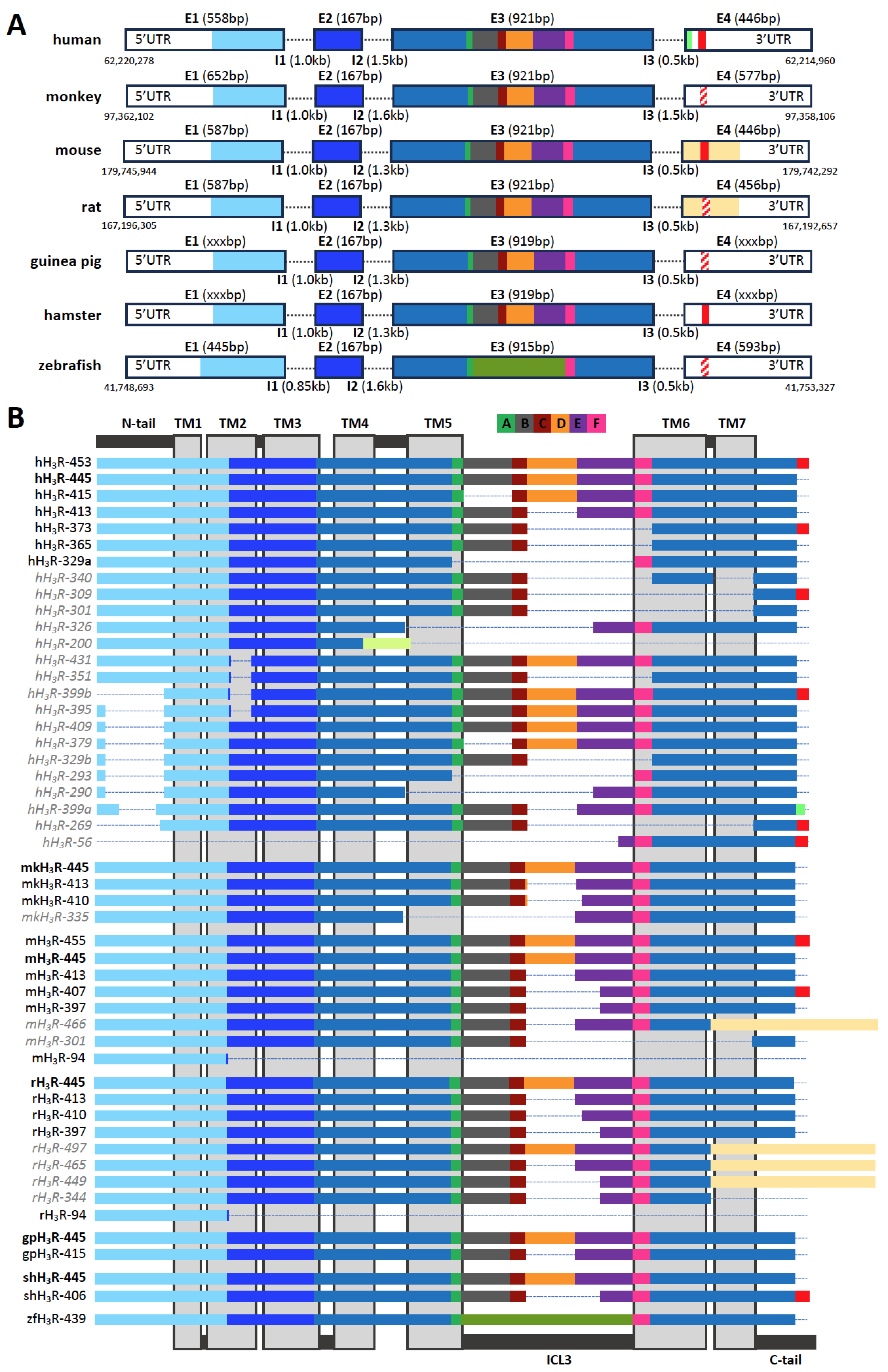
2. Histamine H3 Receptor (H3R) and Its Isoforms: Within and across Species

In the CNS, histamine is produced by neurons located in the tuberomammillary nucleus (TMN) situated in the posterior hypothalamus. These histaminergic neurons send projections throughout the brain, playing a crucial role in the regulation of arousal, wakefulness, and feeding by activating postsynaptic histamine receptors (H1R, H2R, and/or H3R) on glutamatergic, cholinergic and GABAergic neurons [23,24,25,26]. While the roles of H1R, H2R, and H3R are well-documented, the function of the H4R in the CNS remains less understood. Nonetheless, evidence supports the expression of H4R in brain tissues from both rats and patients with Parkinson’s disease (PD) and amyoptrophic lateral sclerosis (ALS), suggesting potential but yet to be fully elucidated roles [27,28,29]. In addition, H4R-deficient mice revealed that H4R influences various neurophysiological processes, such as locomotor activity, nociception, anxiety, and feeding behavior [30]. The H3R was demonstrated for the first time in 1983 as a presynaptic autoreceptor that inhibited histamine release from depolarized rat cerebral cortex slices due to a distinct pharmacological profile as compared to the earlier identified histamine H1 and H2 receptors (H1R and H2R) [31]. Thereafter, this new histamine receptor was shown to be involved in the regulation of a number of other neurotransmitters, i.e., as a heteroreceptor [25].
The human H3R cDNA was first cloned in 1999 from a thalamus cDNA library as a 445 amino acid-long GPCR (hH3R-445) that inhibited cAMP accumulation in transfected cell lines in response to histamine and H3R agonists [15]. In addition, an extended hH3R (hH3R-453) with an eight-amino-acid (446KMKKKTCL453) extended C-terminal tail and a comparable pharmacological profile was independently identified and reported one year later [16]. The human HRH3 gene is located on the antisense strand of chromosome 20 q13.33, spanning 5297 nucleotides, and was initially proposed to consist of three exons separated by two introns [17]. However, the 8-amino-acid C-terminal extension in hH3R-453 results from a splice donor site just before the stop codon in exon 3 that splices out 604 nucleotides [13,20] and consequently defines intron 3 and exon 4 (Figure 1A), instead of those latter two being the 3′ untranslated region (UTR) in exon 3 [17]. Nonetheless, both three- and four-exon HRH3 gene structures (accession codes: NM_00732.3 and XM_01702723.2, respectively) have been annotated in the National Center for Biotechnology Information (NCBI) GenBank database (Gene ID: 11255). For several other species, one or more H3R isoform cDNAs have been subsequently cloned from the hypothalamus (rat and Siberian hamster), thalamus (monkey), striatum (rat), cerebral cortex (guinea pig), or total brain RNA (rat, mouse, monkey, and zebrafish) [32,33,34,35,36,37,38,39,40,41,42,43]. Except for zebrafish, all these species express the H3R-445 isoform (Figure 1B), which is considered to be the canonical H3R variant due to its evolutionary conservation but also highest abundancy in the CNS (vide infra). Interestingly, C-terminal extensions comparable with hH3R-453 (446KMKKKTCL453) were only cloned or detected by RNA sequencing in the Siberian hamster (shH3R-406; 398KMEEKKTRL406) and mouse (mH3R-407/455; 398/446KMEEKKTSSL407/455), respectively [34,44]. Both these extensions also originate from the splice donor site that eliminates 604 base pairs (i.e., intron 3) including the canonical stop codon. Sequences encoding this C-terminal extension can be found in the HRH3 gene of other species, suggesting that they share a similar four-exon structure as humans (Figure 1A). The four-exon gene structure is further supported by 6TM splice variants that have been cloned from rats (rH3R-497, rH3R-465, and rH3R-449) using a 3’UTR reverse primer and RNA sequencing in mice (mH3R-466) [41,44], in which 740 and 686 nucleotides, respectively, are spliced-out after the tyrosine at the end of extracellular loop (ECL) 3 (Y392 in the canonical rH3R-445 and mH3R-445) and an alternative 105-amino-acid-long extracellular C-tail is encoded by exon 4 (Figure 1A,B). Interestingly, an alternative extended C-terminal sequence (NVKGP) was found in hH3R-399a by RNA sequencing, which is also encoded by exon 4. In all HRH3 genes, exon 1 encodes for the extracellular N-terminus, TM1, intracellular loop (ICL) 1, and the intracellular half of TM2. Exon 2 encodes for the extracellular half of TM2, ECL1, and TM3. Exon 3 encodes for the remaining helices, loops, and intracellular C-tail of H3R, and exon 4 encodes for the extended or alternative C-terminus (Figure 1A,B).
Hitherto, twenty-four human H3R isoforms have been identified by cloning and/or RNA sequencing (Table 1) [13,14,15,16,17,18,19,20,21,22,45], with amino acid sequence deletions in the extracellular N-terminal tail, TM2, TM5, TM6, TM7, ECL3, ICL3, or an extension of the C-terminal tail, or a combination of these (Figure 1B). The splice variants with deletions in either the extracellular N-terminus and/or the various TM helices do not conserve the prototypical 7TM GPCR structure and are most likely unable to bind H3R ligands and/or trigger intracellular G protein signaling. Indeed, hH3R-431 was unable to bind [125I]-iodoproxyfan when recombinantly expressed in Chinese hamster ovary K1 subline (CHO-K1) cells, despite a similar subcellular localization as hH3R-445 and hH3R-365 [13], whereas hH3R-301 and hH3R-200 were not responsive to histamine in a functional assay in transfected NIH-3T3 cells [20]. The truncated hH3R-200 has a deletion of 409 nucleotides in exon 3, resulting in a frameshift that might shorten TM4 with one helical turn and create a unique 30-amino-acid-long extracellular C-terminal tail [20]. In both mice and rats, 94-amino-acid-long truncated isoforms have been identified (mH3R-94 and rH3R-94) that consist of the N-terminal tail, TM1, ICL1, and a part of TM2 as a consequence of splicing event 4 nucleotides before the end of exon 1 and the consensus splice site in all other mouse and rat isoforms (i.e., CCTC^GTGGgtaa instead of CCTCGTGG^gtaa, respectively), which might be translated as a single transmembrane protein [20,44].
Exon 3 of HRH3 genes contains multiple cryptic splicing donor and acceptor sequences, allowing the deletion of a pseudo intron and resulting in the existence of numerous isoforms that conserve the 7TM GPCR structure but with a varying length of ICL3 [13,17]. In human H3R isoforms (hH3R-415, hH3R-413, hH3R-365/373, and hH3R-329a), an individual or combination of six ICL3 sequence segments (A–F) is deleted (Figure 1B). The deletion of segment D in hH3R-413 (ΔR274-S305), starting at nucleotide 822 of the coding sequence, is conserved in other species, including monkeys (mkH3R-413), mice (mH3R-413), rats (rH3R-413), and guinea pigs (gpH3R-415). Interestingly, this segment D deletion is extended into a part of segment E in H3R isoforms of the monkey (mkH3R-410), mouse (mH3R-397/407), rat (rH3R-410 and rH3R-397), and Siberian hamster (shH3R-406), whereas, so far only in human isoform hH3R-365/373, the deletion of segment D in combination with E and F (ΔR274-D353) has been encountered. Moreover, also the combined deletion of segments A–E (ΔR227-D353) in hH3R-329a is hitherto unique to humans. Exon 3 pseudo intron deletions are combined with the C-tail extension in hH3R-373, mH3R-407, and shH3R-406, but in humans also with non-7TM deletions in the N-tail/TM1 and/or TM2/TM5/TM6/TM7 (Figure 1B). The H3R isoforms with varying lengths for ICL3 and the C-terminal have garnered interest, as these regions are responsible for the selective interaction with intracellular transducer/scaffold proteins such as G proteins and β-arrestins, as reported for other GPCRs [6,46,47].
Table 1. H3R isoforms from seven species. Alternative names from original reference and GenBank, Uniprot, or the NSTRG, ENST, and ENSMUST RNA sequencing (other) codes from Splice-O-Mat (https://tools.hornlab.org/Splice-O-Mat/; accessed on 23 April 2024), Adult GTEx (https://gtexportal.org/home/transcriptPage; accessed on 23 April 2024), Human Protein Atlas (https://www.proteinatlas.org/about/download; accessed on 23 April 2024), respectively, are provided.
SpeciesIsoformAlternative NamesGenbankUniprotRNA-seqReferences
Human453-XP_054178891- [16,22,45]
445Isoform 1; GPCR97NP_009163.2Q9Y5N1NSTRG_52062.3; ENST00000340177.9[13,15,20,22,48,49]
431H3(TM2,431AA)-- [13]
415H3(Δi3,415AA)-- [13]
413H3S-- [17,49]
409--- [14]
399a399XP_016883112.1-NSTRG_52062.8[22]
399b399XM_017027623.1- [22]
395--- [14]
379--- [14]
373Isoform 4AF321913Q8WXZ9NSTRG_52062.5; ENST00000317393.10[20,22]
365Isoform 2; H3SAF321911Q8WY01 [13,20,48,49]
351--- [18]
340--- [19]
329aH3(Δi3,329AA)-- [13,49]
329b--- [14]
326H3(Δi3+TM5,326AA)AF346903- [13]
309Isoform 6AF346904Q8NI49 [20]
301Isoform 3AF321912Q8WY00 [20]
293--- [14]
290--- [14]
269---NSTRG_52062.7[22]
200Isoform 5AF346903Q8NI50ENST00000611492.1[20,21]
56---NSTRG_52062.6[22]
Monkey (macaca)445-AAO63757.1- [36]
413--- [35]
410--- [35]
335--- [35]
Mouse 466--E9Q540ENSMUST00000163215[44]
455---ENSMUST00000165762[44]
445Isoform 1AY044153P58406ENSMUST00000056480[32,40,44]
413--E9Q5S3ENSMUST00000164442[40,44]
407--E9Q7T5ENSMUST00000165248[44]
397--- [40]
301--E9Q522ENSMUST00000171736[44]
94--E9PZM9ENSMUST00000166724[44]
Rat 497H3DNP_001257495Q2VJ18 [41]
465H3ENP_001257496- [41]
449H3F-- [41]
445H3L; H3AAY009370Q9QYN8 [37,38,42]
413H3S; H3BAY009371Q541U0 [37,38,42]
410isoform 7/8NP_001257498- [38]
397H3C-- [38,50]
344-BAA88768- [51]
94H3(f1);H3T-- [38]
Guinea pig445H3LAF267537Q9JI35 [39]
415H3SAF267538Q9JI36 [39]
Hamster445LongAY855070- [34]
406ShortAY855071- [34]
Zebrafish439-ABF71709- [43]

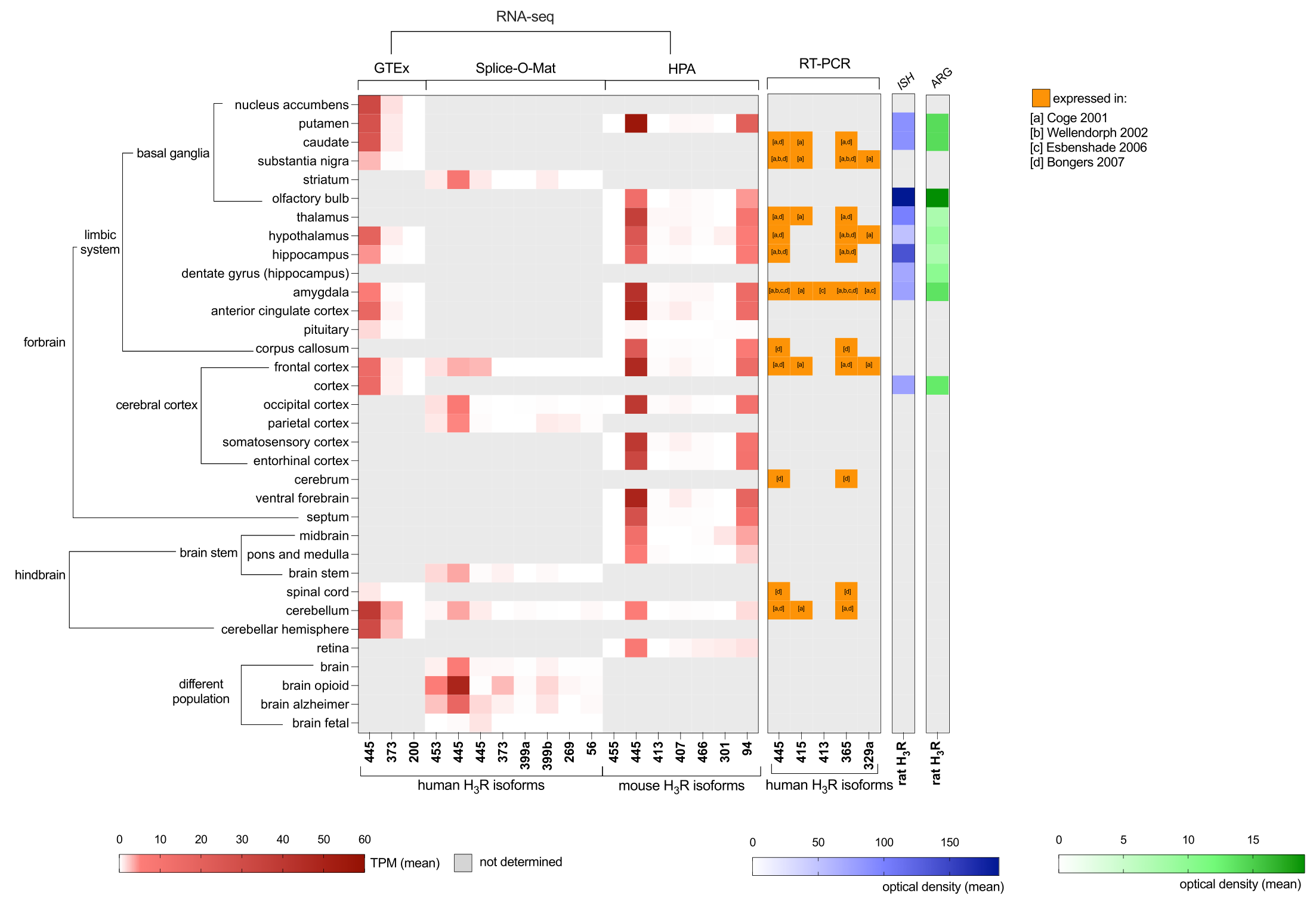
3. Localization of H3R Isoforms in the Central Nervous System

The H3R protein is predominantly expressed in the brain as determined by [3H]Nα-methylhistamine binding to membranes isolated from guinea pig tissues [52]. Highest H3R expression was observed in the cortex, hypothalamus, striatum, and midbrain (54.5, 42.8, 25.3, and 24.1 fmol/mg membrane protein, respectively). In the periphery, H3R is expressed at lower levels in the large intestine, ileum, and pancreas (~5.4 fmol/mg), but is (almost) undetectable in other digestive, respiratory, circulatory, excretory, reproductive, and muscle tissues (<1 fmol/mg).
RNA sequencing (RNA-seq) data from the GTEx consortium and Splice-O-Mat that encompass 53 and 48 tissue sample sites in the human body, respectively, confirmed that H3R is almost exclusively expressed in the CNS with less than 0.1 TPM detection in other tissues (Figure 2) [21,22]. In both human RNA-seq datasets, but also in mice, the canonical H3R-445 is the most abundantly expressed isoform in various brain regions (such as the caudate nucleus, hippocampus, basal ganglia, amygdala, cerebellum, and hypothalamus), which corroborates with the expression profiles by Northern blotting and RT-PCR in earlier studies [13,15,20,48,49]. In contrast to previous studies that reported the abundant expression of hH3R-365 next to the canonical hH3R-445 in the CNS [13,48,49], both the GTEx and Splice-O-Mat RNA-seq data did not detect the hH3R-365 transcripts but instead its C-terminally extended isoform hH3R-373 (Figure 1B and Figure 2). Indeed, the study of Cogé and co-workers did not identify extended extended isoform hH3R-373 as a reverse oligonucleotide primer based on the stop codon of the canonical hH3R-445 was used for cloning [13], whereas the C-terminally extended isoform hH3R-373 was cloned by Wellendorph et al. using 3’UTR reverse primers in addition to hH3R-365 [20]. Nonetheless, the earlier RT-PCR studies all used primers that flanked ICL3 and consequently did not discriminate between hH3R-365 and hH3R-373 [13,20,48,49]. The expression of the hH3R-453, hH3R-399a, hH3R-399b, hH3R-269, hH3R-200, and hH3R-56 isoforms was less abundant as compared to hH3R-445 according to RNA sequencing. RNA sequencing of a mouse brain revealed the expression of four additional mouse isoforms (mH3R-407, mH3R-466, mH3R-301 and, mH3R-94) that were not reported by other techniques before [44]. Surprisingly, the truncated mH3R-94 transcript is highly expressed in most brain regions and might perhaps act as single transmembrane anti-chaperone on the expression of other H3R isoforms (vide infra), as previously observed for the human H4R [53] (Figure 2).
Interestingly, RNA sequencing data from the Splice-O-Mat platform showed that hH3R-445, hH3R-453, hH3R-373, hH3R-399a, hH3R-399b, hH3R-269, and hH3R-56 were expressed at higher levels in the brains of individuals with an opioid use disorder (OUD) or Alzheimer’s disease symptoms as compared to the control population (Figure 2). The latter corroborates with the increased H3R mRNA levels observed in the prefrontal cortex of female subjects with Alzheimer’s disease [55]. Additionally, the upregulated expression of H3R was observed in the dorsolateral prefrontal cortex of individuals with schizophrenia [56], suggesting a potential role for H3R dysregulation in neurodegenerative disorders.
The elevated expression of H3R in OUD individuals may reflect an adaptive response to chronic opioid exposure. One possible mechanism underlying this upregulation could involve the modulation of histaminergic neurotransmission in response to opioid-induced alterations in neuronal activity. Moreover, the observed upregulation of H3R expression in OUD individuals may contribute to the modulation of opioid analgesia and tolerance. Previous studies have demonstrated a synergistic interaction between H3R agonists, RAMH, and the opioid fentanyl in animal models, suggesting that increased H3R expression could enhance the analgesic effects of opioids [57]. This phenomenon may represent a compensatory mechanism aimed at counteracting the development of tolerance to opioid analgesia, whereby the upregulation of H3R serves to potentiate opioid-induced analgesia and mitigate the need for escalating opioid doses. Furthermore, the upregulation of H3R expression in OUD individuals highlights the potential therapeutic relevance of targeting H3R for the treatment of opioid addiction. Given the role of H3R in modulating neurotransmitter release and synaptic plasticity, pharmacological interventions aimed at modulating H3R activity could represent a novel approach for managing OUD and reducing opioid-related harms. However, further research is needed to elucidate the precise mechanisms underlying the dysregulation of H3R in OUD and to evaluate the therapeutic potential of H3R-targeted interventions in the context of opioid addiction.
A comprehensive comparison between autoradiography with the H3R-selective radioligand [125I]iodoproxyfan and in situ hybridization (ISH) using a 33P-labeled riboprobe that recognizes most rH3R isoforms in the brains of the same rats (Figure 2) has revealed some discrepancies between receptor binding sites and mRNA expression [54], which can be explained by mRNA being located in perikarya, whereas presynaptic receptor binding occurs on the axon terminals [58]. Comparable distributions of H3R in the cerebral cortex, nucleus accumbens, striatum, and substantia nigra were observed in other autoradiography studies using R-[3H]-α-methylhistamine and [125I]iodophenpropit for rat [59,60], mouse [61], monkey [62] and human [62] cases. Notably, differential expression patterns of rat isoforms rH3R-445, rH3R-413, and rH3R-397 were observed in the dentate gyrus and hippocampal subfields by ISH [42].
In addition to radioligands, antibodies have been used to detect H3R expression in the CNS. Detection of its isoforms in the brain can be achieved using specific antibodies that target distinct peptide sequences. Antibodies raised against the RLSRDRKVAK peptide, corresponding to amino acids 349–358 within TM6 of the canonical H3R-445 sequence, including the last five amino acids of segment F (Figure 1B), allow for the identification of H3R protein expression across various brain regions, including the cerebral cortex, hippocampus, cerebellum, striatum, olfactory tubercle, substantia nigra, and thalamus. This distribution pattern is consistent with previous studies using mRNA and binding site analyses [63]. Additionally, using a different antibody from commercial resources (epitope not disclosed) revealed the H3R expression in dopamine D1 receptor (D1R)-positive interneurons and vesicular glutamate transporter 1 (VGLUT1)-positive corticostriatal output neurons, suggesting a potential functional role through receptor dimerization in neural networks (vide infra) [64]. The presence of the H3R protein has been confirmed in the ventral tegmental area and substantia nigra, particularly within dopaminergic neurons [65].
The peptide sequence junctions created by alternative splicing could provide unique epitopes for the generation of isoform-specific antibodies. Indeed, the anti-hH3R-329a antibody, raised against a peptide sequence at the junction between TM5 and segment F (CYLNIQ/SFTQR) in hH3R-329a (Figure 1), selectively detected this isoform in transfected cells without cross-reacting with hH3R-365 and hH3R-445 [66]. Similarly, a polyclonal antibody targeting a fourteen amino acid segment in ICL3 of rH3R-445 displays specificity for rH3R-445 without binding to the shorter rH3R-413 or rH3R-397 isoforms that lack this sequence in transfected HEK293 cells [67]. However, this antibody was found to also detect the β subunit of ATPase in H3R knockout mice. Challenges remain, where the anti-hH3R-365 antibody, targeting the peptide sequence including the segment C junction with TM6 (EAMPLH/RKVAKSLAC), has not distinguished between hH3R-365 and hH3R-445 [66]. Despite these difficulties, the combined use of antibodies such as anti-hH3R-365 and anti-hH3R-329a has provided valuable insights into the presence and localization of H3R isoforms in brain regions like dendrites, the tuberomammillary nucleus (TMN), and substantia nigra (SN) neurons. This understanding contributes to the potential functional diversity of H3R isoforms within the brain [66].

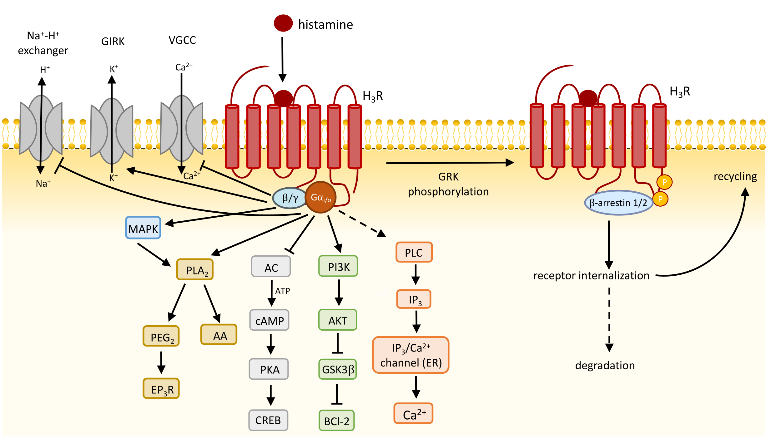
4. Function and Signaling Transduction of H3R

4.1. H3R Constitutively Activates Gi/o Proteins

Pretreatment of rat cerebral cortex membranes with pertussis toxin (PTX) disabled the accumulation of [35S]GTPγS in activated heterotrimeric G proteins upon stimulation with the selective H3R agonists Rα-methylhistamine and Nα-methylhistamine, indicating that endogenous H3R signals via Gαi/o proteins [68]. Indeed, systematic evaluation of histamine-induced hH3R-445 coupling to chimeric Gα subunits that harbor subtype-specific six-amino-acid C-tail substitutions in engineered human embryonic kidney cells (HEK293) by measuring alkaline phosphatase-fused transforming growth factor-α shedding has confirmed selective coupling to Gαi1, Gαi3, and Gαio [69]. Moreover, histamine stimulates the activation of heterotrimeric Gαi1, Gαi2, Gαi3, and Gαio by hH3R-445 with comparable potencies as measured by the dissociation of these Gα from Gβγ subunits using bioluminescence resonance energy transfer (BRET)-based sensors, suggesting that hH3R-445 displays no coupling preference between the PTX-sensitive Gi/o proteins [70].
The H3R-mediated activation of heterotrimeric Gαi/o protein results in various intracellular responses: the inhibition of adenylyl cyclase (AC), voltage-gated calcium channels (VGCC), and the Na+/H+ exchanger; the activation of phosphatidylinositol 3-kinase (PI3K), mitogen-activated protein kinase (MAPK), phospholipase C (PLC), phospholipase A2 (PLA2), and G protein-gated inwardly rectifying potassium (GIRK) channels (vide infra) (Figure 3).
The H3R is one of the few GPCRs that shows constitutive activity in native tissues as revealed by the decrease in [35S]GTPγS accumulation to membranes from mouse cerebral cortex and various rat brain regions, including the cerebral cortex, striatum, hypothalamus, thalamus, hippocampus, and midbrain upon incubation with H3R inverse agonists FUB 465, ciproxifan, and thioperamide [37,71]. A similar decrease in H3R-mediated G protein activation was observed in hippocampus membranes from the ground squirrel upon incubation with the inverse agonist clobenpropit [72]. In addition, recombinant (over)expression of hH3R-445 in Chinese hamster ovary (CHO) cells resulted in a receptor level-dependent increase in constitutive [35S]GTPγS accumulation to activated G proteins, which can be inhibited by inverse agonists ciproxifan [71]. Similarly, increasing rH3R-445 or rH3R-413 density in CHO cells constitutively enhanced [3H]arachidonic acid release and reduced cAMP accumulation [37]. Moreover, agonists and inverse agonists induced an opposite bioluminescence resonance energy transfer (BRET) signal in an intramolecular BRET-based H3R biosensor in which the bioluminescent donor nanoluciferase (Nluc) was fused to the H3R C-terminal tail and ICL3 was substituted from Thr229 to Phe348 with the fluorescent acceptor HaloTag, indicating that the apo receptor is constitutively active and its conformation is shifted into an inactive versus more active one, respectively [73].
The short isoforms hH3R-373 and hH3R-365 display higher constitutive activity than hH3R-445 in transfected cells, resulting agonist-independent [35S]GTPγS binding to activated G proteins, inhibition of cAMP production, and the activation of extracellular signal-regulated kinases (ERK)1/2 MAPK, which could be reduced by inverse agonists [48,74,75]. Interestingly, agonists display higher potencies (pEC50) to activate these responses via hH3R-373 and hH3R-365 as compared to hH3R-445, whereas the opposite was observed for the inhibition of constitutive signaling by inverse agonists that have higher potencies on hH4R-445 [48,74]. These potency differences between agonists and inverse agonists for hH3R-365/373 versus hH3R-445 are in line with their binding affinities for these receptor isoforms [48,74,76] and follow the paradigm that agonists bind preferentially to (constitutive) active receptors, whereas inverse agonists display higher affinity for inactive receptor conformations [77]. In addition, the higher affinity of agonists for hH3R-365 as compared to hH3R-445 is associated with a slower deactivation rate of Gβγ-driven GIRK channel activity in a recombinant Xenopus laevis oocyte model following histamine washout, which was hypothesized to reflect slower ligand dissociation kinetics from a high versus lower affinity binding site and consequently leaving the ternary complex between agonist-bound receptor and G protein longer intact [78].
The hH3R-365/373 (AlphaFold accession code: Q8WY01) isoforms are the only ones in which, in addition to ICL3 (segments D and E), TM6 is shortened by approximately two helical turns (segment F) as compared to the cryo-EM structure of hH3R-445 (PDB accession code: 8YUU) and lacks the aspartate (D353 in hH3R-445) at position 6.30 × 30 (Figure 4) [79]. This aspartate (or glutamate) residue is conserved in many aminergic GPCRs and interacts with arginine at position 3.50 × 50 in the DRY motif within TM3 to form an ionic lock to maintain the receptor in an inactive conformation [80]. Disruption of this ionic lock by site-directed mutagenesis of the aspartate or glutamate at the 6.30 × 30 position increased constitutive activity in, for example, the β2-adrenergic receptor and H1R, and consequently increased agonist binding affinity [80,81]. Indeed, Ala-substitution of D353 in hH3R-445 resulted in slower GIRK deactivation rates than hH3R-445 upon histamine washout in oocytes, suggesting prolonged agonist binding, but not as slow as hH3R-365, indicating that the 80-amino-acid deletion has a larger effect on the receptor conformation [78]. Other isoforms with conserved 7TM, such as hH3R-415, hH3R-453, and hH3R-413, exhibit similar ligand affinity profiles to hH3R-445, which is in line with their comparable level of constitutive activity [74]. Interestingly, the shortest 7TM H3R isoform, hH3R-329a, exhibits increased binding affinities for agonists, while its binding affinity for inverse agonists is comparable to that of hH3R-445. The 116-amino-acid deletion (segments A–E) in hH3R-329a leaves only a four-amino-acid long ICL3 (i.e., 343SFTQ346 in the canonical hH3R-445 sequence), potentially hindering this receptor isoform from adopting a conformation that supports high-affinity binding of inverse agonists, despite the detection of inverse agonism [74].

4.2. Presynaptic H3R Inhibits Neurotransmitter Release and Synthesis

Neurons communicate with other cells by releasing neurotransmitters (e.g., histamine, dopamine, serotonin) in the synaptic cleft between these cells upon arrival of an axonal action potential and depolarization of the presynaptic terminal (Figure 5). This depolarization opens voltage-gated calcium channels (VGCC) resulting in an influx of Ca2+ ions. The increased intracellular Ca2+ concentrations subsequently drive the exocytotic machinery (involving SNARE proteins, SM proteins, and synaptotagim) to fuse vesicles that contain neurotransmitters with the presynaptic plasma membrane, releasing their content into the synaptic cleft [83,84]. In addition to the activation of their cognate receptors on postsynaptic cells, neurotransmitters can activate presynaptic autoreceptors to inhibit further neurotransmitter release by closing the VGCC in a Gβγ-dependent manner [85,86]. Indeed, stimulation of the H3R autoreceptor with agonists inhibited N- and P-type VGCC activity via pertussis toxin-sensitive heterotrimeric Gi/o proteins in depolarized rat tuberomammillary nucleus (TMN) histaminergic neurons [87], consequently attenuating histamine release from depolarized rat and mouse cortical synaptosomes [37]. Moreover, histamine release from these depolarized cortical synaptosomes is increased by H3R inverse agonists FUB 465 and thioperamide, confirming that native H3R is constitutively active and tonically inhibits histamine release [37]. Treatment of mice with the H3R inverse agonist ciproxifan (3 mg/kg) resulted in increased histamine release in the preoptic area and prefrontal cortex as detected with a genetically encoded fluorescent histamine (GRABHA) sensor that was recombinantly expressed in these areas [88].
In non-histaminergic neurons, H3R acts as a presynaptic heteroreceptor and inhibits the release of other neurotransmitters such as acetylcholine, noradrenaline, serotonin, dopamine, glutamate, GABA, and neuropeptides via multiple G protein-mediated pathways (Table 2). For instance, H3R activation can result in a direct Gβγ-induced inhibition of VGCC to attenuate dopamine release, as detected in H3R-transfected nerve growth factor-differentiated rat pheochromacytoma cells, whose phenotype is close to that of sympathetic neurons [89]. The inhibition of norepinephrine exocytosis by H3R stimulation in guinea pig cardiac sympathetic nerve endings was found through the inhibition of the AC/protein kinase A (PKA) pathway to further phosphorylate VGCC [90]. The hyperpolarization of the cell membrane by GIRK channel activation, which allows potassium ion (K+) efflux from neurons, can potentially inhibit the subsequent opening of VGCC, which is necessary for further neurotransmitter release [91]. It has been shown that the GIRK channel blocker tertiapin-Q has effectively prevented the H3R agonist immepip-induced inhibition of glutamate release in a rat corticostriatal synapse as measured by decreased paired-pulse ratios [92]. Furthermore, a mechanistic connection between H3R-induced MAPK activation and subsequent PLA2 phosphorylation, which initiates anti-exocytotic processes, such as prostaglandin E2 (PGE2) production to activate prostaglandin EP3 receptor (EP3R) and block VGCC, ultimately reduces norepinephrine release in guinea pig heart synaptosomes [93]. However, whether the MAPK-PLA2-PGE2-EP3R axis and GIRK channels contribute to autoreceptor H3R-mediated histamine release remains unclear.
In addition, the activation of the presynaptic H3R autoreceptor suppresses the activity of histidine decarboxylase (HDC) and consequently histamine synthesis in the rat cerebral cortex via the inhibition of calcium/calmodulin-dependent protein kinase type II (CaMKII) and PKA-mediated phosphorylation by the Gβγ- and Gαi/o-mediated inhibition of N- and P-type VGCC and adenylyl cyclase, respectively [112,113]. Importantly, H3R inverse agonists such as thioperamide and clobenpropit enhanced histamine synthesis, indicating that the constitutive activity of H3R contributes to the negative feedback regulation of histamine synthesis [112,113]. Notably, H3R activation can also inhibit dopamine synthesis in the rat nucleus accumbens by the inhibition of the PKA pathway to attenuate tyrosine hydroxylase phosphorylation [65], thereby reducing the available amounts of dopamine for release.
Interestingly, the autoreceptor function of rat H3R is proposed to be carried out by the short isoform. The stereoselectivity of NαMe-αClMeHA and R(-)sopromidine enantiomers on cAMP formation on CHO cells transfected with rH3R-413 was found to be more similar to the effects on the rat cortex, striatum, and hypothalamus, which are areas rich in autoreceptors [114]. Other GPCRs, such as the short dopamine D2 receptor (D2R), with a 29-amino-acid deletion in the third intracellular loop, also function as an autoreceptor.

4.3. Postsynaptic H3R Function and Downstream Effects

Activation of postsynaptic H3R was found to diminish the firing rate of melanin-concentrating hormone-producing neurons [115]. In substantia nigra pars reticulata (SNr) GABA projection neurons, H3R activation hyperpolarized and suppressed firing frequency, consequently decreasing the intensity of basal ganglia output [116]. The reduced firing frequency by H3R is related to the reduced phosphorylation level of ERK and the increased A-type K+ current [117,118]. A more recent study, using a chimeric H3R protein in which ICL3 is fused with extracellular and transmembrane domains of rhodopsin to convey light responsiveness, demonstrated that postsynaptic H3R activation in ventral basal forebrain cholinergic neurons is responsible for the inhibition of contextual fear memory retrieval via suppressing the firing frequency [119].
Downstream effects mediated by H3R contribute to neurological processes such as promoting neurogenesis or exerting neuroprotective effects. For instance, thioperamide can protect primary neurons against oxygen–glucose deprivation-induced injury and promote the proliferation of the neural stem cell line NE-4C through stimulation of downstream cAMP response element binding protein (CREB) phosphorylation, aligning with the constitutive activity of H3R in native tissues [120]. H3R activation by the agonist imetit improves the viability of mouse primary cortical neurons that are impaired by oxygen–glucose deprivation/reoxygenation conditions via promoting ERK1/2 phosphorylation in a PTX-sensitive manner [121]. Besides, H3R activation protects cultured rat and mouse cortical neurons from neurotoxic insults by increasing the expression of the anti-apoptotic protein BCl-2 via the Akt–glycogen synthase kinase (GSK) 3β axis [122], which is constitutively activated via Gi/o proteins and phosphoinositide-3-kinase (PI3K) in SK-N-MC cells recombinantly expressing hH3R-445 [123].

4.4. H3R Dimerization

In the striatum, H3R is present on the afferent terminals of glutamatergic and dopaminergic neurons and acts as presynaptic heteroreceptor (vide supra) to negatively regulate glutamate and dopamine release [64,124,125]. However, the majority of striatal H3Rs are expressed as postsynaptic receptors on efferent striato-nigral and striato-pallidal GABAergic medium spiny neurons (MSN) [64,126], where they co-localize in close proximity with D1R and D2R, respectively, as shown by an antibody-based proximity labeling assay (PLA) in rodent striata [127,128,129]. Co-immunoprecipitation of H3R with either D1R or D2R using specific antibodies from rodent striatal lysates provided additional evidence that these receptors might physically interact in MSNs [126,127,128,129]. Further support that H3R might specifically interact with D1R and D2R has been provided by saturable bioluminescence resonance energy transfer (BRET) between one receptor fused to luciferase (BRET donor) expressed at a fixed level in combination with increasing levels of the other receptor fused to fluorescent protein (BRET acceptor) in heterologous HEK293(T) cell lines [130,131].
GPCRs can modulate each other’s trafficking, ligand binding, and/or signaling properties when forming dimeric or multimeric complexes [132,133]. Negative binding cooperativity was observed within H3R/D1R and H3R/D2R heteromers, with H3R agonists decreasing the binding affinity of D1R and D2R agonists [130,131], which might contribute to the H3R agonist-mediated attenuation of D1R and D2R agonist-induced locomotor activity in mice [127,130]. In contrast, H3R inverse agonists potentiated D1R and D2R-mediated locomotor activity in response to their agonists, SKF38393 and quinpirole, respectively, in mice with a depletion in endogenous striatal dopamine, indicating that H3R constitutively attenuates the responsiveness of the associated D1R or D2R protomers [130]. Stimulation of H3R attenuated D2R-mediated Akt–GSK3β signaling in striato-pallidal MSNs in response to D2R agonists in a β-arrestin2-dependent manner [127]. Interestingly, D1R agonists increased cAMP levels in cell lines expressing only the Gs-coupled D1R but decreased forskolin-induced cAMP levels in cell lines co-expressing D1R and the Gi/o-coupled H3R, suggesting that the D1R/H3R heteromer signals through the H3R protomer [128,131]. Cells recombinantly expressing D1R showed agonist-induced extracellular signal-regulated kinase 1/2 (ERK1/2) phosphorylation, whereas cells that only expressed H3R did not activate this response [131]. However, cells co-expressing D1R and H3R showed pERK1/2 in response to stimulation with D1R (SKF 38393 or SKF 81297) or H3R (RAMH or imetit) agonists, which could be cross-antagonized by selective D1R (SCH23390) and H3R (thioperamide) antagonists [128,131]. Similarly, postsynaptic D1R/H3R-mediated signaling to ERK1/2 and cross-antagonism was observed in striatal slices obtained from rats and wild-type mice but not in striata from transgenic mice that lack D1R. Importantly, the disruption of D1R/H3R heteromers in immortalized striatal cells using a synthetic transmembrane 5 peptide (TAT-TM5) abolished cross-antagonism of the D1R agonist (SKF 81297)-induced pERK1/2 by the H3R antagonist thioperamide [129]. In addition, striatal cell death involving the p38 apoptotic pathway, upon overactivation by the D1R agonist SKF 81297 (>30 μM), can be cross-antagonized by the H3R antagonist thioperamide, which was abolished upon disruption of the D1R/H3R heteromer by TAT-TM5 or the downregulation of H3R using shRNA [129].
In a preclinical Huntington’s disease (HD) mice model (HdhQ7/Q111 knock-in), D1R-H3R heteromers were detected by PLA in the striatum, cerebral cortex, and hippocampus slices at 2–4 months of age but were undetectable at early disease states at 6–8 months of age [129]. Indeed, thioperamide can prevent D1R agonist SKF 81297-induced apoptosis in striatal, cortical, and hippocampal organotypic cultures from HD mice at four but not eight months of age, confirming that D1R/H3R heteromerization is required for this cross-antagonism [129]. In humans, D1R/H3R heteromers are present in striatal (caudate putamen) slices of control individuals and individuals with low-grade (0, 1, and 2) HD, but are almost absent in individuals with high-grade (3 and 4) HD [129]. In immortalized mouse striatal cells, the D1R agonist SKF 81297 reduces D1R/H3R heteromerization, which could be prevented by pretreatment with thioperamide [129]. Moreover, the chronic treatment of HD mice with thioperamide has been shown to prevent the loss of D1R/H3R heteromers and cognitive and motor learning deficits at early disease states, but not when D1R/H3R heteromers were already lost in late disease states [129].
Interestingly, cocaine can disrupt the cross-antagonism of the D1R agonist SKF 38393-induced pERK1/2 and apoptosis in the rodent striatum by thioperamide in a sigma-1 receptor (σ1R)-dependent manner, which can be blocked by pretreatment with σ1R antagonist PD 144418 [128]. BRET and sequential resonance energy experiments in transfected cells revealed that σ1R interacts with the D1R protomer in the D1R/H3R heteromer, whereas their close proximity was confirmed in the rodent striatum by PLA and the co-immunoprecipitation of σ1R and H3R with D1R [129]. Hence, antagonizing σ1R restores the protective effect of H3R on D1R signaling in cocaine-induced cell death.
Postsynaptic D1R/H3R heteromers have also been detected in the rodent cerebral cortex, where they interact with ionotropic N-methyl-D-aspartate (NMDA) glutamate receptors [134]. Importantly, H3R antagonist thioperamide cross-antagonized NMDA- and D1R agonist-induced excitotoxic cell death in rodent cortical cultures. In addition, both the D1R and H3R antagonist prevented neurodegeneration resulting from Aβ peptide toxicity in the context of Alzheimer’s disease [134].
Presynaptic H3R physically interacts with adenosine 2A receptors (A2AR) in the terminals of striatopallidal MSNs, as revealed by co-immunoprecipitation experiments [135]. H3R decreased the binding affinity of the A2AR agonist in globus pallidus synaptosomal membranes by two-fold, whereas a two-fold increase in affinity was observed in the opposite direction, suggesting allosteric interaction within the H3R/A2AR heteromer [136]. The Gs-coupled A2AR enhances the VGCC/Ca2+-dependent GABA release from depolarized striatopallidal synaptosomes via the cAMP/PKA pathway, which is counteracted by H3R via the Gai/o-mediated inhibition of adenylyl cyclase [136].
H3R homodimerization is shown in transfected HEK293 cells and cortical neurons using BRET and co-immunoprecipitation, whereas Western blot analysis of rat cortices, cerebella, and hypothalamus membranes suggests the presence of both monomers and dimers [137]. Interestingly, H3R agonists and inverse agonists induced a concentration-dependent decrease and increase in BRET amplitude, which was more pronounced in the cortical neurons as compared to HEK293 cells [137]. These BRET changes were interpreted as conformational changes within dimers and not the association or dissociation of dimers, which was supported by the fact that agonists did not induce dimer dissociation in Western blot analysis.
The detection of endogenous H3R heteromers in tissue slices by Co-IP and PLA methods does not discriminate between H3R isoforms. Hitherto, the dimerization of other H3R isoforms than the canonical H3R-445 in recombinant cells has not been reported to our best knowledge, whereas the ex vivo studies suggest potential interactions. Three rat H3R isoforms (rH3R-497, rH3R-465, and rH3R-449) that have a unique amino acid sequence after TM6, and consequently lack the conserved TM7 and C-tail, are unable to bind H3R ligands or inhibit adenylyl cyclase activity [41]. However, these three isoforms have a dominant negative effect on the trafficking of rH3R-445 and reduces its expression at the cell surface, presumably by engaging into dimers that are retained intracellularly, as observed for truncated histamine H4 receptors (H4R) and α1A-adrenergic receptor splice isoforms [53,138].

4.5. Regulation of H3R Signaling

Agonist-bound GPCRs are phosphorylated at their C-terminal tail and/or intracellular loops by GPCR kinases (GRKs), resulting in the recruitment of cytosolic β-arrestin1 and/or β-arrestin2 [139], which desensitizes the receptor by hindering further G protein coupling and preventing continuous responsiveness [140]. In addition, β-arrestins act as a scaffold for several proteins involved in the endocytic process, including clathrin, to facilitate receptor internalization.
Some ex vivo evidence indicates that pre-exposure to H3R agonists may induce the desensitization of subsequent responsiveness to a secondary stimulation with an agonist. For instance, in the guinea pig ileum, where H3R activation attenuates electrically-induced contraction primarily through the inhibition of acetylcholine release from postganglionic cholinergic neurons, prior exposure to the agonist RAMH has led to reduced potency and efficacy in subsequent agonist applications [141]. Besides, the specific binding of [3H]NAMH to membranes from rat striatal slides was observed to decrease after pre-treatment of the agonist immepip, suggesting that H3R is internalized/downregulated [142]. Furthermore, the reduction in functional responses, including cAMP signal and [35S]GTPγS accumulation, as well as [3H]NAMH binding, was prevented by culturing CHO cells transfected with H3R in a hypertonic medium or incubation at 4 °C, which is known to affect clathrin-dependent endocytosis [143], indicating the involvement of clathrin-dependent internalization in H3R desensitization. GRK2 is suggested to play a major role in this process, as downregulation of GRK2 expression by small interfering RNA (siRNA) in CHO cells attenuates the desensitization of the cAMP signal and the reduction of [3H]NAMH binding due to prolonged agonist exposure [143].
Notably, differential desensitization dynamics are observed between H3R isoforms hH3R-365 or hH3R-445. Studies in CHO cells transfected with either hH3R-365 or hH3R-445 have revealed that hH3R-365 desensitizes more rapidly compared to hH3R-445. However, despite its quicker onset, hH3R-365 reaches a lower maximum extent of desensitization and resensitization. This suggests that while hH3R-365 can quickly become unresponsive to stimuli, it also recovers its responsiveness more swiftly than hH3R-445 [144]. Furthermore, hH3R-415 and hH3R-445 have been shown to effectively recruit β-arrestin2 upon agonist stimulation, facilitating their desensitization and internalization processes. In contrast, hH3R-365 and hH3R-329a demonstrate higher efficacy and potency in recruiting β-arrestin2. This indicates that these isoforms might have a stronger or more immediate regulatory response to agonist binding, leading to more pronounced desensitization [75].
In addition, H3R-445 responsiveness to agonist stimulation is desensitized in CHO-cells by protein kinase C-mediated phosphorylation following the stimulation of Gq-coupled endogenous purinergic P2Y2 receptors with adenosine 5’-triphosphate (ATP), indicating potential cross-regulation between GPCRs [145]. However, hitherto, no heterologous desensitization of H3R has been reported in native tissue.

4.6. Isoform Signaling Bias

Alternative splicing of the intracellular ICL3 and/or C-terminal tail can alter the conformational activity state of the receptor and consequently indirectly affect ligand binding to the extracellular side of the 7TM domain, with, for example, a 20- and 32-fold lower binding affinity of pitolisant on hH3R-373 and hH3R-365, respectively, as compared to the less constitutively active hH3R-445 (vide supra). This significant difference in binding affinity implies the necessity of a more detailed analysis of the signaling capacities of H3R isoforms for future H3R drug discovery efforts.
Isoform-specific functional properties extend beyond binding selectivity. For example, the H3R agonists impentamine and dimethyl-impentamine act as partial agonists on H3R-453, H3R-445, H3R-415, H3R-413, H3R-373, and H3R-329a, but are full agonists on H3R-365, with the same intrinsic activity as histamine, with comparable binding affinities for hH3R-445 and hH3R-365 [74]. Interestingly, hH3R-365 was also found to be unable to further activate GSK3β phosphorylation in CHO-K1 transfected cells with agonist stimulation [146]. Additionally, certain isoforms are specifically modulated in a ligand-directed manner. For instance, the H3R agonists proxyfan and iodoproxyfan elicited a robust response in increasing intracellular Ca2+ concentrations but failed to elicit a response for hH3R-365, indicating the potential for “isoform-biased” agonists [146]. The selectivity of G protein coupling by GPCR is an intricate process that is heavily influenced by the structural variations within the receptor, particularly in the intracellular loops and transmembrane helices. ICL3, which connects TM5 and TM6, plays a pivotal role in this selectivity as there is a dynamic conformational equilibrium of ICL3 between blocking and exposing the G protein-binding site that allows for the autoregulation of receptor activity [47]. Shorter isoforms with a truncated ICL3 might favor coupling with different subtypes of G proteins compared to longer isoforms, which have a more extended ICL3 that can stabilize different conformations. This can result in variations in signaling efficiency and receptor autoinhibition, as seen in other GPCRs, like the dopamine D2 receptor, where the long isoform (D2L) has a higher efficacy of canonical signaling compared to the short isoform (D2S) with a 29-amino-acid deletion in ICL3 [46,147]. Given the varying lengths of ICL3 in H3R isoforms, these isoforms might exhibit distinct G protein coupling profiles, which presumably leads to differential downstream functional outcomes in addition to their Gi/o-mediated inhibition of adenylyl cyclase activity [74]. Furthermore, structural analyses from cryo-EM studies of H3R (PDB ID: 8YUU and 8YUV) show that the lengths of TM5 and TM6 are longer than in H1R and H2R. Machine learning analysis of 98 homology models from GPCRdb revealed that GPCRs with a long TM5 length are more likely to couple to Gi/o proteins as compared to those with a shorter or tilted TM5. Interestingly, TM5 of hH3R-329a is 1.5 helical turns shorter as compared to the hH3R-445 cryo-EM structure. Agonist stimulation of hH3R-329a did not activate pAKT T308/S473 in CHO-K1-transfected cells, whereas a robust response was observed in hH3R-415, hH3R-365, and hH3R-445 [75]. However, whether this is related to its G protein selectively remains to be investigated. Exchange of the intracellular half of TM5, TM6, and ICL3 (P210 to P373) of H3R with the corresponding section of the Gs-coupled H2R (P194 to P249) shifted its coupling from Gi to Gs, confirming the role of these domains in G protein selectivity [79]. The switch from Gi/o to Gs signaling might result in changes downstream for signaling cascades, potentially leading to varied therapeutic effects and side effects, as observed in the μ-opioid receptor (MOR), for instance. The MOR-1D isoform with an extended C-terminus, in comparison to the canonical isoform MOR-1, has been implicated in morphine-induced itch as a side effect [148]. Additionally, chronic exposure to morphine results in the upregulation of MOR isoforms MOR-1B2 and MOR-1C1. These variants undergo phosphorylation at C-terminal sites that are not present in the canonical MOR-1 isoform, which is associated with a shift from the predominantly inhibitory Gi/o coupling pathway to the stimulatory Gs pathway. This shift leads to changes in cellular responses, and this switch can contribute to the development of tolerance, dependence, and other side effects, such as opioid-induced hyperalgesia or itch [149].
The expression of truncated 6TM GPCR isoforms has been extensively documented in human and rodents, including H3R and the delta-opioid receptor [150]. These 6TM isoforms exert their function via interacting with intracellular compartments or affecting other 7TM isoforms. Studying H3R isoforms might uncover new intracellular signaling or regulation mechanisms, such as dimerization that may contribute to overall receptor function. Understanding the structural determinants of G protein selectivity in H3R isoforms is crucial for developing targeted therapies that can modulate specific receptor activities in pathological states.

5. Discussion

In this review, we have discussed the existence of the H3R isoforms that have been identified so far within and across species, which expands the complexity of H3R function in vivo. The research of H3R isoforms holds significant promise and is crucial for advancing our understanding of H3R functionality and its implications for drug development. However, the identification of various H3R isoforms across different species and tissues indicates complexity in H3R-mediated signaling that remains underexplored. As anticipated, the canonical H3R-445 isoform is predominantly expressed in the brain. However, one compelling reason to study H3R isoforms is their potential differential expression in pathological states, as shown by RNA sequencing. Currently, there is limited research on how and where these isoform proteins are expressed in diseased tissues, which could provide insights into their roles in disease progression and response to treatment. Understanding these patterns could lead to the development of isoform-specific drugs, which might offer more precise therapeutic interventions with potentially fewer side effects. In addition, investigation of the physiological roles of individual isoforms in more relevant biological in vivo and/or ex vivo contexts might provide clearer understanding of their functional significance. In particular, considering the observed differences in the constitutive signaling and ligand affinities between some of the isoforms. This knowledge will enhance our ability to design drugs that can specifically target desired signaling pathways, thereby improving therapeutic outcomes. Hence, the development of selective tool ligands and or antibodies to unambiguously identify which isoform protein is mediating an observed effect in native tissue has so far been challenging but is key for our in-depth understanding of their function and therapeutic potential.

Author Contributions

Writing—M.G., J.F.O. and H.F.V.; writing—review and editing, R.L. and H.F.V.; supervision, R.L. and H.F.V. All authors have read and agreed to the published version of the manuscript.

Funding

Meichun Gao is supported by China Scholarship Council (CSC) grant (funding number: 202006310027).

Data Availability Statement

Splice-O-Mat (https://tools.hornlab.org/Splice-O-Mat/ accessed on 23 April 2024), Adult GTEx (https://gtexportal.org/home/transcriptPage accessed on 23 April 2024), Human Protein Atlas (https://www.proteinatlas.org/about/download accessed on 23 April 2024), AlphaFold (https://alphafold.ebi.ac.uk accessed on 5 April 2024).

Conflicts of Interest

The authors declare no conflicts of interest.

References

  1. Marasco, L.E.; Kornblihtt, A.R. The physiology of alternative splicing. Nat. Rev. Mol. Cell Biol. 2023, 24, 242–254. [Google Scholar] [CrossRef] [PubMed]
  2. Olivieri, J.E.; Dehghannasiri, R.; Wang, P.L.; Jang, S.; de Morree, A.; Tan, S.Y.; Ming, J.; Ruohao Wu, A.; Tabula Sapiens, C.; Quake, S.R.; et al. RNA splicing programs define tissue compartments and cell types at single-cell resolution. eLife 2021, 10, e70692. [Google Scholar] [CrossRef]
  3. Markovic, D.; Challiss, R.A. Alternative splicing of G protein-coupled receptors: Physiology and pathophysiology. Cell. Mol. Life Sci. 2009, 66, 3337–3352. [Google Scholar] [CrossRef] [PubMed]
  4. Hauser, A.S.; Attwood, M.M.; Rask-Andersen, M.; Schioth, H.B.; Gloriam, D.E. Trends in GPCR drug discovery: New agents, targets and indications. Nat. Rev. Drug Discov. 2017, 16, 829–842. [Google Scholar] [CrossRef] [PubMed]
  5. Mele, M.; Ferreira, P.G.; Reverter, F.; DeLuca, D.S.; Monlong, J.; Sammeth, M.; Young, T.R.; Goldmann, J.M.; Pervouchine, D.D.; Sullivan, T.J.; et al. Human genomics. The human transcriptome across tissues and individuals. Science 2015, 348, 660–665. [Google Scholar] [CrossRef] [PubMed]
  6. Marti-Solano, M.; Crilly, S.E.; Malinverni, D.; Munk, C.; Harris, M.; Pearce, A.; Quon, T.; Mackenzie, A.E.; Wang, X.; Peng, J.; et al. Combinatorial expression of GPCR isoforms affects signalling and drug responses. Nature 2020, 587, 650–656. [Google Scholar] [CrossRef] [PubMed]
  7. Panula, P.; Chazot, P.L.; Cowart, M.; Gutzmer, R.; Leurs, R.; Liu, W.L.; Stark, H.; Thurmond, R.L.; Haas, H.L. International Union of Basic and Clinical Pharmacology. XCVIII. Histamine Receptors. Pharmacol. Rev. 2015, 67, 601–655. [Google Scholar] [CrossRef] [PubMed]
  8. Cheng, L.; Liu, J.; Chen, Z. The Histaminergic System in Neuropsychiatric Disorders. Biomolecules 2021, 11, 1345. [Google Scholar] [CrossRef] [PubMed]
  9. Harwell, V.; Fasinu, P.S. Pitolisant and Other Histamine-3 Receptor Antagonists-An Update on Therapeutic Potentials and Clinical Prospects. Medicines 2020, 7, 55. [Google Scholar] [CrossRef]
  10. Syed, Y.Y. Pitolisant: First Global Approval. Drugs 2016, 76, 1313–1318. [Google Scholar] [CrossRef]
  11. Ligneau, X.; Perrin, D.; Landais, L.; Camelin, J.C.; Calmels, T.P.; Berrebi-Bertrand, I.; Lecomte, J.M.; Parmentier, R.; Anaclet, C.; Lin, J.S.; et al. BF2.649 [1-3-[3-(4-Chlorophenyl)propoxy]propylpiperidine, hydrochloride], a nonimidazole inverse agonist/antagonist at the human histamine H3 receptor: Preclinical pharmacology. J. Pharmacol. Exp. Ther. 2007, 320, 365–375. [Google Scholar] [CrossRef] [PubMed]
  12. Urquhart, L. FDA new drug approvals in Q3 2019. Nat. Rev. Drug Discov. 2019, 18, 816. [Google Scholar] [CrossRef] [PubMed]
  13. Coge, F.; Guenin, S.P.; Audinot, V.; Renouard-Try, A.; Beauverger, P.; Macia, C.; Ouvry, C.; Nagel, N.; Rique, H.; Boutin, J.A.; et al. Genomic organization and characterization of splice variants of the human histamine H3 receptor. Biochem. J. 2001, 355, 279–288. [Google Scholar] [CrossRef] [PubMed]
  14. Gallagher, M.; Yates, S.L. Histamine H3 Receptor Polynucleotides. WO Patent 2003/042359, 22 May 2003. [Google Scholar]
  15. Lovenberg, T.W.; Roland, B.L.; Wilson, S.J.; Jiang, X.; Pyati, J.; Huvar, A.; Jackson, M.R.; Erlander, M.G. Cloning and functional expression of the human histamine H3 receptor. Mol. Pharmacol. 1999, 55, 1101–1107. [Google Scholar] [CrossRef]
  16. Nakamura, T.; Itadani, H.; Hidaka, Y.; Ohta, M.; Tanaka, K. Molecular cloning and characterization of a new human histamine receptor, HH4R. Biochem. Biophys. Res. Commun. 2000, 279, 615–620. [Google Scholar] [CrossRef]
  17. Tardivel-Lacombe, J.; Morisset, S.; Gbahou, F.; Schwartz, J.C.; Arrang, J.M. Chromosomal mapping and organization of the human histamine H3 receptor gene. Neuroreport 2001, 12, 321–324. [Google Scholar] [CrossRef] [PubMed]
  18. Tsui, P. Human Histamine H3 Gene Variant-2. WO Patent 2001/068703, 20 September 2001. [Google Scholar]
  19. Tsui, P. Human Histamine H3 Gene Variant-3. WO Patent 2001/068816, 20 September 2001. [Google Scholar]
  20. Wellendorph, P.; Goodman, M.W.; Burstein, E.S.; Nash, N.R.; Brann, M.R.; Weiner, D.M. Molecular cloning and pharmacology of functionally distinct isoforms of the human histamine H(3) receptor. Neuropharmacology 2002, 42, 929–940. [Google Scholar] [CrossRef] [PubMed]
  21. GTEx-Consortium. The GTEx Consortium atlas of genetic regulatory effects across human tissues. Science 2020, 369, 1318–1330. [Google Scholar] [CrossRef] [PubMed]
  22. Kuhn, C.K.; Stenzel, U.; Berndt, S.; Liebscher, I.; Schoneberg, T.; Horn, S. The repertoire and structure of adhesion GPCR transcript variants assembled from publicly available deep-sequenced human samples. Nucleic Acids Res. 2024, 52, 3823–3836. [Google Scholar] [CrossRef]
  23. Thakkar, M.M. Histamine in the regulation of wakefulness. Sleep Med. Rev. 2011, 15, 65–74. [Google Scholar] [CrossRef]
  24. Lin, W.; Xu, L.; Zheng, Y.; An, S.; Zhao, M.; Hu, W.; Li, M.; Dong, H.; Li, A.; Li, Y.; et al. Whole-brain mapping of histaminergic projections in mouse brain. Proc. Natl. Acad. Sci. USA 2023, 120, e2216231120. [Google Scholar] [CrossRef] [PubMed]
  25. Panula, P.; Nuutinen, S. The histaminergic network in the brain: Basic organization and role in disease. Nat. Rev. Neurosci. 2013, 14, 472–487. [Google Scholar] [CrossRef] [PubMed]
  26. Khouma, A.; Moeini, M.M.; Plamondon, J.; Richard, D.; Caron, A.; Michael, N.J. Histaminergic regulation of food intake. Front. Endocrinol. 2023, 14, 1202089. [Google Scholar] [CrossRef] [PubMed]
  27. Connelly, W.M.; Shenton, F.C.; Lethbridge, N.; Leurs, R.; Waldvogel, H.J.; Faull, R.L.; Lees, G.; Chazot, P.L. The histamine H4 receptor is functionally expressed on neurons in the mammalian CNS. Br. J. Pharmacol. 2009, 157, 55–63. [Google Scholar] [CrossRef] [PubMed]
  28. Fang, Q.; Xicoy, H.; Shen, J.; Luchetti, S.; Dai, D.; Zhou, P.; Qi, X.R.; Martens, G.J.M.; Huitinga, I.; Swaab, D.F.; et al. Histamine-4 receptor antagonist ameliorates Parkinson-like pathology in the striatum. Brain Behav. Immun. 2021, 92, 127–138. [Google Scholar] [CrossRef] [PubMed]
  29. Shan, L.; Martens, G.J.M.; Swaab, D.F. Histamine-4 Receptor: Emerging Target for the Treatment of Neurological Diseases. Curr. Top. Behav. Neurosci. 2022, 59, 131–145. [Google Scholar] [CrossRef] [PubMed]
  30. Sanna, M.D.; Ghelardini, C.; Thurmond, R.L.; Masini, E.; Galeotti, N. Behavioural phenotype of histamine H(4) receptor knockout mice: Focus on central neuronal functions. Neuropharmacology 2017, 114, 48–57. [Google Scholar] [CrossRef] [PubMed]
  31. Arrang, J.M.; Garbarg, M.; Schwartz, J.C. Auto-inhibition of brain histamine release mediated by a novel class (H3) of histamine receptor. Nature 1983, 302, 832–837. [Google Scholar] [CrossRef]
  32. Chen, J.; Liu, C.; Lovenberg, T.W. Molecular and pharmacological characterization of the mouse histamine H3 receptor. Eur. J. Pharmacol. 2003, 467, 57–65. [Google Scholar] [CrossRef]
  33. Lovenberg, T.W.; Pyati, J.; Chang, H.; Wilson, S.J.; Erlander, M.G. Cloning of rat histamine H(3) receptor reveals distinct species pharmacological profiles. J. Pharmacol. Exp. Ther. 2000, 293, 771–778. [Google Scholar]
  34. Barrett, P.; Ross, A.W.; Balik, A.; Littlewood, P.A.; Mercer, J.G.; Moar, K.M.; Sallmen, T.; Kaslin, J.; Panula, P.; Schuhler, S.; et al. Photoperiodic regulation of histamine H3 receptor and VGF messenger ribonucleic acid in the arcuate nucleus of the Siberian hamster. Endocrinology 2005, 146, 1930–1939. [Google Scholar] [CrossRef] [PubMed]
  35. Strakhova, M.I.; Fox, G.B.; Carr, T.L.; Witte, D.G.; Vortherms, T.A.; Manelli, A.M.; Miller, T.R.; Yao, B.B.; Brioni, J.D.; Esbenshade, T.A. Cloning and characterization of the monkey histamine H3 receptor isoforms. Eur. J. Pharmacol. 2008, 601, 8–15. [Google Scholar] [CrossRef] [PubMed]
  36. Yao, B.B.; Sharma, R.; Cassar, S.; Esbenshade, T.A.; Hancock, A.A. Cloning and pharmacological characterization of the monkey histamine H3 receptor. Eur. J. Pharmacol. 2003, 482, 49–60. [Google Scholar] [CrossRef] [PubMed]
  37. Morisset, S.; Rouleau, A.; Ligneau, X.; Gbahou, F.; Tardivel-Lacombe, J.; Stark, H.; Schunack, W.; Ganellin, C.R.; Schwartz, J.C.; Arrang, J.M. High constitutive activity of native H3 receptors regulates histamine neurons in brain. Nature 2000, 408, 860–864. [Google Scholar] [CrossRef] [PubMed]
  38. Morisset, S.; Sasse, A.; Gbahou, F.; Heron, A.; Ligneau, X.; Tardivel-Lacombe, J.; Schwartz, J.C.; Arrang, J.M. The rat H3 receptor: Gene organization and multiple isoforms. Biochem. Biophys. Res. Commun. 2001, 280, 75–80. [Google Scholar] [CrossRef] [PubMed]
  39. Tardivel-Lacombe, J.; Rouleau, A.; Heron, A.; Morisset, S.; Pillot, C.; Cochois, V.; Schwartz, J.C.; Arrang, J.M. Cloning and cerebral expression of the guinea pig histamine H3 receptor: Evidence for two isoforms. Neuroreport 2000, 11, 755–759. [Google Scholar] [CrossRef] [PubMed]
  40. Rouleau, A.; Heron, A.; Cochois, V.; Pillot, C.; Schwartz, J.C.; Arrang, J.M. Cloning and expression of the mouse histamine H3 receptor: Evidence for multiple isoforms. J. Neurochem. 2004, 90, 1331–1338. [Google Scholar] [CrossRef] [PubMed]
  41. Bakker, R.A.; Lozada, A.F.; van Marle, A.; Shenton, F.C.; Drutel, G.; Karlstedt, K.; Hoffmann, M.; Lintunen, M.; Yamamoto, Y.; van Rijn, R.M.; et al. Discovery of naturally occurring splice variants of the rat histamine H3 receptor that act as dominant-negative isoforms. Mol. Pharmacol. 2006, 69, 1194–1206. [Google Scholar] [CrossRef] [PubMed]
  42. Drutel, G.; Peitsaro, N.; Karlstedt, K.; Wieland, K.; Smit, M.J.; Timmerman, H.; Panula, P.; Leurs, R. Identification of rat H3 receptor isoforms with different brain expression and signaling properties. Mol. Pharmacol. 2001, 59, 1–8. [Google Scholar] [CrossRef]
  43. Peitsaro, N.; Sundvik, M.; Anichtchik, O.V.; Kaslin, J.; Panula, P. Identification of zebrafish histamine H1, H2 and H3 receptors and effects of histaminergic ligands on behavior. Biochem. Pharmacol. 2007, 73, 1205–1214. [Google Scholar] [CrossRef]
  44. Sjostedt, E.; Zhong, W.; Fagerberg, L.; Karlsson, M.; Mitsios, N.; Adori, C.; Oksvold, P.; Edfors, F.; Limiszewska, A.; Hikmet, F.; et al. An atlas of the protein-coding genes in the human, pig, and mouse brain. Science 2020, 367, eaay5947. [Google Scholar] [CrossRef]
  45. Medhurst, A.D.; Atkins, A.R.; Beresford, I.J.; Brackenborough, K.; Briggs, M.A.; Calver, A.R.; Cilia, J.; Cluderay, J.E.; Crook, B.; Davis, J.B.; et al. GSK189254, a novel H3 receptor antagonist that binds to histamine H3 receptors in Alzheimer’s disease brain and improves cognitive performance in preclinical models. J. Pharmacol. Exp. Ther. 2007, 321, 1032–1045. [Google Scholar] [CrossRef] [PubMed]
  46. Reiner-Link, D.; Madsen, J.S.; Gloriam, D.E.; Brauner-Osborne, H.; Hauser, A.S. Differential G protein activation by the long and short isoforms of the dopamine D(2) receptor. Br. J. Pharmacol. 2024. online version of record. [Google Scholar] [CrossRef] [PubMed]
  47. Sadler, F.; Ma, N.; Ritt, M.; Sharma, Y.; Vaidehi, N.; Sivaramakrishnan, S. Autoregulation of GPCR signalling through the third intracellular loop. Nature 2023, 615, 734–741. [Google Scholar] [CrossRef] [PubMed]
  48. Bongers, G.; Krueger, K.M.; Miller, T.R.; Baranowski, J.L.; Estvander, B.R.; Witte, D.G.; Strakhova, M.I.; van Meer, P.; Bakker, R.A.; Cowart, M.D.; et al. An 80-amino acid deletion in the third intracellular loop of a naturally occurring human histamine H3 isoform confers pharmacological differences and constitutive activity. J. Pharmacol. Exp. Ther. 2007, 323, 888–898. [Google Scholar] [CrossRef] [PubMed]
  49. Esbenshade, T.A.; Strakhova, M.I.; Carr, T.L.; Sharma, R.; Witte, D.G.; Yao, B.B.; Miller, T.R.; Hancock, A.A. Differential CNS expression and functional activity of multiple human H(3) receptor isoforms. Inflamm. Res. 2006, 55 (Suppl. S1), S38–S39. [Google Scholar] [CrossRef] [PubMed]
  50. Arrang, J.M.; Drutel, G.; Schwartz, J.C. Characterization of histamine H3 receptors regulating acetylcholine release in rat entorhinal cortex. Br. J. Pharmacol. 1995, 114, 1518–1522. [Google Scholar] [CrossRef] [PubMed]
  51. Strausberg, R.L.; Feingold, E.A.; Grouse, L.H.; Derge, J.G.; Klausner, R.D.; Collins, F.S.; Wagner, L.; Shenmen, C.M.; Schuler, G.D.; Altschul, S.F.; et al. Generation and initial analysis of more than 15,000 full-length human and mouse cDNA sequences. Proc. Natl. Acad. Sci. USA 2002, 99, 16899–16903. [Google Scholar] [CrossRef] [PubMed]
  52. Korte, A.; Myers, J.; Shih, N.Y.; Egan, R.W.; Clark, M.A. Characterization and tissue distribution of H3 histamine receptors in guinea pigs by N alpha-methylhistamine. Biochem. Biophys. Res. Commun. 1990, 168, 979–986. [Google Scholar] [CrossRef] [PubMed]
  53. van Rijn, R.M.; van Marle, A.; Chazot, P.L.; Langemeijer, E.; Qin, Y.; Shenton, F.C.; Lim, H.D.; Zuiderveld, O.P.; Sansuk, K.; Dy, M.; et al. Cloning and characterization of dominant negative splice variants of the human histamine H4 receptor. Biochem. J. 2008, 414, 121–131. [Google Scholar] [CrossRef]
  54. Pillot, C.; Heron, A.; Cochois, V.; Tardivel-Lacombe, J.; Ligneau, X.; Schwartz, J.C.; Arrang, J.M. A detailed mapping of the histamine H(3) receptor and its gene transcripts in rat brain. Neuroscience 2002, 114, 173–193. [Google Scholar] [CrossRef] [PubMed]
  55. Shan, L.; Bossers, K.; Unmehopa, U.; Bao, A.M.; Swaab, D.F. Alterations in the histaminergic system in Alzheimer’s disease: A postmortem study. Neurobiol. Aging 2012, 33, 2585–2598. [Google Scholar] [CrossRef]
  56. Jin, C.Y.; Anichtchik, O.; Panula, P. Altered histamine H3 receptor radioligand binding in post-mortem brain samples from subjects with psychiatric diseases. Br. J. Pharmacol. 2009, 157, 118–129. [Google Scholar] [CrossRef] [PubMed]
  57. Poveda, R.; Fernandez-Duenas, V.; Fernandez, A.; Sanchez, S.; Puig, M.M.; Planas, E. Synergistic interaction between fentanyl and the histamine H3 receptor agonist R-(alpha)-methylhistamine, on the inhibition of nociception and plasma extravasation in mice. Eur. J. Pharmacol. 2006, 541, 53–56. [Google Scholar] [CrossRef] [PubMed]
  58. Schlicker, E.; Kathmann, M. Role of the Histamine H(3) Receptor in the Central Nervous System. Handb. Exp. Pharmacol. 2017, 241, 277–299. [Google Scholar] [CrossRef] [PubMed]
  59. Arrang, J.M.; Garbarg, M.; Lancelot, J.C.; Lecomte, J.M.; Pollard, H.; Robba, M.; Schunack, W.; Schwartz, J.C. Highly potent and selective ligands for histamine H3-receptors. Nature 1987, 327, 117–123. [Google Scholar] [CrossRef] [PubMed]
  60. Pollard, H.; Moreau, J.; Arrang, J.M.; Schwartz, J.C. A detailed autoradiographic mapping of histamine H3 receptors in rat brain areas. Neuroscience 1993, 52, 169–189. [Google Scholar] [CrossRef] [PubMed]
  61. Jansen, F.P.; Mochizuki, T.; Maeyama, K.; Leurs, R.; Timmerman, H. Characterization of histamine H3 receptors in mouse brain using the H3 antagonist [125I]iodophenpropit. Naunyn Schmiedebergs Arch. Pharmacol. 2000, 362, 60–67. [Google Scholar] [CrossRef] [PubMed]
  62. Martinez-Mir, M.I.; Pollard, H.; Moreau, J.; Arrang, J.M.; Ruat, M.; Traiffort, E.; Schwartz, J.C.; Palacios, J.M. Three histamine receptors (H1, H2 and H3) visualized in the brain of human and non-human primates. Brain Res. 1990, 526, 322–327. [Google Scholar] [CrossRef]
  63. Chazot, P.L.; Hann, V.; Wilson, C.; Lees, G.; Thompson, C.L. Immunological identification of the mammalian H3 histamine receptor in the mouse brain. Neuroreport 2001, 12, 259–262. [Google Scholar] [CrossRef]
  64. González-Sepúlveda, M.; Rosell, S.; Hoffmann, H.M.; Castillo-Ruiz, M.d.M.; Mignon, V.; Moreno-Delgado, D.; Vignes, M.; Díaz, J.; Sabriá, J.; Ortiz, J. Cellular distribution of the histamine H3 receptor in the basal ganglia: Functional modulation of dopamine and glutamate neurotransmission. Basal Ganglia 2013, 3, 109–121. [Google Scholar] [CrossRef]
  65. Aquino-Miranda, G.; Escamilla-Sanchez, J.; Gonzalez-Pantoja, R.; Bueno-Nava, A.; Arias-Montano, J.A. Histamine H3 receptor activation inhibits dopamine synthesis but not release or uptake in rat nucleus accumbens. Neuropharmacology 2016, 106, 91–101. [Google Scholar] [CrossRef] [PubMed]
  66. Shan, L.; Bossers, K.; Luchetti, S.; Balesar, R.; Lethbridge, N.; Chazot, P.L.; Bao, A.M.; Swaab, D.F. Alterations in the histaminergic system in the substantia nigra and striatum of Parkinson’s patients: A postmortem study. Neurobiol. Aging 2012, 33, 1488.e1–1488.e13. [Google Scholar] [CrossRef] [PubMed]
  67. Davies, S.; Lujan, K.S.; Rappaport, E.J.; Valenzuela, C.F.; Savage, D.D. Effect of moderate prenatal ethanol exposure on the differential expression of two histamine H3 receptor isoforms in different brain regions of adult rat offspring. Front. Neurosci. 2023, 17, 1192096. [Google Scholar] [CrossRef] [PubMed]
  68. Clark, E.A.; Hill, S.J. Sensitivity of histamine H3 receptor agonist-stimulated [35S]GTP gamma[S] binding to pertussis toxin. Eur. J. Pharmacol. 1996, 296, 223–225. [Google Scholar] [CrossRef] [PubMed]
  69. Inoue, A.; Raimondi, F.; Kadji, F.M.N.; Singh, G.; Kishi, T.; Uwamizu, A.; Ono, Y.; Shinjo, Y.; Ishida, S.; Arang, N.; et al. Illuminating G-Protein-Coupling Selectivity of GPCRs. Cell 2019, 177, 1933–1947.e25. [Google Scholar] [CrossRef] [PubMed]
  70. Schihada, H.; Shekhani, R.; Schulte, G. Quantitative assessment of constitutive G protein-coupled receptor activity with BRET-based G protein biosensors. Sci. Signal 2021, 14, eabf1653. [Google Scholar] [CrossRef] [PubMed]
  71. Rouleau, A.; Ligneau, X.; Tardivel-Lacombe, J.; Morisset, S.; Gbahou, F.; Schwartz, J.C.; Arrang, J.M. Histamine H3-receptor-mediated [35S]GTP gamma[S] binding: Evidence for constitutive activity of the recombinant and native rat and human H3 receptors. Br. J. Pharmacol. 2002, 135, 383–392. [Google Scholar] [CrossRef] [PubMed]
  72. Sallmen, T.; Lozada, A.F.; Anichtchik, O.V.; Beckman, A.L.; Leurs, R.; Panula, P. Changes in hippocampal histamine receptors across the hibernation cycle in ground squirrels. Hippocampus 2003, 13, 745–754. [Google Scholar] [CrossRef]
  73. Schihada, H.; Ma, X.; Zabel, U.; Vischer, H.F.; Schulte, G.; Leurs, R.; Pockes, S.; Lohse, M.J. Development of a Conformational Histamine H(3) Receptor Biosensor for the Synchronous Screening of Agonists and Inverse Agonists. ACS Sens. 2020, 5, 1734–1742. [Google Scholar] [CrossRef]
  74. Gao, M.; Dekker, M.E.; Leurs, R.; Vischer, H.F. Pharmacological characterization of seven human histamine H(3) receptor isoforms. Eur. J. Pharmacol. 2024, 968, 176450. [Google Scholar] [CrossRef] [PubMed]
  75. Rahman, S.N.; Imhaouran, F.; Leurs, R.; Christopoulos, A.; Valant, C.; Langmead, C.J. Ligand-directed biased agonism at human histamine H(3) receptor isoforms across Galpha(i/o)- and beta-arrestin2-mediated pathways. Biochem. Pharmacol. 2023, in press. [Google Scholar] [CrossRef] [PubMed]
  76. Rahman, S.N.; McNaught-Flores, D.A.; Huppelschoten, Y.; da Costa Pereira, D.; Christopoulos, A.; Leurs, R.; Langmead, C.J. Structural and Molecular Determinants for Isoform Bias at Human Histamine H(3) Receptor Isoforms. ACS Chem. Neurosci. 2023, 14, 645–656. [Google Scholar] [CrossRef] [PubMed]
  77. Staus, D.P.; Strachan, R.T.; Manglik, A.; Pani, B.; Kahsai, A.W.; Kim, T.H.; Wingler, L.M.; Ahn, S.; Chatterjee, A.; Masoudi, A.; et al. Allosteric nanobodies reveal the dynamic range and diverse mechanisms of G-protein-coupled receptor activation. Nature 2016, 535, 448–452. [Google Scholar] [CrossRef] [PubMed]
  78. Sahlholm, K.; Nilsson, J.; Marcellino, D.; Fuxe, K.; Arhem, P. Voltage sensitivities and deactivation kinetics of histamine H(3) and H(4) receptors. Biochim. Biophys. Acta 2012, 1818, 3081–3089. [Google Scholar] [CrossRef] [PubMed]
  79. Shen, Q.; Tang, X.; Wen, X.; Cheng, S.; Xiao, P.; Zang, S.K.; Shen, D.D.; Jiang, L.; Zheng, Y.; Zhang, H.; et al. Molecular Determinant Underlying Selective Coupling of Primary G-Protein by Class A GPCRs. Adv. Sci. 2024, 11, e2310120. [Google Scholar] [CrossRef] [PubMed]
  80. Ballesteros, J.A.; Jensen, A.D.; Liapakis, G.; Rasmussen, S.G.; Shi, L.; Gether, U.; Javitch, J.A. Activation of the beta 2-adrenergic receptor involves disruption of an ionic lock between the cytoplasmic ends of transmembrane segments 3 and 6. J. Biol. Chem. 2001, 276, 29171–29177. [Google Scholar] [CrossRef] [PubMed]
  81. Ma, X.; Segura, M.A.; Zarzycka, B.; Vischer, H.F.; Leurs, R. Analysis of Missense Variants in the Human Histamine Receptor Family Reveals Increased Constitutive Activity of E410(6.30x30)K Variant in the Histamine H(1) Receptor. Int. J. Mol. Sci. 2021, 22, 3702. [Google Scholar] [CrossRef] [PubMed]
  82. Jumper, J.; Evans, R.; Pritzel, A.; Green, T.; Figurnov, M.; Ronneberger, O.; Tunyasuvunakool, K.; Bates, R.; Zidek, A.; Potapenko, A.; et al. Highly accurate protein structure prediction with AlphaFold. Nature 2021, 596, 583–589. [Google Scholar] [CrossRef]
  83. Betke, K.M.; Wells, C.A.; Hamm, H.E. GPCR mediated regulation of synaptic transmission. Prog. Neurobiol. 2012, 96, 304–321. [Google Scholar] [CrossRef]
  84. Zurawski, Z.; Yim, Y.Y.; Alford, S.; Hamm, H.E. The expanding roles and mechanisms of G protein-mediated presynaptic inhibition. J. Biol. Chem. 2019, 294, 1661–1670. [Google Scholar] [CrossRef] [PubMed]
  85. Proft, J.; Weiss, N. G protein regulation of neuronal calcium channels: Back to the future. Mol. Pharmacol. 2015, 87, 890–906. [Google Scholar] [CrossRef] [PubMed]
  86. Dolphin, A.C.; Lee, A. Presynaptic calcium channels: Specialized control of synaptic neurotransmitter release. Nat. Rev. Neurosci. 2020, 21, 213–229. [Google Scholar] [CrossRef] [PubMed]
  87. Takeshita, Y.; Watanabe, T.; Sakata, T.; Munakata, M.; Ishibashi, H.; Akaike, N. Histamine modulates high-voltage-activated calcium channels in neurons dissociated from the rat tuberomammillary nucleus. Neuroscience 1998, 87, 797–805. [Google Scholar] [CrossRef] [PubMed]
  88. Dong, H.; Li, M.; Yan, Y.; Qian, T.; Lin, Y.; Ma, X.; Vischer, H.F.; Liu, C.; Li, G.; Wang, H.; et al. Genetically encoded sensors for measuring histamine release both in vitro and in vivo. Neuron 2023, 111, 1564–1576.e6. [Google Scholar] [CrossRef] [PubMed]
  89. Morrey, C.; Estephan, R.; Abbott, G.W.; Levi, R. Cardioprotective effect of histamine H3-receptor activation: Pivotal role of G beta gamma-dependent inhibition of voltage-operated Ca2+ channels. J. Pharmacol. Exp. Ther. 2008, 326, 871–878. [Google Scholar] [CrossRef] [PubMed]
  90. Seyedi, N.; Mackins, C.J.; Machida, T.; Reid, A.C.; Silver, R.B.; Levi, R. Histamine H3-receptor-induced attenuation of norepinephrine exocytosis: A decreased protein kinase a activity mediates a reduction in intracellular calcium. J. Pharmacol. Exp. Ther. 2005, 312, 272–280. [Google Scholar] [CrossRef] [PubMed]
  91. Jeremic, D.; Sanchez-Rodriguez, I.; Jimenez-Diaz, L.; Navarro-Lopez, J.D. Therapeutic potential of targeting G protein-gated inwardly rectifying potassium (GIRK) channels in the central nervous system. Pharmacol. Ther. 2021, 223, 107808. [Google Scholar] [CrossRef]
  92. Vázquez-Vázquez, H.; Gonzalez-Sandoval, C.; Vega, A.V.; Arias-Montaño, J.-A.; Barral, J. Histamine H3 Receptor Activation Modulates Glutamate Release in the Corticostriatal Synapse by Acting at CaV2.1 (P/Q-Type) Calcium Channels and GIRK (KIR3) Potassium Channels. Cell. Mol. Neurobiol. 2020, 42, 817–828. [Google Scholar] [CrossRef]
  93. Levi, R.; Seyedi, N.; Schaefer, U.; Estephan, R.; Mackins, C.J.; Tyler, E.; Silver, R.B. Histamine H3-receptor signaling in cardiac sympathetic nerves: Identification of a novel MAPK-PLA2-COX-PGE2-EP3R pathway. Biochem. Pharmacol. 2007, 73, 1146–1156. [Google Scholar] [CrossRef]
  94. Starke, K.; Gothert, M.; Kilbinger, H. Modulation of neurotransmitter release by presynaptic autoreceptors. Physiol. Rev. 1989, 69, 864–989. [Google Scholar] [CrossRef]
  95. Westerink, B.H.; Cremers, T.I.; De Vries, J.B.; Liefers, H.; Tran, N.; De Boer, P. Evidence for activation of histamine H3 autoreceptors during handling stress in the prefrontal cortex of the rat. Synapse 2002, 43, 238–243. [Google Scholar] [CrossRef]
  96. Li, M.; Hu, J.; Chen, Z.; Meng, J.; Wang, H.; Ma, X.; Luo, X. Evidence for histamine as a neurotransmitter in the cardiac sympathetic nervous system. Am. J. Physiol. Heart Circ. Physiol. 2006, 291, H45–H51. [Google Scholar] [CrossRef]
  97. Sundvik, M.; Kudo, H.; Toivonen, P.; Rozov, S.; Chen, Y.C.; Panula, P. The histaminergic system regulates wakefulness and orexin/hypocretin neuron development via histamine receptor H1 in zebrafish. FASEB J. 2011, 25, 4338–4347. [Google Scholar] [CrossRef]
  98. Alves-Rodrigues, A.; Timmerman, H.; Willems, E.; Lemstra, S.; Zuiderveld, O.P.; Leurs, R. Pharmacological characterisation of the histamine H3 receptor in the rat hippocampus. Brain Res. 1998, 788, 179–186. [Google Scholar] [CrossRef]
  99. Fuder, H.; Muscholl, E. Heteroreceptor-mediated modulation of noradrenaline and acetylcholine release from peripheral nerves. Rev. Physiol. Biochem. Pharmacol. 1995, 126, 265–412. [Google Scholar] [CrossRef]
  100. Schlicker, E.; Werthwein, S.; Zentner, J. Histamine H3 receptor-mediated inhibition of noradrenaline release in the human brain. Fundam. Clin. Pharmacol. 1999, 13, 120–122. [Google Scholar] [CrossRef]
  101. Schlicker, E.; Malinowska, B.; Kathmann, M.; Gothert, M. Modulation of neurotransmitter release via histamine H3 heteroreceptors. Fundam. Clin. Pharmacol. 1994, 8, 128–137. [Google Scholar] [CrossRef]
  102. Schlicker, E.; Behling, A.; Lummen, G.; Gothert, M. Histamine H3A receptor-mediated inhibition of noradrenaline release in the mouse brain cortex. Naunyn Schmiedebergs Arch. Pharmacol. 1992, 345, 489–493. [Google Scholar] [CrossRef]
  103. Schlicker, E.; Fink, K.; Hinterthaner, M.; Gothert, M. Inhibition of noradrenaline release in the rat brain cortex via presynaptic H3 receptors. Naunyn Schmiedebergs Arch. Pharmacol. 1989, 340, 633–638. [Google Scholar] [CrossRef]
  104. Smits, R.P.; Mulder, A.H. Inhibitory effects of histamine on the release of serotonin and noradrenaline from rat brain slices. Neurochem. Int. 1991, 18, 215–220. [Google Scholar] [CrossRef]
  105. Ishikawa, S.; Sperelakis, N. A novel class (H3) of histamine receptors on perivascular nerve terminals. Nature 1987, 327, 158–160. [Google Scholar] [CrossRef]
  106. Schlicker, E.; Betz, R.; Gothert, M. Histamine H3 receptor-mediated inhibition of serotonin release in the rat brain cortex. Naunyn Schmiedebergs Arch. Pharmacol. 1988, 337, 588–590. [Google Scholar] [CrossRef]
  107. Fink, K.; Schlicker, E.; Neise, A.; Gothert, M. Involvement of presynaptic H3 receptors in the inhibitory effect of histamine on serotonin release in the rat brain cortex. Naunyn Schmiedebergs Arch. Pharmacol. 1990, 342, 513–519. [Google Scholar] [CrossRef]
  108. Schlicker, E.; Fink, K.; Detzner, M.; Gothert, M. Histamine inhibits dopamine release in the mouse striatum via presynaptic H3 receptors. J. Neural Transm. Gen. Sect. 1993, 93, 1–10. [Google Scholar] [CrossRef]
  109. Arias-Montano, J.A.; Floran, B.; Garcia, M.; Aceves, J.; Young, J.M. Histamine H(3) receptor-mediated inhibition of depolarization-induced, dopamine D(1) receptor-dependent release of [(3)H]-gamma-aminobutryic acid from rat striatal slices. Br. J. Pharmacol. 2001, 133, 165–171. [Google Scholar] [CrossRef]
  110. Matsubara, T.; Moskowitz, M.A.; Huang, Z. UK-14,304, R(-)-alpha-methyl-histamine and SMS 201-995 block plasma protein leakage within dura mater by prejunctional mechanisms. Eur. J. Pharmacol. 1992, 224, 145–150. [Google Scholar] [CrossRef]
  111. Dimitriadou, V.; Rouleau, A.; Trung Tuong, M.D.; Newlands, G.J.; Miller, H.R.; Luffau, G.; Schwartz, J.C.; Garbarg, M. Functional relationships between sensory nerve fibers and mast cells of dura mater in normal and inflammatory conditions. Neuroscience 1997, 77, 829–839. [Google Scholar] [CrossRef]
  112. Gomez-Ramirez, J.; Ortiz, J.; Blanco, I. Presynaptic H3 autoreceptors modulate histamine synthesis through cAMP pathway. Mol. Pharmacol. 2002, 61, 239–245. [Google Scholar] [CrossRef]
  113. Torrent, A.; Moreno-Delgado, D.; Gomez-Ramirez, J.; Rodriguez-Agudo, D.; Rodriguez-Caso, C.; Sanchez-Jimenez, F.; Blanco, I.; Ortiz, J. H3 autoreceptors modulate histamine synthesis through calcium/calmodulin- and cAMP-dependent protein kinase pathways. Mol. Pharmacol. 2005, 67, 195–203. [Google Scholar] [CrossRef]
  114. Gbahou, F.; Rouleau, A.; Arrang, J.M. The histamine autoreceptor is a short isoform of the H(3) receptor. Br. J. Pharmacol. 2012, 166, 1860–1871. [Google Scholar] [CrossRef]
  115. Parks, G.S.; Olivas, N.D.; Ikrar, T.; Sanathara, N.M.; Wang, L.; Wang, Z.; Civelli, O.; Xu, X. Histamine inhibits the melanin-concentrating hormone system: Implications for sleep and arousal. J. Physiol. 2014, 592, 2183–2196. [Google Scholar] [CrossRef]
  116. Zhou, F.W.; Xu, J.J.; Zhao, Y.; LeDoux, M.S.; Zhou, F.M. Opposite functions of histamine H1 and H2 receptors and H3 receptor in substantia nigra pars reticulata. J. Neurophysiol. 2006, 96, 1581–1591. [Google Scholar] [CrossRef]
  117. Lundius, E.G.; Sanchez-Alavez, M.; Ghochani, Y.; Klaus, J.; Tabarean, I.V. Histamine influences body temperature by acting at H1 and H3 receptors on distinct populations of preoptic neurons. J. Neurosci. 2010, 30, 4369–4381. [Google Scholar] [CrossRef]
  118. Sethi, J.; Sanchez-Alavez, M.; Tabarean, I.V. Kv4.2 mediates histamine modulation of preoptic neuron activity and body temperature. PLoS ONE 2011, 6, e29134. [Google Scholar] [CrossRef]
  119. Zheng, Y.; Fan, L.; Fang, Z.; Liu, Z.; Chen, J.; Zhang, X.; Wang, Y.; Zhang, Y.; Jiang, L.; Chen, Z.; et al. Postsynaptic histamine H3 receptors in ventral basal forebrain cholinergic neurons modulate contextual fear memory. Cell Rep. 2023, 42, 113073. [Google Scholar] [CrossRef]
  120. Wang, N.; Ma, J.; Liu, J.; Wang, J.; Liu, C.; Wang, H.; Liu, Y.; Yan, H.; Jiang, S. Histamine H3 Receptor Antagonist Enhances Neurogenesis and Improves Chronic Cerebral Hypoperfusion-Induced Cognitive Impairments. Front. Pharmacol. 2019, 10, 1583. [Google Scholar] [CrossRef]
  121. Lai, X.; Ye, L.; Liao, Y.; Jin, L.; Ma, Q.; Lu, B.; Sun, Y.; Shi, Y.; Zhou, N. Agonist-induced activation of histamine H3 receptor signals to extracellular signal-regulated kinases 1 and 2 through PKC-, PLD-, and EGFR-dependent mechanisms. J. Neurochem. 2016, 137, 200–215. [Google Scholar] [CrossRef]
  122. Mariottini, C.; Scartabelli, T.; Bongers, G.; Arrigucci, S.; Nosi, D.; Leurs, R.; Chiarugi, A.; Blandina, P.; Pellegrini-Giampietro, D.E.; Passani, M.B. Activation of the histaminergic H3 receptor induces phosphorylation of the Akt/GSK-3 beta pathway in cultured cortical neurons and protects against neurotoxic insults. J. Neurochem. 2009, 110, 1469–1478. [Google Scholar] [CrossRef] [PubMed]
  123. Bongers, G.; Sallmen, T.; Passani, M.B.; Mariottini, C.; Wendelin, D.; Lozada, A.; Marle, A.; Navis, M.; Blandina, P.; Bakker, R.A.; et al. The Akt/GSK-3beta axis as a new signaling pathway of the histamine H(3) receptor. J. Neurochem. 2007, 103, 248–258. [Google Scholar] [CrossRef] [PubMed]
  124. Molina-Hernandez, A.; Nunez, A.; Arias-Montano, J.A. Histamine H3-receptor activation inhibits dopamine synthesis in rat striatum. Neuroreport 2000, 11, 163–166. [Google Scholar] [CrossRef]
  125. Molina-Hernandez, A.; Nunez, A.; Sierra, J.J.; Arias-Montano, J.A. Histamine H3 receptor activation inhibits glutamate release from rat striatal synaptosomes. Neuropharmacology 2001, 41, 928–934. [Google Scholar] [CrossRef]
  126. Moreno, E.; Hoffmann, H.; Gonzalez-Sepulveda, M.; Navarro, G.; Casado, V.; Cortes, A.; Mallol, J.; Vignes, M.; McCormick, P.J.; Canela, E.I.; et al. Dopamine D1-histamine H3 receptor heteromers provide a selective link to MAPK signaling in GABAergic neurons of the direct striatal pathway. J. Biol. Chem. 2011, 286, 5846–5854. [Google Scholar] [CrossRef]
  127. Xu, J.; Pittenger, C. The histamine H3 receptor modulates dopamine D2 receptor-dependent signaling pathways and mouse behaviors. J. Biol. Chem. 2023, 299, 104583. [Google Scholar] [CrossRef]
  128. Moreno, E.; Moreno-Delgado, D.; Navarro, G.; Hoffmann, H.M.; Fuentes, S.; Rosell-Vilar, S.; Gasperini, P.; Rodriguez-Ruiz, M.; Medrano, M.; Mallol, J.; et al. Cocaine disrupts histamine H3 receptor modulation of dopamine D1 receptor signaling: Sigma1-D1-H3 receptor complexes as key targets for reducing cocaine’s effects. J. Neurosci. 2014, 34, 3545–3558. [Google Scholar] [CrossRef]
  129. Moreno-Delgado, D.; Puigdellivol, M.; Moreno, E.; Rodriguez-Ruiz, M.; Botta, J.; Gasperini, P.; Chiarlone, A.; Howell, L.A.; Scarselli, M.; Casado, V.; et al. Modulation of dopamine D(1) receptors via histamine H(3) receptors is a novel therapeutic target for Huntington’s disease. Elife 2020, 9, e51093. [Google Scholar] [CrossRef]
  130. Ferrada, C.; Ferre, S.; Casado, V.; Cortes, A.; Justinova, Z.; Barnes, C.; Canela, E.I.; Goldberg, S.R.; Leurs, R.; Lluis, C.; et al. Interactions between histamine H3 and dopamine D2 receptors and the implications for striatal function. Neuropharmacology 2008, 55, 190–197. [Google Scholar] [CrossRef]
  131. Ferrada, C.; Moreno, E.; Casado, V.; Bongers, G.; Cortes, A.; Mallol, J.; Canela, E.I.; Leurs, R.; Ferre, S.; Lluis, C.; et al. Marked changes in signal transduction upon heteromerization of dopamine D1 and histamine H3 receptors. Br. J. Pharmacol. 2009, 157, 64–75. [Google Scholar] [CrossRef]
  132. Dale, N.C.; Johnstone, E.K.M.; Pfleger, K.D.G. GPCR heteromers: An overview of their classification, function and physiological relevance. Front. Endocrinol. 2022, 13, 931573. [Google Scholar] [CrossRef]
  133. Ferre, S.; Casado, V.; Devi, L.A.; Filizola, M.; Jockers, R.; Lohse, M.J.; Milligan, G.; Pin, J.P.; Guitart, X. G protein-coupled receptor oligomerization revisited: Functional and pharmacological perspectives. Pharmacol. Rev. 2014, 66, 413–434. [Google Scholar] [CrossRef] [PubMed]
  134. Rodriguez-Ruiz, M.; Moreno, E.; Moreno-Delgado, D.; Navarro, G.; Mallol, J.; Cortes, A.; Lluis, C.; Canela, E.I.; Casado, V.; McCormick, P.J.; et al. Heteroreceptor Complexes Formed by Dopamine D(1), Histamine H(3), and N-Methyl-D-Aspartate Glutamate Receptors as Targets to Prevent Neuronal Death in Alzheimer’s Disease. Mol. Neurobiol. 2017, 54, 4537–4550. [Google Scholar] [CrossRef]
  135. Marquez-Gomez, R.; Robins, M.T.; Gutierrez-Rodelo, C.; Arias, J.M.; Olivares-Reyes, J.A.; van Rijn, R.M.; Arias-Montano, J.A. Functional histamine H(3) and adenosine A(2A) receptor heteromers in recombinant cells and rat striatum. Pharmacol. Res. 2018, 129, 515–525. [Google Scholar] [CrossRef]
  136. Morales-Figueroa, G.E.; Rivera-Ramirez, N.; Gonzalez-Pantoja, R.; Escamilla-Sanchez, J.; Garcia-Hernandez, U.; Galvan, E.J.; Arias-Montano, J.A. Adenosine A(2A) and histamine H(3) receptors interact at the cAMP/PKA pathway to modulate depolarization-evoked [(3)H]-GABA release from rat striato-pallidal terminals. Purinergic Signal. 2019, 15, 85–93. [Google Scholar] [CrossRef]
  137. El Khamlichi, C.; Cobret, L.; Arrang, J.M.; Morisset-Lopez, S. BRET Analysis of GPCR Dimers in Neurons and Non-Neuronal Cells: Evidence for Inactive, Agonist, and Constitutive Conformations. Int. J. Mol. Sci. 2021, 22, 10638. [Google Scholar] [CrossRef]
  138. Coge, F.; Guenin, S.P.; Renouard-Try, A.; Rique, H.; Ouvry, C.; Fabry, N.; Beauverger, P.; Nicolas, J.P.; Galizzi, J.P.; Boutin, J.A.; et al. Truncated isoforms inhibit [3H]prazosin binding and cellular trafficking of native human alpha1A-adrenoceptors. Biochem. J. 1999, 343 Pt 1, 231–239. [Google Scholar] [CrossRef]
  139. Wess, J.; Oteng, A.B.; Rivera-Gonzalez, O.; Gurevich, E.V.; Gurevich, V.V. beta-Arrestins: Structure, Function, Physiology, and Pharmacological Perspectives. Pharmacol. Rev. 2023, 75, 854–884. [Google Scholar] [CrossRef]
  140. Underwood, O.; Haider, R.S.; Sanchez, J.; Canals, M. Arrestin-centred interactions at the membrane and their conformational determinants. Br. J. Pharmacol. 2024. online version of record. [Google Scholar] [CrossRef]
  141. Perez-Garcia, C.; Morales, L.; Alguacil, L.F. Histamine H3 receptor desensitization in the guinea-pig ileum. Eur. J. Pharmacol. 1998, 341, 253–256. [Google Scholar] [CrossRef]
  142. Garduno-Torres, B.; Arias-Montano, J.A. Homologous down-regulation of histamine H3 receptors in rat striatal slices. Synapse 2006, 60, 165–171. [Google Scholar] [CrossRef] [PubMed]
  143. Osorio-Espinoza, A.; Escamilla-Sanchez, J.; Aquino-Jarquin, G.; Arias-Montano, J.A. Homologous desensitization of human histamine H(3) receptors expressed in CHO-K1 cells. Neuropharmacology 2014, 77, 387–397. [Google Scholar] [CrossRef] [PubMed]
  144. Garcia-Galvez, A.M.; Escamilla-Sanchez, J.; Flores-Maldonado, C.; Contreras, R.G.; Arias, J.M.; Arias-Montano, J.A. Differential homologous desensitization of the human histamine H(3) receptors of 445 and 365 amino acids expressed in CHO-K1 cells. Neurochem. Int. 2018, 112, 114–123. [Google Scholar] [CrossRef] [PubMed]
  145. Montejo-Lopez, W.; Rivera-Ramirez, N.; Escamilla-Sanchez, J.; Garcia-Hernandez, U.; Arias-Montano, J.A. Heterologous, PKC-Mediated Desensitization of Human Histamine H3 Receptors Expressed in CHO-K1 Cells. Neurochem. Res. 2016, 41, 2415–2424. [Google Scholar] [CrossRef] [PubMed]
  146. Riddy, D.M.; Cook, A.E.; Diepenhorst, N.A.; Bosnyak, S.; Brady, R.; Mannoury la Cour, C.; Mocaer, E.; Summers, R.J.; Charman, W.N.; Sexton, P.M.; et al. Isoform-Specific Biased Agonism of Histamine H3 Receptor Agonists. Mol. Pharmacol. 2017, 91, 87–99. [Google Scholar] [CrossRef] [PubMed]
  147. Giros, B.; Sokoloff, P.; Martres, M.P.; Riou, J.F.; Emorine, L.J.; Schwartz, J.C. Alternative splicing directs the expression of two D2 dopamine receptor isoforms. Nature 1989, 342, 923–926. [Google Scholar] [CrossRef] [PubMed]
  148. Liu, X.Y.; Liu, Z.C.; Sun, Y.G.; Ross, M.; Kim, S.; Tsai, F.F.; Li, Q.F.; Jeffry, J.; Kim, J.Y.; Loh, H.H.; et al. Unidirectional cross-activation of GRPR by MOR1D uncouples itch and analgesia induced by opioids. Cell 2011, 147, 447–458. [Google Scholar] [CrossRef] [PubMed]
  149. Chakrabarti, S.; Liu, N.J.; Gintzler, A.R. Relevance of Mu-Opioid Receptor Splice Variants and Plasticity of Their Signaling Sequelae to Opioid Analgesic Tolerance. Cell Mol. Neurobiol. 2021, 41, 855–862. [Google Scholar] [CrossRef]
  150. Piltonen, M.; Parisien, M.; Gregoire, S.; Chabot-Dore, A.J.; Jafarnejad, S.M.; Berube, P.; Djambazian, H.; Sladek, R.; Geneau, G.; Willett, P.; et al. Alternative Splicing of the Delta-Opioid Receptor Gene Suggests Existence of New Functional Isoforms. Mol. Neurobiol. 2019, 56, 2855–2869. [Google Scholar] [CrossRef]
Figure 1. HRH3 gene structure and identified H3R isoforms in seven species. (A) Genome sequences of human chromosome 20 (NC_000020.11), monkey chromosome 10 (NC_041763.1), mouse chromosome 2 (NC_000068.8), rat chromosome 3 (NC_086021.1), guinea pig (NW_026947488.1), Siberian hamster (JANBXA010000429), and zebrafish chromosome 7 (NC_007118.7) were retrieved from GenBank (https://www.ncbi.nlm.nih.gov/nuccore/; accessed on 1 April 2024)). Exons 1–4 (E1–E4) and introns 1–3 (I1–I3) are depicted as boxes (on scale) and dashed lines (not on scale), respectively. The colors indicate the H3R protein domains that are encoded by the four exons, and, within exon 3, the pseudo intron encoding for ICL3 segments. The six ICL3 segments (A-F) have been defined based on identified human splice variants: A = R227–L233; B = D234–Q263; C = K264–H273; D = R274–S305; E = S306–Q342; F = S343–D353. Hatched red boxes in exon 4 indicate genomic sequences that encode for the extended C-tail but for which transcripts have so far not been detected. (B) Schematic protein sequence alignment of human (h), monkey (mk), mouse (m), rat (r), guinea pig (gp), Siberian hamster (sh) and zebra fish (zf) H3R isoforms identified so far with exons and different segments (A–F) colored according to the DNA coding sequencing. The snake plot on the background indicates the structural domains of the 7TM GPCRs, with the N- and C-terminal tail, transmembrane (TM) domains, and intracellular loop 3 (ICL3) indicated. The H3R isoforms that conserve the prototypical 7TM GPCR folding are indicated in black with reference H3R-445 orthologs in bold, whereas isoforms that do not conserve this 7TM folding due to sequence deletions elsewhere in the protein and/or alternative sequences are depicted in grey and italics.
Biomolecules 14 00761 g001
Figure 2. H3R isoform expression in the CNS of a human, mouse, and rat. Isoform transcripts in the CNS detected by RNA sequencing and extracted from publicly available Adult GTEx [21], Splice-O-Mat [22], and HPA databases [44], in situ hybridization [54] or RT-PCR [13,20,48,49], and protein level by [125I]iodoproxyfan binding [54]. TPM (transcripts per million) represents the relative transcript abundant. Splice-O-Mat: https://tools.hornlab.org/Splice-O-Mat/ (accession date: 23 April 2024); Adult GTEx: https://gtexportal.org/home/transcriptPage (version 8, accession date: 23 April 2024); HPA (mouse): https://www.proteinatlas.org/about/download (version 23.0, accession date: 23 April 2024).
Biomolecules 14 00761 g002
Figure 3. H3R-mediated intracellular signaling pathway and its regulation. H3R activation triggers signaling cascades via Gαi/o or Gβγ subunits of heterotrimeric Gi/o protein to mediate intracellular response. Recruitment of β-arrestin1/2 to the GRK-phosphorylated H3R prevents further G protein coupling and directs the receptor towards internalization to prevent overstimulation.
Biomolecules 14 00761 g003
Figure 4. hH3R-365 has a shorter TM6 and ICL3 than hH3R-445. Comparison between cryo-EM structure of (A) hH3R-445 in complex with histamine (depicted as space filling molecule in light pink) (PDB: 8YUU) and predicted structures of (B) hH3R-365 (Q8WY01) by AlphaFold (https://alphafold.ebi.ac.uk; accessed on 5 April 2024) show the shorter ICL3 and TM6 in hH3R-365. The arginine at position 6.31 × 31 is present in both hH3R-445 (R354) and hH3R-365 (* indicated as R354 according to its number in the reference isoform 445) is depicted in green, whereas aspartate at position 6.30 × 30 in hH3R-445 is depicted in red. The 80-amino-acid deletion of segment DEF in H3R-365 places histidine 273 (indicated in magenta) at the end of segment C next to arginine at position 6.31 × 31. The AlphaFold confidence scores are indicated in blue (very high predicted local distance difference test (pLDDT) > 90), cyan (high 90 > pLDDT > 70), yellow (low > 70 > pLDDT > 50), and orange (very low pLDDT < 50) [82].
Biomolecules 14 00761 g004
Figure 5. Presynaptic and postsynaptic H3R. Depolarization during an action potential opens voltage-gated calcium channels in the presynaptic terminal of an axon. The Ca2+ influx subsequently triggers calcium/calmodulin-dependent kinase II (CaMKII) to stimulate histamine synthesis by phosphorylating histidine decarboxylase (HDC) and triggering histamine release in the synaptic cleft. Histamine activates postsynaptic H1R, H2R and H3R, thereby modulating various neurological processes. Additionally, histamine activates presynaptic H3 autoreceptors to inhibit histamine synthesis and release by promoting the closure of VGCC and reducing the phosphorylation of HDC. These negative feedback loops are indicated in blue.
Biomolecules 14 00761 g005
Table 2. Presynaptic H3R-mediated neurotransmitter release in various species.
TransmitterSpeciesTissuePathwayReferences
HistamineHumanNeocortex↓AC, ↓cAMP[94]
MouseNeocortex[37]
RatNeocortex[37,94,95]
Guinea pigCardiac synaptosomes[96]
HamsterHypothalamus[34]
ZebrafishHypothalamus[97]
AcetylcholineRatCortex↓VGCC[50,98]
Guinea pigIleum[99]
NoradrenalineHumanNeocortex [100]
MouseNeocortex [101,102]
RatNeocortex [103,104]
Guinea pigCardiac synaptosomes↓AC/↓PKA/↓VGCC; ↑MAPK/↑PLA2/↑PEG2/↑EP3R/↓VGCC[90,93]
SerotoninGuinea pigMesenteric artery↓PKA/↓VGCC[105]
RatNeocortex↓AC, ↓cAMP [104,106,107]
DopamineMouseStriatum↓AC, ↓cAMP[108]
GlutamateRatStriatum↑GIRK, ↓VGCC[92]
GABARatStriatum↓AC, ↓cAMP ↓PKA/↓VGCC[109]
NeuropeptidesRatDura mater↓AC, ↓cAMP ↓PKA/↓VGCC[110,111]
Guinea pigDura mater[110]
AC; adenylate cyclase, cAMP; cyclic adenosine monophosphate, PKA; protein kinase A, VGCC; voltage-gated calcium channels; GIRK; G protein-gated inwardly rectifying potassium.
Disclaimer/Publisher’s Note: The statements, opinions and data contained in all publications are solely those of the individual author(s) and contributor(s) and not of MDPI and/or the editor(s). MDPI and/or the editor(s) disclaim responsibility for any injury to people or property resulting from any ideas, methods, instructions or products referred to in the content.

