
Effects of Plant Meristem-Cell-Based Cosmetics on Menopausal Skin: Clinical Data and Mechanisms

 ,
,
Abstract
1. Introduction
2. Materials and Methods
2.1. Patients
2.2. Test Cosmetic Products and Their Actives
2.3. Clinical Study Design
2.4. Clinical Safety and Aesthetic Efficacy Assessment
2.5. Instrumental Assessment of Skin Physiology Parameters
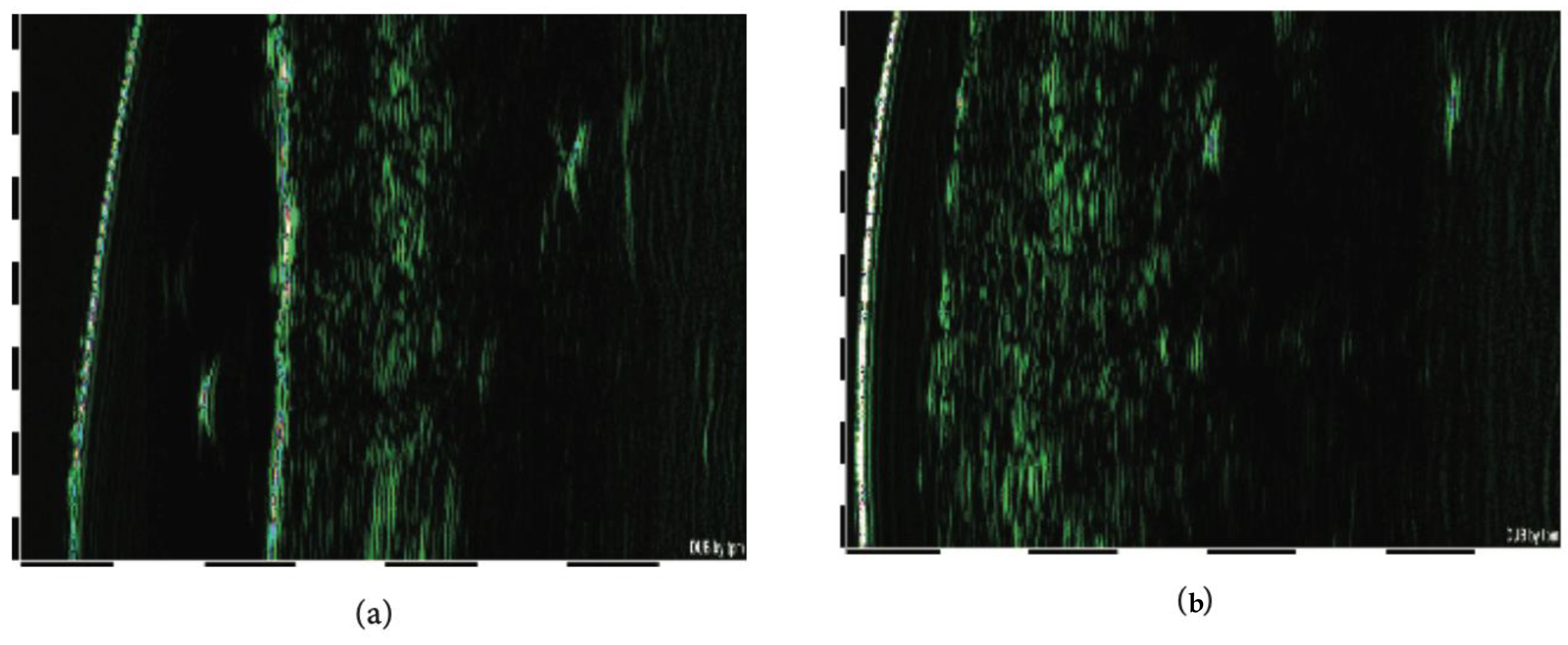
2.6. Assessment of the Ultrasound Properties of the Skin
2.7. Assessment of Direct Anti-Microbial Effects of the Test Cosmetics and of the Skin Microbiota Pattern
- (1)
- A bacterial and yeast cell suspension obtained after overnight culture was spread uniformly on the solid agar medium and left dried at room temperature. The wells were cut using a sterile Pasteur pipet, and the diameter of the wells was the same in each experiment (8 mm). Then, 50 μL of the serum mixture was loaded and kept in chilled conditions for 2 h to allow diffusion into the agar. Then, another 50 μL aliquot of the tested serum mixture was added to the wells. A potassium buffer solution (PBS, pH = 7.2) was used as a negative control, and a mixture of the antibiotics penicillin, streptomycin, and acetic acid was used as a positive control, for bacteria and yeast, respectively. The agar plates were incubated at 37 °C for 24 h for Staphylococcus aureus, Propionbacterium acnes, Escherichia coli, Staphylococcus epidermidis, and Candida albicans. A clear zone diameter around the well, indicating microbial inhibition, was measured at two perpendicular directions. The minimal inhibitory concentration (MIC) was determined by testing different volumes of the mixture diluted by PBS. All measurements were performed in triplicate [48].
- (2)
- Microbiological analyses of the facial skin swabs collected from 12 randomly selected participants were performed by a quantitative real-time polymerase chain reaction (RT-PCR) (differential bacterial counting). For RT-PCR tests, DNA was isolated from samples of skin swabs taken by sterile water-soaked cotton sticks and kept on ice for no more than 12 h. DNA was amplified with iQTM Supermix using the MiniOpticon Real-Time PCR Detection System (Bio-Rad, Hercules, CA, USA). All real-time assays were carried out under the following conditions: 35 cycles of denaturation at 95 °C for 15 s; annealing and extension at 60 °C for 60 s. Melt curve analysis was performed to confirm the specificity of the amplified products. All samples were run in triplicate, and relative expression was determined by normalising samples to housekeeping genes [49].
2.8. Hydroxyproline Assay
2.9. Assessment of Lipid Peroxidation in Skin Lipids
2.10. Statistical Evaluation
3. Results
3.1. Dermatological and Ophthalmological Safety of the Test Cosmetics
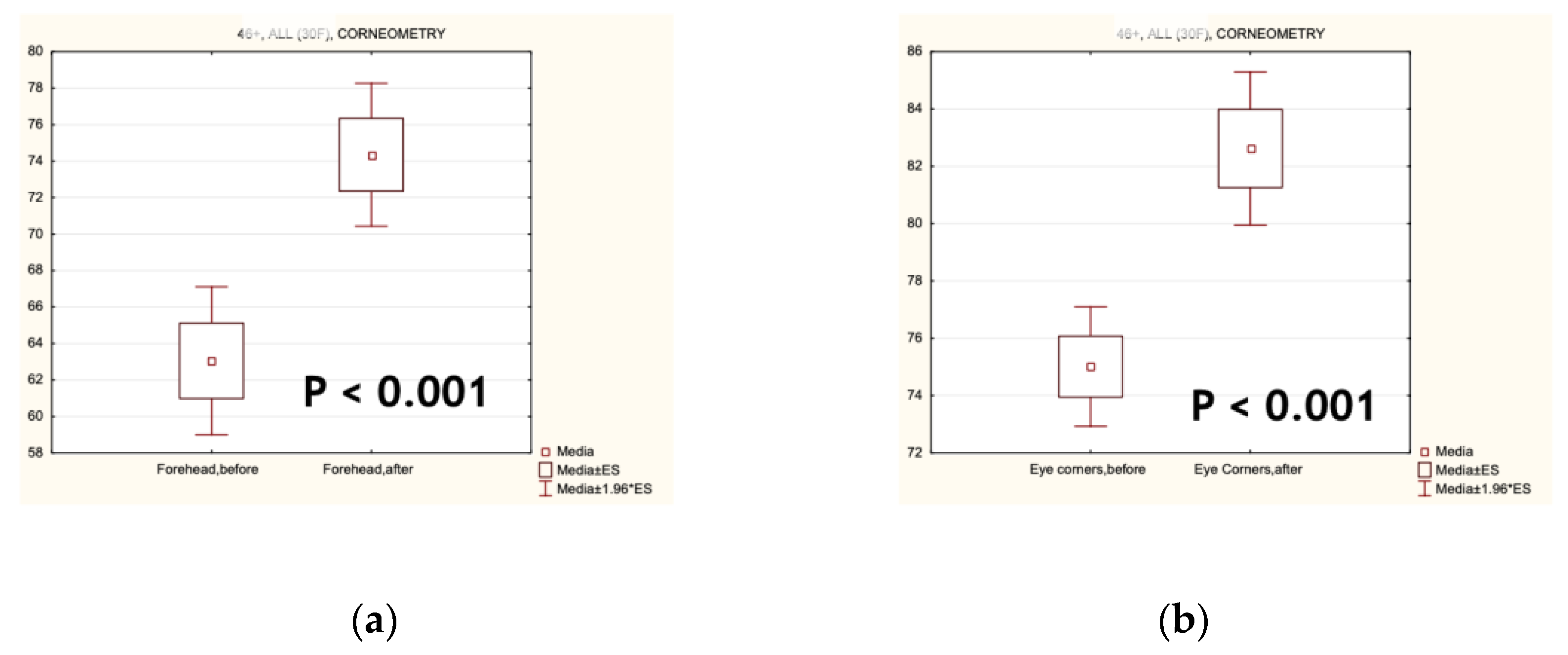
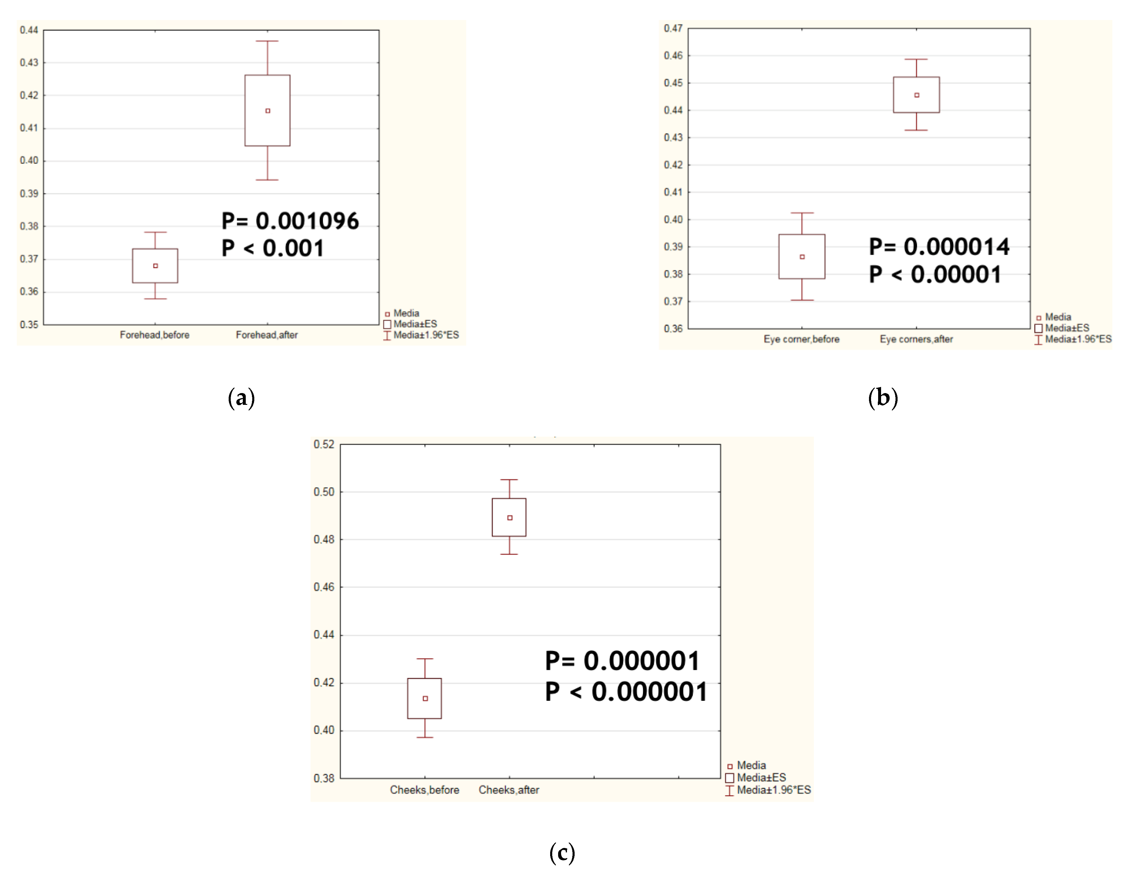
3.2. Effects of the Test Cosmetics on the Facial Skin Physiology and Ultrasonic Properties
3.3. Effects of the Test Cosmetics on the Biochemical Markers of Skin Ageing
3.4. Direct Anti-Microbial Effects, and Changes in the Facial Skin Microbiota after the Regular Four-Week-Long Application of the Test Cosmetics
4. Discussion
5. Conclusions
Author Contributions
Funding
Institutional Review Board Statement
Informed Consent Statement
Data Availability Statement
Acknowledgments
Conflicts of Interest
Appendix A

| Microbes, Cocci | Normal Values | Results, 105 Cells/g | ||
|---|---|---|---|---|
| Before | After | p, Before versus After | ||
| Bacillus cereus | 23 ± 5 | 13 ± 5 | 10 ± 5 | >0.05 |
| Bacillus megaterium | 0 | 0 | 0 | >0.05 |
| Enterococcus spp. | 290 ± 45 | 230 ± 35 | 240 ± 55 | >0.05 |
| Staphylococcus aureus | 0 | 0 | 0 | >0.05 |
| Staphylococcus epidermidis | 120 ± 25 | 104 ± 25 | 90 ± 35 | >0.05 |
| Anaerobs | ||||
| Bacteroides fragilis | 0 | 0 | 0 | >0.05 |
| Bifidobacterium spp. | 5067 ± 450 | 4760 ± 240 | 5010 ± 280 | >0.05 |
| Blautiacoccoides | 0 | 0 | 0 | >0.05 |
| Clostridium spp. | 245 ± 20 | 260 ± 37 | 235 ± 20 | >0.05 |
| Clostridium difficile | 385 ± 30 | 355 ± 50 | 360 ± 40 | >0.05 |
| Cl.hystoluticum/Str.pneumonia | 0 | 0 | 0 | >0.05 |
| Clostridium perfringens | 12 ± 5 | 0 | 0 | >0.05 |
| Clostridium propionicum | 288 ± 30 | 250 ± 30 | 248 ± 30 | >0.05 |
| Clostridium ramosum | 2000 ± 445 | 1550 ± 345 | 1750 ± 445 | >0.05 |
| Eubacterium spp. | 6912 ± 450 | 6912 | 6912 | |
| Eggerthellalenta | 68 ± 15 | 60 ± 15 | 55 ± 12 | >0.05 |
| Fusobacterium spp./Haemophilus spp. | 0 | 0 | 0 | >0.05 |
| Lactobacillus spp. | 6613 ± 450 | |||
| Peptostreptococcus anaerobius 18623 | 0 | 0 | 0 | >0.05 |
| Peptostreptococcus anaerobius 17642 | 0 | 0 | 0 | >0.05 |
| Prevotella spp. | 0-18 | 0 | 0 | >0.05 |
| Ruminicoccus spp. | 0–140 | 0 | 0 | >0.05 |
| Veilonella spp. | 0 | 0 | 0 | >0.05 |
| Actinobacterium | ||||
| Actinomyces spp. | 77 ± 20 | 75 ± 10 | 77 ± 20 | >0.05 |
| Actinomyces viscosus | 1190 ± 120 | 1110 ± 110 | 1090 ± 100 | >0.05 |
| Corynebacterium spp. | 605 ± 30 | 600 ± 50 | 580 ± 30 | >0.05 |
| Nocardia spp. | 262 ± 20 | 242 ± 20 | 232 ± 20 | >0.05 |
| Nocardia asteroids | 274 ± 15 | 274 ± 15 | 274 ± 15 | >0.05 |
| Mycobacterium spp. | 0 | 0 | 0 | >0.05 |
| Pseudonocardia spp. | 70 ± 15 | 80 ± 15 | 72 ± 13 | >0.05 |
| Rhodococcus spp. | 423 ± 45 | 420 ± 45 | 410 ± 35 | >0.05 |
| Streptomyces spp. | 62 ± 15 | 60 ± 10 | 52 ± 15 | >0.05 |
| Streptomyces farmamarensis | 0 | 0 | 0 | >0.05 |
| Enterobacterium | ||||
| Enterobacteriaceae spp. | 0 | 0 | 0 | >0.05 |
| Helicobacter pylori | 0–10 | 0 | 0 | >0.05 |
| Campylobacter mucosalis | 99 ± 35 | 75 ± 15 | 70 ± 15 | >0.05 |
| Gram-negative | ||||
| Alcaligenes spp./Klebsiella spp. | 45 ± 15 | 49 ± 15 | 35 ± 15 | >0.05 |
| Kingella spp. | 10 ± 7 | 0 | 0 | >0.05 |
| Flavobacterium spp. | 0 | 0 | 0 | >0.05 |
| Moraxella spp./Acinetobacter spp. | 0 | 0 | 0 | >0.05 |
| Porphyromonas spp. | 0 | 0 | 0 | >0.05 |
| Pseudomonas aeruginosa | 0 | 0 | 0 | >0.05 |
| Stenotrophomonas maltophilia | 0 | 0 | 0 | >0.05 |
| Micellium, yeasts | ||||
| Aspergillus spp. | 110 ± 25 | 90 ± 15 | 97 ± 17 | >0.05 |
| Candida spp. | 549 ± 70 | 540 ± 50 | 515 ± 50 | >0.05 |
| Microfungi (campsterol) | 842 ± 50 | 790 ± 40 | 812 ± 65 | >0.05 |
| Microfungi (sitosterol) | 384 ± 30 | 355 ± 20 | 390 ± 40 | >0.05 |
References
- Wines, N.; Willsteed, E. Menopause and the skin. Australas. J. Dermatol. 2001, 42, 149–158. [Google Scholar] [CrossRef] [PubMed]
- Monteleone, P.; Mascagni, G.; Giannini, A.; Genazzani, A.R.; Simoncini, T. Symptoms of menopause—Global prevalence, physiology and implications. Nat. Rev. Endocrinol. 2018, 14, 199–215. [Google Scholar] [CrossRef] [PubMed]
- Raine-Fenning, N.J.; Brincat, M.P.; Muscat-Baron, Y. Skin ageing and menopause: Implications for treatment. Am. J. Clin. Dermatol. 2003, 4, 371–378. [Google Scholar] [CrossRef] [PubMed]
- Wilkinson, H.N.; Hardman, M.J. The role of estrogen in cutaneous ageing and repair. Mauritas 2017, 103, 60–64. [Google Scholar] [CrossRef]
- Zouboulis, C.C.; Blume-Peytavi, U.; Kosmadaki, M.; Roo, E.; Vexiau-Robert, D.; Kerob, D.; Goldstein, S.R. Skin, hair and beyond: The impact of menopause. Climacteric 2022, 25, 434–442. [Google Scholar] [CrossRef]
- Calleya-Agius, J.; Brincat, M. The effect of menopause on the skin and other connective tissues. Gynecol. Endocrinol. 2012, 28, 273–277. [Google Scholar] [CrossRef]
- Wang, Z.H. Anti-glycative effects of asiatic acid in human keratinocyte cells. BioMedicine 2014, 4, 19–28. [Google Scholar] [CrossRef]
- Fulop, T.; Larbi, A.; Pawelec, G.; Khalil, A.; Cohen, A.A.; Hirokawa, K.; Witkowski, G.M.; Franceschi, C. Immunology of aging: The birth of inflammaging. Clin. Rev. Allergy Immunol. 2023, 64, 109–122. [Google Scholar] [CrossRef]
- Dugan, B.; Conway, J.; Duggal, N.A. Inflammaging as a target for healthy ageing. Age Ageing 2023, 52, afac328. [Google Scholar] [CrossRef]
- Belkaid, Y.; Hand, T. Role of the microbiota in immunity and inflammation. Cell 2014, 157, 121–141. [Google Scholar] [CrossRef]
- Kharaeva, Z.; Hokonova, T.; Elmurzaeva, J.; Dzamihova, I.; Mayer, W.; De Luca, C.; Trakhtman, I.; Korkina, L. Effects of Heavy Isotopes (2H1 and 18O16) Depleted Water Con-Sumption on Physical Recovery and Metabolic and Immunological Parameters of Healthy Volunteers under Regular Fitness Load. Sports 2021, 9, 110. [Google Scholar] [CrossRef] [PubMed]
- Liu, T.; Li, N.; Yan, Y.-Q.; Liu, Y.; Xiong, K.; Liu, Y.; Xia, Q.-M.; Zhang, H.; Liu, Z.-D. Recent advances in the anti-aging effects of phytoestrogens on collagen, water content, and oxidative stress. Phytother. Res. 2020, 34, 435–447. [Google Scholar] [CrossRef] [PubMed]
- Duarte, G.V.; Trigo, A.C.; Paim de Oliveira, M.d.F. Skin disorders during menopause. Cutis 2016, 97, E16–E23. [Google Scholar] [PubMed]
- Shu, Y.Y.; Maibach, H.I. Estrogen and skin: Therapeutic options. Am. J. Clin. Dermatol. 2011, 12, 297–311. [Google Scholar] [CrossRef]
- Kostyuk, V.; Potapovich, A.; Albuhaydar, A.R.; Mayer, W.; De Luca, C.; Korkina, L. Natural Substances for Prevention of Skin Photoaging: Screening Systems in the Development of Sunscreen and Rejuvenation Cosmetics. Rejuvenation Res. 2018, 21, 91–101. [Google Scholar] [CrossRef] [PubMed]
- Korkina, L.G.; Mayer, W.; De Luca, C. Meristem Plant Cells as a Sustainable Source of Redox Actives for Skin Rejuvenation. Biomolecules 2017, 7, 40. [Google Scholar] [CrossRef]
- Korkina, L.; Kostyuk, V.; Potapovich, A.; Mayer, W.; Talib, N.; De Luca, C. Secondary plant metabolites for sun protective cosmetics: From pre-selection to product formulation. Cosmetics 2018, 5, 32. [Google Scholar] [CrossRef]
- Korkina, L.G. Phenylpropanoids as naturally occurring antioxidants: From plant defence to human health. Cell Mol. Biol. 2007, 53, 13–23. [Google Scholar] [CrossRef]
- Pastore, S.; Lulli, D.; Pascarella, A.; Maurelli, R.; Dellambra, E.; Potapovich, A.; Kostyuk, V.; De Luca, C.; Korkina, L. Resveratrol enhances solar UV-induced responses in normal human epidermal keratinocytes. Photochem. Photobiol. 2012, 88, 1522–1530. [Google Scholar] [CrossRef]
- Alipieva, K.; Korkina, L.; Orhan, I.E.; Georgiev, M.I. Verbascoside—A review of its occurrence, (bio)synthesis, and pharmacological significance. Biotech. Adv. 2014, 32, 1065–1076. [Google Scholar] [CrossRef]
- Vertuani, S.; Beghelli, E.; Scalambra, E.; Malisardi, G.; Copetti, S.; Dal Toso, R.; Baldisserotto, A.; Manfredini, S. Activity and stability studies of verbascoside, a novel antioxidant, in dermo-cosmetic and pharmaceutical topical formulations. Molecules 2011, 16, 7068–7080. [Google Scholar] [CrossRef] [PubMed]
- Sgarbossa, A.; Dal Bosco, M.; Pressi, G.; Cuzzocrea, S.; Dal Toso, R.; Menegazzi, M. Phenylpropanoid glycosides from plant cell cultures induce heme oxygenase 1 gene expression in a human keratinocyte cell line by affecting the balance of NRF2 and BACH1 transcription factors. Chem. Biol. Interact. 2012, 199, 87–95. [Google Scholar] [CrossRef] [PubMed]
- Kostyuk, V.; Potapovich, A.; Stancato, A.; De Luca, C.; Lulli, D.; Pastore, S.; Korkina, L. Photo-oxidation products of skin surface squalene mediate metabolic and inflammatory responses to solar UV in human keratinocytes. PLoS ONE 2012, 7, e44472. [Google Scholar] [CrossRef] [PubMed]
- Li, H.; Meng, X.; Zhang, Y.; Guo, M.; Li, L. Active components of Leontopodium alpinum callus culture extract for blue light damage in human fibroblasts. Molecules 2023, 28, 7319. [Google Scholar] [CrossRef]
- Schwaiger, S.; Cervellati, R.; Seger, C.; Ellmerer, E.P.; About, N.; Renimel, I.; Godenir, C.; André, P.; Gafner, F.; Stuppner, H. Leontopodic acid—A novel highly substituted glucaric acid derivative from Edelweiss (Leontopodium alpinum Cass.) and its antioxidative and DNA protecting properties. Tetrahedron 2005, 61, 4621–4630. [Google Scholar] [CrossRef]
- Meng, X.; Guo, M.; Geng, Z.; Wang, Z.; Zhang, H.; Li, S.; Ling, X.; Li, L. Effects and Mechanism of the Leontopodium alpinum Callus Culture Extract on Blue Light Damage in Human Foreskin Fibroblasts. Molecules 2023, 28, 2172. [Google Scholar] [CrossRef]
- Lulli, D.; Potapovich, A.; Maurelli, R.; Dellambra, E.; Pressi, G.; Kostyuk, V.; Dal Toso, R.; De Luca, C.; Pastore, S.; Korkina, L. Anti-inflammatory effects of concentrated ethanol extracts of Edelweiss (Leontopodium alpinum Cass.) callus cultures towards human keratinocytes and endothelial cells. Med. Inflamm. 2012, 2012, 498373. [Google Scholar] [CrossRef]
- Cho, W.K.; Kim, H.-I.; Kim, S.-Y.; Seo, H.H.; Song, J.; Kim, J.; Shin, D.S.; Jo, Y.; Choi, H.; Lee, J.H.; et al. Anti-Aging Effects of Leontopodium alpinum (Edelweiss) Callus Culture Extract through Transcriptome Profiling. Genes 2020, 11, 230. [Google Scholar] [CrossRef]
- Zhang, D.; Lu, C.; Wang, X.; Yan, L.; Zhang, J.; Li, H.; Wang, J.; Wen, A. Echinacoside alleviates UVB irradiation-mediated skin damage via inhibition of oxidative stress, DNA damage, and apoptosis. Oxid. Med. Cell Longev. 2017, 2018, 6851464. [Google Scholar] [CrossRef]
- Guarnerio, C.F.; Fraccaroli, M.; Gonzo, I.; Pressi, G.; Dal Toso, R.; Guzzo, F.; Levi, M. Metabolomic analysis reveals that the accumulation of specific secondary metabolites in Echinacea angustifolia cells cultured in vitro can be controlled by light. Plant Cell Rep. 2012, 31, 361–367. [Google Scholar] [CrossRef]
- Facino, R.M.; Carini, M.; Aldini, G.; Salbene, L.; Pietta, P.; Mauri, P. Echinacoside and caffeoyl conjugates protect collagen from free radical-induced degradation: A potential use of Echinacea extracts in the prevention of skin photodamage. Planta Med. 1995, 61, 510–514. [Google Scholar] [CrossRef] [PubMed]
- Chen, Y.; Wang, Y.; Song, S.; Zhang, X.; Wu, L.; Li, X. Topical application of baicalin combined with echinacoside ameliorates psoriatic skin lesions by suppressing the inflammation-related TNF signaling pathway and the angiogenesis-related VEGF signaling pathway. ACS Omega 2023, 8, 40260–40276. [Google Scholar] [CrossRef] [PubMed]
- Speroni, E.; Govoni, P.; Guizzardi, S.; Renzulli, C.; Guerra, M.C. Anti-inflammatory and cicatrizing activity of Echinacea pallida Nutt. root extract. J. Ethnopharmacol. 2002, 79, 265–272. [Google Scholar] [CrossRef] [PubMed]
- Han, J.; Sun, Y.; Wu, T.; Hou, X.; Zheng, S.; Zhang, H.; Lin, T.; Liu, H.; Sun, T. Echinacoside-zinc nanomaterial inhibits skin glycation by suppressing the transcriptional activation of the receptor for advanced glycation end-products. ACS Nano 2023, 17, 14123–14135. [Google Scholar] [CrossRef]
- Park, K.S. Pharmacological effects of Centella asiatica on skin diseases: Evidence and possible mechanisms. Evid. Based Complement. Alternat Med. 2021, 2021, 5462633. [Google Scholar] [CrossRef]
- Bylka, W.; Znajdek-Awizen, P.; Studzinska-Sroka, E.; Danczak-Pazdrowska, A.; Brzezinska, M. Centella asiatica in dermatology: An overview. Phytother. Res. 2014, 28, 1117–1124. [Google Scholar] [CrossRef] [PubMed]
- Bylka, W.; Znajdek-Awizen, P.; Studzinska-Sroka, E.; Brzezinska, M. Centella asiatica in cosmetology. Postepy Dermatol. Alergol. 2013, 30, 46–49. [Google Scholar] [CrossRef]
- Jiang, H.; Zhou, X.; Chen, L. Asiaticoside delays senescence and attenuate generation of ROS in UV-exposure cells through regulates TGF-β1/SMAD pathway. Exp. Ther. Med. 2022, 24, 667. [Google Scholar] [CrossRef]
- Lee, J.; Jung, E.; Lee, H.; Seo, Y.; Koh, J.; Park, D. Evaluation of the effects of a preparation containing asiaticoside on periocular wrinkles of human volunteers. Int. J. Cosmet. Sci. 2008, 30, 167–173. [Google Scholar] [CrossRef]
- Lee, Y.; Choi, H.K.; N’deh, K.P.U.; Choi, Y.-J.; Fan, M.; Kim, E.-K.; Chung, K.-H.; An, J.H. Inhibitory effect of Centella asiatica extract on DNCB-induced atopic dermatitis in HaCaT cells and BALB/c mice. Nutrients 2020, 12, 411. [Google Scholar] [CrossRef]
- Lee, J.-C.; Kim, H.-L.; Lee, M.H.; You, K.E.; Kwon, B.-J.; Seo, H.J.; Park, J.-C. Asiaticoside enhances normal human skin cell migration, attachment and growth in vitro wound healing model. Phytomedicine 2012, 19, 1223–1227. [Google Scholar] [CrossRef] [PubMed]
- Liu, Y.; Zhao, J.; Mu, X.; Deng, J.; Wu, X.; He, W.; Liu, Y.; Gu, R.; Han, F.; Nie, X. Asiaticoside-nitric oxide promoting diabetic wound healing through the miRNA-21-5p/TGF-β1/SMAD7/TIMP3 signaling pathway. J. Ethnopharmacol. 2024, 319 Pt 2, 117266. [Google Scholar] [CrossRef] [PubMed]
- Tundis, R.; Loizzo, M.R.; Bonesi, M.; Menichini, F. Potential role of natural compounds against skin aging. Curr. Med. Chem. 2015, 22, 1515–1538. [Google Scholar] [CrossRef]
- Mukherjee, P.K.; Maity, N.; Nema, N.K.; Sarkar, B.K. Bioactive compounds from natural sources against akin aging. Phytomedicine 2011, 19, 64–73. [Google Scholar] [CrossRef]
- Antognoni, F.; Crespi Perellino, N.; Crippa, S.; Dal Toso, R.; Minghetti, A.; Poli, F.; Pressi, G. Irbic acid, a dicaffeoylquinic acid derivative from Centella asiatica cell cultures. Fitoterapia 2011, 87, 950–954. [Google Scholar] [CrossRef] [PubMed]
- Sawant, O.; Khan, T. Management of periorbital hyperpigmentation: An overview of nature-based agents and alternative approaches. Dermatol. Ther. 2020, 33, e13717. [Google Scholar] [CrossRef]
- Bandopadhyay, S.; Mandal, S.; Ghorai, M.; Jha, N.K.; Kumar, M.; Radha; Ghosh, A.; Prockow, J.; Perez de la Lastra, J.M.; Dey, A. Therapeutic properties and pharmacological activities of asiaticoside and madecassoside: A review. J. Cell Mol. Med. 2023, 27, 593–608. [Google Scholar] [CrossRef]
- Kharaeva, Z.F.; Mustafaev, M.S.; Khazhmetov, A.V.; Gazaev, I.H.; Blieva, L.Z.; Steiner, L.; Mayer, W.; De Luca, C.; Korkina, L.G. Anti-Bacterial and Anti-Inflammatory Effects of Toothpaste with Swiss Medicinal Herbs towards Patients Suffering from Gingivitis and Initial Stage of Periodontitis: From Clinical Efficacy to Mechanisms. Dent. J. 2020, 8, 10. [Google Scholar] [CrossRef]
- Kharaeva, Z.; Shokarova, A.; Shomakhova, Z.; Ibragimova, G.; Trakhtman, P.; Trakhtman, I.; Chung, J.; Mayer, W.; De Luca, C.; Korkina, L. Fermented Carica papaya and Morinda citrifolia as Perspective Food Supplements for the Treatment of Post-COVID Symptoms: Randomized Placebo-Controlled Clinical Laboratory Study. Nutrients 2022, 14, 2203. [Google Scholar] [CrossRef]
- Kharaeva, Z.; Gostova, E.; De Luca, C.; Raskovic, D.; Korkina, L. Clinical and biochemical effects of coenzyme Q10, vitamin E and selenium supplementation to psoriasis patients. Nutrition 2009, 25, 295–302. [Google Scholar] [CrossRef]
- Thiele, J.J.; Schroeter, C.; Hsieh, S.N.; Podda, M.; Packer, L. The antioxidant network of the stratum corneum. Curr. Probl. Dermatol. 2001, 29, 26–42. [Google Scholar] [CrossRef] [PubMed]
- Sander, C.S.; Chang, H.; Salzmann, S.; Müller, C.S.; Ekanayake-Mudiyanselage, S.; Elsner, P.; Thiele, J.J. Photoaging is associated with protein oxidation in human skin in vivo. J. Investig. Dermatol. 2002, 118, 618–625. [Google Scholar] [CrossRef] [PubMed]
- Sander, C.S.; Chang, H.; Hamm, F.; Elsner, P.; Thiele, J.J. Role of oxidative stress and the antioxidant network in cutaneous carcinogenesis. Int. J. Dermatol. 2004, 43, 326–335. [Google Scholar] [CrossRef] [PubMed]
- Passi, S.; Grandinetti, M.; Maggio, F.; Stancato, A.; De Luca, C. Epidermal oxidative stress in vitiligo. Pigment. Cell Res. 1998, 11, 81–85. [Google Scholar] [CrossRef] [PubMed]
- Woodby, B.; Penta, K.; Pecorelli, A.; Lila, M.A.; Valacchi, G. Skin Health from the Inside Out. Annu. Rev. Food Sci. Technol. 2020, 11, 235–254. [Google Scholar] [CrossRef]
- Packer, L.; Valacchi, G. Antioxidants and the response of skin to oxidative stress: Vitamin E as a key indicator. Skin. Pharmacol. Appl. Skin. Physiol. 2002, 15, 282–290. [Google Scholar] [CrossRef]
- Korkina, L.; Kostyuk, V. Biotechnologically produced secondary plant metabolites for cancer treatment and prevention. Curr. Pharm. Biotechnol. 2012, 13, 265–275. [Google Scholar] [CrossRef]
- Potapovich, A.I.; Kostyuk, V.A.; De Luca, C.; Mikhal’chik, E.; Korkina, L.G. Plant polyphenols regulate chemokine expression and tissue repair in human keratinocytes through interaction with cytoplasmic and nuclear components of epidermal growth factor receptor system. Antiox Redox Signal 2012, 16, 314–328. [Google Scholar] [CrossRef]
- Pastore, S.; Lulli, D.; Potapovich, A.I.; Korkina, L.G.; Mikhal’chik, E.V.; Suprun, M.V.; Pastore, S.; Dal Toso, R. Molecular mechanisms underlying wound healing and anti-inflammatory properties of naturally occurring biotechnologically produced phenylpropanoid glycosides. Cell Mol. Biol. 2007, 53, 78–83. [Google Scholar]
- Kostyuk, V.A.; Potapovich, A.I.; Suhan, T.O.; De Luca, C.; Korkina, L.G. Antioxidant and signal modulating properties of plant polyphenols in controlling vascular inflammation. Eur. J. Pharmacol. 2011, 658, 248–256. [Google Scholar] [CrossRef]
- Georgiev, M.; Pastore, S.; Lulli, D.; Alipieva, K.; Potapovich, A.; Panetta, M.; Korkina, L. Verbascum xanthopoeniceum-derived phenylethanoid glycosides are potent inhibitors of inflammatory chemokines in dormant and interferon-gamma-stimulated human keratinocytes. J. Ethnopharmacol. 2012, 144, 754–760. [Google Scholar] [CrossRef] [PubMed]
- Hooper, L.V.; Littman, D.R.; Macpherson, A.J. Interactions between the microbiota and the immune system. Science 2012, 336, 1268–1273. [Google Scholar] [CrossRef] [PubMed]
- Souak, D.; Barreau, M.; Courtois, A.; Andre, V.; Duclairoir Poc, C.; Feuilloley, M.G.J.; Gault, M. Challenging cosmetic innovation: The skin microbiota and probiotics protect the skin from UV-induced damage. Microorganisms 2021, 9, 936. [Google Scholar] [CrossRef] [PubMed]
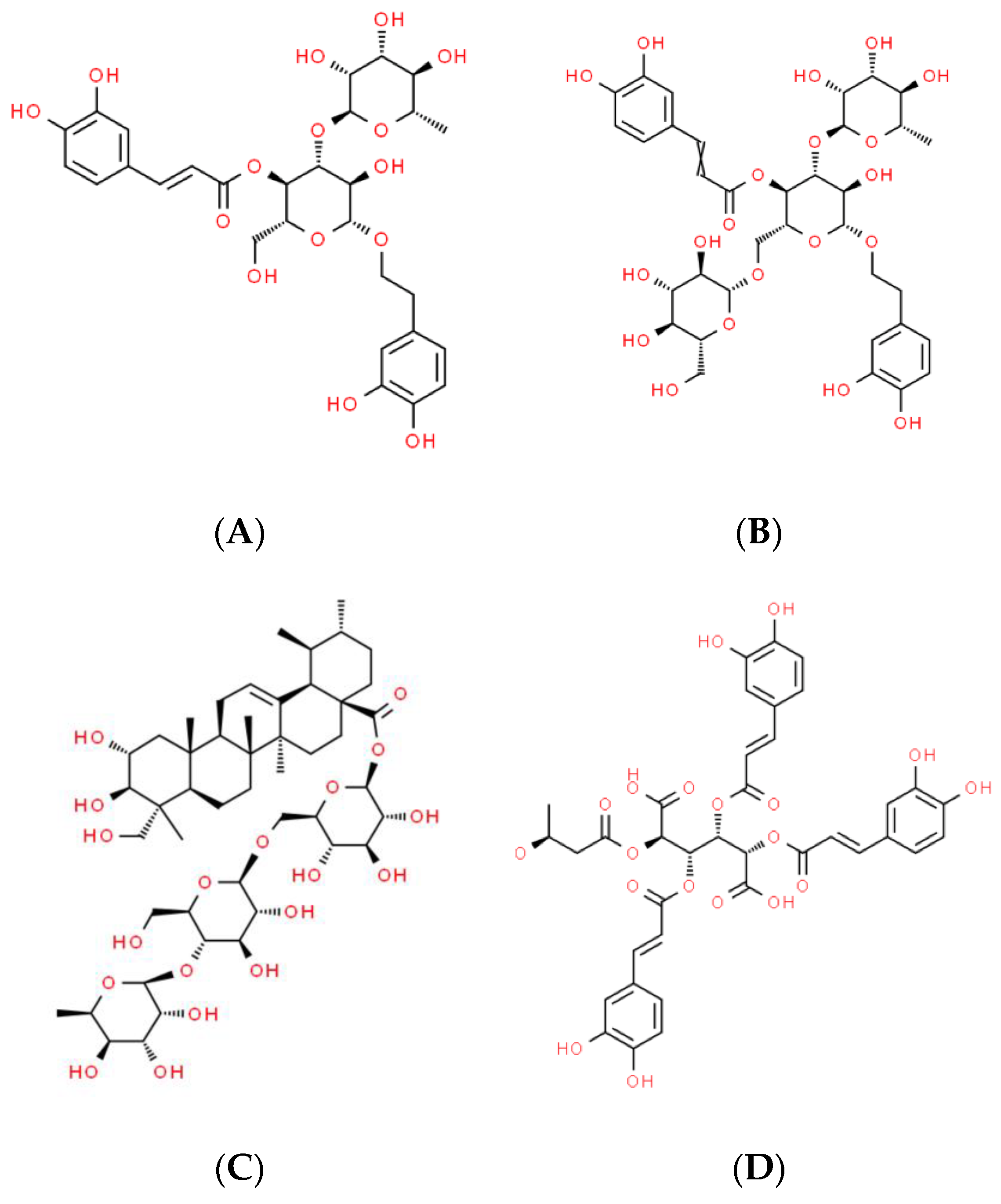


| Plant Meristem Cells | Active Compound IUPAC Name | Effects on Human Skin |
|---|---|---|
| Buddeleja davidii | Verbascoside 2-(3,4-Dihydroxyphenyl)ethyl α-L-rhamnopyranosyl-(1→4)-{5-O-[(2E)-3-(3,4-dihydroxymethyl)prop-2-enoyl]-β-D-glucopyranoside} | UVA, UVB protection [15,16,17] Anti-senescence factors for skin cells in culture [18] Anti-ageing [19] Anti-inflammatory [17] Direct and indirect antioxidants via NRF2 pathway [17,19,20,21,22] Rescue of endogenous antioxidants [23] |
| Leontopodium alpinum | Leontopodic acids A & B 3,4,5-Tris-O-[(2E)-3-(3,4-dihydroxyphenyl)-2-propenoyl]-2-O-[(3S)-3-hydroxybutanoyl]-D-glucaric acid | Antioxidant [24,25] Blue-light-induced damage preventing [24,26] Collagen destruction preventing [26] Anti-inflammatory [27] Anti-chronological ageing [28] |
| Echinacea angustifolia | Echinacoside 2-(3,4-Dihydroxyphenyl)ethyl α-L-rhamnopyranosyl-(1→3)-[β-D-glucopyranosyl-(1→6)]-β-D-glucopyranoside 4-[(2E)-3-(3,4-dihydroxyphenyl)prop-2-enoate] | Anti-photo-ageing by inhibition of oxidative stress and DNA damage [29,30] Anti-photo-damage through inhibition of collagen degradation [31] Anti-inflammatory through TNF pathway suppression [32] Anti-inflammatory and wound healing promoting [33] Anti-angiogenesis via VEGF suppression [32] Cytoprotection from oxidative damage and xenobiotics by NRF2 induction [33] Anti-glycation [34] |
| Centella asiatica | Asiaticoside 6-Deoxy-α-L-mannopyranosyl-(1->4)-β-D-glucopyranosyl-(1->6)-1-O-[(2α,3β)-2,3,23-trihydroxy-28-oxours-12-en-28-yl]-β-D-glucopyranose | Acne, burns, atopic dermatitis, wounds [35,36] Hypertrophic scars [36,37] Photo-ageing skin, cellulite [37,38] Anti-inflammatory, atopic dermatitis [39] Wound healing via accelerated skin cell migration [40,41] Diabetic wound healing [7,42] Anti-chronological ageing/anti-wrinkles [41,43,44,45] Periorbital hyperpigmentation [46] Skin hydration, collagen synthesis [45,47] |
| Parameter, Unit | Before Treatment | After Treatment |
|---|---|---|
| Elasticity, Arb. Units | 33.66 ± 1.21 | 40.26 ± 0.87 ** |
| Smoothness, Arb. Units | 12.15 ± 0.24 | 18.01 ± 0.37 *** |
| Moisture, Arb. Units | 49.03 ± 3.52 | 56.86 ± 3.02 ** |
| TEWL, g/h/m2 | 6.3 ± 0.03 | 4.5 ± 0.07 ** |
| Sebum, Arb. Units | 29.27 ± 4.76 | 56.85 ± 4.04 *** |
| Skin biological age, Years | 51.5 ± 0.4 | 46.3 ± 1.0 * |
| Parameter | Before Treatment | After Treatment |
|---|---|---|
| Thickness, μm | 3900 ± 31 | 4133 ± 28 * |
| Acoustic density, Arb. Units | 5.1 ± 0.2 | 6.3 ± 0.1 * |
| Parameter | Before Treatment | After Treatment |
|---|---|---|
| Thickness, μm | 77.0 ± 0.8 | 77.6 ± 0.9 |
| Acoustic density, Arb. Units | 35.2 ± 2.2 | 35.4 ± 2.0 |
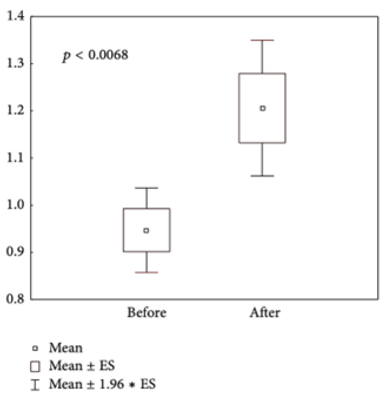
| Cosmetics | Number of Participants (Number of Sebum Samples) | Duration of Application (Days) | MDA (μmol/L Sebum) | |
|---|---|---|---|---|
| Before | After | |||
| All Infinity Serums | 24 (144) | 28 | 26 ± 4 | 15 ± 1 ** |
| Pathogen | Negative Control | Antibiotic Control | Mixture of Infinity Serums | |
|---|---|---|---|---|
| Inhibition Zone (mm) | Inhibition Zone (mm) | Inhibition Zone (mm) | MIC Values (μL) | |
| Propionbacterium acnes | 0 | 10.22 ± 1.31 | 6.15 ± 0.97 | 50 |
| Staphylococcus aureus | 0 | 7.68 ± 0.87 | 4.87 ± 1.11 | 60 |
| Streptococcus epidermidis | 0 | 9.66 ± 0.79 | 5.60 ± 1.23 | 60 |
| Esherichia coli | 0 | 8.32 ± 0.43 | 3.88 ± 1.54 | 75 |
| Candida albicans | 0 | 10.89 ± 1.59 | 6.65 ± 2.04 | 25 |
| Microbes | Normal Values, 105 cells/g | Results, 105 cells/g | ||
|---|---|---|---|---|
| Before the Trial | After the Trial | p, Before/After | ||
| Streptococcus spp. | 249 ± 45 | 205 ± 50 | 160 ± 25 | <0.05 |
| Propionbacterium acnes | 0–42 | 50 ± 5 | 10 ± 3 | <0.05 |
| Propionbacterium freudenechii | 3480 ± 470 | 4200 ± 470 | 2980 ± 200 | <0.05 |
| Propionbacterium jensenii | 38 ± 3 | 38 ± 6 | 28 ± 3 | <0.05 |
Disclaimer/Publisher’s Note: The statements, opinions and data contained in all publications are solely those of the individual author(s) and contributor(s) and not of MDPI and/or the editor(s). MDPI and/or the editor(s) disclaim responsibility for any injury to people or property resulting from any ideas, methods, instructions or products referred to in the content. |
© 2024 by the authors. Licensee MDPI, Basel, Switzerland. This article is an open access article distributed under the terms and conditions of the Creative Commons Attribution (CC BY) license (https://creativecommons.org/licenses/by/4.0/).
Share and Cite
Korkina, L.; Kharaeva, Z.; Shokarova, A.; Barokova, E.; Mayer, W.; Trakhtman, I.; Dal Toso, R.; De Luca, C. Effects of Plant Meristem-Cell-Based Cosmetics on Menopausal Skin: Clinical Data and Mechanisms. Biomolecules 2024, 14, 1176. https://doi.org/10.3390/biom14091176
Korkina L, Kharaeva Z, Shokarova A, Barokova E, Mayer W, Trakhtman I, Dal Toso R, De Luca C. Effects of Plant Meristem-Cell-Based Cosmetics on Menopausal Skin: Clinical Data and Mechanisms. Biomolecules. 2024; 14(9):1176. https://doi.org/10.3390/biom14091176
Chicago/Turabian StyleKorkina, Liudmila, Zaira Kharaeva, Albina Shokarova, Elena Barokova, Wolfgang Mayer, Ilya Trakhtman, Roberto Dal Toso, and Chiara De Luca. 2024. "Effects of Plant Meristem-Cell-Based Cosmetics on Menopausal Skin: Clinical Data and Mechanisms" Biomolecules 14, no. 9: 1176. https://doi.org/10.3390/biom14091176
APA StyleKorkina, L., Kharaeva, Z., Shokarova, A., Barokova, E., Mayer, W., Trakhtman, I., Dal Toso, R., & De Luca, C. (2024). Effects of Plant Meristem-Cell-Based Cosmetics on Menopausal Skin: Clinical Data and Mechanisms. Biomolecules, 14(9), 1176. https://doi.org/10.3390/biom14091176