Article

Identification of the Novel Small Compound Stress Response Regulators 1 and 2 That Affect Plant Abiotic Stress Signaling

1
Department of Bio-Health Convergence, Duksung Women’s University, Seoul 01369, Republic of Korea
2
Department of Biotechnology, Duksung Women’s University, Seoul 01369, Republic of Korea
*
Author to whom correspondence should be addressed.
Biomolecules 2024, 14(9), 1177; https://doi.org/10.3390/biom14091177
Submission received: 28 August 2024 / Revised: 14 September 2024 / Accepted: 17 September 2024 / Published: 19 September 2024

Abstract

:
Abiotic stresses, such as drought, salinity, and extreme temperatures, limit plant growth and development, reducing crop yields. Therefore, a more comprehensive understanding of the signaling mechanisms and responses of plants to changing environmental conditions is crucial for improving sustainable agricultural productivity. Chemical screening was conducted to find novel small compounds that act as regulators of the abiotic stress signaling pathway using the ABA-inducible transgenic reporter line. Small molecules called stress response regulators (SRRs) were isolated by screening a synthetic library composed of 14,400 small compounds, affecting phenotypes such as seed germination, root growth, and gene expression in response to multiple abiotic stresses. Seeds pretreated with SRR compounds positively affected the germination rate and radicle emergence of Arabidopsis and tomato plants under abiotic stress conditions. The SRR-priming treatment enhanced the transcriptional responses of abiotic stress-responsive genes in response to subsequent salt stress. The isolation of the novel molecules SRR1 and SRR2 will provide a tool to elucidate the complex molecular networks underlying the plant stress-tolerant responses.

1. Introduction

Chemical genetics has provided a powerful strategy for understanding the molecular mechanisms underlying plant growth, development, stress responses, and epigenetic regulation using small molecules. Small molecules can modulate complex biological systems in rapid, reversible, conditional, and dose-dependent manners [1,2]. Small molecule-based approaches can thereby overcome the limitations of classical genetic studies, such as mutant lethality and genetic redundancy [3]. Small bioactive molecules that activate cognate targets and signaling pathways have been isolated by the high-throughput screening of diverse chemical libraries, allowing the discovery of novel regulatory components in many phytohormone signaling networks [4,5,6,7]. The plant hormone abscisic acid (ABA) is a pivotal regulator mediating various physiological processes of plant growth, development, and adaptive responses to environmental stresses [8]. Plants produce ABA in response to abiotic stresses, which initiate various adaptive stress responses by controlling the ion channel activities or stress-responsive gene expression [9,10]. Recently, many chemical compounds have been identified to examine the underlying mechanisms for abiotic stress responses in plants. For example, small compounds, such as pyrabactin, quinabactin, ABA ANTAGONIST1 (AA1), S7, and [5-(3,4-dichlorophenyl)furan-2-yl]-piperidine-1-ylmethanethione (DFPM), were isolated by screening a small molecule library to target the critical parts of the ABA signaling pathway [11,12,13,14,15]. Moreover, the structure-based modification on such chemical modulators has resulted in developing promising ABA agonists, such as cyanabactin, opabactin, and ABA mimic 1 fluorine derivative 4 (AMF4), enhancing responses compared to ABA [4,16,17].
Chemical priming has recently been used to improve plant tolerance against a range of individual or combined abiotic stresses [18]. In a process called the chemical priming of plants, exogenous treatments with certain chemical compounds can enhance a broad range of tolerance against abiotic stresses by inducing molecular and physiological defense mechanisms. For example, different chemical priming agents such as phytohormones, reactive oxygen species (ROS), vitamins, osmoprotectants, and mineral elements cause a priming state in plants. Plants under primed conditions can improve cell homeostasis, plant growth, and crop yield because of the increased resistance to multiple abiotic and biotic stresses [19,20,21,22,23,24]. Chemical priming can induce short- or long-term stress memory, making plants more tolerant of additional stress in the current or subsequent generations. In particular, plant stress memory is mediated by epigenetic mechanisms with fundamental roles in maintaining genome stability and controlling gene expression through DNA methylation and histone modifications [25,26]. For example, a positive feedback loop between HSFA2 and the H3K27me3 histone demethylase RELATIVE OF EARLY FLOWERING 6 (REF6) contributes to a transgenerational thermomemory in Arabidopsis [27].
Priming with chemical agents can be applied at different developmental stages of the plant life cycle and to various plant parts or organs [28]. Seed priming technology is a simple, low-cost, ecofriendly, and effective strategy to improve seed and crop performance. This technology has been applied successfully in many field crops, including rice, wheat, barley, maize, sorghum, and soybean [29,30]. Seed priming is a pre-sowing, controlled seed hydration treatment that allows pre-germinative metabolic activities to continue without radical emergence. Primed seeds can rapidly start and revive the seed metabolism after sowing, leading to an improved germination rate, uniformity and vigor of seedlings, a well-developed root system, early crop establishment, and increased tolerance to abiotic and biotic stresses [29,31,32,33]. Furthermore, seed priming with combinations of different priming agents effectively enhanced seed and crop performance [34,35,36]. Despite the various organic and inorganic chemicals being used as seed priming agents [30,37,38], further studies are needed to determine the optimal treatment conditions and application methods in different crop species [39,40].
This study conducted high-throughput chemical screening using the transgenic Arabidopsis expressing pRAB18:GFP to identify novel small compounds that regulate abiotic stress signaling. We hypothesized that isolated small compounds might activate stress response pathways, enhancing the resistance to environmental stress in plants. Small molecules called SRRs (stress response regulators) were isolated by screening the 14,400-compound Maybridge HitFinderTM library, affecting stress responses, including seed germination, root growth, and gene expression in response to multiple abiotic stresses. Interestingly, seed priming with SRR chemicals improved the germination percentage and early radicle emergence of Arabidopsis and tomato seeds under abiotic stress conditions. The SRR-priming treatment affected the transcriptional levels of stress-responsive genes and expression patterns of genes in the epigenetic control process in response to subsequent salt stress. The use of the newly identified small molecules SRR1 and SRR2 may provide an efficient tool for the comprehensive understanding of stress-tolerant responses and developing priming processes toward improved stress-tolerant traits in plants.

2. Materials and Methods

2.1. Root Growth Assay

Arabidopsis seeds (Columbia–0 (Col-0)) were sterilized with 100% ethanol and sown on growth medium (0.5× Murashige and Skoog (MS), 0.05% MES, 1% sucrose, 0.8% plant agar, pH 5.8). After two days of stratification at 4 °C, plants were grown vertically for seven days at 22 °C under long-day conditions (16 h light/8 h dark). Seven-day-old seedlings were transferred to a new solid medium containing each chemical at various concentrations. The ends of each primary root of seedlings were marked and monitored to observe root growth arrest after six more days. All root growth experiments were repeated at least three times in independent experiments.

2.2. Chemicals

SRR1 (HTS13184SC) and SRR2 (JFD02841SC) were isolated from the screening of 14,400 small synthetic compounds in the Maybridge HitFinderTM library (Thermo Fisher Scientific, Waltham, MA, USA) (http://www.maybridge.com/ (accessed on 10 January 2018)). The compound library has high structural diversity and purity levels greater than 90%, following the Lipinski guidelines for drug-likeness. The library compounds were dissolved in 100% DMSO at 10 mM.

2.3. Chemical Library Screening

The homozygous T3 transgenic reporter line expressing pRAB18:GFP in the Col-0 background was used for chemical library screening [14]. Ten-day-old transgenic reporter lines grown in 0.5× MS liquid medium in 96-well plates were incubated for 15 h with each library compound at a final concentration of 50 μM. Eighty different library compounds were stored in the middle of a 96-well plate, and the first and last columns were used as controls without chemicals. Eight positive controls (10 μM ABA) and eight negative controls (distilled water) were located alternately on the first and last columns of the plate to minimize errors. 0.1% DMSO was used as the solvent control for chemicals, and 0.02% ethanol was used as the solvent control for ABA. After the chemical incubation, compound-induced GFP fluorescence was measured using a NIGHTSEA™ stereo microscope fluorescence adapter (NIGHTSEA, Hatfield, PA, USA) with a green filter set. The compounds that induced a significant pRAB18:GFP expression were considered hit compounds. The hit chemicals were reexamined through secondary and tertiary screenings using confocal microscopy.

2.4. Quantitative Real-Time PCR

Arabidopsis, tomato, and radish seedlings grown on growth media for 12 days were treated with DMSO, 10 μM ABA, 200 mM NaCl, or 50 μM SRR compounds for six hours. The total RNA was extracted using TRIsure™ (Meridian Bioscience Inc., Memphis, TN, USA) and treated with DNase I (Thermo Fisher Scientific, Waltham, MA, USA) before reverse transcription using a SensiFAST™ cDNA Synthesis Kit (Meridian Bioscience Inc., Memphis, TN, USA). The mRNA expression of the target gene was measured by RT-qPCR using SensiFAST™ SYBR® Hi-ROX Kit (Meridian Bioscience Inc., Memphis, TN, USA) and normalized relative to the expression of Clathrin (At4g24550), CAC (Solyc08g006960), or ACTIN (Rsa1.0_00546.1) as reference genes (Table S1). Four replicates were examined for each condition.

2.5. RNA-Seq and Co-Expression Analysis

Twelve-day-old Arabidopsis Col-0 seedlings treated with 0.1% DMSO, 10µM ABA, 200 mM NaCl, or 50 µM SRR chemicals for six hours were used for RNA extraction with TRIsure™ (Meridian Bioscience Inc., Memphis, TN, USA) according to the manufacturer’s instructions. Purified RNA was treated with DNase I (Thermo Fisher Scientific, Waltham, MA, USA) and evaluated for its quality by determining the RNA Integrity Number (RIN). Three biological replicates of each treatment group, with a RIN value > 7, were used to construct a cDNA library. Sequencing of the cDNA libraries was performed using an Illumina HiSeq4000 (Illumina, Inc., San Diego, CA, USA) with a 100 bp read length and a sequence depth of ~50 million uniquely mapped reads. After a quality assessment and the pre-processing of raw sequence data, the filtered reads were aligned by HISAT2 to the Arabidopsis TAIR10 genome and assembled into transcripts using StringTie. Differentially expressed genes (DEGs) between each sample were selected based on the false discovery rate (FDR) < 0.05 and |log2 fold-change (FC)| ≥ 2 and subjected to Gene Ontology (GO) functional enrichment (http://www.geneontology.org (accessed on 11 January 2019)) and Kyoto encyclopedia of genes and genomes (KEGG) pathway analysis (http://www.genome.jp/kegg (accessed on 11 January 2019)).
All interaction evidence obtained in the STRING database (version 11, https://string-db.org/ (accessed on 19 October 2022)) was used to retrieve the predicted protein–protein interaction (PPI) networks, including text mining, experiments, databases, co-expression, neighborhood, gene fusion, and co-occurrence [40]. The TAIR homologous proteins of Arabidopsis thaliana were mapped using the STRING system and visualized in the interaction network at a high confidence level of >0.7. The network nodes correspond to the proteins, and the edges represent the predicted functional associations.

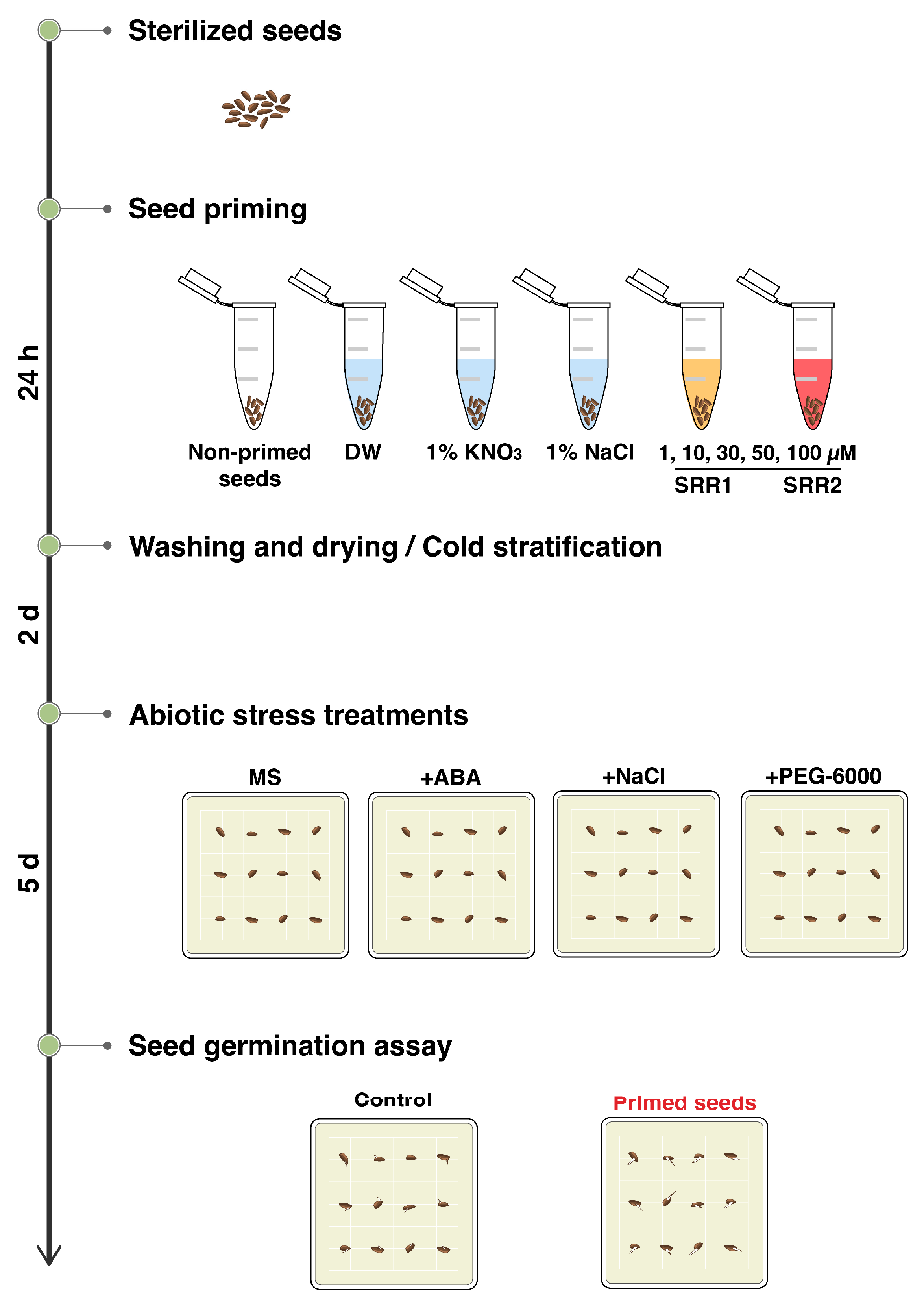
2.6. Seed Priming and Germination Assay

Tomato (Solanum lycopersicum) and rashish (Raphanus sativus) seeds were pretreated with 0.01% rapidase for three hours and washed three times with distilled water. After the pretreatment, the seeds were briefly sterilized with 70% ethanol and rinsed twice with distilled water. The seeds were finally treated with 50% chlorine solution for 30 s, followed by re-washing ten times with distilled water to remove the residual disinfectant. Sterilized Arabidopsis seeds were primed by soaking in different priming solutions for 24 h at room temperature in the dark. Various chemical solutions were used as priming agents: distilled water, 1% KNO3, 1% NaCl, and five concentrations of the SRR chemicals (1, 10, 30, 50, and 100 μM). The seed weight to solution volume ratio (w/v) was 1:5. Distilled water-primed seeds were used as the control. All primed seeds were washed with distilled water for two minutes and re-dried to their original moisture content on filter paper before sowing. After the priming process, seeds were sown on half-strength MS medium supplemented with or without a different concentration of chemicals: 0.5 μM ABA, 150 mM NaCl, 6% PEG-6000, and 200 mM mannitol. After two days of stratification at 4 °C, the seeds were grown for seven days at 22 °C under long-day conditions (16 h light/8 h dark). The germination rate was evaluated six days after sowing, and each treatment was replicated three times with 60 seeds per replicate.

2.7. Transcriptional Analysis for Successive Stress Conditions

Ten-day-old seedlings were pretreated with 100 mM NaCl or each concentration of SRR compounds for 24 h. The primed seedlings were recovered in liquid growth media for 48 h and subjected to second salt stress with 200 mM NaCl for six hours. Distilled water-primed control seedlings were recovered in parallel before being treated with 200 mM NaCl. All plant tissues were harvested after the second stress treatment to determine the transcript level.

3. Results

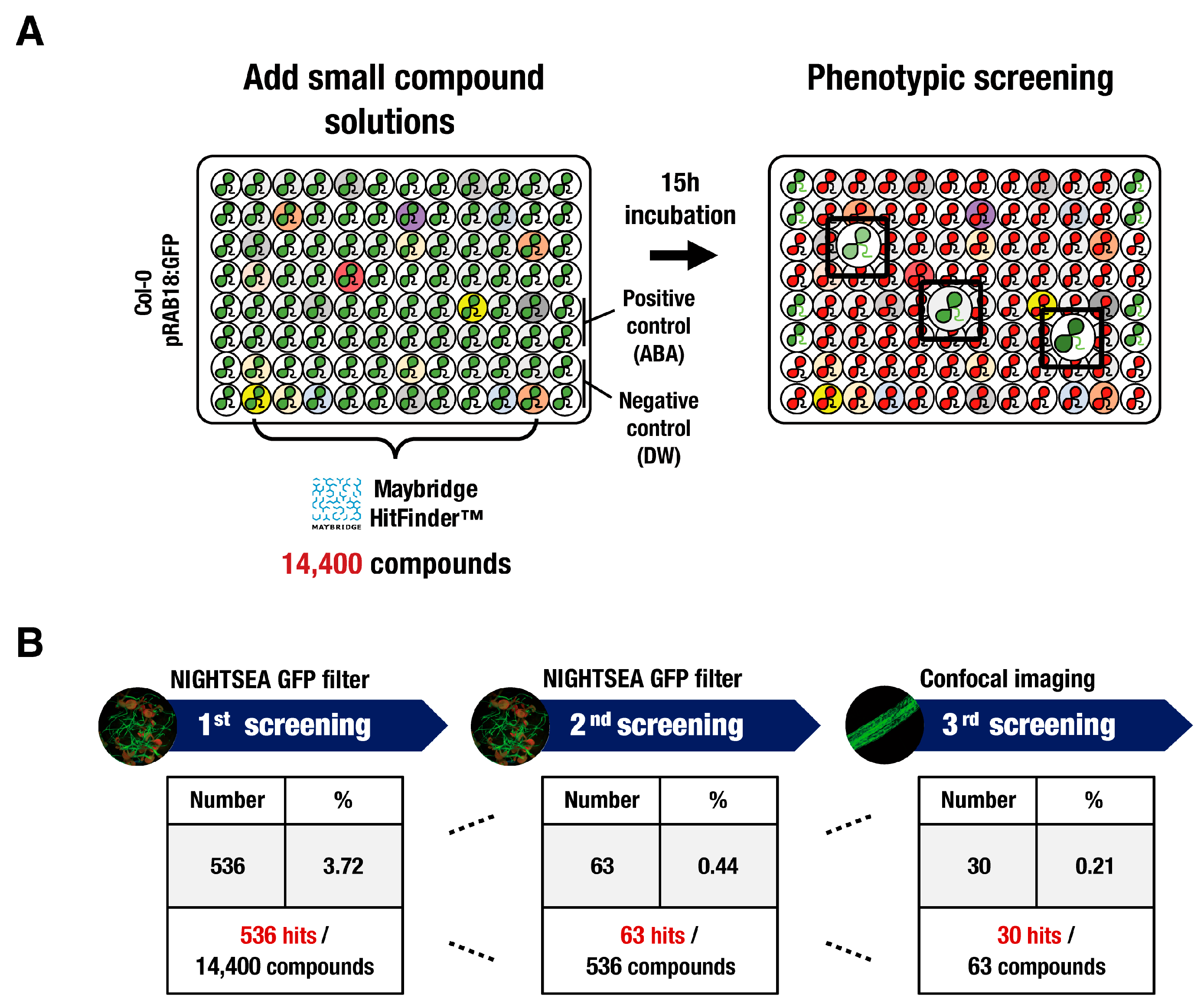
3.1. High-Throughput Screening of Synthetic Chemical Libraries for a Regulator of Abiotic Stress Signaling

Forward chemical genetic screening was performed to identify novel small compounds that potentially regulate the abiotic stress signal transduction using the transgenic Arabidopsis seedlings expressing the ABA-responsive reporter gene pRAB18:GFP. Candidate molecules that induce the ABA-mediated pRAB18:GFP reporter expression were isolated by screening a highly diverse set of 14,400 small compounds from the Maybridge HitFinderTM library (Figure 1A). Ten-day-old transgenic seedlings grown in 96-well tissue culture plates were incubated with each library compound at a concentration of 50 μM for 15 h. After the chemical incubation, chemically induced GFP fluorescence was measured using a NIGHTSEA™ stereo microscope fluorescence adapter. The compounds that induced significant pRAB18:GFP expression in the leaves or roots of the transgenic seedlings were regarded as hit compounds. Five hundred and thirty-six chemicals were identified in the initial screening, corresponding to a 3.72% hit rate among all compounds (Figure 1B). Secondary screening was conducted for candidates with high GFP expression intensity, which was first selected and narrowed down to 63 hit compounds. The selected hit chemicals were re-examined through tertiary screening by confocal microscopy to detect specific GFP expression in the epidermal layer of true leaves or roots. Among the screening compounds, 30 small molecules that produced a significant GFP reporter induction in epidermal cells were analyzed further.

3.2. Small Molecules SRR1 and SRR2 Affect the Physiology of Seed Germination and Root Growth

The expression of several abiotic stress-responsive genes was analyzed to determine if the candidate compounds activate the endogenous expression level of stress-responsive genes in addition to the RAB18 reporter gene. Of the 30 candidate compounds, 22 small molecules significantly increased the expression of genes induced by multiple abiotic stress stimuli such as salt, drought, cold, and ABA (Figure 2A). These small compounds were called stress response regulators (SRRs). In particular, two small molecules were selected for further analysis, designated SRR1 (4-amino-3-{[2-(1-pyrrolidinyl)ethyl]sulfanyl}thieno [2,3-c]isothiazol-5-yl)(phenyl)methanone) and SRR2 (N-(4-{2-[1-(4-fluorobenzyl)-4-pyridinium]vinyl}phenyl)-N-methylmethanamine iodide), which significantly increased the expression levels of several abiotic stress-inducible genes (Figure 2A,B). SRR1 and SRR2 induced RAB18 reporter gene expression to the extent induced by 5 μM ABA in epidermal cells (Figure 2C).
The effects of the selected SRR compounds on root growth and seed germination were analyzed. Seven-day-old seedlings grown on a standard growth medium were transferred to the plates with each concentration of the chemicals and monitored for the production of any root growth arrest phenotype for a week. Treatment with SRR1 caused a substantial decrease in primary root growth while increasing the average lateral root length compared to the DMSO or ABA treatment (Figure 3A,C,E). By contrast, the application of SRR2 severely inhibited primary root growth (Figure 3A) and reduced the number and length of lateral roots at a low concentration of 10 μM (Figure 3C,D). The germination rate was decreased by approximately 73% and 46% after 50 μM SRR1 and SRR2 treatments, respectively (Figure 3F).

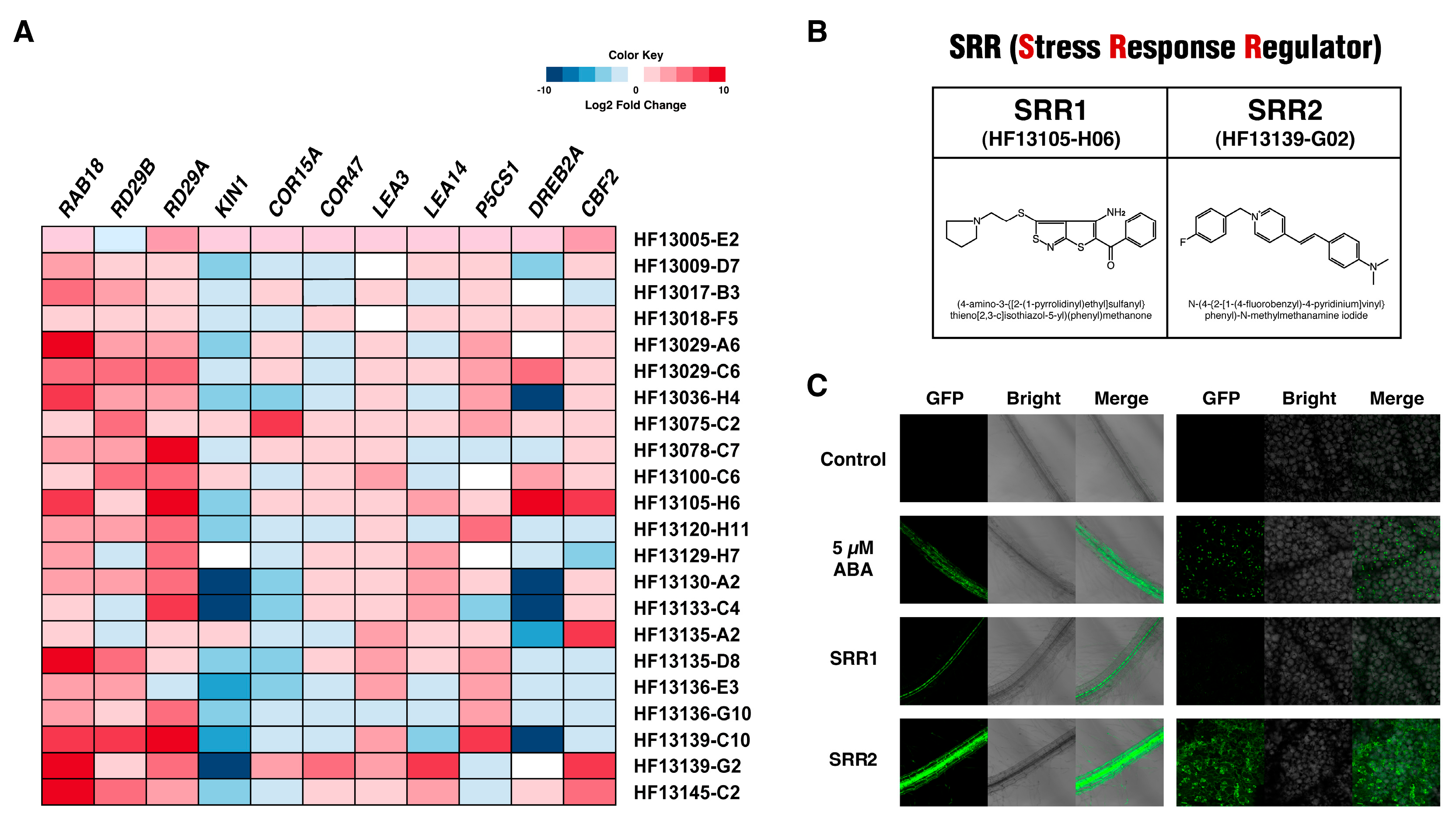
3.3. SRR1 and SRR2 Trigger the Expression of Genes Involved in Abiotic Stress Responses

The selected chemicals SRR1 and SRR2 significantly upregulated the expression of well-characterized marker genes, including RD29A, DREB2A, CBF2, and LEA14, which are responsible for imparting enhanced tolerance to various abiotic stresses [41,42,43,44,45]. RNA-sequencing analysis investigated the large-scale gene expression profiles triggered by SRR1 and SRR2. As a result, 10,614 differentially expressed genes (DEGs) were isolated between each treatment group (Figure 4A). Among the DEGs induced by SRR compounds, the SRR1-treated group contained 3014 DEGs, of which 1637 were upregulated and 1377 were downregulated (Figure 4B). In addition, the SRR2 treatment produced 1855 upregulated DEGs and 1876 downregulated DEGs. In particular, many SRR-induced DEGs exhibited differential expression across ABA or salt treatments, suggesting that the application of SRR compounds induces responses similar to those for abiotic stresses (Figure 4B). Gene Ontology (GO) enrichment analysis for these DEGs presented that the SRR-induced DEGs were enriched significantly in the main terms responsible for controlling the plant resistance to abiotic stresses, such as the oxidation-reduction process, sequence-specific DNA binding, and integral component of the membrane (Figure S1A) [46,47,48]. In addition, the stress-responsive GO enrichments showed that SRR compounds triggered the expression of many genes involved in the response to multiple abiotic stresses (Figure S1B).
In particular, SRR compounds upregulated the transcript levels of several gene family members involved in the essential steps of the abiotic stress response and tolerance, including HSPs and GSTUs (Tables S2 and S3) [49,50,51,52,53,54,55,56,57,58,59,60]. Hence, SRR chemicals may regulate the plant responses to various abiotic stresses through the transcriptional control of the signaling components participating in multiple stress responses.

3.4. Selected SRR Compounds Induce the Abiotic Stress-Related Gene Expression in Crop Plants

In Arabidopsis, SRR1 and SRR2 could influence abiotic stress-induced gene expression. This study examined whether the effects of SRR1 and SRR2 similarly activate abiotic stress signaling in crops such as tomatoes and radishes. Ten-day-old tomato and radish seedlings were treated with 10 µM ABA, 200 mM NaCl, or 50 µM SRR chemicals for six hours to determine the expression levels of the abiotic stress-responsive genes. Overall, the SRR treatments caused transcriptional changes in several genes involved in the abiotic stress responses in crop plants. The transcript levels of MPK3, PR1, and ZAT12, known for mediating multiple stress responses, were increased significantly by SRR1 more than by ABA or salt treatment in tomatoes (Figure 5A) [61,62,63]. The SRR1 treatment increased the expression of critical enzymes required for ABA biosynthesis, such as NCED1 and NCED3 [64]. The SRR2 treatment increased the expression levels of most of the examined genes compared to the control [65,66,67,68,69,70]. In addition, several genes associated with salinity or drought stress tolerance, such as LEA14, GSTU19, and bZIP11, were upregulated significantly by the SRR1 treatment compared to the control in radish (Figure 5B) [44,71,72]. The SRR2 treatment in radish increased the expression level of all investigated genes more than the control. These results suggest that SRR1 and SRR2 could be universal regulators of abiotic stress responses in various crops.

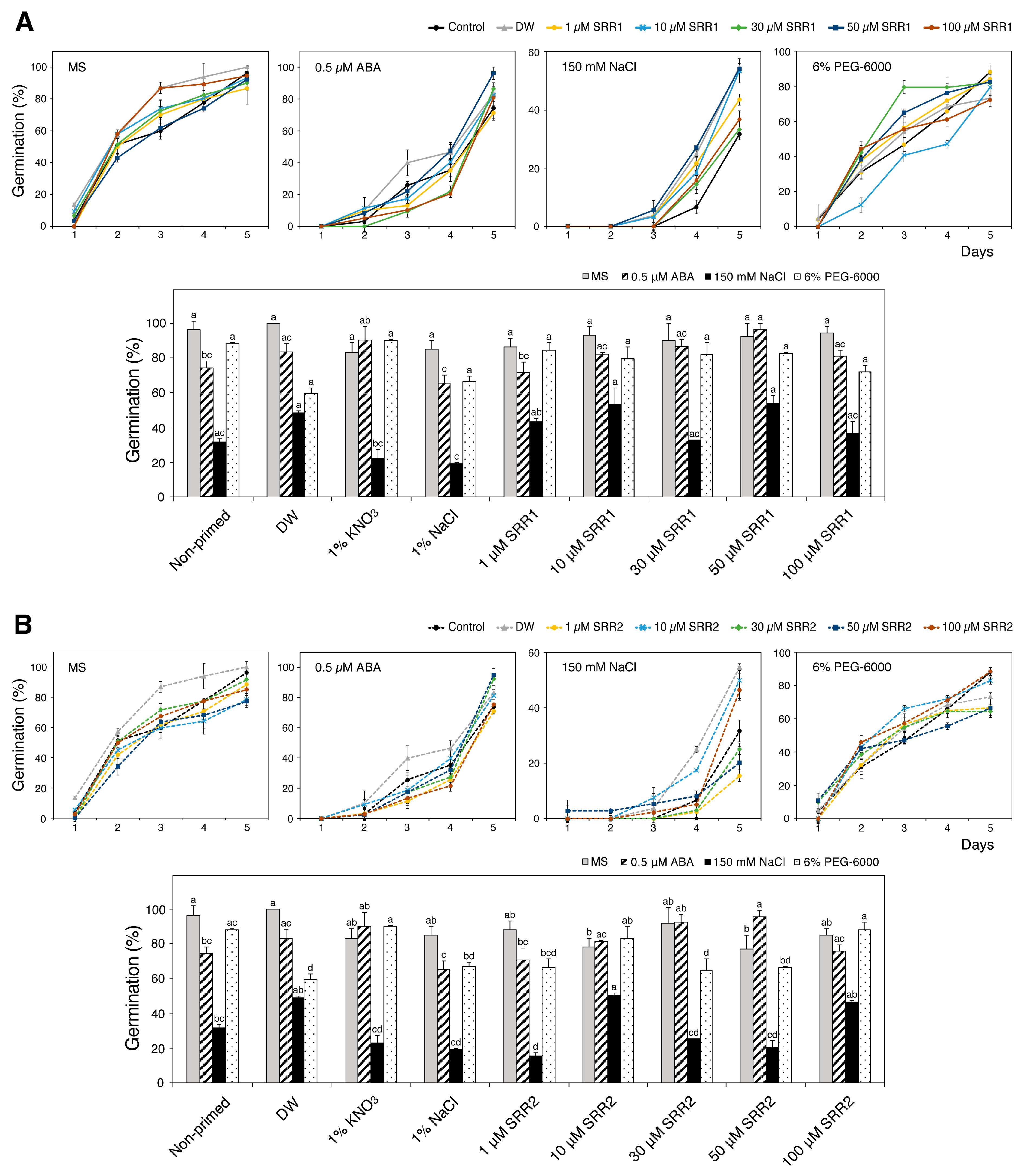
3.5. Seed Priming with SRR1 and SRR2 Affects Germination and Radicle Emergence of Arabidopsis and Tomato Seeds

Based on reports that seed priming using chemical compounds can improve plant germination and growth, this study investigated whether seed priming with SRR1 and SRR2 positively affected the physiology of seed germination under abiotic stress conditions (Figure 6). Sterilized Arabidopsis seeds were primed by soaking in five priming agent solutions for 24 h: distilled water, 1% KNO3, 1% NaCl, and different concentrations of SRR chemicals. The primed seeds were grown on MS medium supplemented with 0.5 μM ABA, 150 mM NaCl, or 6% PEG-6000 and evaluated for germination analysis. As a result, seed priming with selected SRR chemicals generally enhanced the germination performance of Arabidopsis seeds in response to abiotic stresses. The primed seeds with SRR1 concentrations ranging from 10 to 50 µM gradually increased the germination percentage under ABA stress (Figure 7A). Priming with all concentrations of SRR1 resulted in a higher germination rate and faster radicle protrusion compared to the water-primed control during PEG stress. Increasing SRR2 concentrations of up to 50 μM can also promote seed germination in response to ABA stress (Figure 7B). The exogenous application of 30 μM SRR2 significantly improved the salt tolerance during seed germination. Overall, soaking in a 50 μM SRR1 solution had the most pronounced effects on improving seed germination in response to various abiotic stresses compared to other priming agents. SRR2 priming at concentrations of 50 μM was most effective in promoting seed germination under ABA stress conditions. On the other hand, the priming effects of SRR1 against salt stress were not strong compared to the control during seed germination.

3.6. Priming with SRR1 and SRR2 Augments the Induction of Abiotic Stress-Responsive Genes under Salt Stress

After confirming that the priming treatment of SRR1 and SRR2 affected the seed germination response to abiotic stress, this study examined how the SRR1 and SRR2 priming treatments affected the gene expression of abiotic stress-responsive genes in response to subsequent abiotic stress. Ten-day-old Arabidopsis and tomato seedlings were pretreated with 100 mM NaCl or each concentration of the SRR chemical solutions for 24 h. The primed seedlings recovered in growth media for two days were subjected to secondary salt stress with 200 mM NaCl for six hours (Figure 8A). All plant tissues were harvested at corresponding time points to determine the transcript levels. After the priming treatment with 50 μM SRR compounds, the transcript levels of abiotic stress-responsive genes increased to expected levels, followed by a significant decrease in the expression levels in the recovery interval (Figure 8B). The SRR-primed seedlings showed more robust transcriptional reactivation during subsequent salt stress than during the first stress exposure (Figure 8B). The priming treatment with each concentration of SRR1 caused the strong induction of stress-related maker genes, including COR15A, P5CS1, COR47, RD29A, RD22, and KIN1, compared to the control or NaCl treatment (Figure 8C) [73,74]. Similar to the SRR1-priming treatment, priming with SRR2 also increased the expression of all maker genes in subsequent salt stress (Figure 8C). Interestingly, priming treatment with SRR1 significantly increased the levels of COR15A, P5CS1, and RD29A expression in a dose-dependent manner (Figure 8C). On the other hand, most of the genes examined as stress-responsive genes were progressively downregulated with increasing SRR2 concentration during secondary salt stress conditions. In line with the results shown in Arabidopsis, the SRR-priming treatment in tomatoes affected the abiotic stress-induced gene expression under secondary salt stress conditions (Figure 8D). The ZAT12 expression was upregulated slightly by SRR1-priming, whereas the levels of P5CS1 expression were lower than that of the control. Pretreatment with SRR2 greatly enhanced the transcript levels of the HSP90-1, LEA14, NCED3, and ZAT12 genes compared to the effects of the NaCl treatment. Priming with SRR1 and SRR2 might induce mechanisms to increase the transcription of abiotic stress-induced genes for successive stress conditions in Arabidopsis and tomato.

4. Discussion

Climate change has increased the frequency and intensity of abiotic stress combinations, which will have far-reaching impacts on plant growth and agricultural productivity [75,76]. Chemical priming is a promising tool that allows plants to activate more rapid and vigorous responses to mitigate individual or combined abiotic stresses [18]. The novel small molecules SRR1 and SRR2 were isolated by screening a 14,400-compound chemical library based on the ABA-mediated pRAB18:GFP reporter expression (Figure 1 and Figure 2). Phytohormones, termed plant growth regulators, are small molecules that govern all aspects of plant development and growth under optimal and stress conditions. The selected small compound SRRs can also affect the different developmental stages of plants, such as seed germination, root elongation, and lateral root formation. In particular, SRR2 caused a substantial reduction in primary root growth (Figure 3A), lateral root formation (Figure 3C,D), and seed germination (Figure 3F). The application of SRR1 consistently produced inhibitory effects on primary root growth (Figure 3A) and seed germination (Figure 3F). On the other hand, SRR1 reduced the total number of lateral roots (Figure 3D) while increasing the average lateral root length compared to the DMSO control (Figure 3C,E). In particular, the seedlings exposed to 10 μM SRR1 with less abundant but much longer lateral roots had normal green leaf phenotypes similar to that observed in the control (Figure 3B). Several root architectural traits, such as root length, branching and lateral root number, and root hair development, facilitate adaptation to multiple stress factors in different crops [77,78,79]. For example, transgenic Arabidopsis plants overexpressing GmNAC109 exhibited an increased lateral root formation and improved tolerance to drought and salt stress [80]. Maize genotypes with fewer but longer lateral roots showed superior water capture, biomass, and yield under drought stress [81]. These results suggest that SRR compounds might have plant-growth regulatory activities, potentially improving plant tolerance against abiotic stress.
This study reported that SRR1 and SRR2 induce the transcriptional activation of genes, imparting multiple stress tolerances, which was found through comparative transcriptome profiling and co-expression network analysis (Figure 4 and Figures S1–S4). The GO annotation showed that the SRR-triggered DEGs were highly enriched into functional terms, including the response to abiotic stimulus, redox process, and membrane components (Figure S1). Many of these DEGs were enriched in the Kyoto Encyclopedia of Genes and Genomes (KEGG) pathways, particularly in hormone signal transduction, the biosynthesis of secondary metabolites, and metabolic pathways (Figure S2). Transcriptomic studies on gene enrichment and co-expression network analyses suggested that the signaling pathways of SRR molecules might integrate with complex networks involving HSPs, Ca2+ signals, and ROS regulatory systems to modulate abiotic stress responses and generate cross-stress tolerances.
This study assessed the priming effects of different concentrations of the SRR molecules on the germination performance of Arabidopsis seeds under abiotic stress conditions. Among the common seed priming agents, halopriming with 1% KNO3 increased Arabidopsis seed germination in the presence of ABA stress, while hydro-priming appeared to be more effective in improving the germination rate under ABA, NaCl, and normal conditions (Figure 7) [28,29]. Seed priming with the SRR1 compound at concentrations of 10–50 µM caused a dose-dependent increase in Arabidopsis seed germination under ABA stress (Figure 7A). The germination percentage was also increased by the SRR2-priming treatment at 30 μM and 50 μM (Figure 7B). Furthermore, the application of SRR1 and SRR2 positively affected the enhancement and acceleration of seed germination under PEG stress (Figure 7A,B). The priming process triggers the rapid imbibition of water and various metabolic changes at the cellular level that facilitate synchronized and earlier seed germination [28,82]. These metabolic changes enhance the tolerance capability of crop plants, including the synthesis of nucleic acids and proteins, cell division, ATP synthesis, the production of antioxidants, and the activation of DNA repair mechanisms [28,82]. Although most seed priming techniques cause partial seed pre-hydration and early germination events, their efficiency depends on the plant species and the selected priming technique. Furthermore, physical and chemical factors can influence priming effects, including the priming agent, duration, water potential, and temperature [25]. Therefore, the optimal priming conditions for each plant species should be evaluated to determine the potential applications of SRR compounds in the seed priming approach.
Priming can also stimulate stress tolerance mechanisms to subsequent stresses via activating stress memory [82,83]. Epigenetic regulatory mechanisms and chromatin marks have been heavily implicated in priming-dependent stress memory [84]. Recent studies revealed that seed priming improved stress tolerance by inducing epigenetic alterations and stress memory [85]. Overall, transcriptional amplification after the SRR-priming treatment involves more robust abiotic stress-related genes re-inducing under subsequent salt stress in Arabidopsis and tomato (Figure 8). Hence, the SRR-priming may induce stress memory that confers enhanced responses to additional stress exposure, indicating a possible cross-tolerance effect. The duration and inherent features of SRR-induced stress memory and stress tolerant effects must be analyzed further according to the treatment conditions. Future studies on identifying the chromatin and epigenetic marks at SRR-induced memory gene loci under abiotic stress conditions will help better understand the epigenetic-based mechanisms in abiotic stress adaptation and plant stress memory.

5. Conclusions

Based on the hypothesis that plant stress response regulators can be identified through chemical library screening, we have conducted a small compound library screening based on the ABA-inducible transcriptional responses. The selected SRR1 and SRR2 compounds affected the processes of Arabidopsis seed germination and root development and triggered transcriptional changes to multiple abiotic stresses. Furthermore, seed priming with SRR compounds alleviated abiotic stresses during the germination and the emergence of Arabidopsis seeds. Understanding the mechanisms for the specific regulation of stress-responsive gene expressions by SRR priming may be the next significant challenge. The isolation of novel small molecules, such as SRR1 and SRR2, and their possible application as priming agents will provide opportunities to elucidate the complex molecular mechanisms and epigenetic regulation underlying the plant stress-tolerant responses.

Supplementary Materials

The following supporting information can be downloaded at: https://www.mdpi.com/article/10.3390/biom14091177/s1: Figure S1. Gene Ontology (GO) enrichment analysis for the SRR-induced DEGs. Figure S2. Kyoto Encyclopedia of Genes and Genomes (KEGG) enrichment analysis for the SRR-induced DEGs. Figure S3. Co-expression network analysis of GSTU19 and comparison with the SRR-induced DEGs. Figure S4. Co-expression network analysis of HSP17.6 and comparison with the SRR-induced DEGs. Table S1. List of primers used in this study. Table S2. The SRR-induced DEGs are involved in the glutathione metabolic process. Table S3. The SRR-induced DEGs participate in the heat-shock protein/chaperone pathway.

Author Contributions

S.K. and T.-H.K. planned the study and analyzed the data. S.K. performed most of the experiments. S.K. and T.-H.K. wrote the manuscript. All authors have read and agreed to the published version of the manuscript.

Funding

This research was funded by a grant from the Next-Generation BioGreen 21 program (PJ01335001), which is funded by the Rural Development Administration, Republic of Korea, and by a National Research Foundation of Korea (NRF) grant from the Korean government (2021R1F1A1058458).

Institutional Review Board Statement

Not applicable.

Informed Consent Statement

Not applicable.

Data Availability Statement

The data involved in this study are listed in the article and its Supplementary Materials.

Conflicts of Interest

The authors declare no conflicts of interest.

References

  1. Lepri, A.; Longo, C.; Messore, A.; Kazmi, H.; Madia, V.N.; Di Santo, R.; Costi, R.; Vittorioso, P. Plants and Small Molecules: An Up-and-Coming Synergy. Plants 2023, 12, 1729. [Google Scholar] [CrossRef] [PubMed]
  2. Pasquer, Q.T.L.; Tsakoumagkos, I.A.; Hoogendoorn, S. From Phenotypic Hit to Chemical Probe: Chemical Biology Approaches to Elucidate Small Molecule Action in Complex Biological Systems. Molecules 2020, 25, 5702. [Google Scholar] [CrossRef] [PubMed]
  3. Dejonghe, W.; Russinova, E. Plant Chemical Genetics: From Phenotype-Based Screens to Synthetic Biology. Plant Physiol. 2017, 174, 5–20. [Google Scholar] [CrossRef] [PubMed]
  4. Hewage, K.A.H.; Yang, J.F.; Wang, D.; Hao, G.F.; Yang, G.F.; Zhu, J.K. Chemical Manipulation of Abscisic Acid Signaling: A New Approach to Abiotic and Biotic Stress Management in Agriculture. Adv. Sci. 2020, 7, 2001265. [Google Scholar] [CrossRef]
  5. Hu, D.; Wei, L.; Liao, W. Brassinosteroids in Plants: Crosstalk with Small-Molecule Compounds. Biomolecules 2021, 11, 1800. [Google Scholar] [CrossRef]
  6. Chini, A.; Monte, I.; Fernandez-Barbero, G.; Boter, M.; Hicks, G.; Raikhel, N.; Solano, R. A small molecule antagonizes jasmonic acid perception and auxin responses in vascular and nonvascular plants. Plant Physiol. 2021, 187, 1399–1413. [Google Scholar] [CrossRef]
  7. Jiang, K.; Asami, T. Chemical regulators of plant hormones and their applications in basic research and agriculture. Biosci. Biotechnol. Biochem. 2018, 82, 1265–1300. [Google Scholar] [CrossRef]
  8. Chen, K.; Li, G.J.; Bressan, R.A.; Song, C.P.; Zhu, J.K.; Zhao, Y. Abscisic acid dynamics, signaling, and functions in plants. J. Integr. Plant Biol. 2020, 62, 25–54. [Google Scholar] [CrossRef]
  9. Hsu, P.K.; Dubeaux, G.; Takahashi, Y.; Schroeder, J.I. Signaling mechanisms in abscisic acid-mediated stomatal closure. Plant J. 2021, 105, 307–321. [Google Scholar] [CrossRef]
  10. Sah, S.K.; Reddy, K.R.; Li, J. Abscisic acid and abiotic stress tolerance in crop plants. Front. Plant Sci. 2016, 7, 571. [Google Scholar] [CrossRef]
  11. Santiago, J.; Rodrigues, A.; Saez, A.; Rubio, S.; Antoni, R.; Dupeux, F.; Park, S.Y.; Marquez, J.A.; Cutler, S.R.; Rodriguez, P.L. Modulation of drought resistance by the abscisic acid receptor PYL5 through inhibition of clade A PP2Cs. Plant J. 2009, 60, 575–588. [Google Scholar] [CrossRef] [PubMed]
  12. Okamoto, M.; Peterson, F.C.; Defries, A.; Park, S.Y.; Endo, A.; Nambara, E.; Volkman, B.F.; Cutler, S.R. Activation of dimeric ABA receptors elicits guard cell closure, ABA-regulated gene expression, and drought tolerance. Proc. Natl. Acad. Sci. USA 2013, 110, 12132–12137. [Google Scholar] [CrossRef] [PubMed]
  13. Min, M.K.; Kim, R.; Moon, S.J.; Lee, Y.; Han, S.; Lee, S.; Kim, B.G. Selection and functional identification of a synthetic partial ABA agonist, S7. Sci. Rep. 2020, 10, 4. [Google Scholar] [CrossRef] [PubMed]
  14. Kim, T.H.; Hauser, F.; Ha, T.; Xue, S.; Bohmer, M.; Nishimura, N.; Munemasa, S.; Hubbard, K.; Peine, N.; Lee, B.H.; et al. Chemical genetics reveals negative regulation of abscisic acid signaling by a plant immune response pathway. Curr. Biol. 2011, 21, 990–997. [Google Scholar] [CrossRef] [PubMed]
  15. Leung, J. The First Broad-Spectrum Abscisic Acid Antagonist. Plant Physiol. 2017, 173, 1939. [Google Scholar] [CrossRef]
  16. Cao, M.J.; Zhang, Y.L.; Liu, X.; Huang, H.; Zhou, X.E.; Wang, W.L.; Zeng, A.; Zhao, C.Z.; Si, T.; Du, J.; et al. Combining chemical and genetic approaches to increase drought resistance in plants. Nat. Commun. 2017, 8, 1183. [Google Scholar] [CrossRef]
  17. Vaidya, A.S.; Peterson, F.C.; Yarmolinsky, D.; Merilo, E.; Verstraeten, I.; Park, S.Y.; Elzinga, D.; Kaundal, A.; Helander, J.; Lozano-Juste, J.; et al. A Rationally Designed Agonist Defines Subfamily IIIA Abscisic Acid Receptors As Critical Targets for Manipulating Transpiration. ACS Chem. Biol. 2017, 12, 2842–2848. [Google Scholar] [CrossRef]
  18. Sako, K.; Nguyen, H.M.; Seki, M. Advances in Chemical Priming to Enhance Abiotic Stress Tolerance in Plants. Plant Cell Physiol. 2021, 61, 1995–2003. [Google Scholar] [CrossRef]
  19. Sheteiwy, M.S.; An, J.; Yin, M.; Jia, X.; Guan, Y.; He, F.; Hu, J. Cold plasma treatment and exogenous salicylic acid priming enhances salinity tolerance of Oryza sativa seedlings. Protoplasma 2019, 256, 79–99. [Google Scholar] [CrossRef]
  20. Wang, W.; Wang, X.; Zhang, J.; Huang, M.; Cai, J.; Zhou, Q.; Dai, T.; Jiang, D. Salicylic acid and cold priming induce late-spring freezing tolerance by maintaining cellular redox homeostasis and protecting photosynthetic apparatus in wheat. Plant Growth Regul. 2020, 90, 109–121. [Google Scholar] [CrossRef]
  21. Gohari, G.; Alavi, Z.; Esfandiari, E.; Panahirad, S.; Hajihoseinlou, S.; Fotopoulos, V. Interaction between hydrogen peroxide and sodium nitroprusside following chemical priming of Ocimum basilicum L. against salt stress. Physiol. Plant 2020, 168, 361–373. [Google Scholar] [CrossRef] [PubMed]
  22. Joshi, N.C.; Yadav, D.; Ratner, K.; Kamara, I.; Aviv-Sharon, E.; Irihimovitch, V.; Charuvi, D. Sodium hydrosulfide priming improves the response of photosynthesis to overnight frost and day high light in avocado (Persea americana Mill, cv. ‘Hass’). Physiol. Plant 2020, 168, 394–405. [Google Scholar] [CrossRef] [PubMed]
  23. Zulfiqar, F.; Nafees, M.; Chen, J.; Darras, A.; Ferrante, A.; Hancock, J.T.; Ashraf, M.; Zaid, A.; Latif, N.; Corpas, F.J.; et al. Chemical priming enhances plant tolerance to salt stress. Front. Plant Sci. 2022, 13, 946922. [Google Scholar] [CrossRef] [PubMed]
  24. Honig, M.; Roeber, V.M.; Schmulling, T.; Cortleven, A. Chemical priming of plant defense responses to pathogen attacks. Front. Plant Sci. 2023, 14, 1146577. [Google Scholar] [CrossRef]
  25. Liu, H.; Able, A.J.; Able, J.A. Priming crops for the future: Rewiring stress memory. Trends Plant Sci. 2022, 27, 699–716. [Google Scholar] [CrossRef]
  26. Siddique, A.B.; Parveen, S.; Rahman, M.Z.; Rahman, J. Revisiting plant stress memory: Mechanisms and contribution to stress adaptation. Physiol. Mol. Biol. Plants 2024, 30, 349–367. [Google Scholar] [CrossRef]
  27. Liu, J.; Feng, L.; Gu, X.; Deng, X.; Qiu, Q.; Li, Q.; Zhang, Y.; Wang, M.; Deng, Y.; Wang, E.; et al. An H3K27me3 demethylase-HSFA2 regulatory loop orchestrates transgenerational thermomemory in Arabidopsis. Cell Res. 2019, 29, 379–390. [Google Scholar] [CrossRef]
  28. Wiszniewska, A. Priming Strategies for Benefiting Plant Performance under Toxic Trace Metal Exposure. Plants 2021, 10, 623. [Google Scholar] [CrossRef]
  29. Devika, O.S.; Singh, S.; Sarkar, D.; Barnwal, P.; Suman, J.; Rakshit, A. Seed Priming: A Potential Supplement in Integrated Resource Management Under Fragile Intensive Ecosystems. Front. Sustain. Food Syst. 2021, 5, 654001. [Google Scholar] [CrossRef]
  30. Farooq, M.; Usman, M.; Nadeem, F.; Rehman, H.u.; Wahid, A.; Basra, S.M.A.; Siddique, K.H.M. Seed priming in field crops: Potential benefits, adoption and challenges. Crop Pasture Sci. 2019, 70, 731–771, 741. [Google Scholar] [CrossRef]
  31. Liu, X.; Quan, W.; Bartels, D. Stress memory responses and seed priming correlate with drought tolerance in plants: An overview. Planta 2022, 255, 45. [Google Scholar] [CrossRef] [PubMed]
  32. Rehman, A.; Nadeem, F.; Farooq, M. Role of seed priming in root development and crop production. In The Root Systems in Sustainable Agricultural Intensification; Wiley: New York, NY, USA, 2021; pp. 221–243. [Google Scholar]
  33. Yang, Z.; Zhi, P.; Chang, C. Priming seeds for the future: Plant immune memory and application in crop protection. Front. Plant Sci. 2022, 13, 961840. [Google Scholar] [CrossRef] [PubMed]
  34. Afzal, I. Seed priming: What’s next? Seed Sci. Technol. 2023, 51, 379–405. [Google Scholar] [CrossRef]
  35. Singhal, R.K.; Pandey, S.; Bose, B. Seed priming with Mg(NO3)2 and ZnSO4 salts triggers physio-biochemical and antioxidant defense to induce water stress adaptation in wheat (Triticum aestivum L.). Plant Stress 2021, 2, 100037. [Google Scholar] [CrossRef]
  36. Marks, Z.D.; Cowley, J.M.; Burton, R.A.; Bianco-Miotto, T. The role of oxidative stress in seed priming to improve germination and vigour. BioRxiv 2022. [Google Scholar] [CrossRef]
  37. Hasanuzzaman, M.; Fotopoulos, V. Priming and Pretreatment of Seeds and Seedlings; Springer: Berlin/Heidelberg, Germany, 2019. [Google Scholar]
  38. Nile, S.H.; Thiruvengadam, M.; Wang, Y.; Samynathan, R.; Shariati, M.A.; Rebezov, M.; Nile, A.; Sun, M.; Venkidasamy, B.; Xiao, J.; et al. Nano-priming as emerging seed priming technology for sustainable agriculture—Recent developments and future perspectives. J. Nanobiotechnol. 2022, 20, 254. [Google Scholar] [CrossRef]
  39. Wang, W.; He, A.; Peng, S.; Huang, J.; Cui, K.; Nie, L. The Effect of Storage Condition and Duration on the Deterioration of Primed Rice Seeds. Front. Plant Sci. 2018, 9, 172. [Google Scholar] [CrossRef]
  40. Singh, K.P.; Gupta, N.; Dhingra, M. Effect of temperature regimes, seed priming and priming duration on germination and seedling growth on American cotton. J. Environ. Biol. 2018, 39, 83–91. [Google Scholar] [CrossRef]
  41. Szklarczyk, D.; Gable, A.L.; Nastou, K.C.; Lyon, D.; Kirsch, R.; Pyysalo, S.; Doncheva, N.T.; Legeay, M.; Fang, T.; Bork, P.; et al. The STRING database in 2021: Customizable protein-protein networks, and functional characterization of user-uploaded gene/measurement sets. Nucleic Acids Res. 2021, 49, D605–D612. [Google Scholar] [CrossRef]
  42. Kimotho, R.N.; Baillo, E.H.; Zhang, Z. Transcription factors involved in abiotic stress responses in Maize (Zea mays L.) and their roles in enhanced productivity in the post genomics era. PeerJ 2019, 7, e7211. [Google Scholar] [CrossRef]
  43. Mizoi, J.; Kanazawa, N.; Kidokoro, S.; Takahashi, F.; Qin, F.; Morimoto, K.; Shinozaki, K.; Yamaguchi-Shinozaki, K. Heat-induced inhibition of phosphorylation of the stress-protective transcription factor DREB2A promotes thermotolerance of Arabidopsis thaliana. J. Biol. Chem. 2019, 294, 902–917. [Google Scholar] [CrossRef] [PubMed]
  44. Marinho, J.P.; Pagliarini, R.F.; Molinari, M.D.C.; Marcolino-Gomes, J.; Caranhoto, A.L.H.; Marin, S.R.R.; Oliveira, M.C.N.; Foloni, J.S.S.; Melo, C.L.P.; Kidokoro, S.; et al. Overexpression of full-length and partial DREB2A enhances soybean drought tolerance. Agron. Sci. Biotechnol. 2022, 8, 1–21. [Google Scholar]
  45. Jia, F.; Qi, S.; Li, H.; Liu, P.; Li, P.; Wu, C.; Zheng, C.; Huang, J. Overexpression of Late Embryogenesis Abundant 14 enhances Arabidopsis salt stress tolerance. Biochem. Biophys. Res. Commun. 2014, 454, 505–511. [Google Scholar] [CrossRef] [PubMed]
  46. Li, X.; Liang, X.; Li, W.; Yao, A.; Liu, W.; Wang, Y.; Yang, G.; Han, D. Isolation and Functional Analysis of MbCBF2, a Malus baccata (L.) Borkh CBF Transcription Factor Gene, with Functions in Tolerance to Cold and Salt Stress in Transgenic Arabidopsis thaliana. Int. J. Mol. Sci. 2022, 23, 9827. [Google Scholar] [CrossRef] [PubMed]
  47. Sachdev, S.; Ansari, S.A.; Ansari, M.I.; Fujita, M.; Hasanuzzaman, M. Abiotic Stress and Reactive Oxygen Species: Generation, Signaling, and Defense Mechanisms. Antioxidants 2021, 10, 277. [Google Scholar] [CrossRef]
  48. Wang, Z.; Wong, D.C.J.; Chen, Z.; Bai, W.; Si, H.; Jin, X. Emerging Roles of Plant DNA-Binding With One Finger Transcription Factors in Various Hormone and Stress Signaling Pathways. Front. Plant Sci. 2022, 13, 844201. [Google Scholar] [CrossRef]
  49. Saijo, Y.; Loo, E.P.i. Plant immunity in signal integration between biotic and abiotic stress responses. New Phytol. 2020, 225, 87–104. [Google Scholar] [CrossRef]
  50. Kumar, S.; Trivedi, P.K. Glutathione S-Transferases: Role in Combating Abiotic Stresses Including Arsenic Detoxification in Plants. Front. Plant Sci. 2018, 9, 751. [Google Scholar] [CrossRef]
  51. Landi, S.; Capasso, G.; Ben Azaiez, F.E.; Jallouli, S.; Ayadi, S.; Trifa, Y.; Esposito, S. Different Roles of Heat Shock Proteins (70 kDa) During Abiotic Stresses in Barley (Hordeum vulgare) Genotypes. Plants 2019, 8, 248. [Google Scholar] [CrossRef]
  52. di Donato, M.; Geisler, M. HSP90 and co-chaperones: A multitaskers’ view on plant hormone biology. FEBS Lett. 2019, 593, 1415–1430. [Google Scholar] [CrossRef]
  53. McLoughlin, F.; Basha, E.; Fowler, M.E.; Kim, M.; Bordowitz, J.; Katiyar-Agarwal, S.; Vierling, E. Class I and II Small Heat Shock Proteins Together with HSP101 Protect Protein Translation Factors during Heat Stress. Plant Physiol. 2016, 172, 1221–1236. [Google Scholar] [CrossRef] [PubMed]
  54. Stavridou, E.; Voulgari, G.; Michailidis, M.; Kostas, S.; Chronopoulou, E.G.; Labrou, N.E.; Madesis, P.; Nianiou-Obeidat, I. Overexpression of A Biotic Stress-Inducible Pvgstu Gene Activates Early Protective Responses in Tobacco under Combined Heat and Drought. Int. J. Mol. Sci. 2021, 22, 2352. [Google Scholar] [CrossRef] [PubMed]
  55. Kissoudis, C.; Kalloniati, C.; Flemetakis, E.; Madesis, P.; Labrou, N.E.; Tsaftaris, A.; Nianiou-Obeidat, I. Stress-inducible GmGSTU4 shapes transgenic tobacco plants metabolome towards increased salinity tolerance. Acta Physiol. Plant. 2015, 37, 102. [Google Scholar] [CrossRef]
  56. Islam, S.; Rahman, I.A.; Islam, T.; Ghosh, A. Genome-wide identification and expression analysis of glutathione S-transferase gene family in tomato: Gaining an insight to their physiological and stress-specific roles. PLoS ONE 2017, 12, e0187504. [Google Scholar] [CrossRef] [PubMed]
  57. Wu, J.; Zhang, N.; Liu, Z.; Liu, S.; Liu, C.; Lin, J.; Yang, H.; Li, S.; Yukawa, Y. The AtGSTU7 gene influences glutathione-dependent seed germination under ABA and osmotic stress in Arabidopsis. Biochem. Biophys. Res. Commun. 2020, 528, 538–544. [Google Scholar] [CrossRef] [PubMed]
  58. Ding, F.; Wang, C.; Zhang, S.; Wang, M. A jasmonate-responsive glutathione S-transferase gene SlGSTU24 mitigates cold-induced oxidative stress in tomato plants. Sci. Hortic. 2022, 303, 111231. [Google Scholar] [CrossRef]
  59. Guo, L.M.; Li, J.; He, J.; Liu, H.; Zhang, H.M. A class I cytosolic HSP20 of rice enhances heat and salt tolerance in different organisms. Sci. Rep. 2020, 10, 1383. [Google Scholar] [CrossRef]
  60. Wu, J.; Gao, T.; Hu, J.; Zhao, L.; Yu, C.; Ma, F. Research advances in function and regulation mechanisms of plant small heat shock proteins (sHSPs) under environmental stresses. Sci. Total Environ. 2022, 825, 154054. [Google Scholar] [CrossRef]
  61. Hu, J.; Cai, J.; Park, S.J.; Lee, K.; Li, Y.; Chen, Y.; Yun, J.Y.; Xu, T.; Kang, H. N6-Methyladenosine mRNA methylation is important for salt stress tolerance in Arabidopsis. Plant J. 2021, 106, 1759–1775. [Google Scholar] [CrossRef]
  62. Zhang, M.; Zhang, S. Mitogen-activated protein kinase cascades in plant signaling. J. Integr. Plant Biol. 2022, 64, 301–341. [Google Scholar] [CrossRef]
  63. Akbudak, M.A.; Yildiz, S.; Filiz, E. Pathogenesis related protein-1 (PR-1) genes in tomato (Solanum lycopersicum L.): Bioinformatics analyses and expression profiles in response to drought stress. Genomics 2020, 112, 4089–4099. [Google Scholar] [CrossRef] [PubMed]
  64. Kaur, M.; Asthir, B.; Kaur, R.; Chaudhary, A. Zat12 Gene Ameliorates Temperature Stress in Wheat Transgenics by Modulating the Antioxidant Defense System. Stresses 2023, 3, 316–330. [Google Scholar] [CrossRef]
  65. Baek, D.; Kim, W.Y.; Cha, J.Y.; Park, H.J.; Shin, G.; Park, J.; Lim, C.J.; Chun, H.J.; Li, N.; Kim, D.H.; et al. The GIGANTEA-ENHANCED EM LEVEL Complex Enhances Drought Tolerance via Regulation of Abscisic Acid Synthesis. Plant Physiol. 2020, 184, 443–458. [Google Scholar] [CrossRef] [PubMed]
  66. Lestari, R.; Rio, M.; Martin, F.; Leclercq, J.; Woraathasin, N.; Roques, S.; Dessailly, F.; Clement-Vidal, A.; Sanier, C.; Fabre, D.; et al. Overexpression of Hevea brasiliensis ethylene response factor HbERF-IXc5 enhances growth and tolerance to abiotic stress and affects laticifer differentiation. Plant Biotechnol. J. 2018, 16, 322–336. [Google Scholar] [CrossRef]
  67. Xing, L.; Di, Z.; Yang, W.; Liu, J.; Li, M.; Wang, X.; Cui, C.; Wang, X.; Wang, X.; Zhang, R.; et al. Overexpression of ERF1-V from Haynaldia villosa Can Enhance the Resistance of Wheat to Powdery Mildew and Increase the Tolerance to Salt and Drought Stresses. Front. Plant Sci. 2017, 8, 1948. [Google Scholar] [CrossRef]
  68. Thirumalaikumar, V.P.; Gorka, M.; Schulz, K.; Masclaux-Daubresse, C.; Sampathkumar, A.; Skirycz, A.; Vierstra, R.D.; Balazadeh, S. Selective autophagy regulates heat stress memory in Arabidopsis by NBR1-mediated targeting of HSP90.1 and ROF1. Autophagy 2021, 17, 2184–2199. [Google Scholar] [CrossRef]
  69. Vyse, K.; Schaarschmidt, S.; Erban, A.; Kopka, J.; Zuther, E. Specific CBF transcription factors and cold-responsive genes fine-tune the early triggering response after acquisition of cold priming and memory. Physiol. Plant 2022, 174, e13740. [Google Scholar] [CrossRef]
  70. Zhang, Q.; Zhao, Y.; Zhang, J.; Li, X.; Ma, F.; Duan, M.; Zhang, B.; Li, H. The Responses of the Lipoxygenase Gene Family to Salt and Drought Stress in Foxtail Millet (Setaria italica). Life 2021, 11, 1169. [Google Scholar] [CrossRef]
  71. Huang, K.; Peng, L.; Liu, Y.; Yao, R.; Liu, Z.; Li, X.; Yang, Y.; Wang, J. Arabidopsis calcium-dependent protein kinase AtCPK1 plays a positive role in salt/drought-stress response. Biochem. Biophys. Res. Commun. 2018, 498, 92–98. [Google Scholar] [CrossRef]
  72. Han, Y.; Hou, Z.; He, Q.; Zhang, X.; Yan, K.; Han, R.; Liang, Z. Genome-Wide Characterization and Expression Analysis of bZIP Gene Family Under Abiotic Stress in Glycyrrhiza uralensis. Front. Genet. 2021, 12, 754237. [Google Scholar] [CrossRef]
  73. Xu, J.; Tian, Y.S.; Xing, X.J.; Peng, R.H.; Zhu, B.; Gao, J.J.; Yao, Q.H. Over-expression of AtGSTU19 provides tolerance to salt, drought and methyl viologen stresses in Arabidopsis. Physiol. Plant 2016, 156, 164–175. [Google Scholar] [CrossRef] [PubMed]
  74. Wang, J.; Li, Q.; Mao, X.; Li, A.; Jing, R. Wheat Transcription Factor TaAREB3 Participates in Drought and Freezing Tolerances in Arabidopsis. Int. J. Biol. Sci. 2016, 12, 257–269. [Google Scholar] [CrossRef] [PubMed]
  75. Moulick, D.; Bhutia, K.L.; Sarkar, S.; Roy, A.; Mishra, U.N.; Pramanick, B.; Maitra, S.; Shankar, T.; Hazra, S.; Skalicky, M.; et al. The intertwining of Zn-finger motifs and abiotic stress tolerance in plants: Current status and future prospects. Front. Plant Sci. 2022, 13, 1083960. [Google Scholar] [CrossRef] [PubMed]
  76. Jägermeyr, J.; Müller, C.; Ruane, A.C.; Elliott, J.; Balkovic, J.; Castillo, O.; Faye, B.; Foster, I.; Folberth, C.; Franke, J.A.; et al. Climate impacts on global agriculture emerge earlier in new generation of climate and crop models. Nat. Food 2021, 2, 873–885. [Google Scholar] [CrossRef]
  77. Zandalinas, S.I.; Fritschi, F.B.; Mittler, R. Global Warming, Climate Change, and Environmental Pollution: Recipe for a Multifactorial Stress Combination Disaster. Trends Plant Sci. 2021, 26, 588–599. [Google Scholar] [CrossRef]
  78. Grzesiak, M.T.; Hordyńska, N.; Maksymowicz, A.; Grzesiak, S.; Szechyńska-Hebda, M. Variation Among Spring Wheat (Triticum aestivum L.) Genotypes in Response to the Drought Stress. II—Root System Structure. Plants 2019, 8, 584. [Google Scholar] [CrossRef]
  79. Canales, F.J.; Nagel, K.A.; Muller, C.; Rispail, N.; Prats, E. Deciphering Root Architectural Traits Involved to Cope With Water Deficit in Oat. Front. Plant Sci. 2019, 10, 1558. [Google Scholar] [CrossRef]
  80. Fenta, B.A.; Beebe, S.E.; Kunert, K.J.; Burridge, J.D.; Barlow, K.M.; Lynch, J.P.; Foyer, C.H. Field Phenotyping of Soybean Roots for Drought Stress Tolerance. Agronomy 2014, 4, 418–435. [Google Scholar] [CrossRef]
  81. Yang, X.; Kim, M.Y.; Ha, J.; Lee, S.H. Overexpression of the Soybean NAC Gene GmNAC109 Increases Lateral Root Formation and Abiotic Stress Tolerance in Transgenic Arabidopsis Plants. Front. Plant Sci. 2019, 10, 1036. [Google Scholar] [CrossRef]
  82. Munné-Bosch, S.; Alegre, L. Cross-stress tolerance and stress “memory” in plants. Environ. Exp. Bot. 2013, 94, 1–2. [Google Scholar] [CrossRef]
  83. Marthandan, V.; Geetha, R.; Kumutha, K.; Renganathan, V.G.; Karthikeyan, A.; Ramalingam, J. Seed Priming: A Feasible Strategy to Enhance Drought Tolerance in Crop Plants. Int. J. Mol. Sci. 2020, 21, 8258. [Google Scholar] [CrossRef] [PubMed]
  84. Ganie, S.A.; McMulkin, N.; Devoto, A. The role of priming and memory in rice environmental stress adaptation: Current knowledge and perspectives. Plant Cell Environ. 2024, 47, 1895–1915. [Google Scholar] [CrossRef] [PubMed]
  85. Mladenov, V.; Fotopoulos, V.; Kaiserli, E.; Karalija, E.; Maury, S.; Baranek, M.; Segal, N.; Testillano, P.S.; Vassileva, V.; Pinto, G.; et al. Deciphering the Epigenetic Alphabet Involved in Transgenerational Stress Memory in Crops. Int. J. Mol. Sci. 2021, 22, 7118. [Google Scholar] [CrossRef] [PubMed]
Figure 1. Screening of synthetic chemical libraries for a regulator of abiotic stress signaling. (A) Schematic representation of chemical library screening. Ten-day-old pRAB18:GFP-expressing seedlings grown in liquid MS medium on 96-well plates were incubated for 15 h with library compounds at a final concentration of 50 μM. After incubation, the chemical-induced GFP fluorescence signal was analyzed using a NIGHTSEA™ stereo microscope fluorescence adapter and confocal microscopy. A treatment with 10 μM ABA or distilled water was used as a control. (B) Summary of results for each screening. The number and the percentage of chemicals that fall into each screening are shown.
Biomolecules 14 01177 g001
Figure 2. Small molecule SRRs induce the abiotic stress-inducible gene expression. (A) Among the 30 candidate compounds, 22 small molecules caused a significant induction of abiotic stress-responsive gene expression. The expression levels were determined by RT-qPCR analysis. The color key at the top shows a log2-fold change. (B) Chemical structures of the SRR1 and SRR2 compounds. (C) Representative images of pRAB18:GFP reporter expression in epidermal cells of leaves and roots after treatment with DMSO, 5 μM ABA, or 50 μM SRR compounds.
Biomolecules 14 01177 g002
Figure 3. SRR1 and SRR2 affect root growth and seed germination. (A) SRR1 and SRR2 caused the primary root growth arrest. Seven-day-old Col-0 seedlings were treated with SRR and monitored for root growth after six days. (B) Representative photographs of Col-0 seedlings grown on MS media containing DMSO, ABA, SRR1, and SRR2. The black horizontal bars mark the starting point of the root tips when the plants were transferred to plates with the designated chemicals. (C) The lateral root length, (D) lateral root number, and (E) lateral root frequency of plants treated with the selected compounds were measured. (F) SRR1 and SRR2 exhibited inhibitory activities on seed germination. The bar graphs show the seed germination rate percentage after five days of sowing. (G) The photographs were taken seven days after sowing. DMSO treatment was used as a control. The error bars represent the SD of the mean (n = 3 with 12 plants each). Different letters represent significant differences determined by one-way ANOVA with Tukey’s post-hoc test (p < 0.05).
Biomolecules 14 01177 g003
Figure 4. Transcriptome analysis for the differentially expressed genes (DEGs) induced by selected SRR compounds. (A) Cluster heatmap of DEGs under ABA, salt, and SRR chemicals treatments. Twelve-day-old seedlings were treated with DMSO, 10 µM ABA, 200 mM NaCl, and 50 µM SRR chemicals for six hours. Differentially expressed genes (DEGs) were isolated between three biological replicates of each treatment group based on the |log2 FC| ≥ 2 and adjusted for p < 0.05. Treatment with 0.1% DMSO was used as a control. (B) Venn diagrams and bar graphs of up- and downregulated DEGs in different treatment groups.
Biomolecules 14 01177 g004
Figure 5. Analysis of the abiotic stress-responsive gene expression changes in crop plants by SRR1 and SRR2 treatment. The expression level of the abiotic stress-related genes in (A) tomato and (B) radish was determined by RT-qPCR analysis. Ten-day-old seedlings were treated with 10 μM ABA, 200 mM NaCl, or 50 μM SRR compounds for six hours. Treatment with DMSO was used as a control. The error bars indicate the SD of the mean (n = 3). Different letters represent significant differences determined by one-way ANOVA with Tukey’s post-hoc test (p < 0.05).
Biomolecules 14 01177 g005
Figure 6. Schematic diagram of the experimental procedure for seed priming. Sterilized seeds were soaked in different priming solutions for 24 h: distilled water (DW), 1% KNO3, 1% NaCl, and various concentrations of SRR chemicals (1, 10, 30, 50, and 100 μM). The primed seeds were washed and re-dried to their original moisture content before sowing. After two days of stratification at 4 °C, seeds were grown on MS medium supplemented with 0.5 μM ABA, 150 mM NaCl, or 6% PEG-6000. The germination rate was measured after five days of sowing. The distilled water-primed seeds were used as the control.
Biomolecules 14 01177 g006
Figure 7. Effects of seed priming with SRR1 and SRR2 on Arabidopsis seed germination under abiotic stress conditions. Seed priming treatments with (A) SRR1 and (B) SRR2 affected Arabidopsis seed germination. Seeds were primed in different priming solutions for 24 h and grown under ABA, salt, or osmotic stress conditions. The number of germinated seeds was counted from days one to five. The bar graphs show the germination rate five days after sowing. Water-primed seeds were used as a control. The error bars indicate the SD of the mean (n = 3 with 20 plants each). Different letters represent significant differences determined by one-way ANOVA with Tukey’s post-hoc test (p < 0.05).
Biomolecules 14 01177 g007
Figure 8. SRR-induced transcriptional amplification of the abiotic stress-responsive gene expression under subsequent salt stress. (A) Experimental scheme for SRR-induced transcriptional amplification assay. Ten-day-old seedlings were pretreated in distilled water, 100 mM NaCl, or each concentration of SRR compounds for 24 h (priming; primary stress). These primed seedlings were recovered in liquid growth media for 48 h and subjected to the second salt stress with 200 mM NaCl for six hours (triggering; secondary stress). Distilled water-primed seedlings were used as a control. All plant tissues were harvested immediately at the corresponding time points to determine the transcript levels (triangles). (B) Arabidopsis seedlings were pretreated with 50 μM SRR compounds for 24 h and recovered for two days before being treated with 200 mM NaCl. RT-qPCR determined the transcript levels after priming, recovery, and secondary stress treatments (the black triangles represent the time points). (C) Arabidopsis and (D) tomato seedlings were exposed to the treatment conditions as presented in Figure 8A. The transcript levels were determined after the second stress treatment (white triangle). As a control, distilled water-primed seedlings were recovered and used for the second stress treatment. The error bars indicate the SD of the mean (n = 3). Different letters represent significant differences determined by one-way ANOVA with Tukey’s post-hoc test (p < 0.05).
Biomolecules 14 01177 g008
Disclaimer/Publisher’s Note: The statements, opinions and data contained in all publications are solely those of the individual author(s) and contributor(s) and not of MDPI and/or the editor(s). MDPI and/or the editor(s) disclaim responsibility for any injury to people or property resulting from any ideas, methods, instructions or products referred to in the content.

