Article

Phytochemical Profiling of Methanolic Fruit Extract of Gardenia latifolia Ait. by LC-MS/MS Analysis and Evaluation of Its Antioxidant and Antimicrobial Activity

1
Department of Biotechnology, Sri Venkateswara University, Tirupati 517502, A.P., India
2
Seed Biotechnology Laboratory, ICAR-Indian Institute of Seed Science, Mau 275103, Uttar Pradesh, India
3
ICAR-Directorate of Floricultural Research, Pune 411005, Maharashtra, India
4
Electrochemical Research Laboratory, Department of Chemistry, Sri Venkateswara University, Tirupati 517502, A.P., India
5
Nutrition and Bromatology Group, Department of Analytical and Food Chemistry, Faculty of Food Science and Technology, Ourense Campus, University of Vigo, E32004 Ourense, Spain
*
Authors to whom correspondence should be addressed.
Plants 2021, 10(3), 545; https://doi.org/10.3390/plants10030545
Submission received: 15 February 2021 / Revised: 6 March 2021 / Accepted: 8 March 2021 / Published: 13 March 2021

Abstract

:
Gardenia latifolia Ait. (Rubiaceae) is also known as Indian Boxwood is a small deciduous tree often growing in southern states of India. In the present study, phytochemical profiling of methanolic extract of G. latifolia fruits were carried out using FTIR and LC-MS/MS analysis. Besides, its antioxidant and antimicrobial potential have been analysed using DPPH activity, differential pulse voltammetry and resazurin microtiter assay, respectively. Phytochemical profiling revealed the presence of 22 major diversified compounds and main were 3-caffeoyl quinic acid (chlorogenic acid), 3,4-Di-O-caffeoyl quinic acid, 6-O-trans-feruloylgenipin gentiobioside, 10-(6-O-trans sinapoyl glucopyranosyl) gardendiol, isoquercitrin, scortechinones, secaubryenol, iridoids and quercetin 3-rutinoside (rutin). The extract showed antioxidant activity (IC50 = 65.82) and powerful antibacterial activity with lowest minimum inhibitory concentration against Gram-positive Staphylococcus aureus (15.62 µg/µL), Bacillus subtilis (31.25 µg/µL) than gram negative Escherichia coli (62.5 µg/µL), Klebsiella pneumoniae (62.5 µg/µL), Pseudomonas aeruginosa (31.25 µg/µL). This study shows that the fruits of G. latifolia have tremendous potential to be used in food industries, phyto-therapeutics and cosmetic industries.

1. Introduction

Gardenia latifolia Ait. (Indian Boxwood) is a tree species belongs to the family Rubiaceae, which is habituated in southern parts of India particularly in dry forests. It has been used not only as a toy making species but also has medicinal properties owing to the presence of secondary metabolites. G. latifolia fruits are reported to be used as folk medicine for treating various ailments such as haemorrhage in humans, skin diseases, dental caries, stomach pain, snake bite and ephemeral fever in live stocks [1,2,3]. Besides, the fruit extract is also used as dye and food additives due to high content of yellow pigments [4]. G. latifolia fruit is a globose berry having crowned calyx and fruits round the year with rugose seeds. G. latifolia fruit is rich in nutrients and medicinal properties but detailed phytochemical analysis responsible for these medicinal properties has been poorly explored.
Some attempts have been made to elucidate the phytochemical compounds and their potential as antioxidants, antimicrobial, anti-diabetic and anti-inflammatory compounds [5,6]. Reddy et al. [7] identified some phyto-constituents in stem bark such as hederagenin, D-mannitol, sitosterol and siaresinolic, episiaresinolic, oleanolic and spinosic acids. However, detailed exploration of phytochemical compounds in fruit extract of G. latifolia is limited [5,8,9]. Therefore, owing to higher efficiency and solubility of compounds in methanol, delineation of compounds in methanolic fruit extract of G. latifolia is quintessential. Determination of antioxidant activity is generally done by various techniques such as photometric, luminescence measurements, fluorimetric, HPLC, thermogravimetric, MS and GC but these procedures confront several drawbacks such as usage of specific reagents, requirement of exorbitant equipment and take much time and volume in sample preparation [10]. Similarly, to screen natural products for antimicrobial property deployment of reliable, efficient, rapid and cost-effective method is inevitable. Besides, quantity of natural products is meagre and is become a limiting factor for any screening programme [11,12]. Conventionally, disc diffusion method was employed to screen the natural products against antimicrobial activity, but it is time-consuming, require significant quantities of test materials [13]. Thus, efficient methods for screening of antioxidant and antimicrobial methods from natural products is essential. Hence to address these issues, in this study methanolic extract of G. latifolia was analysed by FT-IR and LC-MS/MS. Thereafter, antioxidant activity and antimicrobial properties have been evaluated with differential pulse voltammetry and resazurin assay, which are easy, rapid, and reliable and require very low amount of testing volume and time.

2. Results and Discussion

2.1. Phytochemical Screening of G. latifolia Fruit Extract

Selection of solvent systems for phytochemical extraction from G. latifolia has been done using hexane, chloroform, ethyl acetate and methanol. Among various solvents evaluated in the study, methanolic extract showed presence of alkaloids, saponins, glycosides, flavonoids and particularly phenols and terpenoids. In hexane, no compounds were present, while chloroform manifested the presence of phenols and flavonoids. Ethyl acetate showed presence of phenols, flavonoids, glycosides and terpenoids (Table S1). Presence of majority compounds in methanolic fruit extract implies that the solvent is having potential owing to its higher efficiency and solubility of phytochemical compounds. Hence, characterization of phytochemical compounds from G. latifolia has been done using methanol. Phenolic compounds are important class of secondary metabolites in plants that predominantly help in defense against pathogens, parasites, and predators.
Research reports corroborate that the phenolic compounds possess antioxidants, anti-bacterial, anti-atherosclerotic, anti-cancer, anti-viral and anti-inflammatory activities [14,15,16]. Supplementing phenolics rich diet reduces the risk of cardiovascular diseases [17,18]. Flavonoids showed anti-allergic, anti-inflammatory, anticancer, antithrombotic, antimicrobial, antiviral, and hepato-protective properties owing to their ability in scavenging the free radicals effectively [2,18,19]. Terpenoids have been reported with antibiotic, antiseptic, anti-helminthic and insecticidal activities [18,20].

2.2. Characterization of G. latifolia Methanolic Fruit Extract

2.2.1. FTIR Analysis

The FTIR analysis was used to identify the functional groups of the phytoconstituents present in the extract on the basis of peak value in the infrared region (Figure 1) (Table S2). Methanolic fruit extract of G. latifolia showed the characteristic absorption bands at 3274 cm−1 (OH stretching), 2923 cm−1 (aromatic CH stretching), 2857cm−1 (for functional group), 1710 cm−1 (C=O stretching), 1605 cm−1 (C=C stretching), 1517 cm−1 (C=C stretching), 1442 cm−1 (C=C stretching), 1249 cm−1 (C=O stretching vibration), 1037 cm−1 (C-O-C stretching vibration), respectively [21]. The characteristic absorption band at 3274 cm−1 pertinent to OH stretching showed the presence of hydroxyl group, which is common in all phenolic compounds and implies the presence of phenolic compounds in methanolic extract of G. latifolia.

2.2.2. LC–MS/MS Analysis

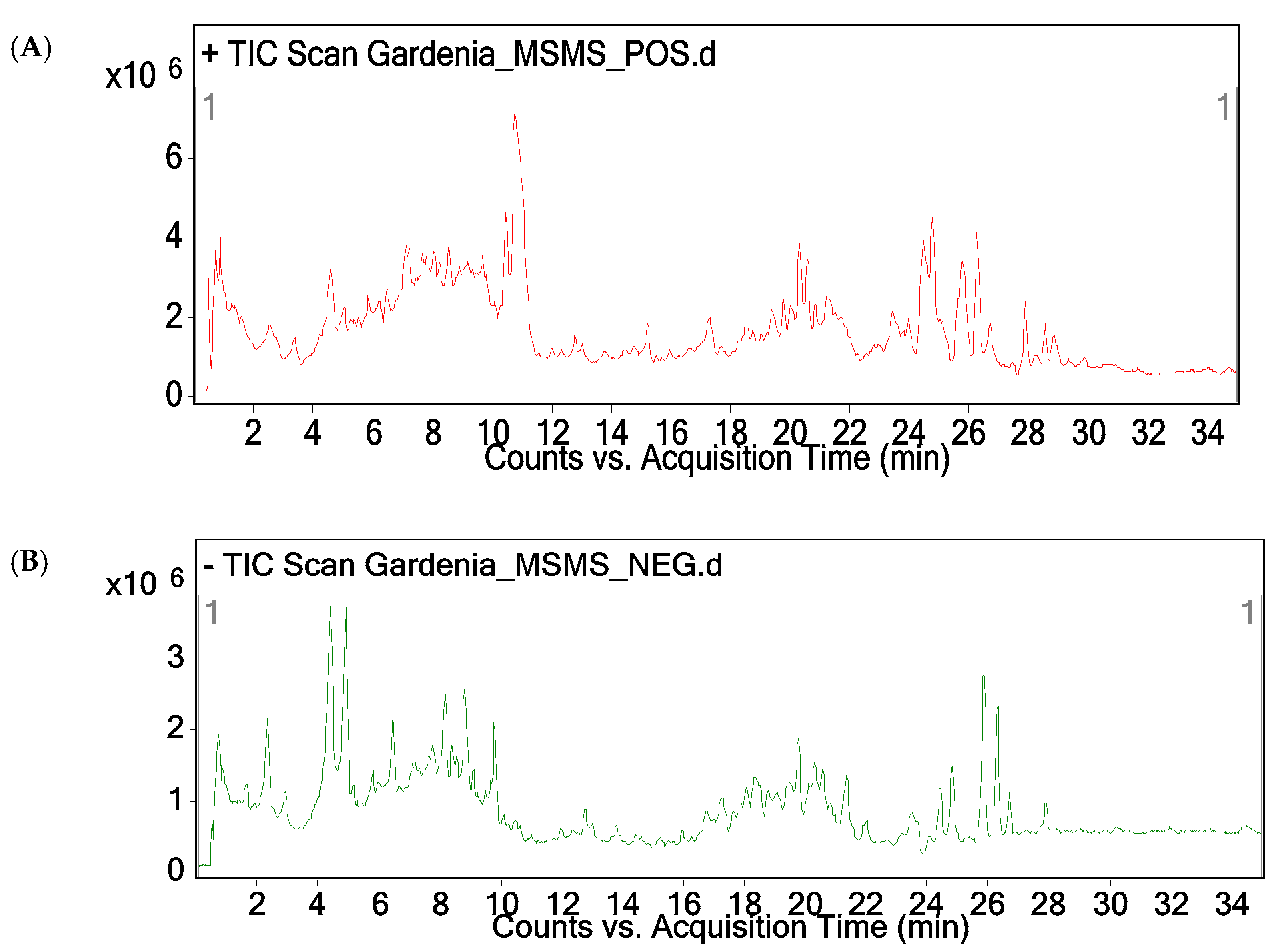
The LC-MS/MS analysis of the methanolic fruit extract of G. latifolia revealed the presence of 22 major compounds. Major peaks attributed in the chromatogram were subjected to both positive and negative modes using ESI-MS. Furthermore, the ion peaks along with MS fingerprint of compound was compared with other literature for identification of compounds. As per the LC-MS/MS analysis, 22 compounds consist of phenols, saponins, glycosides, alkaloids, flavonoids and terpenoids (Figure 2, Table 1). Some of the compounds such as 3,4-Di-O-caffeoylquinic acid, 6″-O-trans-feruloylgenipin gentiobioside, 10-(6-O-trans sinapoyl glucopyranosyl) gardendiol and 3-caffeoyl quinic acid (chlorogenic acid) showed hepato-protective and anti-viral activities and neuro-protective agent for Alzheimer’s disease, respectively [22,23,24].
Some other compounds like 3-caffeoylquinic acid (chlorogenic acid) and isoquercitrin were reported to be used in the treatment of osteoporosis and chemoprotective effects both in vitro and in vivo, against cancer, cardiovascular disorders, diabetes, oxidative stress and allergic reactions [25,26]. Scortechinones showed significant antibacterial activity against methicillin-resistant bacteria Staphylococcus aureus (MRSA) [27]. Secaubryenol is a pentacyclic triterpenoid and has been shown to exhibit a variety of anticancer activities against breast, prostate and cervical cancers [28]. Iridoids showed positive health effects on anti-inflammation, anti-depression, anti-diabetic properties and anti-thrombotic activities [29]. Quercetin-3-rutinoside (rutin), a natural flavonoid glycoside, showed antioxidant, hepatoprotective activities [30].

2.3. In vitro Antioxidant Activity of G. latifolia Methanolic Fruit Extract

2.3.1. DPPH Free Radical Scavenging Assay

In the present study, G. latifolia methanolic fruit extract showed significant inhibition of DPPH free radicals. Dose response curve of DPPH radical scavenging activity of G. latifolia fruit extract in relation to ascorbic acid (standard) has been depicted in Figure 3. The IC50 value of G. latifolia methanolic fruit extract was 65.82 µg/mL, whereas IC50 value of ascorbic acid was 43.03 µg/mL. DPPH (a stable, nitrogen centered free radical) assay is one of the most widely used methods for screening antioxidant activity of plant extracts [31]. The main principle involved in DPPH assay is reduction of purple color methanolic DPPH solution to yellow color diphenyl-picryl hydrazine in the presence of hydrogen donating antioxidants. Decreasing absorbance indicates more antioxidant activity of the extract in terms of hydrogen donating capacity. Antioxidant property of G. latifolia methanolic extract may be attributed to the presence of polyphenol components, which can readily donate either hydrogen atoms or electrons to capture the free radicals.
Phenolic compounds play vital role in scavenging the free radicals. Several studies substantiate the positive correlation of quality of phenolics with the DPPH free radical scavenging effect [32,33]. Although methanolic fruit extract of G. latifolia was significantly less effective than ascorbic acid but showed stronger scavenging activity than those of the fruit extracts of Gardenia volkensii [34] and Gardenia jasminoides [35]. From this study, it is imperative that the methanolic fruit extract of G. latifolia has showed significant antioxidant activity owing to the presence of phenolic compounds.

2.3.2. Differential Pulse Voltammetric Method

Antioxidant potential of methanolic extract and standard ascorbic acid is shown in the Figure 4. The antioxidant potential of vitamin C is 0.060 V, whereas the methanolic extract of G. latifolia is 0.152 V. In cyclic voltammetry analysis, lower the oxidation potential higher the antioxidant activity of the compound [36]. Here the methanolic fruit extract showed higher oxidation potential (0.152 V) than the ascorbic acid (0.060), which indicates that the methanolic fruit extract is having lower antioxidant activity compared to ascorbic acid. Same results have been observed with the DPPH radical assay, which indicates that the method is efficient, reproducible, rapid and consume less time, solvent and energy.

2.3.3. Antimicrobial Assay of G. latifolia Methanolic Fruit Extract

MIC values of methanolic extract against Gram positive and negative bacteria have been depicted in Figure 5. The MIC values of Escherichia coli, Klebsiella pneumoniae, Pseudomonas aeruginosa, Bacillus subtilis and Staphylococcus aureus were 62.5 µg/µL, 62.5 µg/µL, 31.25 µg/µL, 31.25 µg/µL and 15.62 µg/µL, respectively. Resazurin is a non-toxic, non-fluorescent dye, which appears pink and fluorescent upon reduction to resorufin by oxidoreductases in the viable cells. This compound has been used as an indicator for the evaluation of cell growth, particularly in various cytotoxicity assays [37].
Methanolic extract showed significant inhibitory activity against methicillin resistant bacteria such as Staphylococcus aureus, which may be due to the presence of scortechinones. Similar findings were observed in Garcinia scortechinii by Sukpondma et al. [27]. Microtiter plates with pink color after 24 h of incubation in modified resazurin assay indicate the growth, while the microtiter plates with blue signifies no growth. This assay is helpful to screen the natural product bioassay both in vitro and in vivo and also cost-effective, rapid and easy to perform. Ansari et al. [6] have studied antioxidant and antidiabetic properties using the G. latifolia bark, but devoid of fruit extraction studies. Similarly, Sundar and Habibur [5] reported phytochemical compounds and antioxidants from bark, leaf and fruit. However, elucidation of phytochemical compounds of G. latifolia with LC-MS/MS has not reported. Hence, in the current study, phytochemical compounds pertinent to methanolic fruit extract of G. latifolia have been determined with LC-MS/MS, demonstrated antioxidant and antimicrobial activities with differential pulse voltammetry and resazurin tests. This study shows that the fruit extracts of G. latifolia can be used as medicinal extract and also illustrates the application of differential pulse voltammetry and resazurin tests for antioxidant and antimicrobial activities from fruit extracts.

3. Materials and Methods

3.1. Collection of Plant Material

Mature fruits of Gardenia latifolia Ait. were collected during the month of November, 2019 from Bhakarapeta (Latitude: 13°38′48.751″N and Longitude: 79°12′35.574″E) of Seshachalam Biosphere Reserve, Tirupati, A.P., India and authentication was done by Taxonomist, Department of Botany, Sri Venkateswara University, Tirupati. The shade dried and matured fruits were rinsed with running tap water followed by sterile distilled water to remove the dirt on the surface and cut into small pieces. They were then shade dried for 7 days at room temperature.

3.2. Preparation of Extracts

The shade dried fruits of G. latifolia were made into fine powder using electronic blender and kept in desiccator till further use. An amount of 20 g of fruit powder was extracted using 100 mL methanol at room temperature on magnetic stirrer for 72 h. Extract was filtered through Whatman No.1 filter paper, filtrate was collected and the solvent was evaporated by keeping on water bath at 60 °C. The concentrated extract was stored at 4 °C until further analysis.

3.3. Characterization of G. latifolia Methanolic Fruit Extract

3.3.1. FTIR Analysis

FTIR spectrum of methanolic fruit extract was carried out on Alpha FT-IR spectrophotometer (Bruker, Germany) at a frequency of 4000–400 cm−1. About 1 mg of extract was used to record the spectrum.

3.3.2. LC-MS/MS Analysis

LC-MS/MS analysis pertinent to methanolic fruit extract of G. latifolia was done using Agilent LC instrument (1200 infinity series, Santa Clara, California, USA). LC instrument is attached with time-of flight (TOF) mass spectrometer quadrupole (G6540B, Agilent Technologies) coupled with photodiode array detector (PDA) through electron spray ionization (ESI) interface using C18 column (4.6 mm × 250 mm, 5 µm). Mobile phase comprises of water: formic acid (100:0.1) (A) and acetonitrile (B). The gradient (linear) was set at 7–20 % B in 0–10 min, 20–30 % B in 10–13 min, 30 % B in 13–17 min, 30–7 % B in 17–18 min and 7 % in 18–25 min with a flow rate of 1 mL/min using 20 µL of sample injection volume. The column oven temperature was kept at 25 °C. Data acquisition and processing was performed using Mass Hunter Workstation software and the mass analyses were done by both positive and negative ion modes, respectively. Mass spectrometer has been programmed for three consecutive scans, where initial scan was done for full mass with a m/z 100–1000, thereafter finding most abundant ion in full mass (MS2) followed by most abundant ion in the full mass (MS3).

3.4. In vitro Antioxidant Activity of G. latifolia Methanolic Fruit Extract

3.4.1. DPPH Free Radical Scavenging Assay

The DPPH free radical scavenging assay of methanolic extract was determined according to Chang et al. [38] with minor modifications. Stock solutions of methanolic fruit extract of G. latifolia, ascorbic acid and BHT were diluted at concentrations ranging from 20–100 μg/mL with an increment of 20 μg/mL. A volume of about 0.3 mL of standard/ sample was mixed with 2.7 mL of DPPH (0.1 mM) solution and the reaction mixture was incubated at room temperature in dark conditions for 30 min. Thereafter, the absorbance of mixture samples was measured against blank at 517 nm using double beam UV-Visible spectrophotometer (ELICO, Hyderabad, India). An amount of 3 mL of 0.1 mM DPPH solution was treated as control. Percentage (%) of free radical scavenging activity of methanolic fruit extract was calculated as per the formula given below.
: % inhibition = [(absorbance of control − absorbance of test sample)/absorbance of control] × 100
Where, Abs sample refers to the absorbance of plant extract + DPPH radical, Abs control denotes absorbance of Methanol +DPPH radical and RSA implies Radical Scavenging Activity.
The IC50 value, which is defined as the amount of sample concentration required to scavenge 50% DPPH free radicals was deduced from the plot of inhibition (%) against the sample extract concentration. All experiments were carried out in triplicate and the average of the results was reported. BHT and ascorbic acid were used as standards.

3.4.2. Differential Pulse Voltammetric Method

Antioxidant potential of methanolic fruit extract of G. latifolia was determined as per the Amidi et al. [39] method with minor modifications. Differential voltammetric measurements were performed in conventional three electrode system pertinent to a single compartment cell. The three electrode system comprises of saturated calomel reference electrode (SCE), glassy carbon working electrode (GCE) and platinum wire auxiliary electrode. Before measurement of each sample, the working electrode was polished with alumina using polishing cloth and the electrochemical measurements were recorded on Metrohm VA 746 polarograph. The methanolic extract and standard (ascorbic acid) were prepared with a concentration of 5 mg/mL. In DPV experiment, 50 µL of each methanolic and ascorbic acid solution (5 mg/mL) were added to 2950 µL of 0.2 M phosphate buffer having pH 7.0 at 70 mV pulse amplitude and 5 mV/s. The voltammograms were recorded between −0.2 V to 0.6 V.

3.5. Determination of Antimicrobial Potential of Methanolic Fruit Using Resazurin Microtiter Plate Assay

3.5.1. Bacterial Cultures and Culture Media

Bacterial strains such as Gram-positive Staphylococcus aureus (ATCC 25923), Bacillus subtilis (ATCC6633) and Gram-negative Escherichia coli (ATCC 25922), Pseudomonas aeruginosa (ATCC 27853), Klebsiella pneumoniae (ATCC 700603) were obtained from NCIM Resource Centre, Pune, India and were used in the present study. Bacterial cultures were cultivated on Mueller–Hinton broth (MHB) (HiMedia, Mumbai, India) and incubated as per the protocol supplied by NCIM resource centre, Pune, India. Bacterial cultures were suspended in 10 mL of physiological saline solution until the optical density readings reached to 0.5 McFarland standard. Further, to calculate minimum inhibitory concentration (MIC) of bacterial solutions 5 × 10⁵ colony-forming units (cfu) mL were used.

3.5.2. Preparation of Microtiter Plates

The MIC value of methanolic fruit extract was determined using sterile 96-well microtiter plate with resazurin as an indicator for cell growth [11]. A 96 well microtiter plate (Tarsons, Kolkata, India) was prepared by transferring 100 μL of Mueller–Hinton broth under laminar air flow chamber. A volume of 100 μL of methanolic extract (10 mg/mL) in 10% (v/v) DMSO was added into the first row of the plate and serial dilutions were performed. To each well, initially 10 μL of resazurin solution was added followed by 10 μL of bacterial inoculum (5 × 10⁶ cfu/mL). Thereafter, cling film was loosely wrapped to the microtiter plate to avoid bacterial culture dehydration and the plates were incubated at 37 °C for 24 h in an incubator. Colour change in the wells was observed visually, where change from purple to pink colour or colourless was scored as positive. MIC value was determined by taking the lowest concentration of the extract in which the colour change occurred. All the experiments were carried out in triplicates and the average values were measured for the MIC of the test material.

4. Conclusions

Phytochemical compounds of G. latifolia fruits showed several secondary metabolites such as saponins, alkaloids, glycosides, phenols, terpenoids and flavonoids having various putative functions. Antioxidant activity of methanolic fruit extract showed that it has huge potential to be used in food industry. Unlike conventional tests, this study has evaluated the antioxidant activity from the sample using differential pulse voltammetric method and showed reproducible results with DPPH assay. Besides, resazurin tests also showed highly reproducible results, which can be replaced with the conventional tests as they are rapid, reproducible, cost-effective and consume less time, energy and solvents. This study further emphasizes to use the G. latifolia fruit extract for the development of phyto-therapeutics owing to the presence of diversified biologically active compounds.

Supplementary Materials

The following are available online at https://www.mdpi.com/2223-7747/10/3/545/s1, Table S1. Preliminary phytochemical analysis of G.latifolia fruit extracts. Table S2. FTIR spectral peak values and functional groups of the methanolic fruit extract of G.latifolia.

Author Contributions

All authors conceive, design, experiment, write and revise the paper. All authors have read and agreed to the published version of the manuscript.

Institutional Review Board Statement

Not applicable.

Informed Consent Statement

Not applicable.

Acknowledgments

The authors acknowledge the Department of Biotechnology, Sri Venkateswara University, for their cordial support in conducting the experiment.

Conflicts of Interest

The authors declare no conflict of interest.

References

  1. Reddy, K.; Subbaraju, G.; Reddy, C.; Raju, V. Ethnoveterinary medicine for treating livestock in eastern ghats of Andhra Pradesh. Indian J. Tradit. Knowl. 2006, 5, 368–372. [Google Scholar]
  2. Rahman, M.A.; Uddin, S.B.; Wilcock, C.C. Medicinal plants used by chakma tribe in hill tracts districts of Bangladesh. Indian J. Tradit. Knowl. 2007, 6, 508–517. [Google Scholar]
  3. Madhava Chetty, K.; Sivaji, K.; Tulasi Rao, K. Flowering plants of Chittoor District, Andhra Pradesh, India. Publ. Stud. Offset Print. Tirupati 2008, 34–35. [Google Scholar]
  4. Liu, S.J.; Zhang, X.T.; Wang, W.M.; Qin, M.J.; Zhang, L.H. Studies on chemical constituents of Gardenia jasminoides var. radicans. Chin. Tradit. Herb. Drugs 2015, 43, 238–241. [Google Scholar]
  5. Sundar, R.A.; Habibur, R.C. Pharmacognostic, phytochemical and antioxidant studies of Gardenia latifolia Aiton: An ethnomedicinal tree plant. Int. J. Pharmacogn. Phytochem. Res. 2018, 10, 216–228. [Google Scholar]
  6. Ansari, F.; Khare, S.; Dubey, B.K.; Joshi, A.; Jain, A.; Dhakad, S. Phytochemical analysis, antioxidant, antidiabetic and anti-inflammatory activity of Bark of Gardenia latifolia. J. Drug Deliv. Ther. 2019, 9, 141–145. [Google Scholar] [CrossRef] [Green Version]
  7. Reddy, G.C.S.; Ayengar, K.N.N.; Rangaswami, S. Triterpenoids of Gardenia latifolia. Phytochemistry 1975, 307–308. [Google Scholar] [CrossRef]
  8. Galaqin, M.; Koji, U.; Minoru, Y.; Tatsuo, I. Simultaneous analysis of major ingredients of Gardenia fruit by HPLC-MS/TQMS method. Mong. J. Chem. 2016, 17, 34–37. [Google Scholar] [CrossRef] [Green Version]
  9. Saravanakumar, K.; SeonJu, P.; Anbazhagan, S.; Kil-Nam, K.; Su-Hyeon, C.; Arokia, V.A.M.; Myeong-Hyeon, W. Metabolite profiling of methanolic extract of Gardenia jaminoides by LC-MS/MS and GC-MS and its anti-diabetic, and anti-oxidant activities. Pharmaceuticals 2021, 14, 102. [Google Scholar] [CrossRef] [PubMed]
  10. Huang, W.Y.; Cai, Y.Z.; Zhang, Y. Natural phenolic compounds from medicinal herbs and dietary plants: Potential use for cancer prevention. Nutr. Cancer 2010, 62, 1–20. [Google Scholar] [CrossRef] [PubMed]
  11. Sarker, S.D.; Nahar, L.; Kumarasamy, Y. Microtitre plate-based antibacterial assay incorporating resazurin as an indicator of cell growth, and its application in the in vitro antibacterial screening of phytochemicals. Methods 2007, 42, 321–324. [Google Scholar] [CrossRef] [PubMed]
  12. Sahoo, S.; Ghosh, G.; Das, D.; Nayak, S. Phytochemical investigation and in vitro antioxidant activity of an indigenous medicinal plant Alpinia nigra B.L. Burtt. Asian Pac. J. Trop. Biomed. 2013, 3, 871–876. [Google Scholar] [CrossRef] [Green Version]
  13. Drummond, A.J.; Waigh, R.D. The development of microbiological methods for phytochemical screening. Recent Res. Develop. Phytochem. 2000, 4, 143–152. [Google Scholar]
  14. Bhat, R.; Ameran, S.B.; Voon, H.C.; Karim, A.A.; Tze, L.M. Quality attributes of starfruit (Averrhoa carambola L.) juice treated with ultraviolet radiation. Food Chem. 2011, 127, 641–644. [Google Scholar] [CrossRef] [PubMed]
  15. Kumar, A.; Ramesh, K.V.; Chandusingh; Sripathy, K.V.; Agarwal, D.K.; Pal, G.; Kuchlan, M.K.; Singh, R.K.; Ratnaprabha; Kumar, S.P.J. Bio-prospecting nutraceuticals from selected soybean skins and cotyledons. Indian J. Agric. Sci. 2019, 89, 2064–2068. [Google Scholar]
  16. Kumar, S.P.J.; Kumar, A.; Ramesh, K.V.; Singh, C.; Agarwal, D.K.; Pal, G.; Kuchlan, M.K.; Singh, R. Wall bound phenolics and total antioxidants in stored seeds of soybean (Glycine max) genotypes. Indian J. Agric. Sci. 2020, 90, 118–222. [Google Scholar]
  17. Alothman, M.; Bhat, R.; Karim, A.A. Effects of radiation processing on phytochemicals and antioxidants in plant produce. Trends Food Sci. Technol. 2009, 20, 201–212. [Google Scholar] [CrossRef]
  18. Khanam, Z.; Wen, C.S.; Bhat, I.U.H. Phytochemical Screening and Antimicrobial Activity of Root and Stem Extracts of Wild Eurycoma longifolia Jack (Tongkat Ali). J. King Saud Univ. Sci. 2015, 27, 23–30. [Google Scholar] [CrossRef] [Green Version]
  19. Aiyelaagbe, O.O.; Osamudiamen, P.M. Phytochemical screening for active compounds in Mangifera indica leaves from Ibadan. Plant Sci. Res. 2009, 2, 11–13. [Google Scholar]
  20. U.S. Department of Agriculture, Agricultural Research Service. 1992–2016. Dr. Duke’s Phytochemical and Ethnobotanical Databases. Home Page. Available online: http://phytochem.nal.usda.gov/ (accessed on 15 February 2021).
  21. Kumar, S.P.J.; Banerjee, R. Enhanced lipid extraction from oleaginous yeast biomass using ultrasound assisted extraction: A greener and scalable process. Ultrason. Sonochem. 2018, 52, 25–32. [Google Scholar] [CrossRef]
  22. Kim, K.H.; Kim, Y.H.; Lee, K.R. Isolation of quinic acid derivatives and flavonoids from the aerial parts of Lactuca indica L. and their hepatoprotective activity in vitro. Bioorg. Med. Chem. Lett. 2007, 17, 6739–6743. [Google Scholar] [CrossRef] [PubMed]
  23. Yu, Y.; Xie, Z.L.; Gao, H.; Ma, W.W.; Dai, Y.; Wang, Y.; Zhong, Y.; Yao, X.S. Bioactive iridoid glucosides from the fruit of Gardenia jasminoides. J. Nat. Prod. 2009, 72, 1459–1464. [Google Scholar] [CrossRef] [PubMed]
  24. Li, H.-B.; Yu, Y.; Wang, Z.-Z.; Dai, Y.; Gao, H.; Xiao, W.; Yao, X.-S. Iridoid and Bis-Iridoid glucosides from the fruit of Gardenia jasminoides. Fitoterapia 2013, 88, 7–11. [Google Scholar] [CrossRef] [PubMed]
  25. Kwak, S.C.; Lee, C.; Kim, J.-Y.; Oh, H.M.; So, H.-S.; Lee, M.S.; Rho, M.C.; Oh, J. Chlorogenic acid inhibits osteoclast differentiation and bone resorption by down-regulation of receptor activator of nuclear factor kappa-B ligand-induced nuclear factor of activated T cells C1 expression. Biol. Pharm. Bull. 2013, 36, 1779–1786. [Google Scholar] [CrossRef] [PubMed] [Green Version]
  26. Valentová, K.; Vrba, J.; Bancířová, M.; Ulrichová, J.; Křen, V. Isoquercitrin: Pharmacology, toxicology, and metabolism. Food Chem. Toxicol. 2014, 68, 267–282. [Google Scholar] [CrossRef]
  27. Sukpondma, Y.; Rukachaisirikul, V.; Phongpaichit, S. Xanthone and sesquiterpene derivatives from the fruits of Garcinia scortechinii. J. Nat. Prod. 2005, 68, 1010–1017. [Google Scholar] [CrossRef]
  28. Grougnet, R.; Magiatis, P.; Mitaku, S.; Loizou, S.; Moutsatsou, P.; Terzis, A.; Cabalion, P.; Tillequin, F.; Michel, S. Seco-cycloartane triterpenes from Gardenia aubryi. J. Nat. Prod. 2006, 69, 1711–1714. [Google Scholar] [CrossRef]
  29. Xiao, W.; Li, S.; Wang, S.; Ho, C.T. Chemistry and bioactivity of Gardenia jasminoides. J. Food Drug Anal. 2017, 25, 43–61. [Google Scholar] [CrossRef] [Green Version]
  30. Kumar, S.; Pandey, A.K. Chemistry and biological activities of flavonoids: An overview. Sci. World J. 2013, 162750. [Google Scholar] [CrossRef] [PubMed] [Green Version]
  31. Kumar, N.S.S.; Kumar, I.S.; Kumar, S.P.J.; Sarbon, N.M.H.D.; Chintagunta, A.D.; Anvesh, B.S.; Dirisala, V.R. Extraction of bioactive compounds from Psidium guajava leaves and its utilization in preparation of jellies. AMB Express. 2021, 11, 36. [Google Scholar] [CrossRef] [PubMed]
  32. Liu, S.C.; Lin, J.T.; Wang, C.K.; Chen, H.Y.; Yang, D.J. Antioxidant properties of various solvent extracts from lychee (Litchi chinenesis Sonn.) flowers. Food Chem. 2009, 114, 577–581. [Google Scholar] [CrossRef]
  33. Kedare, S.B.; Singh, R.P. Genesis and development of DPPH method of antioxidant assay. J. Food Sci. Technol. 2011, 48, 412–422. [Google Scholar] [CrossRef] [Green Version]
  34. Juma, B.F.; Majinda, R.R.T. Constituents of Gardenia volkensii: Their brine shrimp lethality and dpph radical scavenging properties. Nat. Prod. Res. 2007, 21, 121–125. [Google Scholar] [CrossRef] [PubMed]
  35. Debnath, T.; Park, P.J.; Deb Nath, N.C.; Samad, N.B.; Park, H.W.; Lim, B.O. Antioxidant activity of Gardenia jasminoides Ellis fruit extracts. Food Chem. 2011, 128, 697–703. [Google Scholar] [CrossRef]
  36. Barros, L.; Cabrita, L.; Boas, M.V.; Carvalho, A.M.; Ferreira, I.C.F.R. Chemical, biochemical and electrochemical assays to evaluate phytochemicals and antioxidant activity of wild plants. Food Chem. 2011, 127, 1600–1608. [Google Scholar] [CrossRef]
  37. McNicholl, B.P.; McGrath, J.W.; Quinn, J.P. Development and application of a resazurin-based biomass activity test for activated sludge plant management. Water Res. 2007, 41, 127–133. [Google Scholar] [CrossRef]
  38. Chang, S.T.; Wu, J.H.; Wang, S.Y.; Kang, P.L.; Yang, N.S.; Shyur, L.F. Antioxidant activity of extracts from Acacia confusa bark and heartwood. J. Agric. Food Chem. 2001, 49, 3420–3424. [Google Scholar] [CrossRef]
  39. Amidi, S.; Mojab, F.; Bayandori Moghaddam, A.; Tabib, K.; Kobarfard, F. A simple electrochemical method for the rapid estimation of antioxidant potentials of some selected medicinal plants. Iran. J. Pharm. Res. 2012, 11, 117–121. [Google Scholar] [PubMed]
Figure 1. FTIR spectrum of methanolic fruit extract of G. latifolia.
Plants 10 00545 g001
Figure 2. LC-MS/MS spectrum of methanolic fruit extract of G. latifolia (A) Chromatogram of fruit extract of G. latifolia on positive ionization mode. (B) Chromatogram of fruit extract of G. latifolia on negative ionization mode.
Plants 10 00545 g002
Figure 3. DPPH free radical scavenging assay of methanolic fruit extract of G. latifolia.
Plants 10 00545 g003
Figure 4. Differential pulse voltammograms of vitamin C (Vit-C) and methanolic fruit (MF).
Plants 10 00545 g004
Figure 5. Minimum inhibitory concentrations of methanolic fruit extract of G. latifolia against Gram negative and positive bacteria.
Plants 10 00545 g005
Table 1. Compounds identified in methanolic fruit extract of G. latifolia by LC-MS/MS.
S. No.Molecular
Formula
m/zRTMassName of The CompoundMode
+/−
1.C11 H12 O5225.0752 3.294 224.0679 Sinapinic acid +
2.C16 H12 O6301.0709 11.969 300.0636 4′Hydroxywogonin +
3.C16 H18 O9355.1029 2.281 354.0956 3-caffeoylquinic acid (Chlorogenic acid) +
4.C18 H22 O10397.1154 5.302 398.1228 3-O-sinapoylquinic acid
5.C19 H22 O5331.1553 7.731 330.1479 5-Deoxystrigol +
6.C20 H20 O8389.1234 16.589 388.1161 5 Hydroxy6,7,3′, 4′,5′pentamethoxyflavone+
7.C20 H20 O9403.1044 15.14 404.1118 5,3′Dihydroxy3,6,7,4′, 5′pentamethoxy flavone
8.C20 H32 O2305.2476 22.752 304.2403 2-Ketoepimanool +
9.C21 H20 O12465.10317.685464.0957Isoquercitrin
10.C25 H24 O12517.1344 8.177 516.1269 3,4-Di-O-caffeoylquinic acid +
11.C26 H30 O13549.1627 8.502 550.1699 2′-O-trans-feruloylgardoside (Iridoid glycosides)
12.C26 H30 O14565.159 7.534 566.1661 6′-O-[(E)-caffeoyl]-deacetylasperulosidic acid methyl ester (Iridoid glycosides)
13.C27 H28 O13561.1609 9.684 560.1535 4-O-sinapoyl-5-O-caffeoylquinic acid +
14.C27 H30 O16609.1496 7.451 610.1569 Quercetin-3-rutinoside (Rutin)+
15.C27 H34 O20677.1545 10.708 678.1617 10-(6-O-trans-sinapoylglucopyranosyl) gardendiol
16.C30 H48 O3455.354 23.505 456.3613 Betulinic acid
17.C33 H42 O18725.2302 9.186 726.2369 6″-O-trans-feruloylgenipin gentiobioside (Iridoid glycosides)
18.C34 H40 O10609.271 24.561 608.2637 Scortechinone C+
19.C34 H40 O9593.2769 25.688 592.2695 Scortechinone B+
20.C35 H42 O9607.2923 27.925 606.285 Scortechinone G +
21.C35 H44 O10625.3027 25.124 624.2953 Scortechinone I +
22.C6 H8 O6177.0415.731176.0337Ascorbic acid+
Publisher’s Note: MDPI stays neutral with regard to jurisdictional claims in published maps and institutional affiliations.