Article

Biochemical and Proteomic Changes in the Roots of M4 Grapevine Rootstock in Response to Nitrate Availability

Department of Agricultural and Environmental Sciences—Production, Landscape, Agroenergy (DiSAA), Università degli Studi di Milano, I-20133 Milano, Italy
*
Author to whom correspondence should be addressed.
Plants 2021, 10(4), 792; https://doi.org/10.3390/plants10040792
Submission received: 13 March 2021 / Revised: 12 April 2021 / Accepted: 15 April 2021 / Published: 17 April 2021
(This article belongs to the Special Issue Omic Sciences in the Fields of Crop Quality and Sustainability)

Abstract

:
In agricultural soils, nitrate (NO3) is the major nitrogen (N) nutrient for plants, but few studies have analyzed molecular and biochemical responses involved in its acquisition by grapevine roots. In viticulture, considering grafting, NO3 acquisition is strictly dependent on rootstock. To improve the knowledge about N nutrition in grapevine, this study analyzed biochemical and proteomic changes induced by, NO3 availability, in a hydroponic system, in the roots of M4, a recently selected grapevine rootstock. The evaluation of biochemical parameters, such as NO3, sugar and amino acid contents in roots, and the abundance of nitrate reductase, allowed us to define the time course of the metabolic adaptations to NO3 supply. On the basis of these results, the proteomic analysis was conducted by comparing the root profiles in N-starved plants and after 30 h of NO3 resupply. The analysis quantified 461 proteins, 26% of which differed in abundance between conditions. Overall, this approach highlighted, together with an increased N assimilatory metabolism, a concomitant rise in the oxidative pentose phosphate pathway and glycolysis, needed to fulfill the redox power and carbon skeleton demands, respectively. Moreover, a wide modulation of protein and amino acid metabolisms and changes of proteins involved in root development were observed. Finally, some results open new questions about the importance of redox-related post-translational modifications and of NO3 availability in modulating the dialog between root and rhizosphere.

1. Introduction

Among mineral nutrients, nitrogen (N) is the one required in the greatest amounts by plants, being an integral constituent of many organic compounds, such as nucleic acids, proteins, co-enzymes, chlorophyll, phytohormones and secondary metabolites [1]. In grapevine, N, after H, C and O, is usually the fourth most abundant element, representing 2–5% of plant biomass, and, in many cases, it is the most limiting factor for growth (i.e., vigor) and harvest yield [2]. Many studies conducted on grapevine highlighted the deep impact of N supply on plant reproductive capability as well as on grape berry metabolism and, finally, on berry composition [2,3,4,5,6]. In this view, the tight relationship between the rootstock/scion combination and N availability is emerging [7,8,9]. The N status of grapevine, like other perennial crops, depends on many factors, such as the acquisition of N from the soil, its distribution among annual organs and its reallocation from annual growth parts into both fruits and woody tissues [10,11,12,13,14]. In this context, the N content in grape berries, lost from the vineyard each year, has been estimated to be 2–3 kg of N per ton of fruit matter [2]. Nevertheless, a good management of N in vineyard must consider multifaceted aspects, in light of the fact that an excessive N supply can determine detrimental effects on the quality of grape berries and, therefore, of wine [4,5,8]. The achievement of this goal also comes via the improvement of the knowledge of the physiological, biochemical and molecular processes involved in the N metabolism of grapevine.
Although Vitis species could use different N forms, such as NO3, NH4+, urea and amino acids, NO3, usually the predominant N form in vineyard soils, is the primary source of N for grapevine [2,15,16]. Consistent with the fluctuating availability of this anion in soil solution (from 100 µM to 10 mM and more), plants have evolved different root uptake systems with different affinity (high-affinity transport systems, HATSs, and low-affinity transport systems, LATSs) and with both constitutive and inducible components [17,18,19]. The presence of HATSs rapidly responding to NO3 availability has been recently demonstrated in grapevine [20,21]. According to the proton gradient requirement for sustaining the ≈2H+: 1NO3 symporters, a concomitant induction of plasma membrane H+-ATPase in grapevine roots exposed to NO3 has also been described [20]. Interestingly, the capability to induce NO3 transporters in response to NO3 availability is strictly dependent on the scion/rootstock combination [21].
In grapevine, N assimilation can occur in both roots and leaves [2,22]. The process is performed by the well-characterized pathway that involves the reduction of NO3 to NH4+ through two steps catalyzed by nitrate reductase (NR) and nitrite reductase (NiR) and the subsequent assimilation catalyzed by glutamine synthetase (GS) and glutamate synthase (GOGAT), finally producing glutamic acid [1,22]. The contribution of the two organs depends on several factors, like N availability, C metabolism (i.e., availability of C skeletons), environmental conditions, tissue age and others [2,22,23,24,25]. Moreover, NO3 assimilation shows several connections with other biochemical pathways and responses, among which are amino acid metabolism, redox status and pH homeostasis [2,26,27]. Recently, Cochetel and co-workers published the first paper describing transcriptomic changes occurring in the roots of Riparia Gloire de Montpellier and 1103 Paulsen rootstocks in relation to NO3 availability [28]. Comparing these two genotypes, this study highlighted both specific responses and common changes in the gene expression patterns. Nitrogen-responsive genes involved in the uptake and assimilation of NO3 as well as genes encoding enzymes of the oxidative pentose phosphate (OPP) pathway were identified as being influenced by NO3 availability. According to the literature, the increase in OPP pathway activity is related to the typical metabolic adaptation of root tissues needed to satisfy the demand for reducing power [29,30]. In this context, the entity of the translocation of sugars from the photosynthetic tissues into roots affects the N assimilatory contribution of the two organs [2].
To our knowledge, to date, no proteomic investigation has studied the N responses in grapevine roots. Therefore, improvement of the information at translational and post-translational levels is desirable to better characterize the molecular and biochemical events evoked by changes in N availability in this perennial species. In the present work, we adopted biochemical and proteomic approaches to analyze, in the root of M4 grapevine rootstock, the responses occurring after the addition of NO3 to the nutrient solution in hydroponics. This rootstock genotype was selected on the basis of its good response to abiotic stress [31,32,33,34,35], which is contributing to the expansion of its use in viticulture. The biochemical evaluations were devoted to verifying the role played by roots in the response to NO3 resupply, monitoring key parameters. Moreover, since a holistic approach represents a good strategy to study the biological complexity [18,26,29], proteomic analysis was used to obtain a more comprehensive description of the first metabolic events involved in NO3 acquisition in roots of Vitis.

2. Results and Discussion

The activation of NO3 metabolism, which involves the modulation of specific transport systems and an assimilation pathway, is tightly linked to the external availability of the nutrient [20,21,29]. The rate at which plants induce these activities deeply affects their efficiency in using this mineral element in field conditions [36]. Moreover, studies performed on grapevine highlighted that, during the induction phase, the root organ plays an important role not only in N uptake from the soil, but also in N assimilation [2]. Starting from these considerations, the present work aimed at investigating in the roots of M4 grapevine rootstock the responses to the addition of 10 mM NO3 in the hydroponic nutrient solution after a period of N starvation (see Materials and Methods for further details). First, key biochemical parameters were measured to define the timing of induction of the metabolic processes involved in the acquisition of NO3. Afterwards, the changes in the root proteome were investigated.

2.1. Changes in Biochemical Parameters in Response to Nitrate

Biochemical analyses of key parameters in the roots of M4 were performed in order to define plant adaptations to renewed NO3 availability (Figure 1).
After the exposure of plants to NO3, the contents of the anion in roots increased progressively within the first 30 h (Figure 1A), suggesting that NO3 uptake and accumulation were activated early and maintained throughout the treatment period (30 h). According to the role of NO3 as a signal for the induction of its assimilation [17], a parallel increase in NR abundance, evaluated by immunoblot analysis, occurred (Figure 1B). However, an increase in the content of amino acids was detected only after 30 h of treatment (Figure 1C), suggesting that a full activation of the NO3 assimilatory pathway was reached at this time point. In support of this conclusion, a decrease in reducing sugar and sucrose contents (Figure 1D,E) was measured only after 30 h of treatment, probably resulting from an increase in the use of C skeletons to sustain N assimilation.
This evaluation describes the typical metabolic changes involved in the acquisition and assimilation of NO3 well [18,36]. Moreover, these results allowed us to define, in M4 grapevine rootstock, the timing of these responses, which, interestingly, is highly consistent with those described for other grapevine genotypes [20,21,28]. Starting with this information, we performed the proteomic analysis, comparing the roots of N-starved plants (control plants, 0 h) with those of plants grown in the presence of 10 mM NO3 for 30 h.

2.2. Proteomic Analysis of M4 Root System

The proteomic analysis was performed by means of one-dimensional gel–liquid chromatography–mass spectrometry (GeLC–MS/MS), considering its suitability for the analysis of total proteomes in plant tissues [33,37]. Through this approach, it was possible to study the abundance of 461 proteins, with high reliability in identification and a good degree of comparability among samples and conditions (Table S1).

2.2.1. Functional Distribution of the Identified Proteins

The functional classification of the identified proteins, reported in detail in Table S2, was conducted according to the MapMan4 ontology [38]. The identified proteins fell into 15 functional categories (Figure 2). Many of them belonged to four categories, “Protein” (24%), “Not assigned-annotated” (18%), “Carbon and energy metabolism” (13%) and “Enzymes/Coenzyme metabolism” (8%). Regarding the proteins that fell into the “Not assigned-annotated” category, in order to better define their functional role, a supplemental investigation was performed, using the UniProt database (https://www.uniprot.org/, accessed on 1 December 2020) and the basic local alignment search tool (BLAST). However, no information, or very fragmentary information, was available for proteins classified in the “Not assigned-not annotated” group, which corresponded to 5% of total identified proteins.
Statistical analysis highlighted 119 proteins that significantly changed in abundance after the exposure of the plants to 10 mM NO3 for 30 h. Figure 3 shows their functional distribution. Among these proteins, 68 increased or appeared while 51 decreased or disappeared. Almost a half (46%) of the proteins that increased in abundance belong to the “Protein” category. Interestingly, some other functional categories included proteins that were accumulated in a higher abundance in response to NO3 supply, such as “Carbon and energy metabolism”, “Not assigned-annotated”, “Enzyme/Coenzyme metabolism” and “Amino acid metabolism” (13%, 10%, 9% and 6%, respectively). However, many of the proteins that decreased or disappeared were classified into the categories “Not assigned-annotated”, “Protein”, “Enzyme/Coenzyme metabolism” and “Carbon and energy metabolism” (31%, 16%, 10% and 8%, respectively).
Overall, the functional distribution of the NO3-responsive proteins is consistent with the typical adaptations described in plants, and specifically in root tissues, in response to the availability of this nutrient [26,39,40]. Among them, modifications are evident in pathways related to the production of energy and C skeletons as well as broad changes in protein metabolism. In addition, several other metabolic traits were affected by the nutritional treatment. In details, Table 1 includes all the proteins that changed in abundance in response to NO3.

2.2.2. Proteomic Changes Involved in Nitrogen Acquisition and in Carbon and Energy Metabolism

The proteomic analysis revealed the appearance of ferredoxin-nitrite reductase (#246) in the roots of plants exposed to NO3. According to the increases in NR abundance and in amino acid levels (Figure 1B,D), this result confirms the activation of the primary N assimilation pathway. Moreover, the appearance of ferredoxin-NADP reductase (#136) and glucose-6-phosphate 1-dehydrogenase (#323), as well as the dramatic increase in 6-phosphogluconate dehydrogenase (#64), is in agreement with a higher metabolic demand for NADPH, that in non-photosynthetic tissues is fulfilled by the OPP pathway [26,29]. However, cytosolic glutamine synthetase (#29) decreased in abundance in response to NO3, suggesting a reduction in N recycling. This behavior probably mirrors the effect of NO3 availability on the plant N status, and the consequent change in protein and amino acid catabolism [41].
At the same time, several glycolytic enzymes, such as fructose-bisphosphate aldolase (#28), phosphoglycerate kinase (#23), phosphoglycerate mutase (PGM, #154) and enolase (#81), increased in abundance in response to NO3, according to a higher demand for C skeletons needed for N assimilation [42]. Among them, the peculiar role played by PGM that, even if it catalyzes a non-rate-limiting step in glycolysis, is involved in the recycling of glyceraldehyde-3P produced by the OPP is highlighted [26,29].
Interestingly, the concomitant decrease in the E1 component of pyruvate dehydrogenase (#122) suggests that an activation of anaplerotic reaction(s) occurred, rather than an upsurge of carbon oxidation through the Krebs cycle. In this view, it could be observed that the levels of the β subunit of ATP synthase (#2), mitochondrial aldehyde dehydrogenase (#11), NADH-cytochrome b5 reductase (#119) and NAD(P)H dehydrogenase (#110) also decreased after the addition of NO3. Once again, this behavior could be ascribed to a shift in the balance between N recycling and assimilation.
In support of the above results, the activation of C metabolism in roots was accompanied by a decrease in the content of reducing sugars and sucrose (Figure 1D,E). In this view, it is interesting to observe that only in roots of plants exposed to NO3 was a sucrose synthase (#14) detectable, an enzyme that plays a pivotal role in determining the sink strength necessary to import photoassimilates from the source organs [43]. This result suggests that the sink strength of the root system increases during the early phases of exposure to NO3. Hence, it is possible that this response could have a role in the scion/rootstock relations and in the N use efficiency of different grafting combinations.

2.2.3. Proteomic Changes Involved in Protein and Amino Acid Metabolism

The present study found that many proteins involved in protein synthesis, modification and degradation changed in abundance in response to NO3 availability (Table 1). These include ribosomal proteins (#300, #126, #217, #443, #375) as well as initiation (#37) and elongation factors (#407, #149, #128). These changes agree with recent literature that highlights a resumption of these metabolic activities among the responses of N-starved plants to renewed availability of NO3, in Arabidopsis (Arabidopsis thaliana L.) and maize (Zea mays L.) [26,39,40]. According to an activation of protein synthesis, a few heat shock proteins (#452, #1, #370, #317, #107), known to be involved in protein folding [44], were also more abundant in NO3-treated plants. At the same time, the proteomic analysis revealed a different accumulation of proteins operating in the proteasome (#147, #420, #388) and in ubiquitination (#145, #379), suggesting an increase in protein turnover.
Conversely, several proteases, such as two carboxypeptidases (#93, #195), a putative cysteine protease RD21B (#151) and an aspartic proteinase A1 (#26), decreased in abundance after the reintroduction of NO3. Considering that some of these proteins have extracellular localization, these results support the hypothesis that during N starvation, roots are able to intensify the exudation of proteases, probably in order to mobilize N from peptides and proteins in the rhizosphere [45]. Interestingly, the secretion of such enzymes has also been observed in the absence of microorganisms [45], and, therefore, it is possible that it occurs in plants grown in hydroponic systems.
The metabolism of a few amino acids seemed to be positively affected upon NO3 resupply. The proteomic analysis revealed an increase in the abundance of enzymes involved in the biosynthesis of methionine (#3, 5-methyltetrahydropteroyltriglutamate-homocysteine S-methyltransferase; #38, adenosylhomocysteinase), isoleucine (#73, acetohydroxy-acid reductoisomerase) and serine (#30, D-3-phosphoglycerate dehydrogenase). Moreover, an increase in the level of phospho-2-dehydro-3-deoxyheptonate aldolase (#140) was detected, being the first enzyme of the shikimate pathway, which is involved in the synthesis of aromatic amino acids. This result is in agreement with studies on other plant species that describe an activation of the synthesis of amino acids in order to sustain the reactivation of protein synthesis [26,40,46].

2.2.4. Other Biochemical Functions Affected by Nitrate Resupply

A few enzymes involved in cell wall lignification, such as a caffeoyl-CoA O-methyltransferase (CCOMT, #131) and an omega-hydroxy-palmitate O-feruloyl transferase (OHFT, #54), decreased in abundance in response to NO3 availability. It is interesting to observe that the late embryogenesis abundant protein Lea14-A (#215), described as able to positively regulate the deposition of lignin under stress conditions [47], followed a similar trend, as well as a glucan endo-1,3-beta-D-glucosidase (#105). These results are consistent with the well-documented reprogramming of the root development process that occurs in response to NO3 [39,48,49]. Moreover, the different behaviors of two peroxidases (POXs, #208, #345) are noteworthy, which showed an opposite response to NO3. Although a role of POX in the lignification process was proposed, the trends of CCOMT and OHFT strongly weaken this hypothesis and suggest a context in which this process seems reduced. However, considering the very multifaceted role of these enzymes, it is interesting to highlight that an increase in peroxidase and polyphenol oxidase (PPO) activities was described in grapevine during the adventitious rooting process [50]. Our proteomic analysis identified a PPO (#314) positively affected by NO3 exposure, highlighting a possible analogy.
Other proteins involved in phenolic metabolism changed in abundance in response to NO3. Among them, a decrease in chalcone isomerase (#106), an enzyme that catalyzes the isomerization of naringenin chalcone to naringenin flavanone in the flavonoid pathway, was observed. This result agrees with the well-known reduction in phenylpropanoid metabolism occurring after NO3 resupply to N-starved plants [26,51,52,53,54]. At the same time, we observed an increase in trans-resveratrol di-O-methyltransferase (#41), which suggests different effects on specific traits of the phenylpropanoid metabolism. This enzyme catalyzes the formation of pterostilbene from resveratrol, a compound with antimicrobial and antifungal properties [55,56]. The specific role of this molecule in roots remains to be clarified, however, it was proposed that its increase upon high N availability could participate in promoting root relations with the rhizosphere community. Although further studies are needed, attention could be focused on the interplay occurring between plants and other organisms in the soil. In this view, it is interesting to note that our proteomic analysis identified a proline iminopeptidase (#265) that decreased in abundance in response to NO3. Considering that this peptidase is induced by bacteria and seems to participate in virulence [57], our results suggest that NO3 availability could reduce root susceptibility to pathogens.
Moreover, our proteomic analysis revealed changes of a few typical stress-responsive proteins. Some of them, such as major allergen Pru ar 1 (#199), chitin-binding type-1 (#395) and PHB (#42, #91), decreased, whilst others, such as MLP-like protein 34 and 43 (#89, #165) and annexin D2 (#46), increased in response to NO3. Given the paucity of the current information for many of them [58], it is difficult to fully understand the biological meaning of these results. Recently, Wang and co-workers [59] provided new information about MLP-like protein 43, highlighting its participation in the drought responses mediated by ABA. In detail, these authors described the involvement of this protein in the response to oxidative stress as well as in the modulation of primary metabolism. Further analyses may help to clarify possible roles of this protein in the metabolic changes induced by NO3. Although, for annexin, an interesting role in the differentiation/growth processes at the membrane level is emerging [60], and in view of the morphological responses evoked by NO3 in root system, further work is necessary to define its specific role in grapevine rootstock.
The available literature describes changes in the expression of genes involved in redox metabolism upon NO3 provision in Arabidopsis [26,39]. Our study revealed that NO3 positively affected a glutaredoxin-dependent peroxiredoxin (#269) and cystathionine beta-synthase family protein a (#76), whilst another glutaredoxin-dependent peroxiredoxin (#185) and a thioredoxin h-type (#330) decreased. Considering their involvement in the regulation of the oxidative state of sulfhydryl groups and in the S-glutathionylation of proteins [61,62], this result underlines the crucial role of the redox system in the modulation of the metabolic adjustment evoked by increased NO3 availability. Reinforcing this hypothesis, we also found four glutathione transferases (#108, #337, #448, #232) that showed an upsurge in abundance in response to NO3 supply. Interestingly, future redox proteomics studies could be very useful to better clarify the role of redox-related post-translational modifications in response to N supply in plants.
Overall, our results highlight the important role of roots in the plant adaptations to changes in N availability. As previously described in other plant species, this study confirms that grapevine roots show a typical metabolic activation involved in the acquisition of this macronutrient. Moreover, this investigation brings evidence of interesting relationships between N availability and root–rhizosphere dialog. This new knowledge paves the way for further studies aimed at characterizing key factors involved in N use efficiency in this pluriannual species. Other interesting aspects that deserve attention in the future are the plant responses in field conditions, the adaptability to N availability in different graft combinations and the final outcomes in grape quality.

3. Materials and Methods

3.1. Plant Material and Nutritional Treatments

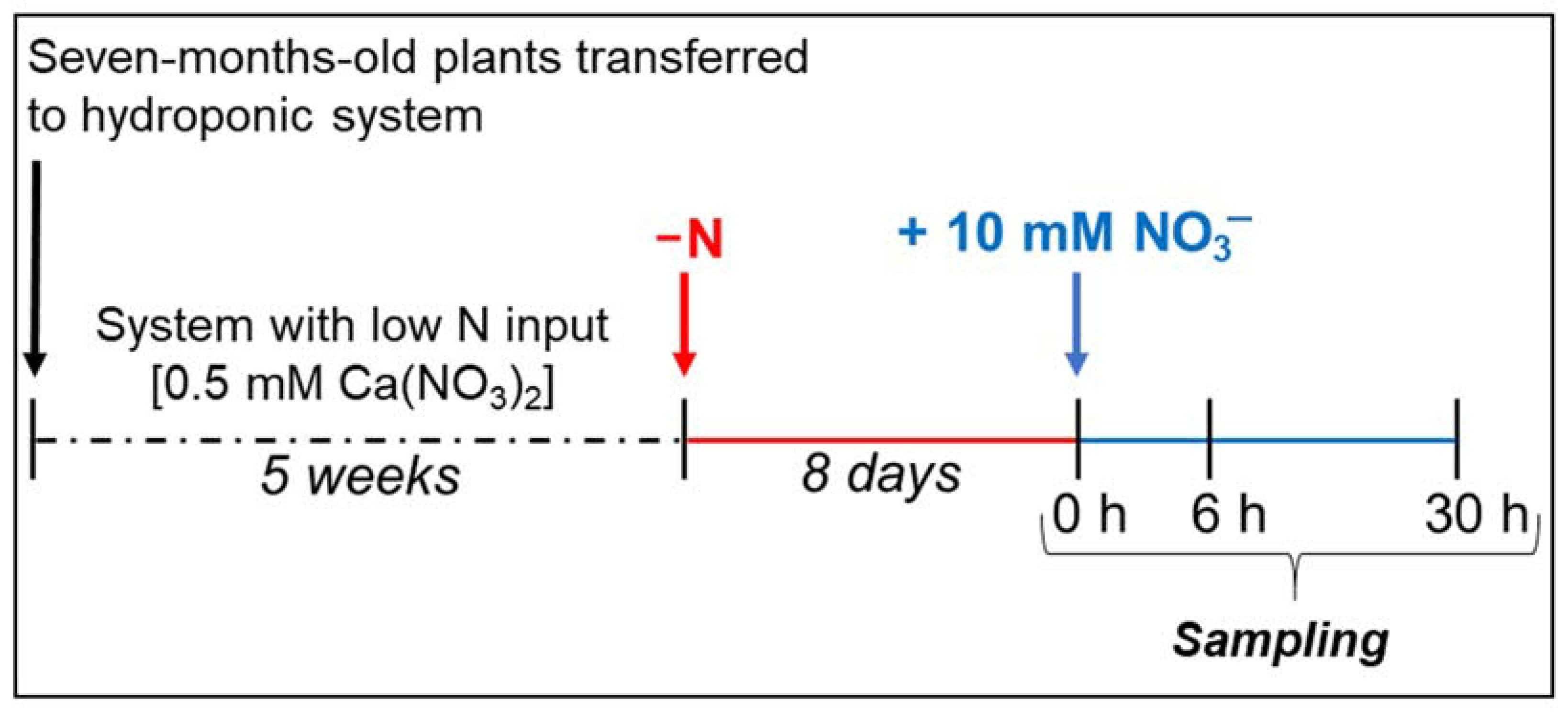
The M4 [(V. vinifera × V. berlandieri) × V. berlandieri cv. Resseguier no. 1] grapevine rootstock genotype was obtained from Vitro Hellas (Niseli Alexandreia, Greece, https://www.vitrohellas.gr/en/home, accessed on 1 August 2020). A scheme of the experimental design is shown in Figure 4. Plants, previously grown in peat soil, were flared, gently washed to remove residual particles of soil from the roots and then transferred to a hydroponic system. The experiments were conducted in a growth chamber with a 16/8 h day/night regime, at 26/22 °C, constant relative humidity of 65% and PPFD of 300 µmol m−2 s−1. All hydroponic solutions were continuously aerated by an electric pump. To allow adaptation to hydroponic conditions, plants were grown for 5 weeks in a nutrient solution (0.77 mM K2SO4, 0.65 mM MgSO4, 0.51 mM KH2PO4, 0.4 mM CaSO4, 100 µM Fe-EDTA, 50 µM KCl, 10 µM H3BO3, 1 µM MnSO4, 0.5 µM CuSO4, 0.5 µM ZnSO4, 0.35 µM Na2MoO4, pH = 6.1) containing 0.25 mM Ca(NO3)2 (i.e., low N input). The solution was replaced weekly. After this period, the plants were transferred into new nutrient solutions without N. After 8 days of N starvation (control plants, 0 h), plants were transferred to a fresh growing solution containing 10 mM KNO3. The start of the experiments coincided with the start of the light period. The plants were sampled at 0 h and after 6 and 30 h of treatment. Roots were rinsed with water, blotted with paper towels, and then immediately frozen in liquid N2. Each biological sample was composed of roots collected from three plants. Samples were stored at −80 °C.

3.2. Determination of the Contents of Nitrate, Amino Acids, Sucrose and Reducing Sugars

Nitrate was extracted from the tissues by homogenizing the samples in 4 volumes of distilled water and heating at 50 °C for 15 min. The homogenate was centrifuged at 12,000× g for 20 min to obtain a clarified supernatant. An aliquot of the supernatant was used for the determination of NO3 concentration, according to Cataldo et al. [63].
Amino acids and total sugars were extracted in perchloric acid (PCA) as previously described by Meggio and co-workers [31]. The contents of total amino acids were measured by the ninhydrin method [64]. The contents of total soluble sugars were determined by boiling an aliquot of the PCA extract for 1 h before neutralization. Sugar concentrations were then measured according to the colorimetric method of Nelson [65]. All the analyses were replicated on three independent biological samples (n = 3) and compared by the ANOVA test (p < 0.05, Holm–Šídák method).

3.3. Protein Extraction

Protein extraction was performed as previously described [33]. Briefly, frozen powdered samples (1 g) of three biological samples for each experimental condition (n = 3) were finely powdered in liquid N2 using a pestle and mortar, adding 5% (w/w) of polyvinylpolypyrrolidone. The total protein fraction was extracted by adding 5 volumes of phenol and an adequate volume of aqueous buffer [0.7 M sucrose, 10 mM Na2-EDTA, 4 mM ascorbic acid, 0.2% (v/v) Triton X-100, 1 mM PMSF, 0.1 mg mL−1 Pefabloc and 0.4% (v/v) β-mercaptoethanol]. The protein fraction was then purified by methanol-based and acetone precipitation [66]. The final pellet was then dissolved in SDS buffer [150 mM Tris-HCl pH 6.8, 10% (w/w) glycerol, 2% (w/w) sodium dodecyl sulfate (SDS), 2% (v/v) β-mercaptoethanol] and incubated at 95 °C for 5 min. The sample was centrifuged at 10,000× g for 10 min and the supernatant stored at −80 °C until further use. The protein concentration was determined by the 2-D Quant Kit (GE Healthcare Europe GmbH, Freiburg, Germany).

3.4. Immunoblot Analyses

Protein samples (10 µg) were diluted with a volume of SDS buffer with 0.01% (w/v) bromophenol blue, heated for 5 min at 95 °C and then separated by SDS-polyacrylamide gel electrophoresis (SDS-PAGE) using 10% (w/v) polyacrylamide gel [67]. The samples were then electrophoretically transferred onto a polyvinylidene difluoride (PVDF) filter using a semidry blotting system (NovaBlot, Pharmacia, Sweden) with a buffer containing 10 mM 3-cyclohexylamino-1-propanesulphonic acid (CAPS, pH 11 with NaOH) and 10% (v/v) methanol. Filters were blocked for 1 h with TBS-T buffer [50 mM Tris–HCl (pH 7.6), 200 mM NaCl, and 0.1% (v/v) Tween 20] supplemented with 3% (w/v) albumin. The TBS-T buffer was used as an incubation medium throughout the procedure. Filters were incubated overnight at 4 °C with primary polyclonal antibodies against nitrate reductase using a 1:1,250 dilution (Agrisera, Vännäs, Sweden, AS08 310). After washing with TBS-T, the filters were incubated for 2 h at room temperature with a secondary antibody (alkaline phosphatase-conjugated anti-rabbit immunoglobulin G). The blot was developed with nitroblue tetrazolium and 5-bromo-4-chloro-3-indolyl phosphate (FAST BCIP/NBT, Sigma). The analysis was performed on three biological samples (n = 3), and the quantification of the signals was conducted through densitometric analysis by using the software ImageJ (https://imagej.net/, accessed on 1 August 2020).

3.5. Gel Electrophoresis and In-gel Digestion

Gel electrophoresis, in-gel digestion and mass spectrometry analyses were performed as previously described [40], with the following refinements. Briefly, 30 µg of proteins were separated on an SDS-PAGE on 16% (w/v) polyacrylamide gel [67], until samples ran a 3 cm length. After Coomassie brilliant blue staining, the blank portions of the gels as well as the regions above 250 kDa or below 12 kDa were removed. Each line was cut into 3 regular slices (10 × 10 × 0.75 mm). Each slice was then treated as an independent sample. In-gel digestion was performed according to Prinsi and co-workers [40]. The extracted peptides were finally dissolved in 0.1% (v/v) formic acid (FA).

3.6. Mass Spectrometry Analysis

All mass spectrometry experiments were conducted with an Agilent 6520 Q-TOF mass spectrometer equipped with an HPLC Chip Cube (Agilent Technologies, Cernusco sul Naviglio, Italy), as previously described [33], with some refinements. In detail, the peptides were eluted by applying a 100 min non-linear gradient of acetonitrile from 5% to 50% (v/v) in acidic conditions (FA 0.1% v/v) at 0.4 µL min−1. The mass spectrometer was run in positive ion mode and MS scans were acquired over a range from 300 to 3000 mass-to-charge ratio (m/z) at 4 spectra s1. MS/MS scans were acquired over a range from 50 to 3000 mass-to-charge ratio (m/z) at 3 spectra s1. Precursor ions were selected by auto-MS/MS with a maximum of 4 precursors per cycle and active exclusion set at 2 spectra for 0.1 min. Sample profiles were reconstructed by combining the chromatograms obtained for all three slices into which they were divided. Analysis of MS/MS spectra was performed by Spectrum Mill MS Proteomics Workbench (Rev B.04.00.127, Agilent Technologies). Carbamidomethylation of cysteine was set as a fixed modification while the oxidation of methionine was a variable modification. Trypsin was selected as the enzyme for digestion, accepting 2 missed cleavages per peptide. For spectrum interpretation, the search was conducted against the Vitis (ID 3603) protein database (December 2020, 167,581 entries) downloaded from UniProtKB/Swiss-Prot (http://www.uniprot.org/, accessed on 1 February 2021) and concatenated with the reverse one. The mass spectrometry proteomics data have been deposited in the ProteomeXchange Consortium [68] via the PRIDE partner repository with the data set identifier PXD025212. The threshold used for protein identification was peptide false discovery rate (FDR) ≤ 1% and number of unique peptides per protein ≥ 2. Peptide quantification was obtained as the spectrum intensity (SI) of the precursor. Protein quantification was obtained by summing the SIs of all the identified peptides in the protein. Protein abundance was normalized as the percentage with respect to the abundance of all validated proteins in the sample (%(SI)), summing all validated peptides in the 3 slices [33]. The analysis was performed using three biological samples for each condition (n = 3). Statistical significance was assessed by a Student’s t-test (p < 0.05). The identified proteins were classified into metabolic functional categories according to the MapMan4 BIN ontology [38].

Supplementary Materials

The following are available online at https://www.mdpi.com/article/10.3390/plants10040792/s1, Table S1: Protein quantification by nLC–nESI–MS/MS in M4 root proteome, Table S2: Functional classification of the identified proteins in M4 rootstock genotype.

Author Contributions

Conceptualization, B.P. and L.E.; methodology, B.P. and L.E.; investigation, B.P., C.M. and L.E.; validation, B.P., C.M. and L.E.; formal analysis, C.M. and B.P.; supervision, L.E.; writing—original draft preparation, L.E.; writing—review and editing, B.P. and L.E. All authors have read and agreed to the published version of the manuscript.

Funding

This research received no external funding.

Institutional Review Board Statement

Not applicable.

Informed Consent Statement

Not applicable.

Data Availability Statement

The data presented in this study are available on request from the corresponding author. The mass spectrometry proteomics data are available via ProteomeXchange with identifier PXD025212.

Acknowledgments

The authors thank colleagues Lucio Brancadoro for procuring the experimental material and Silvia Morgutti for reading through the manuscript.

Conflicts of Interest

The authors declare no conflict of interest.

References

  1. Hawkesford, M.; Horst, W.; Kichey, T.; Lambers, H.; Schjoerring, J.; Møller, I.S.; White, P. Chapter 6-Functions of Macronutrients. In Marschner’s Mineral Nutrition of Higher Plants, 3rd ed.; Marschner, P., Ed.; Academic Press: San Diego, CA, USA, 2012; pp. 135–189. [Google Scholar] [CrossRef]
  2. Keller, M. The Science of Grapevines: Anatomy and Physiology, 2nd ed.; Keller, M., Ed.; Academic Press: Boston, MA, USA, 2015; ISBN 878-0-12-419987-3. [Google Scholar]
  3. Keller, M.; Kummer, M.; Vasconcelos, M.C. Reproductive Growth of Grapevines in Response to Nitrogen Supply and Rootstock. Aust. J. Grape Wine Res. 2001, 7, 12–18. [Google Scholar] [CrossRef]
  4. Bell, S.-J.; Henschke, P.A. Implications of Nitrogen Nutrition for Grapes, Fermentation and Wine. Aust. J. Grape Wine Res. 2005, 11, 242–295. [Google Scholar] [CrossRef]
  5. Gachons, C.P.; Leeuwen, C.V.; Tominaga, T.; Soyer, J.-P.; Gaudillère, J.-P.; Dubourdieu, D. Influence of Water and Nitrogen Deficit on Fruit Ripening and Aroma Potential of Vitis vinifera L cv Sauvignon Blanc in Field Conditions. J. Sci. Food Agr. 2005, 85, 73–85. [Google Scholar] [CrossRef]
  6. Soubeyrand, E.; Basteau, C.; Hilbert, G.; van Leeuwen, C.; Delrot, S.; Gomès, E. Nitrogen Supply Affects Anthocyanin Biosynthetic and Regulatory Genes in Grapevine cv. Cabernet-Sauvignon Berries. Phytochemistry 2014, 103, 38–49. [Google Scholar] [CrossRef] [PubMed]
  7. Lecourt, J.; Lauvergeat, V.; Ollat, N.; Vivin, P.; Cookson, S.J. Shoot and Root Ionome Responses to Nitrate Supply in Grafted Grapevines Are Rootstock Genotype Dependent. Aust. J. Grape Wine Res. 2015, 21, 311–318. [Google Scholar] [CrossRef]
  8. Habran, A.; Commisso, M.; Helwi, P.; Hilbert, G.; Negri, S.; Ollat, N.; Gomès, E.; van Leeuwen, C.; Guzzo, F.; Delrot, S. Roostocks/Scion/Nitrogen Interactions Affect Secondary Metabolism in the Grape Berry. Front. Plant Sci. 2016, 7. [Google Scholar] [CrossRef] [PubMed] [Green Version]
  9. Rossdeutsch, L.; Schreiner, R.P.; Skinkis, P.A.; Deluc, L. Nitrate Uptake and Transport Properties of Two Grapevine Rootstocks with Varying Vigor. Front. Plant Sci. 2021, 11. [Google Scholar] [CrossRef]
  10. Zapata, C.; Deléens, E.; Chaillou, S.; Magné, C. Partitioning and Mobilization of Starch and N Reserves in Grapevine (Vitis Vinifera L.). J. Plant Physiol. 2004, 161, 1031–1040. [Google Scholar] [CrossRef]
  11. Grechi, I.; Vivin, P.H.; Hilbert, G.; Milin, S.; Robert, T.; Gaudillère, J.-P. Effect of Light and Nitrogen Supply on Internal C:N Balance and Control of Root-to-Shoot Biomass Allocation in Grapevine. Environ. Exp. Bot. 2007, 59, 139–149. [Google Scholar] [CrossRef]
  12. Metay, A.; Magnier, J.; Guilpart, N.; Christophe, A. Nitrogen Supply Controls Vegetative Growth, Biomass and Nitrogen Allocation for Grapevine (cv. Shiraz) Grown in Pots. Funct. Plant Biol. 2015, 42, 105–114. [Google Scholar] [CrossRef]
  13. Ferrara, G.; Malerba, A.D.; Matarrese, A.M.S.; Mondelli, D.; Mazzeo, A. Nitrogen Distribution in Annual Growth of ‘Italia’ Table Grape Vines. Front. Plant Sci. 2018, 9. [Google Scholar] [CrossRef] [PubMed] [Green Version]
  14. Vrignon-Brenas, S.; Aurélie, M.; Romain, L.; Shiva, G.; Alana, F.; Myriam, D.; Gaëlle, R.; Anne, P. Gradual Responses of Grapevine Yield Components and Carbon Status to Nitrogen Supply. OENO One 2019. [Google Scholar] [CrossRef]
  15. Jiménez, S.; Gogorcena, Y.; Hévin, C.; Rombolà, A.D.; Ollat, N. Nitrogen Nutrition Influences Some Biochemical Responses to Iron Deficiency in Tolerant and Sensitive Genotypes of Vitis. Plant Soil 2007, 290, 343–355. [Google Scholar] [CrossRef] [Green Version]
  16. Kulmann, M.S.; Sete, P.B.; Paula, B.V.; Stefanello, L.O.; Schwalbert, R.; Schwalbert, R.A.; Arruda, W.S.; Sans, G.A.; Parcianello, C.F.; Nicoloso, F.T.; et al. Kinetic Parameters Govern of the Uptake of Nitrogen Forms in ‘Paulsen’ and ‘Magnolia’ Grapevine Rootstocks. Sci. Hortic. 2020, 264, 109174. [Google Scholar] [CrossRef]
  17. Miller, A.J.; Fan, X.; Orsel, M.; Smith, S.J.; Wells, D.M. Nitrate Transport and Signalling. J. Exp. Bot. 2007, 58, 2297–2306. [Google Scholar] [CrossRef] [PubMed]
  18. Wang, Y.-Y.; Hsu, P.-K.; Tsay, Y.-F. Uptake, Allocation and Signaling of Nitrate. Trends Plant Sci. 2012, 17, 458–467. [Google Scholar] [CrossRef]
  19. Fan, X.; Naz, M.; Fan, X.; Xuan, W.; Miller, A.J.; Xu, G. Plant Nitrate Transporters: From Gene Function to Application. J. Exp. Bot. 2017, 68, 2463–2475. [Google Scholar] [CrossRef]
  20. Pii, Y.; Alessandrini, M.; Guardini, K.; Zamboni, A.; Varanini, Z. Induction of High-Affinity NO3 Uptake in Grapevine Roots Is an Active Process Correlated to the Expression of Specific Members of the NRT2 and Plasma Membrane H+-ATPase Gene Families. Funct. Plant Biol. 2014, 41, 353–365. [Google Scholar] [CrossRef] [Green Version]
  21. Tomasi, N.; Monte, R.; Varanini, Z.; Cesco, S.; Pinton, R. Induction of Nitrate Uptake in Sauvignon Blanc and Chardonnay Grapevines Depends on the Scion and Is Affected by the Rootstock. Aust. J. Grape Wine Res. 2015, 21, 331–338. [Google Scholar] [CrossRef]
  22. Loulakakis, K.A.; Morot-Gaudry, J.F.; Velanis, C.N.; Skopelitis, D.S.; Moschou, P.N.; Hirel, B.; Roubelakis-Angelakis, K.A. Advancements in Nitrogen Metabolism in Grapevine. In Grapevine Molecular Physiology & Biotechnology; Roubelakis-Angelakis, K.A., Ed.; Springer: Dordrecht, The Netherlands, 2009; pp. 161–205. [Google Scholar] [CrossRef]
  23. Zerihun, A.; Treeby, M.T. Biomass Distribution and Nitrate Assimilation in Response to N Supply for Vitis vinifera L. cv. Cabernet Sauvignon on Five Vitis Rootstock Genotypes. Aust. J. Grape Wine Res. 2002, 8, 157–162. [Google Scholar] [CrossRef]
  24. Lang, C.P.; Merkt, N.; Zörb, C. Different Nitrogen (N) Forms Affect Responses to N Form and N Supply of Rootstocks and Grafted Grapevines. Plant Sci. 2018, 277, 311–321. [Google Scholar] [CrossRef]
  25. Lang, C.P.; Bárdos, G.; Merkt, N.; Zörb, C. Expression of Key Enzymes for Nitrogen Assimilation in Grapevine Rootstock in Response to N-Form and Timing. J. Plant Nutr. Soil Sci. 2020, 183, 91–98. [Google Scholar] [CrossRef] [Green Version]
  26. Scheible, W.-R.; Morcuende, R.; Czechowski, T.; Fritz, C.; Osuna, D.; Palacios-Rojas, N.; Schindelasch, D.; Thimm, O.; Udvardi, M.K.; Stitt, M. Genome-Wide Reprogramming of Primary and Secondary Metabolism, Protein Synthesis, Cellular Growth Processes, and the Regulatory Infrastructure of Arabidopsis in Response to Nitrogen. Plant Physiol. 2004, 136, 2483–2499. [Google Scholar] [CrossRef] [Green Version]
  27. Nunes-Nesi, A.; Fernie, A.R.; Stitt, M. Metabolic and Signaling Aspects Underpinning the Regulation of Plant Carbon Nitrogen Interactions. Mol. Plant 2010, 3, 973–996. [Google Scholar] [CrossRef]
  28. Cochetel, N.; Escudié, F.; Cookson, S.J.; Dai, Z.; Vivin, P.; Bert, P.-F.; Muñoz, M.S.; Delrot, S.; Klopp, C.; Ollat, N.; et al. Root Transcriptomic Responses of Grafted Grapevines to Heterogeneous Nitrogen Availability Depend on Rootstock Genotype. J. Exp. Bot. 2017, 68, 4339–4355. [Google Scholar] [CrossRef] [Green Version]
  29. Wang, R.; Okamoto, M.; Xing, X.; Crawford, N.M. Microarray Analysis of the Nitrate Response in Arabidopsis Roots and Shoots Reveals over 1000 Rapidly Responding Genes and New Linkages to Glucose, Trehalose-6-Phosphate, Iron, and Sulfate Metabolism. Plant Physiol. 2003, 132, 556–567. [Google Scholar] [CrossRef] [PubMed] [Green Version]
  30. Lejay, L.; Wirth, J.; Pervent, M.; Cross, J.M.-F.; Tillard, P.; Gojon, A. Oxidative Pentose Phosphate Pathway-Dependent Sugar Sensing as a Mechanism for Regulation of Root Ion Transporters by Photosynthesis. Plant Physiol. 2008, 146, 2036–2053. [Google Scholar] [CrossRef] [PubMed] [Green Version]
  31. Meggio, F.; Prinsi, B.; Negri, A.S.; Lorenzo, G.S.D.; Lucchini, G.; Pitacco, A.; Failla, O.; Scienza, A.; Cocucci, M.; Espen, L. Biochemical and Physiological Responses of Two Grapevine Rootstock Genotypes to Drought and Salt Treatments. Aust. J. Grape Wine Res. 2014, 20, 310–323. [Google Scholar] [CrossRef]
  32. Corso, M.; Vannozzi, A.; Maza, E.; Vitulo, N.; Meggio, F.; Pitacco, A.; Telatin, A.; D’Angelo, M.; Feltrin, E.; Negri, A.S.; et al. Comprehensive Transcript Profiling of Two Grapevine Rootstock Genotypes Contrasting in Drought Susceptibility Links the Phenylpropanoid Pathway to Enhanced Tolerance. J. Exp. Bot. 2015, 66, 5739–5752. [Google Scholar] [CrossRef]
  33. Prinsi, B.; Negri, A.S.; Failla, O.; Scienza, A.; Espen, L. Root Proteomic and Metabolic Analyses Reveal Specific Responses to Drought Stress in Differently Tolerant Grapevine Rootstocks. BMC Plant Biol. 2018, 18, 126. [Google Scholar] [CrossRef] [PubMed]
  34. Prinsi, B.; Failla, O.; Scienza, A.; Espen, L. Root Proteomic Analysis of Two Grapevine Rootstock Genotypes Showing Different Susceptibility to Salt Stress. Int. J. Mol. Sci. 2020, 21, 1076. [Google Scholar] [CrossRef] [Green Version]
  35. Prinsi, B.; Simeoni, F.; Galbiati, M.; Meggio, F.; Tonelli, C.; Scienza, A.; Espen, L. Grapevine Rootstocks Differently Affect Physiological and Molecular Responses of the Scion under Water Deficit Condition. Agronomy 2021, 11, 289. [Google Scholar] [CrossRef]
  36. Xu, G.; Fan, X.; Miller, A.J. Plant Nitrogen Assimilation and Use Efficiency. Annu. Rev. Plant Biol. 2012, 63, 153–182. [Google Scholar] [CrossRef] [PubMed] [Green Version]
  37. Gilmore, J.M.; Washburn, M.P. Advances in Shotgun Proteomics and the Analysis of Membrane Proteomes. J. Proteom. 2010, 73, 2078–2091. [Google Scholar] [CrossRef]
  38. Schwacke, R.; Ponce-Soto, G.Y.; Krause, K.; Bolger, A.M.; Arsova, B.; Hallab, A.; Gruden, K.; Stitt, M.; Bolger, M.E.; Usadel, B. MapMan4: A Refined Protein Classification and Annotation Framework Applicable to Multi-Omics Data Analysis. Mol. Plant 2019, 12, 879–892. [Google Scholar] [CrossRef] [PubMed]
  39. Canales, J.; Moyano, T.C.; Villarroel, E.; Gutiérrez, R.A. Systems Analysis of Transcriptome Data Provides New Hypotheses about Arabidopsis Root Response to Nitrate Treatments. Front. Plant Sci. 2014, 5. [Google Scholar] [CrossRef] [Green Version]
  40. Prinsi, B.; Espen, L. Time-Course of Metabolic and Proteomic Responses to Different Nitrate/Ammonium Availabilities in Roots and Leaves of Maize. Int. J. Mol. Sci. 2018, 19, 2202. [Google Scholar] [CrossRef] [Green Version]
  41. Bernard, S.M.; Habash, D.Z. The Importance of Cytosolic Glutamine Synthetase in Nitrogen Assimilation and Recycling. New Phytol. 2009, 182, 608–620. [Google Scholar] [CrossRef]
  42. Stitt, M. Nitrate Regulation of Metabolism and Growth. Curr. Opin. Plant Biol. 1999, 2, 178–186. [Google Scholar] [CrossRef]
  43. Stein, O.; Granot, D. An Overview of Sucrose Synthases in Plants. Front. Plant Sci. 2019, 10. [Google Scholar] [CrossRef] [Green Version]
  44. Sun, J.-L.; Li, J.-Y.; Wang, M.-J.; Song, Z.-T.; Liu, J.-X. Protein Quality Control in Plant Organelles: Current Progress and Future Perspectives. Mol. Plant 2021, 14, 95–114. [Google Scholar] [CrossRef] [PubMed]
  45. Kohli, A.; Narciso, J.O.; Miro, B.; Raorane, M. Root Proteases: Reinforced Links between Nitrogen Uptake and Mobilization and Drought Tolerance. Physiol. Plant. 2012, 145, 165–179. [Google Scholar] [CrossRef]
  46. Wang, Y.-H.; Garvin, D.F.; Kochian, L.V. Nitrate-Induced Genes in Tomato Roots. Array Analysis Reveals Novel Genes That May Play a Role in Nitrogen Nutrition. Plant Physiol. 2001, 127, 345–359. [Google Scholar] [CrossRef] [PubMed] [Green Version]
  47. Park, S.-C.; Kim, Y.-H.; Jeong, J.C.; Kim, C.Y.; Lee, H.-S.; Bang, J.-W.; Kwak, S.-S. Sweetpotato Late Embryogenesis Abundant 14 (IbLEA14) Gene Influences Lignification and Increases Osmotic- and Salt Stress-Tolerance of Transgenic Calli. Planta 2011, 233, 621–634. [Google Scholar] [CrossRef]
  48. Yu, P.; Li, X.; Yuan, L.; Li, C. A Novel Morphological Response of Maize (Zea mays) Adult Roots to Heterogeneous Nitrate Supply Revealed by a Split-Root Experiment. Physiol. Plant. 2014, 150, 133–144. [Google Scholar] [CrossRef]
  49. Vega, A.; O’Brien, J.A.; Gutiérrez, R.A. Nitrate and Hormonal Signaling Crosstalk for Plant Growth and Development. Curr. Opin. Plant Biol. 2019, 52, 155–163. [Google Scholar] [CrossRef] [PubMed]
  50. Kose, C.; Erdal, S.; Kaya, O.; Atici, O. Comparative Evaluation of Oxidative Enzyme Activities during Adventitious Rooting in the Cuttings of Grapevine Rootstocks. J. Sci. Food Agr. 2011, 91, 738–741. [Google Scholar] [CrossRef]
  51. Fritz, C.; Palacios-Rojas, N.; Feil, R.; Stitt, M. Regulation of Secondary Metabolism by the Carbon–Nitrogen Status in Tobacco: Nitrate Inhibits Large Sectors of Phenylpropanoid Metabolism. Plant J. 2006, 46, 533–548. [Google Scholar] [CrossRef]
  52. He, F.; Mu, L.; Yan, G.-L.; Liang, N.-N.; Pan, Q.-H.; Wang, J.; Reeves, M.J.; Duan, C.-Q. Biosynthesis of Anthocyanins and Their Regulation in Colored Grapes. Molecules 2010, 15, 9057–9091. [Google Scholar] [CrossRef] [Green Version]
  53. Larbat, R.; Bot, J.L.; Bourgaud, F.; Robin, C.; Adamowicz, S. Organ-Specific Responses of Tomato Growth and Phenolic Metabolism to Nitrate Limitation. Plant Biol. 2012, 14, 760–769. [Google Scholar] [CrossRef]
  54. Prinsi, B.; Negrini, N.; Morgutti, S.; Espen, L. Nitrogen Starvation and Nitrate or Ammonium Availability Differently Affect Phenolic Composition in Green and Purple Basil. Agronomy 2020, 10, 498. [Google Scholar] [CrossRef] [Green Version]
  55. Schmidlin, L.; Poutaraud, A.; Claudel, P.; Mestre, P.; Prado, E.; Santos-Rosa, M.; Wiedemann-Merdinoglu, S.; Karst, F.; Merdinoglu, D.; Hugueney, P. A Stress-Inducible Resveratrol O-Methyltransferase Involved in the Biosynthesis of Pterostilbene in Grapevine. Plant Physiol. 2008, 148, 1630–1639. [Google Scholar] [CrossRef] [Green Version]
  56. Ramawat, K.G.; Goyal, S. Co-Evolution of Secondary Metabolites During Biological Competition for Survival and Advantage: An Overview. In Co-Evolution of Secondary Metabolites; Merillon, J.-M., Ramawat, K.G., Eds.; Reference Series in Phytochemistry; Springer International Publishing: Cham, Germany, 2019; pp. 1–15. [Google Scholar] [CrossRef]
  57. González, J.F.; Venturi, V. A Novel Widespread Interkingdom Signaling Circuit. Trends Plant Sci. 2013, 18, 167–174. [Google Scholar] [CrossRef]
  58. Di, C.; Xu, W.; Su, Z.; Yuan, J.S. Comparative Genome Analysis of PHB Gene Family Reveals Deep Evolutionary Origins and Diverse Gene Function. BMC Bioinform. 2010, 11, S22. [Google Scholar] [CrossRef] [Green Version]
  59. Wang, Y.; Yang, L.; Chen, X.; Ye, T.; Zhong, B.; Liu, R.; Wu, Y.; Chan, Z. Major Latex Protein-like Protein 43 (MLP43) Functions as a Positive Regulator during Abscisic Acid Responses and Confers Drought Tolerance in Arabidopsis thaliana. J. Exp. Bot. 2016, 67, 421–434. [Google Scholar] [CrossRef] [Green Version]
  60. Konopka-Postupolska, D.; Clark, G.; Hofmann, A. Structure, Function and Membrane Interactions of Plant Annexins: An Update. Plant Sci. 2011, 181, 230–241. [Google Scholar] [CrossRef] [Green Version]
  61. Yoo, K.S.; Ok, S.H.; Jeong, B.-C.; Jung, K.W.; Cui, M.H.; Hyoung, S.; Lee, M.-R.; Song, H.K.; Shin, J.S. Single Cystathionine β-Synthase Domain–Containing Proteins Modulate Development by Regulating the Thioredoxin System in Arabidopsis. Plant Cell 2011, 23, 3577–3594. [Google Scholar] [CrossRef] [PubMed] [Green Version]
  62. Noctor, G.; Mhamdi, A.; Chaouch, S.; Han, Y.; Neukermans, J.; Marquez-Garcia, B.; Queval, G.; Foyer, C.H. Glutathione in Plants: An Integrated Overview. Plant Cell Environ. 2012, 35, 454–484. [Google Scholar] [CrossRef] [PubMed]
  63. Cataldo, D.A.; Maroon, M.; Schrader, L.E.; Youngs, V.L. Rapid Colorimetric Determination of Nitrate in Plant Tissue by Nitration of Salicylic Acid. Commun. Soil Sci. Plant 1975, 6, 71–80. [Google Scholar] [CrossRef]
  64. Moore, S.; Stein, W.H. A Modified Ninhydrin Reagent for the Photometric Determination of Amino Acids and Related Compounds. J. Biol. Chem. 1954, 211, 907–913. [Google Scholar] [CrossRef]
  65. Nelson, N. A Photometric Adaptation of the Somogyi Method for the Determination of Glucose. J. Biol. Chem. 1944, 153, 375–380. [Google Scholar] [CrossRef]
  66. Prinsi, B.; Negri, A.S.; Pesaresi, P.; Cocucci, M.; Espen, L. Evaluation of Protein Pattern Changes in Roots and Leaves of Zea mays plants in Response to Nitrate Availability by Two-Dimensional Gel Electrophoresis Analysis. BMC Plant Biol. 2009, 9, 113. [Google Scholar] [CrossRef] [Green Version]
  67. Laemmli, U.K. Cleavage of Structural Proteins during the Assembly of the Head of Bacteriophage T4. Nature 1970, 227, 680–685. [Google Scholar] [CrossRef] [PubMed]
  68. Vizcaíno, J.A.; Deutsch, E.W.; Wang, R.; Csordas, A.; Reisinger, F.; Ríos, D.; Dianes, J.A.; Sun, Z.; Farrah, T.; Bandeira, N.; et al. ProteomeXchange Provides Globally Co-Ordinated Proteomics Data Submission and Dissemination. Nat. Biotechnol. 2014, 32, 223–226. [Google Scholar] [CrossRef] [PubMed]
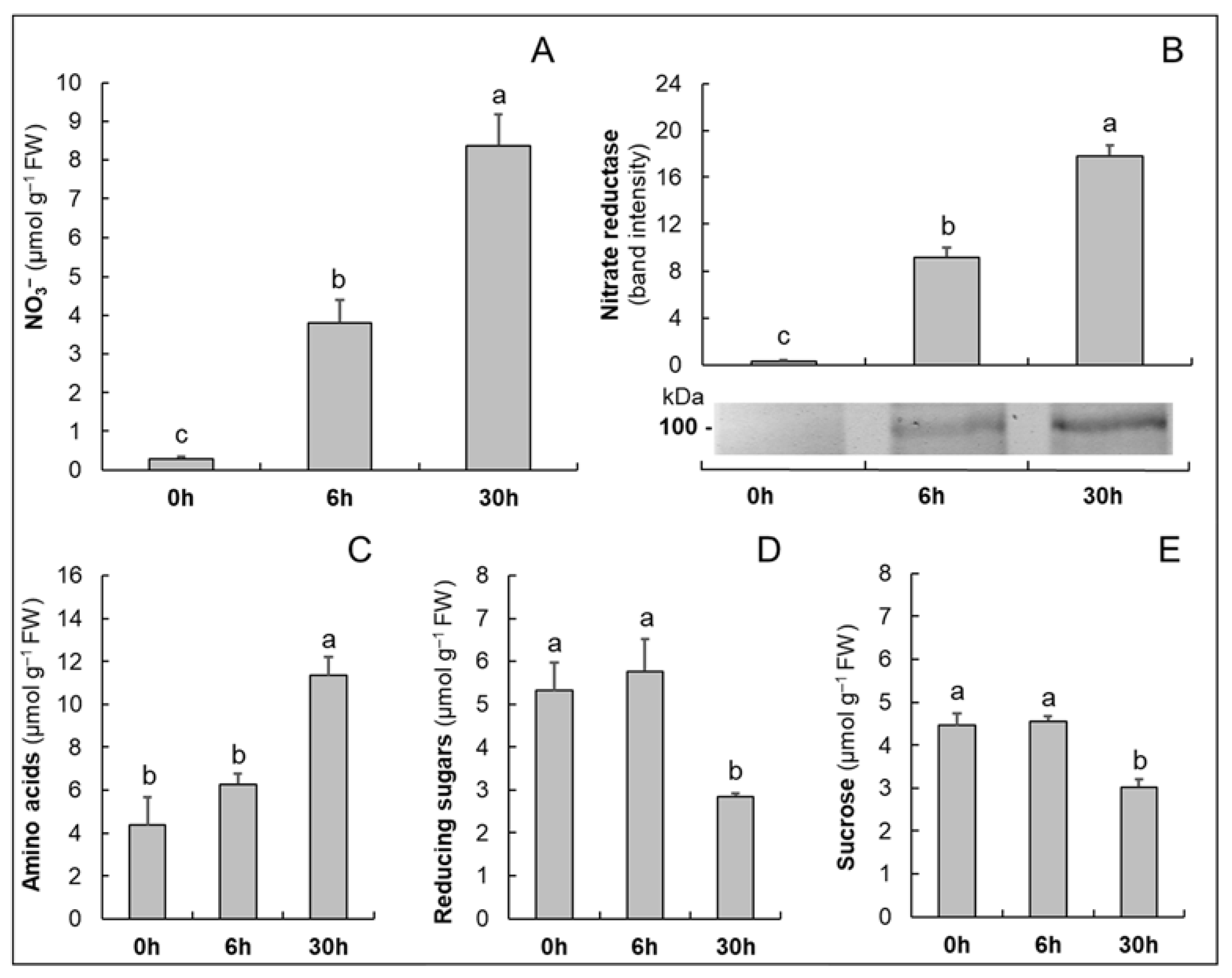
Figure 1. The time course of the changes in the contents of NO3 (A), in the accumulation of nitrate reductase evaluated by immunoblot analysis (B), and in the contents of amino acids (C), reducing sugars (D) and sucrose (E) in the roots of M4 grapevine rootstock, previously grown in the absence of N for 8 d (0 h) and incubated for a further 6 and 30 h in the presence of 10 mM NO3. The values are means ± SE (n = 3). Statistical significance was assessed by one-way ANOVA and the Holm–Šídák method. Different letters indicate significant differences (p ≤ 0.05).
Plants 10 00792 g001
Figure 2. Functional distribution of all the identified proteins in the roots of M4 grapevine rootstock. Proteins were grouped in categories according to BIN ontology.
Plants 10 00792 g002
Figure 3. Functional distribution of the differentially accumulated proteins in roots of M4 grapevine rootstock after exposure of the plants to 10 mM NO3. Proteins were grouped in categories according to BIN ontology (codes reported in brackets). The percentage refers to the total number of proteins having the same trend: blue bars, proteins that increased/appeared; yellow bars, proteins that decreased/disappeared.
Plants 10 00792 g003
Figure 4. Growth experiment design and nutritional treatments. Further details are reported in the Material and Methods.
Plants 10 00792 g004
Table 1. Proteins differentially accumulated in response to 10 mM NO3 for 30 h in roots of M4. Proteins are grouped according to functional classification (Figure 3). Subtitles report functional categories and their bin codes. #: identification number (group). UIP: number of unique identified peptides. Score: MS/MS search score. Δ(nit/con): fold changes expressed as the ratio between the protein abundance in NO3-treated plants (nit, 30 h) and in the control N-starved plants (con, 0 h). Blue cells: proteins that increased in abundance. Yellow cells: proteins that decreased in abundance. s: statistical significance assessed by Student’s t-test (n = 3) (* p < 0.05, ** p < 0.01). a: protein annotated by BLAST. In italics: additional information from UniProt. New: not detected in control plants; d.: disappeared, not detected in NO3-treated plants.
#Accession NumberUIPScoreProtein NameΔ
(nit/con)
s
Carbon and energy metabolism (1, 2, 3)
28A5B11812167.9Fructose-bisphosphate aldolase2.01*
23A5CAF612191.3Phosphoglycerate kinase1.83*
122F6I1P0680.5Pyruvate dehydrogenase E1 component subunit beta0.22**
251A5AY34445.9Oxidored_q6 domain-containing protein4.34*
2A5AYU818330.6ATP synthase subunit beta0.63**
150D7T300654.9ATP synthase subunit O, mitochondrial I anew*
14F6HGZ914160.9Sucrose synthasenew*
257Q1PSI9443.7L-idonate 5-dehydrogenase15.59**
323A0A438D9B1336.8Glucose-6-phosphate 1-dehydrogenasenew**
169A0A438HWY8566.2Probable 6-phosphogluconolactonase0.29*
64F6HGH48120.06-phosphogluconate dehydrogenase, decarboxylating9.40**
239D7TJI9450.4Pyruvate decarboxylase0.34**
77F6HUI7888.3RmlD_sub_bind domain-containing protein3.34*
Cell wall (21)
131A0A438KK24675.1Caffeoyl-CoA O-methyltransferase0.36**
54F6GSZ79119.1Omega-hydroxypalmitate O-feruloyl transferase a0.63*
Amino acid metabolism (4)
3F6HMN818266.75-methyltetrahydropteroyltriglutamate--homocysteine S-methyltransferase2.90*
73A0A438GBL88106.4Acetohydroxy-acid reductoisomerase6.64*
30F6I5Y512159.3D-3-phosphoglycerate dehydrogenase1.66*
140F6H0X2667.6Phospho-2-dehydro-3-deoxyheptonate aldolase2.39**
Lipid metabolism (5)
51F6I1D69142.9Non-specific phospholipase C3 a0.52**
462A0A438FDG5220.2Enoyl-CoA delta isomerase 31.63*
Secondary metabolism (9)
173F6HHQ7565.2Putative acetyl-CoA acetyltransferase, cytosolic 2 a0.39*
314A0A024FS61339.3Polyphenol_oxidase4.73*
415A0A438ESC9225.13-isopropylmalate dehydratase small subunit 30.09*
86F6I0767107.7CN hydrolase domain-containing protein0.05**
Redox homeostasis (10)
185G1JT87559.7Glutaredoxin-dependent peroxiredoxin0.41*
269D7T6T0439.8Glutaredoxin-dependent peroxiredoxin4.16*
330A9UFY2336.1Thioredoxin h-type0.45*
Enzymes/Coenzyme metabolism (50, 7)
136D7TKJ3671.1Ferredoxin-NADP reductase, chloroplasticnew**
148D7SNB1658.9Salutaridine reductase a0.41*
11K9N4H514213.1Mitochondrial aldehyde dehydrogenase 2B80.65*
119A5BHH9680.8NADH-cytochrome b5 reductase0.29*
41F6H5H510156.0Trans-resveratrol di-O-methyltransferase a1.63**
458A0A438CVH9220.5UDP-glycosyltransferase 74F27.77*
105A0A438KGT6698.7Glucan endo-1,3-beta-D-glucosidase0.27**
151A0A438ITG1593.1Putative cysteine protease RD21B0.36**
81A0A438EKJ27116.9Phosphopyruvate hydratase (synonym: Enolase)2.28**
154A0A438D2Y0583.1Phosphoglycerate mutase2.00**
38F6GTM711160.1Adenosylhomocysteinase4.96*
DNA/RNA/Cell cycle (6, 12, 13, 16)
280A5B6U5434.5Proliferating cell nuclear antigen0.05*
378F6GSZ1231.5RRM domain-containing protein0.61*
Protein (17,18,19,23)
300A5AXI6343.060S acidic ribosomal protein P024.11**
126A5BUU4678.240S ribosomal protein SA4.43**
217A5C4J2458.040S ribosomal protein S19-3 a2.38*
443A5AJ83222.4Ribosomal_S10 domain-containing protein4.27*
135A0A438KA42672.8Guanine nucleotide-binding protein subunit beta-like protein3.44**
375F6HLE8233.4Ribosomal_S7 domain-containing protein3.47*
171A0A438C2W6565.7Aspartate-tRNA ligase2.44*
37F6HXZ511164.6Eukaryotic initiation factor 4A-2 a2.50**
407F6GTY8226.2Tr-type G domain-containing protein10.31**
149A0A438CSH7657.0Elongation factor 1-gammanew*
128F6H4T7677.2Tr-type G domain-containing protein20.06**
346A0A438JTD3333.5Dolichyl-diphosphooligosaccharide-protein glycosyltransferase 48 kDa subunitnew*
44D7TBD910143.7Alpha-MPP0.39*
99A5ANH8782.5Probable mitochondrial-processing peptidase subunit beta, mitochondrial a3.78*
176A0A438DUK9563.2Protein disulfide-isomerase13.28**
335A0A438K994335.3Citrulline-aspartate ligase7.32**
47D7SIX710128.0Serine/threonine-protein phosphatase 2A 65 kDa regulatory
subunit A beta isoform a
0.56*
108A0A438G7L8697.4Glutathione S-transferase U102.49*
337F6I510334.9Putative glutathione S-transferase parC a2.94*
448F6GT86221.5Glutathione S-transferase a4.05*
232A0A438KHW4453.1Glutathione transferase1.65*
452F6HYG1221.4Heat shock 70 kDa protein 15-like anew*
1F6HNX520361.7Putative heat shock cognate protein 2 a2.04**
370A0A438K358238.1Hsp70-Hsp90 organizing protein 1new**
317A0A438D490338.2Heat shock cognate protein 80new*
107D7SLM9698.0RuBisCO large subunit-binding protein subunit beta, chloroplastic a5.54**
145F6GUM1665.0E1 ubiquitin-activating enzyme6.77**
379A0A438J7X4231.5Ubiquitin-conjugating enzyme E2-17 kDa1.48*
147A0A438KGZ1662.4Proteasome subunit beta0.52*
420D7SKV3224.4Proteasome subunit beta6.25*
388A0A438EWK5229.326S proteasome regulatory subunit 78.65*
322F6HT17336.9PCI domain-containing protein3.30*
93A0A438JN39793.2Serine carboxypeptidase-like 70.48*
115D7T3Q1685.9Glucose acyltransferase 1 a0.48**
195A5C1I0557.1Carboxypeptidase0.71*
26F6H7H112172.6Aspartic proteinase A1 a0.56*
396A0A438K8Z1228.0Aminopeptidasenew**
400A0A438EKP3227.0Ankyrin repeat domain-containing protein 2A0.21*
228A0A438JPS3453.8GTP-binding nuclear protein Ran1B12.55**
Cytoskeleton organization (20)
4A5ATG817307.8Tubulin beta chain0.78*
347A0A438F6R2333.3T-complex protein 1 subunit gamma4.31**
Vesicle trafficking (22)
144D7T9L8665.0Coatomer subunit delta6.58**
Solute transport/Nutrient uptake (24, 25)
306F6I0Z8340.4Plasma membrane 22 aquaporin2.81**
203Q9FS46470.6Putative aquaporin0.65**
216A5AQ65458.1Mitochondrial outer membrane protein porin 2 a0.38**
325A0A438CTH2336.5Mitochondrial outer membrane protein porin of 34 kDa0.43**
246A0A438FMR0446.7Ferredoxin--nitrite reductase, chloroplasticnew**
29A5AP3812162.2Glutamine synthetase (cytosolic a)0.65*
321A0A438E3X6337.1Ferritin10.77**
Phytohormone action/External stimuli response (11, 26)
224F6H6V6454.5Senescence-associated carboxylesterase 101 a4.18**
Not assigned-annotated (35.1)
46A0A438KRJ610138.5Annexin (D2 a)1.44**
403A0A438JYU9226.9Dipeptide epimerase8.06**
76D7SJF5895.2Cystathionine beta-synthase family protein a5.70**
158A5BM68573.7TCTP domain-containing protein1.81*
48A0A438J6W510124.3Glutelin type-A 20.11**
230A0A438KKU7453.6Stem-specific protein TSJT10.26**
89A0A438JUJ67104.2MLP-like protein 341.49**
165A0A438JUL6568.5MLP-like protein 433.25*
42F6GTA610147.7PHB domain-containing protein0.54**
91D7TNE5797.3PHB domain-containing protein0.51*
106A0A438J2L0698.2Chalcone-flavonone isomerase family protein (synonim: Chalcone isomerase)0.61*
110A0A438BSC8692.9NAD(P)H dehydrogenase (quinone)0.40*
215D7T2N7459.9Late embryogenesis abundant protein Lea14-A, putative a0.58*
265D7T3J3441.3Proline iminopeptidase0.26**
289F6H6H8348.8Glyco_hydro_18 domain-containing proteind.*
387A5BM29229.4NTF2 domain-containing protein0.11**
104D7T7N46102.7RRM domain-containing protein0.28*
112A0A438GQU3691.0Kunitz trypsin inhibitor 20.02**
199A0A438IQU7547.6Major allergen Pru ar 1d.*
180A0A438C6P2562.0Plastid-lipid-associated protein, chloroplastic0.14*
395A5AJB3228.1Chitin-binding type-1 domain-containing proteind.*
208A0A438JVD2464.1Peroxidase0.42**
345F6HIK4333.9Peroxidase4.52*
Not assigned-not annotated (35.2)
72A5C8L88106.9Pyr_redox_2 domain-containing protein5.10**
95D7TA35788.6Usp domain-containing protein4.39*
273D7U4I8438.5Usp domain-containing proteind.**
142A0A438JK35666.9Bifunctional epoxide hydrolase 20.01*
167A5AEX6567.0DLH domain-containing protein0.75*
Publisher’s Note: MDPI stays neutral with regard to jurisdictional claims in published maps and institutional affiliations.