Article

Cold Plasma Affects Germination and Fungal Community Structure of Buckwheat Seeds

1
Biotechnical Faculty, University of Ljubljana, Jamnikarjeva 101, 1000 Ljubljana, Slovenia
2
Jozef Stefan Institute, Jamova 39, 1000 Ljubljana, Slovenia
*
Author to whom correspondence should be addressed.
Plants 2021, 10(5), 851; https://doi.org/10.3390/plants10050851
Submission received: 2 March 2021 / Revised: 18 April 2021 / Accepted: 20 April 2021 / Published: 23 April 2021
(This article belongs to the Special Issue Breeding Buckwheat for Nutritional Quality)

Abstract

:
Crop seeds are frequently colonised by fungi from the field or storage places. Some fungi can cause plant diseases or produce mycotoxins, compromising the use of seeds as seeding material, food or feed. We have investigated the effects of cold plasma (CP) on seed germination and diversity of seed-borne fungi in common and Tartary buckwheat. The seeds were treated with CP for 15, 30, 45, 60, 90, and 120 s in a low-pressure radiofrequency system using oxygen as the feed gas. The fungi from the seed surface and fungal endophytes were isolated using potato dextrose agar plates. After identification by molecular methods, the frequency and diversity of fungal strains were compared between CP treated and chemically surface-sterilised (30% of H2O2) seeds. CP treatments above 60 s negatively affected the germination of both buckwheat species. A significant reduction in fungal frequency and diversity was observed after 90 s and 120 s in common and Tartary buckwheat, respectively. The filamentous fungi of genera Alternaria and Epicoccum proved to be the most resistant to CP. The results of our study indicate that CP treatment used in our study may be applicable in postharvest and food production, but not for further seed sowing.

1. Introduction

Seeds act as primary inoculum for many fungal diseases [1,2]. Fungi infecting cereals and pseudocereals originate from fields or storage. The first are mainly phytopathogenic fungi, referred to as preharvest fungi, with major genera including Alternaria, Fusarium, Cladosporium, Rhizopus, and many others. Storage fungi or postharvest fungi develop during storage, with the genera Aspergillus and Penicillium being by far the most recurrent. [3]. They can affect seed quality by suppressing germination or causing spoilage of the stored seeds, compromising the whole process of plant production [4].
Buckwheat is a traditional crop grown in Europe and Asia. It originates from Central Asia and is often referred to as an alternative crop. In Slovenia and the larger central-eastern European area, it has been cultivated for centuries. Currently, it is attracting much attention since the interest in functional foods connected to human health concerns is growing rapidly [5,6,7]. Buckwheat is used for flour and groat products [8], and because it is gluten-free, it is safe for consumption by celiac patients [9]. Besides, it has numerous positive effects on human health [6,8], as it has a high nutritional value and is rich in phenolics, of which flavonoids, especially rutin, predominate [10]. Two buckwheat species are usually cultivated throughout the world: common buckwheat (CB; Fagopyrum esculentum Moench) and the lesser-known Tartary buckwheat (TB; Fagopyrum tataricum Gaertn.). The former is grown in Europe, North and South America, Australia and most of Asia, while the latter is cultivated mainly in the mountainous regions of China, India, Bhutan and Nepal, in harsh climatic conditions. TB was once grown in some regions of Slovenia, but its production waned in the 1980s [6,7]. Nowadays, buckwheat production is rising in Europe and worldwide, mainly due to modest growing demands, which makes it suitable for organic farming [11] in light of the increasing trends towards sustainable and environmentally friendly agriculture with limited use of chemical fertilisers and pesticides.
Contamination with microorganisms, especially fungi, is a critical concern in the cereal industry, not only as a threat to food safety but also because of significant economic losses and irreversible damage, such as reducing the quality, nutritional value and overall appearance of cereals [12,13,14]. The same is true for buckwheat, as fungi are the leading cause of buckwheat diseases at all stages from seed emergence to harvest. Particularly problematic are the soil parasites responsible for seed decay or the rotting of the seedlings [15].
Information on the structure of the fungal communities of buckwheat seeds is scarce. More than 36 different fungal species belonging to more than 22 genera are reported to colonise buckwheat seeds [15,16,17,18]. Many of them produce toxic secondary metabolites—mycotoxins—that are harmful to humans since they can induce acute intoxication leading to severe sickness and even death [19,20]. Chronic exposure to lower doses of these substances can cause genotoxic and mutagenic effects and even some types of cancer, such as colon and oesophageal cancer [21,22,23,24]. It is thus necessary to prevent fungal infections of seeds, not only those intended for sowing but especially those intended for human consumption. Due to the rapid population growth, which is estimated to reach almost 10 billion people by 2050 [25], new approaches are needed to address global food security concerning crops, cereal grains and their products. Thus far, chemical agents such as fungicides and other xenobiotics have been widely used in crop production to eliminate seed-borne pathogens. In this prospect, there is a great need for new, environmentally friendly and economically sustainable technologies to reduce the use of pesticides and other toxic chemicals in plant production. The recently emerging field of cold plasma technology seems to offer a promising solution.
Plasma is an ionised gas consisting of electrons, atoms, ions, radicals and other molecules coexisting with UV photons and visible light. All these components give plasma unique properties, and although it has no net charge, it conducts electricity [26,27]. Energy must be added to the gas, either as thermal or electrical energy (e.g., electric current, electromagnetic radiation), to generate plasma [28]. Cold plasma (CP), also known as “thermodynamic non-equilibrium” plasma, is characterised by a lower electron density and a lower overall temperature compared to thermal plasma [27]. In recent years, the potential of seed treatment by CP has been identified in agriculture, as it can promote seed germination and serve as a means of surface decontamination [29,30,31]. The active chemical species in plasma have efficient antimicrobial properties and can be utilised for surface sterilisation [13,27]. The results of many previous studies suggest that CP could be used as a novel and alternative technology for seed priming and further processing in the food industry [30].
This work reports on the effects of seed CP treatment on the naturally occurring fungal community structure of CB and TB for the first time. Furthermore, the effect of CP exposure time on the germination of both buckwheat species has been investigated to find the optimum conditions for the inactivation of fungal pathogens.

2. Results

2.1. Effect of CP Treatment on Seed Germination

Statistically significant differences in seed germination between all CP treatments (compared to the control group) were observed during the first three days of germination, with the exception of a 15 s CP treatment in CB (Figure 1). This indicates that CP treatment causes a delay in the germination of buckwheat seeds. This can be seen in Table 1, where mean germination time (MGT) and the time to reach 50% germination (T50) are calculated. Index T50 clearly shows delayed germination with longer CP treatment time in both buckwheat species. This delay was on average at least 1 day in 30 s and longer CP seed treatments. Comparable seed germination percentages were observed after one week (Figure 1) in the control and both buckwheat species treated up to 45 s, while the 60 s treatment caused a significant reduction in germination. Furthermore, the 90 s and 120 s treatments caused a complete suppression of germination in CB and almost complete suppression in TB (4% at 90 s and 1% at 120 s). Interestingly, TB showed a one-day delay in germination compared to CB. In our results only data for untreated seeds (control group) are presented, as the preliminary studies showed no significant difference between the untreated control seeds and the vacuum treated control seeds (data shown in Supplementary Table S1).
Relative seed germination (normalised to the average control value) per specific CP treatment showed that both buckwheat species reacted similarly to 15–45 s CP treatments (Figure 2). However, in the 60 s treatment, TB seed germination was on average 50% higher than that of CB. This indicates that TB seeds tolerate longer CP exposures than CB.

2.2. Effect of Cold Plasma Treatment on Buckwheat Seed-Borne Fungi

2.2.1. Fungal Colonisation

In both buckwheat species, CP treatments affected the level of seed colonisation as measured by the percentage of the Petri dish plate overgrown by fungi after one week (Figure 3). The longest CP exposures (120 s) reduced the level of fungal contamination in both buckwheat species (in CB already the 90 s treatment) to the level of the seeds sterilised with 30% of H2O2 (marked as SC in Figure 3).
The longest exposure of 120 s resulted in a lower average number of fungal morphotypes per plate (Figure 4) in both species. On average, around 1.5 fungal morphotypes were found on each Petri dish in the control seeds of both species and only 0.5 morphotypes after 120 s CP treatment.
The absolute fungal frequencies reveal that lower fungal incidence was found in CB than in TB seeds (Figure 5). In CB, a clear shift was observed from filamentous fungi, which prevailed in shorter CP exposures, to yeasts that were predominant in longer CP exposures. A similar decrease in filamentous fungi was observed in TB seeds. Interestingly, in TB seeds a lower yeast incidence (2.9% of all fungi) was observed than in CB seeds (29.3%).

2.2.2. Fungal Diversity

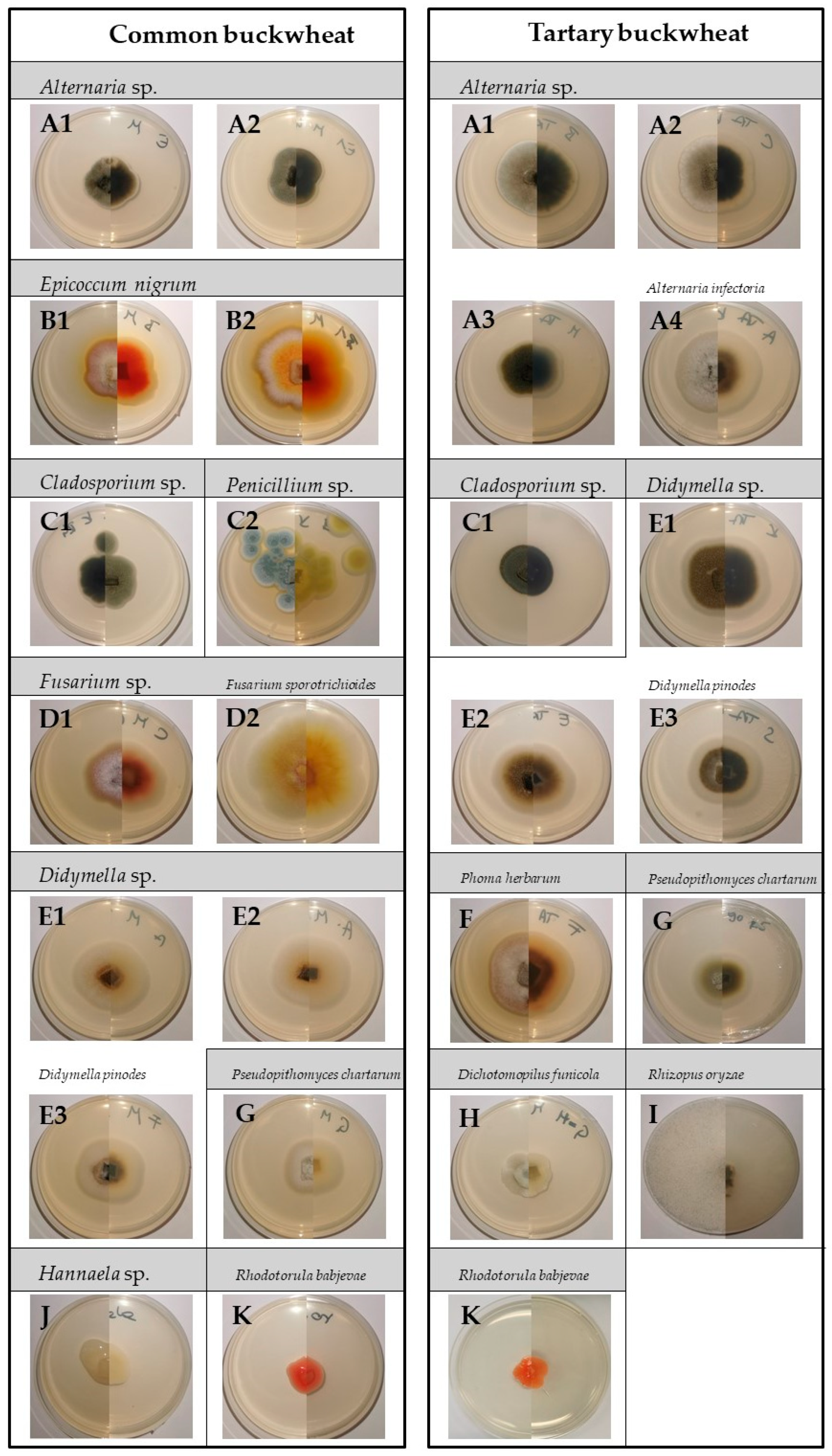
By morphology, the fungal isolates were classified into nine morphotypes and 14 subtypes in CB and seven morphotypes with 13 subtypes in TB (Figure 6). A total of 179 different fungal strains were isolated from buckwheat seeds, of which 75 isolates were obtained from CB and 104 from TB.
Two or three (for more abundantly represented fungi) specimens of each sub-morphotype were selected for molecular identification: a total of 29 from CB and 25 from TB. The sequences were deposited in the GenBank database under accession numbers MW332066—MW332094 (CB isolates) and MW336993—MW337017 (TB isolates) (Supplementary Table S2).
The buckwheat seeds harboured a distinct seed fungal community. In the control seeds of TB, genera Alternaria and Didymella represented the vast majority (90%) of the fungal colonists. In CB, however, there was greater fungal diversity, with the genera Didymella and Epiccocum comprising more than half of all fungal colonists. The CP treatments affected the seed fungal community structure in both buckwheat species. After 120 s CP treatment, less than 50% of seeds of both buckwheat species were colonised by fungi (Figure 7). Overall, most fungi colonising TB seeds were filamentous fungi, while yeasts accounted for up to 6% of all fungal colonisers (genus Rhodotorula), regardless of the CP exposure length. In contrast, yeasts were more common colonisers in CB (predominantly genus Hannaella), accounting for between 15% (in control seeds) and 80% (in 120 s CP treatment) of all colonisers. Among filamentous fungi, the genus Alternaria predominated in TB seeds, accounting for between 40% and almost 84% of the total fungal community in 120 s CP treatment. We have found at least six morphologically distinct types of Alternaria isolates, but only A. infectoria was identified down to the species level. In CB, the predominant colonisers belonged to Didymellaceae, with Epicoccum nigrum accounting for 12%–31% and a species from the genus Didymella (formerly known as Phoma) accounting for 10%–38% of all fungi. At the same time, Didymella species were also the second most common colonisers of TB seeds, accounting for 17% to over 30% of all fungi. As with isolates identified as Alternaria sp., Didymella species were also morphologically diverse and grouped into six submorphotypes. Interestingly, in TB and CB, the isolates of Didymella sp. were found only in up to 90 s CP exposures, which was also true for most other less common fungal genera. This suggests that most of the filamentous fungi contaminating buckwheat seeds may be sensitive to 120 s CP exposure, except for Alternaria sp. and Epicoccum nigrum. Fungi from the genus Cladosporium were also isolated from CB and TB, but less frequently. A common member of the filamentous fungi found in TB was also Rhizopus oryzae, which accounted for about 5% to 10% of the total fungal communities per particular CP treatment. Fungi from the genus Fusarium were also found in CB seeds, but in smaller amounts (up to 9%). Other species, rarely isolated, included Phoma herbarum, Pithomyces chartarum and Dichotomopilus funicola. In H2O2 surface-sterilised group of seeds (SC), only Hannaella sp. (yeasts) was isolated from CB and Alternaria sp. from TB seeds. All molecularly identified taxa of fungi in both buckwheat species are listed in Supplementary Table S1.

3. Discussion

3.1. Effect of Cold Plasma Treatment on Seed Germination

Significant progress in the use of cold plasma in agriculture has been observed in the last 20 years. Many studies reported possible positive effects of CP treatment on seed and grain production, indicating that CP treatment can stimulate germination, improve seedling vigour and growth dynamics, and also significantly inhibit pathogen growth by reducing surface colonisation [31,32,33,34,35,36,37,38,39]. CP treatment affects different morphological, physiological and biochemical properties of the seeds [40] and can therefore significantly influence the overall plant production. However, most of the studies focused only on the most commonly grown and studied crops worldwide, such as wheat, maize, barley, soybean and chickpea. In chickpea, improved germination has been observed at a shorter CP treatment up to 1 min, while treatments of more than 5 min induced a change in the cotyledon morphology and a consequent loss of seed viability and weaker germination [30]. A positive effect of shorter CP treatments on germination rate and early seedling growth was observed in spring wheat and maize, while no effect was found in blue lupine [37]. Similarly, germination and seedling vigour significantly increased in shorter AP CP treatments (20–50 s) for wheat seeds [38]. Some experiments on AP CP treatment of maize seeds indicate that shorter treatments (40–80 s) have a positive effect on germination [34], while other authors found no significant changes in maize germination at shorter treatments (60–120 s) [39]. On the other hand, it is accepted that longer CP exposure causes a significant decrease in germination and later inhibition of growth parameters in both LP and AP CP systems [33,34,37,38,39]. All these studies confirm that the effectiveness of CP treatment for seed germination varies with the type and condition of the seed and environmental factors such as climate, soil conditions and water availability [31].
To our knowledge, Šera et al. [41] are the only authors who studied the effects of CP treatment in common buckwheat seeds. They found significant effects of different CP plasma devices with air as supply gas and CP exposure lengths on buckwheat germination and early seedling growth. A small positive effect of atmospheric pressure (AP) plasma sustained by gliding arc discharge was observed, and a strong negative effect of Surface Dielectric Barrier Discharge (SDBD) plasma after a shorter (180 s) treatment. With increasing exposure (300 s and 600 s), increasing inhibitory effect on germination and initial growth of buckwheat was confirmed, indicating that buckwheat is very sensitive to CP treatment. In control seeds, the germination rate was 63%, which is similar to our results (68%). In contrast to Šera et al. [41], we found no positive effects of CP treatment on germination in either of the buckwheat species. However, for shorter treatments (15–45 s), the germination rate was comparable to the control group in both species after 7 days. It can thus be concluded that CP treatment of up to 45 s is suitable for buckwheat seeds. On the other hand, Šera et al. observed little positive or no significant effect on germination rate after 180 s of treatment for most of the tested CP devices, with the exception of SDBD [41]. In our experiments, a 60 s CP treatment resulted in significantly reduced germination in both buckwheat species, with CB more susceptible than TB. Longer treatments (90 s and 120 s) completely reduced the germination rate in both species. This is probably due to a higher input power producing a higher electron density, more UV radiation and more heat [42], affecting the buckwheat seeds already at shorter treatments. However, we did not measure temperature during our experiments and this is something that needs to be determined and further studied in future research. In general, TB was more tolerant to longer CP treatments, as 80% of the seeds (relative to the control group) still germinated at 60 s treatment, while the relative germination rate of CB was only about 30%. This could be attributed to higher concentrations of polyphenols in TB [6]. CP treatment acts as a “positive stress” for the plants at shorter exposures, affecting the hormonal activity of the seeds [36] and possibly causing oxidative stress at longer exposures. Differences in flavonoid concentrations and flavonoid metabolism between CB and TB have been previously linked to environmental factors such as increased UV-B radiation in locations where TB was originally cultivated [43]. TB plants originate from higher altitudes and contain higher amounts of compounds that protect against UV radiation, possibly making them more tolerant of UV radiation in CP.

3.2. Effect of Cold Plasma Treatment on Fungal Decontamination

The use of gaseous plasma is known as an alternative sterilisation technique [44], and it was suggested as a potential tool for the decontamination of toxic fungi from the seed surface [45]. Plasma sterilisation uses gases that have no germicidal properties and only become biocidal when plasma is formed. Low pressure (LP) plasmas, as in our case, have been considered for biological sterilisation for quite some time [46]. CP treatment generates many antimicrobial active species, including ozone, monoatomic oxygen, free radicals (superoxide, hydroxyl, nitric oxide, etc.) and ultraviolet radiation [45]. Initially, it was proposed that UV photons play a crucial role in microbial sterilisation [47], but a few years later, the contribution of oxygen atoms (as oxidants) was demonstrated [48]. Now it is recognized that microbial destruction results from the synergistic effect of oxygen atoms and UV photons generated by the gas plasma [49]. Microbial sterilisation in CP treatment is, thus, a result of the direct contact of plasma species (positively charged ions and free electrons) and UV radiation, which is still considered the main sterilisation factor of most CP systems.
Some authors have used different types of AP CP devices to reduce native fungi on various types of seeds [30,36,38,39,50]. All conducted studies have found a significant reduction in naturally occurring fungi with increasing duration of CP treatment. However, no study has been yet conducted on the effects of low-pressure CP on native seed-borne fungal microbiota. In our study, a significant reduction of the fungal frequency of seed-borne fungi was observed after 90 s for CB and after 120 s for TB seeds. In both buckwheat species, a fungal reduction greater than 50% compared to the control group was found after 120 s CP treatment. Similarly, a longer treatment time also resulted in a greater fungal reduction in other studies. Some of them were able to completely decontaminate all naturally occurring filamentous fungi and yeasts, as was observed after 120 s treatment in wheat seeds [38] and after 180 s in maize seeds [39] with AP CP. However, we cannot directly compare our results because a different plasma setup system and different seeds were used in our study. Our results also indicate that overall evaluation of fungal contamination of seeds based on Petri dish area covered with fungi is probably not the most accurate, as obvious in the case of CB seeds, where a clear shift between communities of fast-growing filamentous fungi and slowly growing yeast was observed after longer CP exposures.
When considering decontamination by CP, three basic mechanisms synergistically contribute to microbial inactivation in medium and low-pressure CP systems [51]: the direct destruction of genetic material (DNA) by UV radiation; the erosion of microbial matter from the surface of plant cells (atom by atom) by intrinsic photodesorption; and etching caused by reactive species generated in plasma enhanced by UV radiation. In low-pressure plasmas (below 10 mbar), UV photons dominate the microbial inactivation process, resulting in shorter sterilization times. Especially in oxygen-containing plasma mixtures, this effect can be significantly accelerated by the simultaneous erosion of microbial cells by reactive oxygen species. As the erosion of microbial matter progresses, the number of UV photons successfully interacting with microbial DNA increases, resulting in a shorter time required for complete inactivation of the microorganisms [52,53]. This was also confirmed in some studies, all of which indicated that in the first phase, UV radiation plays the primary role, while in the second phase, charged plasma particles become the leading inactivating agent [44,54,55,56].
Our plasma was generated with a radiofrequency (RF) generator in inductively coupled (IC) mode, which is more efficient in microbial decontamination than its alternative, the capacitively coupled mode. This is attributed to higher electron and ion densities formed in IC mode, which ultimately leads to better enhancement of synergistic mechanisms involving electrons (surface etching) [42]. Furthermore, biological materials can be exposed to plasma in two different modes of operation: “direct exposure” (glow), where the treated sample is in direct contact with the plasma and all plasma-generated species are in contact with the sample; and “indirect or remote exposure” (afterglow), in which the sample is placed at a distance from the plasma source or in an adjacent chamber. In the latter, the amount of transmitted heat, as well as some short-lived plasma species reaching the sample, is greatly reduced [46]. In our case, direct exposure was used, leading to greater efficiency of decontamination and resulting in shorter sterilisation times [44,52].
The effectiveness of CP decontamination is, however, also limited by the penetration depth of the UV photons, as they must reach the microbial DNA. Therefore, a shortcoming of CP sterilisation is its dependence on the actual “thickness” of the microorganisms to be inactivated [52] and on the surface structure of the treated material, which can lead to non-homogenous sterilisation [57]. It has been pointed out that the decontamination effect depends not only on the gas used, plasma parameters and exposure length but also on the type of contaminated seeds, where shape and surface play a major role [14,58]. This might be the reason for the better efficacy of CP treatment on CB than on TB in our study. The seeds of TB are more irregularly shaped and have a more wrinkled surface than those of CB, which provides more space for microbial colonisation and makes them less accessible to plasma. Seed wrinkles and crevices have already been mentioned as a possible factor creating a barrier for CP to reach all microflora [59,60]. It was suggested by Mitra et al. [30] to rotate the samples during CP to ensure uniform exposure of the entire seed surface to the plasma. Fungi can also be present in seeds as endophytes, located under the seed coat and cannot be reached by the CP species, strongly influencing the seed sterilization by CP.
Another factor that should be considered when treating thermally sensitive biological samples, such as seeds, is the possible side effect of plasma treatment, namely sample heating. Although it was reported that there was a minimal effect of thermal degradation by LP oxygen CP generated in IC RF mode as in our case [42], some authors pointed out that sample heating could have an influence. Greater sterilisation efficiency with increasing plasma exposure time may depend on oxygen species alone, but sample heating can occur due to radical interactions with the substrate. It was shown that the temperature accelerates the sterilisation and assists the oxygen atoms in the etching process of the bacterial cells. On the other hand, this also limits the exposure time since the surface of the treated object can become strongly heated. Further studies should be conducted to carefully monitor the temperature of biological material during plasma treatment [55].

3.3. Effect of Cold Plasma Treatment on Fungal Diversity

In this study, we attempted to determine how CP treatment affects the fungal community structure of buckwheat seeds. Thus far, the information on fungal colonisers of buckwheat seeds is very scarce [15,16,17,18,61]. Our results suggest that CP affects the fungal community structure of the buckwheat seeds, as fewer fungi were isolated and their diversity was reduced with prolonged CP treatment, especially in CB. Alternaria sp. and Didymella sp. were the predominant fungal colonisers of both buckwheat species. Especially in TB, the genus Alternaria was predominant after all CP exposures, with at least four different morphotypes belonging to four different species. However, only Alternaria infectoria was clearly distinguished from other Alternaria sp. isolates, as it has been discovered that in ITS identification of Alternaria species, A. infectoria can be clearly distinguished from the A. alternaria species complex [62,63]. The latter contains some very similar common plant pathogens indistinguishable in ITS sequencing [64]. For this reason, our isolates were identified as Alternaria sp. but are very likely members of Alternaria sect. alternaria, which includes some of the most common plant saprophytes, such as A. alternaria, A. tenuissima, and A. arborescens. Similarly to our results, the genus Alternaria was, on average, the most common coloniser of freshly harvested buckwheat seeds in previous studies [18] together with genera Botrytis and Cladosporium [17]. Alternaria species were also the predominant colonisers of both CB and TB seeds in the study of Kovačec et al., where their proportion increased during storage in comparison to other genera [16]. That can be problematic as Alternaria species are known phytopathogenic fungi that cause many plant diseases such as black point of some grain cereals, black rot of tomato, black and grey rot of citrus fruits and many others [21]. Epicoccum species were also found on CB seeds [16,17], but not as frequently as in our experiment. Interestingly, we found no Epicoccum sp. on TB seeds. This could be a coincidence or the fact that TB seeds were predominantly colonised by Alternaria sp., which has been shown to be an antagonistic fungus to Epicoccum sp. [16].
Although less frequently isolated, some other genera such as Cladosporium, Penicillium, Phoma, Pithomyces and Rhizopus, all of which have been previously found in buckwheat seeds [17,18], were also identified during our investigation. Fusarium species, the common colonisers of buckwheat seeds [17,18,61] were also confirmed, though, interestingly, only in CP treatments up to 60 s in CB and not found in TB seeds. This is consistent with studies that found Fusarium sp. to be one of the most sensitive fungi to AP CP, with a large or almost complete reduction observed after only 60 s [38,39]. That is a good indication since Fusarium species are fungal pathogens of many agriculturally important host plants occurring worldwide, known for causing the wilting of young and adult plants of many species such as tomato, onion and garlic [65]. Besides that, they are also capable of producing mycotoxins [65] that are harmful for human health. Not only Fusarium sp. but also some other fungi isolated in our experiments, especially the genus Alternaria, are capable of producing mycotoxins, with A. alternata being one of the most problematic, as it can synthesise many different mycotoxins and intoxicate food and feed [23]. A. alternata was also the most frequently isolated fungal pathogen from onion seeds treated with CP, regardless of exposure time [50]. Similarly, it was also the most resistant fungal contaminant of maize seeds to AP CP treatment, with even 300 s treatment time failing to achieve complete devitalisation [39]. In light of this, further studies should be carried out to find the appropriate CP parameters to achieve its complete reduction.
Besides filamentous fungi, yeasts (belonging to the genera Hannaella and Rhodotorula) were frequently isolated from CB seeds and became predominant during prolonged CP exposure, accounting for more than half of all fungal contaminants after 90 s and 120 s of CP treatment. Interestingly, yeasts were only seldomly isolated from TB seeds. It is known that yeasts and spores are more resistant to CP treatment because they have extremely thick polysaccharide cell walls [45]. Some authors who compared the antimicrobial efficacy of CP treatment also concluded that fungi (especially yeasts) are more resistant to CP treatment than bacteria, as the complete destruction of bacteria was found to occur in shorter periods than fungi [39,57].
CP treatment has many advantages over conventional methods of sterilisation. It minimises damage to the treated materials since they are not subjected to high temperature or pressure (as in steam sterilisation) or radiation and toxic chemicals such as ethylene oxide, thus posing little risk to personnel and the environment [44,45]. The results of many studies conducted so far indicate that CP could be utilised as a new and alternative technology for seed processing, under optimal plasma source and treatment parameters. The optimum exposure time that resulted in a significant reduction in naturally occurring seed-borne fungi without affecting seed viability was found at 10 s CP treatment for the wheat [66] and 20 s CP treatment for sweet basil seeds [67], respectively. In both experiments, an AP CP system was used with air as the feed gas. Within our experimental conditions, we failed to find an optimum CP exposure that would significantly reduce fungal colonisation and maintain a sufficient germination rate of buckwheat seeds. Therefore, this type of CP treatment would be suitable for surface decontamination of buckwheat grains for the food industry but not for agricultural use. As already pointed out, longer CP exposures were successful in reducing naturally occurring fungi on various seeds but were not effective in seed enhancement because of their negative influence on seed germination and viability [36]. This calls for further studies on different CP systems and working parameters to adjust and find the optimum CP treatments for each plant species to be used for commercial purposes.

4. Materials and Methods

4.1. Buckwheat Seeds Origin

Seeds of CB (Fagopyrum esculentum Moench) and TB (Fagopyrum tataricum Gaertn.) were obtained from Rangus Mill (located in Šentjernej, south-eastern Slovenia, about 230 m a.s.l.). They were harvested in 2018 and stored under suitable conditions (in a dry and dark environment at room temperature) until the experiments were performed in 2020.

4.2. Cold Plasma Treatment

A custom-made large-scale radiofrequency (RF) plasma system [68] was used in our experiments (Figure 8). The system is pumped with a two-stage rotary vacuum pump of pumping speed 80 m3·h−1. The ultimate pressure achievable in the reactor after the evacuation was about 1 Pa. The system was hermetically tight, so the residual atmosphere contained practically only water vapour. The length of the discharge tube was about 2 m, and the inner diameter 0.18 m, so the volume of the plasma reactor was about 50 litres. A typical optical spectrum is shown in Figure 9. By far the most extensive radiation arises from O-atoms, and there is also a weak molecular band at about 309 nm, which arises from OH-radicals. Other spectral features belong to H-atom transitions (Balmer series). Such a spectrum is typical for highly dissociated oxygen plasma. Seeds, approx. 200 in number, were evenly spread on a metallic mesh and placed inside the tube in the coil area. Plasma was sustained at a pressure of 50 Pa–52 Pa and a power of 1500 W. A copper coil was connected to an RF generator via a matching network. The RF generator operated at a frequency of 27.12 MHz. The power density was, therefore, only 30 W L−1. Oxygen gas with a purity of 99.99% was introduced into the plasma system at a constant flow rate of 202 standard cm3 min−1. The seeds were treated with CP for 15, 30, 45, 60, 90, and 120 s. Immediately after plasma treatment, the seeds were packaged in sterile PVC bags to prevent environmental contamination.

4.3. Germination Test

In germination tests, 20 seeds from a given plasma treatment or untreated seeds were placed into Petri dishes (diameter 70 mm) with two layers of filter paper and moistened with sterile distilled water. Each test was performed in five replicates. The seeds were incubated in plant growth chambers at 22 °C, 60% humidity, in the dark. If necessary, sterile distilled water was added to the Petri dishes to maintain constant humidity during germination. The germination rate (GR%) was measured every 24 h for the first three days and then after one week (on the 7th day). It was calculated according to the following Equation (1). The penetration of the radicle through the seed coat was used as the criterion for germination.
GR   ( % ) = ( number   of   germinated   seeds ) / ( total   number   of   seeds )   ×   100
Mean germination time (MGT) was calculated according to the equation of Ellis and Roberts (1981) [69] as under:
MGT = ΣD × n/Σn
where, n is the number of seeds, which were germinated on day D, and D is the number of days counted from the beginning of germination.
The time to 50% germination (T50) was calculated according to the following formula of Coolbear et al. (1984) modified by Farooq et al. (2005) [70] as:
T50 = ti + [(N/2 − ni) (ti − tj)]/(ni − nj)
where N is the final number of germination and ni, nj cumulative number of seeds germinated by adjacent counts at times ti and tj when ni < N/2 < nj.

4.4. Effect of CP on Seed-Borne Fungi Decontamination

To test the effect of CP treatment on buckwheat seed-borne fungi, seeds from all CP treatments and untreated seeds were analysed as described below. Chemically surface-sterilised seeds (with 30% H2O2 and 2 drops of Tween 80 for 20 min) were analysed in parallel to assess the efficacy of the CP treatment as a surface decontaminant.

4.5. Cultivation of Seed-Borne Fungi

The agar plate or “Ulster” method was used to assess seed colonisation and fungal growth [71]. Seeds were planted on sterile plastic Petri dishes (diameter 70 mm) filled with 2% potato dextrose agar (PDA) supplemented with the antibiotic chloramphenicol (50 mg L−1) to prevent bacterial growth. One buckwheat seed was placed in the centre of each Petri dish to assess fungal growth. Plates were incubated in growth chambers at a temperature of 22 °C in dark conditions for one week.

4.6. Seed Colonisation and Morphological Characterisation of Fungi

After one week of cultivation, the plates were examined to measure the percentage of colonised seeds, average fungal area and fungal diversity (morphotypes). Fungal area per plate was determined at day 7 from the mycelial radius and expressed as the percentage of the colonised area diameter relative to the diameter of the Petri dish. Fungi were visually examined and grouped by their morphological appearance (morphotypes and sub-morphotypes). The percentage of colonised seeds (CS), the average fungal area (FA), the average number of morphotypes (MN) and the frequency of each morphotype (MF) for each treatment were calculated according to Equations (4), (5), (6) and (7), respectively:
CS   ( % ) = ( number   of   colonised   seeds ) / ( total   number   of   seeds )   ×   100
FA   ( % ) = ( diameter   of   fungal   area   [ mm ] ) / ( diameter   of   Petri   dish   [ mm ] )   ×   100
MN = ( number   of   morphotypes   per   plate ) / ( total   number   of   morphotypes )
MF   ( % ) = ( isolates   of   the   morphotype ) / ( all   fungal   isolates )   ×   100
Fungal isolates were repeatedly sub-cultured until pure cultures were obtained.

4.7. Molecular Identification

For molecular identification, at least 2 (3 for more frequent fungi) pure culture isolates of each sub-morphotype were selected (if possible, from different CP treatments). Fungal material was scraped from agar plates and crushed in liquid nitrogen using a mortar and pestle. Fungal genomic DNA was isolated using GenElute® Plant Genomic DNA Miniprep Kit (Sigma-Aldrich, St. Louis, MO, USA) according to the manufacturer’s instructions. Fungal DNA (ITS region) was amplified using the primer pair ITS1f/ITS4 and Taq polymerase in an MJ Research® thermal cycler. The reaction mixtures and PCR conditions were performed according to Likar and Regvar [72]. Purification and sequencing of the PCR products were performed by Macrogen Inc. (Amsterdam, The Netherlands). For further identification, the sequences were subjected to a nucleotide BLAST® search within the NCBI database. After successful identification, the sequence data were processed using the MEGA-X software and submitted to the GenBank database.

4.8. Statistical Analysis

The reported results are expressed as mean ± standard error (SE) of five replicates (for germination tests) and at least ten replicates (for fungal growth analyses). Statistical significance between groups (treatments) was determined using a one-way analysis of variance (ANOVA) with Duncan’s post hoc test (Statistica StatSoft version 7). The significance level was considered at a p-value of less than 0.05.

5. Conclusions

Chemical agents, such as fungicides and other xenobiotics, have been widely used in crop production to eliminate seed-borne pathogens. There is a great need for new, environmentally friendly and economically sustainable technologies to reduce the use of pesticides and other toxic chemicals in plant production.
The results presented in this paper show that low-pressure cold plasma treatment affects germination and seed-borne fungal community structure in seeds of common and Tartary buckwheat species. Shorter exposures (up to 45 s) can be safely applied, as they had no significant effect on seed germination in either of the buckwheat species, while longer exposures reduced the germination rate in both species. TB seeds were found to be more resistant to longer CP exposures than CB seeds.
Only the longest CP exposures (120 s) significantly affected the degree of fungal colonisation and diversity in seeds of both buckwheat species. The genera Alternaria, Didymella and Epicoccum were the most common fungi isolated from the seeds. Alternaria was prevalent in TB, and our results suggest that the genus is (beside genus Epicoccum) also the most resistant to the given CP treatment. This may prove problematic as Alternaria species are known sources of mycotoxins harmful to humans and animals.
CP treatment in general seems to be a promising alternative to remove surface contamination and improve seed germination, thus increasing the overall plant production. However, further research is needed to define the optimum CP treatment parameters and adequately address the problem of contaminated seeds to ensure global food security, provide safer food, and reduce the impact of agricultural production on the environment.

Supplementary Materials

The following are available online at https://www.mdpi.com/article/10.3390/plants10050851/s1, Table S1: Preliminary germination tests, Table S2: Molecular identifications of isolates.

Author Contributions

Conceptualization, J.M. and K.V.-M.; investigation, J.M. and P.S.; writing—original draft preparation, J.M.; writing—review and editing, J.M., K.V.-M., M.M. and M.R.; visualisation, J.M.; supervision, K.V.-M., M.M.; project administration, K.V.-M., M.M. All authors have read and agreed to the published version of the manuscript.

Funding

This research was funded by Slovenian Research Agency (ARRS): Young researcher grants (Jure Mravlje and Pia Starič), program groups (P1-0212-Plant biology and P2-0082-Thin-film structures and plasma surface engineering) and projects J7-9418, N1-0105 and L4-9305.

Data Availability Statement

The genetic data presented in this study are in a publicly accessible repository available in Supplementary Table S1.

Acknowledgments

The authors are grateful to the members of the Laboratory for Plant Physiology (at the Department of Biology), especially our technician Milena Kubelj and students Neja Bizjak, Anja Mavrič, Tjaša Trajbarič, Marija Jovchevska, Miha Lampe and Žiga Doljak who helped us with laboratory work. The buckwheat seeds used in this study were kindly provided by Rangus Mill (Slovenia).

Conflicts of Interest

The authors declare no conflict of interest.

References

  1. Kay, J.G.; Owen, H. Transmission of Rhynchosporium secalis on barley grain. Trans. Br. Mycol. Soc. 1973, 60, 405–411. [Google Scholar] [CrossRef]
  2. Rioux, R.A.; Shultz, J.; Garcia, M.; Willis, D.K.; Casler, M.; Bonos, S.; Smith, D.; Kerns, J. Sclerotinia homoeocarpa Overwinters in turfgrass and is present in commercial seed. PLoS ONE 2014, 9, e110897. [Google Scholar] [CrossRef] [PubMed]
  3. Christensen, C.M. Deterioration of stored grains by fungi. Bot. Rev. 1957, 23, 108–134. [Google Scholar] [CrossRef]
  4. Halloin, J.M. Deterioration resistance mechanisms in seeds. Phytopathology 1983, 73, 335–339. [Google Scholar] [CrossRef]
  5. Bonafaccia, G.; Marocchini, M.; Kreft, I. Composition and technological properties of the flour and bran from common and tartary buckwheat. Food Chem. 2003, 80, 9–15. [Google Scholar] [CrossRef]
  6. Bonafaccia, G.; Fabjan, N. Nutritional comparison of tartary buckwheat with common buckwheat and minor cereals. Zb. Bioteh. Fak. Univ. Ljublj. Kmet 2003, 81, 349–355. [Google Scholar]
  7. Bonafaccia, G.; Gambelli, L.; Fabjan, N.; Kreft, I. Trace elements in flour and bran from common and tartary buckwheat. Food Chem. 2003, 83, 1–5. [Google Scholar] [CrossRef]
  8. Kreft, I.; Fabjan, N.; Yasumoto, K. Rutin content in buckwheat (Fagopyrum esculentum Moench) food materials and products. Food Chem. 2006, 98, 508–512. [Google Scholar] [CrossRef]
  9. Skerritt, J.H. Molecular comparison of alcohol-soluble wheat and buchwheat proteins. Cereal Chem. 1986, 63, 365–369. [Google Scholar]
  10. Kalinova, J.; Triska, J.; Vrchotova, N. Distribution of vitamin e, squalene, epicatechin, and rutin in common buckwheat plants (Fagopyrum esculentum Moench). J. Agric. Food Chem. 2006, 74, 5330–5335. [Google Scholar] [CrossRef]
  11. Popović, V.; Sikora, V.; Berenji, J.; Filipović, V.; Dolijanović, Ž.; Ikanović, J.; Dončić, D. Analysis of buckwheat production in the world and Serbia. Ekon. Poljopr. 2014, 61, 53–62. [Google Scholar] [CrossRef]
  12. Los, A.; Ziuzina, D.; Akkermans, S.; Boehm, D.; Cullen, P.J.; Van Impe, J.; Bourke, P. Improving microbiological safety and quality characteristics of wheat and barley by high voltage atmospheric cold plasma closed processing. Food Res. Int. 2018, 106, 509–521. [Google Scholar] [CrossRef] [Green Version]
  13. Misra, N.; Schluter, O.; Cullen, P. Cold Plasma in Food and Agriculture, 1st ed.; Academic Press: Cambridge, MA, USA, 2016; ISBN 9780128013656. [Google Scholar]
  14. Selcuk, M.; Oksuz, L.; Basaran, P. Decontamination of grains and legumes infected with Aspergillus spp. and Penicillum spp. by cold plasma treatment. Bioresour. Technol. 2008, 99, 5104–5109. [Google Scholar] [CrossRef]
  15. Milevoj, L. Buckwheat diseases. In Fagopyrum (Buckwheat newsletter); Kreft, I., Ed.; Biotehniška Fakulteta: Ljubljana, Yugoslavia, 1989; Volume 9, pp. 31–40. [Google Scholar]
  16. Kovačec, E.; Likar, M.; Regvar, M. Temporal changes in fungal communities from buckwheat seeds and their effects on seed germination and seedling secondary metabolism. Fungal Biol. 2016, 120, 666–678. [Google Scholar] [CrossRef]
  17. Mills, J.T.; Wallace, H.A.H. Microflora of buckwheat seed, changes in storage and effect of seed treatments on seedling emergence. Can. Plant Dis. Surv. 1971, 51, 154–158. [Google Scholar]
  18. Singh, P.N.; Sindhu, I.R.; Singhal, G. Fungi recorded from seeds and seedlings of Fagopyrum esculentum. J. Indian Bot. Soc. 1984, 63, 236–243. [Google Scholar]
  19. Fung, F.; Clark, R.F. Health effects of mycotoxins: A toxicological overview. J. Toxicol. Clin. Toxicol. 2004, 42, 217–234. [Google Scholar] [CrossRef] [PubMed]
  20. Paterson, R.R.M.; Lima, N. Toxicology of Mycotoxins; Luch, A., Ed.; Birkhäuser: Basel, Switzerland, 2010; Volume 100. [Google Scholar]
  21. Logrieco, A.; Moretti, A.; Solfrizzo, M. Alternaria toxins and plant diseases: An overview of origin, occurrence and risks. World Mycotoxin J. 2009, 2, 129–140. [Google Scholar] [CrossRef]
  22. Miller, J.D. Fungi and mycotoxins in grains: Implication for stored roducts research. J. Stored Prod. Res. 1995, 31, 1–16. [Google Scholar] [CrossRef]
  23. Ostry, V. Alternaria mycotoxins: An overview of chemical characterization, producers, toxicity, analysis and occurrence in foodstuffs. World Mycotoxin J. 2008, 1, 175–188. [Google Scholar] [CrossRef]
  24. Soriano, J.M.; Dragacci, S. Occurrence of fumonisins in foods. Food Res. Int. 2004, 37, 985–1000. [Google Scholar] [CrossRef]
  25. FAO United Nations. How to Feed the World in 2050 Executive; FAO United Nations: Rome, Italy, 2009. [Google Scholar]
  26. Piel, A. Plasma Physics; Springer: Berlin/Heidelberg, Germany, 2010; Volume 53, ISBN 9788578110796. [Google Scholar]
  27. Tendero, C.; Tixier, C.; Tristant, P.; Desmaison, J.; Leprince, P. Atmospheric pressure plasmas: A review. Spectrochim. Acta Part B At. Spectrosc. 2006, 61, 2–30. [Google Scholar] [CrossRef]
  28. Conrads, H.; Schmidt, M. Plasma generation and plasma sources. Plasma Sources Sci. Technol. 2000, 9, 441–454. [Google Scholar] [CrossRef] [Green Version]
  29. Filatova, I.; Azharonok, V.; Gorodetskaya, E.; Mel, L.; Shedikova, O.; Shik, A. Plasma-Radiowave Stimulation of Plant Seeds Germination and Inactivation of Pathogenic Microorganisms. IPSC_19. 2009, pp. 4–7. Available online: https://www.semanticscholar.org/paper/Plasma-radiowave-stimulation-of-plant-seeds-and-of-Filatova-Azharonok/503c47acd4f1b4008ec50a6fbf4aac47eb9fe940 (accessed on 3 February 2021).
  30. Mitra, A.; Li, Y.; Klämpfl, T.G.; Shimizu, T.; Jeon, J.; Morfill, G.E.; Zimmermann, J.L. Inactivation of surface-borne microorganisms and increased germination of seed specimen by cold atmospheric plasma. Food Bioprocess Technol. 2014, 7, 645–653. [Google Scholar] [CrossRef] [Green Version]
  31. Randeniya, L.K.; De Groot, G.J.J.B. Non-thermal plasma treatment of agricultural seeds for stimulation of germination, removal of surface contamination and other benefits: A review. Plasma Process. Polym. 2015, 12, 608–623. [Google Scholar] [CrossRef]
  32. Živković, S.; Puač, N.; Giba, Z.; Grubišić, D.; Petrović, Z.L. The stimulatory effect of non-equilibrium (low temperature) air plasma pretreatment on light-induced germination of Paulownia tomentosa seeds. Seed Sci. Technol. 2004, 32, 693–701. [Google Scholar] [CrossRef]
  33. Volin, J.C.; Denes, F.S.; Young, R.A.; Park, S.M.T. Modification of seed germination performance through cold plasma chemistry technology. Crop Sci. 2000, 40, 1706–1718. [Google Scholar] [CrossRef]
  34. Henselová, M.; Slováková, Ľ.; Martinka, M.; Zahoranová, A. Growth, anatomy and enzyme activity changes in maize roots induced by treatment of seeds with low-temperature plasma. Biologia 2012, 67, 490–497. [Google Scholar] [CrossRef]
  35. Bormashenko, E.; Grynyov, R.; Bormashenko, Y.; Drori, E. Cold radiofrequency plasma treatment modifies wettability and germination speed of plant seeds. Sci. Rep. 2012, 2. [Google Scholar] [CrossRef]
  36. Filatova, I.; Azharonok, V.; Shik, A.; Antonuk, A.; Terletskaya, N. Fungicidal Effects of Plasma and Radio-Wave Pre-treatments on Seeds of Grain Crops and Legumes. NATO Sci. Peace Secur. Ser. A Chem. Biol. 2012, 469–479. [Google Scholar] [CrossRef]
  37. Filatova, I.; Azharonok, V.; Lushkevich, V.; Zhukovsky, A.; Gadzhieva, G.; Spasi, K. Plasma Seeds Treatment as a Promising Technique for Seed Germination Improvement. 31st ICPIG. 2013, pp. 4–7. Available online: https://www.semanticscholar.org/paper/Plasma-seeds-treatment-as-a-promising-technique-for-Filatova-Azharonok/5e45e5f0a890d8e65a4936292d55173fdddd6c95 (accessed on 3 February 2021).
  38. Zahoranová, A.; Henselová, M.; Hudecová, D.; Kaliňáková, B.; Kováčik, D.; Medvecká, V.; Černák, M. Effect of cold atmospheric pressure plasma on the wheat seedlings vigor and on the inactivation of microorganisms on the seeds surface. Plasma Chem. Plasma Process. 2016, 36, 397–414. [Google Scholar] [CrossRef]
  39. Zahoranová, A.; Hoppanová, L.; Šimončicová, J.; Tučeková, Z.; Medvecká, V.; Hudecová, D.; Kaliňáková, B.; Kováčik, D.; Černák, M. Effect of cold atmospheric pressure plasma on maize seeds: Enhancement of seedlings growth and surface microorganisms inactivation. Plasma Chem. Plasma Process. 2018, 38, 969–988. [Google Scholar] [CrossRef]
  40. Starič, P.; Vogel-Mikuš, K.; Mozetič, M.; Junkar, I. Effects of nonthermal plasma on morphology, genetics and physiology of seeds: A review. Plants 2020, 9, 1736. [Google Scholar] [CrossRef] [PubMed]
  41. Šerá, B.; Gajdová, I.; Černák, M.; Gavril, B.; Hnatiuc, E.; Kováčik, D.; Kříha, V.; Sláma, J.; Šerý, M.; Špatenka, P. How various plasma sources may affect seed germination and growth. In Proceedings of the 2012 13th International Conference on Optimization of Electrical and Electronic Equipment (OPTIM), Brasov, Romania, 24–26 May 2012; pp. 1365–1370. [Google Scholar] [CrossRef]
  42. Bol’shakov, A.A.; Cruden, B.A.; Mogul, R.; Rao, M.V.V.S.; Sharma, S.P.; Khare, B.; Meyyappan, M. Radio-frequency oxygen plasma as a sterilization source. AIAA J. 2004, 42, 823–832. [Google Scholar] [CrossRef]
  43. Regvar, M.; Bukovnik, U.; Likar, M.; Kreft, I. UV-B radiation affects flavonoids and fungal colonisation in Fagopyrum esculentum and F. tataricum. Cent. Eur. J. Biol. 2012, 7, 275–283. [Google Scholar] [CrossRef]
  44. Moreira, A.J.; Mansano, R.D.; De Jesus Andreoli Pinto, T.; Ruas, R.; Da Silva Zambon, L.; Da Silva, M.V.; Verdonck, P.B. Sterilization by oxygen plasma. Appl. Surf. Sci. 2004, 235, 151–155. [Google Scholar] [CrossRef]
  45. Montie, T.C.; Kelly-Wintenberg, K.; Reece Roth, J. An overview of research using the one atmosphere uniform glow discharge plasma (OAUGDP) for sterilization of surfaces and materials. IEEE Trans. Plasma Sci. 2000, 28, 41–50. [Google Scholar] [CrossRef]
  46. Laroussi, M. Low temperature plasma-based sterilization: Overview and state-of-the-art. Plasma Process. Polym. 2005, 2, 391–400. [Google Scholar] [CrossRef]
  47. Chau, T.T.; Kao, K.C.; Blank, G.; Madrid, F. Microwave plasmas for low-temperature dry sterilization. Biomaterials 1996, 17, 1273–1277. [Google Scholar] [CrossRef]
  48. Kelly-Wintenberg, K.; Montie, T.C.; Brickman, C.; Roth, J.R.; Carr, A.K.; Sorge, K.; Wadsworth, L.C.; Tsai, P.P.Y. Room temperature sterilization of surfaces and fabrics with a one atmosphere uniform glow discharge plasma. J. Ind. Microbiol. Biotechnol. 1998, 20, 69–74. [Google Scholar] [CrossRef]
  49. Moreau, S.; Moisan, M.; Tabrizian, M.; Barbeau, J.; Pelletier, J.; Ricard, A.; Yahia, L. Using the flowing afterglow of a plasma to inactivate Bacillus subtilis spores: Influence of the operating conditions. J. Appl. Phys. 2000, 88, 1166–1174. [Google Scholar] [CrossRef]
  50. Kopacki, M.; Pawlat, J.; Terebun, P.; Kwiatkowski, M.; Starek, A.; Kiczorowski, P. Efficacy of non-thermal plasma fumigation to control fungi occurring on onion seeds. In Proceedings of the 2017 International Conference on Electromagnetic Devices and Processes in Environment Protection with Seminar Applications of Superconductors (ELMECO & AoS), Naleczow, Poland, 3–6 December 2017; pp. 1–4. [Google Scholar] [CrossRef]
  51. Moisan, M.; Barbeau, J.; Moreau, S.; Pelletier, J.; Tabrizian, M.; Yahia, L.H. Low-temperature sterilization using gas plasmas: A review of the experiments and an analysis of the inactivation mechanisms. Int. J. Pharm. 2001, 226, 1–21. [Google Scholar] [CrossRef]
  52. Philip, N.; Saoudi, B.; Crevier, M.C.; Moisan, M.; Barbeau, J.; Pelletier, J. The respective roles of UV photons and oxygen atoms in plasma sterilization at reduced gas pressure: The case of N2-O2 mixtures. IEEE Trans. Plasma Sci. 2002, 30, 1429–1436. [Google Scholar] [CrossRef]
  53. Moisan, M.; Barbeau, J.; Crevier, M.; Pelletier, J.; Philip, N.; Saoudi, B. Plasma sterilization. Methods and mechanisms. Pure Appl. Chem. 2002, 74, 349–358. [Google Scholar] [CrossRef]
  54. Rossi, F.; Kylián, O.; Hasiwa, M. Decontamination of surfaces by low pressure plasma discharges. Plasma Process. Polym. 2006, 3, 431–442. [Google Scholar] [CrossRef]
  55. Cvelbar, U.; Vujoševič, D.; Vratnica, Z.; Mozetič, M. The influence of substrate material on bacteria sterilization in an oxygen plasma glow discharge. J. Phys. D Appl. Phys. 2006, 39, 3487–3493. [Google Scholar] [CrossRef]
  56. Vicoveanu, D.; Popescu, S.; Ohtsu, Y.; Fujita, H. Competing inactivation agents for bacterial spores in radio-frequency oxygen plasmas. Plasma Process. Polym. 2008, 5, 350–358. [Google Scholar] [CrossRef]
  57. Lee, K.; Paek, K.H.; Ju, W.T.; Lee, Y. Sterilization of bacteria, yeast, and bacterial endospores by atmospheric-pressure cold plasma using helium and oxygen. J. Microbiol. 2006, 44, 269–275. [Google Scholar] [PubMed]
  58. Basaran, P.; Ozcan, M. Occurrence of aflatoxins in various nuts commercialized in Turkey. J. Food Saf. 2009, 29, 95–105. [Google Scholar] [CrossRef]
  59. Caetano-Anollés, G.; Favelukes, G.; Bauer, W.D. Optimization of surface sterilization for legume seed. Crop. Sci. 1990, 30, 708–712. [Google Scholar] [CrossRef]
  60. Charkowski, A.O.; Sarreal, C.Z.; Mandrell, R.E. Wrinkled alfalfa seeds harbor more aerobic bacteria and are more difficult to sanitize than smooth seeds. J. Food Prot. 2001, 64, 1292–1298. [Google Scholar] [CrossRef]
  61. Kalinova, J.; Voženilkova, B.; Moudry, J. Occurrence of Fusarium spp and bacteria on surface of buckwheat achenes (Fagopyrum esculentum Moench). In Proceedings of the 9th International Symposium on Buckwheat, Prague, Czech Republic, 18–22 August 2004; pp. 491–493. [Google Scholar]
  62. Pastor, F.J.; Guarro, J. Alternaria infections: Laboratory diagnosis and relevant clinical features. Clin. Microbiol. Infect. 2008, 14, 734–746. [Google Scholar] [CrossRef] [Green Version]
  63. De Hoog, G.S.; Horré, R. Molecular taxonomy of the Alternaria and Ulocladium species from humans and their identification in the routine Molekulare Taxonomie humaner Isolate von Alternaria und Ulocladium -Arten und ihre Identifizierung im Routinelabor. Mycoses 2002, 276, 259–276. [Google Scholar] [CrossRef] [Green Version]
  64. Woudenberg, J.H.C.; Seidl, M.F.; Groenewald, J.Z.; de Vries, M.; Stielow, J.B.; Thomma, B.P.H.J.; Crous, P.W. Alternaria section Alternaria: Species, formae speciales or pathotypes? Stud. Mycol. 2015, 82, 1–21. [Google Scholar] [CrossRef] [Green Version]
  65. Stankovic, S.; Levic, J.; Petrovic, T.; Logrieco, A.; Moretti, A. Pathogenicity and mycotoxin production by Fusarium proliferatum isolated from onion and garlic in Serbia. Eur. J. Plant Pathol. 2007, 118, 165–172. [Google Scholar] [CrossRef]
  66. Kordas, L.; Pusz, W.; Czapka, T.; Kacprzyk, R. The effect of low-temperature plasma on fungus colonization of winter wheat grain and seed quality. Pol. J. Environ. Stud. 2015, 24, 433–438. [Google Scholar]
  67. Ambrico, P.F.; Šimek, M.; Morano, M.; De Miccolis Angelini, R.M.; Minafra, A.; Trotti, P.; Ambrico, M.; Prukner, V.; Faretra, F. Reduction of microbial contamination and improvement of germination of sweet basil (Ocimum basilicum L.) seeds via surface dielectric barrier discharge. J. Phys. D Appl. Phys. 2017, 50. [Google Scholar] [CrossRef]
  68. Holc, M.; Primc, G.; Iskra, J.; Titan, P.; Kovač, J.; Mozetič, M.; Junkar, I. Effect of oxygen plasma on sprout and root growth, surface morphology and yield of garlic. Plants 2019, 8, 462. [Google Scholar] [CrossRef] [Green Version]
  69. Ellis, R.A.; Roberts, E.H. The quantification of aging and survival in orthodox seeds. Seed Sci. Technol. 1981, 9, 373–409. [Google Scholar]
  70. Farooq, M.; Basra, S.M.A.; Ahmad, N.; Hafeez, K. Thermal hardening: A new seed vigor enhancement tool in rice. J. Integr. Plant Biol. 2005, 47, 187–193. [Google Scholar] [CrossRef]
  71. Muskett, A.E.; Malone, J.P. The ulster method for the examination of flax seed for the presence of seed—borne parasites. Ann. Appl. Biol. 1941, 28, 8–13. [Google Scholar] [CrossRef]
  72. Likar, M.; Regvar, M. Isolates of dark septate endophytes reduce metal uptake and improve physiology of Salix caprea L. Plant Soil 2013, 370, 593–604. [Google Scholar] [CrossRef]
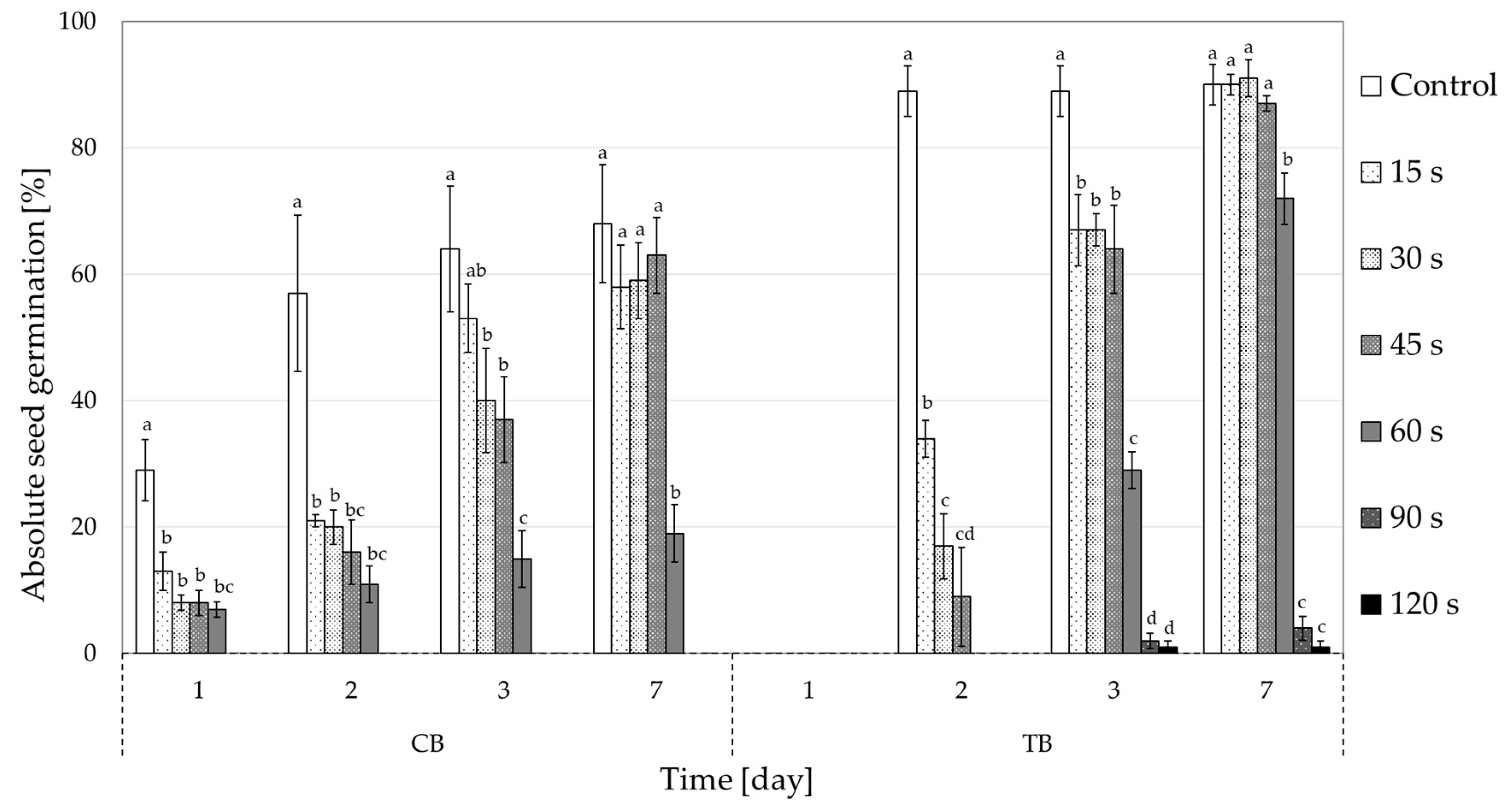
Figure 1. The effects of cold plasma treatments (15–120 s) on seed germination (% of all seed) in both buckwheat species (CB—common buckwheat; TB—Tartary buckwheat) on days 1, 2, 3 and 7. Different letters above the columns represent statistically significant differences between the control group and all cold plasma treatments on a specific day in each buckwheat species. There was no germination in CB after 90 and 120 s CP treatment.
Plants 10 00851 g001
Figure 2. Comparison of the effects of cold plasma treatments (15–120 s) on relative seed germination (% of average control seeds) between both buckwheat species (CB—common buckwheat and TB—Tartary buckwheat) after one week. ** represent a statistically significant difference between buckwheat species (p < 0.01).
Plants 10 00851 g002
Figure 3. The fungal colonisation level (as mean % of Petri dish area overgrown by fungi) per seed at different cold plasma treatments for each buckwheat species (CB—common buckwheat; TB—Tartary buckwheat). SC—surface-sterilised seeds. Different letters represent statistically significant differences between control and all cold plasma treatments for each buckwheat species (letters a–d for CB and x, y for TB).
Plants 10 00851 g003
Figure 4. Average number of fungal morphotypes per Petri dish at different cold plasma treatments for each buckwheat species (CB—common buckwheat; TB—Tartary buckwheat). SC—surface-sterilised seeds. Different letters represent statistically significant differences between cold plasma treatments for each buckwheat species (letters a–c for CB and x–z for TB).
Plants 10 00851 g004
Figure 5. Fungal frequencies per different cold plasma treatments. SC—surface-sterilised seeds. (a) Common buckwheat; (b) Tartary buckwheat.
Plants 10 00851 g005
Figure 6. Cultures of fungal morphotypes isolated from common and Tartary buckwheat seeds on potato dextrose agar plates after seven days of cultivation at 22 °C in the dark. The left part of each image shows the front view, and the right part the back view of each morphotype grown in a Petri dish. Different letters represent different morphotypes and numbers indicate different sub-types of each morphotype.
Plants 10 00851 g006
Figure 7. Seed-borne fungal community structure at different cold plasma treatments for (a) common buckwheat and (b) Tartary buckwheat. SC—surface-sterilised seeds.
Plants 10 00851 g007
Figure 8. Scheme of the low-pressure cold plasma system using O2 as input gas.
Plants 10 00851 g008
Figure 9. Optical spectrum of oxygen plasma at 50 Pa and 1500 W.
Plants 10 00851 g009
Table 1. Mean germination time (MGT) and time to reach 50% germination (T50) calculated for both buckwheat species per each CP treatment. Different letters indicate statistically significant differences between control and CP treatments per each buckwheat species. n.d.—indices could not be calculated due to very low germination (only few seeds per treatment). Different letters (superscripts) represent statistically significant differences between the control group and cold plasma treatments.
CBTB
TreatmentMGTT50MGTT50
Control3.77 a1.30 a4.01 a1.51 a
15 s4.26 bc1.56 a4.71 b2.33 ab
30 s4.54 cd2.41 b4.99 b2.54 b
45 s4.82 d2.88 b5.17 b2.62 b
60 s3.98 ab2.40 b5.86 c3.63 c
90 sn.d.n.d.5.89 c3.67 c
120 sn.d.n.d.n.d.n.d.
Publisher’s Note: MDPI stays neutral with regard to jurisdictional claims in published maps and institutional affiliations.