Article

Identification of Susceptibility Genes in Castanea sativa and Their Transcription Dynamics following Pathogen Infection

Dipartimento di Scienze Agrarie, Forestali e Alimentari-DISAFA, Università degli Studi di Torino, Largo Paolo Braccini 2, Grugliasco, 10095 Torino, Italy
*
Author to whom correspondence should be addressed.
Plants 2021, 10(5), 913; https://doi.org/10.3390/plants10050913
Submission received: 31 March 2021 / Revised: 26 April 2021 / Accepted: 28 April 2021 / Published: 2 May 2021
(This article belongs to the Section Plant Genetics, Genomics and Biotechnology)

Abstract

:
Castanea sativa is one of the main multipurpose tree species valued for its timber and nuts. This species is susceptible to two major diseases, ink disease and chestnut blight, caused by Phytophthora spp. and Cryphonectria parasitica, respectively. The loss-of-function mutations of genes required for the onset of pathogenesis, referred to as plant susceptibility (S) genes, are one mechanism of plant resistance against pathogens. On the basis of sequence homology, functional domain identification, and phylogenetic analyses, we report for the first time on the identification of S-genes (mlo1, dmr6, dnd1, and pmr4) in the Castanea genus. The expression dynamics of S-genes were assessed in C. sativa and C. crenata plants inoculated with P. cinnamomi and C. parasitica. Our results highlighted the upregulation of pmr4 and dmr6 in response to pathogen infection. Pmr4 was strongly expressed at early infection phases of both pathogens in C. sativa, whereas in C. crenata, no significant upregulation was observed. The infection of P. cinnamomi led to a higher increase in the transcript level of dmr6 in C. sativa compared to C. crenata-infected samples. For a better understanding of plant responses, the transcript levels of defense genes gluB and chi3 were also analyzed.

1. Introduction

The Castanea genus belongs to the Fagaceae family and includes four major species of commercial and ecosystemic interest: Castanea sativa Mill. (European chestnut), Castanea crenata Sieb. et Zucc. (Japanese chestnut), Castanea mollissima Bl. (Chinese chestnut), and Castanea dentata Borkh (American chestnut). C. sativa is a woody species common in all Mediterranean countries and Asia Minor. It has been widely used since ancient times, not only for the consumption of its edible nuts, but also for wood and the products of its ecosystem, such as mushrooms and honey. It is a forest tree, relevant for landscape ecology and biodiversity of mountain and rural areas [1].
Over the last century, the number of chestnut trees decreased in growing areas in Europe due to the depopulation of mountains, climate change, and the spread of two severe diseases: ink disease and chestnut blight [2,3]. Ink disease is caused by the Oomycete Phytophthora cinnamomi and Phytophthora cambivora. Both species are pathogenic to C. sativa, although P. cinnamomi generally displays greater virulence than P. cambivora [4,5]. Among Castanea species, only C. crenata exhibits high tolerance to P. cinnamomi [6]. The disease, which affects both young and old trees, leads to subcortical necrosis of the root system and the basal part of the stem; this is followed by the appearance of wasting symptoms in the foliage until the total desiccation and death of the plant occur [7,8,9,10]. These pathogens spread mainly through the movement of soil harboring inoculum and the dissemination of asexual flagellated spores (i.e., zoospores) that can actively travel short distances or passively travel long distances in flowing water [10,11]. The use of resistant rootstocks represents one possible solution to protect against these pathogens, although, at present, only tolerant selections obtained from hybridization between C. sativa and C. crenata are available [12].
Chestnut blight stands among the most destructive fungal tree diseases ever [10,13]. The causal agent, Cryphonectria parasitica, infects trees through dead plant tissue and wounds, including those caused by pruning, graft, and hail [13,14]. The symptoms involve bark cankers that can develop on suckers, young branches, and adult branches and trunks [15]. Chestnut blight was one of the causes of the abandonment of chestnut orchards in Europe until the end of the 1970s, when the natural spread of the hypovirulent form of the fungus favored a slow but progressive recovery of chestnut orchards and coppices. However, the fungus still represents a relevant problem in many areas of Europe. It is very harmful to young grafted trees in particular, hampering the establishment of new orchards in many areas [10,13].
C. dentata forests in Eastern North America were wiped out by C. parasitica in the early 20th century [16]. Extensive studies and breeding activities have been carried out to restore the American chestnut species introgressing resistance genes of C. mollissima [17,18]. More recently, researchers discovered that the onset of the disease is associated with the release of oxalic acid by the pathogen during infection. Blight-resistant C. dentata trees were obtained by transferring a wheat gene that encodes oxalate oxidase [19].
Recently, a new interest and sensitivity towards the preservation of the local landscape generated a growing interest in silviculture and chestnut trees [20]. Moreover, the market demand for chestnuts in European countries has been strong in the last two decades and has often been supplied by importations. This has been due in part to the gall wasp (Dryocosmus kuriphilus Yasumatsu) infestation, which only recently has been controlled effectively [1,21], and to the general difficulty of developing a modern chestnut industry based on quality cultivars of C. sativa that are more tolerant to pathogens. The elucidation of the genetic mechanism behind host–pathogen interaction could thus be useful for the development of novel breeding strategies aimed at achieving resistance or higher tolerance to these pathogens.
Plants take advantage of different defense mechanisms during pathogen attack, and pathogens trigger counter-defense mechanisms. Plants carry pattern recognition receptors (PRRs) able to perceive pathogen-associated molecular patterns (PAMPs); this perception leads to intracellular signal transduction culminating in PAMP-triggered immunity (PTI). PTI is characterized by the production of reactive oxygen species (ROS), the secretion of antimicrobial compounds, and hydrolytic enzymes targeting the pathogen cell wall (chitinase and glucanase) and local cell wall fortifications (through callose deposition) [22].
To suppress PTI, pathogens developed effector molecules able to facilitate pathogen infection by manipulating the host response to support compatibility. Plant resistance (R) genes can detect effectors and trigger effector-triggered immunity (ETI) [23]. The recognition between R genes and effectors causes a cascade of responses involving jasmonic acid (JA) and salicylic acid (SA), culminating in a hypersensitive response (HR) [24].
Most pathogens require the cooperation of the host to establish a compatible interaction. Plant genes supporting compatibility and facilitating infection are called susceptibility (S) genes. S-genes can be divided into three main classes: (a) genes required for the early pathogen infection step (basic compatibility); (b) genes encoding negative regulators of plant immunity; (c) genes necessary for pathogen proliferation (sustained compatibility) [22].
The mutation or loss of an S-gene can thus limit the ability of the pathogen to infect the host and the spread of the disease. The resistance mediated by the S-gene mutation can be pathogen-specific or broad-spectrum. In the former case, the pathway can be implicated in the penetration phase; in the latter, one of the target genes can be involved in constitutive defense responses [22]. Resistance due to the loss of S-genes is generally recessive, differing from the generally dominant resistance mediated by R genes.
Among the S-genes, Mildew resistance locus O (mlo1), Powdery mildew resistance 4 (pmr4), Downy Mildew Resistance 6 (dmr6), and Defense no death (dnd1) have been characterized in many plant species. The Mlo gene family, encoding seven transmembrane domain proteins, has been characterized in many plant species [25]. Some mlo homologs act as PM susceptibility factors, as their loss of function results in a distinguished type of resistance known as mlo resistance. Originally discovered in barley (Hordeum vulgare L.), mlo resistance was later shown to occur in several monocot and eudicot species, namely Arabidopsis, tomato (Solanum lycopersicum L.), pea (Pisum sativum L.), pepper (Capsicum annum L.), tobacco (Nicotiana tabacum L.), and wheat (Triticum aestivum L.) [26,27] plants. The callose synthase encoded by pmr4 is responsible for the production of callose in response to biotic and abiotic stresses. In tomato and potato plants, the knockout and silencing of pmr4 led to Oidium neolycopersici and Phytophthora infestans tolerance [28,29]. Dmr6 is involved in the conversion of salicylic acid (SA) to 2,3-dihydroxybenzoic acid (2,3-DHBA) and negatively regulates defense gene expression [30]. Its silencing caused resistance to hemi-biotrophic Phythophthora capsici, Hyaloperonospora arabidopsidis, and Pseudomonas syringae [31]. Mutants of dnd1, encoding for a cyclic nucleotide-gated cation channel, showed P. infestans resistance [29].
At the moment, studies on S-genes in woody plant species have been carried out only for mlo genes in rubber trees [32], poplar trees [33], apple trees, and grapevines [34]. In our work, we report on the S-genes identification and characterization in C. sativa on the basis of sequence homology, functional domain detection and phylogenetic relationships. In addition, the expression dynamics of S-genes were assessed in C. sativa and C. crenata plants inoculated with the two pathogens, P. cinnamomi and C. parasitica, belonging to different kingdoms. Using the same plant material, the transcription levels of key genes involved in pathogen resistance, chi3 (acidic 26 kDa endochitinase) and gluB (glucan endo-1,3-beta- glucosidase), were also determined (S1 File). Our analysis revealed the strong activation of pmr4 and dmr6 genes in response to infection by both P. cinnamomi and C. parasitica.

2. Results

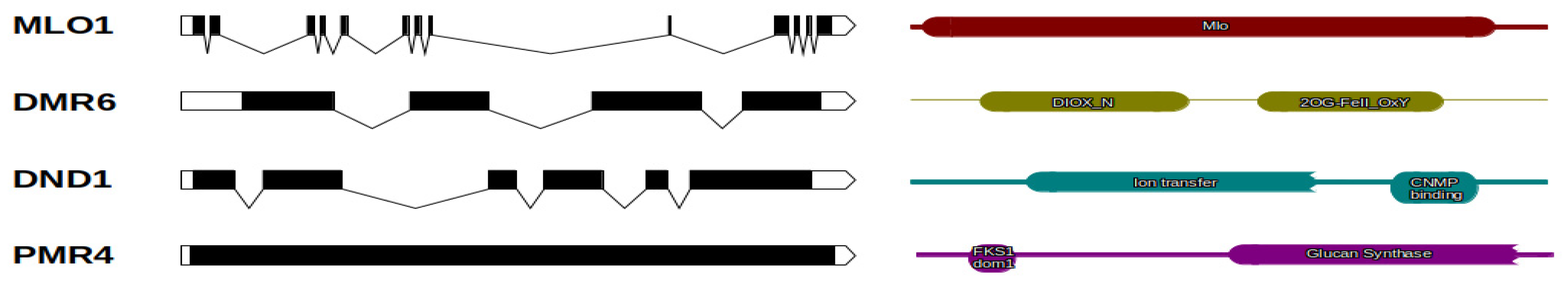
2.1. Genes Identification and Structure

Chestnut susceptible (S) genes were identified in the C. mollissima v1.1 reference genome using available coding sequences of gene orthologs as a query (S2 File). Based on the blastn survey, four loci with high similarity were identified and attributed to different subclasses of S-genes due to the presence of specific domains: mlo1, dmr6, dnd1, and pmr4 (Figure 1). The coding sequence length of mlo1 is 1425 bp (composed of 13 exons); the protein size is 474 amino acids (aa) (Table 1). A single Mlo domain (PF03094) is present within the protein sequence. The Dmr6 gene, whose coding sequence is 1128 bp, contains four exons and is translated into a 375 aa protein (Table 1). Two specific domains are characterized: 2OG-FeII_Oxy and DIOX_N (PF03171; PF14226). Dnd1 is 1407 bp in length, codes for 468 aa proteins, and is composed of six exons. Two structural domains, cNMP_binding and Ion_trans, were highlighted (PF00027; PF00520) (Figure 1; Table 1). The Pmr4 gene is characterized by one single 5346 bp exon. The protein size is 1781 aa, and the structural domains are FKS1 dom1 and Glucan_synthase (PF14288; PF02364) (Table 1).

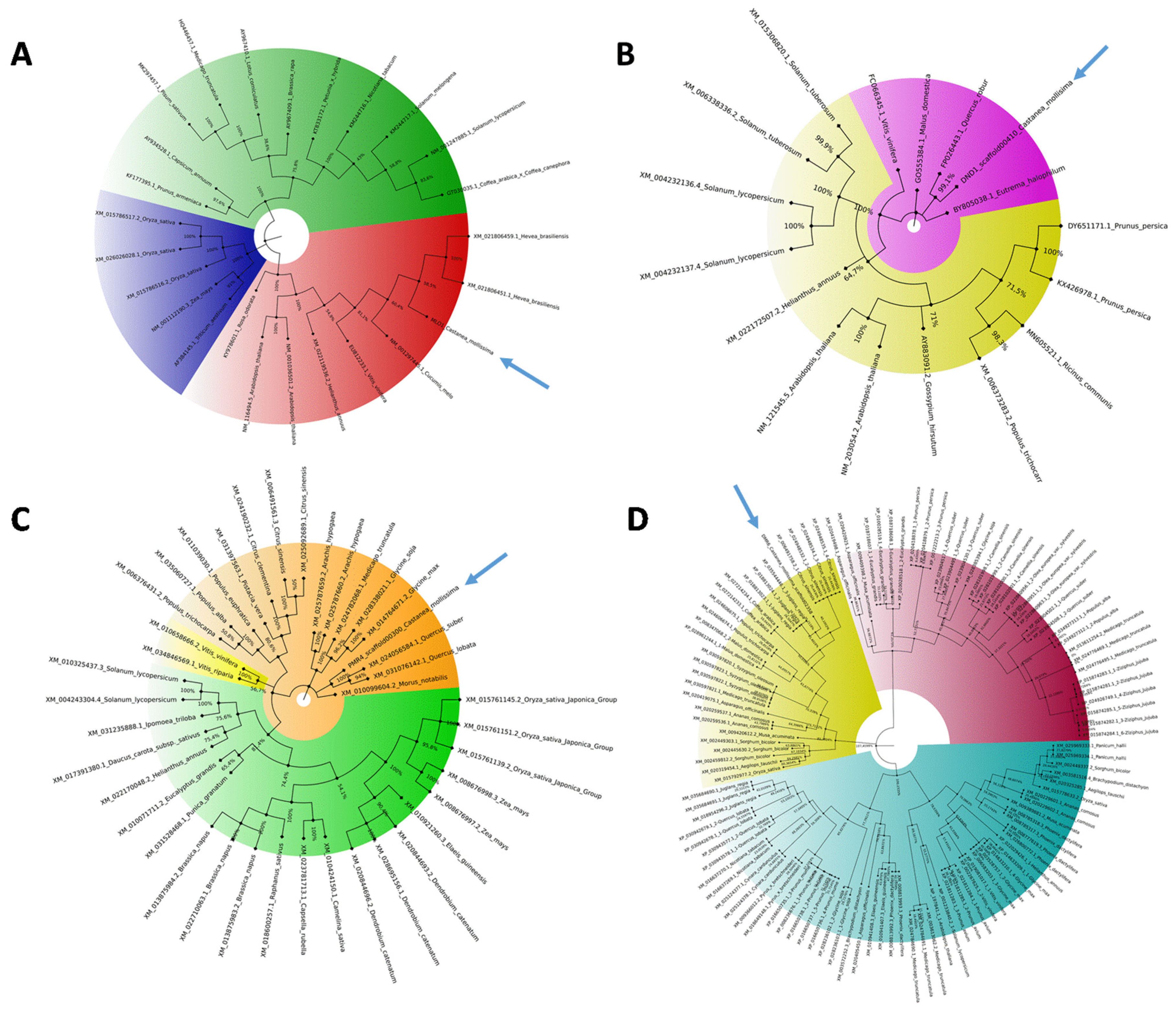
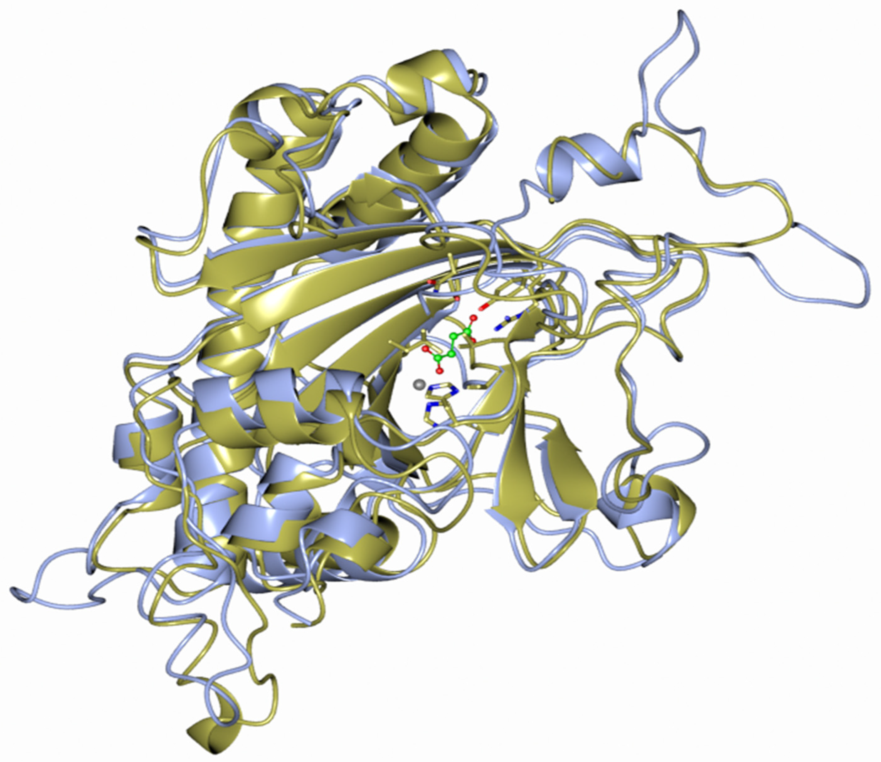
2.2. Phylogenetic and Modeling Analysis

Available full-length NCBI S-gene coding sequence orthologues (S3 File) were used for phylogenetic tree construction. The resulting unrooted maximum-likelihood trees are shown in Figure 2, and all the phylogenetic trees are available separately in the S4 File.
The mlo1 tree was grouped into three clades (blue, green, red) with 100% bootstrap value. The monocot proteins formed a separate clade (blue) with respect to those of the dicotyledonous species. CmMlo1 is located in the red clade with Vitis vinifera and Hevea Brasiliensis orthologs (Figure 2A). The 17 dnd1 coding sequences were divided into two subclades; no monocotyledon genes were included. CmDnd1 is in the violet subclade, phylogenetically close to Quercus robur ortholog (Figure 2B). For the construction of pmr4 and dmr6 trees, a greater number of coding sequences were available: 40 and 115, respectively. The Pmr4 phylogenetic tree showed the division in three main clades, with monocots in the green clade intermixed with dicots. CmPmr4 is located in the orange clade and clusters together with Quercus lobata and Quercus spp. orthologs (Figure 2C). The Dmr6 tree is divided into three clades intermixed with monocot/dicot proteins. CmDmr6 is in the yellow clade, phylogenetically close to the Juglans regia ortholog gene (99% bootstrap value) (Figure 2D).
By comparing the 3D protein structure model of C. mollissima DMR6 with Arabidopsis thaliana DMR6 via Modeller software, a high degree of structural conservation was observed (Figure 3). DMR6 is a putative oxygenase involved in the conversion of salicylic acid (SA) to 2,3-dihydroxybenzoic acid (2,3-DHBA), and its catalytic activity is probably necessary to suppress plant immunity. The catalytic triad that binds the iron atom (grey sphere) is made by two histidines (H212 and H269), and an aspartic acid residue (D214). Zeilmaker et al. [31] demonstrated, by removing the histidines, that these residues were fundamental for the catalytic activity of DMR6, as well as for its role in immune suppression.

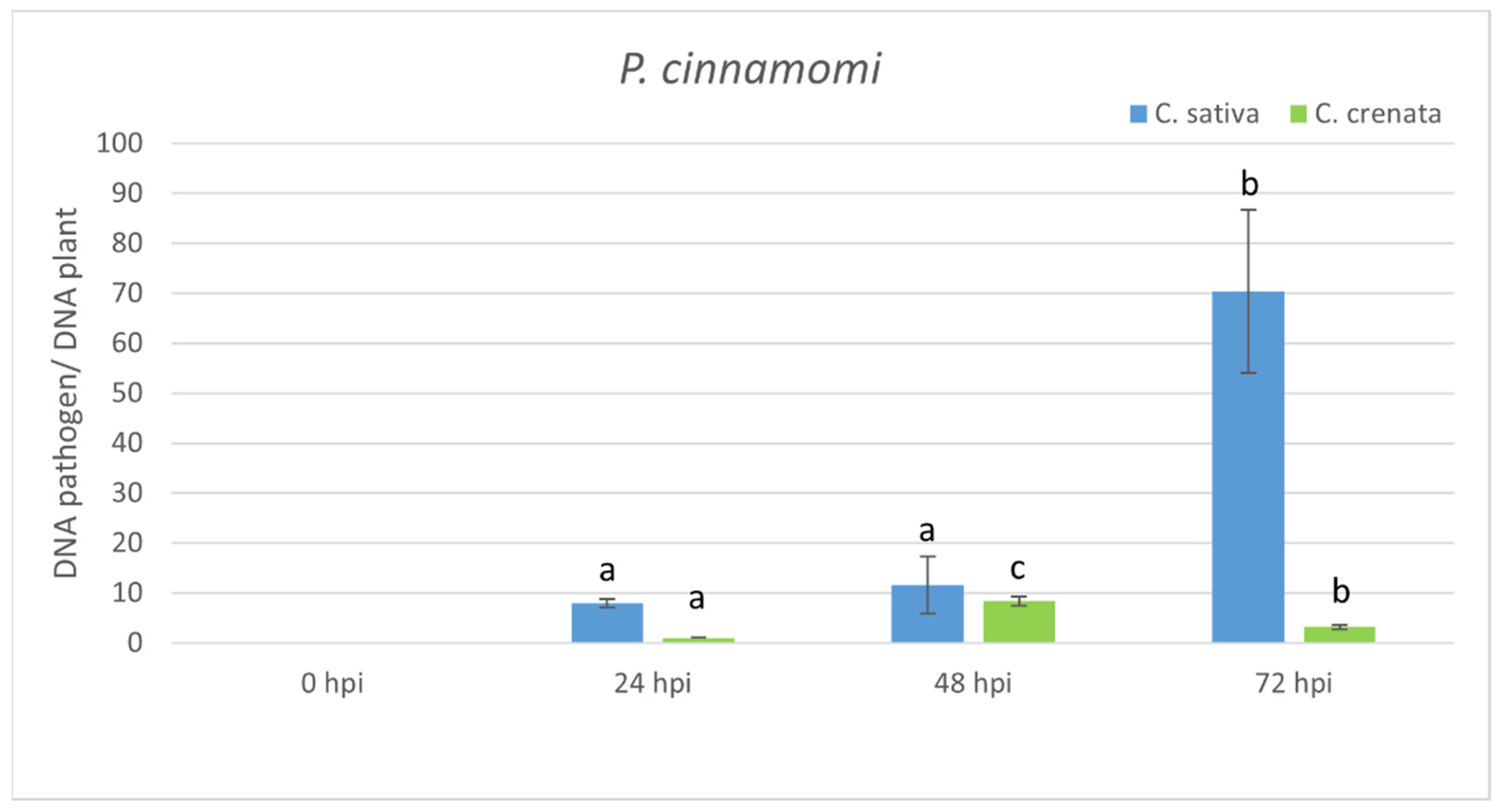
2.3. Transcriptional Profiling in Response to P. cinnamomi Infection

S-genes are genes related to plant–pathogen interaction and are supposed to be activated during the early stages of infection before symptoms emerge. The susceptible species C. sativa was used as a reference to define the onset of symptoms due to P. cinnamomi inoculation on the stem. Five days after inoculation, lesions [35] were observed, followed by total leaf desiccation. Based on the evidence from the preliminary inoculation tests, C. sativa and C. crenata species inoculated with P. cinnamomi and samples were collected at 0, 3, 6, 12, 24, 48, and 72 h post-inoculation (hpi). The wound area left a lesion of 0.5 cm2. C. sativa plants inoculated with P. cinnamomi showed an enlargement of the lesion of 0.6 cm2 at 24, 48, and 72 hpi compared to the initial area of the lesion at time 0 (control). No visible enlargement of lesions was recorded in the case of C. crenata.
Quantitative PCR from infected stem tissues was applied to quantify P. cinnamomi and the assay confirmed a higher amount of the pathogen in plant tissues at 72 hpi (Figure 4). C. crenata showed a lower abundance of the pathogen compared to C. sativa at all tested experimental time points. S-gene expression was analyzed using the same time points (Figure 5A). Mlo1 was mainly expressed at early infection phases, peaking at 6 and 3 hpi in C. sativa and C. crenata, respectively. Dnd1 showed an analogous trend in both plant species, and its transcription was strongest at 6 hpi. Regarding pmr4, a differential transcript regulation in response to P. cinnamomi infection was highlighted in C. sativa and C. crenata. In C. sativa, pmr4 was strongly expressed at 3 and 6 hpi, with transcript level around 6-fold higher compared to 0 hpi. On the contrary, the expression of pmr4 remained very limited in C. crenata infected tissues, and a significant downregulation at 48 and 72 hpi was observed. Significant increases in the transcript level of dmr6 were observed only at 12 hpi (for C. crenata) and at 3 and 12 hpi (for C. sativa). For a better understanding of the plant response against P. cinnamomi, genes coding for pathogenesis-related (PR) proteins, chi3 and gluB, were analyzed (Figure 5B). Chi3 peaked at 6 hpi both in C. sativa and in C. crenata, but the increment was higher in the former species (~3 fold vs. ~2 fold). GluB increased with the progression of infection in C. sativa, whereas a higher upregulation was observed during the early infection phases (at 3 and 6 hpi) in C. crenata.

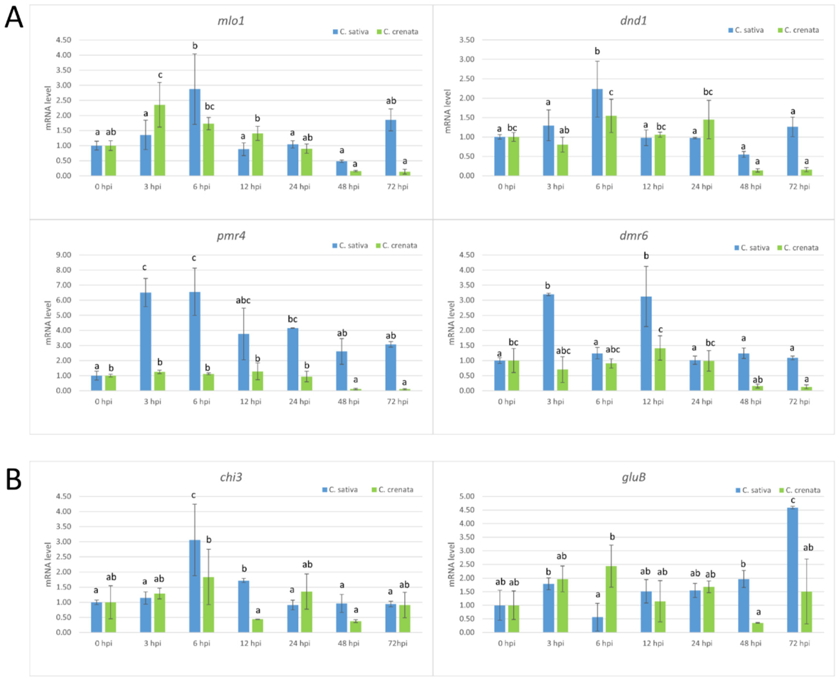
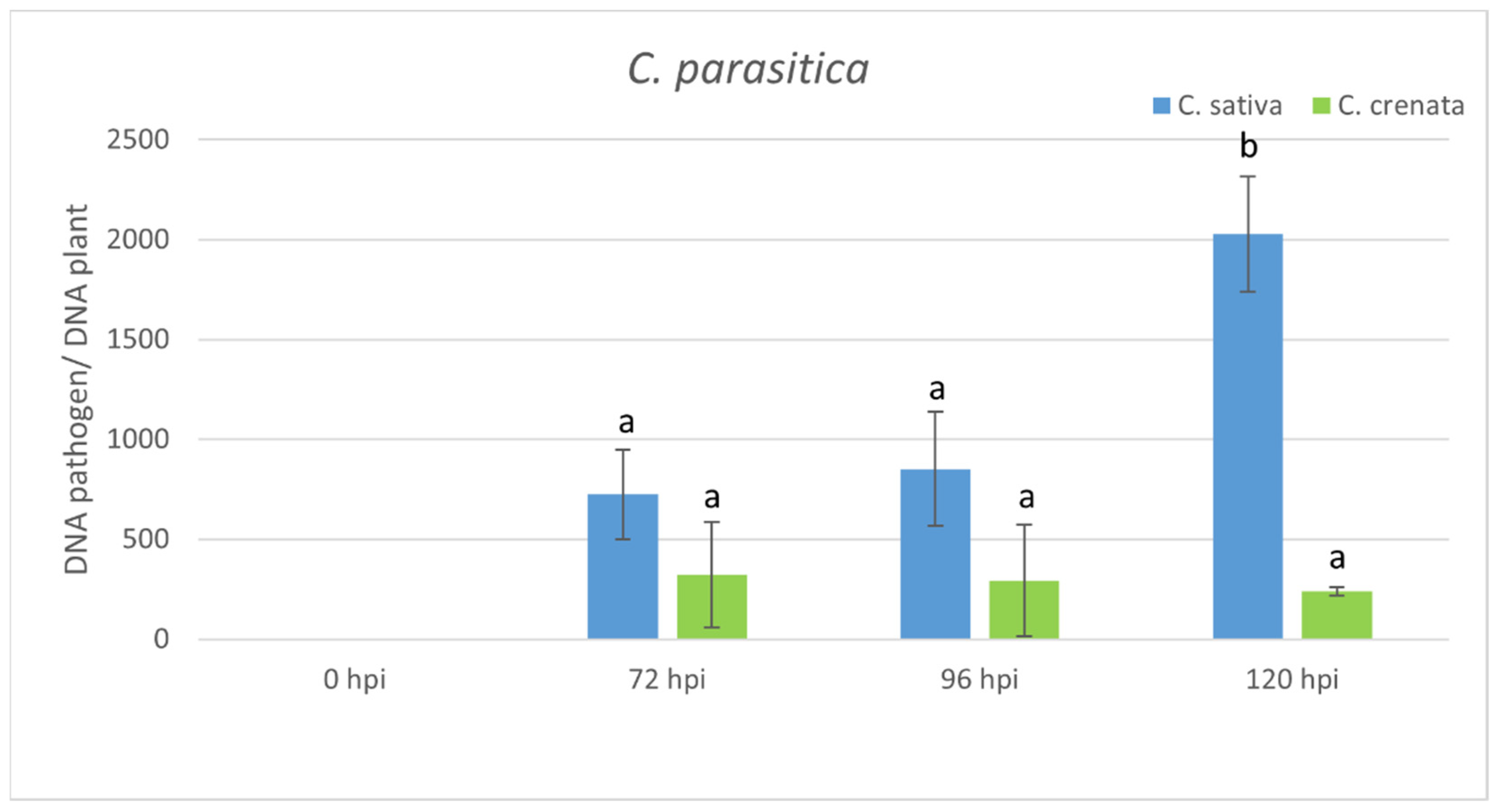
2.4. Transcriptional Profiling in Response to C. parasitica Infection

As described for P. cinnamomi, a preliminary stem inoculation assay on C. sativa plants was done with C. parasitica. Seven days after inoculation, a necrotic lesion around the inoculation point and orange fruiting bodies were observed in C. sativa. Based on the results of the preliminary inoculation test, C. sativa and C. crenata plants were inoculated and sampled at 0, 12, 24, 48, 72, 96, and 120 hpi. The size of the wound area was 0.5 cm2. C. sativa plants inoculated with C. parasitica showed enlargements of the lesion of 0.5 cm2 (at 48 and 120 hpi) and of 1 cm2 (at 96 hpi) compared to the initial wound area at 0 hpi. No visible enlargement of the inoculation lesions was recorded in C. crenata at 72 and 120 hpi, and a limited enlargement of 0.5 cm2 was observed at 96 hpi.
In C. sativa, qPCR analysis showed an increase in the abundance of pathogen inoculum with time elapsing from infection, peaking at 72 hpi. In C. crenata, no statistical differences were observed among the different experimental times (from 72 to 120 hpi) (Figure 6). The transcript levels of S-genes were analyzed using the same time points (Figure 7A). Mlo1 was mainly expressed at 24 hpi in C. sativa. No significant upregulation of mlo1 was observed in C. crenata-infected tissues. An upregulation of dnd1 was detected at 24 hpi in C. sativa infected tissues. In C. sativa, pmr4 was strongly expressed at the early infection phases (12 and 24 hpi), with transcript levels around 3-fold higher as compared with the level at 0 hpi. On the contrary, pmr4 was downregulated at all experimental times in inoculated C. crenata plants. A significant upregulation of dmr6 was observed only at late infection phases, 48 hpi in C. crenata and 24 hpi in C. sativa. The transcription of chi3 was strongest at 96 hpi in C. sativa and at 48 hpi in C. crenata. The transcript level of gluB increased until reaching 96 hpi in C. sativa and 24 hpi in C. crenata (Figure 7B).

3. Discussion

C. sativa is a European woody tree species commonly used across the globe in the food and timber industries. This chestnut species is susceptible to the two major pathogens, P. cinnamomi and C. parasitica [10,36]. In contrast, the Asian chestnut species C. crenata and C. mollissima exhibit higher tolerance to P. cinnamomi and C. parasitica [6]. Achieving tolerance or resistance to pathogens is the major aim of rootstock breeding. Blight-resistant trees were obtained through backcross breeding of introgression genes from Asian species into American chestnut trees. [37]. However, this approach, although successful in developing blight-resistant American chestnut selections has been slowed by a lack of genetic tools. In Europe, ink disease tolerant hybrids were obtained through interspecific crosses between C. sativa and C. crenata, although the nut quality produced by these hybrids is below current market standards [38,39].
It has long been recognized that a deep understanding of a pathogen’s biology, host–pathogen interactions, and the resistance mechanisms are fundamental to improving breeding programs. Genomic and transcriptomic analyses have provided the first genetic insights into mechanisms underlying susceptible and resistant chestnut species responses to P. cinnamomi and C. parasitica [37,38,40,41,42]. Santos et al. [40] reported the upregulation of a set of candidate genes (e.g., Cast_Gnk2-like and Calcium-dependent protein kinase) after P. cinnamomi infection, which may trigger HR-like cell death in C. crenata cells. A significant number of genes involved in the defense against chestnut blight were identified [37].
Traditionally, the introduction of resistance gene analogues into plants was the most promising approach to facilitate the acquisition of resistance. However, it did not prove to be durable enough because the widespread use of R genes caused the selection of pathogens capable of overcoming them [24]. Susceptibility (S) genes can be interesting candidates to be used in target breeding programs [22,23,24]. Furthermore, on the basis of previous studies, it was highlighted that the disabling of susceptibility genes may facilitate durable resistance since the pathogen needs to gain a new function to replace the lost host factor it was exploiting [43].
In woody species, the investigation of S-genes has been performed only for MLO genes in rubber trees [32], poplar trees [33], apple trees, and grapevines [34,44]. Due to the absence of a C. sativa genome, highly similar S-genes were selected using the C. mollissima v 1.1 genome. Based on the blastn survey, four loci with high similarity were identified in the C. mollissima genome and attributed to different subclasses of S-genes [31,45,46,47] due to the presence of specific domains: mlo1, dmr6, dnd1, and pmr4 (Figure 1, Table 1). As previously observed [31], in the phylogenetic trees, monocot proteins formed a separate clade with respect to those of dicotyledonous species, supporting the hypothesis that an independent evolution occurred for these genes (Figure 2). Quantitative PCR analysis has been carried out to identify the differential expression of candidate S-genes in response to P. cinnamomi and C. parasitica in the stems of a susceptible species, C. sativa, and of a tolerant one, C. crenata. Lesion analysis and DNA quantification of the pathogen (Figure 4 and Figure 6) confirmed the higher tolerance level of C. crenata in response to both P. cinnamomi and C. parasitica infection. Our qPCR results highlighted the main upregulation of pmr4 and dmr6 in response to infection by both P. cinnamomi and C. parasitica. As expected, a greater increase in the transcription of these susceptibility genes was observed in the susceptible species C. sativa. Remarkably, pmr4 was strongly expressed at early infection phases of both pathogens in C. sativa; in the tolerant C. crenata, significant upregulation was observed (Figure 5 and Figure 7). Pmr4 codifies for a callose synthase, which is necessary to create a physical barrier to avoid pathogen penetration and is also implicated in plant-triggered immunity suppression. Pmr4 is thus not only involved in the synthesis of callose, but it also acts as a negative regulator of the salicylic acid pathway [28].
Huibers et al. [48] demonstrated that resistance due to the silencing of Pmr4 is associated with salicylic acid (SA) accumulation rather than with the callose deposition absence. Salicylic acid signaling plays a key role protecting against biotrophic pathogens through the establishment of a hypersensitive response (HR). Saiz-Fernandez et al. [49] revealed the increment of SA levels in P. cinnamomi inoculated stems, indicating that P. cinnamomi activates a defense response similar to that triggered by biotrophic pathogens. Inoculation with both virulent and hypovirulent strains of C. parasitica led to SA accumulation in European chestnut plantlets that were grown in vitro [50]. Transcriptome analyses carried out in both C. dentata and C. mollissima highlighted activation of salicylic-acid-related genes in canker tissues [37].
In chestnut trees, callose deposition around P. cinnamomi hyphae was detected early in the infection process; however, it does not seem to play a key role in the associated interactions since the pathogen can reach the vascular cylinder in both susceptible and resistant plant genotypes [51]. This result was validated by transcriptomes analyses of C. sativa and C. crenata, in which no overexpression of Callose synthases after P. cinnamomi infection was observed [38].
Based on our results and the literature, we can hypothesize that callose accumulation due to the pmr4 upregulation in inoculated C. sativa lines may not be responsible for controlling P. cinnamomi colonization. We suggest that the upregulation of pmr4 could lead to a negative regulation of the SA pathway that in turn provokes the susceptibility of C. sativa to both P. cinnamomi and C. parasitica. A clear link with SA pathway has emerged even with the other chestnut gene candidate dmr6 (downy mildew resistance 6). The mutation of Arabidopsis dmr6 gene, associated with salicylic acid (SA) homeostasis [31], results in the generation of plants that are resistant to bacteria and oomycetes. Dmr6 is involved in the conversion of salicylic acid (SA) to 2,3-dihydroxybenzoic acid (2,3-DHBA) and negatively regulates the expression of defense genes (PR-1, PR-2, and PR-5) [30].
The expression trend of the Dmr6 gene in response to P. cinnamomi infection turned out to be similar to the profile of pmr4. Indeed, dmr6 was strongly expressed at early infection phases of P. cinnamomi in C. sativa; in C. crenata no significant upregulation was detected (Figure 5). No upregulation of dmr6 in response to C. parasitica was highlighted in both plant species (Figure 7). We can thus hypothesize that dmr6 upregulation observed in C. sativa could negatively regulate defense gene expression, leading to susceptibility to P. cinnamomi.
Plants produce a variety of hydrolytic defense enzymes against pathogens, including chitinases, proteases, and also glucanases [52]. The genes coding for several pathogenesis-related (PR) proteins, Acidic 26 kDa endochitinase gene (chi3) and Glucan endo-1,3-beta-glucosidase B gene (gluB), were selected in our analysis because they are considered as responsive to SA-dependent signaling [53,54]. Chi3 and gluB are enzymes that cause the lysis of hyphae of various pathogens, resulting in growth inhibition [55,56,57].
In both C. sativa and C. crenata plants inoculated with C. parasitica, chi3 and gluB were significantly upregulated. The transcription of chi3 was higher in C. crenata than in C. sativa, presumably as a consequence of the improved defense mechanism against C. parasitica. Our results are in agreement with Shain et al. [58], who demonstrated the involvement of b-1,3-glucanase and chitinase in chestnut species affected by C. parasitica. Studies on the role of chitinase in blight infection mostly involved C. sativa as a model system [50,59]. In both C. dentata and C. mollissima, transcripts of several compounds expressing chitinase accumulated more in canker tissues than healthy stem tissues [37]. In order to obtain chestnut plants with potentially increased resistance/tolerance to chestnut blight, the endogenous Ch3gene encoding a chitinase-like protein was over-expressed in the European chestnut through Agrobacterium-mediated transformation [60].
The emergent CRISPR/Cas9 technology is expected to play a key role in future crop breeding as it allows highly efficient gene editing and generates genetic changes indistinguishable from those arising spontaneously in nature or through conventional breeding [61]. Several examples of edited plants resistant to fungal pathogens have been described [62,63]. For example, genome editing was successfully applied to knock out mlo S-genes, leading to Powdery mildew (PM) resistance [44,64,65,66]. Pmr4 and dmr6 loss-of-function through CRISPR/Cas reduced the susceptibility to PM in tomato plants [28,67]. In our laboratory, we are setting up a CRISPR/Cas9 transformation protocol in Castanea sativa. Our future goal will be to perform the functional characterization using the CRISPR/Cas9 approach of the two candidate genes (dmr6 and pmr4), while checking if the two genes may also play a role in the interaction between C. sativa and the emergent nut rot and canker agent Gnomoniopsis castaneae [68].

4. Materials and Methods

4.1. Identification of Chestnut S-genes Orthologues

S-gene sequences (S2 File), available in the NCBI database (https://www.ncbi.nlm.nih.gov/, accessed on 31 March 2021), were used as a query in the BLAST+ program (blastn task) against Castanea mollissima v1.1 reference genome (https://www.hardwoodgenomics.org/Genome-assembly/1962958, accessed on 31 March 2021) to find chestnut S-gene orthologs. Hits were filtered using the e-value cut-off of 1 × 10⁻⁵.
The domain structures of chestnut S-genes were predicted using Pfam (pfam.xfam.org/, accessed date 31 March 2021) and Uniprot (https://www.uniprot.org/, accessed on 31 March 2021) databases. The graphical gene structure with exons/introns representation was realized using the script accessible at http://wormweb.org/exonintron (accessed on 31 March 2021).

4.2. Phylogenetic Analysis

The alignment of chestnut S-gene coding sequences (mlo, pmr4, dmr6, and dnd1) and of known related S-genes in other plant species were generated via multiple sequence alignment using the ClustalW algorithm (http://www.clustal.org/, accessed on 31 March 2021). All the sequences used for tree construction are accessible in S3 File. MEGAX software (https://www.megasoftware.net/, accessed on 31 March 2021) was used for phylogenetic tree construction, applying maximum likelihood algorithms. To obtain a confidence level for each branch, bootstrap analysis was performed with 1000 iterations. All the phylogenetic trees are available in S4 File.

4.3. Protein Modeling

The Modeller (https://salilab.org/modeller/, accessed on 31 March 2021) software was applied to generate 3D protein structure models. The Modeller software generates the 3D structure of a given target protein sequence based on its alignment with a known protein structure (templates) [69].
The alignment file of Arabidopsis thaliana (Q9FL0) and C. mollissima protein sequences was obtained using the Emboss Needle online tool (https://www.ebi.ac.uk/Tools/psa/emboss_needle/, accessed on 31 March 2021). The 3D model was developed with an automodel class using the 3D A. thaliana model and the alignment file. Ccp4mg software (www.ccp4.ac.uk/, accessed on 31 March 2021) was used for protein 3D structure visualization, which is useful for studies of catalytic and regulatory domain conservation/divergence.

4.4. Pathogens Inoculation and Samples Collection

P. cinnamomi (ID_C4) and C. parasitica (ID_5183 L2d) isolates used in the experiment were originally isolated from symptomatic C. sativa trees in Piedmont and Aosta Valley, northwestern Italy, respectively, and preserved in the plant pathogen culture collection at DISAFA (University of Turin). Isolates were subcultured in potato dextrose agar (PDA) before inoculations. The inoculation trial was carried out on Castanea sativa and Castanea crenata plants (1-year-old) grafted on C. sativa and C. sativa x C. crenata rootstocks, respectively. Plants were grown in pots under greenhouse conditions. The identity of the plant material was preliminary checked through marker analysis using 10 SSR loci from Marinoni et al. [70]. Plants were 80–100 cm high and 0.9–1.5 cm in diameter at the collar.
Plants were inoculated 20 cm above the collar by placing a colonized plug of PDA (0.5 cm diameter) in a slit, superficially cleaned with 70% ethanol, obtained by excising a small portion of the bark with a sterile scalpel according to the methods described by Zampieri et al. [71]. After the inoculation process, the inoculation point was wrapped with parafilm to prevent tissue dryness and external contamination [71,72,73]. As negative controls (0 hpi), plants were wounded in the same way but inoculated with a sterile PDA plug. Plants were incubated in greenhouse conditions at 28 ± 2 °C with a 16-h photoperiod.
S-genes are genes related to plant–pathogen interaction and are expressed during the first step of inoculation, before symptoms manifest. The time points used in our analysis were selected using the susceptible C. sativa as a reference on the basis of the onset of evident symptoms, i.e., bark necrosis and leaf dryness/browning, on C. sativa reference plants inoculated with each pathogens. The selected time points were 5 days after inoculation for P. cinnamomi and 7 days after inoculation for C. parasitica. Three biological replicates for seven experimental time points, including 0 hpi (control), were tested both for Castanea sativa and Castanea crenata and for the two pathogens (84 plants in total).
Plant material was harvested at 0, 3, 6, 12, 24, 48, 72 h after P. cinnamomi inoculation. For the C. parasitica experiment, material was collected at 0, 12, 24, 48, 72, 96, and 120 h after inoculation. For each time point, two disks of the stem were cut 0.5 cm above and below the wound. Bark was then removed to reduce the polyphenol contamination of RNA, and samples were frozen in liquid nitrogen to preserve RNA integrity. All samples were stored at −80 °C before RNA extraction.

4.5. RNA Extraction and Real-Time qPCR Quantification

RNA was extracted from both inoculated and control (0 hpi) samples. A total of 0.1 g of frozen tissue was manually ground into a fine powder and liquid nitrogen was added. RNA was extracted using Spectrum Plant Total RNA Kit (Sigma-Aldrich) following the manufacturer’s protocol. Extracted RNA was treated with DNase I (Thermo Fisher Scientific) following the manufacturer’s instructions.
RNA was quantified by a NanoDrop spectrophotometer (Thermo Scientific, Hudson, NH, USA). cDNA was synthesized from 2 µg RNA using the High-Capacity cDNA Reverse Transcription Kit (Thermo Fisher Scientific, Hudson, NH, USA). Primer sequences for candidate S-genes were designed using the Primer3 online tool (https://primer3.ut.ee/, accessed on 31 March 2021) and are available in S5 File. All primers were in silico tested through Primer-BLAST program (https://www.ncbi.nlm.nih.gov/tools/primer-blast/, accessed on 31 March 2021).
Chi3 and gluB genes, coding for several pathogenesis-related (PR) proteins, were also analyzed in order to observe their role in the defense response of chestnut trees [74]. Transcript abundance was quantified in three biological replicates by the StepOnePlus Real-Time PCR System (Applied Biosystems). Real-Time qPCR was performed using the Power SYBR®® Green Master Mix added with bovine serum albumin (BSA) to reduce the action of PCR inhibitors. The amplification protocol included an initial denaturation step at 95 °C for 5 min, followed by 40 cycles of 95 °C/5 s and 60 °C/1 min. Data were quantified using the 2−ΔΔCt method based on Ct values of candidate genes and actin (as a housekeeping gene) [38]. IBM SPSS statistical software was used to carry out a one-way analysis of variance test (ANOVA). Each value represented the mean of three biological replicates, which were compared using Tukey’s HSD test (p ≤ 0.05).

4.6. Pathogen Quantification

Samples inoculated with P. cinnamomi (24, 48, 72 h) and C. parasitica (72, 96, 120 h) were used for DNA extraction and pathogen quantification. Plants were debarked and the necrotic area in the cambium layer was measured using ImageJ v. 1.8.0 software.
The DNA extraction was performed using an E.Z.N.A.®® Stool DNA Kit following the manufacturer’s protocol. Standard curves were prepared for the quantification of DNAs by qPCR using primers designed as follows: 7-actin for chestnut DNA, the ypt gene for P. cinnamomi DNA, and the mf1 species-specific gene for C. parasitica DNA [75] (S5 File). All the inoculated and control (0 hpi) samples were analyzed through real-time qPCR both with pathogens genes (ypt and mf1) and with 7-actin. The results, normalized by standard curves, were used for the calculation of the ratio of DNA fungus/plant DNA. Real-time qPCR was performed using the experimental conditions previously described: initial denaturation step at 95 °C for 5 min, followed by 40 cycles at 95 °C/5 s and 60 °C/1 min. Data were quantified through the 2−ΔΔCt method based on Ct values of pathogen genes and actin-7 as a housekeeping gene [38].
IBM SPSS statistical software was applied to perform a one-way analysis of variance test (ANOVA). Each value represented the mean of three biological replicates compared using Tukey’s HSD Test (p ≤ 0.05).

Supplementary Materials

The following are available online at https://www.mdpi.com/article/10.3390/plants10050913/s1, S1 File: experiments pipeline, S2 File: S-gene coding sequences available in the NCBI database (https://www.ncbi.nlm.nih.gov/, accessed on 31 March 2021) used for S-gene detection in the C. mollissima genome, S3 File: S-gene coding sequences used for tree construction, S4 File: phylogenetic trees, S5 File: primer sequences.

Author Contributions

Conceptualization, R.B., A.M., P.G. and D.T.M.; methodology, A.M. and V.P.; software, A.M., V.P. and E.C.-G.; validation, A.M. and V.P.; formal analysis, A.M. and V.P.; investigation, A.M. and V.P.; resources R.B. and P.G.; data curation, A.M. and V.P.; writing—original draft preparation, A.M. and V.P.; writing—review and editing, R.B., P.G., D.T.M., A.M. and V.P.; visualization, A.M. and V.P.; supervision, A.M., R.B., P.G. and D.T.M.; project administration, R.B.; funding acquisition, R.B. All authors have read and agreed to the published version of the manuscript.

Funding

This research was funded by Fondazione CRT. This research was also partially funded by the “MONGEFITOFOR” Project Interreg Cooperation Program Va ITA-CH 2014/2020.

Conflicts of Interest

The authors declare no conflict of interest.

References

  1. Torello Marinoni, D.; Nishio, S.; Valentini, N.; Shirasawa, K.; Acquadro, A.; Portis, E.; Alma, A.; Akkak, A.; Pavese, V.; Cavalet-Giorsa, E.; et al. Development of High-Density Genetic Linkage Maps and Identification of Loci for Chestnut Gall Wasp Resistance in Castanea spp. Plants 2020, 9, 1048. [Google Scholar] [CrossRef] [PubMed]
  2. Conedera, M.; Krebs, P.; Tinner, W.; Pradella, M.; Torriani, D. The cultivation of Castanea sativa (Mill.) in Europe, from its origin to its diffusion on a continental scale. Veg. Hist. Archaeobot. 2004, 13, 161–179. [Google Scholar] [CrossRef] [Green Version]
  3. Müllerová, J.; Hédl, R.; Szabó, P. Coppice abandonment and its implications for species diversity in forest vegetation. For. Ecol. Manag. 2015, 343, 88–100. [Google Scholar] [CrossRef] [PubMed] [Green Version]
  4. Zentmyer, G.A. Phytophthora cinnamomi and The Diseases it Causes; The American Phytopathological Society: St. Paul, MN, USA, 1980; Volume 10, p. 96. [Google Scholar]
  5. Cristinzio, G.; Grassi, G. Valutazione di resistenza a Phytophthora cambivora e Phytophthora cinnamomi in cultivar di Castanea sativa. Monti Boschi 1993, 1, 54–58. [Google Scholar]
  6. Serrazina, S.; Machado, H.; Costa, R.; Duque, P.; Malhó, R. Expression of Castanea crenata Allene Oxide Synthase in Arabidopsis Improves the Defense to Phytophthora cinnamomi. bioRxiv 2020. [Google Scholar] [CrossRef]
  7. Maurel, M.; Robin, C.; Simonneau, T.; Loustau, D.; Dreyer, E.; Desprez-Loustau, M.-L. Stomatal conductance and root-to-shoot signalling in chestnut saplings exposed to Phytophthora cinnamomi or partial soil drying. Funct. Plant Biol. 2004, 31, 41–51. [Google Scholar] [CrossRef]
  8. Vannini, A.; Vettraino, A.M. Ink disease in chestnuts: Impact on the European chestnut. For. Snow Landsc. Res. 2002, 76, 345–350. [Google Scholar]
  9. Robin, C.; Smith, I.; Hansen, E.M. Phythophthora cinnamomi. For. Phytophthoras 2012, 2, 1. [Google Scholar] [CrossRef]
  10. Gonthier, P.; Robin, C. Diseases. In The Chestnut Handbook: Crop and Forest Management; Beccaro, G., Alma, A., Bounous, G., Gomes-Laranjo, J., Eds.; CRC Press, Taylor & Francis Group: Boca Raton, FL, USA, 2019; pp. 297–316, Chapter 11; ISBN 9781138334021. [Google Scholar]
  11. Hayden, K.J.; Garbelotto, M.; Dodd, R.; Wright, J.W. Scaling up from greenhouse resistance to fitness in the field for a host of an emerging forest disease. Evol. App. 2013, 6, 970–982. [Google Scholar] [CrossRef]
  12. Bounous, G. Il Castagno: Risorsa Multifunzionale in Italia e nel Mondo; Edagricole: Bologna, Italy, 2014. [Google Scholar]
  13. Rigling, D.; Prospero, S. Cryphonectria parasitica, the causal agent of chestnut blight: Invasion history, population biology and disease control. Mol. Plant Pathol. 2018, 1, 7–20. [Google Scholar] [CrossRef] [Green Version]
  14. Lione, G.; Giordano, L.; Turina, M.; Gonthier, P. Hail-Induced Infections of the Chestnut Blight Pathogen Cryphonectria parasitica Depend on Wound Size and May Lead to Severe Diebacks. Phytopathology 2020, 110, 1280–1293. [Google Scholar] [CrossRef]
  15. Trapiello, E.; González-Varela, G.; González, A.J. Chestnut Blight Control by Agrochemicals in Castanea sativa under Managed Conditions. J. Plant Dis. Prot. 2015, 122, 120–124. [Google Scholar] [CrossRef]
  16. Westbrook, J.W.; Zhang, Q.; Mandel, M.K.; Jenkins, E.V.; Barth, L.E.; Jenkins, J.W.; Grimwood, J.; Schmutz, J.; Holliday, J.A. Genomic selection analyses reveal tradeoff between blight tolerance and genome inheritance from American chestnut (Castenea dentata) in [C. dentata x C. mollissima] x C. dentata backcross populations. BioRxiv 2019. [Google Scholar] [CrossRef] [Green Version]
  17. Kubisiak, T.L.; Hebard, F.V.; Nelson, C.D.; Zhang, J.; Bernatzky, R.; Huang, H.; Anagnostakis, S.L.; Doudrick, R.L. Molecular Mapping of Resistance to Blight in an Interspecific Cross in the genus Castanea. Phytopathology 1997, 87, 751–759. [Google Scholar] [CrossRef] [PubMed] [Green Version]
  18. Kubisiak, T.L.; Nelson, C.D.; Staton, M.E.; Zhebentyayeva, T.; Smith, C.; Olukolu, B.A.; Fang, G.-C.; Hebard, F.V.; Anagnostakis, S.; Wheeler, N.; et al. A transcriptome-based genetic map of Chinese chestnut (Castanea mollissima) and identification of regions of segmental homology with peach (Prunus persica). Tree Genet. Genomes 2012, 9, 557–571. [Google Scholar] [CrossRef] [Green Version]
  19. Zhang, B.; Oakes, A.D.; Newhouse, A.E.; Baier, K.M.; Maynard, C.A.; Powell, W.A. A threshold level of oxalate oxidase transgene expression reduces Cryphonectria parasitica-induced necrosis in a transgenic American chestnut (Castanea dentata) leaf bioassay. Transgenic Res. 2013, 22, 973–982. [Google Scholar] [CrossRef] [PubMed] [Green Version]
  20. Marcolin, E.; Manetti, M.C.; Pelleri, F.; Conedera, M.; Pezzatti, G.B.; Lingua, E.; Pividori, M. Seed regeneration of sweet chestnut (Castanea sativa Miller) under different coppicing approaches. For. Ecol. Manag. 2020, 472, 118273. [Google Scholar] [CrossRef]
  21. Aebi, A.; Schönrogge, K.; Melika, G.; Alma, A.; Bosio, G.; Quacchia, A.; Picciau, L.; Abe, Y.; Moriya, S.; Yara, K.; et al. Parasitoid recruitment to the globally invasive chestnut gall wasp Dryocosmus kuriphilus. In Ecology and Evolution of Galling Arthropods and Their Associates; Ozaki, K., Yukwa, J., Ohgushi, T., Price, P.W., Eds.; Springer: Tokyo, Japan, 2006; pp. 103–121. [Google Scholar]
  22. Van Schie, C.C.; Takken, F.L. Susceptibility genes 101: How to be a good host. Annu. Rev. Phytopathol. 2014, 52, 551–581. [Google Scholar] [CrossRef] [PubMed]
  23. Zaidi, S.S.; Mukhtar, M.S.; Mansoor, S. Genome editing: Targeting susceptibility genes for plant disease resistance. Trends Biotechnol. 2018, 36, 898–906. [Google Scholar] [CrossRef] [PubMed]
  24. Pavan, S.; Jacobsen, E.; Visser, R.G.; Bai, Y. Loss of susceptibility as a novel breeding strategy for durable and broad-spectrum resistance. Mol. Breed. 2010, 25, 1–12. [Google Scholar] [CrossRef] [Green Version]
  25. Devoto, A.; Hartmann, H.A.; Piffanelli, P.; Elliott, C.; Simmons, C.; Taramino, G.; Goh, C.S.; Cohen, F.E.; Emerson, B.C.; Schulze-Lefert, P.; et al. Molecular phylogeny and evolution of the plant-specific seven-transmembrane MLO family. J. Mol. Evol. 2003, 56, 77–88. [Google Scholar] [CrossRef] [PubMed]
  26. Büschges, R.; Hollricher, K.; Panstruga, R.; Simons, G.; Wolter, M.; Frijters, A.; van Daelen, R.; van der Lee, T.; Diergaarde, P.; Groenendijk, J.; et al. The barley Mlo gene: A novel control element of plant pathogen resistance. Cell 1997, 88, 695–705. [Google Scholar] [CrossRef] [Green Version]
  27. Appiano, M.; Pavan, S.; Catalano, D.; Zheng, Z.; Bracuto, V.; Lotti, C.; Visser, R.G.; Ricciardi, L.; Bai, Y. Identification of candidate MLO powdery mildew susceptibility genes in cultivated Solanaceae and functional characterization of tobacco NtMLO1. Transgenic Res. 2015, 24, 847–858. [Google Scholar] [CrossRef] [PubMed] [Green Version]
  28. Santillán Martínez, M.I.; Bracuto, V.; Koseoglou, E.; Appiano, M.; Jacobsen, E.; Visser, R.G.F.; Wolters, A.A.; Bai, Y. CRISPR/Cas9-targeted mutagenesis of the tomato susceptibility gene PMR4 for resistance against powdery mildew. BMC Plant Biol. 2020, 20, 284. [Google Scholar] [CrossRef]
  29. Sun, K.; Wolters, A.M.A.; Loonen, A.E.; Huibers, R.P.; van der Vlugt, R.; Goverse, A.; Jacobsen, E.; Visser, R.G.; Bai, Y. Down-regulation of Arabidopsis DND1 orthologs in potato and tomato leads to broad-spectrum resistance to late blight and powdery mildew. Transgenic Res. 2016, 25, 123–138. [Google Scholar] [CrossRef] [PubMed] [Green Version]
  30. Van Damme, M.; Huibers, R.P.; Elberse, J.; Van Den Ackerveken, G. Arabidopsis DMR6 encodes a putative 2OG-Fe(II) oxygenase that is defense-associated but required for susceptibility to downy mildew. Plant J. 2008, 54, 785–793. [Google Scholar] [CrossRef] [PubMed]
  31. Zeilmaker, T.; Ludwig, N.R.; Elberse, J.; Seidl, M.F.; Berke, L.; Van Doorn, A.; Schuurink, R.C.; Snel, B.; Van den Ackerveken, G. Downy mildew resistant 6 and DMR6-like oxygenase 1 are partially redundant but distinct suppressors of immunity in Arabidopsis. Plant J. 2015, 81, 210–222. [Google Scholar] [CrossRef]
  32. Liyanage, K.K.; Khan, S.; Herath, V.; Brooks, S.; Mortimer, P.E.; Nadir, S.; Hyde, K.D.; Xu, J. Genome Wide Identification of the MLO Gene Family Associated with Powdery Mildew Resistance in Rubber Trees (Hevea brasiliensis). Trop. Plant Biol. 2020, 13, 331–342. [Google Scholar] [CrossRef]
  33. Filiz, E.; Vatansever, R. Genome-wide identification of mildew resistance locus O (MLO) genes in tree model poplar (Populus trichocarpa): Powdery mildew management in woody plants. Eur. J. Plant Pathol. 2018, 152, 95–109. [Google Scholar] [CrossRef]
  34. Pessina, S.; Lenzi, L.; Perazzolli, M.; Campa, M.; Dalla Costa, L.; Urso, S.; Valè, G.; Salamini, F.; Velasco, R.; Malnoy, M. Knockdown of MLO genes reduces susceptibility to powdery mildew in grapevine. Hortic. Res. 2016, 3, 16016. [Google Scholar] [CrossRef] [Green Version]
  35. Jung, T.; Pérez-Sierra, A.; Durán, A.; Jung, M.H.; Balci, Y.; Scanu, B. Canker and decline diseases caused by soil- and airborne Phytophthora species in forests and woodlands. Pers. Mol. Phylogeny Evol. Fungi 2018, 40, 182–220. [Google Scholar] [CrossRef] [PubMed] [Green Version]
  36. Pereira-Lorenzo, S.; Ballester, A.; Corredoira, E.; Bounous, G.; Botta, R.; Beccaro, G.L.; Kubisiak, T.L.; Conedera, M.; Krebs, P.; Yamamoto, T.; et al. Chestnut. In Fruit Breeding; Badenes, M.L., Byrne, D.H., Eds.; Springer Science + Business Media: New York, NY, USA, 2012; pp. 729–769. [Google Scholar]
  37. Barakat, A.; Staton, M.; Cheng, C.H.; Park, J.; Yassin, N.B.M.; Ficklin, S.; Yeh, C.C.; Hebard, F.; Baier, K.; Powell, W.; et al. Chestnut resistance to the blight disease: Insights from transcriptome analysis. BMC Plant Biol. 2012, 12, 38. [Google Scholar] [CrossRef] [PubMed] [Green Version]
  38. Serrazina, S.; Santos, C.; Machado, H.; Pesquita, C.; Vicentini, R.; Pais, M.S.; Sebastiana, M.; Costa, R. Castanea root transcriptome in response to Phytophthora cinnamomi challenge. Tree Genet. Genomes 2015, 11, 19. [Google Scholar] [CrossRef]
  39. Santos, C.; Machado, H.; Correia, I.; Gomes, F.; Gomes-Laranjo, J.; Costa, R. Phenotyping Castanea hybrids for Phytophthora cinnamomi resistance. Plant Pathol. 2015, 64, 901–910. [Google Scholar] [CrossRef]
  40. Santos, C.; Duarte, S.; Tedesco, S.; Fevereiro, P.; Costa, R.L. Expression profiling of Castanea genes during resistant and susceptible interactions with the oomycete pathogen Phytophthora cinnamomi reveal possible mechanisms of immunity. Front. Plant Sci. 2017, 8, 515. [Google Scholar] [CrossRef] [Green Version]
  41. Santos, C.; Nelson, C.D.; Zhebentyayeva, T.; Machado, H.; Gomes-Laranjo, J.; Costa, R.L. First interspecific genetic linkage map for Castanea sativa x Castanea crenata revealed QTLs for resistance to Phytophthora cinnamomi. PLoS ONE 2017, 12, e0184381. [Google Scholar] [CrossRef] [Green Version]
  42. Barakat, A.; DiLoreto, D.S.; Zhang, Y.; Smith, C.; Baier, K.; Powell, W.A.; Wheeler, N.; Sederoff, R.; Carlson, J.E. Comparison of the transcriptomes of American chestnut (Castanea dentata) and Chinese chestnut (Castanea mollissima) in response to the chestnut blight infection. BMC Plant Biol. 2009, 9, 51. [Google Scholar] [CrossRef] [Green Version]
  43. van Esse, H.P.; Reuber, T.L.; van der Does, D. Genetic modification to improve disease resistance in crops. New Phytol. 2020, 225, 70–86. [Google Scholar] [CrossRef] [Green Version]
  44. Wan, D.Y.; Guo, Y.; Cheng, Y.; Hu, Y.; Xiao, S.; Wang, Y.; Wen, Y.Q. CRISPR/Cas9-mediated mutagenesis of VvMLO3 results in enhanced resistance to powdery mildew in grapevine (Vitis vinifera). Hortic. Res. 2020, 7, 116. [Google Scholar] [CrossRef]
  45. Clough, S.J.; Fengler, K.A.; Yu, I.C.; Lippok, B.; Smith, R.K.; Bent, A.F. The Arabidopsis dnd1 “defense, no death” gene encodes a mutated cyclic nucleotide-gated ion channel. Proc. Natl. Acad. Sci. USA 2000, 97, 9323–9328. [Google Scholar] [CrossRef] [Green Version]
  46. Nishimura, M.T.; Stein, M.; Hou, B.H.; Vogel, J.P.; Edwards, H.; Somerville, S.C. Loss of a callose synthase results in salicylic acid-dependent disease resistance. Science 2003, 301, 969–972. [Google Scholar] [CrossRef]
  47. Zhang, Y.; Zhao, L.; Zhao, J.; Li, Y.; Wang, J.; Guo, R.; Gan, S.; Liu, C.J.; Zhang, K. S5H/DMR6 encodes a salicylic acid 5-hydroxylase that fine-tunes salicylic acid homeostasis. Plant Physiol. 2017, 175, 1082–1093. [Google Scholar] [CrossRef] [PubMed] [Green Version]
  48. Huibers, R.P.; Loonen, A.E.; Gao, D.; Van den Ackerveken, G.; Visser, R.G.; Bai, Y. Powdery mildew resistance in tomato by impairment of SlPMR4 and SlDMR1. PLoS ONE 2013, 8, e67467. [Google Scholar] [CrossRef] [PubMed] [Green Version]
  49. Saiz-Fernández, I.; Milenković, I.; Berka, M.; Černý, M.; Tomšovský, M.; Brzobohatý, B.; Kerchev, P. Integrated Proteomic and Metabolomic Profiling of Phytophthora cinnamomi Attack on Sweet Chestnut (Castanea sativa) Reveals Distinct Molecular Reprogramming Proximal to the Infection Site and Away from It. Int. J. Mol. Sci. 2020, 21, 8525. [Google Scholar] [CrossRef]
  50. Lovat, C.-A.; Donnelly, D.J. Mechanisms and metabolomics of the host-pathogen interactions between Chestnut (Castanea species) and Chestnut blight (Cryphonectria parasitica). For. Pathol. 2019, 49, e12562. [Google Scholar] [CrossRef]
  51. Fernandes, P.; Machado, H.; Silva, M.D.C.; Costa, R.L. A Histopathological Study Reveals New Insights Into Responses of Chestnut (Castanea spp.) to Root Infection by Phytophthora cinnamomi. Phytopathology 2021. [Google Scholar] [CrossRef] [PubMed]
  52. Veluthakkal, R.; Dasgupta, M.G. Pathogenesis-related genes and proteins in forest tree species. Trees 2010, 24, 993–1006. [Google Scholar] [CrossRef]
  53. Puthoff, D.P.; Holzer, F.M.; Perring, T.M.; Walling, L.L. Tomato Pathogenesis-related Protein Genes are Expressed in Response to Trialeurodes vaporariorum and Bemisia tabaci Biotype B Feeding. J. Chem. Ecol. 2010, 36, 1271–1285. [Google Scholar] [CrossRef] [Green Version]
  54. Molinari, S.; Fanelli, E.; Leonetti, P. Expression of tomato salicylic acid (SA)-responsive pathogenesis-related genes in Mi-1-mediated and SA-induced resistance to root-knot nematodes. Mol. Plant Pathol. 2014, 15, 255–264. [Google Scholar] [CrossRef]
  55. Broglie, K.; Chet, I.; Holliday, M.; Cressman, R.; Biddle, P.; Knowlton, S.; Mauvais, C.J.; Broglie, R. Transgenic plants with enhanced resistance to the fungal pathogen, Rhizoctonia solani. Science 1991, 254, 1194–1197. [Google Scholar] [CrossRef] [PubMed]
  56. Mauch, F.; Staehelin, L.A. Functional Implications of the Subcellular Localization of Ethylene-Induced Chitinase and [beta]-1,3-Glucanase in Bean Leaves. Plant Cell 1989, 1, 447–457. [Google Scholar] [CrossRef] [PubMed]
  57. Schlumbaum, A.; Mauch, F.; Vögeli, U.; Boller, T. Plant chitinases are potent inhibitors of fungal growth. Nature 1986, 324, 365–367. [Google Scholar] [CrossRef]
  58. Shain, L.; Miller, J.B.; Spalding, R.J. Responses of American and Chinese Chestnut to Cryphonectria parasitica and Ethylene. In Proceedings of the International Chestnut Conference; Double, M.L., MaCDonald, D.H., Eds.; West Virginia University Press: Morgantown, WV, USA, 1994; pp. 97–101. [Google Scholar]
  59. Vannini, A.; Caruso, C.; Leonardi, L.; Rugini, E.; Chiarot, E.; Caporale, C.; Buonocore, V. Antifungal properties of chitinases from Castanea sativa against hypovirulent and virulent strains of the chestnut blight fungus Cryphonectria parasitica. Physiol. Mol. Plant Pathol. 1999, 55, 29–35. [Google Scholar] [CrossRef]
  60. Corredoira, E.; San José, M.C.; Vieitez, A.M.; Allona, I.; Aragoncillo, C.; Ballester, A. Agrobacterium-mediated transformation of European chestnut somatic embryos with a Castanea sativa (Mill.) endochitinase gene. New For. 2016, 47, 669–684. [Google Scholar] [CrossRef]
  61. Zhu, H.; Li, C.; Gao, C. Applications of CRISPR–Cas in agriculture and plant biotechnology. Nat. Rev. Mol. Cell Biol. 2020, 21, 661–677. [Google Scholar] [CrossRef]
  62. Schenke, D.; Cai, D. Applications of CRISPR/Cas to Improve Crop Disease Resistance: Beyond Inactivation of Susceptibility Factors. iScience 2020, 23, 101478. [Google Scholar] [CrossRef] [PubMed]
  63. Tyagi, S.; Kesiraju, K.; Saakre, M.; Rathinam, M.; Raman, V.; Pattanayak, D.; Sreevathsa, R. Genome editing for resistance to insect pests: An emerging tool for crop improvement. ACS Omega 2020, 5, 20674–20683. [Google Scholar] [CrossRef] [PubMed]
  64. Wang, Y.; Cheng, X.; Shan, Q.; Zhang, Y.; Liu, J.; Gao, C.; Qiu, J.L. Simultaneous editing of three homoeoalleles in hexaploid bread wheat confers heritable resistance to powdery mildew. Nat. Biotechnol. 2014, 32, 947–951. [Google Scholar] [CrossRef]
  65. Malnoy, M.; Viola, R.; Jung, M.H.; Koo, O.-J.; Kim, S.; Kim, J.-S.; Velasco, R.; Kanchiswamy, C.N. DNA-Free Genetically Edited Grapevine and Apple Protoplast Using CRISPR/Cas9 Ribonucleoproteins. Front. Plant Sci. 2016, 7, 1904. [Google Scholar] [CrossRef]
  66. Nekrasov, V.; Wang, C.; Win, J.; Lanz, C.; Weigel, D.; Kamoun, S. Rapid generation of a transgene-free powdery mildew resistant tomato by genome deletion. Sci. Rep. 2017, 7, 482. [Google Scholar] [CrossRef] [Green Version]
  67. de Toledo Thomazella, D.P.; Brail, Q.; Dahlbeck, D.; Staskawicz, B.J. CRISPR-Cas9 mediated mutagenesis of a DMR6 ortholog in tomato confers broad-spectrum disease resistance. bioRxiv 2016, 1–23. [Google Scholar] [CrossRef] [Green Version]
  68. Lione, G.; Danti, R.; Fernandez-Conradi, P.; Ferreira-Cardoso, J.V.; Lefort, F.; Marques, G.; Meyer, J.B.; Prospero, S.; Radòcz, L.; Robin, C.; et al. The emerging pathogen of chestnut Gnomoniopsis castaneae: The challenge posed by a versatile fungus. Eur. J. Plant Pathol. 2019, 153, 671–685. [Google Scholar] [CrossRef]
  69. Webb, B.; Sali, A. Comparative Protein Structure Modeling Using MODELLER. Curr. Protoc. Bioinform. 2016, 54, 5.6.1–5.6.37. [Google Scholar] [CrossRef] [PubMed] [Green Version]
  70. Marinoni, D.; Akkak, A.; Bounous, G.; Edwards, K.J.; Botta, R. Development and characterization of microsatellite markers in Castanea sativa (Mill.). Mol. Breed. 2003, 11, 127–136. [Google Scholar] [CrossRef]
  71. Zampieri, E.; Giordano, L.; Lione, G.; Vizzini, A.; Sillo, F.; Balestrini, R.; Gonthier, P. A nonnative and a native fungal plant pathogen similarly stimulate ectomycorrhizal development but are perceived differently by a fungal symbiont. New Phytol. 2017, 213, 1836–1849. [Google Scholar] [CrossRef]
  72. Robin, C.; Morel, O.; Vettraino, A.M.; Perlerou, C.; Diamondis, S.; Vannini, A. Genetic variation in susceptibility to Phytophthora cambivora in European chestnut (Castanea sativa). For. Ecol. Manag. 2006, 226, 199–207. [Google Scholar] [CrossRef]
  73. Dinis, L.-T.; Peixoto, F.; Zhang, C.; Martins, L.; Costa, R.; Gomes-Laranjo, J. Physiological and biochemical changes in resistant and sensitive chestnut (Castanea) plantlets after inoculation with Phytophthora cinnamomi. Physiol. Mol. Plant Pathol. 2011, 75, 146–156. [Google Scholar] [CrossRef]
  74. Wu, C.T.; Leubner-Metzger, G.; Meins, F. Class I beta-1,3-glucanase and chitinase are expressed in the micropylar endosperm of tomato seeds prior to radicle emergence. Plant Physiol. 2001, 126, 1299–1313. [Google Scholar] [CrossRef] [Green Version]
  75. Popov, A.P.; Tsvetkov, I.L.; Belov, A.A.; Konichev, A.S.; Ivanushkina, N.E.; Kochkina, G.A.; Ozerskaya, S.M. Molecular genetic identification of the phytopathogenic fungus Cryphonectria parasitica. Microbiology 2010, 79, 223–228. [Google Scholar] [CrossRef]
Figure 1. Chestnut S-genes and their protein structures. The graphical representations of gene exon/intron structures were generated using the http://wormweb.org/exonintron tool (accessed on 31 March 2021) and are shown in the left panel. The exons are indicated with black boxes, whereas introns are shown with lines. In the right panel, the protein structural domains are displayed.
Plants 10 00913 g001
Figure 2. Phylogenetic analysis of the S-genes. The 4 phylogenetic trees of mlo1 (A), dnd1 (B), pmr4 (C), and dmr6 (D) were constructed using MEGAX software by aligning chestnut S-gene coding sequences with NCBI S-gene ortholog coding sequences (available in S3 File). The colors indicate the main clades detected, and the arrows underline the location of C. mollissima. To visualize details, all the phylogenetic trees are available in S4 File.
Plants 10 00913 g002
Figure 3. The DMR6 3D protein model created using Modeller software and visualized using Ccp4mg software. The C. mollissima (Cm) DMR6 (blue) protein and A. thaliana (At) DMR6 (yellow) protein are shown.
Plants 10 00913 g003
Figure 4. qRT-PCR pathogen DNA quantification after P. cinnamomi inoculation. Data were quantified using the 2−ΔΔCt method based on the Ct values of pathogen genes (ypt and mf1) and actin-7 as a housekeeping gene. Data are the means of three biological replicates ± SE. C. sativa data are normalized with C. sativa 0 hpi control; C. crenata data are normalized with C. crenata 0 hpi control. Different letters associated with the set of means indicate a significant difference based on Tukey’s HSD test (p ≤ 0.05).
Plants 10 00913 g004
Figure 5. qRT-PCR-based transcription profiling after P. cinnamomi inoculation. (A) The S-gene transcription profiles in C. sativa (blue) and C. crenata (green) chestnut species. (B) The expression analysis of genes coding for several pathogenesis-related (PR) proteins in C. sativa (blue) and C. crenata (green) species. In all analyses, Cm7-actin was used as a housekeeping gene. Data are the means of three biological replicates ± SE. C. sativa data are normalized with C. sativa 0 hpi control; C. crenata data are normalized with C. crenata 0 hpi control. Different letters associated with the set of means indicate a significant difference based on Tukey’s HSD test (p ≤ 0.05).
Plants 10 00913 g005
Figure 6. qRT-PCR pathogen DNA quantification after C. parasitica inoculation. Data were quantified using the 2−ΔΔCt method based on the Ct values of fungal genes (ypt and mf1) with actin-7 as a housekeeping gene. Data are the means of three biological replicates ± SE. C. sativa data are normalized with the C. sativa 0 hpi control; C. crenata data are normalized with C. crenata 0 hpi control. Different letters associated with the set of means indicate a significant difference based on Tukey’s HSD test (p ≤ 0.05).
Plants 10 00913 g006
Figure 7. qRT-PCR-based transcription profiling after C. parasitica inoculation. (A) The S-gene transcription profile in C. sativa (blue) and C. crenata (green) chestnut species. (B) The expression analysis of genes coding for several pathogenesis-related (PR) proteins of C. sativa (blue) and C. crenata (green) species. In all the analyses, Cm7-actin was used as the housekeeping gene. The data are the means of three biological replicates ± SE. C. sativa data are normalized with C. sativa 0 hpi control; C. crenata data are normalized with C. crenata 0 hpi control. Different letters associated with the set of means indicate a significant difference based on Tukey’s HSD test (p ≤ 0.05).
Plants 10 00913 g007
Table 1. S-genes detected in the C. mollissima v1.1 genome and protein domain annotations.
Gene NameScaffoldORF Length (bp)N° ExonsSize (aa)DomainsPFAM DOMAINS
MLO1scaffold00101142513474MloPF03094
DMR6scaffold02358112843752OG-FeII_Oxy; DIOX_NPF03171;PF14226
DND1scaffold0041014076468cNMP_binding; Ion_transPF00027;PF00520
PMR4scaffold00300534611781FKS1_dom1; Glucan_synthasePF14288;PF02364
Publisher’s Note: MDPI stays neutral with regard to jurisdictional claims in published maps and institutional affiliations.

