Article

Deciphering the Molecular Mechanism Responsible for Efficiently Inhibiting Metastasis of Human Non-Small Cell Lung and Colorectal Cancer Cells Targeting the Matrix Metalloproteinases by Selaginella repanda

1
Department of Biology, College of Science, University of Hail, Hail P.O. Box 2440, Saudi Arabia
2
Section of Histology-Cytology, Medicine Faculty of Tunis, University of Tunis El Manar, La Rabta-Tunis 1007, Tunisia
3
Department of Clinical Nutrition, College of Applied Medial Sciences, University of Hail, Hail P.O. Box 2440, Saudi Arabia
4
Department of Oral Radiology, College of Dentistry, University of Hail, Hail P.O. Box 2440, Saudi Arabia
5
School of Forensic and Applied Sciences, University of Central Lancashire, Preston PR1 2HE, UK
6
Department of Microbiology and Immunology, College of Medicine and Health Sciences, UAE University, Al Ain 17666, United Arab Emirates
7
Bapalal Vaidya Botanical Research Centre, Department of Biosciences, Veer Narmad South Gujarat University, Surat 394230, India
*
Authors to whom correspondence should be addressed.
Plants 2021, 10(5), 979; https://doi.org/10.3390/plants10050979
Submission received: 28 March 2021 / Revised: 7 May 2021 / Accepted: 8 May 2021 / Published: 14 May 2021

Abstract

:
Selaginella species are known to have antimicrobial, antioxidant, anti-inflammatory, anti-diabetic as well as anticancer effects. However, no study has examined the cytotoxic and anti-metastatic efficacy of Selaginella repanda (S. repanda) to date. Therefore, this study aimed to evaluate the potential anti-metastatic properties of ethanol crude extract of S. repanda in human non-small-cell lung (A-549) and colorectal cancer (HCT-116) cells with possible mechanisms. Effect of S. repanda crude extract on the growth, adhesion, migration and invasion of the A-549 and HCT-116 were investigated. We demonstrated that S. repanda crude extract inhibited cell growth of metastatic cells in a dose and time dependent manner. Incubation of A-549 and HCT-116 cells with 100–500 µg/mL of S. repanda crude extract significantly inhibited cell adhesion to gelatin coated surface. In the migration and invasion assay, S. repanda crude extract also significantly inhibited cellular migration and invasion in both A-549 and HCT-116 cells. Moreover, reverse transcription-polymerase chain reaction, and real-time PCR (RT-PCR) analysis revealed that the activity and mRNA level of matrix metalloproteinase-9 (MMP-9), matrix metalloproteinase-2 (MMP-2) and membrane type 1-matrix metalloproteinase (MT1-MMP) were inhibited. While the activity of tissue inhibitor matrix metalloproteinase 1 (TIMP-1); an inhibitor of MMPs was stimulated by S. repanda crude extract in a concentration-dependent manner. Therefore, the present study not only indicated the inhibition of motility and invasion of malignant cells by S. repanda, but also revealed that such effects were likely associated with the decrease in MMP-2/-9 expression of both A-549 and HCT-116 cells. This further suggests that S. repanda could be used as a potential source of anti-metastasis agent in pharmaceutical development for cancer therapy.

1. Introduction

Over the past few years, cancer is the leading cause of death globally, resulting in about 9.6 million deaths in 2018. In men, commonly occurring cancers are lung, prostate, colorectal, stomach and liver, whereas, in women, cervical, lung, colorectal and thyroid cancers are most common [1]. Metastasis is the most devastating hallmark of cancer with more than 90% of deaths worldwide and a major obstacle in the treatment of cancers, which is defined as the emergence of secondary and tertiary tumors in tissues and organs aside from the origin [2,3]. Options available for the treatment of various types of cancers are chemotherapy, radiation therapy and surgery. All of these treatment options have harmful and severe adverse effects [4,5]. Therefore, in recent times, back to nature approach is highly accepted. A large number of plant-based molecules are developed and approved for anticancer and anti-metastatic therapies [6,7]. Hence, to find out the feasible course of action to have safe and to lower the adverse effects prompted by chemotherapy, the continuous exploration for anticancer phytochemicals plays a critical role.
The cosmopolitan genus Selaginella P. Beauv. also acknowledged as a “spike moss” belongs to the family Selaginellaceae, possessing about 700 to 750 species distributed around the globe. The Selaginella plants are usually used by a tribal community to cure sore throat, hepatic disorders, fever, cirrhosis, jaundice, cholecystitis, cough of lungs, diarrhea and various other related ailments. Moreover, it is also used for promoting the blood circulation, removing blood stasis, prevention of external bleeding after separation of the umbilical cord and trauma [8,9]. This is due to the presence of high content of different phytochemicals including, carbohydrates, chromones, benzenoids, alkaloids, coumarins, lignans, flavonoids, phenylpropanoids, steroids, pigments and quinoids [10,11,12,13,14,15,16,17,18,19]. Although the members of Selaginella are reported for their efficient medicinal applications, the cytotoxicity and anti-metastatic activity of S. repanda (Desv. &Poir.) against lung and colon cancers have not been described. Furthermore, matrix metalloproteinases (MMPs) are believed to be intricated in cancer development and progression. MMPs are known to play a vital role in tumor metastasis, angiogenesis and invasion, and are considered as a potential anticancer target. Therefore, the present study aimed to find out the cytotoxic and anti-metastatic efficacy of traditionally treasured plant S. repanda on human non-small cell lung and colorectal cancer with possible mechanisms explaining the expression of MMP-2/-9, MT1-MMP and TIMP-1 with and without treatment with S. repanda.

2. Results

2.1. High Resolution-Liquid Chromatography-Mass Spectroscopy (HR-LC-MS) Identification of S. repanda Phytoconstituents

Comprehensive phytoconstituent analysis from crude extract of S. repanda was carried out to analyze the plant’s total extract obtained from 85% aqueous ethanol obtained from the whole plant of S. repanda via UHPLC-PDA-ESI-MS/MS. A diverse class of bioactive compounds was revealed, which are known to be potent and linked to various biological activities. Table 1 listed the identified phytoconstituents in S. repanda crude extract and are in agreement with previous studies [20,21]. A total of fifty-four constituents were tentatively identified. The molecular mass, mass spectral fragmentation patterns of the constituents, and standard samples were used for the purpose. Out of all the identified compounds, phenols and flavonoids were the major constituents. Identified phenols were isoferulic acid, chlorogenic acid, 4-coumaric acid, 7-hydroxycoumarine, coumarin, caffeic acid, formononetin, 4-methoxycinnamic acid and 2-amino-1,3,4-octadecanetriol. Identified potent flavonoid compounds were glycitin, vitexin, rhamnetin, kaempferol, luteolin, rutin, diosmetin, apigenin H, genistein. Identified potent terpenoids, alkaloids, benzenoids, monocarboxylic acid and ester compounds were urocanic acid, guvacoline, hordenine, sedanolide, scopoletin, norharman, citral, methylcinnamate, 8-hydroxyquinoline, pulegone and 4-hydroxyphenylacetic acid [9].
Overall, LC-MS analysis endorsed the rich nature of the S. repanda plant in terms of varying chemical classes of compounds present in it, whereby the majority of all the identified compounds are flavonoids and phenolics in nature as previously described [9,20,21]. Flavonoids and natural phenolic acids are one of the most prevalent and pharmacologically active groups of plant secondary metabolites. Whereas, alkaloids are nitrogenous compounds that are widely distributed from prokaryotes to eukaryotes and are well-known for their different biological activities like anti-microbial, anti-HIV, anticancer and antiparasitic. These compounds play a crucial role in the prevention of cancer via various mechanisms at a molecular level. This may include impeding the signaling pathways, migration, differentiation and proliferation inhibition, gene regulation, carcinogen metabolism, induction of apoptosis via arresting cell cycle, etc. [22,23]. Therefore, the presence of diverse biologically active constituents contributed immensely to the plant’s bioactive potential, and perhaps the ensuing better anticancer and anti-metastatic bioactivities.

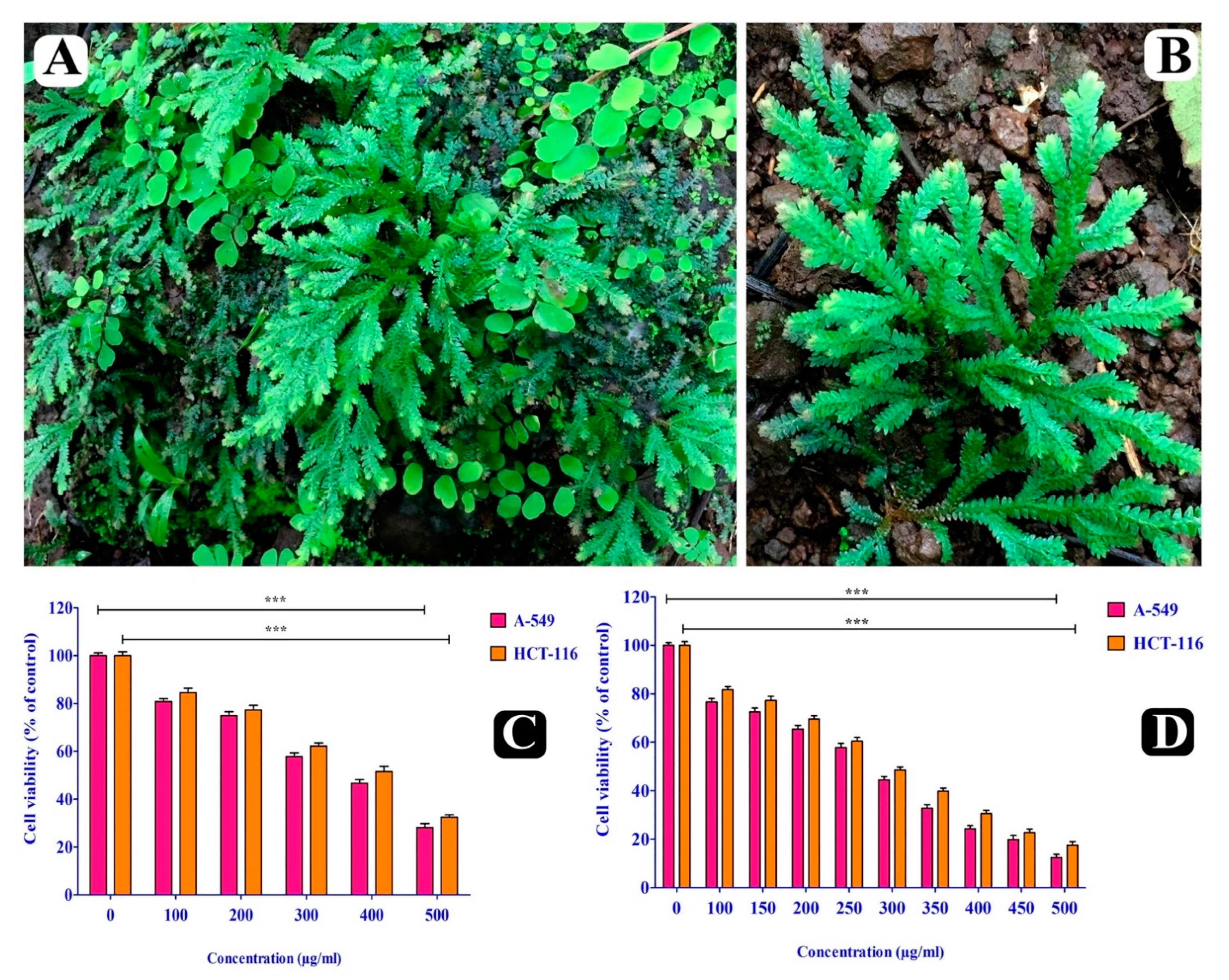
2.2. Cytotoxic Effect of S. repanda Crude Extract

Cytotoxicity of S. repanda crude extract was evaluated against A549 and HCT-116 cancer cell lines by MTT assay at 24 and 48 h. Significant inhibition of both cancer cells’ viability was observed in a time and dose-dependent manner. The IC50 values for 24 h were found to be 341.1 μg/mL and 378.8 μg/mL (Figure 1C) and for 48 h were found to be 326.7 μg/mL and 351.4 μg/mL for A549 and HCT-116 cells (Figure 1D).

2.3. Anti-Migratory Effect of S. repanda Crude Extract

The most important metastatic event is the motility of cells, which takes place in different epithelial cells during the progression of cancer. Wound closure assay was carried out to study the effect of S. repanda crude extract on the migration of cells over wound scratch, made on culture plates. The migration of both A549 and HCT-116 cancer cells was evidently inhibited by the treatment of S. repanda crude extract in a time as well as dose-dependent manner, when compared to untreated control cells. Hence, S. repanda crude extract evidently inhibited the anchorage and spread of both cell lines along the edge of wound scratch (Figure 2A,B).

2.4. Anti-Invasion Effect of S. repanda Crude Extract

Transwell® cell culture inserts coated with Matrigel matrix were used to evaluate the ability of S. repanda crude extract to inhibit the invasion of A549 and HCT-116 cells, following treatment with different concentrations (100–500 μg/mL). The cellular invasion of both A549 and HCT-116 cancer cells was significantly inhibited by the treatment of S. repanda crude extract (100 μg/mL) in a time as well as dose-dependent manner, when compared to untreated control cells (Figure 2 C).

2.5. Anti-Adhesion Effect of S. repanda Crude Extract

Adhesion to endothelial cells by malignant cells is known to be mediated by extracellular matrix proteins (ECM), thereby, we evaluated the adhesion property of both A549 and HCT-116 cancer cells to gelatin coated surfaces both in the presence and absence of S. repanda crude extract. The adhesion of A549 and HCT-116 cells were reduced by 75.67% and 72.0% (500 μg/mL) at the incubation of 6 h. Relative adherence was measured by setting the number of adherent cells at 6 h to 100% (Figure 3A,B).

2.6. Changes in Transcriptional Level of Metastasis Related Genes

The expression level of metastasis related genes MMP-2, MMP-9, MT1-MMP and TIMP-1 in both A549 and HCT-116 malignant cells, which were induced by S. repanda crude extract were determined by real time PCR. Firstly, the expression level of MMP-2 and MMP-9 genes were evaluated and was found to be decreased in both cells treated with S. repanda crude extract after 24 h incubation, when compared to untreated cells (Figure 4A–D). After the analysis of MMP-2 and MMP-9, we further examined the effect of S. repanda crude extract on the expression of activator and inhibitor genes of MMPs (MT1-MMP and TIMP-1). The expression level of TIMP-1 was found to be increased and of MT1-MMP gene was decreased in both cells after the treatment of S. repanda crude extract, when compared to untreated cells after 24 h incubation (Figure 5A,B). These results show that S. repanda crude extract can regulate the expression of MMP-2 and MMP-9 genes, which can possibly further control the cascade of metastasis.

3. Discussion

Regardless of all current progress in oncology, cancer is still one of the most life-threatening diseases around the globe [1]. It takes place as a localized disease, but can extend to different sites of the human body through migration, invasion and metastasis [2]. Metastasis is a multifaceted process involving an array of complicated mechanisms, which begins with extrication, accumulation and motility of cancer cells, followed by sticking to endothelial cells and the start of cancerous growth at different sites [24]. Progression of metastasis occurs after the degradation of ECM with cancer cells via different proteases like serine proteinase, cathepsins, MMPs and plasminogen activator, which prompts the separation of the intercellular matrix to promote the mobility of cancer cells [25]. Amongst involved proteases, MMP-9 and MMP-2 are profoundly implicated in cancer invasion and metastasis as they are the most essential for the degradation of base membranes [26,27,28].
Due to resistance to apoptosis and cytotoxic agents, metastasis is the dominant basis for cancer associated death. The percentage of morbidity and mortality in metastatic cancer patients is high, due to the failure of current chemotherapy agents to selectively and effectively kill cancer cells without destroying healthy cells at the sites of metastasis [29]. Metastasis is still a crucial clinical challenge in cancer treatment for researchers around the globe. Up till now, no such strong cleanse for cancer and its other catastrophic presentations have been found [29]. Currently, radiation therapy, chemotherapy, surgery are the conventional treatments for cancer. These treatments come with a range of side effects to human health; therefore, the importance of traditional medicines may decline. Thus, metastasis found the greatest challenging obstacle for successful cancer management and can be viewed as the last edge of cancer research [24].
Since ancient times, natural products have been recognized as an excellent source of bioactive compounds. They have been the primary source of general medicines, and can also be used directly as medicines [30,31,32,33,34,35]. Therefore, the pursuit and curiosity in the identification of medicinal plants and their derived natural products for evolving the novel cancer therapeutic strategies expanded vastly in recent times. In our previous study [9], a comprehensive phytochemical analysis from the crude extract of S. repanda was carried out via HR-LC-MS. A chromatogram was obtained with both positive and negative run and different types of phytochemicals were identified. In-depth profiling revealed various classes of metabolites such as alkaloids, flavonoids, sugars, vitamins, amino acids, phenols, terpenoids, phenols, etc. Though their biological activities are known, it can be evidently said that these compounds are somehow directly or indirectly responsible for the possible anti-metastatic effect on lung and colon cancer cells. However, individual testing of each identified phytochemical is required to reveal and interpret the actual involvement of these S. repanda compounds in anti-metastasis.
Plants in the genus Selaginella have diverse therapeutic potential on different cancer cells, comprising induction of apoptosis, inhibition of cell proliferation and arrest of cell cycle. There are species of Selaginella (S. delicatula, S. tamariscina, S. moellendorffii), which are known to possess potent antioxidant and antitumor activities, related to activation of apoptosis through DNA fragmentation and nucleus clotting [36], by inducing the expression of p53 and G1 arrest [37], through obstruction in fatty acid synthesis [38], inhibiting transactivation of iNOS and COX-2 via inactivating the NF-kB and avoiding the p65 translocation [39]. Several studies revealed the potent antitumor activities by S. uncinata and S. tamariscina, while moderate activity was found against Bel-7402 and HeLa cells by S. moellendorfii. Furthermore, out of all Selaginella species in the world, S. tamariscina is considered one of the most biologically potent plants, which is very well-known to inhibit the growth of various cancers (breast, leukemia, gastric, lung) [36,37,38,39]. Additionally, in osteosarcoma cells, it also possesses the anti-metastatic activity by down-regulating the MMP-2 and MMP-9 secretions and increasing the TIMP-1 and TIMP-2 expressions via Akt-dependent and p38 pathways [37,38,40,41,42].
A similar study with S. delicatula revealed the cytotoxicity against cancer cells of P-388, HT-29 [43] Raji, Calu-1, lymphoma and leukemia [44], due to the presence of phytocompounds robustaflavone and amentoflavone or its derivatives. Few other studies using S. moellendorfii show the growth inhibition of OVCAR-3, HeLa and FS-5 cancer cells [45,46], and anti-metastasis activity in lung cancer cells [39]. However, no studies on cytotoxicity and anti-metastasis of S. repanda crude extract against human cancer cells exist to date, and the mechanism of anticancer potential also remains unclear. Therefore, our study was designed to potentially identify the anti-metastatic activity of S. repanda crude extract against A549 and HCT-116 cancer cells with a possible molecular mechanism.
S. repanda crude extract displayed significant cytotoxic potential on both malignant A549 and HCT-116 cancer cells, and their IC50 were 341.1 μg/mL and 378.8 μg/mL, respectively. We performed an in vitro wound healing assay to evaluate the effect of S. repanda crude extract on cell migration, as migration is an important event in the progression of metastasis and cancer. S. repanda crude extract significantly inhibited the migration of cancer cells in the direction of the wounded area. Such results of the present study displayed that S. repanda crude extract impeded cell migration, which is crucial during the early phase of wound healing. Furthermore, S. repanda crude extract also remarkably retarded the invasion of both malignant cancer cells. Therefore, in the present study using wound healing, adhesion and invasion assays, we have shown that S. repanda crude extract efficiently inhibits the metastasis in both malignant cells in vitro.
To further elucidate the possible molecular mechanism behind the anti-metastatic potential of S. repanda crude extract, the mRNA expression level of MMP-2 and MMP-9 genes involved in the metastasis process was investigated. Both MMP-2 and MMP-9 genes mRNA expression levels were significantly decreased in a dose-dependent manner in both malignant cells. In metastasis, MMP-9 is considered the most significant protease, and its expression is connected with the growth of local tumor, invasion and metastasis in the majority of the carcinomas [47]. Therefore, increasing evidence suggests that a particular suppression of MMP-9 activity might avert metastasis [48]. Moreover, the expression of MMP genes is mainly controlled by their activators and inhibitors at the transcriptional, post-transcriptional and at the protein level [26,49]. TIMPs are assumed to play an immense role in the inhibition of MMPs [50]. In the present study, A549 and HCT-116 cells treated with S. repanda crude extract collectively up-regulated the expression of TIMP-1 gene. Apart from TIMP-1, MT1-MMP is another key enzyme among the regulation of MMPs, whose overexpression has the main effect on the growth of tumors [51]. It is mainly responsible for the activation of MMP2 [52,53]. The expression of MT1-MMP gene was significantly found to be inhibited in both A549 and HCT-116 cells treated with S. repanda crude extract. Hence, these results revealed that anti-metastatic effect of S. repanda crude extract is linked to the inhibition of enzymatically degradative processes of tumor metastasis.
Natural products are produced by all organisms, but plants are the major contributors. All of these organisms co-exist in the ecosystem and interact with each other in various ways in which chemistry plays a major role. Various approaches to understand the taxonomy of plants have been evolved over the years, which include morphological, anatomical and chemotaxonomic classification. However, morphological and anatomical classification system is considered as traditional approach, whereas, the science of chemotaxonomy or chemical taxonomy is a modern approach to classify the plants, especially on the basis of their chemical constituents [54,55]. The phenolics, alkaloids, terpenoids and non-protein amino acids are the four important and widely exploited groups of compounds utilized for chemotaxonomic classification [56]. These groups of compounds exhibit a wide variation in chemical diversity, distribution and function [56,57]. From this study, S. repanda have been recognized as an excellent source of phytoconstituents with diverse chemical class, hence our data can also be used by ethnopharmacologists, taxonomists and ethnobotanists for chemotaxonomic importance to solve the selected taxonomical problems.
Furthermore, as we stated earlier that our study is the first study to report the phytochemistry of S. repanda and the identified constituents can be seen in Table 1. In the literature, around 130 chemically defined natural products are reported from 32 species of Selaginella, which are belonging to the classes of pigments, benzenoids, alkaloids, carbohydrates, coumarins, flavonoids, chromones, oxygen heterocycle, lignans, phenylpropanoids, quinoids and steroids. Here, we have discussed the phytoconstituents of other Selaginella species in comparison to S. repanda. Chao et al. 1987 identified hordenine, hordenine-[6-O-(4-hydroxy-cinnamoyl)-β-D-glucosyl]-(1,3)-α-L-rhamnoside, hordenine-O-α-L-rhamnopyranoside, -methyltyramine-O-α-L-rhamnoside as alkaloids in S. doederleinii [58]. Similarly, in S. moellendorfii, identified alkaloids were selaginellic acid, 5-hydroxyselaginellic acid, 5-hydroxy-N8,N8-dimethylpseudophrynaminol, N-selaginelloyl-L-phenylalanine, N-(5-hydroxyselaginelloyl)-L-phenylalanine, neoselaginellic acid, and N-(5-Hydroxyneoselaginelloyl)-L-phenylalanine [59]. In S. tamariscina, adenosine and guanosine were identified as alkaloids [60]. Likewise, 4-hydroxy-benzoic acid [61], arbutin, vanillic acid, syringic acid [60,62] are identified as benzoids in S. pulvinata and S. tamariscina respectively.
Additionally, in carbohydrate class, selaginose was identified in S. adunca, S. asperula, S. epirrhizos, S. galeotti, S. geniculata, S. kraussiana, S. marginata, S. parkeri, S. plumosa, S. sanguinolenta, S. stellate and S. sulcata [63]; 2-carboxy-arabinitol was identified in S. mertensii [64]; and mycose was identified in S. pulvinata [61]. Moreover, 8-methyl-eugenitol, uncinoside A, uncinoside B was identified as chromone in S. uncinata [65,66]. On the other hand, numerous flavonoids were identified in various Selaginella species. For example, 2,3-dihydroamentoflavone, 2″,3″-dihydroamentoflavone, tetrahydro-amentoflavone was found in S. bryopteris [67]; amentoflavone-7,4,7,4-tetramethylether was found in S. moellendorfii [68]; 4′,7″-di-O-methyl-amentoflavone was found in S. sinensis [69]; 4′,7″-di-O-methyl-amentoflavone was found in S. willdenowii [70]; amentoflavone was identified in various species of Selaginella i.e., S. braunii, S. davidii, S. delicatula, S. denticulata, S. kraussiana, S. moellendorfii, S. pulvinata, S. rupestris, S. sanguinolenta, S. selaginoides, S. sinensis, S. stauntoniana, S. tamariscina, S. uncinata, S. willdenowii [44,69,70,71,72,73,74,75]; apigenin-7-O-β-neohesperidoside, apigenin-8-C- β-D-glucopyranoside, 6,8-di-C-β-D-glucopyranosyl-apigenin, 6-C-β-D-glucopyranosyl-8-C-β-D-xylopyranosyl-apigenin, 6-C-β-D-xylopyranosyl-8-C-β-D-glucopyranosyl-apigenin was identified in S. moellendorfii [76,77]; 2″,3″-dihydro-4′,7,7″-trimethylether-robustaflavone, 2,3-dihydro-4′,7,7″-trimethylether-robustaflavone, 2″,3″-dihydro-4′,7,-dimethylether-robustaflavone, 4′,7-dimethylether-robustaflavone, 4′-methylether-robustaflavone was identified in S. delicatula [44,78]; and sumaflavone was identified in S. tamariscina [79].
Lignan was also found in other Selaginella species. 5-acethyl-dihydro-2-(3′,5′-dimethoxy-4′-hydroxy-phenyl)-7-methoxybenzofuran was found in S. tamariscina [60]; (-)-lirioresinol A, (-)-lirioresinol B, (+)-matairesinol was found in S. doederleinii [18]; syringaresinol, tamariscinoside B, tamariscinoside C was found in S. tamariscina [60,62]. Some important pigments are also known to be found in various Selaginella species, i.e., selaginellin in S. sinensis [80], selaginellin A, selaginellin B in S. tamariscina [81], selaginellin C, selaginellin D, selaginellin E, selaginellin F, selaginellin G, selaginellin H in S. pulvinata [82,83,84]. Quinoids are also known to be found in some Selaginella species. Chrysophanic acid, emodin, physcion are identified in S. stauntoniana [85]; 1-methoxy-3-methylanthraquinone was identified in S. tamariscina [85]. Similarly, steroids are also known to be present in numerous Selaginella species, i.e., cholesterol, 22-dehydrocampesterol in S. delicatula, S. doederleinii [86], 3β-16α-dihydroxy-(5α)-cholestan-21-oic acid in S. pulvinata [87], 24α-ethyl-cholest-5-en-3β-ol, 24α-methyl-cholest-5-en-3β-ol, 24β-methyl-cholest-5-en-3β-ol, 24α-ethyl-cholesta-5,22-dien-3β-ol in S. delicatula and S. doederleinii [86] and β-sitostero in S. doederleinii, S. moellendorfii and S. pulvinata [61,88,89]. Therefore, our results can also be correlated and compared with the possibility to identify other Selaginella species for their anti-metastatic and anticancer efficacy against various carcinomas, due to the presence of diverse and potential phytoconstituents.

4. Materials and Methods

4.1. Plant Material Collection and Extraction

Whole plant of S. repanda was collected from the wild regions of Gujarat state, India during the July–August period of 2019. The voucher specimen (BVBRC035) was deposited at Bapalal Vaidya Botanical Garden, Department of Biosciences, Veer Narmad South Gujarat University, Surat, Gujarat, India. The collected plant material was dried in an oven and then grounded into fine powder followed by storage in airtight containers. A total of 20 g of S. repanda powder was soaked in 85% ethanol for 24 h at 37 °C with vigorous shaking. The ethanol phase was filtered with Whatman no. 1 filter paper and then concentrated using a rotary evaporator to get the dried residue. All the assays were performed using a stock solution of crude ethanol extract.

4.2. HR-LC–MS Analysis

Phytochemistry of S. repanda crude extract was analyzed using UHPLC-PDA-Detector Mass Spectrophotometer (HR-L = CMS 1290 Infinity UHPLC System), Agilent Technologies®, Santa Clara, CA, USA. The liquid chromatographic system consisted of the HiP sampler, binary gradient solvent pump, column compartment and quadrupole time of flight mass spectrometer (MS Q-TOF) with the dual Agilent Jet Stream Electrospray (AJS ES) ion source. Of the sample, 10 µL was injected into the system, followed by separation in the SB-C18 column (2.1 mm × 50 mm, 1.8 µm particle size). Solvent A (1% formic acid in deionized water) and solvent B (acetonitrile) were used as solvents. A flow rate of 0.350 mL/min was used, while MS detection was performed in MS Q-TOF. Compounds were identified via their mass spectra and their unique mass fragmentation patterns. Compound Discoverer 2.1, ChemSpider and PubChem were used as the main tools for the identification of the phytochemical constituents [9].

4.3. Cell culture and Treatment

Human lung (A549) and colon (HCT-116) cancer cell lines were obtained from National Centre for Cell Science (NCCS), India and propagated in a humidified atmosphere with 5% CO2 at 37 °C. Cells were maintained in 25 cm2 flask having Dulbecco’s Modified Eagle’s Medium (DMEM) supplemented with 10% Fetal Bovine Serum (FBS). They were grown up to 80% confluence for future analyses. Cells were treated with different concentrations of S. repanda crude extract (100–500 μg/mL).

4.4. Cell Viability Analysis Using MTT Assay

To determine the cytotoxicity of S. repanda crude extract, an MTT colorimetric assay was performed. For seeding, 96-well plates were used for both human cancer cell lines with incubation in humidified atmosphere comprising of 5% CO2 at 37 °C up to adherence. Different concentrations of S. repanda crude extract (100–500 μg/mL) were then used to treat the cells for 24 h, followed by washing with PBS solution. Cells were then subjected with 100 μL of MTT solution (3-(4,5-dimethylthiazolyl-2)-2,5-diphenyltetrazolium bromide) (5 mg/mL), followed by 4-h incubation. The medium was then removed and to solubilize the formazan crystals, 100 µL of dimethyl sulfoxide (DMSO) was added. Using ELISA reader, the amount of formazan crystal was determined by measuring the absorbance at 570 nm. All assays were carried out in triplicate, and 50% cytotoxic concentration (IC50) was calculated. 5-Fluorouracil was used as a positive control.

4.5. Wound Closure Assay

The effect of S. repanda crude extract on the motility of A-549 and HCT-116 cells was carried out using the wound healing assay. Confluent monolayer cell culture grown in 6 well plates was used to perform the assay. Cells were seeded at 1 × 10⁶ cells/mL in 3 mL final volume of growth medium. With the help of a sterile 1 mL pipette tip, an injury line was made in the central area of culture. After washing with phosphate-buffered saline (PBS), DMEM medium containing 2% FBS with and without S. repanda crude extract (100–500 μg/mL) was added into the well, floating cells were discarded. Plates were then incubated for 48 h at 37 °C. Wound closure through cell migration was measured at 0, 24 and 48 h under a microscope. The cells migration towards the wound scratches was expressed as migrated cells percentage and calculated as [90]:
Migrated cells percentage = [(At = 0 h − At = Δh)/At = 0 h] × 100
where,
At = 0 h is the area of wound measured immediately after scratching
At = Δh is the area of wound measured 24 or 48 h after scratching

4.6. Invasion Assay

The effect of S. repanda crude extract on the invasion ability of A549 and HCT-116 cells were observed using Transwell® chambers with 6.5 mm polycarbonate filters of 8 μm pore size (TCP152, Himedia®, India). A 50 μL aliquot of the Matrigel (E1270, Merck®, India) was pipetted into the upper chamber (culture insert), which was placed in a lower chamber with sterile forceps and incubated for the solidification of the gel at 37 °C for 30 min. Cells were seeded into the upper chamber with 1 × 10⁶ cells/mL consisting of 100 μL of serum free media. Immediately this chamber was then transferred into the lower chamber consisting of 600 μL of medium (2% FBS) with and without S. repanda crude extract (100–500 μg/mL). The plates were then incubated for 24 h at 37 °C. Afterward, the upper chamber was taken out, and using the cotton buds, non-migrated cells on its inner surface were wiped off. Furthermore, the migrated cells on the outer surface of the upper chamber were fixed using 70% methanol for 10 min. It was then followed by staining with 0.2% crystal violet, washing with distilled water and air drying at room temperature [91]. Under an inverted microscope, invaded cells were then observed. The cells invasion through the permeable matrix gel was expressed as percentage of invading cells and calculated as:
% invading cells = [Mean number of cells invading through the permeable matrix gel/Mean number of cells migrating through uncoated culture insert] × 100
Mean number of cells migrating through uncoated culture insert.

4.7. Cell Adhesion Assay

The effect of S. repanda crude extract on the adhesion ability of A549 and HCT-116 cells was carried out according to the method described previously by Burg–Roderfeld et al., (2007) [92]. Firstly, 0.1% gelatin was coated on the surface of the 6 well culture plates and left to air dry at 37 °C for 30 min. Both A549 and HCT-116 cells (1 × 10⁶ cells/mL) were harvested by trypsin and resuspended in a medium with and without S. repanda crude extract (100–500 μg/mL). Then, cells were dispensed into the respective wells and further incubated at 37 °C for 2, 4 and 6 h in 5% CO2. At the end of incubation, attached cells were gently washed with PBS twice and counted time dependently under an inverted microscope.

4.8. Expression Levels Determination of Metastasis Related Genes

Using the TriPure Isolation Reagent (Sigma-Aldrich®, India) and according to the manufacturer’s instructions, cellular RNA was isolated. RNA was quantified electrophoretically using 1.2% agarose gel, staining with ethidium bromide and visualizing under UV light. Firstly, RT-first strand synthesis kit (Qiagen®, CA, USA) was used to reverse transcribe the 1 μg of isolated RNA, and then the relative expression of metastasis related genes was determined by SYBR green based qRT PCR method (Applied Biosystems® 7500 Fast Real-Time PCR machine, CA, USA). ΔΔCt method was then followed to analyze the data, and values were expressed in terms of fold change relative to control [93,94]. Cycling conditions for relative expression of genes were as follows: initial reverse transcription at 55 °C for 45 min, 1 cycle denaturation of 95 °C with 10 min hold, followed by 40 cycles of 95 °C with 15 s hold, annealing temperature at 60 °C (MMP-2, MMP-9, MT1-MMP, TIMP-1 and GAPDH) with a 60 s hold. Four pairs of primers were separately used (Table 2). Samples were run in triplicate and their relative expression was determined by normalizing the expression of each target GAPDH.

4.9. Statistical Analysis

All the results are expressed as mean ± SD of the number of experiments performed. A significance test was carried out among the treatments by one way ANOVA followed by Tukey’s post hoc test at p < 0.05. Statistical analysis was conducted with software GraphPad Prism 5.0.

5. Conclusions

As per our knowledge, this study is the first one that demonstrates the anti-metastatic effect of S. repanda crude extract on lung and colon cancer cells. In conclusion, this study revealed that S. repanda crude extract exerts an inhibitory effect on various crucial steps of metastasis such as cell adhesion, invasion and migration via modulating the activities of metastasis related proteases and their activators and inhibitors. This reveals that S. repanda can be used/recommended as a potential anti-metastatic agent for drug development and therapy in cancer treatment.

Author Contributions

Conceptualization, M.A., M.A.K. and M.P.; methodology, M.S. (Mejdi Snoussi), A.J., A.J.S. and S.A.A.; validation, M.S. (Manojkumar Sachidanandan), M.S. (Mejdi Snoussi), W.S.H. and A.M.A.; formal analysis, M.P., M.A., M.S. (Mejdi Snoussi) and A.J.S.; investigation, R.B., A.J., S.A.A. and A.M.A.; data curation, M.P., A.J., W.S.H., S.A.A. and M.S. (Manojkumar Sachidanandan); writing—original draft preparation, M.P. and M.A.; writing—review and editing, M.A.K., M.A. and S.H.; visualization, M.P., R.B. and W.S.H.; supervision, M.A., S.H. and M.A.K.; project administration, S.H., M.A.K. and M.A. All authors have read and agreed to the published version of the manuscript.

Funding

This research has been funded by Scientific Research Deanship at University of Ha’il Saudi Arabia through project number RG-191194.

Institutional Review Board Statement

Not applicable.

Informed Consent Statement

Not applicable.

Data Availability Statement

All data generated or analyzed during this study are included in this article.

Acknowledgments

I would like to thank and appreciate all the support and technical assistance provided by Mushtaq’s lab at UAEU during this study. I further extend my gratitude to UoH for graciously providing numerous contributions and making all endeavors possible.

Conflicts of Interest

The authors declare no conflict of interest. The funders had no role in the design of the study; in the collection, analyses, or interpretation of data; in the writing of the manuscript, or in the decision to publish the results.

References

  1. Sung, H.; Ferlay, J.; Siegel, R.L.; Laversanne, M.; Soerjomataram, I.; Jemal, A.; Bray, F. Global cancer statistics 2020: GLOBOCAN estimates of incidence and mortality worldwide for 36 cancers in 185 countries. CA Cancer J. Clin. 2021. [Google Scholar] [CrossRef]
  2. Coghlin, C.; I Murray, G. Current and emerging concepts in tumour metastasis. J. Pathol. 2010, 222, 1–15. [Google Scholar] [CrossRef]
  3. Adnan, M.; Khan, S.; Al-Shammari, E.; Patel, M.; Saeed, M.; Hadi, S.; Information, P.E.K.F.C. In pursuit of cancer metastasis therapy by bacteria and its biofilms: History or future. Med. Hypotheses 2017, 100, 78–81. [Google Scholar] [CrossRef] [PubMed]
  4. Sak, K. Chemotherapy and Dietary Phytochemical Agents. Chemother. Res. Pr. 2012, 2012, 282570. [Google Scholar] [CrossRef] [Green Version]
  5. Baskar, R.; Dai, J.; Wenlong, N.; Yeo, R.; Yeoh, K.-W. Biological response of cancer cells to radiation treatment. Front. Mol. Biosci. 2014, 1, 24. [Google Scholar] [CrossRef] [Green Version]
  6. Chikara, S.; Nagaprashantha, L.D.; Singhal, J.; Horne, D.; Awasthi, S.; Singhal, S.S. Oxidative stress and dietary phyto-chemicals: Role in cancer chemoprevention and treatment. Cancer Lett. 2018, 413, 122–134. [Google Scholar] [CrossRef] [PubMed]
  7. Singh, S.; Sharma, B.; Kanwar, S.S.; Kumar, A. Lead Phytochemicals for Anticancer Drug Development. Front. Plant Sci. 2016, 7, 1667. [Google Scholar] [CrossRef] [Green Version]
  8. Adnan, M.; Siddiqui, A.J.; Jamal, A.; Hamadou, W.S.; Awadelkareem, A.M.; Sachidanandan, M.; Patel, M. Evidence-Based Medicinal Potential and Possible Role of Selaginella in the Prevention of Modern Chronic Diseases: Ethnopharmacological and Ethnobotanical Perspective. Rec. Nat. Prod. 2021, 15, 330–355. [Google Scholar] [CrossRef]
  9. Adnan, M.; Siddiqui, A.J.; Hamadou, W.S.; Patel, M.; Ashraf, S.A.; Jamal, A.; Awadelkareem, A.M.; Sachidanandan, M.; Snoussi, M.; De Feo, V. Phytochemistry, Bioactivities, Pharmacokinetics and Toxicity Prediction of Selaginella repanda with Its Anticancer Potential against Human Lung, Breast and Colorectal Carcinoma Cell Lines. Molecules 2021, 26, 768. [Google Scholar] [CrossRef] [PubMed]
  10. Han, B.-H.; Chi, H.-J.; Han, Y.-N.; Ryu, K.-S. Screening on the anti-inflammatory activity of crude drugs. Korean J. Pharmacogn. 1972, 3, 205–209. [Google Scholar]
  11. Itokawa, H.; Mihashi, S.; Watanabe, K.; Natsumoto, H.; Hamanaka, T. Studies on the constituents of crude drugs having inhibitory activity against contraction of the ileum caused by histamine or barium chloride (1) screening test for the activ-ity of commercially available crude drugs and the related plant materials. Shoyakugaku Zasshi 1983, 37, 223–228. [Google Scholar]
  12. Macfoy, C.A.; Sama, A.M. Medicinal plants in pujehun district of sierra leone. J. Ethnopharmacol. 1983, 8, 215–223. [Google Scholar] [CrossRef]
  13. Han, D.S.L.; Lee, H.K. Ethnobotanical survey in Korea. In Proceedings of the Fifth Asian Symposium on Medicinal Plants and Spices, Seoul, Korea, 20–24 August 1984; Volume 5, p. 125. [Google Scholar]
  14. Winkelman, M. Frequently used medicinal plants in Baja California Norte. J. Ethnopharmacol. 1986, 18, 109–131. [Google Scholar] [CrossRef]
  15. Darias, V.; Bravo, L.; Rabanal, R.; Mateo, C.; Luis, R.; Pérez, A. New contribution to the ethnopharmacological study of the canary islands. J. Ethnopharmacol. 1989, 25, 77–92. [Google Scholar] [CrossRef]
  16. Ono, K.; Nakane, H.; MENG, Z.-M.; OSE, Y.; SAKAI, Y.; MIZUNO, M. Differential inhibitory effects of various herb ex-tracts on the activities of reverse transcriptase and various deoxyribonucleic acid (DNA) polymerases. Chem. Pharm. Bull. 1989, 37, 1810–1812. [Google Scholar] [CrossRef] [PubMed] [Green Version]
  17. Meng, Z.; Sakai, Y.; Ose, Y.; Sato, T.; Nagase, H.; Kito, H.; Matsuda, H.; Sato, M.; Mizuno, M. Antimutagenic activity of medical plants in traditional Chinese medicines. Mutat. Res. Mutagen. Relat. Subj. 1988, 203, 378–379. [Google Scholar] [CrossRef]
  18. Lin, R.; Skaltsounis, A.-L.; Seguin, E.; Tillequin, F.; Koch, M. Phenolic Constituents of Selaginella doederleinii. Planta Medica 1994, 60, 168–170. [Google Scholar] [CrossRef]
  19. De Sá, P.G.S.; Nunes, X.P.; De Lima, J.T.; Filho, J.A.D.S.; Fontana, A.P.; Siqueira, J.D.S.; Quintans-Júnior, L.J.; Damasceno, P.K.F.; Branco, C.R.C.; Branco, A.; et al. Antinociceptive effect of ethanolic extract of Selaginella convoluta in mice. BMC Complement. Altern. Med. 2012, 12, 187. [Google Scholar] [CrossRef] [Green Version]
  20. Almeida, J.R.G.; de Sa, P.G.S.; Macedo, L.A.R.; Filho, J.A.; Oliviera, V.R.; Filho, J.M.B. Phytochemistry of the genus Sela-ginella (Selaginellaceae). J. Med. Plants Res. 2013, 7, 1858–1868. [Google Scholar]
  21. Harbone, J.B.; Williams, C.A. ChemInform Abstract: Advances in Flavonoid Research since 1992. Phytochemistry 2010, 32, 481–504. [Google Scholar] [CrossRef]
  22. Pellati, F.; Benvenuti, S.; Magro, L.; Melegari, M.; Soragni, F. Analysis of phenolic compounds and radical scavenging activity of Echinacea spp. J. Pharm. Biomed. Anal. 2004, 35, 289–301. [Google Scholar] [CrossRef]
  23. Lan, L.; Wang, Y.; Pan, Z.; Wang, B.; Yue, Z.; Jiang, Z.; Li, L.; Wang, C.; Tang, H. Rhamnetin induces apoptosis in human breast cancer cells via the miR-34a/Notch-1 signaling pathway. Oncol. Lett. 2018, 17, 676–682. [Google Scholar] [CrossRef] [PubMed] [Green Version]
  24. Lee, Y.-C.; Lin, H.-H.; Hsu, C.-H.; Wang, C.-J.; Chiang, T.-A.; Chen, J.-H. Inhibitory effects of andrographolide on migra-tion and invasion in human non-small cell lung cancer A549 cells via down-regulation of PI3K/Akt signaling pathway. Eur. J. Pharmacol. 2010, 632, 23–32. [Google Scholar] [CrossRef] [PubMed]
  25. Yeh, C.-B.; Hsieh, M.-J.; Lin, C.-W.; Chiou, H.-L.; Lin, P.-Y.; Chen, T.-Y.; Yang, S.-F. The antimetastatic effects of resvera-trol on hepatocellular carcinoma through the downregulation of a metastasis-associated protease by SP-1 modulation. PLoS ONE 2013, 8, e56661. [Google Scholar] [CrossRef] [Green Version]
  26. Yang, S.-F.; Chen, M.-K.; Hsieh, Y.-S.; Yang, J.-S.; Zavras, A.-I.; Hsieh, Y.-H.; Su, S.-C.; Kao, T.-Y.; Chen, P.-N.; Chu, S.-C. Antimetastatic effects of Terminalia catappa L. on oral cancer via a down-regulation of metastasis-associated prote-ases. Food Chem. Toxicol. 2010, 48, 1052–1058. [Google Scholar] [CrossRef]
  27. Giannelli, G.; Bergamini, C.; Fransvea, E.; Marinosci, F.; Quaranta, V.; Antonaci, S. Human Hepatocellular Carcinoma (HCC) Cells Require Both α3β1 Integrin and Matrix Metalloproteinases Activity for Migration and Invasion. Lab. Investig. 2001, 81, 613–627. [Google Scholar] [CrossRef] [Green Version]
  28. Patel, M.; Sachidanandan, M.; Adnan, M. Serine arginine protein kinase 1 (SRPK1): A moonlighting protein with theranostic ability in cancer prevention. Mol. Biol. Rep. 2018, 46, 1487–1497. [Google Scholar] [CrossRef]
  29. Liotta, L.A. An attractive force in metastasis. Nat. Cell Biol. 2001, 410, 24–25. [Google Scholar] [CrossRef]
  30. Reddy, M.N.; Adnan, M.; Alreshidi, M.M.; Saeed, M.; Patel, M. Evaluation of Anticancer, Antibacterial and Antioxidant Properties of a Medicinally Treasured Fern Tectaria coadunata with its Phytoconstituents Analysis by HR-LCMS. Anti-Cancer Agents Med. Chem. 2020, 20, 1845–1856. [Google Scholar] [CrossRef]
  31. Siddiqui, A.J.; Danciu, C.; Ashraf, S.A.; Moin, A.; Singh, R.; Alreshidi, M.; Patel, M.; Jahan, S.; Kumar, S.; Alkhinjar, M.I. Plants-derived biomolecules as potent antiviral phytomedicines: New insights on ethnobotanical evidences against coro-naviruses. Plants 2020, 9, 1244. [Google Scholar] [CrossRef]
  32. Patel, M.; Ashraf, M.S.; Siddiqui, A.J.; Ashraf, S.A.; Sachidanandan, M.; Snoussi, M.; Adnan, M.; Hadi, S. Profiling and Role of Bioactive Molecules from Puntius sophore (Freshwater/Brackish Fish) Skin Mucus with Its Potent Antibacterial, Antiadhesion, and Antibiofilm Activities. Biomolecules 2020, 10, 920. [Google Scholar] [CrossRef] [PubMed]
  33. Surti, M.; Patel, M.; Adnan, M.; Moin, A.; Ashraf, S.A.; Siddiqui, A.J.; Snoussi, M.; Deshpande, S.; Reddy, M.N. Ili-maquinone (marine sponge metabolite) as a novel inhibitor of SARS-CoV-2 key target proteins in comparison with sug-gested COVID-19 drugs: Designing, docking and molecular dynamics simulation study. RSC Adv. 2020, 10, 37707–37720. [Google Scholar] [CrossRef]
  34. Adnan, M.; Patel, M.; Deshpande, S.; Alreshidi, M.; Siddiqui, A.J.; Reddy, M.N.; Emira, N.; De Feo, V. Effect of Adiantum philippense Extract on Biofilm Formation, Adhesion with Its Antibacterial Activities Against Foodborne Pathogens, and Characterization of Bioactive Metabolites: An in vitro-in silico Approach. Front. Microbiol. 2020, 11, 823. [Google Scholar] [CrossRef]
  35. Mseddi, K.; Alimi, F.; Noumi, E.; Veettil, V.N.; Deshpande, S.; Adnan, M.; Hamdi, A.; Elkahoui, S.; Alghamdi, A.; Kadri, A.; et al. Thymus musilii Velen. as a promising source of potent bioactive compounds with its pharmacological properties: In vitro and in silico analysis. Arab. J. Chem. 2020, 13, 6782–6801. [Google Scholar] [CrossRef]
  36. Ahn, S.-H.; Mun, Y.-J.; Lee, S.-W.; Kwak, S.; Choi, M.-K.; Baik, S.-K.; Kim, Y.-M.; Woo, W.-H. Selaginella tamariscina Induces Apoptosis via a Caspase-3-Mediated Mechanism in Human Promyelocytic Leukemia Cells. J. Med. Food 2006, 9, 138–144. [Google Scholar] [CrossRef]
  37. Lee, I.-S.; Nishikawa, A.; Furukawa, F.; Kasahara, K.-I.; Kim, S.-U. Effects of Selaginella tamariscina on in vitro tumor cell growth, p53 expression, G1 arrest and in vivo gastric cell proliferation. Cancer Lett. 1999, 144, 93–99. [Google Scholar] [CrossRef]
  38. Lee, J.S.; Lee, M.S.; Oh, W.K.; Sul, J.Y. Fatty acid synthase inhibition by amentoflavone induces apoptosis and antiprolif-eration in human breast cancer cells. Biol. Pharm. Bull. 2009, 32, 1427–1432. [Google Scholar] [CrossRef] [Green Version]
  39. Woo, E.-R.; Pokharel, Y.R.; Yang, J.W.; Lee, S.Y.; Kang, K.W. Inhibition of nuclear factor-κB activation by 2′, 8″-biapigenin. Biol. Pharm. Bull. 2006, 29, 976–980. [Google Scholar] [CrossRef] [Green Version]
  40. Setyawan, A.D. Review: Natural products from Genus Selaginella (Selaginellaceae). Nusant. Biosci. 2016, 3, 3. [Google Scholar] [CrossRef]
  41. Yang, J.-S.; Lin, C.-W.; Hsieh, Y.-S.; Cheng, H.-L.; Lue, K.-H.; Yang, S.-F.; Lu, K.-H. Selaginella tamariscina (Beauv.) pos-sesses antimetastatic effects on human osteosarcoma cells by decreasing MMP-2 and MMP-9 secretions via p38 and Akt signaling pathways. Food Chem. Toxicol. 2013, 59, 801–807. [Google Scholar] [CrossRef]
  42. Yang, J.-S.; Lin, C.-W.; Hsin, C.-H.; Hsieh, M.-J.; Chang, Y.-C. Selaginella tamariscina Attenuates Metastasis via Akt Pathways in Oral Cancer Cells. PLoS ONE 2013, 8, e68035. [Google Scholar] [CrossRef] [PubMed] [Green Version]
  43. Chen, J.-J.; Duh, C.-Y.; Chen, J.-F. New cytotoxic biflavonoids from Selaginella delicatula. Planta Med. 2005, 71, 659–665. [Google Scholar] [CrossRef] [PubMed]
  44. Lin, L.-C.; Kuo, Y.-C.; Chou, C.-J. Cytotoxic Biflavonoids from Selaginella delicatula. J. Nat. Prod. 2000, 63, 627–630. [Google Scholar] [CrossRef]
  45. Sun, C.-M.; Syu, W.-J.; Huang, Y.-T.; Chen, C.-C.; Ou, J.-C. Selective cytotoxicity of ginkgetin from Selaginella moellendorffii. J. Nat. Prod. 1997, 60, 382–384. [Google Scholar] [CrossRef] [PubMed]
  46. Su, Y.; Sun, C.-M.; Chuang, H.-H.; Chang, P.-T. Studies on the cytotoxic mechanisms of ginkgetin in a human ovarian adenocarcinoma cell line. Naunyn-Schmiedeberg’s Arch. Pharmacol. 2000, 362, 82–90. [Google Scholar] [CrossRef]
  47. Sato, H.; Takino, T.; Okada, Y.; Cao, J.; Shinagawa, A.; Yamamoto, E.; Seiki, M. A matrix metalloproteinase expressed on the surface of invasive tumour cells. Nat. Cell Biol. 1994, 370, 61–65. [Google Scholar] [CrossRef] [PubMed]
  48. Yeh, C.-B.; Hsieh, M.-J.; Hsieh, Y.-H.; Chien, M.-H.; Chiou, H.-L.; Yang, S.-F. Antimetastatic effects of norcantharidin on hepatocellular carcinoma by transcriptional inhibition of MMP-9 through modulation of NF-kB activity. PLoS ONE 2012, 7, e31055. [Google Scholar] [CrossRef]
  49. Ghiso, J.A.A.; Alonso, D.F.; Farías, E.F.; Gomez, D.E.; Joffè, E.B.D.K. Deregulation of the signaling pathways controlling urokinase production. Its relationship with the invasive phenotype. JBIC J. Biol. Inorg. Chem. 1999, 263, 295–304. [Google Scholar] [CrossRef]
  50. Hwang, E.-S.; Lee, H.J. Allyl isothiocyanate and its N-acetylcysteine conjugate suppress metastasis via inhibition of inva-sion, migration, and matrix metalloproteinase-2/-9 activities in SK-Hep1 human hepatoma cells. Exp. Biol. Med. 2006, 231, 421–430. [Google Scholar] [CrossRef] [PubMed]
  51. Määttä, M.; Soini, Y.; Liakka, A.; Autio-Harmainen, H. Differential expression of matrix metalloproteinase (MMP)-2, MMP-9, and membrane type 1-MMP in hepatocellular and pancreatic adenocarcinoma: Implications for tumor progres-sion and clinical prognosis. Clin. Cancer Res. 2000, 6, 2726–2734. [Google Scholar]
  52. Ogata, R.; Torimura, T.; Kin, M.; Ueno, T.; Tateishi, Y.; Kuromatsu, R.; Shimauchi, Y.; Sakamoto, M.; Tamaki, S.; Sata, M.; et al. Increased expression of membrane type 1 matrix metalloproteinase and matrix metalloproteinase-2 with tumor dedifferentiation in hepatocellular carcinomas. Hum. Pathol. 1999, 30, 443–450. [Google Scholar] [CrossRef]
  53. Tam, E.M.; Moore, T.R.; Butler, G.S.; Overall, C.M. Characterization of the Distinct Collagen Binding, Helicase and Cleavage Mechanisms of Matrix Metalloproteinase 2 and 14 (Gelatinase A and MT1-MMP). J. Biol. Chem. 2004, 279, 43336–43344. [Google Scholar] [CrossRef] [Green Version]
  54. Reynolds, T. The evolution of chemosystematics. Phytochemistry 2007, 68, 2887–2895. [Google Scholar] [CrossRef] [PubMed]
  55. Larsen, T.O.; Smedsgaard, J.; Nielsen, K.F.; Hansen, M.E.; Frisvad, J.C. Phenotypic taxonomy and metabolite profiling in microbial drug discovery. Nat. Prod. Rep. 2005, 22, 672–695. [Google Scholar] [CrossRef] [PubMed]
  56. Fellows, L.; Smith, P.M. The Chemotaxonomy of Plants. Kew Bull. 1978, 32, 806. [Google Scholar] [CrossRef]
  57. Hegnauer, R. Phytochemistry and plant taxonomy—An essay on the chemotaxonomy of higher plants. Phytochemistry 1986, 25, 1519–1535. [Google Scholar] [CrossRef]
  58. Chao, L.R.; Seguin, E.; Tillequin, F.; Koch, M. New Alkaloid Glycosides from Selaginella doederleinii. J. Nat. Prod. 1987, 50, 422–426. [Google Scholar] [CrossRef]
  59. Wang, Y.-H.; Long, C.-L.; Yang, F.-M.; Wang, X.; Sun, Q.-Y.; Wang, H.-S.; Shi, Y.-N.; Tang, G.-H. Pyrrolidinoindoline Alkaloids from Selaginella moellendorfii. J. Nat. Prod. 2009, 72, 1151–1154. [Google Scholar] [CrossRef]
  60. Zheng, X.-K.; Bi, Y.-F.; Feng, W.-S.; Shi, S.-P.; Wang, J.-F.; Niu, J.-Z. Study on chemical constituents of Selaginella tamariscina (Beauv.) Spring. Acta Pharm. Sin. 2004, 39, 266–268. [Google Scholar]
  61. Zheng, X.; Liao, D.F.; Zhu, B.Y.; Tuo, Q.H.; Xu, Y.L. Study on chemical constituents of Selaginella pulvinata. Zhongcaoyao 2001, 32, 17–18. [Google Scholar]
  62. Bi, Y.-F.; Zheng, X.-K.; Feng, W.-S.; Shi, S.-P. Isolation and structural identification of chemical constituents from Selaginella tamariscina (Beauv.) Spring. Acta Pharm. Sin. 2004, 39, 41–45. [Google Scholar]
  63. Fischer, M.; Kandler, M. Identifizierung von selaginose und deren verbreitung in der gattung Selaginella. Phytochemistry 1975, 14, 2629–2633. [Google Scholar] [CrossRef]
  64. Moore, B.D.; Isidoro, E.; Seemann, J.R. Distribution of 2-carboxyarabinitol among plants. Phytochemistry 1993, 34, 703–707. [Google Scholar] [CrossRef]
  65. Ma, L.-Y.; Ma, S.-C.; Wei, F.; Lin, R.-C.; But, P.P.-H.; Lee, S.H.-S.; Lee, S.F. Uncinoside A and B, Two New Antiviral Chromone Glycosides from Selaginella uncinata. Chem. Pharm. Bull. 2003, 51, 1264–1267. [Google Scholar] [CrossRef] [PubMed] [Green Version]
  66. Ma, L.Y.; Wei, F.; Ma, S.C.; Lin, R.C. Two new chromone glycosides from Selaginella uncinata. Chin. Chem. Lett. 2002, 13, 748–751. [Google Scholar]
  67. Kunert, O.; Swamy, R.C.; Kaiser, M.; Presser, A.; Buzzi, S.; Rao, A.A.; Schühly, W. Antiplasmodial and leishmanicidal activity of biflavonoids from Indian Selaginella bryopteris. Phytochem. Lett. 2008, 1, 171–174. [Google Scholar] [CrossRef]
  68. Cao, Y.; Tan, N.H.; Chen, J.J.; Zeng, G.Z.; Ma, Y.B.; Wu, Y.P.; Yan, H.; Yang, J.; Lu, L.F.; Wang, Q. Bioactive flavones and biflavones from Selaginella moellendorfii Hieron. Fitoterapia 2010, 81, 253–258. [Google Scholar] [CrossRef]
  69. Ma, S.-C.; But, P.P.-H.; Ooi, V.E.-C.; He, Y.-H.; Lee, S.H.-S.; Lee, S.-F.; Lin, R.-C. Antiviral Amentoflavone from Selaginella sinensis. Biol. Pharm. Bull. 2001, 24, 311–312. [Google Scholar] [CrossRef] [Green Version]
  70. Silva, G.L.; Chai, H.; Gupta, M.P.; Farnsworth, N.R.; Cordell, G.A.; Pezzuto, J.M.; Beecher, C.W.; Kinghorn, A.D. Cytotoxic biflavonoids from Selaginella willdenowii. Phytochemistry 1995, 40, 129–134. [Google Scholar] [CrossRef]
  71. López-Sáez, J.A.; Alonso, M.J.P.-; Negueruela, A.V. Biflavonoids of Selaginella denticulata Growing in Spain. Z. Nat. C 1994, 49, 267–270. [Google Scholar] [CrossRef]
  72. Qasim, M.A.; Roy, S.K.; Kamil, M.; Ilyas, M. Phenolic constituents of Selaginellaceae. Indian J. Chem. 1985, 24, 220. [Google Scholar]
  73. Chakravarthy, B.K.; Rao, Y.V.; Gambhir, S.S.; Gode, K.D. Isolation of Amentoflavone from Selaginella rupestris and its Pharmacological Activity on Central Nervous System, Smooth Muscles and Isolated Frog Heart Preparations. Planta Med. 1981, 43, 64–70. [Google Scholar] [CrossRef] [PubMed]
  74. Huneck, S.; Khaidav, T. Amentoflavone from Selaginella sanguinolenta. Die Pharm. 1985, 40, 431. [Google Scholar]
  75. Lee, I.R.; Song, J.Y.; Lee, Y.S. Cytotoxicity of folkloric medicines in murine and human cancer cells. Korean J. Pharmacogn. 1992, 23, 132–136. [Google Scholar]
  76. Feng, W.S.; Li, K.K.; Zheng, X.K. A new norlignan lignanoside from Selaginella moellendorfii Hieron. Acta Pharm. Sin. B 2011, 1, 36–39. [Google Scholar] [CrossRef] [Green Version]
  77. Zhu, T.M.; Chen, K.L.; Zhou, W.B. A new flavones glycoside from Selaginella moellendorfii Hieron. Chin. Chem. Lett. 2008, 19, 1456–1458. [Google Scholar] [CrossRef]
  78. Lin, L.C.; Chou, C.J. Three new biflavonoids from Selaginella delicatula. Chin. J. Pharm. 2000, 52, 211–218. [Google Scholar]
  79. Yang, J.W.; Pokharel, Y.R.; Kim, M.-R.; Woo, E.-R.; Choi, H.K.; Kang, K.W. Inhibition of inducible nitric oxide synthase by sumaflavone isolated from Selaginella tamariscina. J. Ethnopharmacol. 2006, 105, 107–113. [Google Scholar] [CrossRef]
  80. Zhang, L.-P.; Liang, Y.-M.; Wei, X.-C.; Cheng, D.-L. A New Unusual Natural Pigment from Selaginella sinensis and Its Noticeable Physicochemical Properties. J. Org. Chem. 2007, 38, 3824–3921. [Google Scholar] [CrossRef]
  81. Cheng, X.-L.; Ma, S.-C.; Yu, J.-D.; Yang, S.-Y.; Xiao, X.-Y.; Hu, J.-Y.; Lu, Y.; Shaw, P.-C.; But, P.P.-H.; Lin, R.-C. Selaginellin A and B, Two Novel Natural Pigments Isolated from Selaginella tamariscina. Chem. Pharm. Bull. 2008, 56, 982–984. [Google Scholar] [CrossRef] [Green Version]
  82. Tan, G.-S.; Xu, K.-P.; Li, F.-S.; Wang, C.-J.; Li, T.-Y.; Hu, C.-P.; Shen, J.; Zhou, Y.-J.; Li, Y.-J. Selaginellin C, a new natural pigment from Selaginella pulvinata Maxim (Hook et Grev.). J. Asian Nat. Prod. Res. 2009, 11, 1001–1004. [Google Scholar] [CrossRef] [PubMed]
  83. Cao, Y.; Chen, J.J.; Tan, N.H.; Oberer, L.; Wagner, T.; Wu, Y.P.; Zeng, G.Z.; Yan, H.; Wang, Q. Antimicrobial selaginellin derivatives from Selaginella pulvinata. Bioorganic Med. Chem. Lett. 2010, 20, 2456–2460. [Google Scholar] [CrossRef] [PubMed]
  84. Cao, Y.; Yang, J.; Wang, Q.; Chen, J.-J.; Tan, N.-H.; Wu, Y.-P. Structure determination of selaginellins G and H from Selaginella pulvinata by NMR spectroscopy. Magn. Reson. Chem. 2010, 48, 656–659. [Google Scholar] [CrossRef] [PubMed]
  85. Liu, J.-F.; Xu, K.-P.; Li, F.-S.; Shen, J.; Hu, C.-P.; Zou, H.; Yang, F.; Liu, G.-R.; Xiang, H.-L.; Zhou, Y.-J.; et al. A New Flavonoid from Selaginella tamariscina (Beauv.) Spring. Chem. Pharm. Bull. 2010, 58, 549–551. [Google Scholar] [CrossRef] [Green Version]
  86. Chiu, P.-L.; Patterson, G.W.; Salt, T.A. Sterol composition of pteridophytes. Phytochemistry 1988, 27, 819–822. [Google Scholar] [CrossRef]
  87. Zheng, X.; Du, J.; Xu, Y.; Zhu, B.; Liao, D. A new steroid from Selaginella pulvinata. Fitoterapia 2007, 78, 598–599. [Google Scholar] [CrossRef] [PubMed]
  88. Chen, P.; Sun, J.Y.; Xie, N.G.; Shi, Y.G. Chemical constituents of daeycai (Selaginella doederleinii). Zhongcaoyao 1995, 26, 397–399. [Google Scholar]
  89. Che, D.H.; Yu, J.G. Analysis on the chemical constituents of jiangnanjuanbai (Selaginella moellendorfii Hieron). Chung Tsao Yao 1986, 17, 4. [Google Scholar]
  90. Baraya, Y.S.; Wong, K.K.; Yaacob, N.S. Strobilanthes crispus inhibits migration, invasion and metastasis in breast cancer. J. Ethnopharmacol. 2019, 233, 13–21. [Google Scholar] [CrossRef] [PubMed]
  91. Justus, C.R.; Leffler, N.; Ruiz-Echevarria, M.; Yang, L.V. In vitro Cell Migration and Invasion Assays. J. Vis. Exp. 2014, e51046. [Google Scholar] [CrossRef] [Green Version]
  92. Burg-Roderfeld, M.; Roderfeld, M.; Wagner, S.; Henkel, C.; Grötzinger, J.; Roeb, E. MMP-9-hemopexin domain hampers adhesion and migration of colorectal cancer cells. Int. J. Oncol. 2007, 30, 985–992. [Google Scholar] [CrossRef] [PubMed]
  93. Adnan, M.; Morton, G.; Hadi, S. Analysis of rpoS and bolA gene expression under various stress-induced environments in planktonic and biofilm phase using 2ΔΔCT method. Mol. Cell. Biochem. 2011, 357, 275–282. [Google Scholar] [CrossRef] [PubMed]
  94. Siddiqui, A.J.; Bhardwaj, J.; Goyal, M.; Prakash, K.; Adnan, M.; Alreshidi, M.M.; Patel, M.; Soni, A.; Redman, W. Immune responses in liver and spleen against Plasmodium yoelii pre-erythrocytic stages in Swiss mice model. J. Adv. Res. 2020, 24, 29–41. [Google Scholar] [CrossRef] [PubMed]
  95. Byambaragchaa, M.; De La Cruz, J.; Yang, S.H.; Hwang, S.-G. Anti-metastatic Potential of Ethanol Extract of Saussurea involucrata against Hepatic Cancer in vitro. Asian Pac. J. Cancer Prev. 2013, 14, 5397–5402. [Google Scholar] [CrossRef] [PubMed]
Figure 1. S. repanda plant and its anticancer activity. (A) Plant in wild (B) Close view of plant (C) Anticancer activity of S. repanda crude extract against A-549 and HCT-116 cancer cells for 24 h. (D) Anticancer activity of S. repanda crude extract against A-549 and HCT-116 cancer cells for 48 h. Error bars indicate SDs (± standard deviation) of three independent experiments. Significance; ns > 0.05, * p < 0.05, ** p < 0.005, *** p < 0.0005.
Plants 10 00979 g001
Figure 2. Anti-migration and anti-invasion effects of S. repanda crude extract. (A) Anti-migration activity on A-549 cancer cells (B) Anti-migration activity on HCT-116 cancer cells. The number of migrated cells was quantified in five different fields from three independent experiments. (C) Anti- invasion activity on A-549 and HCT-116 cancer cells. The invading cells were counted in five random fields under microscopes. Error bars indicate SDs (± standard deviation) of three independent experiments. Significance; ns > 0.05, * p < 0.05, ** p < 0.005, *** p < 0.0005.
Plants 10 00979 g002
Figure 3. Anti-adhesion effects of S. repanda crude extract on gelatin coated surfaces. (A) Anti-adhesion activity on A-549 cancer cells. (B) Anti-adhesion activity on HCT-116 cancer cells. Error bars indicate SDs (± standard deviation) of three independent experiments. Significance; ns > 0.05, * p < 0.05, ** p < 0.005, *** p < 0.0005.
Plants 10 00979 g003
Figure 4. Effect of S. repanda crude extract on metastasis related genes. (A) Reverse transcription PCR to analyze the mRNA expression level of MMP-2 and MMP-9 in A-549 cancer cells. (B) Quantitative real-time PCR. (C) Reverse transcription PCR to analyze the mRNA expression level of MMP-2 and MMP-9 in HCT-116 cancer cells. (D) Quantitative real-time PCR. Significance; ns > 0.05, * p < 0.05, ** p < 0.005, *** p < 0.0005.
Plants 10 00979 g004
Figure 5. Effect of S. repanda crude extract on metastasis related genes. (A) Reverse transcription PCR to analyze the mRNA expression level of MT1-MMP and TIMP-1 in A-549 cancer cells. (B) Quantitative real-time PCR. (C) Reverse transcription PCR to analyze the mRNA expression level of MT1-MMP and TIMP-1 in HCT-116 cancer cells. (D) Quantitative real-time PCR. Significance; ns > 0.05, * p < 0.05, ** p < 0.005, *** p < 0.0005.
Plants 10 00979 g005
Table 1. Phytochemical composition of S. repanda crude extract identified by HR-LC-MS technique [9].
PhytocompoundsFormulaClassm/zRT (min)Mass
Urocanic acidC6H6N2O2monocarboxylic acid131.224.845138.04267
HordenineC10H15NOalkaloid167.223.071169.23958
OleamideC18H35NOfatty acid286.120.177281.27135
HexadecanamideC16H33NOfatty acid amide250.419.849255.25575
Arachidonic acidC20H32O2polyunsaturated fatty acid301.818.601304.2397
4-Coumaric acidC9H8O3phenol168.218.265164.04718
2-Arachidonoyl glycerolC23H38O4fatty acid derivative371.016.012378.27635
ValineC5H11NO2amino acid118.413.901117.07901
GenisteinC15H10O5isoflavone275.313.542270.05214
DiosmetinC16H12O6flavonoid294.313.534300.06279
2-Amino-1,3,4-octadecanetriolC18H39NO3phenol311.513.499317.29231
GlyciteinC16H12O5flavonoid288.913.477284.06807
RhamnetinC16H12O7flavonoid311.212.452316.05766
FormononetinC16H12O4phenol265.312.157268.07329
LuteolinC15H10O6flavonoid278.611.735286.04723
ApigeninC15H10O5flavone272.411.177270.05235
GlycitinC22H22O10isoflavone442.211.088446.12065
(-)-Caryophyllene oxideC15H24Oepoxide228.410.861220.18227
KuromaninC21H20O11pigment458.18.976448.09996
KaempferolC15H10O6flavonoid294.68.662286.04726
SedanolideC12H18O2isobenzofuran199.58.578194.13039
α-Pinene-2-oxideC10H16Oterpenoid148.98.494152.11989
Quercetin-3β-D-glucosideC21H20O12flavonoid260.38.438464.09465
QuercetinC15H10O7flavonoid308.68.414302.04192
VitexinC21H20O10flavonoid436.88.319432.10497
RutinC27H30O16flavonoid615.48.291610.15239
4-Methoxycinnamic acidC10H10O3phenol186.27.754178.06275
NorharmanC11H8N2alkaloid164.76.725168.06847
4-HydroxycoumarinC9H6O3benzopyrone158.86.568162.0314
Methyl cinnamateC10H10O2cinnamic acid ester164.56.458162.06775
Isoferulic acidC10H10O4phenol198.66.367194.05762
ScopoletinC10H8O4coumarin196.35.969192.04198
CitralC10H16Oterpenoid145.85.575152.11989
PulegoneC10H16Oterpenoid158.94.94152.11989
Caffeic acidC9H8O4phenol186.24.692180.04181
7-HydroxycoumarineC9H6O3phenol169.44.651162.0314
Chlorogenic acidC16H18O9phenol360.54.646354.09435
Kynurenic acidC10H7NO3quinoline carboxylic acid181.73.823189.04239
CoumarinC9H6O2phenol148.63.784146.0365
3-MethylcrotonylglycineC7H11NO3amino acid152.63.325157.0737
4-Hydroxyphenylacetic acidC8H8O3benzenoid156.33.314152.04714
8-HydroxyquinolineC9H7NOalkaloid153.22.965145.05255
MaltolC6H6O3sugar130.42.278126.03161
GuvacolineC7H11NO2pyridine alkaloid145.21.539141.07878
L-PhenylalanineC9H11NO2amino acid164.21.375165.07883
L-NorleucineC6H13NO2amino acid136.51.134131.09453
L-Pyroglutamic acidC5H7NO3amino acid133.11.04129.0425
D-GlucosamineC6 H13 NO5amino sugar183.30.946179.079
BetaineC5H11NO2amino acid122.20.935117.07901
L(-)-CarnitineC7H15NO3amino acid derivative157.60.93161.10489
AcetylcholineC7H15 NO2essential nutrient (vitamin)149.70.850145.11
α-LactoseC12H22O11sugar349.90.839342.11521
CholineC5H13N Oessential nutrient (vitamin)111.40.798103.09988
Table 2. List of primers used for metastasis related genes [95].
Sl. NoPrimerSequence
1MMP-2sense–5′-GGCCCTGTCACTCCTGAGAT-3′
antisense–5′-GGCATCCAGGTTATCGGGGA-3′
2MMP-9sense–5′-
CGGAGCACGGAGACGGGTAT-3′
antisense–5′-
TGAAGGGGAAGACGCACAGC-3′
3MT1-MMPsense–5′-TGGGTAGCGATGAAGTCTTC-3′
antisense–5′-AGTAAAGCAGTCGCTTGGGT-3′
4TIMP-1sense–5′-
GATCCAGCGCCCAGAGAGACACC-3′
antisense–5′-TTCCACTCCGGGCAGCATT-3′
5GAPDHsense–5′-
CGAGATCCCTCCAAAATCAA-3′
antisense–5′-AGGTCCACCACTGACACGTT-3′
Publisher’s Note: MDPI stays neutral with regard to jurisdictional claims in published maps and institutional affiliations.

